Article

The Transport and Uptake of Resveratrol Mediated via Glucose Transporter 1 and Its Antioxidant Effect in Caco-2 Cells

1
Key Lab of New Animal Drug Project of Gansu Province, Key Lab of Veterinary Pharmaceutical Development of Ministry of Agriculture and Rural Affairs, Lanzhou Institute of Husbandry and Pharmaceutical Sciences of CAAS, Lanzhou 730050, China
2
College of Life Sciences, South China Agricultural University, Guangzhou 510642, China
*
Author to whom correspondence should be addressed.
These authors contributed equally to this work.
Molecules 2023, 28(12), 4569; https://doi.org/10.3390/molecules28124569
Submission received: 15 May 2023 / Revised: 29 May 2023 / Accepted: 2 June 2023 / Published: 6 June 2023
(This article belongs to the Special Issue Natural Products in the Antioxidant Drug Discovery Process)

Abstract

:
Resveratrol has anti-inflammatory, anti-cancer, and anti-aging pharmacological activities. There is currently a gap in academic research regarding the uptake, transport, and reduction of H2O2-induced oxidative damage of resveratrol in the Caco-2 cell model. This study investigated the role of resveratrol in the uptake, transport, and alleviation of H2O2-induced oxidative damage in Caco-2 cells. In the Caco-2 cell transport model, it was observed that the uptake and transport of resveratrol (10, 20, 40, and 80 μM) were time dependent and concentration dependent. Different temperatures (37 °C vs. 4 °C) could significantly affect the uptake and transportation of resveratrol. The apical to basolateral transport of resveratrol was markedly reduced by STF-31, a GLUT1 inhibitor, and siRNA intervention. Furthermore, resveratrol pretreatment (80 μM) improves the viability of Caco-2 cells induced by H2O2. In a cellular metabolite analysis combined with ultra-high performance liquid chromatography-tandem mass spectrometry, 21 metabolites were identified as differentials. These differential metabolites belong to the urea cycle, arginine and proline metabolism, glycine and serine metabolism, ammonia recycling, aspartate metabolism, glutathione metabolism, and other metabolic pathways. The transport, uptake, and metabolism of resveratrol suggest that oral resveratrol could prevent intestinal diseases caused by oxidative stress.

1. Introduction

In humans and animals, the gastrointestinal tract digests food and absorbs nutrients [1]. After oral administration, many Chinese herbal extracts and Chinese herbal monomers will be absorbed by the small intestine after passing through the stomach and large intestine [2,3]. However, intestinal absorption and bioavailability are also affected by a variety of factors (uptake, absorption time, and apparent permeation coefficient, Papp) [4,5]. The apparent permeability coefficient (Papp) reflects the ability of drugs to penetrate monolayer cells and the speed of absorption. There are many cell models used because they are reproducible, cheap, and have short cycle times. Drug transport and absorption studies are increasingly using in vitro models, such as the human colon adenocarcinoma monolayer model and the human umbilical vein endothelial cell model [6,7]. Particularly, the Caco-2 cell model correlates well with in vivo studies and is effective for quick and early drug screening [8]. Intestinal absorption and drug absorption pathways can be determined by calculating the Papp value. The cell monolayer model was used to determine the bidirectional transportation permeability (apical side to basolateral side and basolateral side to apical side, hereinafter referred to as AP–BL and BL–AP) [9,10].
Resveratrol is a natural polyphenol found in red grapes, peanuts, berries, and pomegranates [11,12]. Studies have shown that resveratrol has antioxidant, anti-inflammatory, anti-cardiovascular, anti-apoptotic, anti-diabetic, anti-neurodegenerative, anti-aging, and anti-cancer effects [13,14,15]. The functional properties of resveratrol also make it a popular nutritional supplement [16]. Furthermore, resveratrol has been reported to have cardioprotective properties against ischemia-reperfusion injury in animal models [17]. Resveratrol protects PC12 cells from oxidative damage caused by high glucose via activating the PI3K/Akt signaling pathways [18]. Furthermore, resveratrol inhibited colon cancer cell proliferation via upregulating BMP7 and inhibiting PI3K/Akt signaling [19].
In the present study, the transport and uptake of resveratrol were investigated by successfully constructing a Caco-2 cell monolayer culture model. The antioxidant effect of resveratrol was investigated by the H2O2-induced Caco-2 cell oxidative damage model. Evidence was presented that resveratrol is absorbed, transported, and acts as an antioxidant in the gut.

2. Results

2.1. Effects of Different Concentrations of Resveratrol on the Viability of Caco-2 Cells

After incubating the Caco-2 cells with different concentrations of resveratrol (10, 20, 40, and 80 μM) for 24 h, a CCK-8 kit was used to detect the viability of the Caco-2 cells. The results showed that resveratrol had no significant effect on the activity of the Caco-2 cells at 24 h (Figure 1).

2.2. Transport Experiment Results

The transport of resveratrol gradually increased with time in the Caco-2 cell model at 37 °C (Figure 2A). The effect of temperature on resveratrol transport was studied at 37 °C and 4 °C. A significant reduction in transport volume was observed at 4 °C compared to 37 °C in the Caco-2 cells (Figure 2B). As the concentration of resveratrol in the Caco-2 cells increased, the number of transporters increased (Figure 3A). On the BL–AP and AP-BL sides, the transmembrane transport rates were concentration dependent at 37 °C (Figure 3B). The BL–AP side of the Caco-2 cells transported resveratrol at 8.57% under 37 °C, and when comparing BL–AP to AP–BL, the percentages were significantly higher (Figure 3C). The above results showed that the transepithelial transport of resveratrol from the AP–BL and BL–AP sides were time dependent, and temperature significantly affects the transport capacity.

2.3. Papp of the Transport of Resveratrol

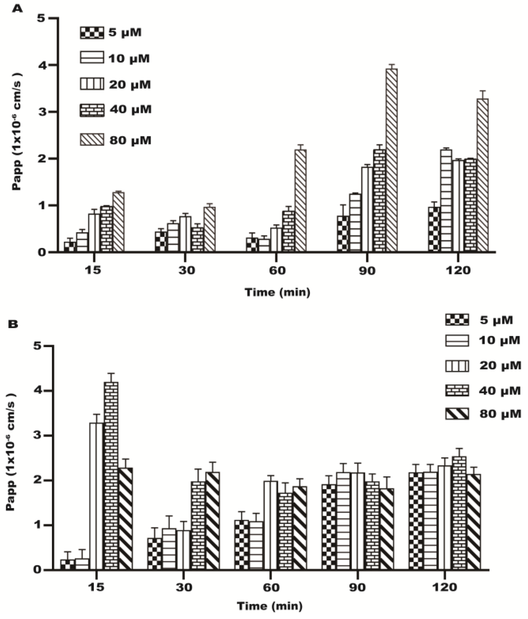
Some studies have shown that the Papp value of drugs or additives can reflect the absorption capacity of substances in the intestine [20]. During the transport of resveratrol at different concentrations on the AP–BL side, the Papp value increased with time and concentration (Figure 4A). In the transport process of the BL–AP side, the Papp values of each concentration group decreased gradually with the increase in time, and the Papp values of each time group decreased gradually with the increase in concentrations from 20~80 μM (Figure 4B). The results indicated the existence of the energy-consuming active transport of resveratrol in the Caco-2 cells during transport.

2.4. Uptake Test Results

Resveratrol uptake increased with time in the Caco-2 monolayer cells at 37 °C (Figure 5A). A significant reduction in resveratrol intake is shown in Figure 5B compared to 37 °C. Low temperatures may affect the fluidity of cell membranes, thereby reducing the Caco-2 cells’ intake of resveratrol. In addition, low temperatures can promote cell-to-cell communication by opening up dense connections between cells. Therefore, when the temperature is lowered from 37 °C to 4 °C, the intake of resveratrol by Caco-2 cells will decrease (Figure 5B). When resveratrol was added to Caco-2 monolayer cells under 37 °C and 120 min, the uptake rate decreased with the concentration increase (Figure 5C). The above results showed that resveratrol uptake by Caco-2 cells is time-dependent, and the temperature has an impact on the uptake of resveratrol.

2.5. Effect of GLUT1 Inhibition on Resveratrol Transport and Uptake

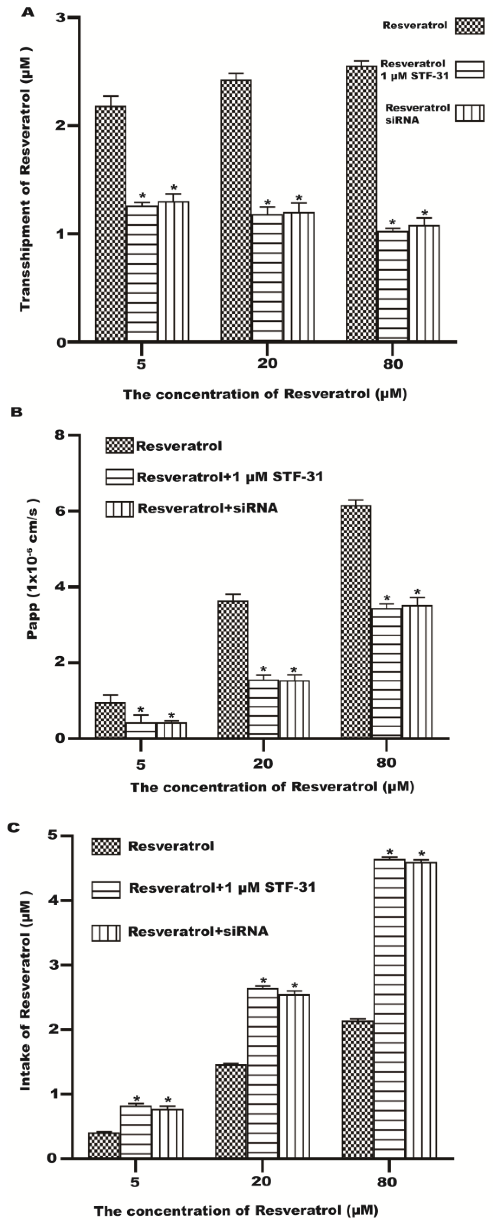
In order to demonstrate that GLUT1 may be involved in the transport and uptake of resveratrol across the monolayer of Caco-2 cells, we investigated the effects of STF-31 (GLUT1 inhibitor) and siRNA on the transmembrane transport and uptake of resveratrol. Pretreatment of cells with STF-31 and GLUT1 siRNA could significantly reduce the transport of resveratrol (Figure 6A,B). Compared with the control group, the intake of resveratrol was significantly increased in the inhibitor group and the siRNA group (Figure 6C). The results showed that GLUT1 was necessary for the excretion of resveratrol from the Caco-2 cells.

2.6. Resveratrol Protects the Cell Viability of H2O2-Stimulated Caco-2 Cells

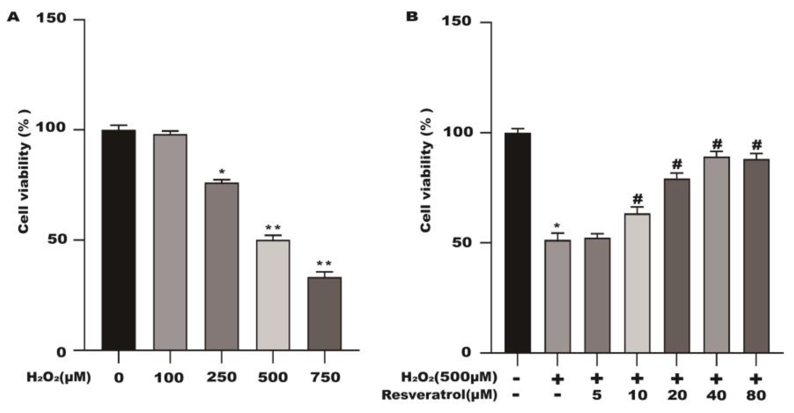
In Figure 1, different concentrations of resveratrol had no significant effect on the activity of the Caco-2 cells at 24 h. The activity of the Caco-2 cells pretreated with different concentrations of H2O2 was significantly decreased. Compared with the control group, the cell viability was reduced to 50.5% after pretreatment of the Caco-2 cells with 500 μM H2O2 for 3 h (Figure 7A). As shown in Figure 7B, resveratrol was able to effectively increase H2O2-induced cell viability in a dose-dependent manner.

2.7. Metabolomics Analysis of Resveratrol Effect on H2O2-Induced Caco-2 Cells

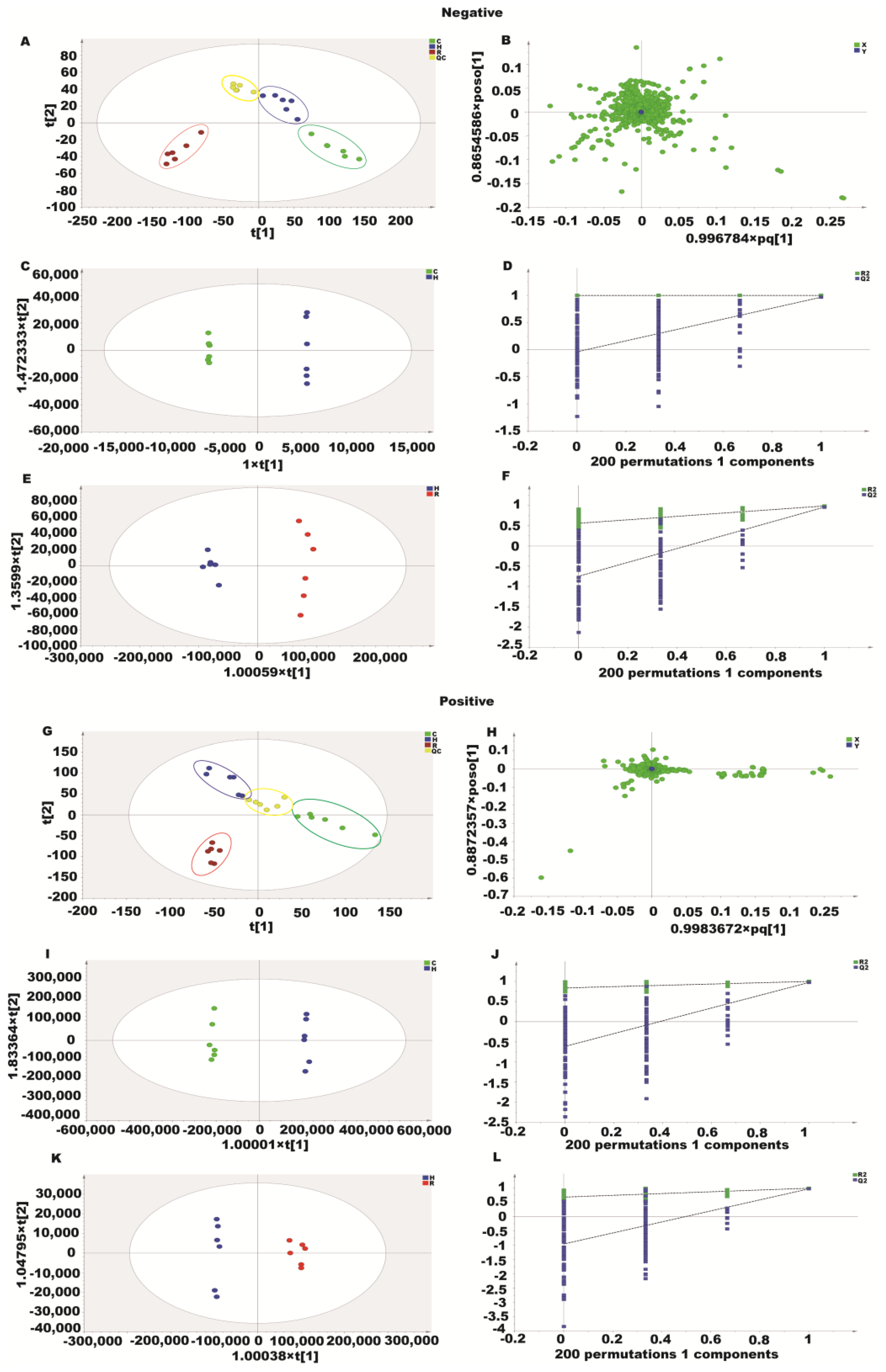
Three experimental groups were analyzed with unsupervised PCA in this study. Positive and negative modes explained 86.8% and 72.6% of the total variance, respectively. The PCA plots (Figure 8A,B,G,H) showed clear separation between the three groups in both positive and negative ion modes. We used supervised orthogonal partial least squares discriminant analysis (OPLS–DA) to separate and identify the metabolites further. The OPLS–DA score plots clearly distinguished the H2O2 group from the other groups without any overlap in either positive or negative modes (Figure 8C,E,I,K). Based on the R2X, R2Y, and Q2 values, the OPLS–DA model was robust and predictive (Figure 8D,F,J,L).
Twenty-one metabolites were identified as potential metabolites, including pipecolinic acid, adenine, uridine, bergenin, glycitin, arginine, citrulline, phosphatidylethanolamine 18, glycine, glutamine, phosphatidylethanolamine 15, threonic acid, methylxanthine, coumaroyl hexoside, malic acid, feruloyl quinic acid, pyroglutamic acid, L-ornithine, L-serine, gluconic acid, and L-glutamic acid (Table 1). As a result of resveratrol treatment, either up- or downregulation of these metabolites occurred.
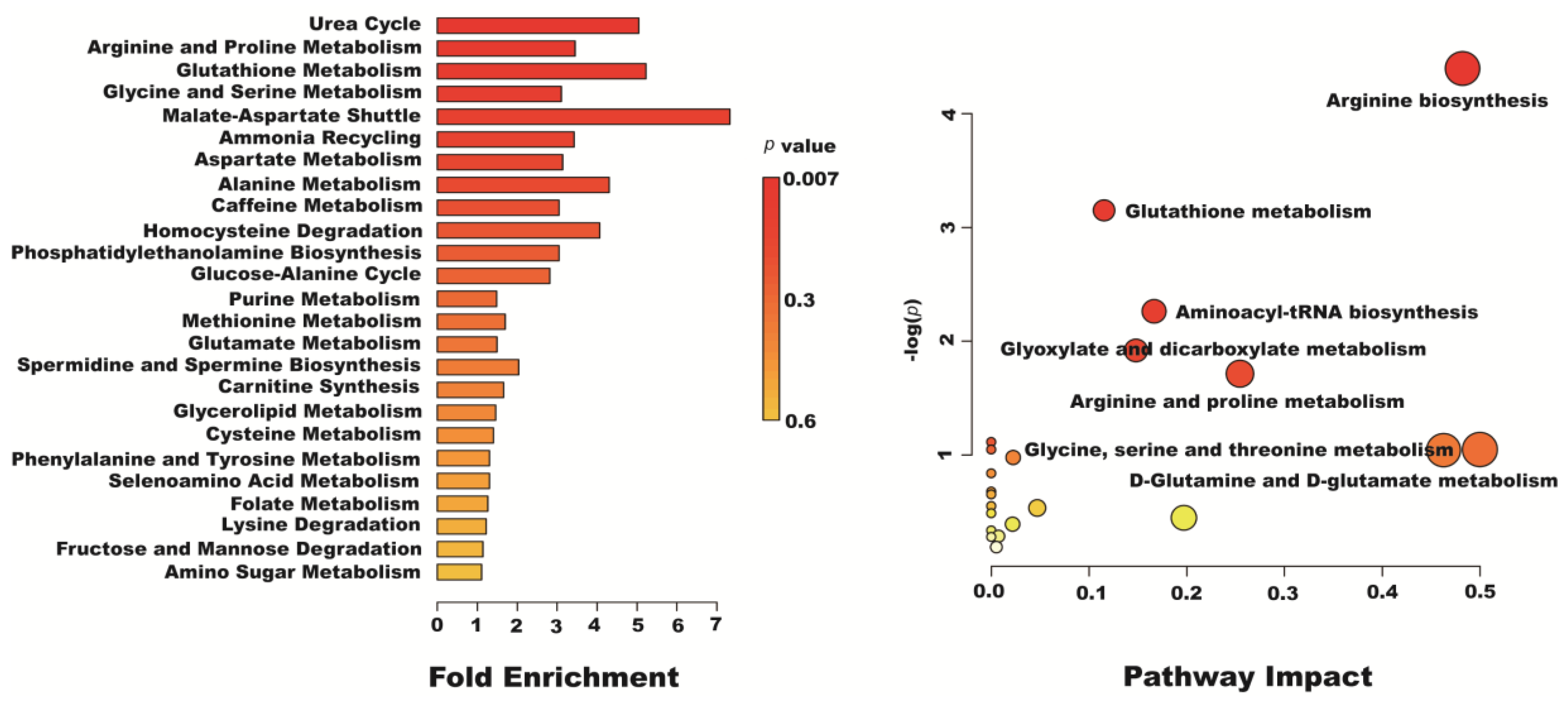
The metabolic pathway analysis data are shown as a bar chart and a bubble chart in Figure 9. According to the KEGG enrichment analysis of differentially expressed metabolites, 25 differentially expressed metabolite pathways were enriched: urea cycle, arginine and proline metabolism, glycine and serine metabolism, ammonia recycling, aspartate metabolism, glutathione metabolism, etc. (p < 0.05). The influence of the path is mainly concentrated in arginine biosynthesis, glutathione metabolism, aminoacyl-tRNA biosynthesis, glyoxylate and dicarboxylate metabolism, arginine and proline metabolism, glycine, serine and threonine metabolism, and D-glutamine and D-glutamate metabolism. The above results show that the oxidative injury induced by H2O2 caused metabolic disorder, and resveratrol could effectively regulate this imbalance (Figure 10).

3. Discussion

Polyphenols are organic compounds found in plant foods [21,22]. Polyphenolic compounds such as resveratrol have many functions [23,24]. Following metabolism, resveratrol is excreted through the urine and feces when consumed orally [25,26]. In the body, resveratrol has relatively low bioavailability. There is approximately 1% bioavailability of resveratrol metabolites in the small intestine and liver. In mammals, resveratrol is metabolized primarily as sulfated and glucuronidated products. A Caco-2 cell model was used in this study to observe the transport and uptake of resveratrol. GLUT1 was found to mediate resveratrol transport in the study. However, some studies have shown that GLUT1 is not necessary for resveratrol transport, but an ATP-binding cassette transporter is necessary [27]. This study found that PappBA polarized resveratrol absorption, and that transporter carriers transported resveratrol. Resveratrol transport was significantly inhibited by STF-31 and GLUT1 siRNA, and it was shown that GLUT1 was most likely involved in resveratrol transport.
As a result of this experiment, resveratrol’s Papp in the Caco-2 cell monolayer ranged from 1.0 × 10⁻⁶ to 4.0 × 10⁻⁶ cm·s−1. The results could predict the mechanism by which resveratrol is absorbed in the intestinal tract in reality. In accordance with current international judging standards on substance absorption, a transport rate of 89.62% was observed for resveratrol in the Caco-2 cells, and resveratrol was readily absorbed. There is evidence that macromolecules are transported in Caco-2 cells via transcellular or paracellular pathways [28]. Paracellular pathways open tight junctions so molecules can pass between cells. This study found that TEER did not decrease significantly, suggesting that resveratrol rarely crosses cellular membranes via paracellular routes.
Studies have shown that trans-resveratrol crosses the cells’ apical membrane through passive transport, while trans-piceid crosses through active transport [29]. In Caco-2 cells, there is evidence that the multidrug-related protein 2 (MRP2), an efflux pump, plays a role in stilbene efflux. An MRP inhibitor, MK-571, appears to inhibit trans-piceid efflux and trans-resveratrol efflux from the apical membrane [30]. It is possible that resveratrol’s oral bioavailability will be limited as enterocytes efficiently metabolize it, mostly by sulfate conjugation and to a lesser extent by glucuronidation.
Temperature changes can significantly affect the fluidity of cell membranes. When the external temperature decreases significantly, the fluidity of the cell membrane also decreases significantly. At low temperatures, enzyme activity in the free cells slows and the fluidity of the cell membrane is decreased, and this interferes with the transport mechanisms. When the Caco-2 cells were at 37 °C, the transport and uptake of resveratrol were normal. However, when the Caco-2 cells were in a low-temperature (4 °C) environment, the transport and uptake of resveratrol significantly decreased. The decrease in temperature not only affects the fluidity of cell membranes, but it also affects the vitality of cells. When a cell is at a lower temperature, its vitality is also affected. When cell viability decreases, the transport and uptake of drugs also decrease accordingly. In this study, whether the fluidity of cell membranes and cell viability affect uptake and transport remains a scientific issue that we will continue to explore in the future.
In cellular metabolomics, levels of endogenous small molecule metabolites are studied in response to various stimuli in order to detect changes in metabolic pathways. A cellular metabolomic analysis of Caco-2 cells pretreated with resveratrol revealed 21 differential metabolites. These differential metabolites were involved in amino acid metabolism, carbohydrate metabolism, energy metabolism, and the metabolism of cofactors. The arginine, citrulline, serine, and glutamic acid concentrations in the H2O2 group were lower, while the amino acid concentrations were significantly higher in the resveratrol group. In the urea cycle, citrulline plays an important role [31,32]. As a result of oxidative stress, arginine, synthesized from citrulline, was found to play an important role in the regulation of immune responses [33]. An enhanced arginine–citrulline–nitric oxide pathway reduces the oxidative damage caused by H2O2. In the H2O2 group, the adenine levels were significantly higher. In oxidative stress studies, adenine levels are often increased [34,35]. In this case, however, it is also possible that they represent ATP loss as part of tissue turnover [36]. It has been suggested that the increase in ornithine and serine over the course of life can contribute to an increased risk of apoptosis, mitochondrial dysfunction, inflammation, lipid metabolism, autophagy, as well as antioxidant stress [37]. Compared with the control group, the level of bergenin was significantly decreased in the H2O2 group; however, resveratrol could significantly increase its level. Antioxidant and anti-inflammatory properties have been demonstrated for bergenin in studies [38,39]. Bergenin hinders the production of extracellular matrix in glomerular mesangial cells and mitigates diabetic nephropathy in mice by suppressing oxidative stress through the mTOR/β-TrcP/Nrf2 pathway [40]. Through immunomodulatory and antioxidant effects, bergenin protects Balb/c mice against cyclophosphamide-induced immunosuppression [41]. In another study, lipopolysaccharide-induced mastitis was observed in mice; inflammatory effects are exerted by bergenin via regulating MAPK and NF-κB signaling pathways [42]. In the H2O2 group, pyroglutamic acid and glutamic acid levels were significantly higher. In glutathione metabolism, glutamic acid is an intermediate. An elevated glutamic acid level affects glutathione metabolism [43,44]. According to the data, resveratrol reduced glutamic acid, while H2O2 increased it, indicating the protective effect of resveratrol in normalizing glutathione metabolism. In the cell membrane, phospholipids called phosphatidylethanolamines play an important role [45,46]. As compared with the control group, the levels of phosphatidylethanolamine 18 and 15 were significantly increased in the H2O2 group. The phosphatidylethanolamine levels were significantly reduced by resveratrol. The majority of these differential metabolites are involved in phosphatidylethanolamine and phosphatidylcholine biosynthesis. The oxidative stress indicator threonic acid is a product of the degradation of ascorbic acid. In the H2O2 group, the threonine levels were significantly lower than in the control group. However, after the resveratrol treatment, the threonine level was significantly recovered. In the metabolism of methylxanthine, 7-Methylxanthine is formed as a purine component. 7-methylxanthine may be closely related to resveratrol’s antioxidant and anti-inflammatory properties. A multitude of cellular processes are affected by malic acid, including the glyoxylate and citric acid cycles [47,48]. Resveratrol was able to significantly increase the level of malic acid compared with the H2O2 group. The results showed that resveratrol can accelerate the citric acid cycle, which speeds up the production of ATP. The glutathione metabolic pathway includes a differential metabolite, pyroglutamate, which is altered by H2O2 and reversed by resveratrol. In maintaining redox homeostasis, glutathione protects cells against oxidative damage and toxicity from exogenous electrophiles. Glutathione metabolism is impaired by elevated levels of pyroglutamate, an intermediate in glutathione synthesis. Resveratrol decreased the level of this metabolite, which was increased by H2O2. The results suggest that resveratrol appears to normalize glutathione metabolism in a protective manner. The results of the metabolomics analysis showed that resveratrol might improve H2O2 induced oxidative damage by increasing the antioxidant level.
Metabolomics analyses using UPLC/MS are limited in their ability to identify many differentially altered metabolites. In the present study, based on the metabolomics analysis conducted with UPLC/MS, the results of the present study explained the biological changes between the groups only partially. There is certainly a need for further research on the interactions between resveratrol and these metabolic pathways.

4. Materials and Methods

4.1. Chemicals

Transwell TM cell culture dishes were obtained from Corning Costar Corp (Cambridge, MA, USA). The CCK-8 kit was purchased from Beyotime (Shanghai, China). The MEM glucose medium, Hank’s buffer, and cell culture flask were obtained from Gibco (Grand Island, NY, USA). The Lucifer yellow was from Solarbio (Beijing, China). The MS-gradeacetonitrile was purchased from Thermo Fisher Scientific (Waltham, MA, USA). The formic acid (98.0%, for LC-MS) was purchased from Tokyo Chemical Industry (Shanghai, China). The glucose transporter 1 siRNA sequence: 5′-AUUCCAUUCACAGUGC UGAUCGCUC-3′; for the scrambled control siRNA: 5′-UACUCAG AACGAGUCUCGUUT-3′. The siRNA specifically targeting glucose transporter 1 and the control siRNA were purchased from Jikai Gene Chemical (Shanghai, China). The STF-31 was purchased from MCE (Shanghai, China). The resveratrol (purity greater than 99%) was purchased from Merck (Darmstadt, Germany).

4.2. Cell Culture

The cell cultures of Caco-2 were grown as before [49]. The Caco-2 cell line, derived from human epithelial colorectal adenocarcinoma, was cultured in Caco-2 cell culture medium. This medium consisted of DMEM high glucose medium supplemented with 10% FBS, 2% L-glutamine, 1% NEAA, and 1% penicillin-streptomycin mix. Before 500 μM H2O2 was used to intervene in the cells, the Caco-2 cells were pretreated with resveratrol for 24 h.

4.3. Cell Viability

Cell viability was determined via CCK-8 assay [50]. The Caco-2 cells were inoculated into 96-well plates and intervened with resveratrol and H2O2. After 24 h, 10 µL of CCK-8 solution was added to each well. After 2 h, the optical density value of each hole at 450 nm was measured using a microplate reader (Bio-Rad 550, Hercules, CA, USA).

4.4. Establishment and Evaluation of the Caco-2 Cell Uptake Model

As previously published, the Caco-2 cell model was established [49]. In brief, the Caco-2 cells were seeded onto polycarbonate 6-well Transwell inserts at a density of 2 × 10⁶ cells/well; then, the confluent monolayers (21 days) were used for permeability studies. The transepithelial electrical resistance (TEER) of the monolayer was measured using an epithelial voltohmmeter (World Precision Instruments, Sarasota, FL, USA) to determine the formation of the monolayer and its integrity during the experiment. The monolayers were washed with Hank’s balanced salt solution (HBSS) prior to the experiment, after which 0.5 and 1.2 mL of HBSS were placed into the upper and lower compartments, respectively. A total volume of 100 μL of solution was taken from the lower compartment at regular intervals over 120 min and replaced with the same volume of fresh buffer.

4.5. Analytical Methods

4.5.1. HBSS Buffer Sample Extraction

Approximately 250 μL HBSS buffer sample containing resveratrol was collected, and 250 μL methanol was added into it and mixed evenly. The mixture was centrifuged in a 14,000× g centrifuge for 10 min. After collecting and evaporating the supernatant, it was reconstituted in 200 μL methanol and analyzed using ultra-high liquid chromatography (UHPLC).

4.5.2. Liquid Chromatography Analysis

The resveratrol was analyzed on an Agilent 1290 UHPLC system (Agilent Technologies, Waldbronn, Germany). In the mobile phase, 0.2% phosphoric acid and methanol (40:60, v/v) was combined at a flow rate of 1 mL/min in an acid solution. A constant wavelength of 306 nm was used to record resveratrol elution.

4.6. Transport Assay of Caco-2 Cells

The AP and BL sides were tested separately as part of the resveratrol transport experiment. Different concentrations of resveratrol solutions (5, 10, 20, 40, and 80 μM) were added to the cells. Approximately 250 μL of sample solution was collected on the BL side at different times. Approximately 250 μL of HBSS was added immediately. We investigated the effect on the transport rate of resveratrol at 37 °C and 4 °C, respectively.

4.7. Caco-2 Cells Uptake Experiment

Different concentrations of resveratrol solution (5, 10, 20, 40, and 80 μM) were added to the cells. The cells were collected at different time periods. Similar to the above steps, the effect of temperature (4 °C) on resveratrol uptake was studied.
The uptake rate (%) = (Resveratrol content in cells/Total amount of resveratrol) × 100%.

4.8. Metabonomic Analysis

4.8.1. Cellular Metabolite Extraction

The Caco-2 cells were treated with resveratrol and H2O2 and collected into centrifuge tubes. After, 80% methanol was added into the centrifuge tube; the sample was mixed and frozen at −80 °C for 5 min. The mixture was lysed with two freeze–thaw cycles (frozen in liquid nitrogen and thawing at 37 °C). Quality control (QC) samples were prepared from 20 μL of supernatant from each sample.

4.8.2. UHPLC–QE Orbitrap/MS/MS Conditions

An HPLC system coupled to an Orbitrap MS was used for the LC–MS/MS analysis. In mobile phase A, 0.1% formic acid was added to water, while in mobile phase B, acetonitrile was added. In this experiment, the elution gradient was set as follows: 0 min, 2% B; 1 min, 2% B; 18 min, 100% B; 22 min, 100% B; 22.1 min, 2% B; and 25 min, 2% B. The flow rate was 0.3 mL/min. The injection volume was 2 μL. During the LC/MS experiments, a QE mass spectrometer was used for acquiring MS/MS spectra on an information-dependent basis (IDA). The ESI source conditions were as follows: the aux gas flow rate as 16 Arb, the collision energy as 25 eV in the NCE model, and the spray voltage as −3.0 kV or 3.6 kV.

4.8.3. Qualitative Analysis of Metabolites

ABF Converter software was used to convert the raw data into “Analysis Base File” (ABF) files. In the data processing, MSDIAL 2.2.62 software was used to detect peaks, deconvolve them, and align them. SIMCA (version 14.1, Umetrics AB, Umeå, Sweden) was used to import the data obtained. A PCA and an OPLS–DA analysis was performed on the SIMCA data. Mass values of differential metabolites were retrieved from the Human Metabolome Database (HMDB). MetabolAnalyst 5.0 was used to analyze differential metabolites in clusters and pathway analyses.

4.9. Statistical Analysis

The data were analyzed using GraphPad Prism 8 (Graphpad Software, LaJolla, CA, USA). Means and standard deviations were presented for all data. In addition to one-way ANOVA, Duncan’s multiple comparisons was used to analyze differences between the treatment groups. Statistical significance was considered at p < 0.05.

5. Conclusions

In the Caco-2 cell model, resveratrol is transported and absorbed mainly by GLUT1. The results of cell metabolomics show that resveratrol can play an important role in the prevention of intestinal diseases caused by oxidative stress as an additive.

Author Contributions

Conceptualization, Y.-J.Y.; methodology, Z.Q.; software, X.-W.L.; re-sources, Q.T.; data curation, Q.T.; writing—original draft preparation, Z.-D.Z.; writing—review and editing, Z.-D.Z.; visualization, S.-H.L.; supervision, J.-Y.L.; project administration, W.-B.G.; funding acquisition, L.-X.B. All authors have read and agreed to the published version of the manuscript.

Funding

This study was supported by the Gansu Youth Science and Technology Fund Program (Grant/Award Numbers: 21JR7RA031).

Institutional Review Board Statement

Not applicable.

Informed Consent Statement

Not applicable.

Data Availability Statement

The data used to support the findings of this study are included within the article.

Conflicts of Interest

The authors declare no conflict of interest.

Sample Availability

The datasets used and/or analyzed during the current study are available from the corresponding author on reasonable request.

References

  1. Jacobson, A.; Yang, D.; Vella, M.; Chiu, I.M. The intestinal neuro-immune axis: Crosstalk between neurons, immune cells, and microbes. Mucosal Immunol. 2021, 14, 555–565. [Google Scholar] [CrossRef] [PubMed]
  2. Gao, Y.; Liang, Z.; Lv, N.; Shan, J.; Zhou, H.; Zhang, J.; Shi, L. Exploring the total flavones of Abelmoschus manihot against IAV-induced lung inflammation by network pharmacology. BMC Complement. Med. Ther. 2022, 22, 36. [Google Scholar] [CrossRef] [PubMed]
  3. Kriseldi, R.; Johnson, J.A.; Walk, C.L.; Bedford, M.R.; Dozier, W.A., 3rd. Influence of exogenous phytase supplementation on phytate degradation, plasma inositol, alkaline phosphatase, and glucose concentrations of broilers at 28 days of age. Poult. Sci. 2021, 100, 224–234. [Google Scholar] [CrossRef] [PubMed]
  4. Gianchecchi, E.; Fierabracci, A. Insights on the Effects of Resveratrol and Some of Its Derivatives in Cancer and Autoimmunity: A Molecule with a Dual Activity. Antioxidants 2020, 9, 91. [Google Scholar] [CrossRef] [PubMed] [Green Version]
  5. Carr, A.C.; Rowe, S. Factors Affecting Vitamin C Status and Prevalence of Deficiency: A Global Health Perspective. Nutrients 2020, 12, 1963. [Google Scholar] [CrossRef]
  6. Efenberger-Szmechtyk, M.; Nowak, A.; Nowak, A. Cytotoxic and DNA-Damaging Effects of Aronia melanocarpa, Cornus mas, and Chaenomeles superba Leaf Extracts on the Human Colon Adenocarcinoma Cell Line Caco-2. Antioxidants 2020, 9, 1030. [Google Scholar] [CrossRef]
  7. Aleksiejczuk, M.; Gromotowicz-Poplawska, A.; Marcinczyk, N.; Stelmaszewska, J.; Dzieciol, J.; Chabielska, E. Aldosterone Increases Vascular Permeability in Rat Skin. Cells 2022, 11, 2707. [Google Scholar] [CrossRef]
  8. Cui, Y.; Claus, S.; Schnell, D.; Runge, F.; MacLean, C. In-Depth Characterization of EpiIntestinal Microtissue as a Model for Intestinal Drug Absorption and Metabolism in Human. Pharmaceutics 2020, 12, 405. [Google Scholar] [CrossRef]
  9. Morresi, C.; Vasarri, M.; Bellachioma, L.; Ferretti, G.; Degl Innocenti, D.; Bacchetti, T. Glucose Uptake and Oxidative Stress in Caco-2 Cells: Health Benefits from Posidonia oceanica (L.) Delile. Mar. Drugs 2022, 20, 457. [Google Scholar] [CrossRef]
  10. Arena, A.; Romeo, M.A.; Benedetti, R.; Masuelli, L.; Bei, R.; Gilardini Montani, M.S.; Cirone, M. New Insights into Curcumin- and Resveratrol-Mediated Anti-Cancer Effects. Pharmaceuticals 2021, 14, 1068. [Google Scholar] [CrossRef]
  11. Summerlin, N.; Soo, E.; Thakur, S.; Qu, Z.; Jambhrunkar, S.; Popat, A. Resveratrol nanoformulations: Challenges and opportunities. Int. J. Pharm. 2015, 479, 282–290. [Google Scholar] [CrossRef] [PubMed]
  12. Sales, J.M.; Resurreccion, A.V. Resveratrol in peanuts. Crit. Rev. Food Sci. Nutr. 2014, 54, 734–770. [Google Scholar] [CrossRef] [PubMed]
  13. De Oliveira, M.R.; Nabavi, S.F.; Manayi, A.; Daglia, M.; Hajheydari, Z.; Nabavi, S.M. Resveratrol and the mitochondria: From triggering the intrinsic apoptotic pathway to inducing mitochondrial biogenesis, a mechanistic view. Biochim. Biophys. Acta 2016, 1860, 727–745. [Google Scholar] [CrossRef] [PubMed]
  14. Wang, X.; Xie, Y.; Zhang, T.; Bo, S.; Bai, X.; Liu, H.; Li, T.; Liu, S.; Zhou, Y.; Cong, X.; et al. Resveratrol reverses chronic restraint stress-induced depression-like behaviour: Involvement of BDNF level, ERK phosphorylation and expression of Bcl-2 and Bax in rats. Brain Res. Bull. 2016, 125, 134–143. [Google Scholar] [CrossRef] [PubMed]
  15. Kalantari, H.; Das, D.K. Physiological effects of resveratrol. Biofactors 2010, 36, 401–406. [Google Scholar] [CrossRef]
  16. Frazzi, R.; Tigano, M. The multiple mechanisms of cell death triggered by resveratrol in lymphoma and leukemia. Int. J. Mol. Sci. 2014, 15, 4977–4993. [Google Scholar] [CrossRef] [Green Version]
  17. Ling, L.; Gu, S.; Cheng, Y. Resveratrol activates endogenous cardiac stem cells and improves myocardial regeneration following acute myocardial infarction. Mol. Med. Rep. 2017, 15, 1188–1194. [Google Scholar] [CrossRef] [Green Version]
  18. Liu, M.H.; Yuan, C.; He, J.; Tan, T.P.; Wu, S.J.; Fu, H.Y.; Liu, J.; Yu, S.; Chen, Y.D.; Le, Q.F.; et al. Resveratrol Protects PC12 Cells from High Glucose-Induced Neurotoxicity Via PI3K/Akt/FoxO3a Pathway. Cell. Mol. Neurobiol. 2015, 35, 513–522. [Google Scholar] [CrossRef]
  19. Zeng, Y.H.; Zhou, L.Y.; Chen, Q.Z.; Li, Y.; Shao, Y.; Ren, W.Y.; Liao, Y.P.; Wang, H.; Zhu, J.H.; Huang, M.; et al. Resveratrol inactivates PI3K/Akt signaling through upregulating BMP7 in human colon cancer cells. Oncol. Rep. 2017, 38, 456–464. [Google Scholar] [CrossRef] [Green Version]
  20. JanssenDuijghuijsen, L.M.; Grefte, S.; de Boer, V.C.J.; Zeper, L.; van Dartel, D.A.M.; van der Stelt, I.; Bekkenkamp-Grovenstein, M.; van Norren, K.; Wichers, H.J.; Keijer, J. Mitochondrial ATP Depletion Disrupts Caco-2 Monolayer Integrity and Internalizes Claudin 7. Front. Physiol. 2017, 8, 794. [Google Scholar] [CrossRef] [Green Version]
  21. Mena, J.; Elgueta, E.; Espinola-Gonzales, F.; Cardenas, H.; Orihuela, P.A. Hydroethanolic Extracts of the Aristotelia Chilensis (Maqui) Berry Reduces Cellular Viability and Invasiveness in the Endometrial Cancer Cell Line Ishikawa. Integr. Cancer Ther. 2021, 20, 15347354211007560. [Google Scholar] [CrossRef]
  22. Romani, A.; Campo, M.; Urciuoli, S.; Marrone, G.; Noce, A.; Bernini, R. An Industrial and Sustainable Platform for the Production of Bioactive Micronized Powders and Extracts Enriched in Polyphenols From Olea europaea L. and Vitis vinifera L. Wastes. Front. Nutr. 2020, 7, 120. [Google Scholar] [CrossRef] [PubMed]
  23. Gonzalez, A.; Krieg, R.; Massey, H.D.; Carl, D.; Ghosh, S.; Gehr, T.W.B.; Ghosh, S.S. Sodium butyrate ameliorates insulin resistance and renal failure in CKD rats by modulating intestinal permeability and mucin expression. Nephrol. Dial. Transpl. 2019, 34, 783–794. [Google Scholar] [CrossRef] [PubMed]
  24. Yang, N.; Gao, J.; Hou, R.; Xu, X.; Yang, N.; Huang, S. Grape Seed Proanthocyanidins Inhibit Migration and Invasion of Bladder Cancer Cells by Reversing EMT through Suppression of TGF-beta Signaling Pathway. Oxid. Med. Cell. Longev. 2021, 2021, 5564312. [Google Scholar] [CrossRef]
  25. Bullon-Vela, V.; Abete, I.; Zulet, M.A.; Xu, Y.; Martinez-Gonzalez, M.A.; Sayon-Orea, C.; Ruiz-Canela, M.; Toledo, E.; Sanchez, V.M.; Estruch, R.; et al. Urinary Resveratrol Metabolites Output: Differential Associations with Cardiometabolic Markers and Liver Enzymes in House-Dwelling Subjects Featuring Metabolic Syndrome. Molecules 2020, 25, 4340. [Google Scholar] [CrossRef]
  26. Zortea, K.; Franco, V.C.; Guimarães, P.; Belmonte-de-Abreu, P.S. Resveratrol Supplementation Did Not Improve Cognition in Patients with Schizophrenia: Results from a Randomized Clinical Trial. Front. Psychiatry 2016, 7, 159. [Google Scholar] [CrossRef] [PubMed] [Green Version]
  27. Hu, Y.; Li, C.; Li, H.; Li, M.; Shu, X. Resveratrol-mediated reversal of tumor multi-drug resistance. Curr. Drug Metab. 2014, 15, 703–710. [Google Scholar] [CrossRef] [PubMed]
  28. Tomassen, M.M.M.; Govers, C.; Vos, A.P.; de Wit, N.J.W. Dietary fat induced chylomicron-mediated LPS translocation in a bicameral Caco-2cell model. Lipids Health Dis. 2023, 22, 4. [Google Scholar] [CrossRef]
  29. Bilotta, S.; Arbogast, J.; Schart, N.; Frei, M.; Lorentz, A. Resveratrol Treatment Prevents Increase of Mast Cells in Both Murine OVA Enteritis and IL-10(-/-) Colitis. Int. J. Mol. Sci. 2022, 23, 1213. [Google Scholar] [CrossRef]
  30. Henry, C.; Vitrac, X.; Decendit, A.; Ennamany, R.; Krisa, S.; Merillon, J.M. Cellular uptake and efflux of trans-piceid and its aglycone trans-resveratrol on the apical membrane of human intestinal Caco-2 cells. J. Agric. Food Chem. 2005, 53, 798–803. [Google Scholar] [CrossRef]
  31. Subramanian, P.; Oh, B.J.; Mani, V.; Lee, J.K.; Lee, C.M.; Sim, J.S.; Koo, J.C.; Hahn, B.S. Differential Metabolic Profiles during the Developmental Stages of Plant-Parasitic Nematode Meloidogyne incognita. Int. J. Mol. Sci. 2017, 18, 1351. [Google Scholar] [CrossRef] [PubMed] [Green Version]
  32. Tan, W.; He, J.; Deng, J.; Yang, X.; Cui, L.; Ran, R.; Du, G.; Jiang, X. Small molecule metabolite biomarkers for hepatocellular carcinoma with bile duct tumor thrombus diagnosis. Sci. Rep. 2018, 8, 3309. [Google Scholar] [CrossRef] [Green Version]
  33. Stoessel, D.; Stellmann, J.P.; Willing, A.; Behrens, B.; Rosenkranz, S.C.; Hodecker, S.C.; Sturner, K.H.; Reinhardt, S.; Fleischer, S.; Deuschle, C.; et al. Metabolomic Profiles for Primary Progressive Multiple Sclerosis Stratification and Disease Course Monitoring. Front. Hum. Neurosci. 2018, 12, 226. [Google Scholar] [CrossRef] [PubMed]
  34. Liu, Z.J.; Zhong, J.; Zhang, M.; Chen, Z.H.; Wang, J.Y.; Chen, H.Y.; Wang, X.Q.; Zhang, B. The Alexipharmic Mechanisms of Five Licorice Ingredients Involved in CYP450 and Nrf2 Pathways in Paraquat-Induced Mice Acute Lung Injury. Oxid. Med. Cell. Longev. 2019, 2019, 7283104. [Google Scholar] [CrossRef] [PubMed]
  35. Hsu, C.N.; Hou, C.Y.; Chang-Chien, G.P.; Lin, S.; Yang, H.W.; Tain, Y.L. Perinatal Resveratrol Therapy Prevents Hypertension Programmed by Maternal Chronic Kidney Disease in Adult Male Offspring: Implications of the Gut Microbiome and Their Metabolites. Biomedicines 2020, 8, 567. [Google Scholar] [CrossRef]
  36. O’Brien, W.G., 3rd; Berka, V.; Tsai, A.L.; Zhao, Z.; Lee, C.C. CD73 and AMPD3 deficiency enhance metabolic performance via erythrocyte ATP that decreases hemoglobin oxygen affinity. Sci. Rep. 2015, 5, 13147. [Google Scholar] [CrossRef] [Green Version]
  37. Chak, C.M.; Lacruz, M.E.; Adam, J.; Brandmaier, S.; Covic, M.; Huang, J.; Meisinger, C.; Tiller, D.; Prehn, C.; Adamski, J.; et al. Ageing Investigation Using Two-Time-Point Metabolomics Data from KORA and CARLA Studies. Metabolites 2019, 9, 44. [Google Scholar] [CrossRef] [Green Version]
  38. De Sa Hyacienth, B.M.; Tavares Picanco, K.R.; Sanchez-Ortiz, B.L.; Barros Silva, L.; Matias Pereira, A.C.; Machado Goes, L.D.; Sousa Borges, R.; Cardoso Ataide, R.; Dos Santos, C.B.R.; de Oliveira Carvalho, H.; et al. Hydroethanolic extract from Endopleura uchi (Huber) Cuatrecasas and its marker bergenin: Toxicological and pharmacokinetic studies in silico and in vivo on zebrafish. Toxicol. Rep. 2020, 7, 217–232. [Google Scholar] [CrossRef]
  39. Zeng, Q.; Zhou, T.; Zhao, F.; Xiong, D.; He, B.; Hua, Q.; Lin, M.; Deng, L.; Sang, X.; Xie, W.; et al. p62-Nrf2 Regulatory Loop Mediates the anti-Pulmonary Fibrosis Effect of Bergenin. Antioxidants 2022, 11, 307. [Google Scholar] [CrossRef]
  40. Qiao, S.; Liu, R.; Lv, C.; Miao, Y.; Yue, M.; Tao, Y.; Wei, Z.; Xia, Y.; Dai, Y. Bergenin impedes the generation of extracellular matrix in glomerular mesangial cells and ameliorates diabetic nephropathy in mice by inhibiting oxidative stress via the mTOR/beta-TrcP/Nrf2 pathway. Free Radic. Biol. Med. 2019, 145, 118–135. [Google Scholar] [CrossRef]
  41. Qi, Q.; Dong, Z.; Sun, Y.; Li, S.; Zhao, Z. Protective Effect of Bergenin against Cyclophosphamide-Induced Immunosuppression by Immunomodulatory Effect and Antioxidation in Balb/c Mice. Molecules 2018, 23, 2668. [Google Scholar] [CrossRef] [Green Version]
  42. Gao, X.J.; Guo, M.Y.; Zhang, Z.C.; Wang, T.C.; Cao, Y.G.; Zhang, N.S. Bergenin Plays an Anti-Inflammatory Role via the Modulation of MAPK and NF-kappaB Signaling Pathways in a Mouse Model of LPS-Induced Mastitis. Inflammation 2015, 38, 1142–1150. [Google Scholar] [CrossRef] [PubMed]
  43. Hong, Y.; Li, B.; Zheng, N.; Wu, G.; Ma, J.; Tao, X.; Chen, L.; Zhong, J.; Sheng, L.; Li, H. Integrated Metagenomic and Metabolomic Analyses of the Effect of Astragalus Polysaccharides on Alleviating High-Fat Diet-Induced Metabolic Disorders. Front. Pharmacol. 2020, 11, 833. [Google Scholar] [CrossRef] [PubMed]
  44. Huang, Y.; Lu, X.; Chen, R.; Chen, Y. Comparative study of the effects of gold and silver nanoparticles on the metabolism of human dermal fibroblasts. Regen. Biomater. 2020, 7, 221–232. [Google Scholar] [CrossRef] [PubMed] [Green Version]
  45. Wang, S.; Liu, F.; Tan, K.S.; Ser, H.L.; Tan, L.T.; Lee, L.H.; Tan, W. Effect of (R)-salbutamol on the switch of phenotype and metabolic pattern in LPS-induced macrophage cells. J. Cell. Mol. Med. 2020, 24, 722–736. [Google Scholar] [CrossRef] [PubMed] [Green Version]
  46. Zhao, Y.Y.; Wang, H.L.; Cheng, X.L.; Wei, F.; Bai, X.; Lin, R.C.; Vaziri, N.D. Metabolomics analysis reveals the association between lipid abnormalities and oxidative stress, inflammation, fibrosis, and Nrf2 dysfunction in aristolochic acid-induced nephropathy. Sci. Rep. 2015, 5, 12936. [Google Scholar] [CrossRef] [Green Version]
  47. Kranawetter, C.; Zeng, S.; Joshi, T.; Sumner, L.W. A Medicago truncatula Metabolite Atlas Enables the Visualization of Differential Accumulation of Metabolites in Root Tissues. Metabolites 2021, 11, 238. [Google Scholar] [CrossRef]
  48. Mihasan, M.; Babii, C.; Aslebagh, R.; Channaveerappa, D.; Dupree, E.; Darie, C.C. Proteomics based analysis of the nicotine catabolism in Paenarthrobacter nicotinovorans pAO1. Sci. Rep. 2018, 8, 16239. [Google Scholar] [CrossRef] [Green Version]
  49. Zhang, Z.D.; Tao, Q.; Qin, Z.; Liu, X.W.; Li, S.H.; Bai, L.X.; Yang, Y.J.; Li, J.Y. Uptake and Transport of Naringenin and Its Antioxidant Effects in Human Intestinal Epithelial Caco-2 Cells. Front. Nutr. 2022, 9, 894117. [Google Scholar] [CrossRef]
  50. Huang, M.Z.; Yang, Y.J.; Liu, X.W.; Qin, Z.; Li, J.Y. Aspirin Eugenol Ester Reduces H2O2-Induced Oxidative Stress of HUVECs via Mitochondria-Lysosome Axis. Oxid. Med. Cell. Longev. 2019, 2019, 8098135. [Google Scholar] [CrossRef] [Green Version]
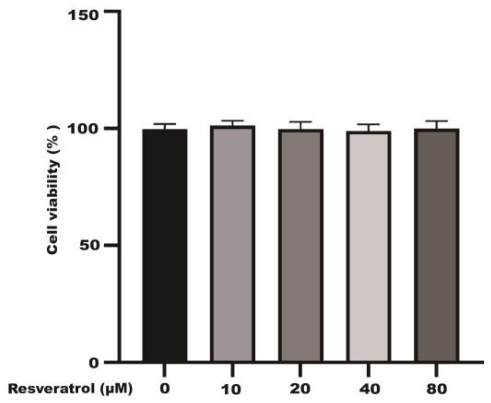
Figure 1. The effects of different concentrations of resveratrol (10~80 μM) on Caco-2 cell viability.
Molecules 28 04569 g001
Figure 2. The transport of resveratrol at different times and temperature. (A) The transport of AP–BL and BL–AP of resveratrol (80 μM) at different times. (B) The transport of AP–BL and BL–AP of resveratrol at different temperatures. * p < 0.05 compared with 37 °C.
Molecules 28 04569 g002
Figure 3. The transport of resveratrol with different concentrations (5~80 μM). (A) The transport of resveratrol in the AP–BL. (B) The transport of resveratrol in the BL–AP. (C) The transport rate of resveratrol. * p < 0.05 compared with AP-BL side.
Molecules 28 04569 g003
Figure 4. The Papp of each concentration (5~80 μM) of resveratrol during 120 min. (A) The Papp of AP–BL. (B) The Papp of BL–AP.
Molecules 28 04569 g004
Figure 5. The uptake of resveratrol with different concentrations (5~80 μM). (A) The uptake of resveratrol (80 μM) at different times. (B) The uptake of resveratrol at different temperatures. (C) The uptake rate of resveratrol of Caco-2 cells. * p < 0.05 compared with 4 °C.
Molecules 28 04569 g005
Figure 6. The effect of STF-31 and siRNA on resveratrol transport and uptake in Caco-2 cells. (A) The transport of different concentrations of resveratrol. (B) The Papp of each concentration of resveratrol. (C) The intake of different concentrations of resveratrol. * p < 0.05 compared with the resveratrol group.
Molecules 28 04569 g006
Figure 7. Resveratrol protects the cell viability of H2O2-stimulated Caco-2. (A) Effects of different concentrations of H2O2 on cell viability. (B) Effects of different concentrations of resveratrol pretreatment on H2O2-induced cell viability. * p < 0.05 compared with the control group; ** p < 0.01 compared with the control group; # p < 0.05 compared with the H2O2 group.
Molecules 28 04569 g007
Figure 8. Metabolomics analysis of the effect of resveratrol on H2O2-induced oxidative injury in Caco-2 cells. (A,G) PCA score plots based on the Caco-2 cells of the control, H2O2 and resveratrol groups in positive and negative modes. (B,H) The loading plot of resveratrol and H2O2 groups in positive and negative modes. (C,I) OPLS–DA score plots of the resveratrol and H2O2 groups in positive and negative modes. (D,J) Permutation test of the OPLS–DA model. (E,K) OPLS–DA score plots of the control and H2O2 groups in positive and negative modes. (F,L) Permutation test of the OPLS–DA model.
Molecules 28 04569 g008
Figure 9. The results of fold enrichment and pathway analysis of potential metabolites in Caco-2 cells.
Molecules 28 04569 g009
Figure 10. Overview of altered concentrations of metabolites and related metabolic pathways.
Molecules 28 04569 g010
Table 1. Statistics of differential metabolites in Caco-2 cells.
No.RTVIPFormulaMetabolitesSMm/zFold Change
C/HH/R
11.0725323.38C6H11NO2Pipecolinic acidESI+129.1570.422.91
27.0664921.31C5H5N5AdenineESI+680.3441.382.42
31.8355332.55C9H12N2O6UridineESI+245.07591.212.95
44.2388713.65C14H16O9BergeninESI+328.270.682.56
50.9139324.42C22H22O10GlycitinESI+485.0150.634.27
60.9474921.86C6H14N4O2ArginineESI+174.200.223.83
70.9184165.23C6H13N4O3CitrullineESI+193.1310.412.36
88.7362212.23C40H80NO8PPhosphatidylethanolamine 18ESI+734.0391.580.84
91.4366671.38C2H5NO2GlycineESI+76.039841.854.57
101.5764421.51C10H20N4O6Glutamine ESI+185.0290.842.48
113.9236142.31C41H78NO8PPhosphatidylethanolamine 15ESI+7441.380.14
124.8362291.33C8H14CaO10Threonic acidESI+310.270.592.84
131.8801092.41C6H6N4O27-MethylxanthineESI+167.05492.780.89
145.3027833.02C9H3Cl6NO2Coumaroyl HexosideESI−369.830.521.83
151.3761723.06C4H6O5Malic acidESI−135.02730.722.88
164.2372093.17C17H20O9Feruloyl quinic acid ESI−368.33530.272.98
170.9941672.44C5H7NO3Pyroglutamic acidESI−130.04972.670.66
180.5826132.82C5H13N2O2L-ornithineESI−132.160.782.86
190.9965384.25C3H7NO3L-serineESI−106.050.973.75
201.8623352.99C6H11O7Gluconic acidESI−195.1480.421.56
211.6418691.72C5H9NO4L-Glutamic acidESI−148.06030.862.52
RT: retention time; VIP: variable importance in the projection; SM: scan mode; +: metabolites identified in positive mode; −: metabolites identified in negative mode. Metabolites identified in both positive and negative modes; C/H: control group compared with the H2O2 group; R/H: resveratrol group compared with the H2O2 group.
Disclaimer/Publisher’s Note: The statements, opinions and data contained in all publications are solely those of the individual author(s) and contributor(s) and not of MDPI and/or the editor(s). MDPI and/or the editor(s) disclaim responsibility for any injury to people or property resulting from any ideas, methods, instructions or products referred to in the content.