Article

Preliminary Study on Rapid and Simultaneous Detection of Viable Escherichia coli O157:H7, Staphylococcus aureus, and Salmonella by PMA-mPCR in Food

1
School of Pharmacy and Food Science, Zhuhai College of Science and Technology, Zhuhai 519041, China
2
School of Environmental Ecology and Biological Engineering, Wuhan Institute of Technology, Wuhan 430205, China
3
Nanchang Agricultural Technology Popularization Center, Nanchang 330299, China
4
Faculty of Medicine, Macau University of Science and Technology, Taipa, Macao 999078, China
*
Authors to whom correspondence should be addressed.
Molecules 2023, 28(15), 5835; https://doi.org/10.3390/molecules28155835
Submission received: 8 June 2023 / Revised: 25 July 2023 / Accepted: 31 July 2023 / Published: 3 August 2023
(This article belongs to the Special Issue Recent Advances in Food Microbiology Control)

Abstract

:
Escherichia coli O157:H7, Staphylococcus aureus, and Salmonella are major foodborne pathogens that are widespread in nature and responsible for several outbreaks of food safety accidents. Thus, a rapid and practical technique (PMA-mPCR) was developed for the simultaneous detection of viable E. coli O157:H7, S. aureus, and Salmonella in pure culture and in a food matrix. To eliminate false positive results, propidium monoazide (PMA) was applied to selectively suppress the DNA amplification of dead cells. The results showed the optimum concentration of PMA is 5.0 µg/mL. The detection limit of this assay by mPCR was 103 CFU/mL in the culture broth, and by PMA-mPCR was 104 CFU/mL both in pure culture and a food matrix (milk and ground beef). In addition, the detection of mixed viable and dead cells was also explored in this study. The detection sensitivity ratio of viable and dead counts was less than 1:10. Therefore, the PMA-mPCR assay proposed here might provide an efficient detection tool for the simultaneous detection of viable E. coli O157:H7, S. aureus, and Salmonella and also have great potential for the detection and concentration assessment of VBNC cells.

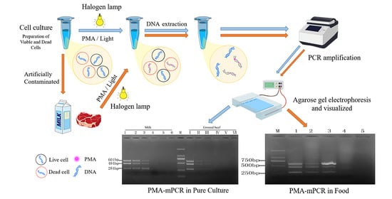
Graphical Abstract

1. Introduction

In recent years, with the frequent emergence of various types of food safety incidents, foodborne pathogens have increased food safety risks and posed a great threat to people’s health and public safety [1]. Escherichia coli O157:H7, Staphylococcus aureus, and Salmonella are considered to be the three most important foodborne pathogens, which widely exist in many kinds of foods and are frequently reported as agents causing food poisoning [2,3]. E. coli O157: H7 is the most well-known and most studied serotype, which can cause gastroenteric infections and lead to serious kidney damage and even death at a low infectious dose. According to other studies, S. aureus is also the cause of frequent food poisoning. In 2017, an estimated 119,247 S. aureus bloodstream infections with 19,832 associated deaths occurred in the United States [4]. Among foodborne disease outbreaks, Salmonella is ubiquitous in a wide range of foods and is the second most commonly reported foodborne pathogen, and leads to bacterial gastroenteritis [5,6,7]. CDC estimates Salmonella bacteria cause about 1.35 million infections, 26,500 hospitalizations, and 420 deaths in the United States every year [8]. However, dead pathogenic bacteria will lose their pathogenicity, but the intact DNA of dead cells can persist for a considerable time and easily lead to a high incidence of false-positive results. Therefore, the detection of the presence and content of viable pathogens is the focus of attention for the management and monitoring of food safety.
The traditional detection methods of foodborne pathogens rely on microbial isolation, culture, and biochemical identification technology. However, culture-based methods are so time-consuming and laborious that it is difficult to meet the new challenges of food companies and the demands for fast and accurate detection of foodborne pathogens by regulatory authorities [9]. Moreover, conventional methods also easily fail to detect viable but non-culturable (VBNC) cells that are pathogenic but non-colony forming. Hence, it is urgent to establish a rapid and accurate method for the simultaneous detection of foodborne pathogens.
To date, polymerase chain reaction (PCR) has become the most widely used technology in classical microbiological methods because of its advantages of short analysis time, strong specificity, and a high degree of automation [10]. Compared with ordinary PCR, multiplex polymerase chain reaction (mPCR) can amplify multiple fragments in a single reaction system for the simultaneous detection of multiple pathogenic bacteria, which has more advantages in saving time, laboratory costs, and reagents [11]. However, normal mPCR is unable to differentiate viable cells from dead cells because the DNA from dead bacteria has a certain persistence, which can also serve as a template during the PCR reaction and lead to a high incidence of false-positive results [12,13]. Thus, this is a major challenge for detecting viable cells in mixed samples of dead and viable microbial cells. In order to overcome this drawback, nucleic acid intercalating dyes, such as ethidium monoazide (EMA) or propidium monoazide (PMA), has been used as sample pre-treatment to remove the interference of dead cells [14,15]. PMA (or EMA) is a high-affinity dye for DNA that cannot enter through intact cell membranes but can selectively permeate into membrane-compromised or dead cells and form cross-links with DNA through exposure to strong visible light. This modification can inhibit PCR amplification to eliminate positive signals from dead bacteria [16,17]. However, it has been reported that EMA has higher toxicity to viable cells than PMA and could partly penetrate into the membranes of viable cells [18,19,20,21]. Therefore, in this study, PMA was selected as pretreatment in combination with mPCR for the fast and simultaneous detection of viable E. coli O157:H7, S. aureus, and Salmonella. To achieve the objective of this study, we selected the O157 antigen gene (rfbE) of E. coli O157:H7, the thermostable nuclease gene (nuc) of S. aureus, and an invasive gene (invA) of Salmonella as highly specific and sensitive primers to detect the three foodborne pathogens. Meanwhile, the PMA-mPCR assay was evaluated in artificially contaminated food products.

2. Results

2.1. Optimization of Concentration of PMA Treatment

PMA concentration is an important factor in the detection of viable target bacteria. Because there are some differences between cell membranes and cell walls of different microorganisms, the optimal PMA concentration may also be different. Therefore, a series of PMA concentrations were conducted on the three viable and dead cell samples in this study. For E. coli O157: H7, two concentration gradients (108 CFU/mL, 106 CFU/mL) were explored. The results are shown in Figure 1 and Figure 2, respectively. It can be seen from Figure 1A that PMA had no significant inhibition on viable E. coli O157:H7 cells. Whereas for the 108 CFU/mL of dead cells, the PMA amplification of its target band appeared significantly weakened but did not completely disappear (Figure 1B). It is indicated that the concentration of dead cells may exceed the maximum range of DNA molecules with which PMA can be combined. When E. coli O157:H7 cells were about 106 CFU/mL, PMA had a slight inhibitory effect on viable cells after the PMA concentration reached 10 µg/mL (Figure 2A). Therefore, the PMA concentration range for cells could not be over 10 µg/mL. As for the concentration of dead bacteria, the amplification of DNA was completely inhibited when PMA at a concentration of 5.0 µg/mL or higher was applied (Figure 2B). According to the study of PMA on the impact of E. coli O157:H7, we directly explored the effect of PMA on the viable and dead bacteria of 106 CFU/mL S. aureus and Salmonella. All the results can be seen in Figure 3 and Figure 4; there was no obvious difference between the amplification of target DNA derived from viable cells of S. aureus and Salmonella treated with a series of PMA concentrations. For the dead S. aureus and Salmonella cells, when the concentration of PMA was (or was over) 3.0 and 5.0 µg/mL, respectively, the amplification of DNA can be completely inhibited (Figure 3B and Figure 4B). Hence, the optimum concentration of PMA for distinguishing those target bacteria DNA from dead and viable cells by the PMA-mPCR is 5.0 µg/mL.

2.2. Specificity and Sensitivity of PMA-mPCR in Pure Culture

To investigate whether the presence of non-target bacteria will interfere with the detection of the three target bacteria, non-target bacteria (Listeria ivanovii subsp. about 106 CFU/mL) was mixed with 106 CFU/mL of the three target bacteria to determine the specificity of the PMA-m PCR assay. The results presented in Figure 5 indicate that non-target bacteria did not disturb the identification and detection of the target bacteria, and there was also no cross-reactivity among the different strains. Moreover, all the amplified fragments were consistent with our expectations. For instance, the amplified fragment of E. coli O157:H7 was 601 bp, of S. aureus near 500 bp was 484 bp, and of Salmonella within 250–500 bp was 284 bp. It also presented that each primer set was highly species-specific.
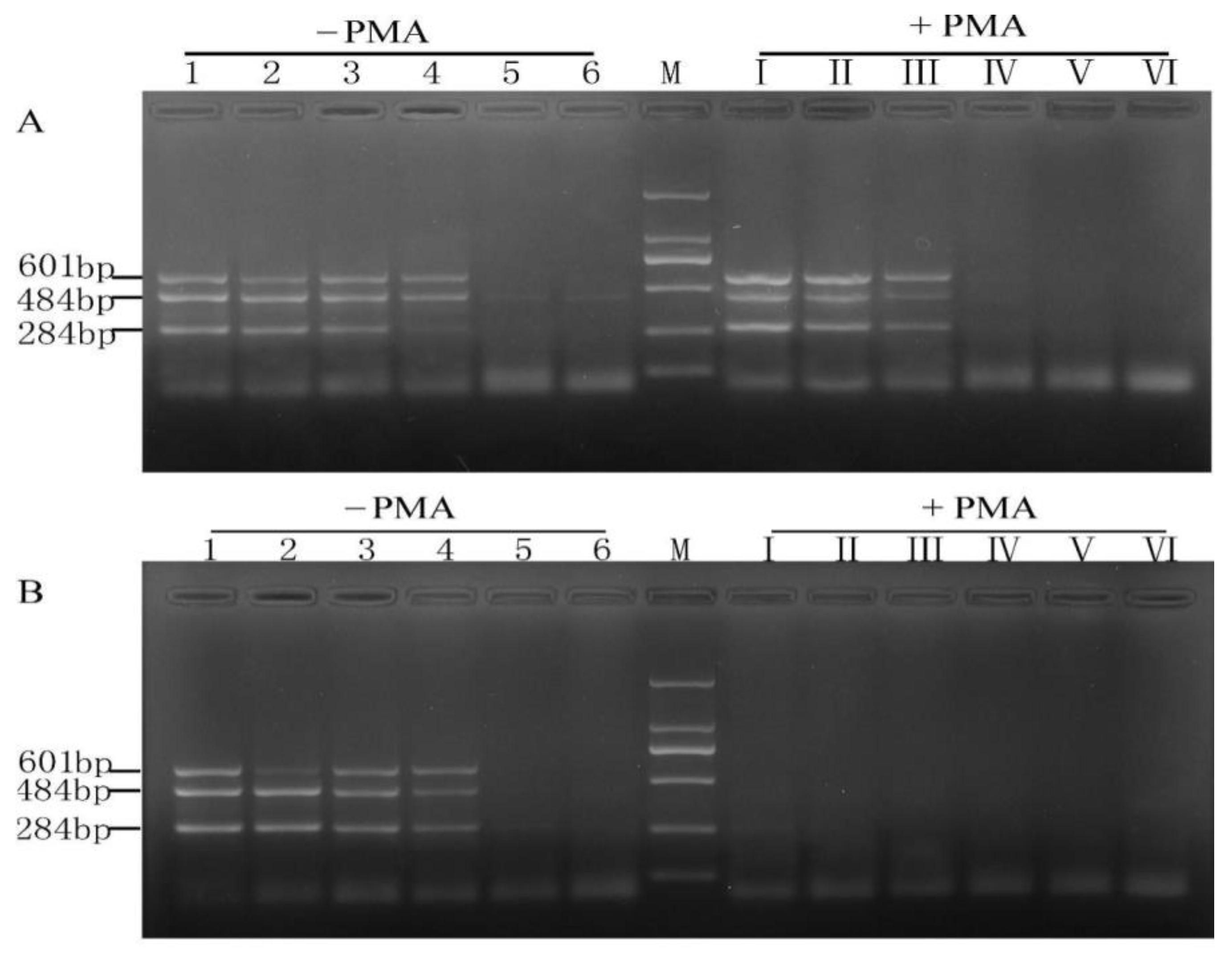
In order to explore whether the detection limits of mPCR were affected by PMA treatment, all the ten-fold serial dilutions of the three mixed target bacterial cultures were divided into two groups in this study. One group was subjected to PMA treatment, while the other was a control. Without PMA treatment of viable and dead bacteria, the detection limits in pure culture were 103 CFU/mL (4.2 × 10³ CFU/mL for E. coli O157:H7, 2.1 × 10³ CFU/mL for S. aureus, 5.4 × 10³ CFU/mL for Salmonella), as shown in Figure 6A,B. The detection limit of the viable bacteria treated by PMA was 104 CFU/mL, and all the dead bacteria treated by PMA had no signals. This result was consistent with that reported by Zhang et al. [12] and also indicated that PMA treatment of cells was efficient and essential before DNA extraction.

2.3. Sensitivity of the PMA-mPCR Assay in Artificial Contaminated Food Products

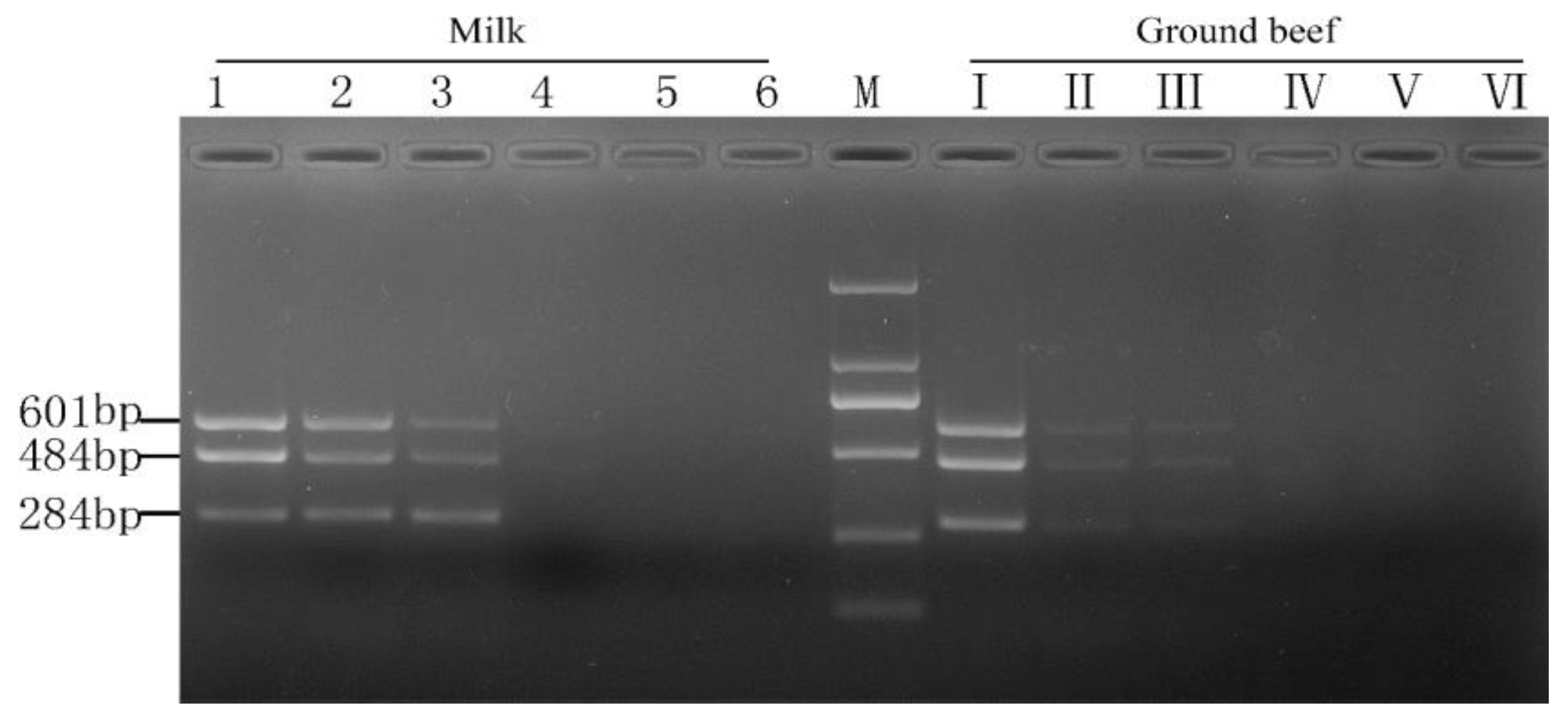
The efficiency of PMA-mPCR was further confirmed by using artificially contaminated aseptic deluxe pure milk and ground beef with concentrations of three mixed viable target bacterial cultures ranging from 101 to 106 CFU/mL to demonstrate real sample application. Figure 7 shows that the detection limits of E. coli O157:H7, S. aureus, and Salmonella in both food matrices all were 104 CFU/mL. The composition of aseptic deluxe pure milk and ground beef was more complex than that of the pure culture system, but the detection limits of three viable target bacteria were consistent with the pure medium utilized in this experiment. The result confirmed that the PMA-mPCR assay could effectively detect three viable target bacteria in food samples.

2.4. Detection of Mixed Viable and Dead Cells

The bacteria present in food do not just exist in a viable or dead form; some of them exist in the form of a mixture of viable and dead cells. Moreover, according to some researchers’ previous studies on viable but non-culturable bacteria, many bacterial cells entering the VBNC state might also be in the same mixed state [22,23]. Hence, the detection of mixed viable and dead cells was carried out in this study. As can be seen from Figure 8, when the proportion of viable and dead bacteria (106 CFU/mL) was lower than 1:10, there was no signal found by PMA-mPCR conducted at a concentration of 5 μg/mL. It indicated that the limit of detection of the three target viable cells was about 105 CFU/mL in the mixture of viable and dead cells culture. Additionally, it also suggests that the amplification of DNA from both dead and viable cells may be simultaneously inhibited when the ratio of viable and dead cell counts is less than 1:10. Because the difference between the final concentration of viable cells in the mixture of viable and dead cells culture and the single viable cells is not so great, the viable amplification is a certain correlation between the ratio of viable: dead cells. Liu and Mustapha also reported that the low concentrations (104–100 cells/g) of viable and 106 dead cells/g contaminating the ground beef sample also generated negative results using PMA real-time PCR [14]. Furthermore, Zhong et al. [24] also got similar results.

3. Materials and Methods

3.1. Bacterial Strains and Cultivation

Reference strains of Salmonella (ATCC 13076), S. aureus (ATCC 27664), and E. coli O157:H7 (ATCC 43895) were used to establish the PMA-mPCR assay in this study. Listeria ivanovii subsp (ATCC 19119) was used as a negative control. All bacterial strains were inoculated on Difco Tryptic Soy Agar (TSA) at 37 °C for 24 h and then cultured in Bacto Tryptic Soy Broth (TSB) at 37 °C in a rotary shaker at 190 rpm for 12 h. After incubation at 37 °C for 24 h, the bacterial count was determined by the plate counting dilution method.

3.2. Preparation of Viable and Dead Cells

To obtain fresh bacterial suspensions, the strains were cultured in Bacto Tryptic Soy Broth in a rotary shaker at 190 rpm for 12 h, and the plates were incubated at 37 °C for 24 h before enumeration. Then, 1 mL of overnight culture of fresh cells suspension (about 108 CFU/mL) was transferred into a 1.5 mL microcentrifuge tube and diluted with sterile normal saline to make a bacterial concentration of 106 CFU/mL through serial decimal dilutions. Following this, all the bacterial suspensions were centrifuged at 10,000× g for 5 min, washed three times, and then resuspended in equal volumes of physiological saline as the displaced TSB. In order to get dead cells, all the cell suspension was killed by exposure to 100 °C for 5 min in a water bath. Then the treated bacterial suspension was cooled to room temperature. The method for confirming dead cells refers to the literature [2,6,14], performing the plate count method on TSA and culturing in TSB after incubation at 37 °C for 24 h. Meanwhile, three parallel experiments were performed to ensure all the cells had died.

3.3. PMA Treatment and DNA Extraction

PMA (Biotium, Inc., Hayward, CA, USA) was dissolved in 20% (v/v) dimethyl sulfoxide (DMSO) (Hayashi Pure Chemical Industries, Ltd., Oakville, Osaka, Japan) to obtain a PMA stock solution (5.0 mg/mL) and stored at 4 °C in the dark. To obtain a PMA working solution with a final concentration of 0.5 mg/mL, the PMA stock solution needed to be diluted 10-fold before it was used. The PMA treatment method was slightly modified according to the procedures described in previous research [24,25]. Briefly, 500 μL of the prepared bacterial suspension in a light-transparent 1.5 mL sterile centrifuge tube was mixed with a series of concentrations of PMA solution and then stored on ice in the dark for 10 min. This promoted PMA entering the dead cells and intercalating with the DNA. After incubation in the dark, all the sample mixtures were placed horizontally on ice and were exposed to a 650 W halogen lamp (Osram, Germany) for 5 min at a 20 cm distance from the light source. During exposure, in order to guarantee homogeneous light exposure, all the tubes were shaken every 30 s. After the light exposure, all the PMA-treated cells were centrifuged at 1000× g for 5 min and washed three times with sterilized saline water.
DNA extraction was based on thermal lysis according to the protocol described in a previous study [25]. Briefly, the PMA-treated pelleted cells (viable or dead) were prepared for DNA extraction. The cells were resuspended with 500 μL Tris–EDTA Triton buffer solution and were boiled in a water bath for 10 min, followed by immediate immersion in ice for 10 min. Then, the samples were centrifuged at 10,000× g for 10 min, and the supernatant was used as a DNA template.

3.4. Multiplex PCR Conditions

In our previous study, when the annealing temperature was at 57 °C and rfbE primer, nuc primer, and invA primer were 0.1, 0.2, and 0.4 μM respectively, we obtained the most specific and sensitive amplification bands for the three target genes [26]. The primer sequences are listed in Table 1. The primers were synthesized and purified by Tianyi Biotech (Wuhan, China).
The mPCR reaction was carried out in a 25 μL volume of the reaction mixture, which contained 0.5 μL of TaKaRa TaqTM DNA polymerase (5 U/μL), 2.0 μL of dNTP Mixture (2.5 mmoL/L) (TaKaRa Biotech, Dalian, China), 2.5 μL of TaKaRa TaqTM 10 × PCR Buffer (Mg2+ plus), 3 μL of DNA templates, 0.1 μM of each forward and reverse rfbE primer, 0.2 μM of each forward and reverse nuc primer, and 0.4 μM of each forward and reverse invA primer. Then, ultra-pure water was filled up to a final volume of 25 μL. The mPCR conditions were as follows: pre-incubation at 94 °C for 5 min, followed by 30 cycles of denaturation at 94 °C for 30 s, annealing at 57 °C for 30 s, and elongation at 72 °C for 30 s, with a final extension at 72 °C for 10 min in a Life Pro Thermal Cycler (Hangzhou Bioer Technology Co. Ltd., Hangzhou, China). After the reaction was completed, the PCR amplification products were subjected to 2% agarose gel electrophoresis and visualized with Golden View dye by the BD-3000 Gel Image Analysis System (Beijing QHBODA Technology Co., Ltd., Beijing, China).

3.5. Specificity and Sensitivity of the PMA-mPCR

First, we prepared a mixed solution of the three target bacteria. We took 100 μL each of Escherichia coli O157: H7, Staphylococcus aureus, and Salmonella bacteria with a concentration of 107 CFU/mL-102 CFU/mL, respectively. They were added to 700 μL of normal saline one after another, stirred well, and a mixed solution with a target bacterial concentration of 106 CFU/mL–101 CFU/mL was obtained. To verify the specificity of target primers and investigate whether the presence of non-target bacteria will disturb the identification and the simultaneous detection of the three target bacteria, non-target cells Listeria ivanovii subsp (viable or dead) were tested by PMA-mPCR assay. At the same time, the negative controls and samples without PMA treatment in mPCR reaction were also subjected to evaluation. Additionally, in order to further test the limit of detection (LOD) of the PMA-mPCR assay, according to Shekar, A. et al. described in [2], tenfold serially diluted target bacteria (viable or dead) ranging from 106 to 101 CFU/mL were washed three times and resuspended in sterilized saline water. The suspensions were subjected to PMA treatment with the best-optimized concentration, and DNA was extracted using the above-mentioned method, followed by the mPCR assay. Furthermore, to explore whether the PMA treatment would have an effect on the sensitivity, non-PMA treated, and negative control samples were also subjected to the mPCR assay.

3.6. Detection of Viable Target Pathogens in Artificially Contaminated Food Products

Samples were treated according to Li et al. described previously with a few modifications [28]. Ground beef (Yayouwang Food Co., Ltd., Shantou, China) and aseptic deluxe pure milk (Inner Mongolia Mengniu Dairy Co., Ltd., Hohhot, China) were used for artificially contaminated studies. First, 5 g of ground beef was added to 45 mL of LB to obtain the sample mixture and homogenized in a mortar to produce a 1:10 homogenate. Then, we took another 7.0 mL of the two food homogenates and added 1.0 mL of the E. coli O157: H7, Staphylococcus aureus, and Salmonella bacteria cultures with a concentration ranging from 107 CFU/mL to 102 CFU/mL, respectively, and shook them well to obtain two food samples with three target bacterial concentrations of 106 CFU/mL to 101 CFU/mL. When all the samples (ground beef, aseptic deluxe pure milk) were verified negative for 3 target pathogens by traditional culture method and standard PCR, these samples were used for further artificial contamination experiments. Afterward, both food samples received the target bacteria, and they were uniformly mixed by shaking to obtain 3 target bacterial concentrations ranging from 101 CFU/mL to 106 CFU/mL, respectively. Subsequently, all samples were subjected to DNA extraction and mPCR sensitivity tests, as mentioned above. All of the tests were conducted in triplicate.

3.7. Detection of Mixed Viable and Dead Cells

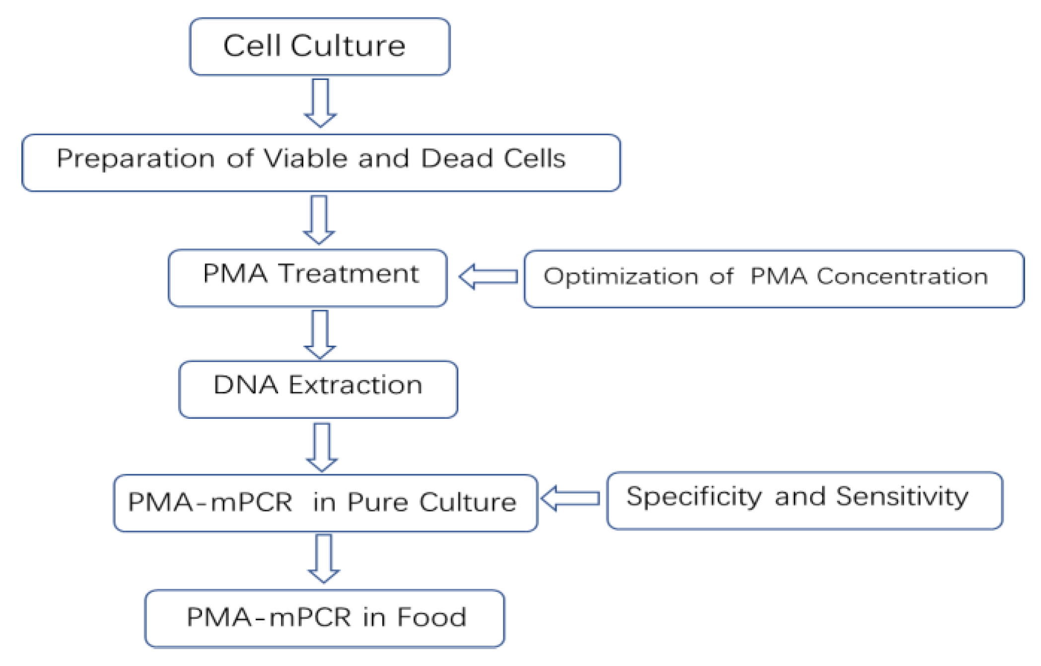
To evaluate whether the PMA-PCR method has an effect on the detection of mixed viable and dead cells, all dead target bacteria (about 106 CFU/mL) were mixed with a series of corresponding viable target bacteria to obtain different proportions (1:1, 1:2, 1:5, 1:10, 1:100, and 1:1000) of viable and dead bacteria mixture suspensions. Thereafter, all the different ratios of viable and dead bacteria mixture suspensions were treated with an optimized concentration of PMA and then used for the PMA-mPCR assay. In this study, we sought to establish a simple method of rapid detection of viable Escherichia coli O157:H7, Staphylococcus aureus, and Salmonella by PMA-mPCR in food, as shown in Figure 9.

4. Discussion and Conclusions

According to previous reports, E. coli O157:H7, S. aureus, and Salmonella are common causative agents of food poisoning outbreaks, resulting in major public health issues and substantial economic burdens [29]. Although some methods for the detection of foodborne bacteria have been reported, many of them could not discriminate viable cells from dead cells. Therefore, it is urgent to establish a rapid and accurate method to detect these viable foodborne pathogens.
In recent years, melt curve-based real-time quantitative PCR assays have emerged as powerful tools for the detection of various pathogens, but it also has a disadvantage in that it requires expensive probes and fluorescent dye for the detection of various bacteria and will greatly increase the cost of the experiment. In addition, there is also a major challenge in detecting viable cells in mixed samples of viable and dead microbial cells by ordinary real-time quantitative PCR. Owing to the convenience and rapid characteristics of mPCR and being cheaper than real-time PCR [30,31], we investigated and established a PMA-mPCR assay that could simultaneously detect viable E. coli O157:H7, S. aureus, and Salmonella. Considering that PMA treatment is a key step to eliminate the false-positive results from dead cells, we therefore explored the optimal concentration of PMA from 0 to 20 µg/mL. In our study, the results show that PMA has less suppression for 108 CFU/mL of dead E. coli O157:H7 cells, even though the PMA concentration is 15 µg/mL. Nevertheless, when PMA is at 5 µg/mL concentration, the results suggest that it can effectively inhibit the dead cell signals from 106 CFU/mL of E. coli O157:H7, S. aureus, and Salmonella. At the same time, there is no difference between with or without PMA treatment on those three viable target cells, which is in accordance with previous studies by Yang et al. [6] and Forghani et al. [32]. However, the concentration of PMA (5 µg/mL) used in our study is low, which also can reduce the cost per assay compared to other reported studies [2,11]. Based on the optimized PMA concentration, the PMA-mPCR assay was successfully subjected to detect E. coli O157:H7, S. aureus, and Salmonella in both pure culture and artificial contaminated model food systems. The concentration of PMA depends on the number of background dead cells. Therefore, when the dosage of PMA is determined, the appropriate concentration of the dead bacteria does not cause false-positive results. If the concentration of dead cells exceeds the appropriate range, false positive results will occur. In this study, a PMA-mPCR assay was developed to rapidly detect the DNA from viable target bacteria in food samples, which cannot detect RNA directly. The total time required for analysis is about 4–6 h, including sample preparation. The detection sensitivity for those three pathogenic bacteria by mPCR is 103 CFU/mL in pure culture, whereas it is 104 CFU/mL in both pure culture and model food systems by PMA-mPCR. These detection sensitivity levels are consistent with our previous studies by mPCR [26] and similar to Zhong et al. [9]. The sensitivity of this study is not very high. This result can be explained by the fact that the PMA may also have little suppression for viable cells, or the thermal lysis method to extract DNA is not efficient. Nevertheless, the thermal lysis method could save time and eliminate the need for intensive labor [12], and the sensitivity of PMA-mPCR in our assay is similar to the result of Li et al. using a DNA extraction kit [28]. They also indicated that the PMA-mPCR detection system could effectively eliminate the signals from dead bacteria and has good stability and sensitivity in both pure culture and model food systems. When conducting foodborne pathogen detection in actual food samples, the difficulty of the pre-enrichment step mainly arises from the competition of other microorganisms present in the samples, the complexity of the food samples, and the low enrichment efficiency. To address these challenges, the following approaches can be adopted: optimizing the enrichment medium, such as using the method of immunomagnetic bead adsorption [6], optimizing the enrichment conditions [11,33], and using automated enrichment equipment based on the same principle of immunomagnetic bead adsorption. For rapid detection of foodborne pathogens, Kim’s group recently developed an HRPzyme-integrated PCR colorimetric detection platform and a paper chip device-based recombinase polymerase amplification method, which provided simple, fast, cost-effective and user-friendly detection assays for DNA as a target analyte [34,35,36]. The PMA-mPCR developed in this study may be expanded for quantitative analysis or semi-quantitative analysis, if needed, by combining with the application of ImageJ in colorimetric image data acquisition.
Viable but non-culturable (VBNC) foodborne pathogens are characterized by a loss of culture ability on enriched agar media and exhibit detectable metabolic functions, which may retain their ability to express toxic genes [37]. VBNC cells can easily fail detection and pose a risk to public health. Thus, many investigators have conducted VBNC induction studies on pathogenic bacteria. For instance, Zhao et al. [22] reported that VBNC cell counts still approximated 106 cells/mL while the total cell counts of 108 cells/mL declined to below 0.1 CFU/mL to undetectable levels. It indicates that the VBNC state is a mixture of viable and dead cells. Hence, we used the PMA-mPCR to detect a series of different proportions of the mixture of viable and dead cells. Meanwhile, we obtained that the detection limit of this assay to the viable cells mixed with dead cells (106 CFU/mL) was 105 CFU/mL. Namely, this method can be applied to the detection of VBNC cells at concentrations 105 CFU/mL or higher. Although the method has some shortcomings and the detection limit is not low, it will have great potential for the detection and concentration assessment of VBNC cells.
In conclusion, a sensitive, specific, and convenient PMA-mPCR assay was developed to simultaneously detect viable E. coli O157:H7, S. aureus, and Salmonella, which was also applied to complex food matrices. The results of the PMA-mPCR method were consistent with traditional mPCR assay. This PMA-mPCR method might provide an efficient detection tool for monitoring food contaminants from these three pathogens to reduce the potential hazards of these harmful pathogens. Thus, it is anticipated that the method can be a sensitive, accurate, cost-effective, and potential tool for the rapid identification of multiple viable foodborne pathogens.

Author Contributions

Conceptualization, H.W.; Methodology, C.W., T.J. and X.Z.; Investigation, Y.L., S., X.L. and Y.D.; Data curation, Y.L.; Writing—original draft, Y.L.; Supervision, X.Z. and T.Z. All authors have read and agreed to the published version of the manuscript.

Funding

This work was supported by the Basic and Applied Basic Research Foundation Project of Guangdong Province (2023A1515011473), Innovation and Entrepreneurship Training Program for College students, Zhuhai University of Science and Technology (DC2022091), and the Science and Technology Development Funds (FDCT) of Macao (0104/2021/A2).

Institutional Review Board Statement

This article does not contain any studies with human participants or animals performed by any of the authors.

Informed Consent Statement

Not applicable.

Data Availability Statement

Data are available from the corresponding author.

Conflicts of Interest

All authors declare that they have no conflicts of interest.

Sample Availability

Not available.

References

  1. Zhao, X.; Lin, C.W.; Wang, J.; Oh, D.H. Advances in rapid detection methods for foodborne pathogens. J. Microbiol. Biotechnol. 2014, 24, 297–312. [Google Scholar] [CrossRef] [Green Version]
  2. Shekar, A.; Babu, L.; Ramlal, S.; Sripathy, M.H.; Batra, H. Selective and concurrent detection of viable Salmonella spp., E. coli, Staphylococcus aureus, E. coli O157:H7, and Shigella spp., in low moisture food products by PMA-mPCR assay with internal amplification control. LWT—Food Sci. Technol. 2017, 86, 586–593. [Google Scholar] [CrossRef]
  3. Kim, J.H.; Rhim, S.R.; Kim, K.T.; Paik, H.D.; Lee, J.Y. Simultaneous Detection of Listeria monocytogenes, Escherichia coli O157:H7, Bacillus cereus, Salmonella spp., and Staphylococcus aureus in Low-fatted Milk by Multiplex PCR. Korean J. Food Sci. Anim. Resour. 2014, 34, 717–723. [Google Scholar] [CrossRef] [Green Version]
  4. Kourtis, A.P.; Hatfield, K.; Baggs, J.; Mu, Y.; See, I.; Epson, E.; Nadle, J.; Kainer, M.A.; Dumyati, G.; Petit, S.; et al. Vital Signs: Epidemiology and Recent Trends in Methicillin-Resistant and in Methicillin-Susceptible Staphylococcus aureus Bloodstream Infections—United States. Morb. Mortal. Wkly. Rep. 2019, 68, 214–219. [Google Scholar] [CrossRef] [Green Version]
  5. Wang, L.; Ye, C.; Xu, H.; Aguilar, Z.P.; Xiong, Y.; Lai, W.; Wei, H. Development of an SD-PMA-mPCR assay with internal amplification control for rapid and sensitive detection of viable Salmonella spp., Shigella spp. and Staphylococcus aureus in food products. Food Control 2015, 57, 314–320. [Google Scholar] [CrossRef]
  6. Yang, Y.; Xu, F.; Xu, H.; Aguilar, Z.P.; Niu, R.; Yuan, Y.; Sun, J.; You, X.; Lai, W.; Xiong, Y. Magnetic nano-beads based separation combined with propidium monoazide treatment and multiplex PCR assay for simultaneous detection of viable Salmonella Typhimurium, Escherichia coli O157:H7 and Listeria monocytogenes in food products. Food Microbiol. 2013, 34, 418–424. [Google Scholar] [CrossRef]
  7. Zhao, X.H.; Zhao, F.H.; Wang, J.; Zhong, N.J. Biofilm formation and control strategies of foodborne pathogens: Food safety perspectives. RSC Adv. 2017, 7, 36670–36683. [Google Scholar] [CrossRef] [Green Version]
  8. Dewey-Mattia, D.; Manikonda, K.; Hall, A.J.; Wise, M.E.; Crowe, S.J. Surveillance for Foodborne Disease Outbreaks—United States, 2009–2015. Mmwr Surveill. Summ. 2018, 67, 1–11. [Google Scholar] [CrossRef]
  9. Zhong, J.L.; Zhao, X.H. Isothermal Amplification Technologies for the Detection of Foodborne Pathogens. Food Anal. Methods 2018, 11, 1543–1560. [Google Scholar] [CrossRef]
  10. Cantekin, Z.; Ergun, Y.; Solmaz, H.; Özmen, G.Ö.; Demir, M.; Saidi, R. PCR assay with host specific internal control for Staphylococcus aureus from bovine milk samples. Maced. Vet. Rev. 2015, 38, 97–100. [Google Scholar] [CrossRef] [Green Version]
  11. Yu, S.; Yan, L.; Wu, X.; Li, F.; Wang, D.; Xu, H. Multiplex PCR coupled with propidium monoazide for the detection of viable Cronobacter sakazakii, Bacillus cereus, and Salmonella spp. in milk and milk products. J. Dairy Sci. 2017, 100, 7874–7882. [Google Scholar] [CrossRef] [Green Version]
  12. Zhang, Z.; Wang, L.; Xu, H.; Aguilar, Z.P.; Liu, C.; Gan, B.; Xiong, Y.; Lai, W.; Xu, F.; Wei, H. Detection of non-emetic and emetic Bacillus cereus by propidium monoazide multiplex PCR (PMA-mPCR) with internal amplification control. Food Control 2014, 35, 401–406. [Google Scholar] [CrossRef]
  13. Zhao, X.H.; Li, M.; Xu, Z.B. Detection of Foodborne Pathogens by Surface Enhanced Raman Spectroscopy. Front. Microbiol. 2018, 9, 1236. [Google Scholar] [CrossRef] [Green Version]
  14. Liu, Y.; Mustapha, A. Detection of viable Escherichia coli O157:H7 in ground beef by propidium monoazide real-time PCR. Int. J. Food Microbiol. 2014, 170, 48–54. [Google Scholar] [CrossRef] [PubMed]
  15. Liu, J.Y.; Zhou, R.; Li, L.; Peters, B.M.; Li, B.; Lin, C.W.; Chuang, T.L.; Chen, D.Q.; Zhao, X.H.; Xiong, Z.Y.; et al. Viable but non-culturable state and toxin gene expression of enterohemorrhagic Escherichia coli 0157 under cryopreservation. Res. Microbiol. 2017, 168, 188–193. [Google Scholar] [CrossRef] [PubMed]
  16. Nocker, A.; Cheung, C.-Y.; Camper, A.K. Comparison of propidium monoazide with ethidium monoazide for differentiation of live vs. dead bacteria by selective removal of DNA from dead cells. J. Microbiol. Methods 2006, 67, 310–320. [Google Scholar] [CrossRef] [PubMed]
  17. Rudi, K.; Moen, B.; Drømtorp, S.M.; Holck, A.L. Use of ethidium monoazide and PCR in combination for quantification of viable and dead cells in complex samples. Appl. Environ. Microbiol. 2005, 71, 1018–1024. [Google Scholar] [CrossRef] [Green Version]
  18. Pan, Y.; Breidt, F., Jr. Enumeration of viable Listeria monocytogenes cells by real-time PCR with propidium monoazide and ethidium monoazide in the presence of dead cells. Appl. Environ. Microbiol. 2008, 73, 8028–8031. [Google Scholar] [CrossRef] [Green Version]
  19. Zhu, R.G.; Li, T.P.; Jia, Y.F.; Song, L.F. Quantitative study of viable Vibrio parahaemolyticus cells in raw seafood using propidium monoazide in combination with quantitative PCR. J. Microbiol. Methods 2012, 90, 262–266. [Google Scholar] [CrossRef]
  20. Gerard, À.; Marta, G.; Sergio, I.; Vanessa, B.; Rubén, L. Method to quantify live and dead cells in multi-species oral biofilm by real-time PCR with propidium monoazide. Amb Express 2013, 3, 1. [Google Scholar]
  21. Zhao, X.; Zhong, J.; Wei, C.; Lin, C.-W.; Ding, T. Current Perspectives on Viable but Non-culturable State in Foodborne Pathogens. Front. Microbiol. 2017, 8, 580. [Google Scholar] [CrossRef] [Green Version]
  22. Zhao, F.; Bi, X.; Hao, Y.; Liao, X. Induction of viable but nonculturable Escherichia coli O157: H7 by high pressure CO2 and its characteristics. PLoS ONE 2013, 8, e62388. [Google Scholar] [CrossRef] [Green Version]
  23. Liu, Y.; Gilchrist, A.; Zhang, J.; Li, X.F. Detection of Viable but Nonculturable Escherichia coli O157:H7 Bacteria in Drinking Water and River Water. Appl. Environ. Microbiol. 2008, 74, 1502–1507. [Google Scholar] [CrossRef] [PubMed] [Green Version]
  24. Zhong, J.L.; Zhao, X.H. Detection of viable but non-culturable Escherichia coli O157:H7 by PCR in combination with propidium monoazide. 3 Biotech 2017, 8, 28. [Google Scholar] [CrossRef] [PubMed]
  25. Zhao, X.; Wang, J.; Forghani, F.; Park, J.H.; Park, M.S.; Seo, K.H.; Oh, D.H. Rapid detection of viable Escherichia coli O157 by coupling propidium monoazide with loop-mediated isothermal amplification. J. Microbiol. Biotechnol. 2013, 23, 1708–1716. [Google Scholar] [CrossRef] [PubMed]
  26. Wei, C.; Zhong, J.; Hu, T.; Zhao, X. Simultaneous detection of Escherichia coli O157: H7, Staphylococcus aureus and Salmonella by multiplex PCR in milk. 3 Biotech 2018, 8, 76. [Google Scholar] [CrossRef]
  27. Xu, Y.; Cheng, W.; Chen, F. Detection of Salmonella spp., Escherichia coli and Staphylococcus aureus by Multiplex PCR. J. Microbiol. 2006, 33, 89–94. [Google Scholar]
  28. Li, F.; Xie, G.; Zhou, B.; Yu, P.; Yu, S.; Aguilar, Z.P.; Wei, H.; Xu, H. Rapid and simultaneous detection of viable Cronobacter sakazakii, Staphylococcus aureus, and Bacillus cereus in infant food products by PMA-mPCR assay with internal amplification control. LWT—Food Sci. Technol. 2016, 74, 176–182. [Google Scholar] [CrossRef]
  29. Vugia, D.; Cronquist, A.; Cartter, M.; Tobin-D’Angelo, M.; Blythe, D.; Smith, K.; Lathrop, S.; Morse, D.; Cieslak, P.; Dunn, J. Preliminary FoodNet Data on the incidence of infection with pathogens transmitted commonly through food—10 States, 2008. Mmwr Morb. Mortal. Wkly. Rep. 2010, 59, 418–422. [Google Scholar]
  30. Saeki, E.K.; Alves, J.; Bonfante, R.C.; Hirooka, E.Y. Multiplex PCR (mPCR) for the Detection of Salmonella spp. and the Differentiation of the Typhimurium and Enteritidis Serovars in Chicken Meat. J. Food Saf. 2013, 33, 25–29. [Google Scholar] [CrossRef]
  31. Park, Y.S.; Lee, S.R.; Kim, Y.G. Detection of Escherichia coli O157:H7, Salmonella spp., Staphylococcus aureus and Listeria monocytogenes in kimchi by multiplex polymerase chain reaction (mPCR). J. Microbiol. 2006, 44, 92–97. [Google Scholar] [PubMed]
  32. Forghani, F.; Langaee, T.; Eskandari, M.; Seo, K.H.; Chung, M.J.; Oh, D.H. Rapid detection of viable Bacillus cereus emetic and enterotoxic strains in food by coupling propidium monoazide and multiplex PCR (PMA-mPCR). Food Control 2015, 55, 151–157. [Google Scholar] [CrossRef]
  33. Chen, J.; Tang, J.; Liu, J.; Cai, Z.; Bai, X. Development and evaluation of a multiplex PCR for simultaneous detection of five foodborne pathogens. J. Appl. Microbiol. 2012, 112, 823–830. [Google Scholar] [CrossRef] [PubMed]
  34. Kim, S.U.; Batule, B.S.; Mun, H.; Shim, W.B.; Kim, M.G. Ultrasensitive colorimetric detection of Salmonella enterica Typhimurium on lettuce leaves by HRPzyme-Integrated polymerase chain reaction. Food Control 2018, 84, 522–528. [Google Scholar] [CrossRef]
  35. Ahn, H.; Batule, B.S.; Seok, Y.; Kim, M.G. Single-Step Recombinase Polymerase Amplification Assay Based on a Paper Chip for Simultaneous Detection of Multiple Foodborne Pathogens. Anal. Chem. 2018, 90, 10211–10216. [Google Scholar] [CrossRef]
  36. Batule, B.S.; Kim, S.U.; Mun, H.; Shim, W.-B.; Kim, M.-G. Development of HRPzyme-Integrated PCR Platform for Colorimetric Detection of Foodborne Pathogens. In Biosensing Technologies for the Detection of Pathogens; Rinken, T., Ed.; IntechOpen Limited: London, UK, 2018. [Google Scholar]
  37. Li, L.; Mendis, N.; Trigui, H.; Oliver, J.D.; Faucher, S.P. The importance of the viable but non-culturable state in human bacterial pathogens. Front. Microbiol. 2014, 5, 258. [Google Scholar] [CrossRef] [Green Version]
Figure 1. Concentrations of PMA for inhibiting the amplification of DNA from 108 CFU/mL of Escherichia coli O157: H7 viable cells (A) and dead cells (B). (A) Lanes 1~8, varying concentrations of PMA (0, 1, 2, 3, 5, 10, 15, 20 µg/mL, lane 9, negative control. M, DL 2000 DNA Marker; (B) Lanes 1~8, varying concentrations of PMA (0, 1, 2, 3, 5, 7, 10,15 µg/mL), lane 9, negative control.
Molecules 28 05835 g001
Figure 2. Concentrations of PMA for inhibiting the amplification of DNA from106 CFU/mL of Escherichia coli O157: H7 viable cells (A) and dead cells (B). (A) Lanes 1~8, varying concentrations of PMA (0, 1, 2, 3, 5, 10, 15, 20 µg/mL, lane 9, negative control. M, DL 2000 DNA Marker; (B) Lanes 1~8, varying concentrations of PMA (0, 1, 2, 3, 5, 7, 10, 15 µg/mL), lane 9, negative control.
Molecules 28 05835 g002
Figure 3. Concentrations of PMA for inhibiting the amplification of DNA from106 CFU/mL of Staphylococcus aureus viable cells (A) and dead cells (B). (A) Lanes 1~8, varying concentrations of PMA (0, 1, 2, 3, 5, 10, 15, 20 µg/mL, lane 9, negative control. M, DL 2000 DNA Marker; (B) Lanes 1~8, varying concentrations of PMA (0, 1, 2, 3, 5, 7, 10, 15 µg/mL), lane 9, negative control.
Molecules 28 05835 g003
Figure 4. Concentrations of PMA for inhibiting the amplification of DNA from106 CFU/mL of Salmonella viable cells (A) and dead cells (B). (A) Lanes 1~8, varying concentrations of PMA (0, 1, 2, 3, 5, 10, 15, 20 µg/mL, lane 9, negative control. M, DL 2000 DNA Marker; (B) Lanes 1~8, varying concentrations of PMA (0, 1, 2, 3, 5, 7, 10, 15 µg/mL), lane 9, negative control.
Molecules 28 05835 g004
Figure 5. Specificity test of PMA-mPCR. M: DL 2000 DNA Marker; lane 1, viable mixture (containing 106 CFU/mL viable Listeria ivanovii subsp.) without PMA, lane 2, with PMA viable mixture (containing 106 CFU/mL viable Listeria ivanovii subsp.), lane 3, dead bacteria mixture (containing 106 CFU/mL of dead Listeria ivanovii subsp.) without PMA, lane 4, dead bacteria mixture (containing 106 CFU/mL dead Listeria ivanovii subsp.) with PMA, lane 5, negative control.
Molecules 28 05835 g005
Figure 6. The limit of detection of multiplex PCR assay using 10-fold serially diluted viable target cells(A) and dead target cells (B) with (+) and without (−) PMA treatment. M, DL 2000 DNA Marker; lanes 1~6 (or Ⅰ~Ⅵ), varying concentrations of bacterial suspension (106, 105, 104, 103, 102, and 101 CFU/mL).
Molecules 28 05835 g006
Figure 7. The limit of detection of PMA-mPCR in the detection of viable target bacteria in milk and ground beef. M, DL 2000 DNA Marker; lanes 1~6 (or Ⅰ~Ⅵ), varying concentrations of bacterial suspension (106, 105, 104, 103, 102, and 101 CFU/mL).
Molecules 28 05835 g007
Figure 8. The results of PMA-mPCR in the mixture of viable and dead cells. M, DL 2000 DNA Marker; lanes 1~6, viable bacteria accounted for the proportion of dead bacteria (106 CFU/mL) were 1: 1, 1: 2, 1: 5, 1:10, 1: 100, 1:1000.
Molecules 28 05835 g008
Figure 9. Schematic diagram of the PMA-mPCR in food in this study.
Molecules 28 05835 g009
Table 1. Primers used in this study.
MicroorganismTarget GenePrimer Sequence (5′-3′)G + C (%)Tm (°C)Amplicon Length (bp)Reference
E. coli
O157:H7
rfbE
(S83460)
F:GCCACCCCCATTTTCGTTG
R:TCCTCTCTTTCCTCTGCGGT
57.9
47.4
63.2
51.7
601[26]
S. aureusnuc
(AP017922)
F:TACAGGTGACTGCGGGCTTATC
R:CTTACCGGGCAATACACTCACTA
50
45.4
60.2
58.3
484[27]
SalmonellainvA
(M90846)
F:CTTTAGCCAAGCCTTGACGAAC
R:AAAGGCAATACGCAAAGAGGT
54.5
47.8
62.1
60.6
284[27]
Disclaimer/Publisher’s Note: The statements, opinions and data contained in all publications are solely those of the individual author(s) and contributor(s) and not of MDPI and/or the editor(s). MDPI and/or the editor(s) disclaim responsibility for any injury to people or property resulting from any ideas, methods, instructions or products referred to in the content.