Review

Structural Modification Endows Small-Molecular SN38 Derivatives with Multifaceted Functions

1
College of Pharmaceutical Science, Anhui Xinhua University, Hefei 230088, China
2
Department of Chemistry, University of Science and Technology of China, Hefei 230026, China
*
Author to whom correspondence should be addressed.
Molecules 2023, 28(13), 4931; https://doi.org/10.3390/molecules28134931
Submission received: 29 May 2023 / Revised: 21 June 2023 / Accepted: 21 June 2023 / Published: 22 June 2023

Abstract

:
As a camptothecin derivative, 7-ethyl-10-hydroxycamptothecin (SN38) combats cancer by inhibiting topoisomerase I. SN38 is one of the most active compounds among camptothecin derivatives. In addition, SN38 is also a theranostic reagent due to its intrinsic fluorescence. However, the poor water solubility, high systemic toxicity and limited action against drug resistance and metastasis of tumor cells of SN38 indicates that there is great space for the structural modification of SN38. From the perspective of chemical modification, this paper summarizes the progress of SN38 in improving solubility, increasing activity, reducing toxicity and possessing multifunction and analyzes the strategies of structure modification to provide a reference for drug development based on SN38.

1. Introduction

Natural products are an important source for developing chemotherapeutic agents. However, natural products, except for paclitaxel, are seldom used directly for the treatment of cancer in clinic due to their intrinsic defects such as poor water solubility, systemic toxicity, weak bioactivity, etc. Therefore, the structural modification of natural products is always a research focus for chemists and pharmaceutical scientists. For instance, many research projects are carried out to structurally modify vinblastine, tropolones, oridonin, etc. [1,2,3,4,5]. Camptothecin derivatives, another important source for the development of antitumor drugs, exert antitumor activity by inhibiting topoisomerase 1. Based on the skeleton of camptothecin, some drugs have been developed for the treatment of cancers, such as topotecan, irinotecan, rubitecan, etc. [6] (Figure 1). The research on camptothecin derivatives, especially 7-ethyl-10-hydroxycamptothecin (SN38), has always attracted much attention from medicinal chemists.
As an active metabolite of irinotecan, SN38 shares a similar anticancer mechanism (inhibition of topo 1) with other camptothecin derivatives and is considered to be one of the most potent antineoplastic agents among camptothecin derivatives. However, the deficiency of SN38, such as poor water solubility, instability, systemic toxicity and weak inhibitory effect on tumor resistance and metastasis, has greatly limited its clinical applications as an anticancer drug [7]. Therefore, structural modification of SN38 is in demand for new drug development. In recent years, a variety of structural modifications of SN38 for addressing the above problems have been reported. We searched the literature through SciFinder using 7-ethyl-10-hydroxycamptothecin as the substance identifier, and 3734 articles were obtained. Then, the articles were refined by document type (Journal and Letter) to obtain 2137 articles, followed by being refined again in accordance with publication year (2018–2023) to obtain 493 articles. We reviewed these articles and recapitulated them. Structural modification of SN38 was classified into five categories: (1) structural modification at position 10 of SN38; (2) structural modification at position 20 of SN38; (3) structural modification at position 10 and 20 of SN38; (4) structural modification at position 9 of SN38; and (5) lactone E ring-opening of SN38. These obtained SN38 derivatives exhibited multifaceted functions, as shown in Table 1. In addition, this also probes into the strategy of the current study concerned with the structural modification of SN38, providing a reference for the further development of SN38.

2. Modification of SN38 at C-10 Position

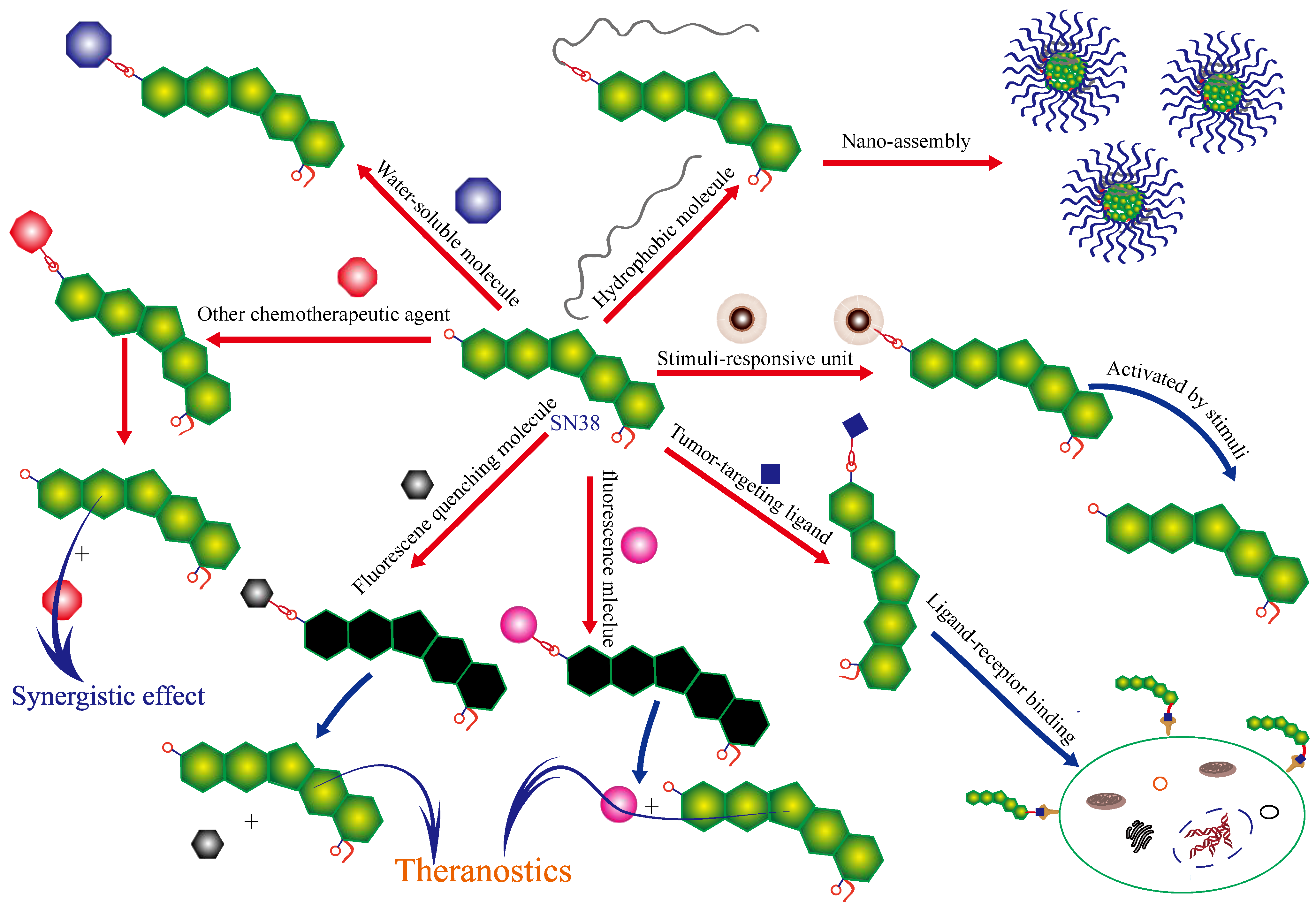
The hydroxyl group at C-10 of camptothecin derivatives plays an important role in maintaining the stability of the parent structure and exerting anticancer activity [8,9]. Therefore, designing a prodrug of SN38 is one of the best design strategies due to the release of parent SN38 and other functional groups from the prodrugs. The small-molecule SN38 derivatives based on the modification at C-10 of SN38 account for a high proportion of all small-molecule SN38 derivatives. These small-molecule SN38 derivatives possess stimuli-responsive release of drugs, tumor targeting, synergism between different drugs, improvement of water solubility and theranostic function, as shown in Figure 2.

2.1. Stimuli-Responsive Release of Drugs

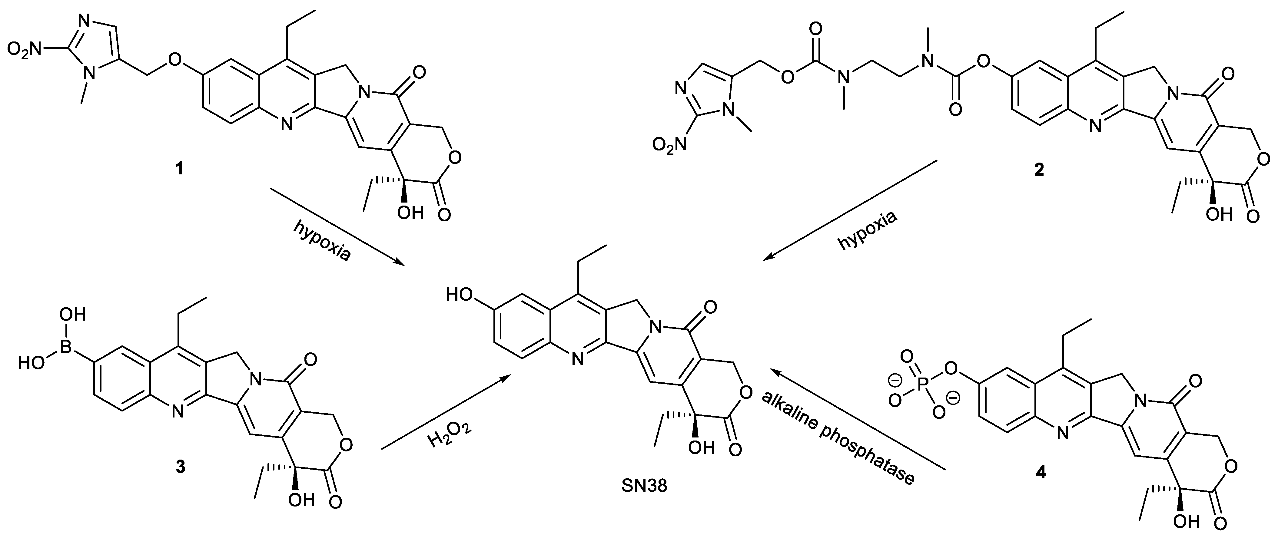
Designing prodrugs capable of stimuli-responsive release is of great value for improving the selectivity of drugs to cancer cells to achieve high efficiency and low toxicity [10,11,12]. Modification of SN38 also follows the principle to utilize the special microenvironment of cancer cells for the design of SN38 derivatives. Upon cellular internalization, these SN38 derivatives can release the active parent SN38 in response to special stimuli in cancer cells. Compared with normal cells, the microenvironment of tumor cells is hypoxia, high hydrogen peroxide and high expression of esterase, etc. [13,14,15]. Based on these findings, compounds 14 were designed and synthesized (Figure 3). Compounds 1 and 2 both contain hypoxia-responsive moiety of (1-methyl-2-nitro-1H-imidazole-5-yl) methanol and can release the parent SN38 in responsive to hypoxia presented in cancer cells. The linkage between (1-methyl-2-nitro-1H-imidazole-5-yl) methanol and SN38 was ether bond for compound 1, while bis-carbamate for compound 2, which significantly affected their pharmacokinetic properties. Compound 1 was considered to be superior. The inhibitory effect of compound 1 on human colorectal cancer H460 cells and human colon cancer HT-29 cells was stronger than that of compound 2 due to its higher selectivity to hypoxia, even 10 folds stronger than that of evofosfamide [16].
Compound 3 was prepared by replacing the hydroxyl group at C-10 of SN38 with boric acid. Utilizing the high levels of H2O2 in cancer cells, compound 3 could be activated to release SN38. The inhibitory effect of compound 3 on topo 1 was stronger than that of SN38. The cell survival assay showed that compound 3 effectively inhibited the growth of human colon cancer HCT-15 cells, human breast cancer MCF-7 cells, human breast cancer MDA-MB-231 cells, human astroblastoma U87MG cells, human glioma U251 cells and HT-29 cells. The cytotoxicity of compound 3 against MCF-7 cells and U251 cells was comparable to that of SN38, but for HT-29 cells, the cytotoxicity of compound 3 was stronger than that of SN38. The in vivo experiment demonstrated compound 3 at a dose of 2.0 mg/kg significantly inhibited the tumor growth of U87MG tumor-bearing mice, indicating that the feasibility of boron acid being responsive to H2O2 [17].
Based on the high expression level of alkaline phosphatase on the membrane of human-induced pluripotent stem cells (hiPSCs) and neural stem cells [18], compound 4 containing phosphate at position 10 was designed. The high water solubility caused compound 4 not to be taken up effectively by normal neural cells and to show low cytotoxicity due to absence of alkaline phosphatase on the membrane of these normal cells. Compared with normal neural cells, hiPSCs and neural stem cells could make compound 4 be hydrolyzed to release SN38 of high penetration and cytotoxicity. Therefore, compound 4 can selectively eliminate proliferative hiPSCs and neural stem cells from neural cell mixtures. It is thus evident that simple phosphorylation of SN38 provided a cost-effective tool for decontaminating hiPSC-derived neurons [19].
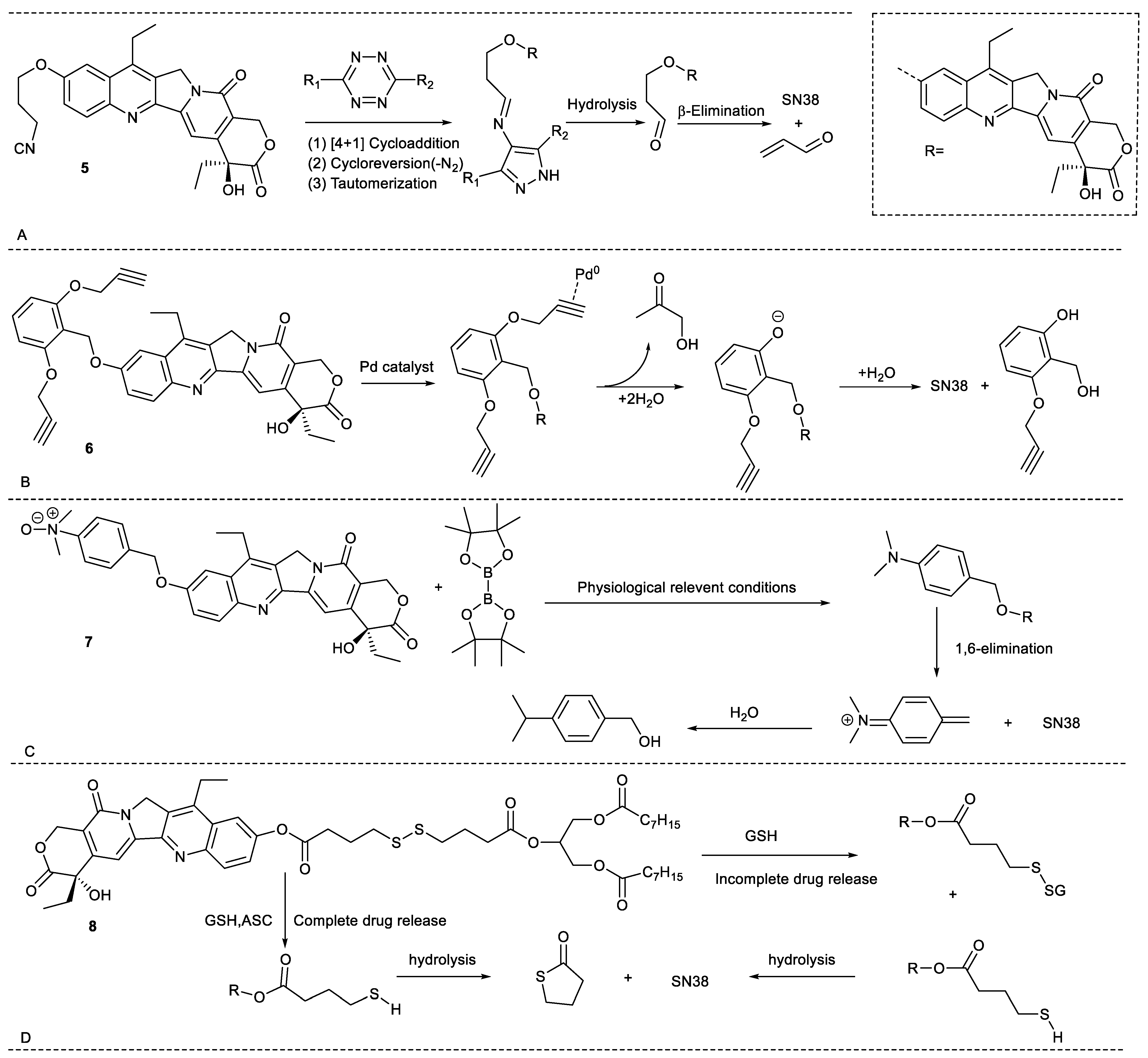
Apart from endogenous substances in cancer cells, some exogenous substances can also activate SN38 prodrugs. For example, compound 5 was prepared through connecting the 3-isocyanopropyl group to C10-OH of SN38. The compound and tetrazines underwent dissociative bioorthogonal reactions to near-quantitatively release SN38, providing an alternative for the controlled release of SN38 (Figure 4A) [20]. Another example is compound 6, obtained by the substitution of C10-OH of SN38 with 2,6-bis(propargyloxy)benzylgroup. Based on biorthogonal reactions, compound 6 cannot release SN38 in the absence of exogenous palladium, while it can release SN38 in the presence of exogenous palladium (Figure 4B). Hence, compared with the cytotoxicity of SN38, the cytotoxicity of compound 6 in the absent of exogenous palladium significantly decrease by 44-fold,16-fold and 35-fold for human colon cancer HCT-116 cells, human glioma U87 cells and U251 cells, respectively. Once co-existed with exogenous palladium, the compound showed cytotoxicity comparable to SN38. In addition, the combination of compound 6 and the Pd-activated 5-FU prodrug can synergistically inhibitthe growth of cancer cells through common palladium catalysis. These findings demonstrate that the combination of compound 6 with tumor targeting nanopalladium can realize the controlled release of SN38 in cancer cells and decrease toxic and side effects on other organs [21]. Another example was compound 7, which was synthesized by introduction of benzylic alcohol containing a tertiary amino group in the form of N-oxide to SN38. The introduction of the group not only significantly enhanced the water solubility of SN38 (300-fold) but also weakened its cytotoxicity against human non-small-cell lung cancer A549 cells (10-fold). Upon exposure to bis(pinacolato)diboron, the compound was activated via a click reaction to release SN38 with fast reaction kinetics and complete cleavage both in vitro and in vivo (Figure 4C) [22]. These findings show that the bioorthogonal prodrug strategy presents significant advances.
Additionally, inspired by the specific absorption mechanism of triglyceride fat, compound 8 was synthesized by conjugation of SN38 with a triglyceride fat containing a disulfide bond. As a structured lip mimetic oral prodrug, compound 8 had a better oral bioavailability than that of SN38. When the compound was coadministrated with ascorbic acid (ASC), it could more efficiently release SN38 via activation by GSH and ASC, which provided a reference for the difficult-for-oral chemotherapeutics (Figure 4D) [23].

2.2. Tumor Targeting

Based on the overexpressed receptors or polysaccharides on the surface of tumor cells, the relevant ligands are selected to decorate small-molecule drugs for achieving active targeting conjugates [24,25]. This strategy has also been successfully applied in the design of the tumor-targeting SN38 derivatives. Sialic acid is expressed highly on the surface of cancer cells and has a high affinity with phenylboronic acid [26,27]. Therefore, introduction of phenylboronic acid moiety into SN38 can prepare the tumor-targeting SN38 derivatives, such as compounds 9 and 10 (Figure 5). Compared with uptake of irinotecan, the uptake of compounds 9 and 10 by human hepatoma HepG2 cells was enhanced 3-fold and 7-fold, respectively. However, the cytotoxicity of compounds 9 and 10 was comparable to that of irinotecan due to the urethane linker between phenylboronic acid and SN38 [28]. The linker was too stable to effectively release SN38, indicating that more attention should be paid to the linker between ligands and drugs for the highly efficient release of parent drugs.
Compound 11 was a dual-targeting prodrug (Figure 5). The tumor-targeting ability and parent drug release were both taken into consideration for the design of compound 11, which contained a folate as a tumor-targeting moiety, due to the overexpression of folate receptors on the tumor surface, and a tetrapeptide Gly-Phe-Leu-Gly (GFLG) as an enzyme-responsive substrate, which was responsive to cathepsin B, which is highly expressed in tumor cells and beneficial for enhancement of water solubility. For human hepatoma SK-Hep-1 cells, human cervical cancer HeLa cells and human cervical cancer SiHa cells with high folate receptor expression, the IC50 value of compound 11 was 2–3 µM, while it has little effect on A549 cells and human bronchial epithelioid 16HBE cells with a negative folate receptor, indicating its good cell selectivity [29]. Compound 12 was prepared by decoration of SN38 with Kyoto probe 1 that selectively labels human-induced pluripotent stem cells (hiPSCs) (Figure 5). Compound 12 can selectively induce the death of hiPSCs, which provides a good strategy for eliminating the residual undifferentiated stem cells to ensure safety and decrease the tumorigenic risk in stem cell therapy [30].

2.3. Improvement of Water Solubility

To address the issue of poor water solubility of SN38, there are mainly two methods reported in the literature: one is insertion of water-soluble moiety into SN38, and the other is the preparation of more hydrophobic SN38 derivatives for facilitating nanodelivery via encapsulation (Figure 5). For example, compound 13 and compound 14 were both combinations of SN38 with glucuronic acid via a self-immolative linker, and they were responsive to β-glucuronidase to release SN38 (Figure 6). The IC50 values of compound 13 and compound 14 against HeLa cells were 0.6 µM and 2 µM, respectively, in the absence of β-glucuronidase, while 59 nM and 82 nM, respectively, in the presence of β-glucuronidase, demonstrating the self-immolative capacity of the linker [31]. Compound 15 and compound 16 also had higher water solubility than SN38 (Figure 6). The former was the insertion of 4-methylpiperidine into SN38, and the latter was the introduction of fluoroalkyl into SN38. The solubility of compound 15 reached 5.73 µg/mL in water. Moreover, compound 15 had stronger cytotoxicity toward A549 cells, HCT-116 cells, human colon carcinoma LoVo cells, human colon carcinoma Colo205 cells, HT-29 cells and HepG2 cells than SN38. The in vivo experiment showed that compound 15 significantly inhibited tumor growth in A549 tumor-bearing mice at dosages of 0.4 and 2.0 mg/kg and exhibited minimum lethal doses comparable to those of irinotecan [32]. The water solubility of compound 16 was enhanced by 17-fold in phosphate-buffered saline compared with SN38. Though the in vitro cytotoxicity of compound 16 toward prostate cancer, PC-3 cells were around half that of SN38, and compound 16 had a higher inhibitory effect on tumor growth in PC-3 tumor-bearing mice than SN38, even at lower dosage [33]. More importantly, fluoride can be tracked in cells by NMR to investigate its disposition, which deserves further study.
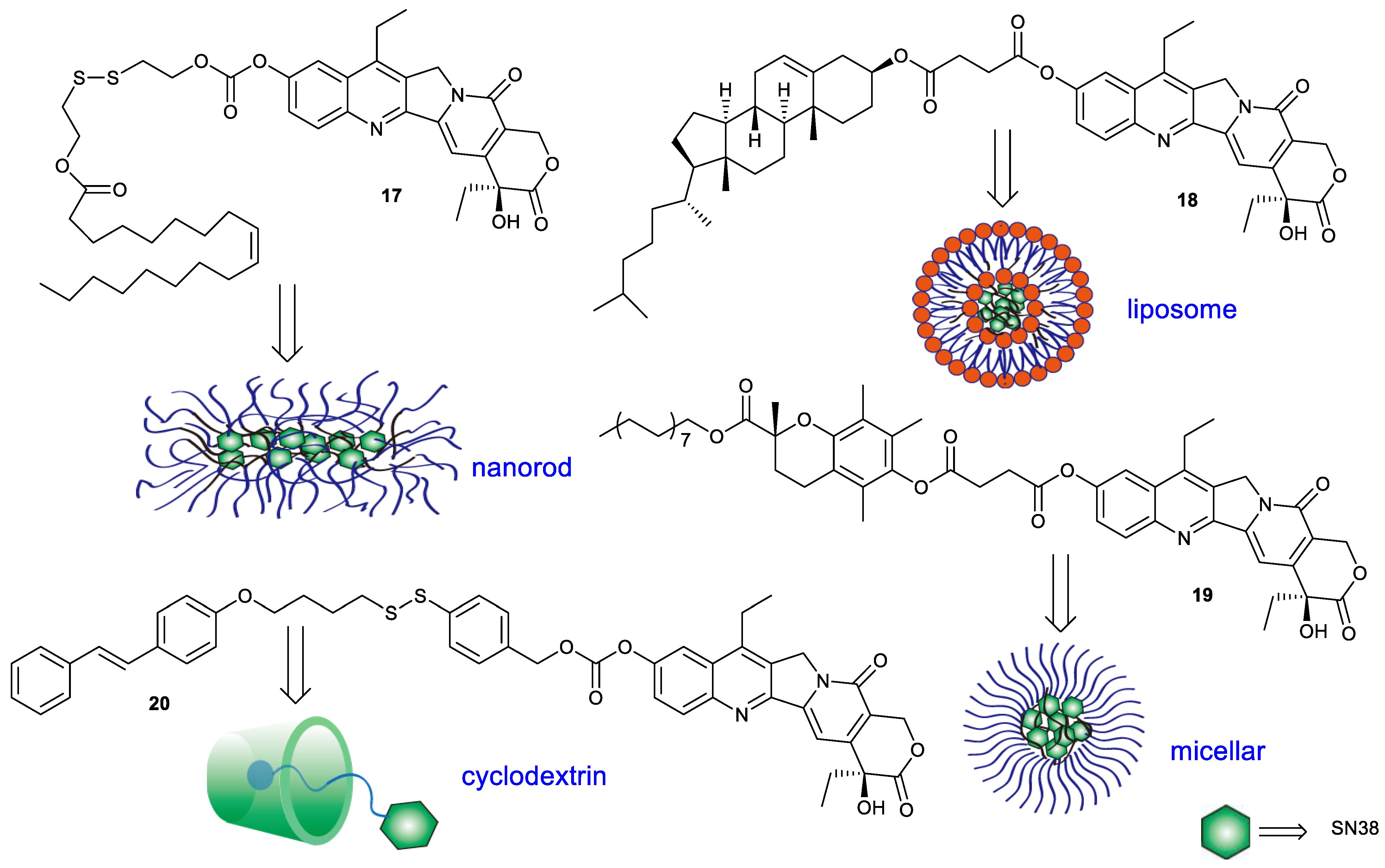
More hydrophobic SN38 derivatives can be encapsulated easily by carrier material to facilitate efficient SN38 delivery (Figure 7). Compound 17 was synthesized by conjugating SN38 with oleic acid via disulfanyl–ethyl carbonate. The encapsulation of compound 17 with DSPE-mPEG2000 could form nanorods and demonstrate colloidal stability. Upon exposure to thiols, the nanorods can rapidly release SN38. For instance, the nanorods could release 1% SN38 within 1 h in phosphate buffer (pH 7.4), versus 100% in 10 mM DTT. The in vitro cytotoxicity of the nanorods toward mouse breast carcinoma 4T1 cells, mouse melanoma B16-F10 cells and mouse colon cancer CT26 cells was comparable to free SN38 and around 90-fold more potent than irinotecan. In addition, the nanorods had a higher inhibitory effect on tumor growth in CT26 tumor-bearing mice than irinotecan [34]. Compound 18 was an esterase-responsive prodrug which was prepared by conjugation of SN38 with cholesterol via succinic acid. The introduction of cholesterol endowed compound 18 with elevated miscibility with liposomal compositions and was beneficial for the formation of nanoliposomes. The resulting nanoliposomes with the drug loading of 9.79% exhibited a size of around 78 nm and a zeta potential of around −28.9 mV. The in vitro cytotoxicity of the nanoliposomes against A549 cells, human lung cancer PC-9 cells, LoVo cells and MCF-7 cells was slightly weaker than that of SN38 alone, while stronger than that of irinotecan. The tumor inhibitory rate of the nanoliposomes in A549 tumor-bearing mice was enhanced by around 1.5-fold compared with that of irinotecan. Moreover, the form of drug delivery significantly improved drug tolerability [35]. Compound 19, a hybrid of SN38 and vitamin E analogue, was synthesized using succinic acid as a linker. Due to high lipid solubility, compound 19 could form nanomicelles with Tween 80 and PEG200. The resulting nanomicelles had much stronger cytotoxicity against human ovarian cancer A2780 cells, A549 cells, HT-29 cells and HepG2 cells than irinotecan [36]. Compound 20 was prepared by conjugation of SN38 with azobenzene moiety via a redox-sensitive self-immolative linker containing a disulfide bond. The hydrophobic compound was delivered through inclusion of beta-cyclodextrin, which widely exhibited applications in many areas, such as the pharmaceutical industry, food technology, cosmetics and dyes, etc., due to its nontoxicity, aqueous solubility, biodegradability and biocompatibility [37]. Compound 20 could be wrapped by beta-cyclodextrin to further self-assemble into tubular or vesicular. The obtained tubular or vesicular could release the parent drug in two ways: one was dependent on the change in cis–trans isomerization of azobenzene exposed to ultraviolet light, and the other was dependent on the reduction in disulfide bond by GSH [38]. The design ideas for these compounds provide a reference for the delivery of other hydrophobic drugs.

2.4. Synergism between Different Drugs

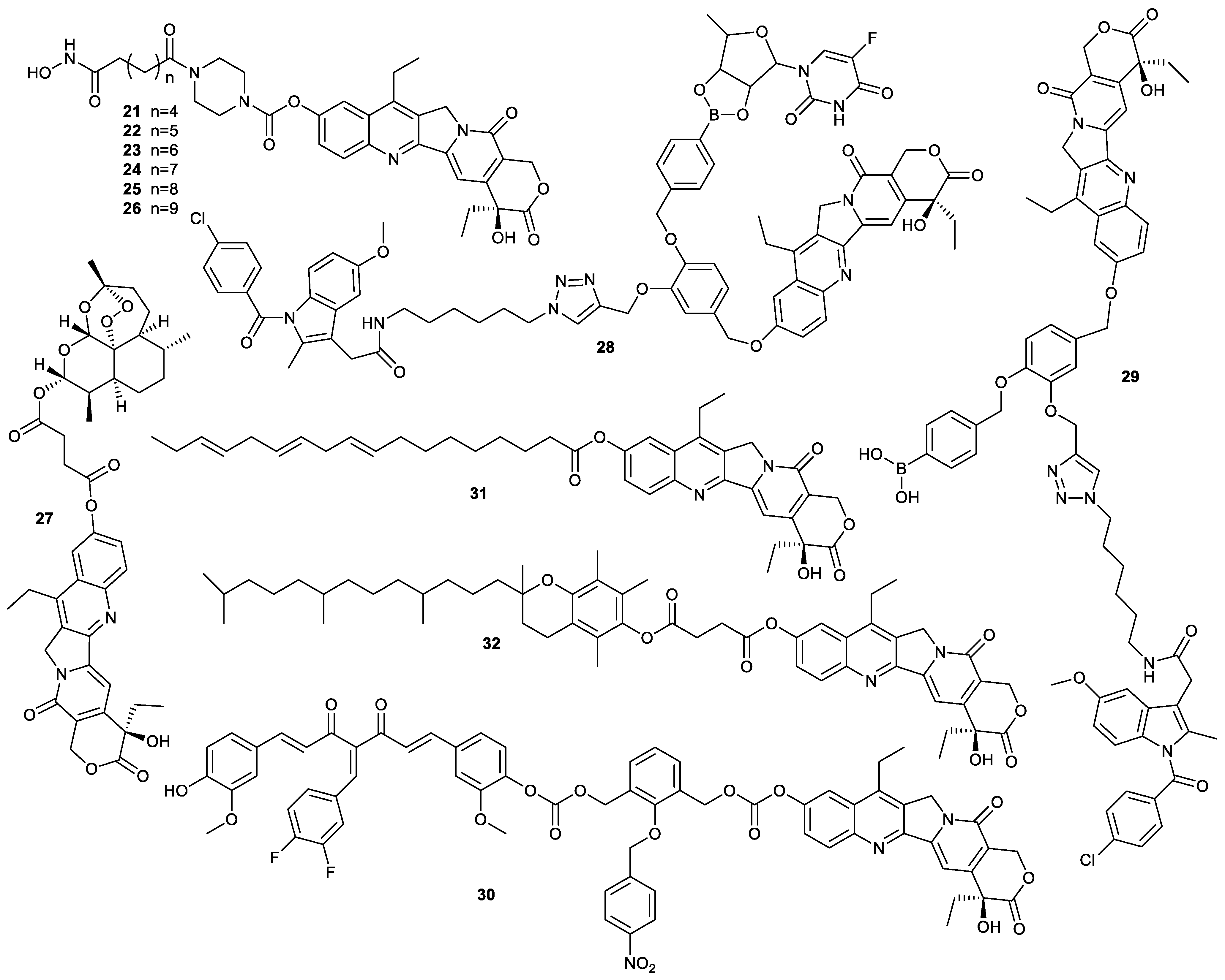
A single drug is generally ineffective for the treatment of some cancers due to the complexity of the initiation and progression of human tumors. Designing multitarget drugs for the treatment of cancer is an important strategy for developing novel antitumor drugs [39]. The introduction of moieties capable of combating cancers in SN38 to prepare a prodrug of SN38 is beneficial for the synergism of different drugs (Figure 8). Upon cellular internalization, this type of prodrug can release parent drugs to synergistically inhibit the growth of cancer cells. Aberrant histone deacetylases (HDACs) have a significant correlation with tumors. Hydroxamic acid derivatives can suppress the growth of cancer cells via inhibition of histone deacetylases. Based on these findings, compounds 2126 were prepared by the introduction of hydroxamic acid moieties into SN38. These compounds were stable in buffers or plasma. However, their inhibitory effect on HDAC was weaker than that of SAHA and was concerned with the length of the carbon chain in hydroxamic acid moieties; the stronger the inhibitory effect on HDAC6, the longer the carbon chain. Due to the difficulty in the release of parent SN38, these compounds exhibited lower cytotoxicity toward A549 cells and HCT-116 cells than SN38 and SAHA, indicating that ensuring the release of parent SN38 was crucial in the design of SN38-based prodrugs [40].
Compound 27, a hybrid of SN38 and artesunic acid, significantly suppressed the growth of the yeast Saccharomyces cerevisiae EKY3 strain via inhibition of the overexpressed human topo 1. The IC50 values of compound 27 against human melanoma RPMI7851 cells and human melanoma SK-MEL 24 cells were 0.05 ± 0.02 µM and 0.32 ± 0.03 µM, respectively, while those of SN38 were 0.51 ± 0.26 µM and 0.48 ± 0.07 µM, demonstrating that compound 27 had more cytotoxicity than SN38 [41]. Compound 28 was another prodrug formed by conjugation of SN38 with 5′-deoxy-5-fluorouridine (5′-DFUR) and indomethacin (IMC). Upon cellular internalization, compound 28 containing borate ester was responsive to H2O2 to release SN38, 5′-DFUR and IMC to inhibit topo 1, thymidylate synthase and COX2, respectively. Due to a strong synergetic effect, compound 28 showed higher cytotoxicity toward COX2-positive human pancreatic carcinoma MIA Paca-2 cells than compound 29, only containing the IMC unit and SN38 unit, indicating that the hybrid containing multiple drugs deserved further investigation. In addition, compound 28 had marginal inhibitory effect on COX2-negative human intestinal Caco-2 cells, exhibiting the tumor-targeting ability [42].
Compound 30 was another hybrid of SN38 and 3,4-difluorobenzylidene curcumin, which could be activated in hypoxic conditions to release SN38 as an inhibitor of topo 1 and 3,4-difluorobenzylidene curcumin as an inhibitor of cancer stem-like cells (CSCs). The in vitro and in vivo experiments showed that the compound could inhibit the metastasis and multidrug resistance of triple-negative breast cancer via eradication of CSCs and downregulation of multidrug resistance [43].
Apart from SN38-based hybrids, codelivery of SN38 derivatives with other chemotherapeutic agents also can create a synergistic effect. For example, SN38 was esterified with α-linolenic acid to obtain compound 31, then compound 31 and the combretastatin-A4 derivative were encapsulated by mPEG5k−PLA16k to prepare nanoparticles. The resulting nanoparticles could inhibit the growth of cancer in a cocktail manner and showed much stronger inhibitory effect on HT-29 cells and HT-29 tumor-bearing mice than free SN38 or its encapsulation. In addition, the nanoparticles could inhibit the metastasis of HUVEC [44]. Another example was compound 32, formed by the conjugation of SN38 with α- tocopherol. The as-prepared prodrug could block detrimental intercellular connections, which were the driving force for the acquired drug resistance and proliferative burst. Compared with MCF-7 cells, the paclitaxel-resistant MCF-7 cells (MCF-7/PTX) absorbed compound 32 more efficiently, which led to the rapid release of parent SN38 to exerted higher cytotoxicity. Similar results were exhibited in the cisplatin-resistant HeLa cells and HeLa cells. However, due to strong hydrophobicity, free compound 32 had difficulties circulating in blood, which could be overcome by encapsulation of compound 32 with amphiphilic block copolymers PEG5k-b-PMASSV. Besides high drug-loading capacity and stability, the obtained nanoparticles had significant anticancer efficacy against both drug-resistant cancer cells and drug-sensitive cancer cells via the above-mentioned antitumor mechanism [45]. Hence, exploiting nanodelivery systems is an alternative for hydrophobic agents.

2.5. Theranostics

Owing to the demand for precision medicine, theranostics is a new trend of developing anticancer drugs, especially for small-molecular theranostics which can be prepared easily and have high tumor-targeting ability and low side effects [46]. Since the introduction of a strong electron-withdrawing group into SN38 at C-10 position will quench or attenuate the intrinsic fluorescence of the conjugated fluorophores via donor-excited photoinduced electron transfer (d-PeT), it is convenient for developing the “turn-on” theranostics based on SN38. As an electron-withdrawing group, the sulfonic group was usually used as the attachment group, such as the sulfonic esters of N-oxyimides, known for a wide range of biological activities [47]. Compound 33, a conjugate prepared by the introduction of 2,4-dinitrobenzene sulfonyl into SN38 at C-10 position, could not produce the fluorescence due to d-PeT. Upon exposure to GSH or some sulfhydryl compounds, compound 33 could release SN38 and produce its intrinsic fluorescence to simultaneously exert therapy and diagnosis via the hydrolyzation of sulfonate moiety (Figure 9). The in vitro cytotoxicity showed that the inhibitory effect of compound 33 on HeLa cells and m-Cherry + OCSC1-F2 cells were comparable to those of SN38 [48].
Compound 34 was synthesized by the introduction of dinitrobenzene into SN38, and its intrinsic fluorescence and activity was significantly quenched by the dinitrobenzene group. Like compound 33, the compound could be responsive to hydrogen sulfide to release SN38 to obtain significantly enhanced fluorescence and activity for real-time monitoring SN38 release and antiproliferation, respectively, which was exhibited in HCT-116 cells and 4T1 cells (Figure 10). It showed stronger cytotoxicity against cancer cells than irinotecan, while exhibiting marginal cytotoxicity against normal human embryonic kidney HEK-293T cells, which was attributed to overexpression of H2S in cancer cells in comparison with normal cells [49].
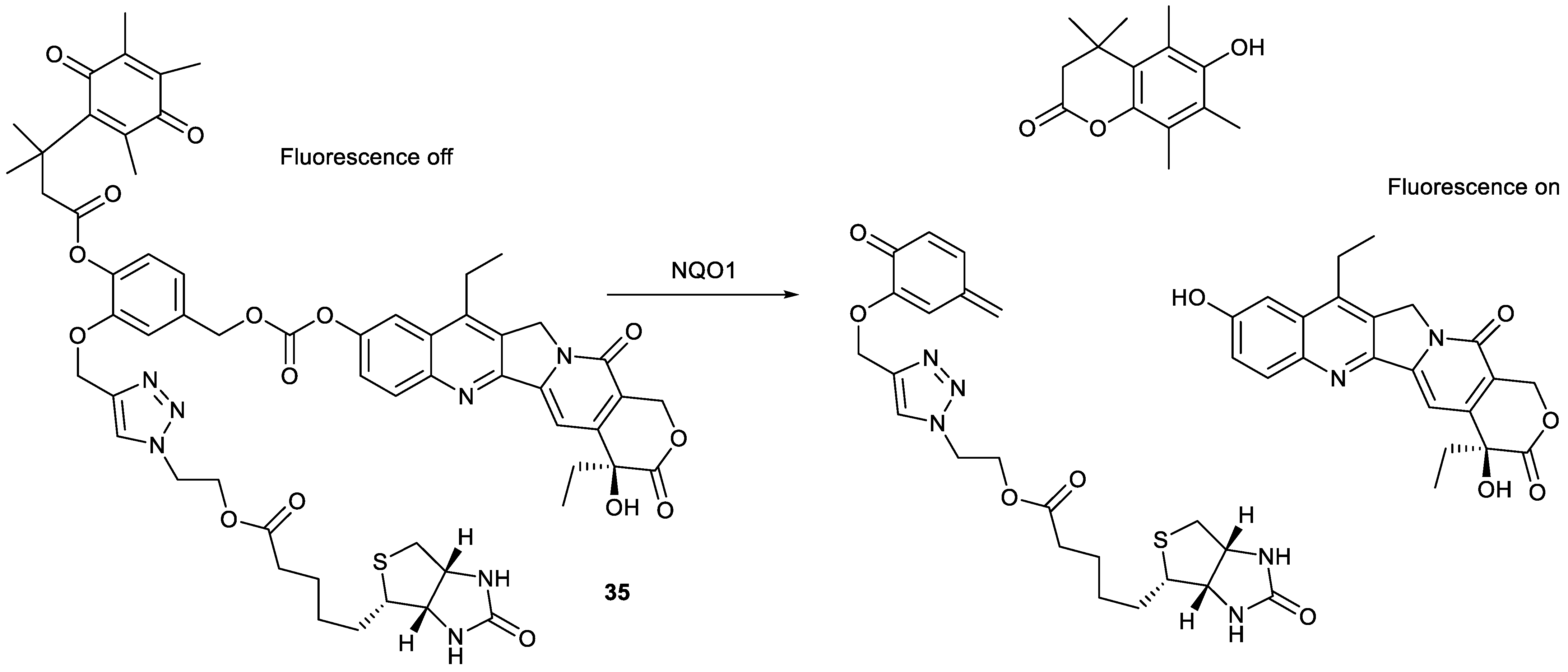
Theranostics with tumor-targeting ability are desirable for the precise diagnosis and treatment of cancer. Based on the overexpression of NAD(P)H:quinone oxidoreductase-1 (NQO1) and the biotin receptor in cancer cells, compound 35 was designed by the introduction of both biotin and quinone propionic acid into SN38 at C-10 position. Due to the insertion of quinone propionic acid, the resulting compound could not produce fluorescence in comparison with SN38. Upon exposure to NQO1, compound 35 could be trigged via quinone propionic acid unit to release parent SN38 as a theranostic agent (Figure 11). The biotin moiety endowed compound 35 with the tumor-targeting ability. For example, compound 35 had significant cytotoxicity against A549 cells and HeLa cells, comparable to SN38, while it demonstrated a marginal effect on human normal fibroblast WI-38 cells and human normal fibroblast BJ cells. Encouragingly, the tumor-targeting ability of compound 35 was verified in the A549 tumor-bearing mice model, showing that compound 35 was mainly accumulated in the tumor organ and had stronger antitumor activity than SN38 [50].
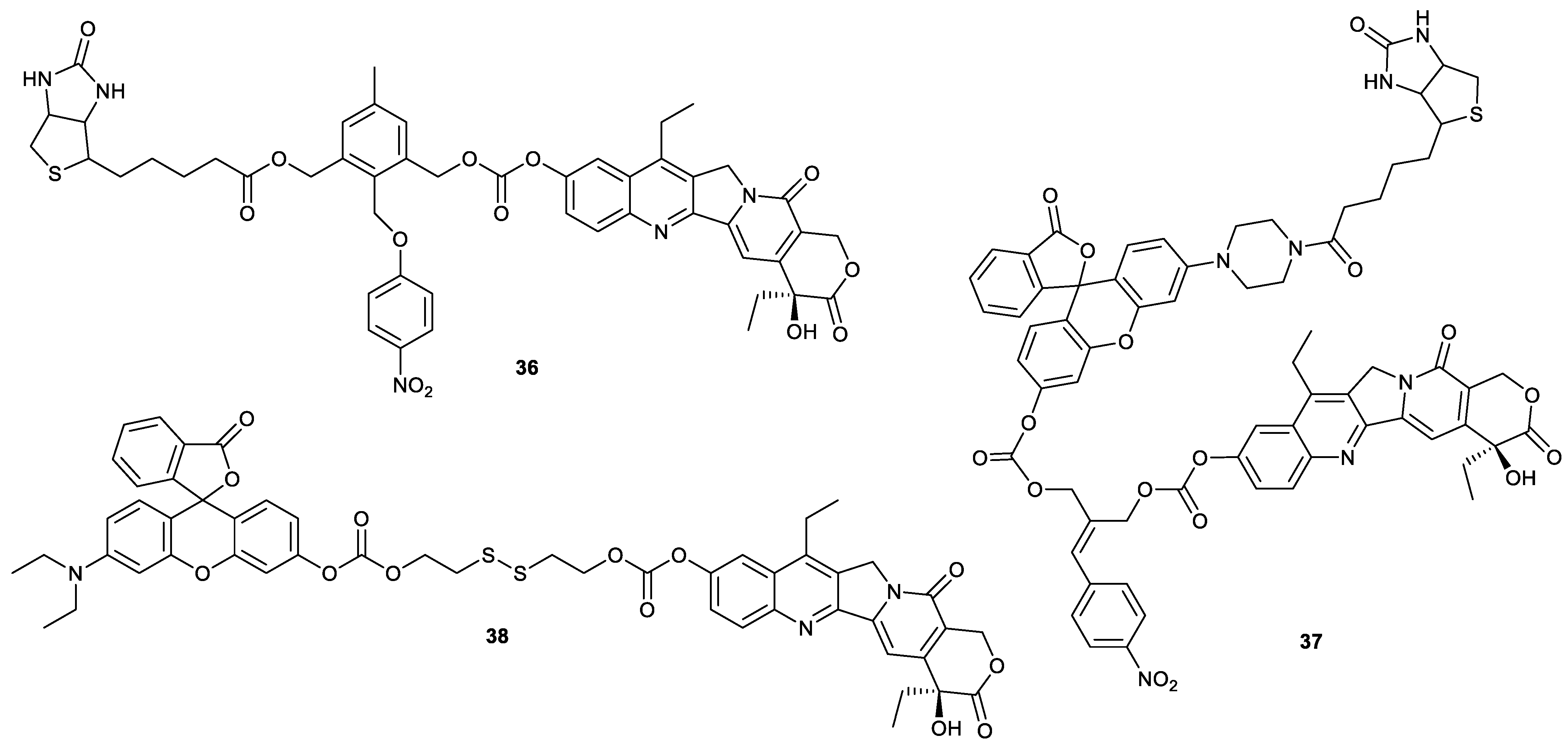
Another theranostic agent was compound 36 (Figure 12), containing biotin as the tumor-targeting unit and 4-nitrobenzene as the hypoxic-responsive unit. Compound 36 was activated in the presence of nitroreductase and NADH in the hypoxic environments of solid tumors, where it was reduced to release SN38 as a theranostic agent. Compound 36 showed a much stronger cytotoxicity against A549 cells and HeLa cells than against normal BJ cells and WI-38 cells, similar to compound 35. In addition, compound 36 had stronger antitumor activity than SN38 in a HeLa tumor-bearing mice model. The fluorescence imaging demonstrated compound 36 could penetrate tissue deeply and be specially accumulated in tumor tissue and activated by hypoxia to release SN38 [51]. Compound 37 was prepared by the introduction of rhodol into compound 36 (Figure 12). There was only a difference in the fluorescence imaging unit between compound 36 and compound 37. The former was a utilization of SN38 as a fluorophore, and the latter was a utilization of rhodol as a fluorophore. Responsive to hypoxia, compound 37 released SN38 to inhibit topo 1 and rhodol to track the distribution of drug. The biotin moiety endowed compound 37 with enhanced cytotoxicity against HeLa cells and decreased cytotoxicity against mouse embryonic fibroblast NIH3T3 cells [52].
Apart from the insertion of a tumor-targeting unit into SN38 derivatives, encapsulation of SN38-based theranostic agents with carrier material capable of targeting tumors can also render the tumor-targeting ability of theranostics possible. For example, though compound 38 formed by conjugation of SN38 with rhodol via disulfide bond did not have tumor-targeting ability (Figure 12), the “turn-on” theranostic prodrug was encapsulated by biotinylated poly(vinyl alcohol) to form a tumor-targeting nanoparticle. Upon cellular internalization, compound 38 was activated by GSH to release SN38 and rhodol to induce apoptosis and monitor the delivery of drugs, respectively. These nanoparticles showed remarkably higher stability in blood serum and much stronger selectivity to cancer cells. The nanoparticles significantly inhibited the proliferation of HeLa cells in a dose-dependent manner while showing only marginal cytotoxicity toward NIH3T3 cells [53].

2.6. Other Functions

For exploiting SN38 derivatives with higher anticancer activity, compound 39 and compound 40 were synthesized. They showed a strong inhibitory effect on tumor cells, especially compound 40, with IC50 values of 0.002, 0.003 and 0.081 µM toward human B lymphoma Raji cells, HCT-116 cells, A549 cells and LoVo cells, respectively. Apart from stronger antitumor activity than SN38, compound 40 could be administrated orally with the bioavailability of 22.4% for mice and significantly suppress the HCT-116-xenograft tumor growth in the nude-mice model at dosages of 0.5, 2.0 and 8.0 mg/kg. When compound 40 was administered intraperitoneally to Raji tumor-bearing mice at dosages of 2.0 and 4.0 mg/kg, Raji xenograft tumor growth was completely inhibited. In addition, the minimum lethal doses of compound 40 was two-fold that of SN38, indicating that compound 40 had much higher safety than SN38 [54]. If the substitution of C10-OH of SN38 with fluorine was carried out, compound 41 was obtained. As a topo 1 inhibitor, compound 41 showed more potent in vitro cytotoxicity against serval types of cancer cells (human colon cancer SW480, human pancreatic cancer SW1990, human hepatoma Hep3B, HepG2, A549, A2780, HeLa and human cholangiocarcinoma QBC cells) and inhibitory effect on tumor growth in HCC and ICC mouse models than topotecan, while showing only marginal toxicity to mouse noncancerous tissues [55].
Compounds 4244, whose IC50 values for human leukemia K562 cells were higher than that of SN38, were prepared by the introduction of 2-nitrobenzene, 3-nitobenzene or 4-nitrobenzene into SN38 via ether bond [56]. Compounds 4551 reported by SQ Zhang and coworkers had an inhibitory effect on A549 cells and HCT-116 cells comparable to that of SN38 [32]. Compounds 5257 showed slightly lower cytotoxicity toward PC-3 cells than SN38 [33]. Compound 58 was synthesized by the introduction of amino acid to SN38. The compound showed higher cytotoxicity against human colon cancer SW1116 and human colon cancer LS174T cell than irinotecan [57].
The rapid release of the parent SN38 from SN38-based prodrug is the key criterion for designing these drugs, which deserves more attention. For example, compound 59, formed by conjugation of SN38 with 2-(aminomethyl)pyrrolidine via carbamate, was able to rapidly release SN38 in a self-immolative manner. When compound 59 was incubated in a DMSO/acetate buffer mixture (pH 5.5) at 37 °C, it released SN38 with a half-life (t1/2) of 1.6 h, more rapidly than compound 60 (t1/2 = 10.6 h) and compound 61 (t1/2 = 38.9 h) [58].
Compounds 6264 were SN38 homodimeric prodrugs with different linkers containing disulfide bonds (Figure 13). These compounds exhibited different chemical and self-assembly stability due to different linker length. Among them, compound 64 possessed good chemical and self-assembly stability. The homodimeric prodrug nanoassemblies (HDPNs) formed by compound 64 had a higher inhibitor effect on 4T1 cells than SN38. Additionally, compound 64 significantly accumulated in tumor tissue to exert its antitumor activity in 4T1 tumor-bearing mice [59]. These findings provided a reference for the design of HDPNs.

3. Modification of SN38 at C-20 Position

Ensuring the structural integrity of camptothecin analogues is very important for the maintenance of antitumor activity. The hydrolysis of the E-ring of camptothecin derivatives will give rise to decreased antitumor activity [60]. It is well established that a prodrug with ester bond as a linker can be hydrolyzed to release a parent drug by esterase in cells. The esterification of C20-OH of camptothecin derivatives not only enhances their stability due to high steric hindrance but also endows them with multifunction, attributed to the introduction of other molecules [61]. The structural modification of SN38 at C-20 position has made progress and obtained some derivatives with the enhancement of synergism, tumor-targeting activity or solubility in recent years.

3.1. Synergism between Different Drugs

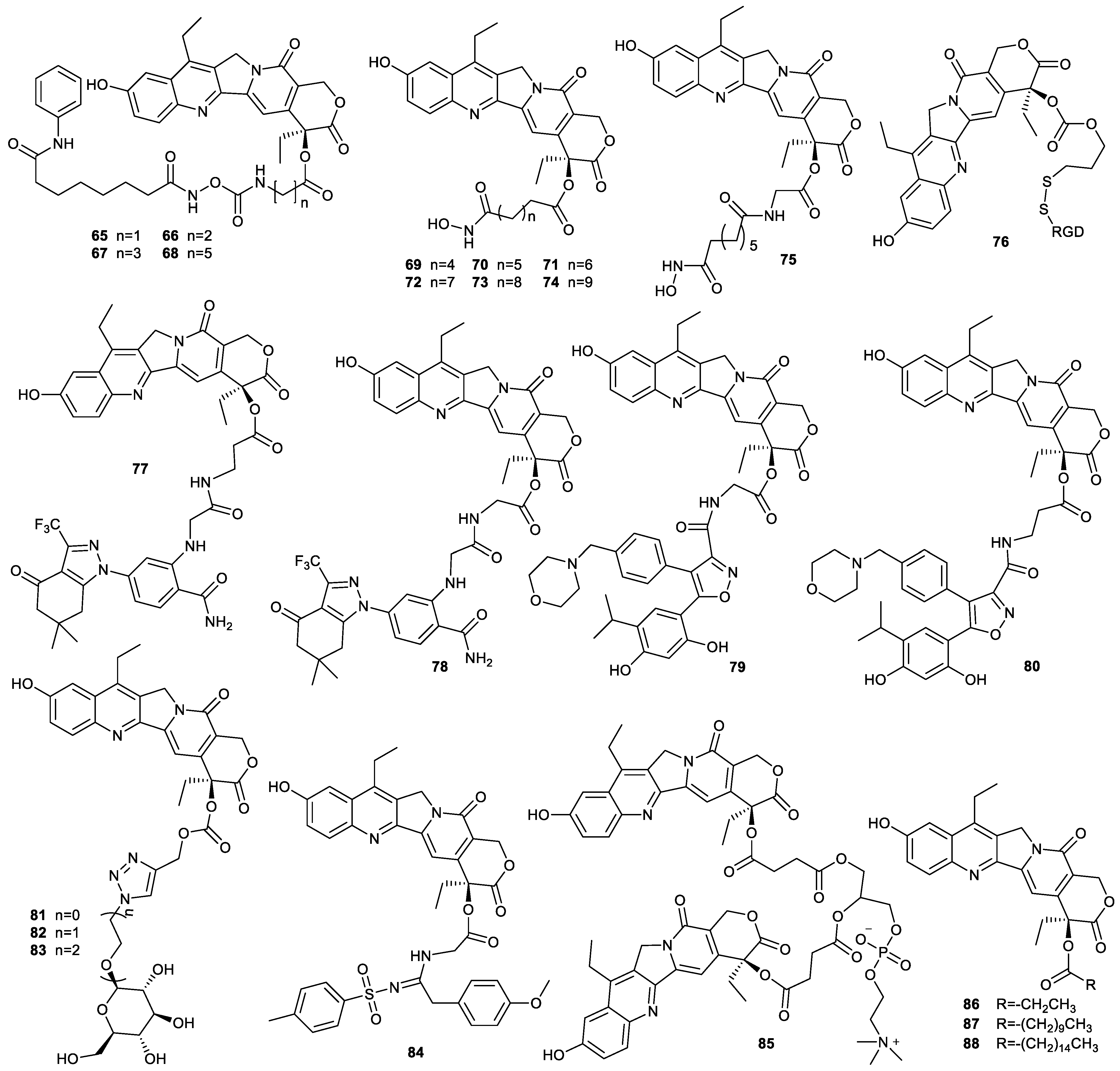
Owing to the complexity of the initiation and progression of human tumors, the combination of different drugs for the treatment of cancer is an effective strategy. Histone deacetylase inhibitors can sensitize chemotherapeutic drugs, including camptothecin-based drugs [62,63]. For example, compounds 6568 synthesized by conjugation of SN38 with SAHA via the amino acids of different chain length demonstrated that their cytotoxicity against A549 cells and HCT-116 cells increased with decreasing amino acid chain length, which was consistent with the results of their release rate of parent drugs from these prodrugs, that is, the release rate increased gradually with decreasing amino acid chain length. While compound 65, the strongest antitumor compound among these prodrugs, showed a slightly weaker antitumor activity than SN38, it exhibited a significantly stronger cytotoxicity than SAHA [64]. The hydroxamic acid moiety of SAHA played a key role in inhibiting histone deacetylase. The direct conjugation of hydroxamic acid moiety with SN38 was carried out to finish the synthesis of compounds 6975. The inhibitory effect of these compounds on HDAC decreased with the increased chain length of C-20 position and was slightly lower than that of SAHA. Though these compounds demonstrated lower inhibition of HDAC than SAHA, they all showed higher cytotoxicity than SAHA, especially compound 75, which could rapidly release SN38. These findings indicated that these as-prepared compounds exhibited synergistic antitumor activity by inhibiting both topo 1 and HDAC [40].

3.2. Tumor Targeting

The enhancement of tumor-targeting ability is beneficial for increasing antitumor activity and decreasing side effects. The decoration of SN38 with tumor-targeting units such as biotin, folic acid, small peptide and antibody is an effective strategy to enhance the tumor-targeting ability of chemotherapeutic agents [24,25]. For example, trastuzumab deruxtecan, an antibody–drug conjugate based on camptothecin, was approved by the US Food and Drug Administration for the treatment of several cancers [65]. SN38 was conjugated with iRGD (small cyclic peptidic motif, CRGDK/RGPD/EC) via disulfide bond to prepare compound 76. The introduction of iRGD not only enhanced the water solubility of compound 76 but also inhibited cancer cell metastasis by targeting αv integrins and neuropilin-1 overexpressed on the surface of several kinds of cancer cells [66,67]. Upon cellular internalization, compound 76 was responsive to GSH to rapidly release SN38 and iRGD. The inhibitory effect of compound 76 on high metastatic human hepatocellular carcinoma HCC-LM3 cells, human hepatocellular carcinoma BEL-7402 cells, HepG2 cells and A549 cells was 1–2 orders of magnitude higher than that of irinotecan, and its antimetastasis was also demonstrated in HCC-LM3 cells. Additionally, the iRGD released from compound 67 could block the metastasis of HCC-LM3 cells. The antiproliferation and antimetastasis of compound 76 was also verified to be stronger than that of irinotecan in HCC-LM3 tumor-bearing mice [68].
The introduction of heat shock protein 90 (HSP90) inhibitors NVP-AUY922 and SNX-5422 into SN38 prepared compounds 7780, which were targeted to cancer cells via extracellular HSP90-mediated endocytosis. These compounds showed excellent stability in buffers at pH 6.0 and 7.4 and in human plasma. However, the in vitro HSP90 binding affinity assay showed that conjugation of SNX-5422 with SN38 had negligible impacts on HSP90 activity, while conjugation of NVP-AUY922 with SN38 did not affect the binding affinity of the hybrids to HSP90, indicating that compounds 79 and 80 deserved further investigation, especially compound 79, which showed strong cytotoxicity against A549 cells, HCT-116 cells, MIA-Paca-2 cells and human pancreatic cancer Capan-1 cells with IC50 values of 56 nM, 22 nM, 46 nM and 36 nM, respectively. In addition, compound 79 also showed strong antitumor activity in HCT-116 tumor-bearing mice and Capan-1 tumor-bearing mice, indicating that compound 79 is a promising new candidate for cancer therapy [69].
In regard to the overexpression of glucose transporters in cancer cells, compounds 8183 were synthesized by conjugation of SN38 with glucose via click reaction. The introduction of glucose to SN38 simultaneously enhanced water solubility and tumor-targeting ability of the SN38 derivatives. Among them, compound 82 exhibited high cytotoxicity and selectivity for HCT-116 cells, which was also demonstrated in an HCT-116 xenograft model [70]. The design idea is also applicable to structural modification of other hydrophobic chemotherapeutic agents.

3.3. Improvement of Solubility

The introduction of water-soluble groups into SN38 can enhance the water solubility of SN38. For example, compound 84, synthesized by the introduction of sulfonyl amidine into SN38, had higher water solubility than SN38. The cytotoxicity of compound 84 against A549 cells, oral epithelial carcinoma KB cells and multidrug-resistant KB cells was significantly stronger than that of irinotecan. In addition, compound 84 could reverse the multidrug resistance of KB cells [71]. Compound 85 was obtained by the decoration of SN38 with L-α-glycerophosphorylcholine. This compound could self-assembled into spherical liposomes with a size of ~140 nm, a potential of −21.6 ± 3.5 mV and a loading rate of 65.2%. The resulting liposomes were stable in a neutral environment but degraded to release SN38 in a weakly acidic condition. Moreover, the liposome had a longer blood retention time. After endocytosis, compound 85 showed strong cytotoxicity, comparable to that of SN38 [72].
Another strategy for improving the solubility of SN38 is the insertion of long-chain fatty acids into SN38, which can improve solubility of the as-prepared compound in lipid excipients. For example, the solubility of compounds 8688 in long-chain triglycerides was increased up to 444-fold (Figure 14). Their stability was controlled, and transmucosal permeability was enhanced. These compounds were stable in simulated gastric fluids but exhibited different rates of hydrolysis in simulated intestinal fluids (in the presence of enzymes) depending on the alkyl chain length [73]. Due to improvement in transepithelial drug transport and cellular uptake, these compounds were beneficial for oral administration.

4. Modification of SN38 at Both C-10 Position and C-20 Position

4.1. Tumor Targeting

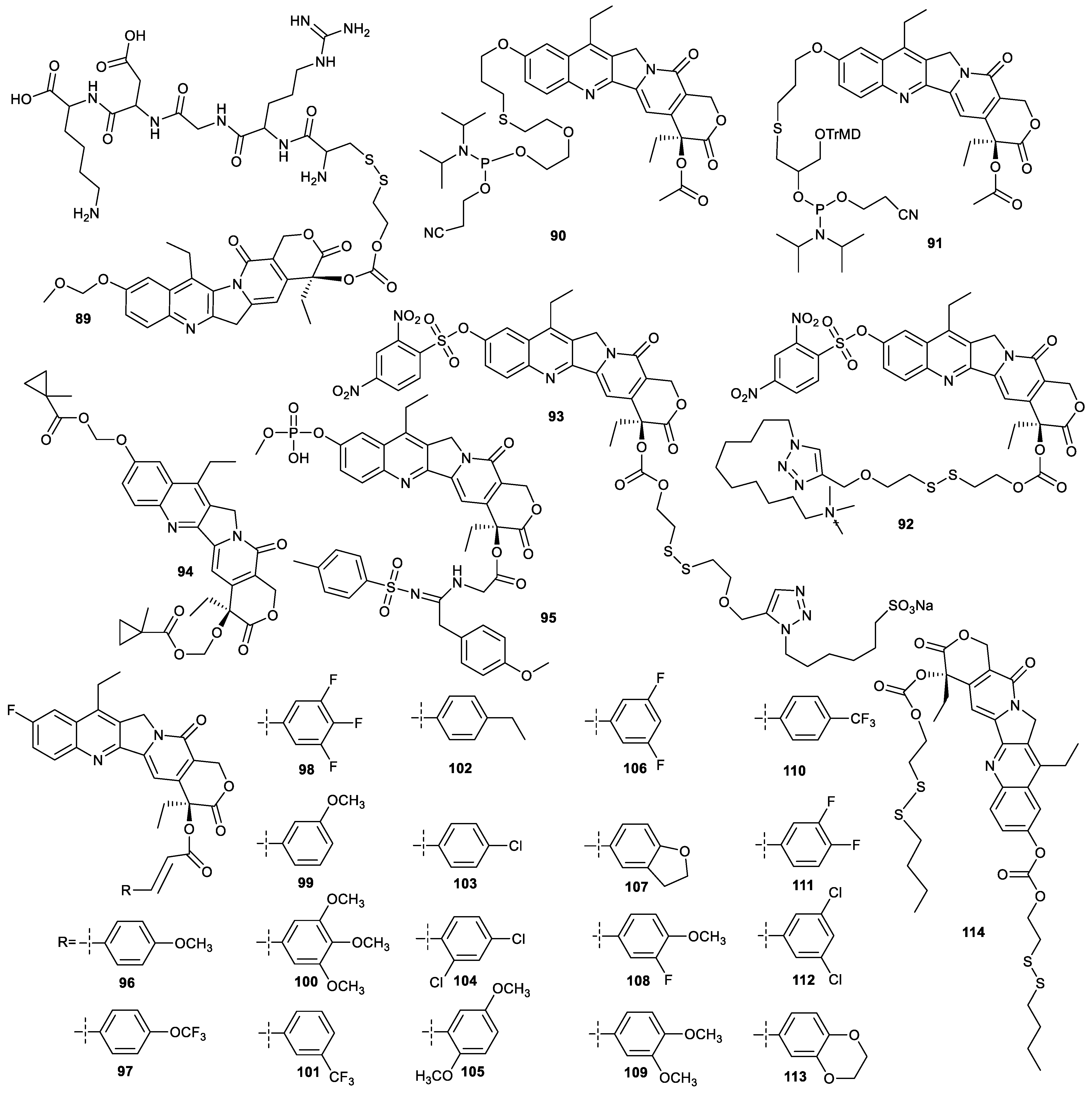
Two methods are reported to prepare tumor-targeting SN38 derivatives decorated at both C-10 position and C-20 position. The front method is that C-10 is confined via insertion of a small group, and C-20 is conjugated with a tumor-targeting moiety, and the behind method is the reverse. Compound 89 was synthesized by the introduction of methoxymethyl ether at C-10 position and an arginine–glycine–aspartic acid–lysine (RGDK) peptide at the C-20 position of SN38. It could be targeted to the neuropilin-1 receptor and responsive to GSH. In addition, compound 89 could self-assemble into stable micelles with a hydrodynamic diameter around 110 nm and a fixed drug loading rate of 35%. The micelles prolonged the retention time in plasma and enhanced accumulation in the tumor tissue of compound 89. Though the in vitro cytotoxicity of compound 89 against A549 was comparable to that of SN38, it showed much higher antitumor activity in a A549 xenograft mice model than irinotecan [74]. Compounds 90 and 91 containing phosphoramidites could be conjugated with aptamer automatically by solid-phase synthesis to prepare aptamer–SN38 conjugates with the tumor-targeting ability. For example, the conjugation of the two compounds with aptamer sgc8 could target the as-prepared compounds to HCT-116 cells. Moreover, as prodrugs, they also could release parent SN38 to induce apoptosis of cancer cells [75].

4.2. Theranostics

Exploiting SN38 derivatives with theranostic function is based on the intrinsic fluorescence of SN38. The direct conjugation of SN38 with 2,4-dinitro-benzene sulfonyl (DNS) at C-10 position causes the original fluorescence quenching, such as compounds 92 and 93. In addition, compounds 92 and 93 had a quaternary ammonium moiety and sodium sulfonate moiety at C-20 position, respectively. Compound 92 could be fully threaded into the hydrophobic cavity of pillar [5]arene to form a supramolecular amphiphilic structure which self-assembles into nanoparticles, with an average hydrodynamic diameter of 253 nm and an encapsulation efficiency of 92.2%. Upon exposure to the high level of GSH in cancer cells, the nanoparticles could release SN38 to exert its antitumor activity and track its distribution in tissues due to the cleavage of their disulfide bonds and the DNS groups. Though compound 92 exhibited cytotoxicity against MCF-7 cells and HepG2 cells comparable to SN38, it demonstrated much lower cytotoxicity against NIH3T3 cells than SN38, indicating that compound 92 had excellent selectivity to cancer cells [76]. Compound 93 was encapsulated by another pillar [5]arene with tetraphenylethene functionalized on the bridged methylene group of the pillararene skeleton. Due the aggregation-induced emission (AIE) of pillar [5]arene and the intrinsic fluorescence of SN38, upon cellular internalization, the inclusions were responsive to GSH in cancer cells to release SN38 to inhibit cell proliferation, accompanied by fluorescence change from dim yellow to brilliant yellow for tracking their tumor tissue distribution. The inclusions showed their theranostic function in MDA-MB-231 cells and MDA-MB-231 tumor-bearing mice. The cytotoxicity of the inclusions toward MDA-MB-231 cells were stronger than that of SN38. Moreover, the inclusions showed marginal cytotoxicity against normal human embryonic lung fibroblast MRC-5 cells, as the pillar [5]arene [77].

4.3. Other Functions

The designing of compound 94 mainly paid more attention to the effective release of SN38. Compound 94, incubated with a split BS2 esterase from Bacillus subtilis, could be effectively inverted into SN38 with a conversion rate of >95% and showed potent cytotoxicity against MDA-MB-231 cells in the presence of BS2, albeit with structural modification at both the C-10 position and C-20 position of SN38 [78]. The aim of designing compound 95 was to enhance the water solubility of SN38. Hence, phosphate and sulfonylamidine moieties were selected to conjugate with SN38 at C-10 position and C-20 position for preparation of compound 95. Compound 95 exhibited 30–100-fold higher cytotoxicity against KB cells and multidrug resistant KB cells than irinotecan, demonstrating that compound 95 could reverse the multidrug resistance of KB cells [71]. Compounds 96113 was synthesized by substitution of C10-OH with fluorine and esterification of C20-OH of SN38. Among them, compound 106 showed broad-spectrum cytotoxicity toward HepG2 cells, SW480 cells, A2780 cells and Hucct1 cells with IC50 values of 0.03, 0.09, 0.22 and 0.32 µM, respectively. In addition, compound 106 showed higher selectivity to cancer cells than topotecan. For example, the ratio of IC50 values to normal human hepatic LO2 cells and HepG2 cells was 113.20 for compound 106, while 85.60 for topotecan. The high antitumor activity and low side effects of compound 106 were verified in a HepG2 tumor-bearing mouse model [79]. Due to the presence of fluorine, compound 106 in cells can be tracked by NMR, which merits further investigation.
The esterification of SN38 at position 10 and position 20 with fatty acid containing a disulfide bond was used to prepare more hydrophobic compound 114 (Figure 15). Then, the obtained compound was encapsulated with DSPE-PEG2000 to prepare nanoparticles which were responsive to GSH to release SN38. The in vitro cytotoxicity against human pancreatic carcinoma Panc-1 and BxPC-3 cells of the nanoparticles was remarkably higher than that of irinotecan. Furthermore, the nanoparticles showed a higher inhibitory effect on tumor growth and higher safety than irinotecan in a Panc-1 xenograft model [80].

5. Modification of SN38 at C-9 Position and the Opening of E-Ring

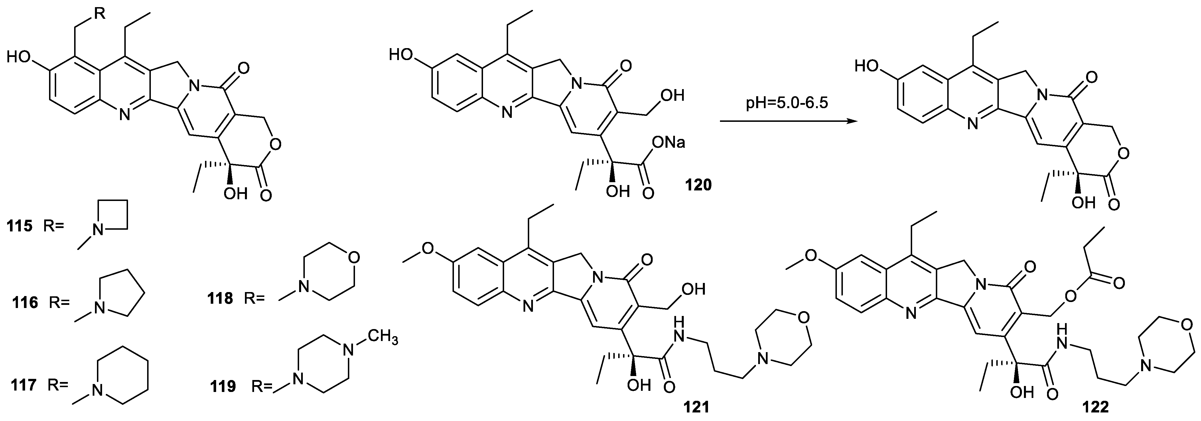
The structural modification of SN38 at C-9 position is seldomly reported. Inspired by the structure of irinotecan, Naumczuk inserted azetidine, pyrrolidine and piperidine into SN38 at C-9 position, respectively, to prepare compounds 115119. These compounds not only inhibited topo 1 but also made DNA alkylation. These compounds showed potent cytotoxicity toward human leukemia HL-60 cells, MCF-7 cells, MDA-MB-231 cells, Caco-2 cells, HT-29 cells and A549 cells comparable to SN38. Compared with the IC50 value of SN38 toward normal CRL-179 cells, the IC50 values of these compounds, especially compounds 116, 118 and 119, were enhanced by around 200-fold, indicating that structural modification at C-9 position endowed SN38 with selectivity to cancer cells [81].
It is well established that the intact E-ring of SN38 is very important for the maintenance of biological activity. Though camptothecin derivatives with opened E-rings were inactive, their carboxylates could undergo lactonization to form the active molecules in the weak acid microenvironment of cancer cells. Hence, ensuring the delivery of the carboxylates of SN38 to tumor tissue is crucial [6]. For example, compound 120 could be adsorbed by cationic supramolecular organic frameworks and effectively delivered to tumor tissue. The half-life of the conversion of compound 120 was 22 h at pH = 6.5, which was shorter than the time gap for repeated administrations of clinically used irinotecan (14 d) or topotecan (21 d) [82]. These findings indicated that the opening of E-rings was not an absolute obstacle to maintain the biological activity of SN38, and ensuring the delivery of SN38 with the opening of E-rings to tumor is curial.
Compounds 121 and 122 also demonstrated that the opening of the E-ring of SN38 could retain its antitumor activity (Figure 16). Compound 121 was prepared by the amidation of a carboxyl group resulting from the opening of the E-ring of SN38. The compound showed more potent cytotoxicity against A549 cells and HT-29 cells than SN38. Once the hydroxyl group of compound 121 was esterified, the resulting compound 122 only had cytotoxicity comparable to that of irinotecan, indicating once again that the intact E-ring was not indispensable to maintain antitumor activity for SN38 [83].

6. Conclusions and Perspectives

Though SN38 has poor water solubility and some side effects, it demonstrates potent antitumor activity and intrinsic fluorescence, which deserves to be further investigated. The aims of the structural modification of SN38 are mainly improving water solubility and enhancing tumor-targeting ability. The conjugation of SN38 with water-soluble molecules and tumor-targeting molecules is an effective strategy to address the above issues. Sometimes, the preparation of more hydrophobic SN38 derivatives for encapsulation by carrier materials is also adopted to improve water solubility. The structural modification of SN38 reported in the literature has been concerned with position 10 and position 20 of SN38 in recent years. Moreover, preparing a prodrug via ester bond is the main form of structural modification of SN38. Modification on other positions such as position 9 and the E-ring of SN38 is rarely reported, owing to a possible decrease in antitumor activity caused by the change in structure. Regardless of how to design the prodrug of SN38, it is crucial for SN38-based prodrugs to be able to quickly release the parent SN38. Hence, the linker between SN38 and other units must be broken to release parent drugs, and the linker responsive to endogenous material in cancer cells is even more noteworthy for designing novel SN38 derivatives, owing to its selectivity for cancer cells.
With the demand for precision medicine, exploiting SN38-based theranostic agents is an important research field due to the intrinsic fluorescence of SN38. However, the wavelength used to excite the fluorescence of SN38 is too short to penetrate deep tissues. Hence, extending the conjugated system of SN38 is worth further investigation on the premise of the maintenance of its antitumor activity. In addition, the resistance and metastasis of cancer cells during chemotherapy is difficult to be cured by a single drug. Hence, designing multitarget drugs for cancer is an important trend for the development of antitumor drugs, which is also applicable for the structural modification of SN38. In summary, the structural modification on SN38 endows its small-molecular derivatives with multifaceted functions, which is beneficial for exploiting novel antitumor drugs.

Author Contributions

Conceptualization, Y.D.; writing—original draft preparation, Y.D.; writing—review and editing, Y.D.; literature search and preparation of the graphics and tables, M.Q. and Y.L.; supervision, M.Q. and Y.L.; funding acquisition, Y.D. All authors have read and agreed to the published version of the manuscript.

Funding

This work was supported by the Natural Science Research Foundation of the Department of Education of Anhui Province (KJ2019A0873), the Scientific Research Team of Anui Xinhua University (No. kytd202211), the Pharmaceutical Institute of Anui Xinhua University (No. yjs202107).

Institutional Review Board Statement

Not applicable.

Informed Consent Statement

Not applicable.

Data Availability Statement

The data presented in this study are shown in this paper.

Conflicts of Interest

The authors declare no conflict of interest.

Sample Availability

Not applicable.

References

  1. Gusakov, E.A.; Topchu, I.A.; Mazitova, A.M.; Dorogan, I.V.; Bulatov, E.R.; Serebriiskii, I.G.; Abramova, Z.I.; Tupaeva, I.O.; Demidov, O.P.; Toan, D.N.; et al. Design, synthesis and biological evaluation of 2-quinolyl-1,3-tropolone derivatives as new anti-cancer agents. RSC Adv. 2021, 11, 4555–4571. [Google Scholar] [CrossRef] [PubMed]
  2. Mayer, S.; Nagy, N.; Keglevich, P.; Szigetvari, A.; Dekany, M.; Szantay, C.; Hazai, L. Synthesis of Novel Vindoline-Chrysin Hybrids. Chem. Biodivers. 2022, 19, e2100725. [Google Scholar] [CrossRef]
  3. Keglevich, A.; Danyi, L.; Rieder, A.; Horvath, D.; Szigetvari, A.; Dekany, M.; Szantay, C.; Latif, A.D.; Hunyadi, A.; Zupko, I.; et al. Synthesis and Cytotoxic Activity of New Vindoline Derivatives Coupled to Natural and Synthetic Pharmacophores. Molecules 2020, 25, 1010. [Google Scholar] [CrossRef] [PubMed] [Green Version]
  4. Pradhan, P.; Das, I.; Debnath, S.; Parveen, S.; Das, T. Synthesis of Substituted Tropones and Advancement for the Construction of Structurally Significant Skeletons. Chemistryselect 2022, 7, e202200440. [Google Scholar] [CrossRef]
  5. Guan, Y.F.; Liu, X.J.; Pang, X.J.; Liu, W.B.; Yu, G.X.; Li, Y.R.; Zhang, Y.B.; Song, J.; Zhang, S.Y. Recent progress of oridonin and its derivatives for cancer therapy and drug resistance. Med. Chem. Res. 2021, 30, 1795–1821. [Google Scholar] [CrossRef]
  6. Khaiwa, N.; Maarouf, N.R.; Darwish, M.H.; Alhamad, D.W.M.; Sebastian, A.; Hamad, M.; Omar, H.A.; Orive, G.; Al-Tel, T.H. Camptothecin’s journey from discovery to WHO Essential Medicine: Fifty years of promise. Eur. J. Med. Chem. 2021, 223, 113639. [Google Scholar] [CrossRef]
  7. Si, J.; Zhao, X.; Gao, S.; Huang, D.; Sui, M. Advances in delivery of Irinotecan (CPT-11) active metabolite 7-ethyl-10-hydroxycamptothecin. Int. J. Pharm. 2019, 568, 118499. [Google Scholar] [CrossRef]
  8. Tanizawa, A.; Kohn, K.W.; Kohlhagen, G.; Leteurtre, F.O.; Pommier, Y. Differential Stabilization of Eukaryotic DNA Topoisomerase I Cleavable Complexes by Camptothecin Derivatives. Biochemistry 1995, 34, 7200–7206. [Google Scholar] [CrossRef]
  9. Bala, V.; Rao, S.; Boyd, B.J.; Prestidge, C.A. Prodrug and nanomedicine approaches for the delivery of the camptothecin analogue SN38. J. Control. Release 2013, 172, 48–61. [Google Scholar] [CrossRef]
  10. Wu, F.; Qiu, F.; Wai-Keong, S.A.; Diao, Y. The Smart Dual-Stimuli Responsive Nanoparticles for Controlled Anti-Tumor Drug Release and Cancer Therapy. Anticancer Agents Med. Chem. 2021, 21, 1202–1215. [Google Scholar] [CrossRef]
  11. Cheng, W.; Gu, L.; Ren, W.; Liu, Y. Stimuli-responsive polymers for anti-cancer drug delivery. Mater. Sci. Eng. C 2014, 45, 600–608. [Google Scholar] [CrossRef] [PubMed]
  12. Chen, H.; Liu, D.; Guo, Z. Endogenous Stimuli-responsive Nanocarriers for Drug Delivery. Chem. Lett. 2016, 45, 242–249. [Google Scholar] [CrossRef] [Green Version]
  13. Thakkar, S.; Sharma, D.; Kalia, K.; Tekade, R.K. Tumor microenvironment targeted nanotherapeutics for cancer therapy and diagnosis: A review. Acta Biomater. 2020, 101, 43–68. [Google Scholar] [CrossRef]
  14. Petrova, V.; Annicchiarico-Petruzzelli, M.; Melino, G.; Amelio, I. The hypoxic tumour microenvironment. Oncogenesis 2018, 7, 10. [Google Scholar] [CrossRef] [Green Version]
  15. Gulzar, A.; Xu, J.; Wang, C.; He, F.; Yang, D.; Gai, S.; Yang, P.; Lin, J.; Jin, D.; Xing, B. Tumour microenvironment responsive nanoconstructs for cancer theranostic. Nano Today 2019, 26, 16–56. [Google Scholar] [CrossRef]
  16. Jin, C.; Zhang, Q.; Lu, W. Synthesis and biological evaluation of hypoxia-activated prodrugs of SN-38. Eur. J. Med. Chem. 2017, 132, 135–141. [Google Scholar] [CrossRef]
  17. Wang, L.; Xie, S.; Ma, L.; Chen, Y.; Lu, W. 10-Boronic acid substituted camptothecin as prodrug of SN-38. Eur. J. Med. Chem. 2016, 116, 84–89. [Google Scholar] [CrossRef] [PubMed]
  18. Singh, U.; Quintanilla, R.H.; Grecian, S.; Gee, K.R.; Rao, M.S.; Lakshmipathy, U. Novel live alkaline phosphatase substrate for identification of pluripotent stem cells. Stem Cell Rev. Rep. 2012, 8, 1021–1029. [Google Scholar] [CrossRef] [Green Version]
  19. Mao, D.; Chung, X.K.W.; Andoh-Noda, T.; Qin, Y.; Sato, S.I.; Takemoto, Y.; Akamatsu, W.; Okano, H.; Uesugi, M. Chemical decontamination of iPS cell-derived neural cell mixtures. Chem. Commun. 2018, 54, 1355–1358. [Google Scholar] [CrossRef] [PubMed]
  20. Tu, J.; Xu, M.; Parvez, S.; Peterson, R.T.; Franzini, R.M. Bioorthogonal Removal of 3-Isocyanopropyl Groups Enables the Controlled Release of Fluorophores and Drugs in Vivo. J. Am. Chem. Soc. 2018, 140, 8410–8414. [Google Scholar] [CrossRef] [PubMed]
  21. Adam, C.; Perez-Lopez, A.M.; Hamilton, L.; Rubio-Ruiz, B.; Bray, T.L.; Sieger, D.; Brennan, P.M.; Unciti-Broceta, A. Bioorthogonal Uncaging of the Active Metabolite of Irinotecan by Palladium-Functionalized Microdevices. Chemistry 2018, 24, 16783–16790. [Google Scholar] [CrossRef] [PubMed] [Green Version]
  22. Zhou, Z.; Feng, S.; Zhou, J.; Ji, X.; Long, Y.Q. On-Demand Activation of a Bioorthogonal Prodrug of SN-38 with Fast Reaction Kinetics and High Releasing Efficiency In Vivo. J. Med. Chem. 2022, 65, 333–342. [Google Scholar] [CrossRef]
  23. Wang, H.; Lu, Q.; Miao, Y.; Song, J.; Zhang, M.; Wang, Z.; Zhang, H.; He, Z.; Tian, C.; Sun, J. Boosting SN38-based oral chemotherapy to combine reduction-bioactivated structured lipid-mimetic prodrug with ascorbic acid. Nano Res. 2022, 15, 9092–9104. [Google Scholar] [CrossRef]
  24. Yoo, J.; Park, C.; Yi, G.; Lee, D.; Koo, H. Active Targeting Strategies Using Biological Ligands for Nanoparticle Drug Delivery Systems. Cancers 2019, 11, 640. [Google Scholar] [CrossRef] [Green Version]
  25. Gu, W.; Meng, F.; Haag, R.; Zhong, Z. Actively targeted nanomedicines for precision cancer therapy: Concept, construction, challenges and clinical translation. J. Control. Release 2021, 329, 676–695. [Google Scholar] [CrossRef]
  26. Pearce, O.M.; Laubli, H. Sialic acids in cancer biology and immunity. Glycobiology 2016, 26, 111–128. [Google Scholar] [CrossRef] [Green Version]
  27. Bull, C.; Stoel, M.A.; den Brok, M.H.; Adema, G.J. Sialic acids sweeten a tumor’s life. Cancer Res. 2014, 74, 3199–3204. [Google Scholar] [CrossRef] [Green Version]
  28. Zhang, Q.; Che, R.; Lu, W. Enhanced cellular uptake efficiency of DCM probes or SN38 conjugating with phenylboronic acids. Bioorganic Med. Chem. 2020, 28, 115377. [Google Scholar] [CrossRef] [PubMed]
  29. Jin, X.; Zhang, J.; Jin, X.; Liu, L.; Tian, X. Folate Receptor Targeting and Cathepsin B-Sensitive Drug Delivery System for Selective Cancer Cell Death and Imaging. ACS Med. Chem. Lett. 2020, 11, 1514–1520. [Google Scholar] [CrossRef] [PubMed]
  30. Mao, D.; Ando, S.; Sato, S.I.; Qin, Y.; Hirata, N.; Katsuda, Y.; Kawase, E.; Kuo, T.F.; Minami, I.; Shiba, Y.; et al. A Synthetic Hybrid Molecule for the Selective Removal of Human Pluripotent Stem Cells from Cell Mixtures. Angew. Chem. 2017, 56, 1765–1770. [Google Scholar] [CrossRef] [Green Version]
  31. Walther, R.; Jarlstad Olesen, M.T.; Zelikin, A.N. Extended scaffold glucuronides: En route to the universal synthesis of O-aryl glucuronide prodrugs. Org. Biomol. Chem. 2019, 17, 6970–6974. [Google Scholar] [CrossRef]
  32. Yang, X.Y.; Zhao, H.Y.; Lei, H.; Yuan, B.; Mao, S.; Xin, M.; Zhang, S.Q. Synthesis and Biological Evaluation of 10-Substituted Camptothecin Derivatives with Improved Water Solubility and Activity. ChemMedChem 2021, 16, 1000–1010. [Google Scholar] [CrossRef]
  33. Doi, H.; Kida, T.; Nishino, K.; Nakatsuji, M.; Sakamoto, S.; Shimizu, S.; Teraoka, Y.; Tamura, Y.; Kataoka, Y.; Inui, T. Solubility-Improved 10-O-Substituted SN-38 Derivatives with Antitumor Activity. ChemMedChem 2017, 12, 1715–1722. [Google Scholar] [CrossRef]
  34. Zheng, Y.; Yan, X.; Wang, Y.; Duan, X.; Wang, X.; Chen, C.; Tian, D.; Luo, Z.; Zhang, Z.; Zeng, Y. Hydrophobized SN38 to redox-hypersensitive nanorods for cancer therapy. J. Mater. Chem. B 2019, 7, 265–276. [Google Scholar] [CrossRef] [PubMed]
  35. Shi, L.; Wu, X.; Li, T.; Wu, Y.; Song, L.; Zhang, W.; Yin, L.; Wu, Y.; Han, W.; Yang, Y. An esterase-activatable prodrug formulated liposome strategy: Potentiating the anticancer therapeutic efficacy and drug safety. Nanoscale Adv. 2022, 4, 952–966. [Google Scholar] [CrossRef] [PubMed]
  36. Zhang, X.; Cao, M.; Xing, J.; Liu, F.; Dong, P.; Tian, X.; Xu, H.; Zhang, L.; Gu, H.; Yang, L.; et al. TQ-B3203, a potent proliferation inhibitor derived from camptothecin. Med. Chem. Res. 2017, 26, 3395–3406. [Google Scholar] [CrossRef]
  37. Poulson, B.G.; Alsulami, Q.A.; Sharfalddin, A.; El Agammy, E.F.; Mouffouk, F.; Emwas, A.-H.; Jaremko, L.; Jaremko, M. Cyclodextrins: Structural, Chemical, and Physical Properties, and Applications. Polysaccharides 2021, 3, 1–31. [Google Scholar] [CrossRef]
  38. Sun, T.; Wang, Q.; Bi, Y.; Chen, X.; Liu, L.; Ruan, C.; Zhao, Z.; Jiang, C. Supramolecular amphiphiles based on cyclodextrin and hydrophobic drugs. J. Mater. Chem. B 2017, 5, 2644–2654. [Google Scholar] [CrossRef]
  39. Ramsay, R.R.; Popovic-Nikolic, M.R.; Nikolic, K.; Uliassi, E.; Bolognesi, M.L. A perspective on multi-target drug discovery and design for complex diseases. Clin. Transl. Med. 2018, 7, 3. [Google Scholar] [CrossRef] [Green Version]
  40. Zhu, Q.; Yu, X.; Shen, Q.; Zhang, Q.; Su, M.; Zhou, Y.; Li, J.; Chen, Y.; Lu, W. A series of camptothecin prodrugs exhibit HDAC inhibition activity. Bioorganic Med. Chem. 2018, 26, 4706–4715. [Google Scholar] [CrossRef]
  41. Botta, L.; Filippi, S.; Zippilli, C.; Cesarini, S.; Bizzarri, B.M.; Cirigliano, A.; Rinaldi, T.; Paiardini, A.; Fiorucci, D.; Saladino, R.; et al. Artemisinin Derivatives with Antimelanoma Activity Show Inhibitory Effect against Human DNA Topoisomerase 1. ACS Med. Chem. Lett. 2020, 11, 1035–1040. [Google Scholar] [CrossRef] [PubMed]
  42. Jangili, P.; Won, M.; Kim, S.J.; Chun, J.; Shim, I.; Kang, C.; Ren, W.X.; Kim, J.S. Binary Drug Reinforced First Small-Molecule-Based Prodrug for Synergistic Anticancer Effects. ACS Appl. Bio Mater. 2019, 2, 3532–3539. [Google Scholar] [CrossRef]
  43. Kim, J.H.; Park, J.M.; Jung, E.; Lee, J.; Han, J.; Kim, Y.J.; Kim, J.Y.; Seo, J.H.; Kim, J.S. A synchronized dual drug delivery molecule targeting cancer stem cells in tumor heterogeneity and metastasis. Biomaterials 2022, 289, 121781. [Google Scholar] [CrossRef] [PubMed]
  44. Wang, H.; Wu, J.; Xie, K.; Fang, T.; Chen, C.; Xie, H.; Zhou, L.; Zheng, S. Precise Engineering of Prodrug Cocktails into Single Polymeric Nanoparticles for Combination Cancer Therapy: Extended and Sequentially Controllable Drug Release. ACS Appl Mater Interfaces 2017, 9, 10567–10576. [Google Scholar] [CrossRef] [PubMed]
  45. Yu, Y.; Xiang, K.; Xu, M.; Li, Y.; Cui, J.; Zhang, L.; Tang, X.; Zhu, X.; Qian, L.; Zhang, M.; et al. Prodrug Nanomedicine Inhibits Chemotherapy-Induced Proliferative Burst by Altering the Deleterious Intercellular Communication. ACS Nano 2021, 15, 781–796. [Google Scholar] [CrossRef]
  46. Kumar, R.; Shin, W.S.; Sunwoo, K.; Kim, W.Y.; Koo, S.; Bhuniya, S.; Kim, J.S. Small conjugate-based theranostic agents: An encouraging approach for cancer therapy. Chem. Soc. Rev. 2015, 44, 6670–6683. [Google Scholar] [CrossRef]
  47. Kijewska, M.; Sharfalddin, A.A.; Jaremko, L.; Cal, M.; Setner, B.; Siczek, M.; Stefanowicz, P.; Hussien, M.A.; Emwas, A.H.; Jaremko, M. Lossen Rearrangement of p-Toluenesulfonates of N-Oxyimides in Basic Condition, Theoretical Study, and Molecular Docking. Front. Chem. 2021, 9, 662533. [Google Scholar] [CrossRef]
  48. Whang, C.-H.; Yoo, E.; Hur, S.K.; Kim, K.S.; Kim, D.; Jo, S. A highly GSH-sensitive SN-38 prodrug with an “OFF-to-ON” fluorescence switch as a bifunctional anticancer agent. Chem. Commun. 2018, 54, 9031–9034. [Google Scholar] [CrossRef]
  49. Huang, K.; Xie, S.; Wang, W.; Wu, Z.-S.; Wu, J.; Jiang, L.; Chen, J.; Li, J. A hydrogen sulfide-responsive prodrug for monitoring real-time release and improving therapeutic effects of anticancer drug SN-38. Sens. Actuators B Chem. 2022, 373, 132750. [Google Scholar] [CrossRef]
  50. Shin, W.S.; Han, J.; Verwilst, P.; Kumar, R.; Kim, J.H.; Kim, J.S. Cancer Targeted Enzymatic Theranostic Prodrug: Precise Diagnosis and Chemotherapy. Bioconjugate Chem. 2016, 27, 1419–1426. [Google Scholar] [CrossRef]
  51. Kumar, R.; Kim, E.J.; Han, J.; Lee, H.; Shin, W.S.; Kim, H.M.; Bhuniya, S.; Kim, J.S.; Hong, K.S. Hypoxia-directed and activated theranostic agent: Imaging and treatment of solid tumor. Biomaterials 2016, 104, 119–128. [Google Scholar] [CrossRef] [PubMed]
  52. Koo, S.; Bobba, K.N.; Cho, M.Y.; Park, H.S.; Won, M.; Velusamy, N.; Hong, K.S.; Bhuniya, S.; Kim, J.S. Molecular Theranostic Agent with Programmed Activation for Hypoxic Tumors. ACS Appl. Bio Mater. 2019, 2, 4648–4655. [Google Scholar] [CrossRef] [PubMed]
  53. Dutta, D.; Alex, S.M.; Bobba, K.N.; Maiti, K.K.; Bhuniya, S. New Insight into a Cancer Theranostic Probe: Efficient Cell-Specific Delivery of SN-38 Guided by Biotinylated Poly(vinyl alcohol). ACS Appl. Mater. Interfaces 2016, 8, 33430–33438. [Google Scholar] [CrossRef] [PubMed]
  54. Fan, S.; Cao, Y.-X.; Li, G.-Y.; Lei, H.; Attiogbe, M.K.I.; Yao, J.-C.; Yang, X.-Y.; Liu, Y.-J.; Hei, Y.-Y.; Zhang, H.; et al. F10, a new camptothecin derivative, was identified as a new orally–bioavailable, potent antitumor agent. Eur. J. Med. Chem. 2020, 202, 112528. [Google Scholar] [CrossRef]
  55. Zhang, M.; Fu, W.; Zhu, L.-Z.; Liu, X.-F.; Li, L.; Peng, L.-Z.; Kai, G.-Y.; Liu, Y.-Q.; Zhang, Z.-J.; Xu, C.-R. Anti-tumor effects and mechanism of a novel camptothecin derivative YCJ100. Life Sci. 2022, 311 Pt A, 121105. [Google Scholar] [CrossRef]
  56. Liang, D.; Wu, X.; Hasinoff, B.B.; Herbert, D.E.; Tranmer, G.K. Evaluation of Nitrobenzyl Derivatives of Camptothecin as Anti-Cancer Agents and Potential Hypoxia Targeting Prodrugs. Molecules 2018, 23, 2041. [Google Scholar] [CrossRef] [Green Version]
  57. Zhao, D.; Wu, D.; Zhang, G.; Li, Y.; Shi, W.; Zhong, B.; Yu, H. A novel irinotecan derivative ZBH-1207 with different anti-tumor mechanism from CPT-11 against colon cancer cells. Mol. Biol. Rep. 2022, 49, 8359–8368. [Google Scholar] [CrossRef]
  58. Dal Corso, A.; Borlandelli, V.; Corno, C.; Perego, P.; Belvisi, L.; Pignataro, L.; Gennari, C. Fast Cyclization of a Proline-Derived Self-Immolative Spacer Improves the Efficacy of Carbamate Prodrugs. Angew. Chem. Int. Ed. 2020, 59, 4176–4181. [Google Scholar] [CrossRef]
  59. Wang, X.; Liu, T.; Huang, Y.; Dong, F.; Li, L.; Song, J.; Zuo, S.; Zhu, Z.; Kamei, K.I.; He, Z.; et al. Critical roles of linker length in determining the chemical and self-assembly stability of SN38 homodimeric nanoprodrugs. Nanoscale Horiz. 2023, 8, 235–244. [Google Scholar] [CrossRef]
  60. Martino, E.; Della Volpe, S.; Terribile, E.; Benetti, E.; Sakaj, M.; Centamore, A.; Sala, A.; Collina, S. The long story of camptothecin: From traditional medicine to drugs. Bioorganic Med. Chem. Lett. 2017, 27, 701–707. [Google Scholar] [CrossRef]
  61. de Groot, F.; Busscher, G.; Aben, R.; Scheeren, H. Novel 20-carbonate linked prodrugs of camptothecin and 9-aminocamptothecin designed for activation by tumour-associated plasmin. Bioorganic Med. Chem. Lett. 2002, 12, 2371–2376. [Google Scholar] [CrossRef]
  62. Guerrant, W.; Patil, V.; Canzoneri, J.C.; Yao, L.P.; Hood, R.; Oyelere, A.K. Dual-acting histone deacetylase-topoisomerase I inhibitors. Bioorganic Med. Chem. Lett. 2013, 23, 3283–3287. [Google Scholar] [CrossRef] [PubMed] [Green Version]
  63. Cincinelli, R.; Musso, L.; Artali, R.; Guglielmi, M.B.; La Porta, I.; Melito, C.; Colelli, F.; Cardile, F.; Signorino, G.; Fucci, A.; et al. Hybrid topoisomerase I and HDAC inhibitors as dual action anticancer agents. PLoS ONE 2018, 13, e0205018. [Google Scholar] [CrossRef] [PubMed]
  64. Liu, S.; Hu, Z.; Zhang, Q.; Zhu, Q.; Chen, Y.; Lu, W. Co-Prodrugs of 7-Ethyl-10-hydroxycamptothecin and Vorinostat with in Vitro Hydrolysis and Anticancer Effects. ACS Omega 2019, 5, 350–357. [Google Scholar] [CrossRef]
  65. Andrikopoulou, A.; Zografos, E.; Liontos, M.; Koutsoukos, K.; Dimopoulos, M.A.; Zagouri, F. Trastuzumab Deruxtecan (DS-8201a): The Latest Research and Advances in Breast Cancer. Clin. Breast Cancer 2021, 21, e212–e219. [Google Scholar] [CrossRef] [PubMed]
  66. Sugahara, K.N.; Teesalu, T.; Karmali, P.P.; Kotamraju, V.R.; Agemy, L.; Girard, O.M.; Hanahan, D.; Mattrey, R.F.; Ruoslahti, E. Tissue-penetrating delivery of compounds and nanoparticles into tumors. Cancer Cell 2009, 16, 510–520. [Google Scholar] [PubMed] [Green Version]
  67. Sugahara, K.N.; Braun, G.B.; de Mendoza, T.H.; Kotamraju, V.R.; French, R.P.; Lowy, A.M.; Teesalu, T.; Ruoslahti, E. Tumor-penetrating iRGD peptide inhibits metastasis. Mol. Cancer Ther. 2015, 14, 120–128. [Google Scholar] [CrossRef] [Green Version]
  68. Xie, H.; Xu, X.; Chen, J.; Li, L.; Wang, J.; Fang, T.; Zhou, L.; Wang, H.; Zheng, S. Rational design of multifunctional small-molecule prodrugs for simultaneous suppression of cancer cell growth and metastasis in vitro and in vivo. Chem. Commun. 2016, 52, 5601–5604. [Google Scholar] [CrossRef] [Green Version]
  69. Zhu, S.; Shen, Q.; Gao, Y.; Wang, L.; Fang, Y.; Chen, Y.; Lu, W. Design, Synthesis, and Biological Evaluation of HSP90 Inhibitor-SN38 Conjugates for Targeted Drug Accumulation. J. Med. Chem. 2020, 63, 5421–5441. [Google Scholar] [CrossRef]
  70. Yang, C.; Xia, A.J.; Du, C.H.; Hu, M.X.; Gong, Y.L.; Tian, R.; Jiang, X.; Xie, Y.M. Discovery of highly potent and selective 7-ethyl-10-hydroxycamptothecin-glucose conjugates as potential anti-colorectal cancer agents. Front. Pharmacol. 2022, 13, 1014854. [Google Scholar] [CrossRef]
  71. Song, Z.-L.; Wang, M.-J.; Li, L.; Wu, D.; Wang, Y.-H.; Yan, L.-T.; Morris-Natschke, S.L.; Liu, Y.-Q.; Zhao, Y.-L.; Wang, C.-Y.; et al. Design, synthesis, cytotoxic activity and molecular docking studies of new 20(S)-sulfonylamidine camptothecin derivatives. Eur. J. Med. Chem. 2016, 115, 109–120. [Google Scholar] [CrossRef] [PubMed] [Green Version]
  72. Du, Y.; Zhang, W.; He, R.; Ismail, M.; Ling, L.; Yao, C.; Fu, Z.; Li, X. Dual 7-ethyl-10-hydroxycamptothecin conjugated phospholipid prodrug assembled liposomes with in vitro anticancer effects. Bioorganic Med. Chem. 2017, 25, 3247–3258. [Google Scholar] [CrossRef] [PubMed]
  73. Bala, V.; Rao, S.; Li, P.; Wang, S.; Prestidge, C.A. Lipophilic Prodrugs of SN38: Synthesis and in Vitro Characterization toward Oral Chemotherapy. Mol. Pharm. 2016, 13, 287–294. [Google Scholar] [CrossRef]
  74. Wang, J.; Hu, S.; Mao, W.; Xiang, J.; Zhou, Z.; Liu, X.; Tang, J.; Shen, Y. Assemblies of Peptide-Cytotoxin Conjugates for Tumor-Homing Chemotherapy. Adv. Funct. Mater. 2019, 29, 1807446. [Google Scholar] [CrossRef]
  75. Gao, F.; Zhou, J.; Sun, Y.; Yang, C.; Zhang, S.; Wang, R.; Tan, W. Programmable Repurposing of Existing Drugs as Pharmaceutical Elements for the Construction of Aptamer-Drug Conjugates. ACS Appl Mater Interfaces 2021, 13, 9457–9463. [Google Scholar] [CrossRef]
  76. Sun, G.; He, Z.; Hao, M.; Xu, Z.; Hu, X.Y.; Zhu, J.J.; Wang, L. Bifunctional supramolecular prodrug vesicles constructed from a camptothecin derivative with a water-soluble pillar [5]arene for cancer diagnosis and therapy. Chem. Commun. 2019, 55, 10892–10895. [Google Scholar] [CrossRef]
  77. Tian, X.; Zuo, M.; Niu, P.; Velmurugan, K.; Wang, K.; Zhao, Y.; Wang, L.; Hu, X.Y. Orthogonal Design of a Water-Soluble meso-Tetraphenylethene-Functionalized Pillar [5]arene with Aggregation-Induced Emission Property and Its Therapeutic Application. ACS Appl. Mater. Interfaces 2021, 13, 37466–37474. [Google Scholar] [CrossRef]
  78. Jones, K.A.; Kentala, K.; Beck, M.W.; An, W.; Lippert, A.R.; Lewis, J.C.; Dickinson, B.C. Development of a Split Esterase for Protein-Protein Interaction-Dependent Small-Molecule Activation. ACS Cent. Sci. 2019, 5, 1768–1776. [Google Scholar] [CrossRef]
  79. Bai, Y.P.; Yang, C.J.; Deng, N.; Zhang, M.; Zhang, Z.J.; Li, L.; Zhou, Y.; Luo, X.F.; Xu, C.R.; Zhang, B.Q.; et al. Design and synthesis of novel 7-ethyl-10-fluoro-20-O-(cinnamic acid ester)-camptothecin derivatives as potential high selectivity and low toxicity topoisomerase I inhibitors for hepatocellular carcinoma. Biochem Pharm. 2022, 200, 115049. [Google Scholar] [CrossRef]
  80. Zhong, Z.X.; Li, X.Z.; Liu, J.T.; Qin, N.; Duan, H.Q.; Duan, X.C. Disulfide Bond-Based SN38 Prodrug Nanoassemblies with High Drug Loading and Reduction-Triggered Drug Release for Pancreatic Cancer Therapy. Int. J. Nanomed. 2023, 18, 1281–1298. [Google Scholar] [CrossRef]
  81. Naumczuk, B.; Wiktorska, K.; Lubelska, K.; Kawęcki, R.; Bocian, W.; Bednarek, E.; Sitkowski, J.; Chilmonczyk, Z.; Kozerski, L. New generation of camptothecin derivatives spontaneously alkylating DNA. New J. Chem. 2016, 40, 7978–7985. [Google Scholar] [CrossRef]
  82. Yan, M.; Peng, W.; Wang, H.; Zhang, D.; Li, Z. Supramolecular Organic Framework Loading for Camptothecin Open-Ring Carboxylates and Their Lactonization Kinetics. Chin. J. Org. Chem. 2019, 39, 2567. [Google Scholar] [CrossRef]
  83. Zheng, C.; Li, M.Z.; You, T.P.; Tang, W.P.; Lou, L.G. Synthesis and antitumor activity of a series of lactone-opened camptothecin derivatives. J. Asian Nat. Prod. Res. 2019, 21, 51–61. [Google Scholar] [CrossRef] [PubMed]
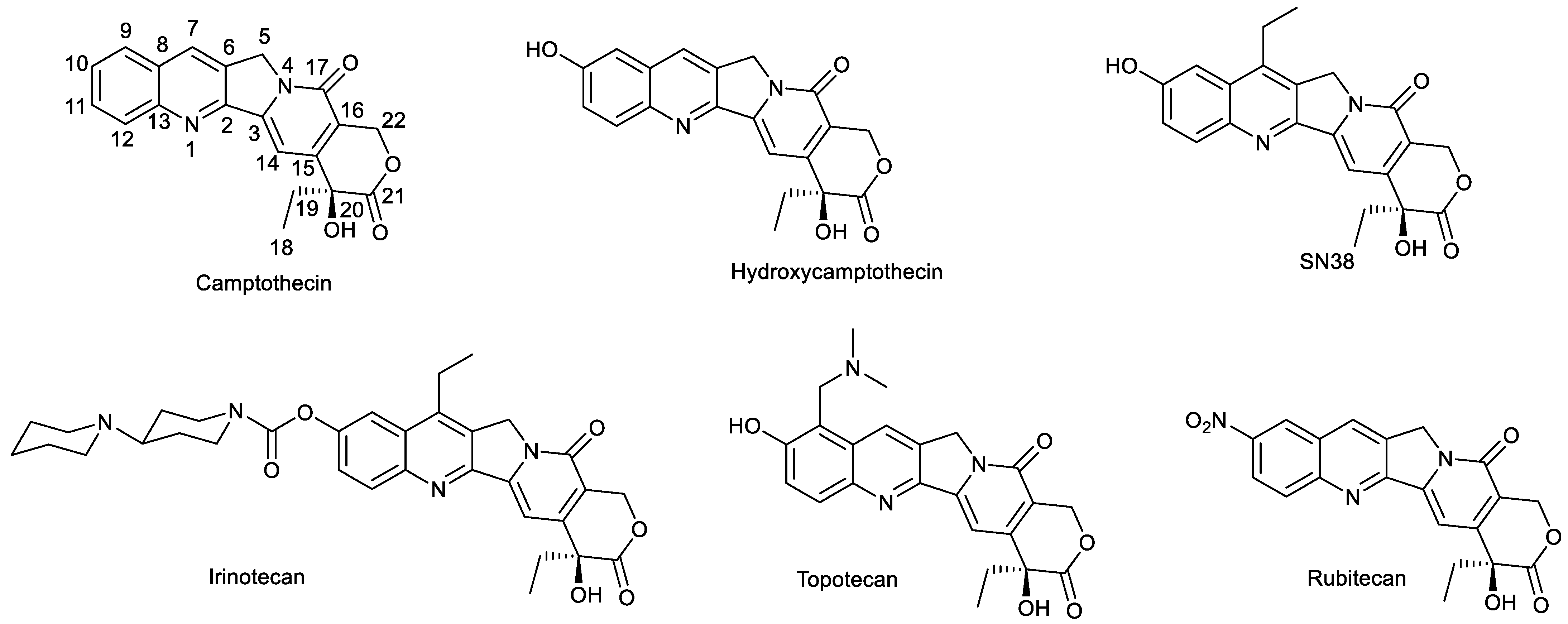
Figure 1. Camptothecin, hydroxycamptothecin, SN38 and clinical drugs based on camptothecin.
Molecules 28 04931 g001
Figure 2. Modification of SN38 at C-10 position with small-molecule units to exert their functions.
Molecules 28 04931 g002
Figure 3. Endo-stimuli-responsive SN38 prodrugs based on modification at C-10 position of SN38.
Molecules 28 04931 g003
Figure 4. Release mechanism of compounds 58. (A): Responsive to tetrazines, compound 5 underwent [4+1] cycloaddition, cycloreversion, tautomerization and β-elimination to release SN38; (B): Responsive to nanopalladium, compound 6 underwent palladium-mediated O-dealkylation to release SN38; (C): Responsive to bis(pinacolato)diboron, compound 7 underwent click reaction to release SN38; (D): Responsive to GSH and ASC, compound 8 underwent reduction and hydrolysis to release SN38.
Molecules 28 04931 g004
Figure 5. Structures of compounds 912.
Molecules 28 04931 g005
Figure 6. Structures of compounds 1316.
Molecules 28 04931 g006
Figure 7. Modification of SN38 at position 10 with hydrophobic groups and their transportation.
Molecules 28 04931 g007
Figure 8. Structures of compounds 2132.
Molecules 28 04931 g008
Figure 9. Fluorescence imaging mechanism of compound 33.
Molecules 28 04931 g009
Figure 10. Fluorescence imaging mechanism of compound 34.
Molecules 28 04931 g010
Figure 11. Fluorescence imaging mechanism of compound 35.
Molecules 28 04931 g011
Figure 12. Structures of compounds 3638.
Molecules 28 04931 g012
Figure 13. Structures of compounds 3964.
Molecules 28 04931 g013
Figure 14. Structures of compounds 6588.
Molecules 28 04931 g014
Figure 15. Structures of compounds 89114.
Molecules 28 04931 g015
Figure 16. Structures of compounds 115122.
Molecules 28 04931 g016
Table 1. Structural Modification of SN38 Reported during 2018–2023.
Types of Structural ModificationAchievable FunctionsCurrent Disadvantages
Modification of C-10 PositionStimuli-responsive Release of DrugsDependent on the type of cell lines or exogenous stimuli
Tumor TargetingDependent on the type of cell lines
Improvement of Water SolubilityThe occuring bottleneck of designing the novel small-molecular SN38 derivatives with improvement of water solubility
Synergism between Different DrugsLittle consideration of the ratio between drugs
TheranosticsWeak tissue penetration due to short excitation wavelength; photobleaching
Other FunctionsNull
Modification of C-20 PositionSynergism between Different DrugsLittle consideration of the ratio between drugs
Tumor TargetingDependent on the type of cell lines
Improvement of SolubilityThe occuring bottleneck of designing the novel small-molecular SN38 derivatives with improvement of water solubility
Modification of C-10 Position and C-20 PositionTumor TargetingDependent on the type of cell lines
TheranosticsWeak tissue penetration due to short excitation wavelength; photobleaching
Oher FunctionsNull
Modification of C-9 PositionMultitarget Inhibition of Cancer CellsA relatively challenging modification
The Opening of E-ringMaintenance of Anticancer ActivityThe usual reduced anticancer activity
Disclaimer/Publisher’s Note: The statements, opinions and data contained in all publications are solely those of the individual author(s) and contributor(s) and not of MDPI and/or the editor(s). MDPI and/or the editor(s) disclaim responsibility for any injury to people or property resulting from any ideas, methods, instructions or products referred to in the content.