Article

Antibacterial Activity of a Novel Oligosaccharide from Streptomyces californics against Erwinia carotovora subsp. Carotovora

by
Maysoon Abdulrahman Al-Zubairy
1,*,
Khaled Hussein
2,
Salwa H. Alkhyat
1,
Abdullah Yahya Al-Mahdi
3,
Saeed Munassar Alghalibi
1,
Adel Ali Al-Gheethi
4,*,
Muhanna Mohammed Al-Shaibani
4,
Hesham Ali El Enshasy
5,6 and
Nik Marzuki Sidik
7,*
1
Microbiology Section, Biological Sciences Department, Faculty of Sciences, Sana’a University, Sana’a 12544, Yemen
2
Chemistry Department, Faculty of Sciences, Sana’a University, Sana’a 12544, Yemen
3
Department of Microbiology, Faculty of Medicine, Lincoln University College, Petaling Jaya 47301, Malaysia
4
Micro-Pollutant Research Centre (MPRC), Department of Civil Engineering, Faculty of Civil Engineering & Built Environment, Universiti Tun Hussein Onn Malaysia, Parit Raja, Batu Pahat 86400, Malaysia
5
Institute of Bioproducts Development (IBD), Universiti Teknologi Malaysia (UTM), Skudai 81310, Malaysia
6
City of Scientific Research and Technology Applications (SRTA), New Burg Al Arab, Alexandria 21934, Egypt
7
Faculty of Agro-Based Industry, Universiti Malaysia Kelantan, Jeli 17600, Malaysia
*
Authors to whom correspondence should be addressed.
Molecules 2022, 27(8), 2384; https://doi.org/10.3390/molecules27082384
Submission received: 17 February 2022 / Revised: 25 March 2022 / Accepted: 3 April 2022 / Published: 7 April 2022
(This article belongs to the Special Issue Drug Discovery and Development: New Options for Old Diseases)

Abstract

:
The present study aims to characterize and predict models for antibacterial activity of a novel oligosaccharide from Streptomyces californics against Erwinia carotovora subsp. carotovora using an adaptive neuro-fuzzy inference system and an artificial neural network. The mathematical predication models were used to determine the optimal conditions to produce oligosaccharide and determine the relationship between the factors (pH, temperature, and time). The characteristics of the purified antibacterial agent were determined using ultraviolet spectroscopy (UV/Vis), infrared spectroscopy (FT-IR), nuclear magnetic resonance spectroscopy (1H- and 13C-NMR), and mass spectrometry (MS). The best performances for the model were 39.45 and 35.16 recorded at epoch 1 for E. carotovora Erw5 and E. carotovora EMCC 1687, respectively. The coefficient (R2) of the training was more than 0.90. The highest antimicrobial production was recorded after 9 days at 25 °C and a pH of 6.2, at which more than 17 mm of the inhibition zone was obtained. The mass spectrum of antimicrobial agent (peak at R.T. = 3.433 of fraction 6) recorded two molecular ion peaks at m/z = 703.70 and m/z = 338.30, corresponding to molecular weights of 703.70 and 338.30 g/mol, respectively. The two molecular ion peaks matched well with the molecular formulas C29H53NO18 and C14H26O9, respectively, which were obtained from the elemental analysis result. A novel oligosaccharide from Streptomyces californics with potential activity against E. carotovora EMCC 1687 and E. carotovora Erw5 was successfully isolated, purified, and characterized.

1. Introduction

Streptomyces are the group of Gram-positive filamentous bacteria growing in various natural environments and classified among the richest natural sources for antibacterial and antitumor activity, as well as antifungals, antivirals, anti-hypertensives, and immunosuppressives [1]. It has been reported that most of the medically useful antibiotics were produced by Streptomyces spp. [2]. The novel antimicrobial products from Streptomyces spp. have received high attention in recent years due to the increasing antimicrobial resistance among the pathogenic bacteria toward currently used antibiotics such as chloramphenicol, tetracycline, macrolide, and vancomycin, which was also isolated from diverse Streptomyces spp. [3]. The new trends in the antimicrobial activity research looking to find novel compounds with high activity against the infectious agents but with a mechanism differ from those reported for currently used antibiotics. That aside, researchers have focused mainly on developing a prediction model to expect the time required to develop resistance mechanisms among the pathogenic bacteria against the new antimicrobial compounds from Streptomyces spp.
The application of antimicrobial compounds from Streptomyces spp. as a biological control is a very promising alternative to pesticides. Studies have detected undesirable chemical residues in the food chain with adverse effects on human health [4]. The utilization of natural antagonism between microorganisms to protect plants has been reported in several studies [5]. Erwinia carotovora subsp. carotovora is one of the most destructive plant pathogen bacteria [6], as the bacterium strain causes bacterial soft rot in potatoes and other crops [7]. It is the major pathogen affecting potato seed tuber pieces after cultivation, during vegetative growth, and in the storage period [8]. Erwinia sp. is a heterologous group of Gram-negative, rod-shaped motiles with peritrichous flagella, non-sporing, and facultative anaerobic pectolytic plant pathogens [9]. The bacterium species are classified in Pectobacterium as P. carotovorum subsp. carotovorum, P. carotovorum subsp. atrosepticum, P. chrysanthemiDickeya spp.” [10,11], Bacillus thuringiensis, B. cereus, B. subtilis, B. megaterium, B. pumilus [12], Cyanodermella sp. [13], Paenobacillus polymyxa [14], B. subtilis, P. fluorescens, Rhizobium leguminosarum, T. harzianum, A. flavus [15], P. fluorescens, B. subtilis, B. thuringiensis [16], and Rhizopus stolonifer [17]. Baz et al. [18] revealed that Streptomyces sp. OE7 showed the potential to control soft rot on potato slices. However, studies on the utilization of biological control agents active against blackleg and soft rot bacteria are still at the lab scale. The main limitation lies in the requirement for the antagonist to satisfy several criteria [19]. The technology transfer from small-scale to large-scale field testing is difficult due to the annual variations in the weather, resulting in a lack of consistency in the results. The application of biological control strains should consider the environmental stresses affecting microbial activities. Therefore, the exploring an effective indigenous agent is one of the most effective solutions [20].
The inactivation mechanism of the antimicrobial agents from the biocontrol microorganisms belongs to alkaloids, elimoklavine, festuklavine, agroklavine, ergometrine, bacteriocins, and iturins [21,22,23]. Cladera-Olivera et al. [24] reported that the bacteriocin-like substance produced by B. licheniformis exhibited bactericidal activity against E. carotovora subsp. carotovora. The lethal mechanisms act by interacting with cell membrane lipids, provoking lysis of the cells.
The adaptive neuro-fuzzy inference system (ANFIS) and artificial neural network (ANN) are among the advanced and powerful modeling tools used in medical and environmental applications. Nevertheless, very limited studies utilized mathematical predication modeling for investigating the production of antimicrobial agents from natural sources. These models are used to simulate the behaviors of antimicrobial substance activities toward infectious agents as a response to the production parameters at a large scale, since these models provide accurate predictions for the degradation process of antibiotics [25].
The current work aims to characterize a new antimicrobial compound from Streptomyces sp. and its biological control activity against E. carotovora subsp. Carotovora.
The mathematical predication models, which include an ANFIS and ANN, are employed to determine the optimal conditions for antimicrobial substance production and activation against E. carotovora subsp. Carotovora and determine the interactions between the factors (pH, temperature, and time). These models provide more details on the nature of the production process and reveal the response of E. carotovora subsp. Carotovora to the activity of antimicrobial substances in different conditions.

2. Results and Discussion

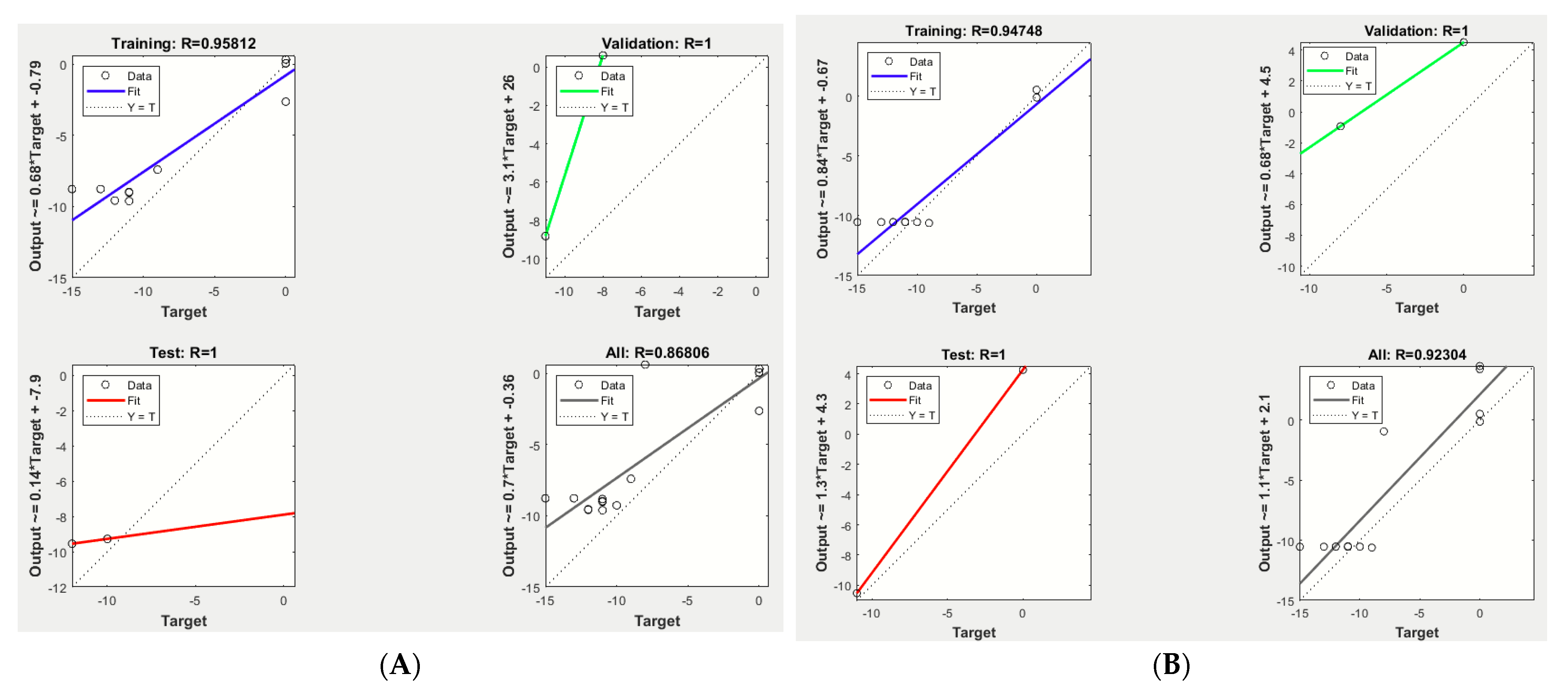
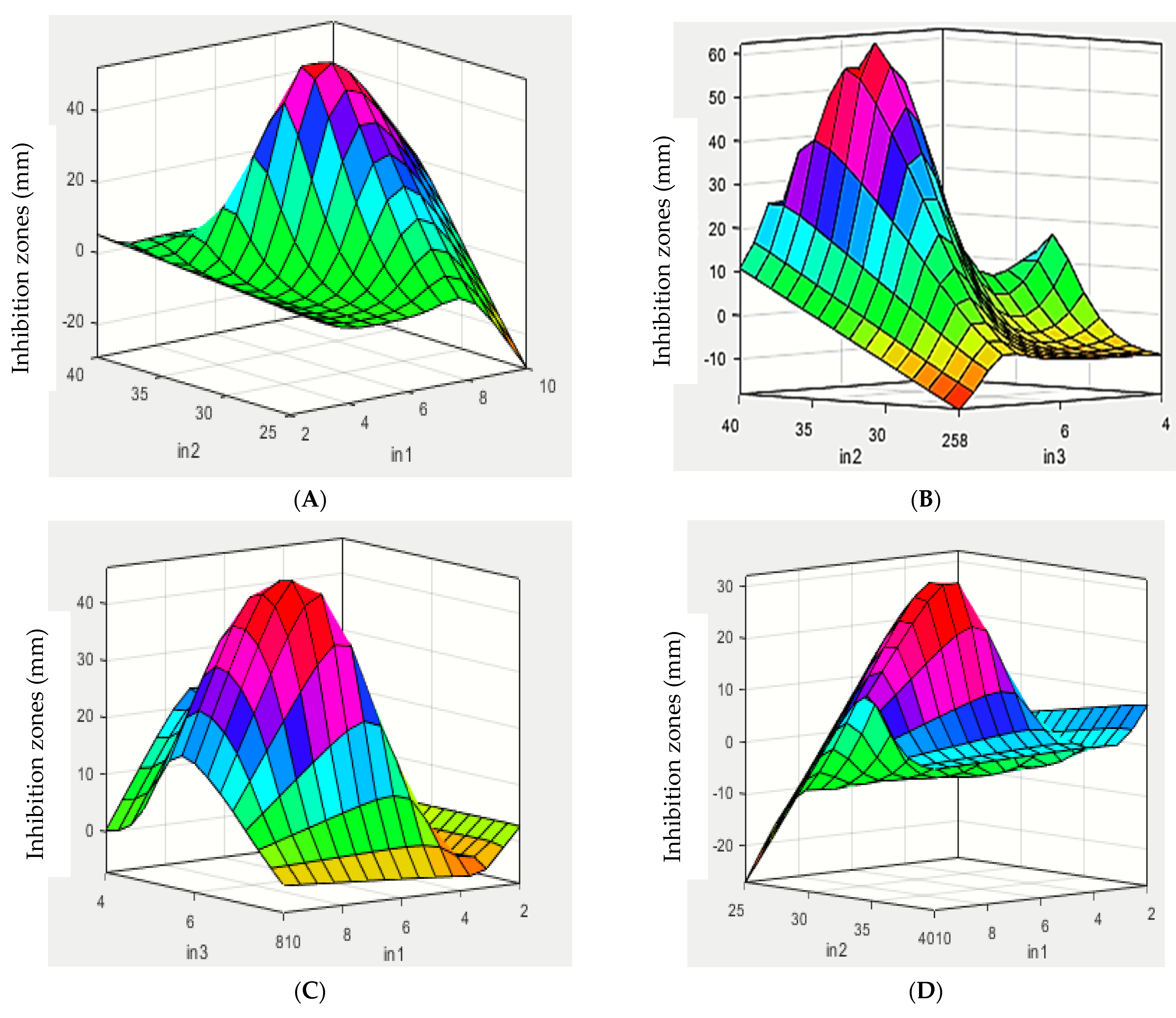
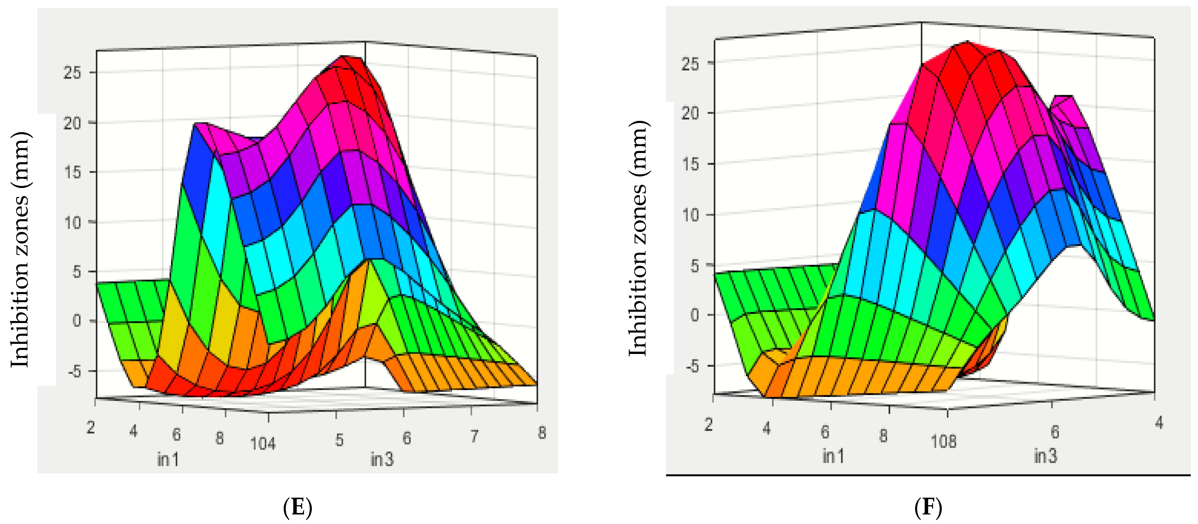
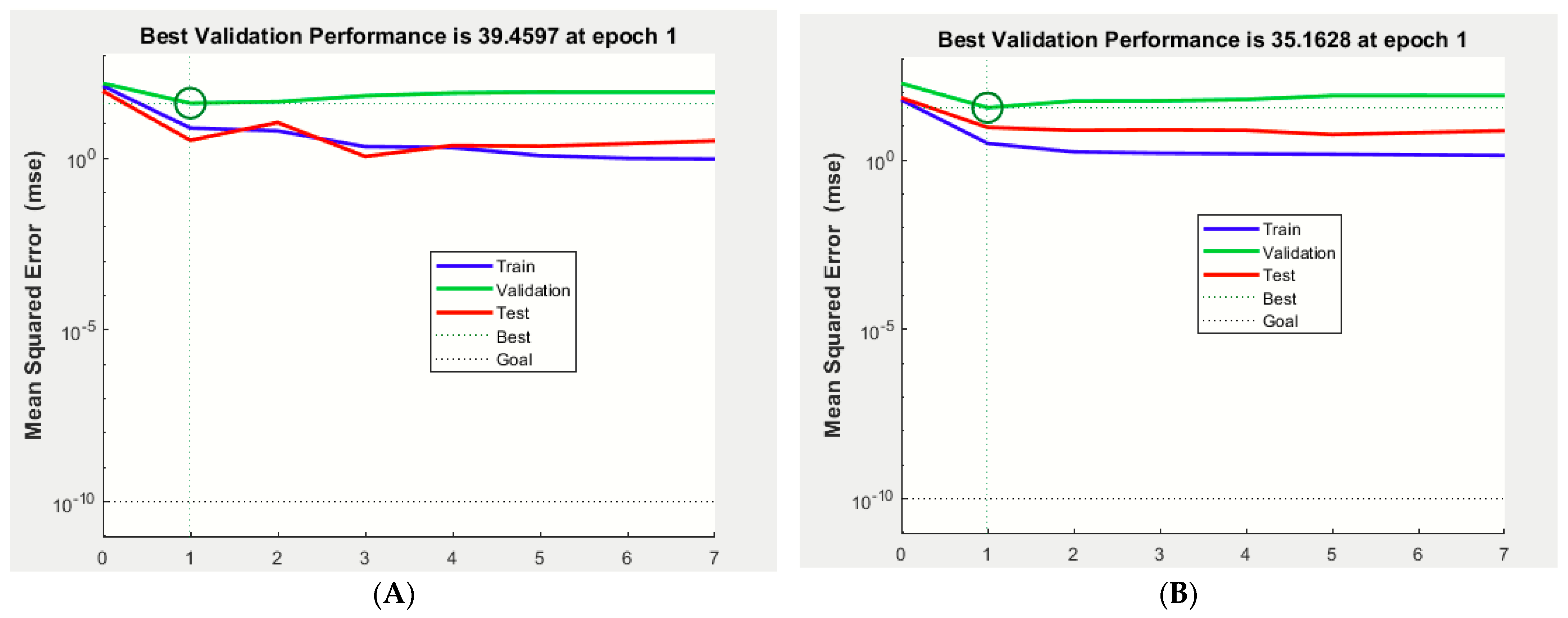
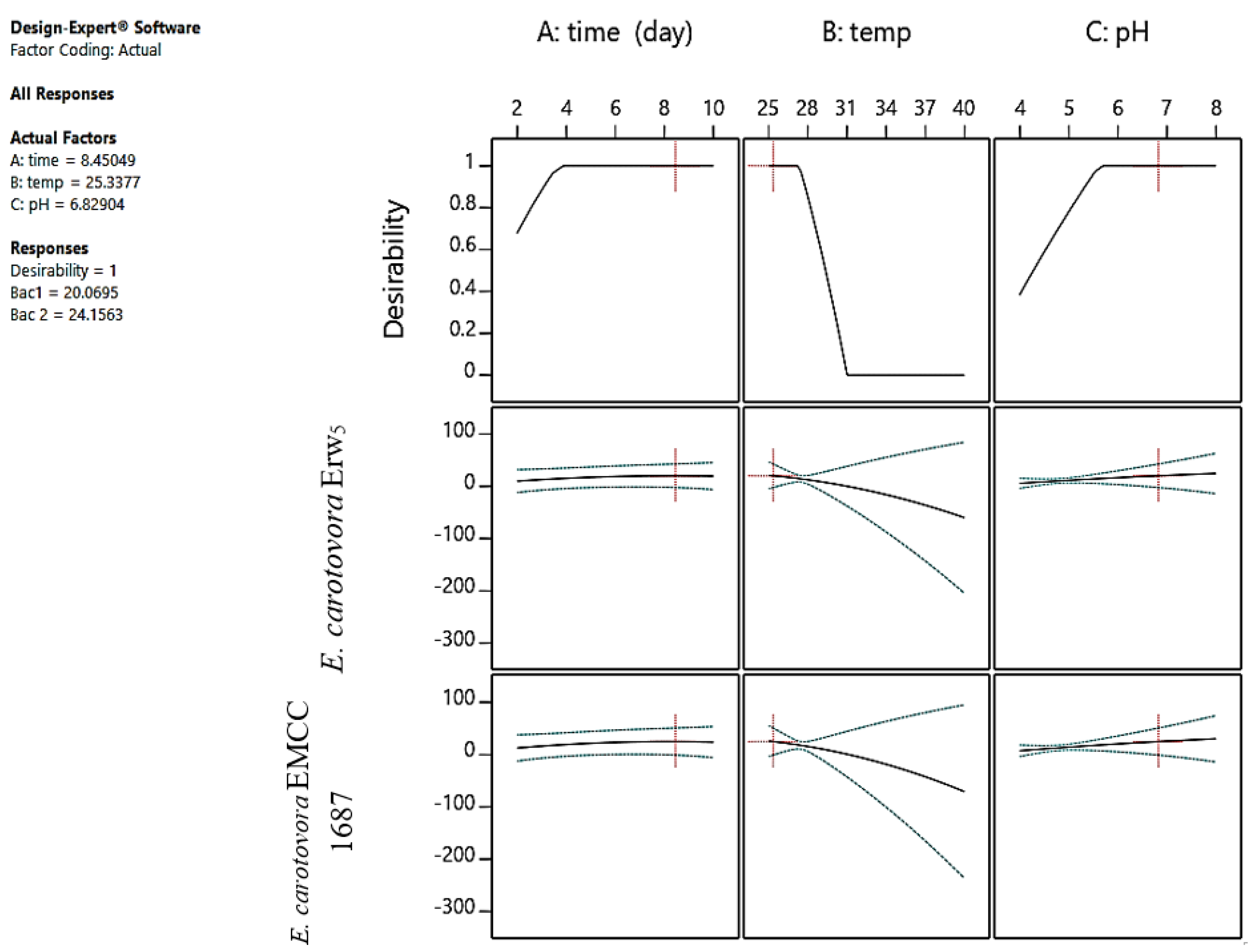
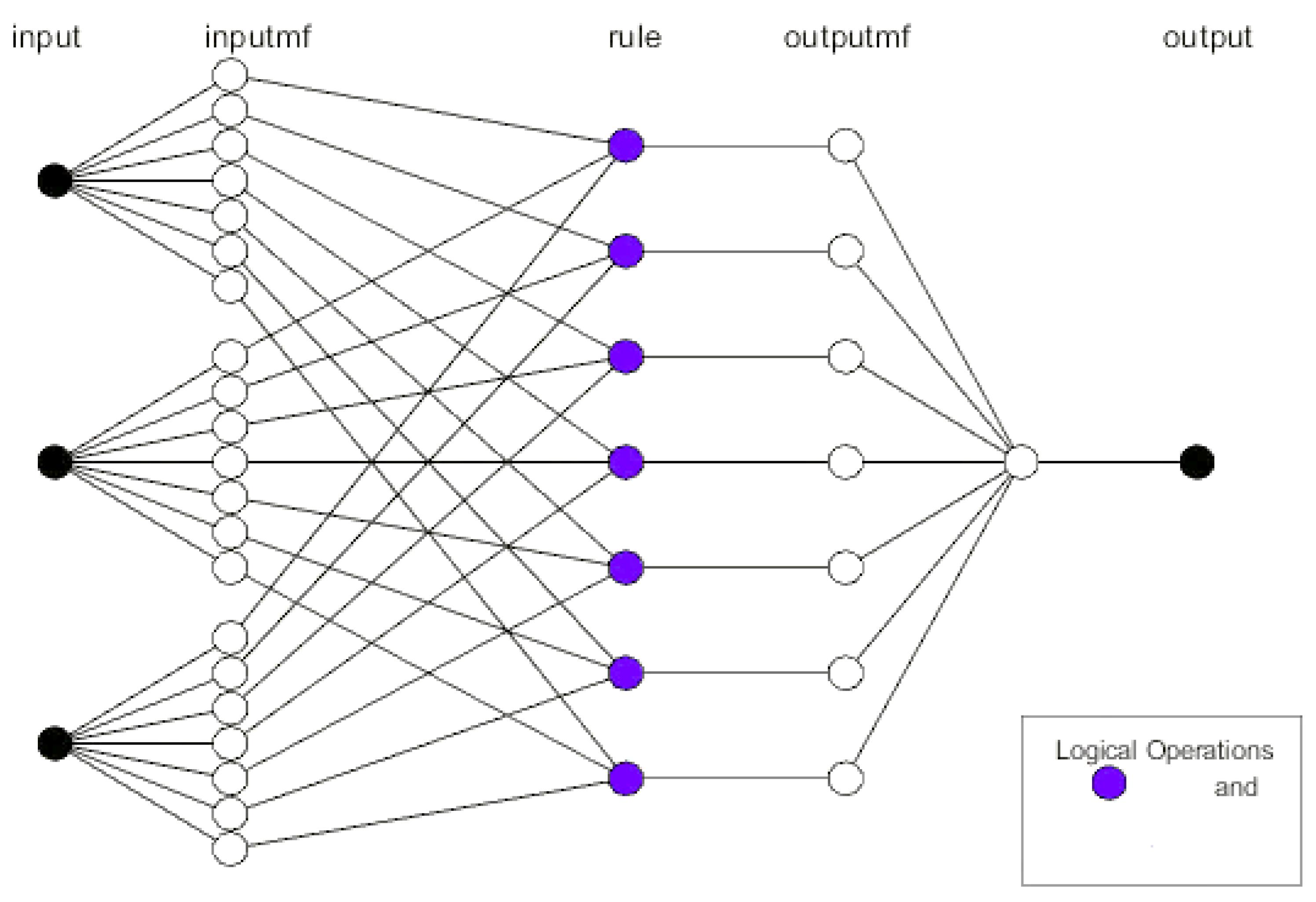
2.1. Optimization and Prediction Models Using ANFIS and ANN Simulations

The mathematical prediction model simulations were performed using an ANFIS with 7 epochs, and the results revealed that the best validation performances were 39.45 and 35.16, recorded at epoch 1 for E. carotovora Ewr5 and E. carotovora subsp. carotovora EMCC 1687, respectively, at which point the training, validation, and testing data exhibited similar mean square errors (MSEs) (Figure 1). The MSE is a statistical indicator for estimating the average of the squares of the errors [26]. The low values indicate the model’s accuracy in the prediction of antimicrobial production. In this investigation, the lowest value of MSE was for training. The coefficient (R2) for training was 0.9581 vs. 0.9474 for E. carotovora Ewr5 and E. carotovora subsp. carotovora EMCC 1687, respectively, as detected using ANN analysis (Figure 2 and Figure 3), which indicated the close results between the actual experimental and predicted results, since the coefficient (R2) was more than 0.7 [27].
The optimization of antimicrobial production by S. californics (22/30a) was performed using ANFIS with three independent factors, including temperature (25–40 °C), pH (4–8), and time (2–10 days) (Figure 4 and Figure S1). The results revealed that the production of antibacterial agents increased with the increasing incubation period, with diameter of inhibition zones of 11 and 13 mm against E. carotovora Ewr5 and E. carotovora EMCC 1687, respectively. The highest production of antibacterial agents was observed at 28 °C, with the highest inhibition zones being 13 and 15 mm against both E. carotovora Ewr5 and E. carotovora EMCC 1687, respectively. At 30 °C, the inhibition zones were 11 and 13 mm against both of E. carotovora Ewr5 and E. carotovora EMCC 1687, respectively, whereas no antibacterial activity was observed at the 25, 37, or 40 °C incubation temperatures. The effect of different initial pH values of the fermentation medium showed that the antibacterial activity increased with the decrease in pH, with the highest inhibition zones being 15 and 17 mm against both E. carotovora Ewr5 and E. carotovora EMCC 1687, respectively.
The effects of different carbon and nitrogen sources on the antibacterial production and activity against E. carotovora Ewr5 and E. carotovora EMCC 1687 are presented in Figure S2. It was noted that the maximum antibacterial inhibition against both strains occurred when the cultures were supplied with starch as a carbon source (12 vs. 14 mm for the inhibition zones in E. carotovora Ewr5 and E. carotovora EMCC 1687, respectively). On the other hand, the maximum antibacterial activity effect was observed when the culture was provided with KNO3 as a nitrogen source. An assay on the antibacterial activity of fractions of S. californics (22/30a) against E. carotovora Erw5 showed inhibition zones of 12 mm and 14 mm for E. carotovora EMCC 1687, as presented in Figure S3.
The results revealed that time exhibited more influence compared with the temperature, which had more of an effect than the pH level. In contrast, the pH exhibited more influence than time on the production of antimicrobial agents. The temperature and time as well as time and pH exhibited non-significant (p > 0.05) positive interactions, while temperature and pH had a non-significant negative interaction (p < 0.05). The first and quadratic models for the interactions between these factors are presented in Equations (1) and (2) with coefficient R2 being 0.7528 and 0.7614 for E. carotovora Ewr5 and E. carotovora EMCC 1687, respectively:
y 1 = 2.39 + 4.8 x 1 29.79 x 2 15.42 x 3 26 x 1 x 3 4.0 x 1 2 12.17 x 2 2 1.4 x 3 2
y 2 = 2.62 + 5.6 x 1 35.36 x 2 18.24 x 3 31.01 x 1 x 3 5.14 x 1 2 14.16 x 2 2
The best operating parameters for antimicrobial production were predicted using ANFIS, with the highest antimicrobial production recorded after 9 days at 25 °C and a pH of 6.2, at which 17.8 and 21.34 mm inhibition zones against E. carotovora Ewr5 and E. carotovora EMCC 1687 were obtained (Figure 4).
The potential of Streptomyces spp. as a biological control against soilborne bacteria has been reported because of secondary metabolite production with the bioactivity [27]. However, secondary metabolite production relies on internal factors, such as carbon and nitrogen sources, and external factors, such as time, temperature, and pH level [28]. The production of secondary metabolites is affected by the availability of nutrients [29,30,31,32]. The maximum production of antimicrobial metabolite by Streptomyces sp. MNK7 was recorded after 10 days [33,34]. However, Singh and Rai [35] claimed that antibiotic production by S. rimosus MTCC 10792 was recorded after 24 hrs. These differences might be related to the microbe strain and the source of isolation. The best production of antimicrobial compounds by Streptomyces sp. is obtained at 28 °C [35], 30 °C [32], or 35 °C [33]. The highest production of antimicrobial agents from Streptomyces sp. was reported at pH levels of 5 [33], 7.0 and 7.5 [32,36], and 8 [37]. S. griseocarneus produces antimicrobial activity with glucose as a carbon source [34,38]. However, a quickly metabolized substance like carbohydrates enhances the production of antimicrobial production [39]. Nitrate, soybean meal, and peptone are among the best nitrogen sources for antimicrobial agent productions [29,36].
The antibacterial agent extracted from S. californics 22/30a was soluble only in water, DMSO, and hot methanol. This result agreed with Zamanian et al. [28], who reported that the active substance of S. plicatus (Strain 101) was water soluble and insoluble in chloroform, hexane, and dichloromethane. Kang et al. [29] found that the final dry extract from Streptomyces sp. strain JJ45 was soluble in water and insoluble in alcohol or ethyl acetate.

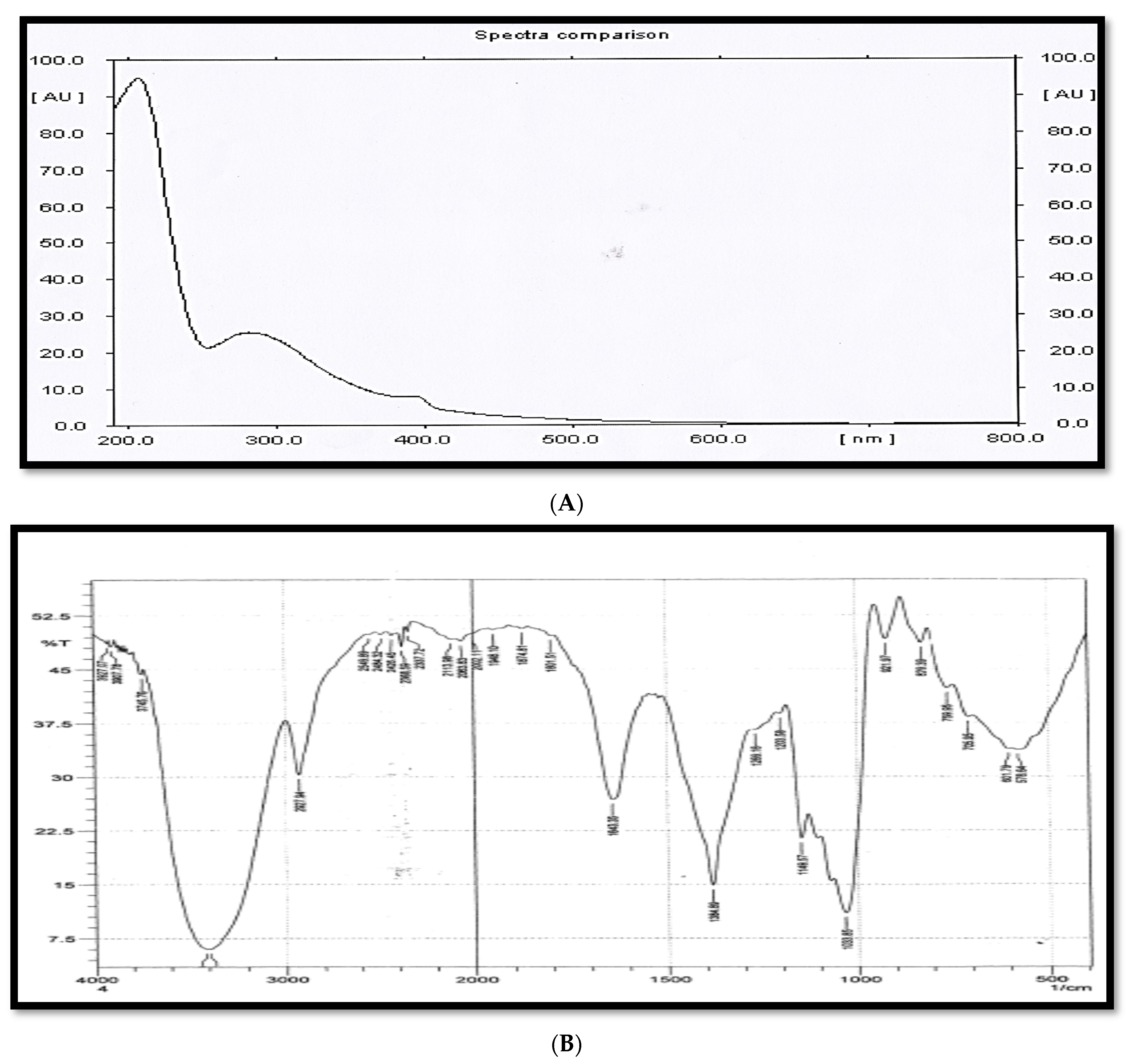
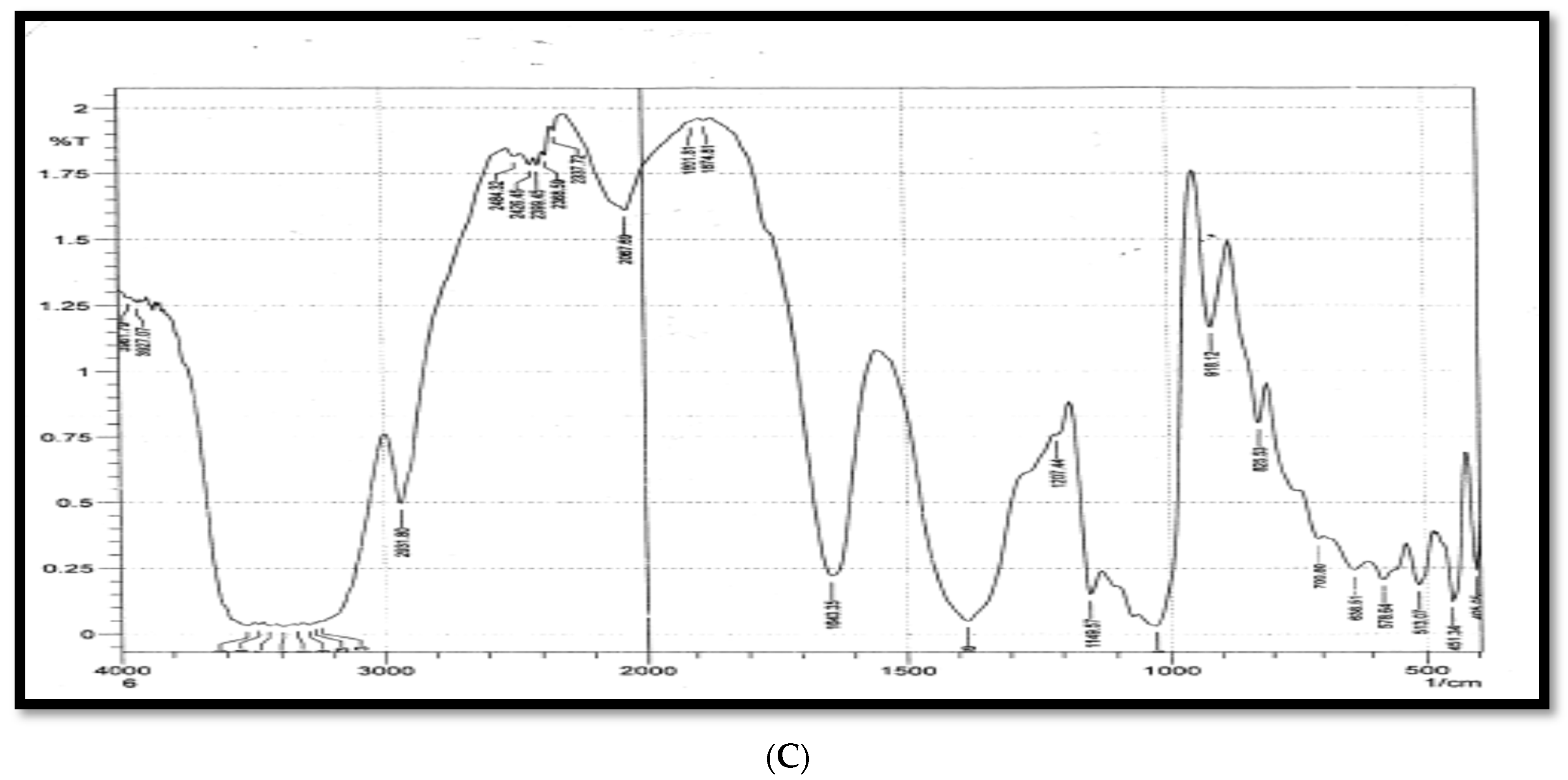
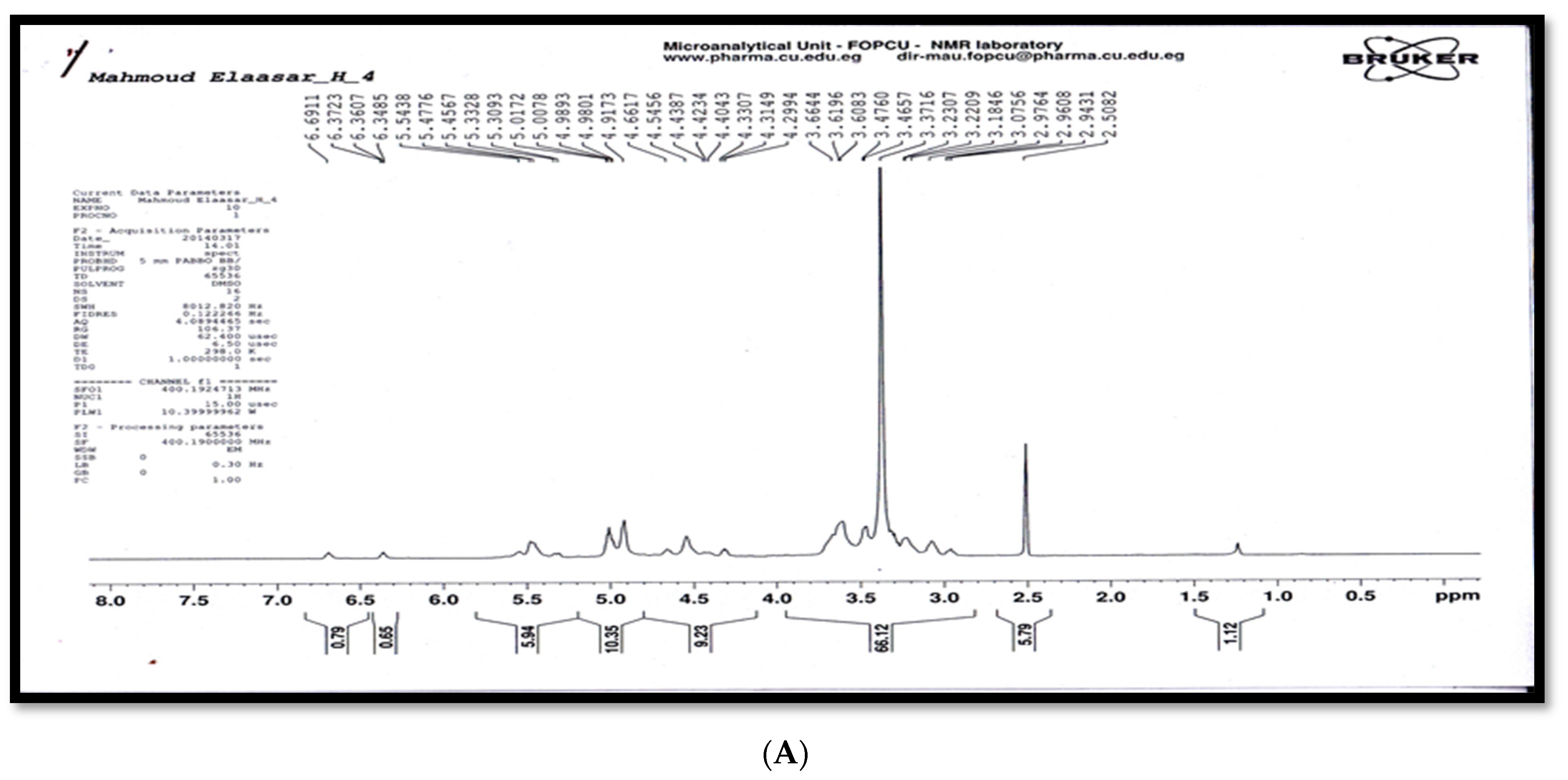
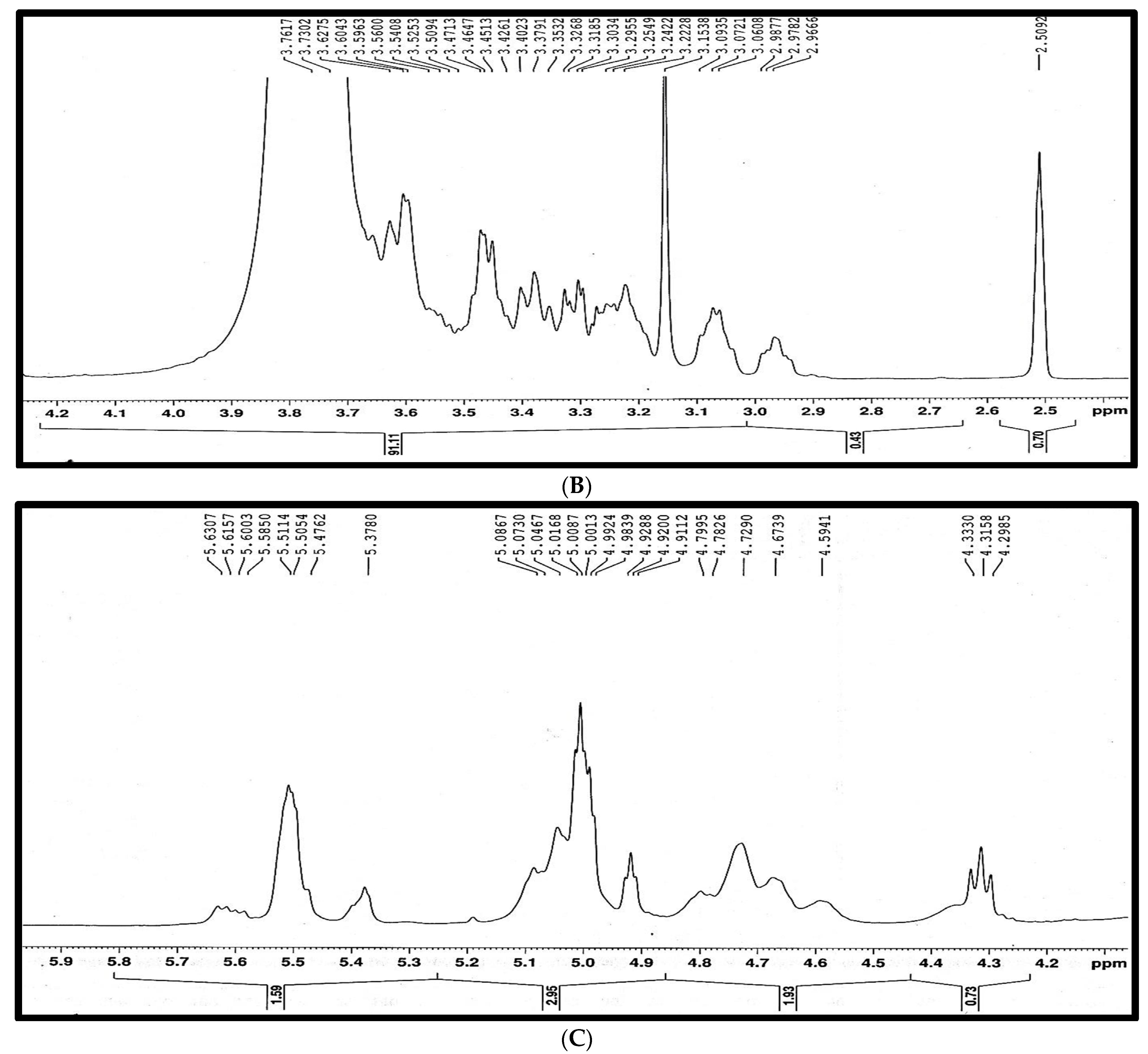
2.2. Characteristics of Antibacterial Agent from S. californics (22/30a)

The maximum activity of antibacterial extract from S. californics (22/30a) against E. carotovora Erw5 (A) and E. carotovora EMCC 1687 was recorded at a pH of 3 with inhibition zones of 19 and 22 mm, respectively (Figure S3). The antibacterial filtrate of S. californics (22/30a) exhibited stabile activity at room and boiling temperatures with inhibition zones of 12 and 14 mm, respectively (Figure S3). The antibacterial activity of crude extract was the highest, with inhibition zone diameters of 19 and 20 mm against E. carotovora Erw5 (A) and E. carotovora EMCC 1687, respectively. The crude antibacterial agent of S. californics (22/30a) had a light brown color, gummy nature, aromatic odor, and solubility in water, DMSO, and hot methanol, with 134–141 °C as the melting point.
The crude antibacterial extract was analyzed using TLC with different eluting systems to determine the suitable mobile phase for column chromatography (CC). Acetone-methanol-water (4:6:3) was the best eluting system which showed the presence of at least two different components of the crude antibacterial agent of S. californics (22/30a). Based on the column chromatography analysis, among 78 fractions, only 6 fractions (41, 43, 44, 45, 47, and 50) showed antibacterial activity against E. carotovora Erw5 and E. carotovora EMCC 1687, with an inhibition zone of 24 mm against E. carotovora EMCC 1687 in the case of fraction 47 and 19 mm against Erw5 in the case of fraction 44 (Figure S3). All fractions were analyzed using TLC and showed that all fractions were like each other and consisted of two components. The fractions with antibacterial activity against both E. carotovora Erw5 and E. carotovora EMCC 1687 were divided into six fractions using the fractional crystallization method. Only two fractions (4 and 6) showed antibacterial activity against both of Erw5 and E. carotovora subsp. carotovora EMCC 1687 (Figure S3). Fraction 6 of the antibacterial agent was chosen to be identified using certain chemical reagents and spectroscopic analysis.
To identify the chemical type of the antimicrobial agent, qualitative analysis was carried out using certain chemical testes (Molisch, ninehydrine, and sodium nitrobrocide tests). The reaction of the antimicrobial agent with the sodium nitrobrocide reagent returned a negative result, whereas the reaction of the antimicrobial agent with ninehydrine reagents gave positive results. With the Molisch reagent, and before carrying out any chemical treatments for the antimicrobial agent, this fraction did not give a positive result, while afterward it was broken down into monosaccharides by acid, giving a positive test result. These results reveal that the antimicrobial agent contained nitrogen atoms in its structure which belonged to carbohydrate compounds.
The UV/Vis spectrum of the purified component showed that the spectrum of the pure compound was determined in the region (200–400 nm) by using methanol as a solvent. The UV/Vis spectrum recorded a maximum absorption band at λmax 210 and 279 nm (Figure 5A). The compound in its IR spectrum exhibited bands at 3670–3050, 2931, 1643, 1384, 1269, 1207, 1149, 1033, 918, and 825–513 cm−1, from which the presence of a primary amine, hydroxyl groups, and glycosides bond bands were inferred (Figure 5B).
The UV/Vis and FT-IR spectral data of C29H53NO18 were in complete agreement with those reported in the literature for oligosaccharides and polysaccharides [37,38,39,40,41,42,43].
The presence of a broad and distorted absorption band in the range of 3670–3050 cm−1 in the IR spectrum of the antibacterial agent was due to overlap between the absorption bands of alcoholic O-H and N-H of the primary amine’s stretching vibration, whereas the absorption band at 2931 cm−1 was due to sp3 C-H stretching vibration.
An important indication of this interference was the disappearance of only this distortion in the infrared spectrum of component 4 (ret. time: 3.350) of fraction 6 (Figure 5C), which did not contain in its structure the amino group (NH2). In addition, the appearance of the strongest absorption band at 1643 cm−1 in the IR spectrum of component 6 (Figure 5B) was due to the N-H bending vibration of the primary amine [44], whereas the presence of a moderate (less intense) absorption band at the same position (1643 cm−1) in the IR spectrum of component 4 (Figure 5C) was related to absorbed water [40].
The occurrence of a slightly broad absorption band at 1384 cm−1 corresponded to overlapping between the absorption bands of the C-N stretching vibration of the amine and sp3 C-H bending vibration, as well as the O-H bending vibration. The presence of ether (pyran and furan rings and methoxy) and glycoside bonds in the chemical structure of component 6 was confirmed by the appearance of a group of absorption bands at 1269, 1207, 1149, and 1033 cm−1, which corresponded to the stretching vibration of the C-O ether and C-O-C acetal bonds. The absorption bands at 918 and 709 cm−1 were due to O-H and N-H out-of-plane (oop) bending vibrations, respectively. The absorption band at 825 cm−1 indicates that the connection between the oligosaccharide units was β-glycosidic bonds [40]. The appearance of an absorption band at 578 cm−1 was attributed to a pyran-type sugar ring.
The results of the GC analysis of fraction 6 are shown in a GC chromatogram (Figure S4), which shows two overlapping peaks in retention time at 3.350 and 3.433 min. The mass spectrum of the antibacterial agent (peak at R.T. = 3.433 for fraction 6) recorded two molecular ion peaks at m/z = 703.70 and m/z = 338.30, corresponding to molecular weights of 703.70 and 338.30 g/mol (Figure S4B). The two molecular ion peaks matched well with the molecular formulae C29H53NO18 and C14H26O9, respectively, obtained from the elemental analysis. The mass errors for the two compounds were calculated based on the isotopic atomic masses of the elements and not based on their relative atomic masses.
The different types of hydrogen and carbon were recorded from the signals in the 1HNMR and 13CNMR spectra of antibacterial agents, respectively, and matched well with the chemical structure in Figure 6A–C. However, the 1HNMR spectrum (400 MHz, DMSO) δ (ppm) (Figure 6A–C) exhibited signals at 2.9782 and 2.95818 (2 H, dd, J = 3.6 and 6.76 Hz, Ha), 3.0828 and 3.0529 (2H, dd, J = 8.56 and 6.32 Hz, Hb), 3.1538 (21H, s, Hc [O-CH3]), 3.18885 and 3.16875 (1H, dd, J = 6.12 and 3.56 Hz, Hd), 3.2509 and 3.21515 (2H, dd, J = 3.2 and 6.12 Hz, He), 3.3711 and 3.29325 (2H, dd, J = 6.4 and 3.32 Hz, Hf), 3.3942 and 3.29945 (2H, dd, J = 6.44 and 3.16 Hz Hg), 3.468 and 3.44365 (2H, dd, J = 3.64 and 6.12 Hz, Hh), 3.61975 and 3.6003 (2H, dd, J = 6.2 and 3.2 Hz Hi), 4.3158–4.2985 and 4.3330–4.3158 (2H, t, J = 6.39 and 6.68 Hz, Hj), 4.5941 (1H, br., s. Hk [OH]), 4.79105 and 4.6632 (1H, dd, J = 6.7 and 8.5 Hz, Hl), 4.7290 (1H, br., s. Hk’ [OH]), 4.9200 (1H, t, J = 3.52 Hz, Hm), 5.0013 (4H, m, Hn), 5.3780 (2H, br., s, Ho [NH2), 5.4762–5.4606 (2H, d, J = 6.24 Hz, Hp), 5.5114 (2H, br., s, Hq [2OH]), 5.6003–5.5850 (1H, d, J = 6.13 Hz, Hr), and 5.6307–5.6157 (1H, d, J = 6 Hz, Hs). The 13CNMR spectrum (100.63 MHz, DMSO) δ (ppm) exhibited signals as described in the chemical structure below and a distribution of carbon atoms in the compound. Further investigation of the chemical structure of the active component was carried out using 1HNMR and 13CNMR (Figure 6 and Figure 7, respectively) and matched well with the chemical structure in Figure 8. These findings agree with Kang et al. [29], who isolated α-L-sorbofuranose (3→2)-β-D-altrofuranose from Streptomyces sp. strain JJ45 with antibacterial activity. Many antibiotics consist of glycosides since the glycosidic residues are crucial for antibiotic activity. The glycosylation also improves the pharmacokinetic parameters of the antibiotics [44]. Carbohydrate-based antibiotics have received much attention in recent years as a new approach for antibiotic resistance [45]. Therefore, dimeric aminoglycosides represent best the candidates for carbohydrate-based antibiotics [46].

3. Materials and Methods

3.1. Streptomyces sp. Strain

The Streptomyces sp. were isolated from soil samples collected from Yemen according to the method described by Johnson and Curel [44]. The morphological characteristics of the Streptomyces spp. strain grown on starch casein agar medium (SCA) were described after a 10-day incubation period at 28 °C. The molecular analysis was conducted at The Regional Centre for Mycology and Biotechnology (RCMB) at Al Azhar University (Egypt). The strain was identified as Streptomyces californics.

3.2. Test Bacterial Strain

E. carotovora subsp. carotovora EMCC 1687 was used as a control, while E. carotovora subsp. Carotovora, a local strain (E. carotovora Erw5), was isolated from a potato tuber infected with soft rot as described by Johnson [45]. The bacterial strain was identified based on the morphological characteristics.

3.3. Production of Antibacterial Agent from Streptomyces sp. Strain

The production of an antibacterial agent from S. californics 22/30a was conducted in a starch nitrate broth medium containing (g L−1) 20.0 of KNO3, 20.0 of K2HPO4, 1.0 of KCl, 0.5 of MgSO4, 0.5 of FeSO4, and 0.01 and 2 of CaCO3. The pH was adjusted to 4 using 0.1 N of HCl. The inoculated media (100 mL) with S. californics 22/30a was incubated on a rotary shaker incubator (200 rpm) at 28 °C for 10 days. The culture broth was subjected to centrifugation at 4000 rpm for 15 min, and 100 µL of supernatants was transferred into a well of 7 mm made in NA plates seeded with target E. carotovora strains. The plates were kept in a refrigerator for 2 h and then incubated at 28 °C for 24 h. The antibacterial activity was recorded in terms of the inhibition zone of the target E. carotovora strains around the well of the supernatant [46].

3.4. Adaptive Neuro-Fuzzy Inference System (ANFIS) and Artificial Neural Network (ANN) Analysis

The ANFIS and ANN models were used in the current work to investigate the production of antimicrobial agents from S. californics 22/30a as a response to the temperature (25–40 °C), pH (4–8), and time (2–10 days). The developed models consisted of three layers, as shown in Figure 1. The input layer consisted of five neurons (parameters) which were represented by input 1 (time, day ( I n 1 ) , temperature (°C, I n 2 ), and pH ( I n 3 )   (Figure 9)). The optimization process was conducted with the hybrid and 7 epochs to attain the best model. The proposed ANFIS and ANN model was executed in MATLAB R2021a. The experimental data were divided into training data (70%) and testing data (30%). The training, testing, and validation of the experimental data values were performed using an ANN and evaluated based on R2, while the MSE error was used to evaluate the proposed model’s performance.

3.5. Extraction, Separation, and Purification

The cell-free culture medium was mixed with ethanol (50:50 v/v) and centrifuged at 4000 rpm to remove the biomass residues. The supernatant was subjected to drying by evaporating the liquids using a dryer with a vacuum at 50 °C [47]. To test the activity of the crude extract, a fixed weight of the dry extract (0.1 g mL−1) was dissolved in distilled water, and 50 μL of the crude extract was assayed in triplicate as mentioned above (Section 3.1). The physical properties of the crude antibacterial agent (appearance, color, odor, and melting point) were determined using a Stuart Scientific Melting Point Apparatus (SMP3, UK).
A thin layer chromatography (TLC) aluminum plate of 20 × 20 cm coated with silica gel 60 F254 (Merck F254) was used to analyze the crude extract and to determine the suitable mobile phase for column chromatography (CC). The plate of TLC was developed at 25 ± 2 °C in a glass chamber saturated (v/v) with acetone-water (4:1), acetonitrile-water (4:1), methanol-water (4:1), and acetone-methanol-water (4:2:1, 4:5:3, 4:6:3, 6:2:3, 2:6:3, 2:4:4, 3:4:4, and 2:2:1) as mobile phases. The TLC plates were visualized under ultraviolet light at λ254 and λ366 nm [29,47].
The column chromatography (CC) loaded with silica gel (silica gel 60 with a 0.2–0.5-mm mesh) was used for a stepwise elution with five solvent systems in increasing order of polarity (in terms of volume ratio (v/v)). Seventy-eight fractions were obtained, and 2 (1–2), 24 (3–26), 24 (27–50), 24 (51–74), and 4 (75–78) fractions were eluted with pure acetone-methanol (from 6:0.5 to 3.5:3 (v/v)), acetone-methanol-water (from 3:3:0.5 to 0.5:3:3 (v/v)), methanol-water (from 3:3.5 to 0.5:6 (v/v)), and pure water in 20–25 mL for each fraction, respectively. The separated fractions (78 fractions) were concentrated and combined into 9 pools (1–9) based on TLC monitoring on silica gel 60 F254 using acetone-methanol-water (4:6:1 (v/v/v)) as a mobile phase. The TLC plates were visualized as mentioned above. The combined fractions (pool 1–9) were tested for their antibacterial activity against E. carotovora strains using the agar well diffusion method [28].
The purified combined fractions were subjected to further purification using fractional crystallization (FC) to 6 fractions at The Regional Centre for Mycology and Biotechnology (RCMB) at Al-Azhar University in Nasr City, Egypt. These fractions were screened via TLC using acetone-methanol-water (4:6:1 v/v/v) as the elution system to determine the purified fraction of the antibacterial agent. Two fractions were obtained and tested for their antibacterial activity against E. carotovora strains as described above (Section 3.3). Fraction number 6 was chosen for chemical characterization because of its activity against the pathogenic bacteria.

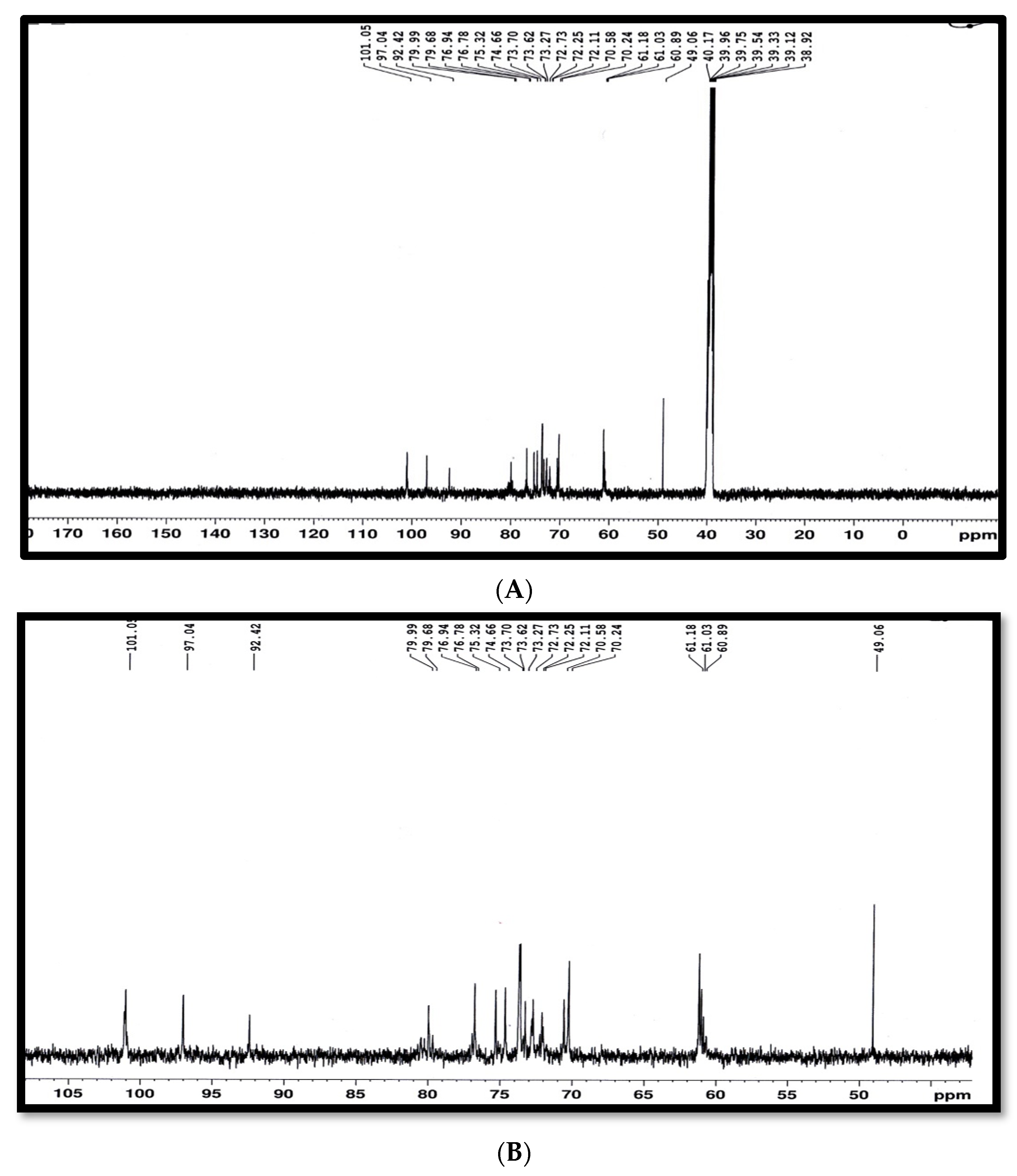
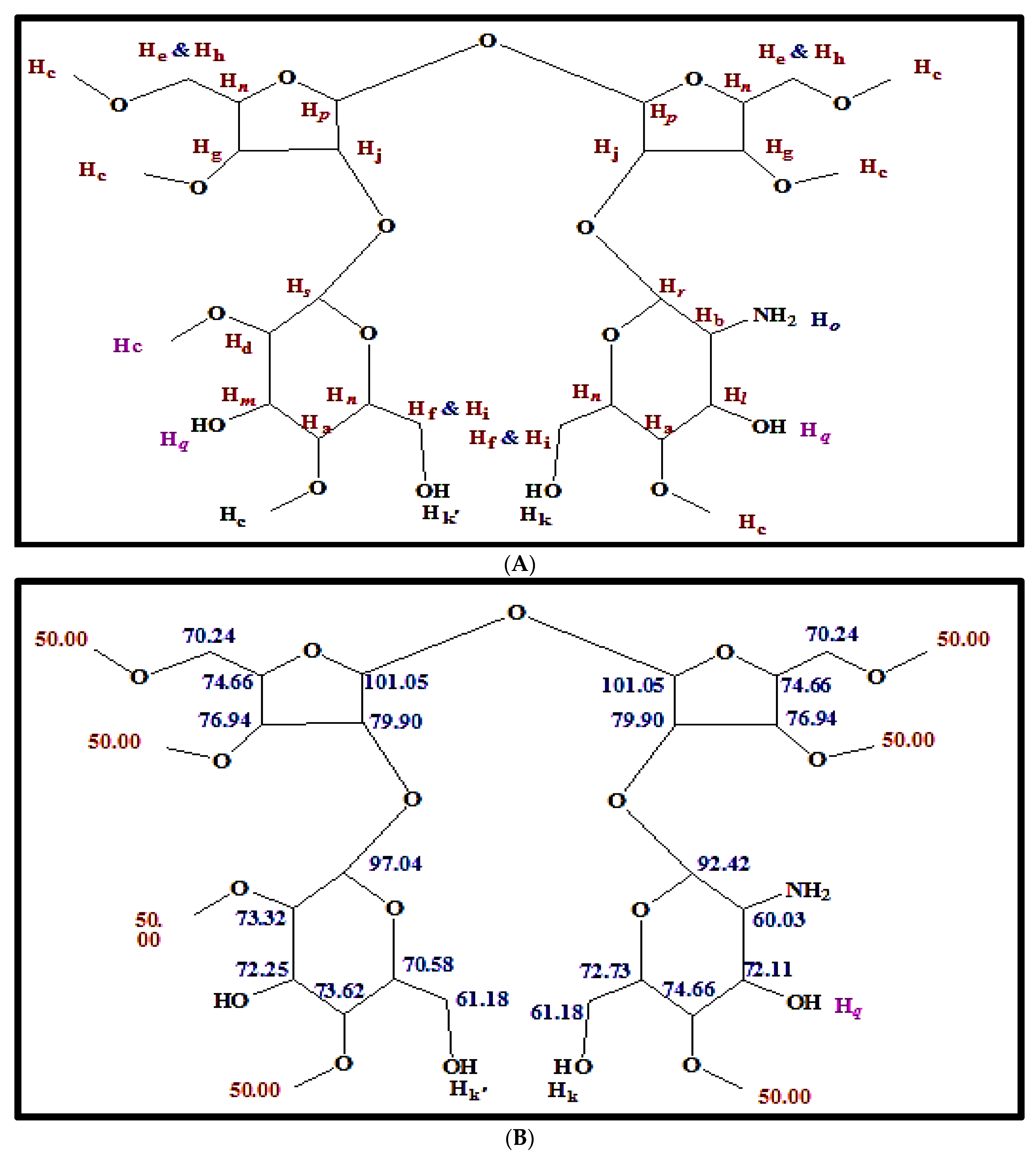
3.6. Characterization and Structural Elucidation of the Purified Antibacterial Agent

Spectroscopic analysis of the purified antibacterial agent was performed using ultraviolet spectroscopy (UV/Vis), infrared spectroscopy (FT-IR), and nuclear magnetic resonance spectroscopy (1H- and 13C-NMR), as well as mass spectrometry (EI-MS). The UV/Vis spectrum of the compound that was developed on a TLC plate (silica gel G-60 aluminum sheet, Merk, Darmstadt, Germany) was recorded using a CAMMAG TLC scanner system at the RCMB at Al-Azhar University in Egypt. The infrared spectra (FT-IR) were recorded using the potassium bromide disc technique with a Perkin-Elmer 1650 FT-IR spectrometer at the Microanalytical Center at Cairo University in Egypt.
The nuclear magnetic resonance (NMR) spectra were recorded on a Bruker high-performance digital FT-NMR spectrometer (1H 400 MHz; 13C 100.63 MHz) at 298.1 K using dimethylsulfoxide (DMSO) as the solvent and TMS as the internal reference, and the chemical shift was expressed in δ units (ppm) relative to the TMS. The coupling constants (J) were expressed in Hertz. The abbreviations used for multiplicities were s = singlet, d = doublet, t = triplet, q = quartet, and m = multiplet. All NMR experiments were conducted at the Microanalytical Unit (FOPCU) of the Faculty of Pharmacy at Cairo University in Cairo, Egypt (www.pharma.cu.edu.eg (accessed on 16 February 2022)). Mass spectral analysis was carried out on a direct Inlet part DI-50 to a mass analyzer by a Shimadzu GC/MS-QP5050 at the Regional Centre for Mycology and Biotechnology (RCMB), Al Azhar University, Egypt.
The gas chromatography of GC/MS was equipped with a 30 m × 0.25 mm (inside diameter) (df = 0.25 μm) bonded phase DB-5 wax cross–linked fused silica capillary column covered with a film thickness of 0.5 μm of polydimethylsiloxane. The oven temperature was automatically increased from 30 °C for 3 minutes with a rate increment from 3 °C/min to 200 °C/min and isothermally held for 20 min at 200 °C. The linear helium carrier gas flow rate was fixed at 1 mL/min. The injector temperature was 140 °C, ion source temperature rest was 200 °C, and detector temperature was 200 °C. An Agilent model 6890 gas chromatograph interfaced with an Agilent 5791A mass selective detector (GC–MS) was used for mass spectral analysis of the GC components, and the mass spectrometer was an electron impact (EI) type with an MS ionization voltage of 70 electron volts, computerized from m/e 50 to m/e 800 (National Research Centre, Dokki, Cairo, Egypt).

4. Conclusions

A novel oligosaccharide from Streptomyces californics with activity against E. carotovora EMCC 1687 and E. carotovora Erw5 was successfully isolated, purified, and characterized. The optimization of the production process was successfully simulated and predicted using an ANFIS and ANN with R2 0.9581 vs. 0.9474 for both bacterial strains, respectively. The best conditions for antimicrobial production and activity were obtained after 9 days at 25 °C and a pH of 6.2, at which there were 17.8- and 21.34-mm inhibition zones for E. carotovora Ewr5 and E. carotovora EMCC 1687, respectively. The mass spectrum determined two molecular ion peaks at m/z = 703.70 and m/z = 338.30, corresponding to molecular weights of 703.70 and 338.30 g/mol. The elemental analysis revealed that the molecular formulae of these compounds were C29H53NO18 and C14H26O9. The different types of hydrogen and carbon were determined by 1HNMR and 13CNMR. The findings of the present study indicate the efficiency of oligosaccharides, as carbohydrate-based antibiotics can use them as an alternative to glycoside-based antibiotics. The application of an ANFIS and ANN in the production simulation reflects the applicability of the production of antimicrobial agents on a large scale.

Supplementary Materials

The following supporting information can be downloaded at: https://www.mdpi.com/article/10.3390/molecules27082384/s1. Figure S1: Effect of carbon and nitrogen sources at different temperatures on the antibacterial activity from S. californics (22/30a) against of E. carotovora Erw5 and E. carotovora EMCC 1687. (A) Carbon sources. (B) Nitrogen sources. Figure S2: Effects of different pH levels (A) and temperatures (B) of S. californics (22/30a) filtrate on antibacterial activity against E. carotovora Erw5 and E. carotovora EMCC 1687. Figure S3: Assay of antibacterial activity of fractions of S. californics (22/30a) against E. carotovora Erw5 and E. carotovora EMCC 1687. (A) Fractions 41 and 50. (B) E. fractions 4 and 6. Figure S4: GC chromatogram of fraction No. 6 (A) and mass spectrum of antibacterial agent fraction No. 6 (ret. time: 3.433) (B).

Author Contributions

Conceptualization, M.A.A.-Z.; methodology, K.H. and A.A.A.-G.; software; writing—original draft preparation, K.H. and S.H.A.; validation, A.Y.A.-M. and S.M.A.; formal analysis, M.M.A.-S.; resources, M.M.A.-S., H.A.E.E. and N.M.S.; writing—original draft preparation, M.A.A.-Z. and A.A.A.-G.; writing—review and editing, K.H., S.H.A., A.Y.A.-M. and S.M.A.; visualization and supervision, H.A.E.E. and N.M.S. All authors have read and agreed to the published version of the manuscript.

Funding

This research was funded by the Faculty of Agro-based Industry at Universiti Malaysia Kelantan. R.M.C. of Universiti Teknologi Malaysia (UTM) for financial support from industrial grant No. R.J130000.7609.4C284 and R.J130000.7609.4C240.

Institutional Review Board Statement

Not applicable.

Informed Consent Statement

Not applicable.

Data Availability Statement

Not applicable.

Acknowledgments

The authors acknowledge the Ph.D. thesis of Maysoon Abdulrahman Al Zubairy (Antibacterial activity of a novel oligosaccharide from Streptomyces californics against Grwinia carotovora subsp. carotovora; optimization, biological and chemical characterization study), which is the thesis from which the current manuscript was derived. The authors also acknowledge Maysoon Abdulrahman Al Zubairy, who created Figure 5, Figure 7, Figure 8 and Figure 9 and confirmed that they used no third-party material in the manuscript. The authors would like to thank the Faculty of Agro-based Industry at Universiti Malaysia Kelantan for their financial support.

Conflicts of Interest

The authors declare that they have no conflict of interest.

Sample Availability

Not available.

References

  1. Balagurunathan, R.; Subramanian, A. Antagonistic streptomycetes from marine sediments. Adv. Biosci. 2001, 20, 71–76. [Google Scholar]
  2. Quinn, G.A.; Banat, A.M.; Abdelhameed, A.M.; Banat, I.M. Streptomyces from traditional medicine: Sources of new innovations in antibiotic discovery. J. Med. Microbiol. 2020, 69, 1040. [Google Scholar] [CrossRef] [PubMed]
  3. Al-Shaibani, M.M.; Radin Mohamed, R.M.S.; Sidik, N.M.; Enshasy, H.A.E.; Al-Gheethi, A.; Noman, E.; Al-Mekhlafi, N.A.; Zin, N.M. Biodiversity of secondary metabolites compounds isolated from phylum actinobacteria and its therapeutic applications. Molecules 2021, 26, 4504. [Google Scholar] [CrossRef] [PubMed]
  4. Rather, I.A.; Koh, W.Y.; Paek, W.K.; Lim, J. The sources of chemical contaminants in food and their health implications. Front. Pharmacol. 2017, 8, 830. [Google Scholar] [CrossRef] [PubMed] [Green Version]
  5. Alizadeh, M.; Vasebi, Y.; Safaie, N. Microbial antagonists against plant pathogens in Iran: A review. Open Agric. 2020, 5, 404–440. [Google Scholar] [CrossRef]
  6. Kyeremeh, A.G.; Kikumoto, T.; Chuang, D.Y.; Gunji, Y.; Takahara, Y.; Ehara, Y. Biological control of soft rot of Chinese Cabbage using single and mixed treatments of bacteriocin-producing avirulent mutants of Erwinia carotovora subsp. carotovora. J. Gen. Plant Pathol. 2000, 66, 264–268. [Google Scholar] [CrossRef]
  7. Salem, E.A.; Abd El-Shafea, Y.M. Biological control of potato soft rot caused by Erwinia carotovora subsp. carotovora. Egypt. J. Biol. Pest Control. 2018, 28, 1–5. [Google Scholar] [CrossRef]
  8. Fiers, M.; Edel-Hermann, V.; Chatot, C.; Le Hingrat, Y.; Alabouvette, C.; Steinberg, C. Potato soil-borne diseases. A review. Agron. Sustain. Dev. 2012, 32, 93–132. [Google Scholar] [CrossRef] [Green Version]
  9. Gnanamanickam, S.S. (Ed.) Plant-Associated Bacteria; Springer: Dordrecht, The Netherlands, 2006; Volume 1. [Google Scholar]
  10. Hauben, L.; Moore, E.R.; Vauterin, L.; Steenackers, M.; Mergaert, J.; Verdonck, L.; Swings, J. Phylogenetic position of phytopathogens within the Enterobacteriaceae. Syst. Appl. Microbiol. 1998, 21, 384–397. [Google Scholar] [CrossRef]
  11. McGregor, I.; Vreugdenhil, D. The Fresh Potato Market, Potato Biology and Biotechnology: Advances and Perspectives; Publisher-Elsevier B.V: Amsterdam, The Netherlands, 2007; p. 823. [Google Scholar]
  12. Darsanaki, R.K.; Rokhi, M.L.; Aliabadi, M.A.; Issazadeh, K. Antimicrobial activities of Lactobacillus strains isolated from fresh vegetables. Middle-East J. Sci. Res. 2012, 11, 1216–1219. [Google Scholar]
  13. Ho, M.Y.; Chung, W.C.; Huang, H.C.; Chung, W.H.; Chung, W.H. Identification of endophytic fungi of medicinal herbs of Lauraceae and Rutaceae with antimicrobial property. Taiwania 2012, 57, 229–241. [Google Scholar]
  14. Niu, B.; Vater, J.; Rueckert, C.; Blom, J.; Lehmann, M.; Ru, J.J.; Chen, X.H.; Wang, Q.; Borriss, R. Polymyxin P is the active principle in suppressing phytopathogenic Erwinia spp. by the biocontrol rhizobacterium Paenibacillus polymyxa M-1. BMC Microbiol. 2013, 13, 1–13. [Google Scholar] [CrossRef] [PubMed] [Green Version]
  15. Rashid, M.; Chowdhury, M.S.M.; Sultana, N. In-vitro screening of some chemicals and biocontrol agents against Erwinia carotovora subsp. carotovora, the causal agent of soft rot of potato (Solanum tuberosum). Agriculturists 2013, 11, 1–9. [Google Scholar]
  16. Algeblawi, A.; Adam, F. Biological control of Erwinia carotovora subsp. carotovora by Pseudomonas fluorescens, Bacillus subtilis and Bacillus thuringiensis. Int. J. Chem. Environ. Biol. Sci. IJCEBS Vol. 2013, 1, 770–774. [Google Scholar]
  17. Iqbal, M.; Amin, M.; Iqbal, Z.; Bibi, H.; Iqbal, A.; Din, Z.U.; Suleman, M.; Shah, H.U. Antimicrobial, cytotoxic and phytotoxic potency of ethyl acetate extract of Rhizopus stolonifer culture. Trop. J. Pharm. Res. 2014, 13, 87–92. [Google Scholar] [CrossRef] [Green Version]
  18. Baz, M.; Tran, D.; Kettani-Halabi, M.; Samri, S.E.; Jamjari, A.; Biligui, B.; Meimoun, P.; El-Maarouf-Bouteau, H.; Garmier, M.; Saindrenan, P.; et al. Calcium-and ROS-mediated defence responses in BY2 tobacco cells by nonpathogenic Streptomyces sp. J. Appl. Microbiol. 2012, 112, 782–792. [Google Scholar] [CrossRef]
  19. Li, J.; Hu, M.; Xue, Y.; Chen, X.; Lu, G.; Zhang, L.; Zhou, J. Screening, identification and efficacy evaluation of antagonistic bacteria for biocontrol of soft rot disease caused by Dickeya zeae. Microorganisms 2020, 8, 697. [Google Scholar] [CrossRef]
  20. Köhl, J.; Kolnaar, R.; Ravensberg, W.J. Mode of action of microbial biological control agents against plant diseases: Relevance beyond efficacy. Front. Plant Sci. 2019, 10, 845. [Google Scholar] [CrossRef] [Green Version]
  21. Jubair, N.; Rajagopal, M.; Chinnappan, S.; Abdullah, N.B.; Fatima, A. Review on the antibacterial mechanism of plant-derived compounds against multidrug-resistant bacteria (MDR). Evid.-Based Complement. Altern. Med. 2021, 2021, 3663315. [Google Scholar] [CrossRef]
  22. Maget-Dana, R.; Peypoux, F. Iturins, a special class of pore-forming lipopeptides: Biological and physicochemical properties. Toxicology 1994, 87, 151–174. [Google Scholar] [CrossRef]
  23. Bekemakhanova, N.E.; Shemshura, O.N. Alkaloids of Microscopic Fungi for Plant Protection. Bioactive Fungal Metabolites. Impact and Exploitation; International Symposium by British Mycological Society: Manchester, UK, 2001; pp. 22–27. [Google Scholar]
  24. Cladera-Olivera, F.; Caron, G.R.; Motta, A.S.; Souto, A.A.; Brandelli, A. Bacteriocin-like substance inhibits potato soft rot caused by Erwinia carotovora. Can. J. Microbiol. 2006, 52, 533–539. [Google Scholar] [CrossRef] [PubMed]
  25. Al-Gheethi, A.; Noman, E.; Mohamed, R.M.S.R.; Talip, B.; Vo, D.V.N.; Algaifi, H.A. Cephalexin removal by a novel Cu–Zn bionanocomposite biosynthesized in secondary metabolic products of Aspergillus arenarioides EAN603 with pumpkin peels medium: Optimization, kinetic and artificial neural network models. J. Hazard. Mater. 2021, 419, 126500. [Google Scholar] [CrossRef] [PubMed]
  26. Dey, P.; Das, A.K. A utilization of GEP (gene expression programming) metamodel and PSO (particle swarm optimization) tool to predict and optimize the forced convection around a cylinder. Energy 2016, 95, 447–458. [Google Scholar] [CrossRef]
  27. Shahmansouri, A.A.; Bengar, H.A.; Jahani, E. Predicting compressive strength and electrical resistivity of eco-friendly concrete containing natural zeolite via GEP algorithm. Constr. Build. Mater. 2019, 229, 116883. [Google Scholar] [CrossRef]
  28. Zamanian, S.; Shahidi, G.H.; Saadoun, I. First report of antibacterial properties of a new strain of Streptomyces plicatus (strain 101) against Erwinia carotovora subsp. carotovora from Iran. Biotechnology 2005, 4, 114–120. [Google Scholar]
  29. Kang, Y.S.; Lee, Y.; Cho, S.K.; Lee, K.H.; Kim, B.J.; Kim, M.; Lim, Y.; Cho, M. Antibacterial activity of a disaccharide isolated from Streptomyces sp. strain JJ45 against Xanthomonas sp. FEMS Microbiol. Lett. 2009, 294, 119–125. [Google Scholar] [CrossRef] [Green Version]
  30. Kong, L.R.; Tzeng, D.D.; Yang, C.H. Generation of PCR-based DNA fragments for specific detection of Streptomyces saraceticus N45. Proc. Natl. Sci. Counc. Repub. China. Part B Life Sci. 2001, 25, 119–127. [Google Scholar]
  31. Vilella, D.; Sánchez, M.; Platas, G.; Salazar, O.; Genilloud, O.; Royo, I.; Cascales, C.; Martin, I.; Diez, T.; Silverman, K.C.; et al. Inhibitors of farnesylation of Ras from a microbial natural products screening program. J. Ind. Microbiol. Biotechnol. 2000, 25, 315–327. [Google Scholar] [CrossRef] [PubMed]
  32. Arasu, M.V.; Duraipandiyan, V.; Agastian, P.; Ignacimuthu, S. In vitro antimicrobial activity of Streptomyces spp. ERI-3 isolated from Western Ghats rock soil (India). J. Mycol. Médicale 2009, 19, 22–28. [Google Scholar] [CrossRef]
  33. Valanarasu, M.; Kannan, P.; Ezhilvendan, S.; Ganesan, G.; Ignacimuthu, S.; Agastian, P. Antifungal and antifeedant activities of extracellular product of Streptomyces spp. ERI-04 isolated from Western Ghats of Tamil Nadu. J. De Mycol. Médicale 2010, 20, 290–297. [Google Scholar] [CrossRef]
  34. Saha, M.R.; Ripa, F.A.; Islam, M.Z.; Khondkar, P. Optimization of conditions and in vitro antibacterial activity of secondary metabolite isolated from Streptomyces sp. MNK7. J. Appl. Sci. Res. 2010, 6, 453–459. [Google Scholar]
  35. Singh, N.E.H.A.; Rai, V.I.B.H.U.T.I. Optimization of cultural parameters for antifungal and antibacterial metabolite from microbial isolate; Streptomyces rimosus MTCC 10792 from soil of Chhattisgarh. Int. J. Pharm. Pharmaceut. Sci. 2012, 4, 94–101. [Google Scholar]
  36. Noaman, N.H.; Fattah, A.; Khaleafa, M.; Zaky, S.H. Factors affecting antimicrobial activity of Synechococcus leopoliensis. Microbiol. Res. 2004, 159, 395–402. [Google Scholar] [CrossRef] [PubMed]
  37. Zheng, Z.; Zeng, W.; Huang, Y.; Yang, Z.; Li, J.; Cai, H.; Su, W. Detection of antitumor and antimicrobial activities in marine organism associated actinomycetes isolated from the Taiwan Strait, China. FEMS Microbiol. Lett. 2000, 188, 87–91. [Google Scholar] [CrossRef] [PubMed]
  38. Fukuda, T.; Matsumoto, A.; Takahashi, Y.; Tomoda, H.; Ōmura, S. Phenatic acids A and B, new potentiators of antifungal miconazole activity produced by Streptomyces sp. K03-0132. J. Antibiot. 2005, 58, 252–259. [Google Scholar] [CrossRef] [Green Version]
  39. Xie, R.; Li, M.; Ma, S.; Liu, J.; Long, M. Efficient analysis of monosaccharides and oligosaccharides from hydrolyzed hemicellulose of Spartina anglica. Bioresources 2020, 15, 7628–7639. [Google Scholar] [CrossRef]
  40. Yang, K.; Zhang, Y.; Cai, M.; Guan, R.; Neng, J.; Pi, X. In vitro prebiotic activities of oligosaccharides from the by-products in Ganoderma lucidum spore polysaccharide extraction. R. Soc. Chem. 2020, 10, 14794–14802. [Google Scholar] [CrossRef] [Green Version]
  41. Křen, V.; Řezanka, T. Sweet antibiotics—The role of glycosidic residues in antibiotic and antitumor activity and their randomization. FEMS Microbiol. Rev. 2008, 32, 858–889. [Google Scholar] [CrossRef] [Green Version]
  42. Ritter, T.K.; Wong, C.H. Carbohydrate-based antibiotics: A new approach to tackling the problem of resistance. Angew. Chem. Int. Ed. 2001, 40, 3508–3533. [Google Scholar] [CrossRef]
  43. Agnelli, F.; Sucheck, S.J.; Marby, K.A.; Rabuka, D.; Yao, S.L.; Sears, P.S.; Liang, F.S.; Wong, C.H. Dimeric aminoglycosides as antibiotics. Angew. Chem. 2004, 116, 1588–1592. [Google Scholar] [CrossRef]
  44. Johnson, L.F.; Curl, E.A. Methods for Research on the Ecology of Soil-Borne Plant Pathogens; Minneapolis, Burgess Publishing Co. CABI: Wallingford, UK, 1972; p. 247. [Google Scholar]
  45. Johnson, S.B. Blackleg and Bacterial Soft Rot; Maine Cooperative Extension Service; University of Maine: Orono, ME, USA, 1999; Volume 2493. [Google Scholar]
  46. Pisano, M.A.; Sommer, M.J.; Taras, L. Bioactivity of chitinolytic actinomycetes of marine origin. Appl. Microbiol. Biotechnol. 1992, 36, 553–555. [Google Scholar] [CrossRef]
  47. Al-Shaibani, M.M.; Radin Mohamed, R.M.S.; Zin, N.M.; Al-Gheethi, A.; Al-Sahari, M.; El Enshasy, H.A. Enhanced Pharmaceutically Active Compounds Productivity from Streptomyces SUK 25: Optimization, Characterization, Mechanism and Techno-Economic Analysis. Molecules 2021, 26, 2510. [Google Scholar] [CrossRef] [PubMed]
Figure 1. Best validation performance for ANFIS analysis with low MSE and minimal training RMSE was 0.365148. (A) E. carotovora Erw5. (B) E. carotovora EMCC 1687.
Molecules 27 02384 g001
Figure 2. Coefficient (R2) of training, testing, and validation data for antimicrobial production as determined by ANN analysis. (A) E. carotovora Erw5. (B) E. carotovora EMCC 1687.
Molecules 27 02384 g002
Figure 3. Antimicrobial agent production by Streptomyces californics as response to independent factors and predicted using ANFIS (time, day ( I n 1 ) , temperature (°C, I n 2 ), and pH ( I n 3 ) . (AC) E. carotovora Erw5. (DF) E. carotovora EMCC 1687.
Molecules 27 02384 g003aMolecules 27 02384 g003b
Figure 4. Best operating parameters for antimicrobial production by S. californics (22/30a) and antibacterial activity against E. carotovora Erw5 and E. carotovora EMCC 1687, as predicted by RSM analysis with central composite design (CCD).
Molecules 27 02384 g004
Figure 5. (A) UV/Vis spectrum. (B) The FTIR spectrum of the compound. (C) FTIR spectrum of fraction 6.
Molecules 27 02384 g005aMolecules 27 02384 g005b
Figure 6. 1H-NMR spectrum of the antibacterial agent (fraction 6), (A) Full spectrum, (B) Expansion of 1H-NMR spectrum partially from 2.00 ppm to 4.00 ppm, (C) Expansion of 1H-NMR spectrum partially from 4.20 ppm to 5.80 ppm.
Molecules 27 02384 g006aMolecules 27 02384 g006b
Figure 7. 13C-NMR spectrum of the antibacterial agent (fraction 6), (A) showing full spectrum. (B) expansion of 13C-NMR spectrum partially from 45.00 ppm to 105.00 ppm.
Molecules 27 02384 g007
Figure 8. (A) Chemical structure of antibacterial agent (fraction 6) and chemical shift values of hydrogen (1H-NMR) by letters. (B) Chemical shift values of the carbon atoms (13C-NMR) displayed in antibacterial compound.
Molecules 27 02384 g008
Figure 9. The input layer (time, day ( I n 1 ) , temperature (°C, I n 2 ) and pH ( I n 3 ) ) and output layer (inhibition zone of Erwinia carotovora) used in the adaptive neuro-fuzzy inference system (ANFIS) and artificial neural network (ANN) analysis.
Molecules 27 02384 g009
Publisher’s Note: MDPI stays neutral with regard to jurisdictional claims in published maps and institutional affiliations.

