Article

Pentacyclic Triterpenoids from Sabia discolor Dunn and Their α-Glycosidase Inhibitory Activities

1
State Key Laboratory of Functions and Applications of Medicinal Plants, Guizhou Medical University, Guiyang 550014, China
2
The Key Laboratory of Chemistry for Natural Products of Guizhou Province and Chinese Academy of Sciences, Guiyang 550014, China
3
School of Pharmacy, Guizhou Medical University, Guiyang 550025, China
4
Kunming Institute of Botany, Chinese Academy of Sciences (CAS), Kunming 650201, China
*
Author to whom correspondence should be addressed.
Molecules 2022, 27(7), 2161; https://doi.org/10.3390/molecules27072161
Submission received: 24 February 2022 / Revised: 24 March 2022 / Accepted: 25 March 2022 / Published: 27 March 2022

Abstract

:
Four new pentacyclic triterpenoids named Sabiadiscolor A–D (1 and 79) together with eleven known ones were isolated by repeated column chromatography. Their structures were identified and characterized by NMR and MS spectral data as 6 oleanane-type pentacyclic triterpenoids (16), 7 ursane-type ones (713), and 2 lupanane-type ones (1415). Except for compound 15, all other compounds were isolated from Sabia discolor Dunn for the first time. Their α-glycosidase inhibitory activities were evaluated, which showed that compounds 1, 3, 8, 9, 13, and 15 implied remarkable activities with IC50 values ranging from 0.09 to 0.27 μM, and the preliminary structure–activity relationship was discussed.

1. Introduction

Diabetes mellitus (DM) is one of the most prevalent metabolic diseases worldwide. This disease is a chronic metabolic disease mainly characterized by hyperglycemia caused by a variety of factors, among which type 2 diabetes mellitus (T2DM) is the most common, accounting for 90% of the total number of diabetic patients. T2DM is a type of diabetes that is associated with an imbalance in glucagon/insulin homeostasis that leads to the formation of amyloid deposits in the brain, in pancreatic islet cells, and possibly in the kidney glomerulus. With increasing human material resources and improvements in living standards, the incidence of T2DM is increasing, which seriously affects human health and quality of life. When diet and exercise fail to control hyperglycemia, patients are forced to start therapy with antidiabetic agents. Currently, long-term medication remains an important tool for T2DM control and treatment, and these drugs are characterized by low bioavailability and immediate drug release, resulting in the need to increase the frequency of administration to achieve therapeutic goals. It is inconvenient for the patient [1]. Therefore, there is no ideal drug for the treatment of this disease and it is still urgent and necessary to develop new candidates with improved clinical therapeutic effects. Natural products, particularly those derived from plants, have been proven to exert anti-diabetic effects via diverse mechanisms [2,3]. However, these drugs present several drawbacks that can affect the course of treatment. α-Glucosidase inhibitors are an important class of drugs that can be used for the treatment of T2DM and widely exist in fruits, leaves, seeds, and other tissues and organs of plants. In the past 30 years, research on α-GI from Chinese herbal medicine has become active at home and abroad, and has gradually become a hot spot in the prevention and treatment of diabetes [4]. Based on this situation, in our continuous discovery of structurally interesting and biologically active triterpenes from medicinal plants [5,6,7], four new pentacyclic triterpenes (1 and 79), including one new oleanane and three new ursane-type triterpenes, as well as eleven known triterpenes, were isolated from the dried stems of S. discolor Dunn. Furthermore, the α-glycosidase inhibitory activities of these fifteen triterpenoids were screened by an enzyme-inhibitor model using maltose as a substrate. Herein, we mainly describe the structural elucidation of four new pentacyclic triterpenes (1 and 79) and the α-glycosidase inhibitory activities of all triterpenoids obtained from S. discolor Dunn. It will be of great significance to provide a scientific basis for the utilization and development of plant resources of genus Sabia.

2. Results and Discussion

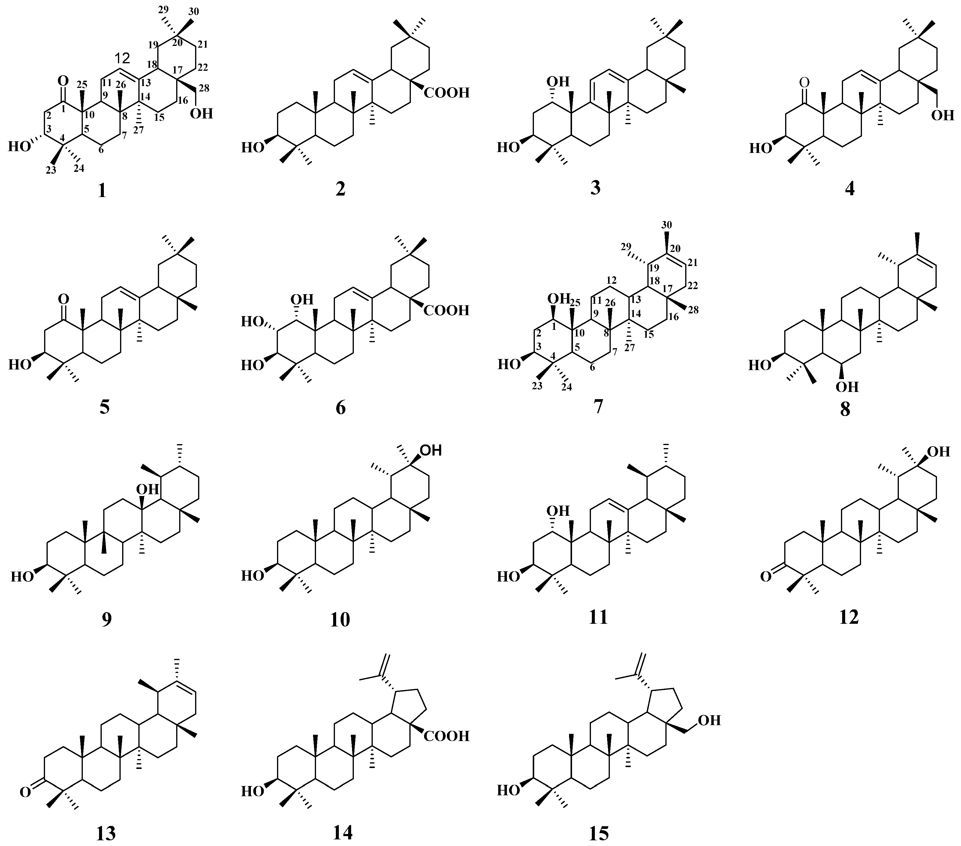
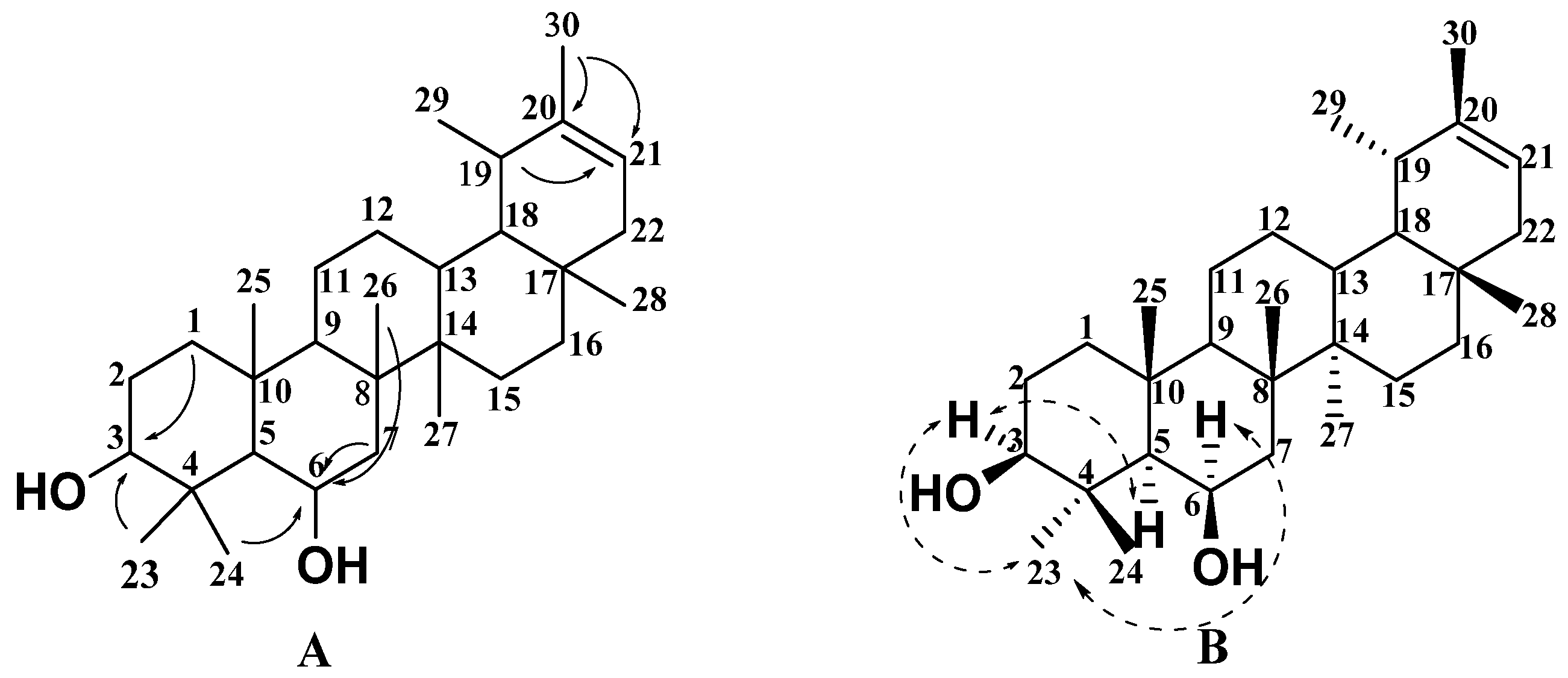
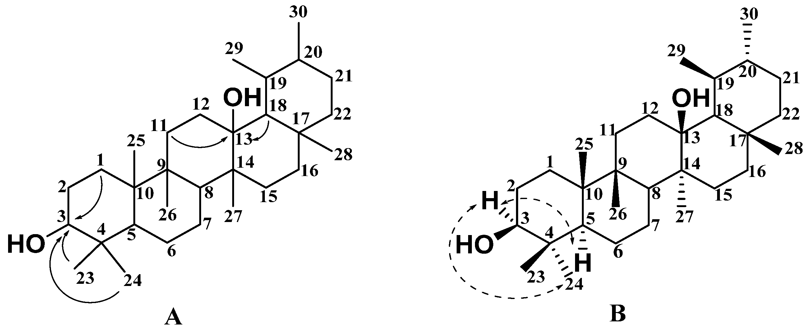
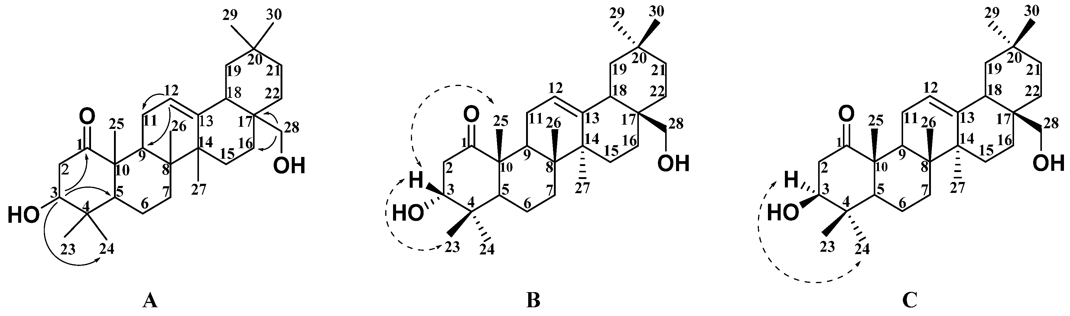
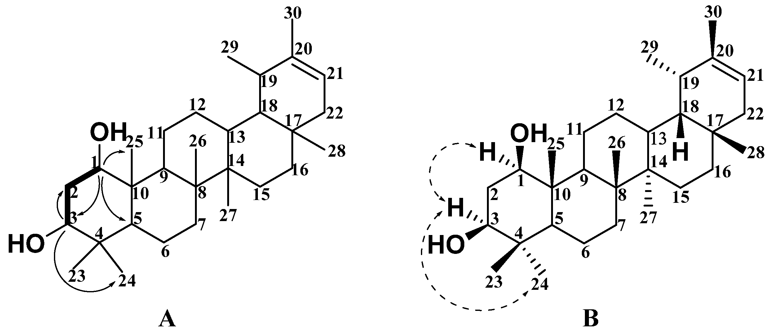
2.1. Structural Analysis of New Compounds

The crude petroleum ether extract of S. discolor Dunn was isolated and purified by various column chromatography techniques, including MCI gel, Sephadex LH-20, silica gel, RP-C18 silica gel, and a semipreparative HPLC column, allowing for the isolation of four new pentacyclic triterpenes, namely Sabiadiscolor A–D (1 and 79), along with eleven known compounds. Their structures are shown in Figure 1. Compared with the literature, eleven of these structures were known compounds based on their NMR and MS data, and were identified as ursolic acid (2) [8], juglangenin A (3) [9], 3β, 28-dihydroxy-12-oleanene-l-one (4) [10], 3-hydroxyolean-12-en-1-one (5) [11], 1α, 2α, 3β-trihydroxyl-olean-12-en-28-oic acid (6) [12], dandelion alkan-3β, 20β-diol (10) [13], olean-12-ene-1, 3-diol (11) [14], 3-oxo-20S-hydroxytaraxastane (12) [15], ψ-taraxasterone (13) [16], betulinic acid (14) [17], and birch ester alcohol (15) [18].
Compound 1 was obtained as a white solid and its molecular formula was inferred to be C30H48O3 by HR-ESI-MS with m/z 457.3671 [M + H]+ (calc. 457.3676). The IR spectrum showed absorption bands for the presence of a hydroxy group (3477 cm−1) and a ketone carbonyl group (1713 cm−1). 1H NMR (Table 1 and Figure S1), 13C NMR (Table 2 and Figure S2), and DEPT (Figure S3) spectral data revealed the presence of eight quaternary carbons, five methine groups, ten methylene groups, and seven methyl groups, including a carbonyl group (δC 214.6) and a trisubstituted olefinic unit (δC 122.9 (CH) and δH 5.20 (dd, J = 4.5, 2.9 Hz, 1H); δC 144.1 (C)). According to 1H NMR (Table 1) spectral data, the compound has seven methyl groups at δH 0.88 (3H, s, H-30), 0.90 (3H, s, H-29), 1.00 (3H, s, H-26), 1.03 (3H, s, H-24), 1.10 (3H, s, H-23), 1.21 (3H, s, H-27), and 1.32 (3H, s, H-25). The comparison of the NMR data of compound 1 with those of the known compound 4 [10] in Table 1 and Table 2 showed that the two compounds should share the same basic skeleton and that both were very similar. According to further HMBC (Figure S5) correlations in Figure 2A, H-3 (δH 3.88, m, 1H) was correlated with C-1 (δC 214.6), C-5 (δC 51.3), and C-24 (δC 22.3), and H-28 (δH 3.30 and 3.21, 2H) was correlated with C-16 (δC 21.9), C-17 (δC 36.9), and C-18 (δC 42.5). Based on this data, it was predicted that the two hydroxyl groups should be at positions C-3 and C-28. Therefore, the planar structure of compound 1 was the same as that of compound 4, as shown in Figure 2A. The relative configuration of compound 1 was further determined according to the NOESY (Figure S6) correlation spectrum in Figure 2B. The correlation signals between H-23, H-25, and H-3 indicate that H-3 is in the β configuration and the 3-substituted hydroxyl group has an α configuration. The relative configuration of compound 4 was determined according to the NOESY (Figure S7) correlation spectrum in Figure 2C. The correlation signals between H-24 and H-3 indicated that H-3 was the α configuration and the 3-substituted hydroxyl group has a β configuration. Therefore, it was confirmed that compound 1 and compound 4 are isomers, and compound 1 is 3α, 28-dihydroxy-12-oleanene-l-one, named Sabiadiscolor A.
Compound 7 was obtained as a white solid and its molecular formula was inferred to be C30H50O2 with 6 degrees of unsaturation by HR-ESI-MS with 465.3701 [M + Na]+ (calc. 465.3703). The IR spectrum of this compound revealed the presence of a hydroxyl group (3368 cm−1). As shown in Table 1 and Table 2, its 1H NMR (Figure S11), 13C NMR (Figure S12), and DEPT (Figure S13) spectral data showed that compound 7 contained thirty carbons, including six quaternary carbons, eight methine groups, eight methylene groups, and eight methyl groups. 1H NMR (Table 1) spectral data at δH 0.80 (3H, s, H-28), 0.99 (3H, s, H-30), 1.01 (3H, s, H-27), 1.09 (3H, s, H-24), 1.17 (3H, s, H-26), 1.24 (3H, s, H-23), 1.26 (3H, s, H-25), and 1.65 (3H, s, H-29) showed seven methyl groups at the sp3 quaternary carbons and one methyl group at the sp3 tertiary carbon. According to the above data, compound 7 was inferred to be a five-membered ring triterpene with one olefinic unit (δC 140.0 (C); δC 119.3 (CH) and δH 5.33 (dd, J = 6.7, 2.0 Hz, 1H)) and two oxygen-substituted methine groups (δC 79.7 (CH) and δH 3.80 (dd, J = 11.1, 4.2 Hz, 1H); δC 75.5 (CH) and δH 3.63 (dd, J = 12.0, 4.4 Hz, 1H)). Compared with the chemical structure of the pentacyclic triterpenes isolated from the genus Sabia, all spectral data showed that the compound was an ursane-type pentacyclic triterpene and was structurally similar to the known compound, which is 20-taraxastene-3β, 22α-diol [19]. By further analyzing the HSQC (Figure S14) and HMBC (Figure S15) correlation signals of compound 7 in Figure 3A, it was shown that H-3 (δH 3.63, dd, J = 12.0, 4.4 Hz) was correlated with C-2 (δC 39.5) and C-24 (δC 16.2), while H-1 (δH 3.63, dd, J = 12.0, 4.4 Hz) was correlated with C-3 (δC 75.5), C-5 (δC 52.1), and C-25 (δC 13.2). Based on the above-mentioned data, it was predicted that the two hydroxyl groups should be at positions C-1 and C-3. According to the 1H-1H COSY (Figure S16) signal in Figure 3A, both H-3 (δH 3.63) and H-1 (δH 3.80) are correlated with H-2 (δH 2.36), as shown in Figure 3A. The relative configuration of compound 7 was further determined according to the NOESY (Figure S17) correlation spectrum in Figure 3B and Figure S16. The correlation signals between H-3 and H-24 indicate that H-3 has an α configuration. The correlation signals between H-3 and H-1 indicate that H-1 has an α configuration. Both substituted hydroxyl groups at C-1 and C-3 had a β configuration. Therefore, the structure of the compound can be determined as 20-taraxastene-1β, 3β -diol, named Sabiadiscolor B.
Compound 8 was obtained as a white solid and its molecular formula was inferred to be C30H50O2 by HR-EI-MS with m/z 442.3806 [M]+. The IR spectrum absorption at 3413 cm−1 revealed the presence of the hydroxyl group. In Table 1 and Table 2, the 1H NMR (Figure S21), 13C NMR (Figure S22), and DEPT (Figure S23) spectral data of compound 8 showed that it also contained 30 carbons, including six quaternary carbons, six methine groups, and ten methylene and eight methyl groups. Two sp3 methines (δC 77.9 (CH) and δH 3.48 (m, 1H); δC 75.0 (CH) and δH 3.26 (dd, J = 12.4, 4.3 Hz, 1H)) were typical of oxygen-bearing groups. Similar to compound 7, eight methyl groups at δH 0.74 (3H, s, H-28), 0.76 (3H, s, H-27), 0.93 (3H, s, H-23), 0.96 (3H, s, H-24), 0.96 (3H, s, H-25), 0.99 (3H, s, H-29), 1.06 (3H, s, H-26), and 1.57 (3H, s, H-30), and one trisubstituted olefinic unit (δC 118.8 (CH) and δH 5.30 (s, 1H); δC 139.9 (C)) existed in compound 8. According to NMR spectral data, compound 8 was found to be very similar to the known compound pseudotaraxasterol [20]. By comparing their NMR data, it was found that the main difference lies in the chemical shift values of C-6, C-7, C-23, and C-25. Therefore, it was speculated that another hydroxyl group might be at C-6 or C-7. The HSQC (Figure S24) and HMBC (Figure S25) spectra of compound 8 showed that H-3 (δH 3.50) was correlated with C-1 (δC 38.1) and C-23 (δC 12.0). H-6 (δH 3.29) is associated with C-7 (δC 17.9), C-24 (δC 27.8), and C-26 (δC 16.3). The relative configuration of compound 8 was determined according to its NOESY (Figure S27) correlation spectrum, as shown in Figure 4B. The strong correlations of Me-23/H-3/H-5 and H-6/Me-23 indicated that these protons or methyl groups were α-orientations. Thus, Me-23, H-3, H-5, and H-6 were arbitrarily assigned α-orientations, while two hydroxyl groups on the third and sixth carbons were β-oriented. Therefore, the structure of compound 8 can be determined to be 6β-pseudotaraxasterol, named Sabiadiscolor C.
Compound 9 was obtained as a white solid and its molecular formula was inferred to be C30H52O2 by HR-ESI-MS with m/z 443.3339 [M − H]. The IR spectrum revealed the presence of the hydroxyl group (3388 cm−1). All 30 carbons observed in the 1H NMR (Figure S31), 13C NMR (Figure S32), and DEPT (Figure S33) spectral data could be classified into six sp3 quaternary carbons, eight sp3 methine groups, eight sp3 methylene groups, and eight methyl groups, as shown in Table 1 and Table 2. Among them, one sp3 methine (δC 77.9 (CH) and δH 3.49 (dd, J = 10.6, 5.6 Hz, 1H)) and one sp3 quaternary carbon(δC 73.8 (C)) were ascribed as bearing oxygen atoms. According to 1H NMR data (Table 1), the compound has eight methyl groups at δH 0.90 (3H, s, H-24), 1.01 (3H, s, H-28), 1.03 (3H, d, H-29), 1.03 (3H, d, H-30), 1.08 (3H, s, H-25), 1.27 (3H, s, H-23), 1.33 (3H, s, H-26), and 1.42 (3H, s, H-27). The comparison of the NMR data of compound 9 with the known compound ursan-3β, which is 5α-diol [21], suggested that compound 9 possessed an ursane-type pentacyclic triterpene skeleton. The HSQC (Figure S34) and HMBC (Figure S35) spectra of compound 9 showed that C-13 (δC 73.8) was correlated with H-14 (δH 1.42) and H-18 (δH 1.33), which revealed that the hydroxyl group should be assigned at C-13. The relative configuration of compound 9 was determined according to its NOESY (Figure S37) correlation spectrum, as shown in Figure 5B. The strong correlations of Me-24/H-3/H-5 indicated that Me-24, H-3, and H-5 were α-orientations, while the hydroxyl group on the third carbon was β-oriented. Therefore, the structure of compound 9 can be determined as ursan-3β, 13β-diol, named Sabiadiscolor D.

2.2. α-Glycosidase Inhibitory Activities

All compounds (115) isolated from S. discolor Dunn were evaluated for their α-glycosidase inhibitory activity. As shown in Table 3, compounds 1, 3, 8, 9, 13, and 15 showed remarkable activities with IC50 values from 0.09 to 0.27 μM, while compound 7 showed weak activity with an IC50 value of 0.56 ± 0.0331 μM. The other compounds had low inhibitory activity against α-glycosidase and are not listed in Table 3.

2.3. Discussion

By modern natural medicinal chemistry experiments, fifteen natural pentacyclic triterpenoids (115) were obtained and identified from the traditional Chinese ethnic medicinal plant named S. discolor Dunn, collected from minority areas, and four of them (1 and 79) were new compounds. Their preliminary α-glycosidase inhibitory activities were evaluated. The results showed that six compounds (1, 3, 8, 9, 13, and 15) showed remarkable activities with IC50 values of 0.27 ± 0.0499, 0.11 ± 0.0222, 0.23 ± 0.0135, 0.23 ± 0.0307, 0.26 ± 0.0383, and 0.09 ± 0.0045 μM, respectively. It was found that ursane-type pentacyclic triterpenes have better hypoglycemic activities and especially new compounds 1, 8, and 9 have more significant activity than the positive control (Acarbose), which revealed that they might be a class of potential α-glycosidase inhibitors. According to the structure and activity data of these pentacyclic triterpenoids with potential hypoglycemic activity, it is speculated that the number and location of hydroxyl groups as well as double-bond groups might contribute more greatly to the inhibition rate of α-glycosidase, and both oleanane-type pentacyclic triterpenoids and lupanane-type ones should be the potential α-glycosidase inhibitor. Due to limited quantity of the isolated compounds, it is not possible to systematically discuss the structure–activity relationship of such compounds, but compound 15 with the lupanane-type pentacyclic triterpenoid skeleton was implied to have the best activity.
S. discolor Dunn is one of the most important species in the genus Sabia, which is rich in resources in the minority areas of southwest China. The medical plant is used to treat rheumatism, bone pain, bruises, hepatitis, and other diseases in the folk [22]. The main types of chemical constituents in the genus Sabia included pentacyclic triterpenoids, alkaloids, benzene derivatives, and fatty acids [23], but there was less literature reporting on the chemical composition of S. discolor Dunn. As our research suggested its hypoglycemic activity for the first time, it provided an important basis for the comprehensive utilization of this plant resource.

3. Materials and Methods

3.1. General Experimental Materials

One-dimensional and 2D NMR spectra were measured on a Bruker AM-600 spectrometer. HRESIMS data were obtained by Q EXACTIVE FOCUS (Thermo Fisher Technologies Co. Ltd., Waltham, MA, USA) spectrometers. Electrospray ionization (ESI) data were obtained by an HP 1100SMD. Preparative HPLC separations were run on a SEP system (Beijing Sepuruisi Scientific Co., Ltd., Beijing, China) equipped with a variable-wavelength UV detector using a YMC-Pack ODS-A column (250 × 20 mm, 5 µm). IR spectra were obtained using a Bruker Tensor-27 instrument (Bruker, Munich, Germany). The extract was obtained through a 300 L extraction tank (JF21060, Jiangsu Jufeng Machinery Co. Ltd., Huaian, China). Column chromatography (CC) was performed on silica gel (40–80 mesh, 200–300 mesh, and 300–400 mesh, Qingdao Haiyang Chem. Ind. Ltd., Qingdao, China), silica gel H (40–80 μm mesh, Qingdao, China), Sephadex LH-20 (40–70 μm, Amersham Pharmacia Biotech AB, Uppsala, Sweden), MCI gel (CHP20P, 75–150 μm, Mitsubishi Chemical Industry Co. Ltd., Kenyworth, NJ, USA), and C18 reversed-phase silica gel (20–45 μm, Merck, Darmstadt, Germany). TLC plates were precoated with silica gel GF254 (Qingdao Haiyang Chem. Ind. Ltd., Qingdao, China). All solvents were of analytical grade (Anergy Chemical Reagents Co. Ltd., Shanghai, China; Shanghai Titan Technology Co. Ltd., Shanghai, China).
Altogether, dimethyl sulfoxide (DMSO; Beijing Mr. Lai Treasure Company, Beijing, China); phosphate buffer solution (PBS; HyClone); centrifuge tubes, 96 cell culture plates, and other consumables (NEST Biotechnology Co. Ltd., Beijing, China); and α-glucosidase (Sigma Company, St. Louis, MO, USA) from saccharomyces cerevisiae were obtained for this study. Acarbose (Shanghai Yuanye Biotechnology Co. Ltd., Shanghai, China) was used as a positive control. The absorbance was read using a microplate reader (Varioskan LUX, Thermo, Waltham, MA, USA) at 405 nm. The results were obtained for at least three independent experiments.

3.2. Plant Materials

S. discolor Dunn was collected from Zhutou Mountain, Guangxi Province. The dried stems and leaves were identified by Professor Qing-Wen Sun of Guizhou University of Traditional Chinese Medicine. The samples were stored in the Key Laboratory of Chemistry for Natural Products, Chinese Academy of Sciences, Guizhou Province.

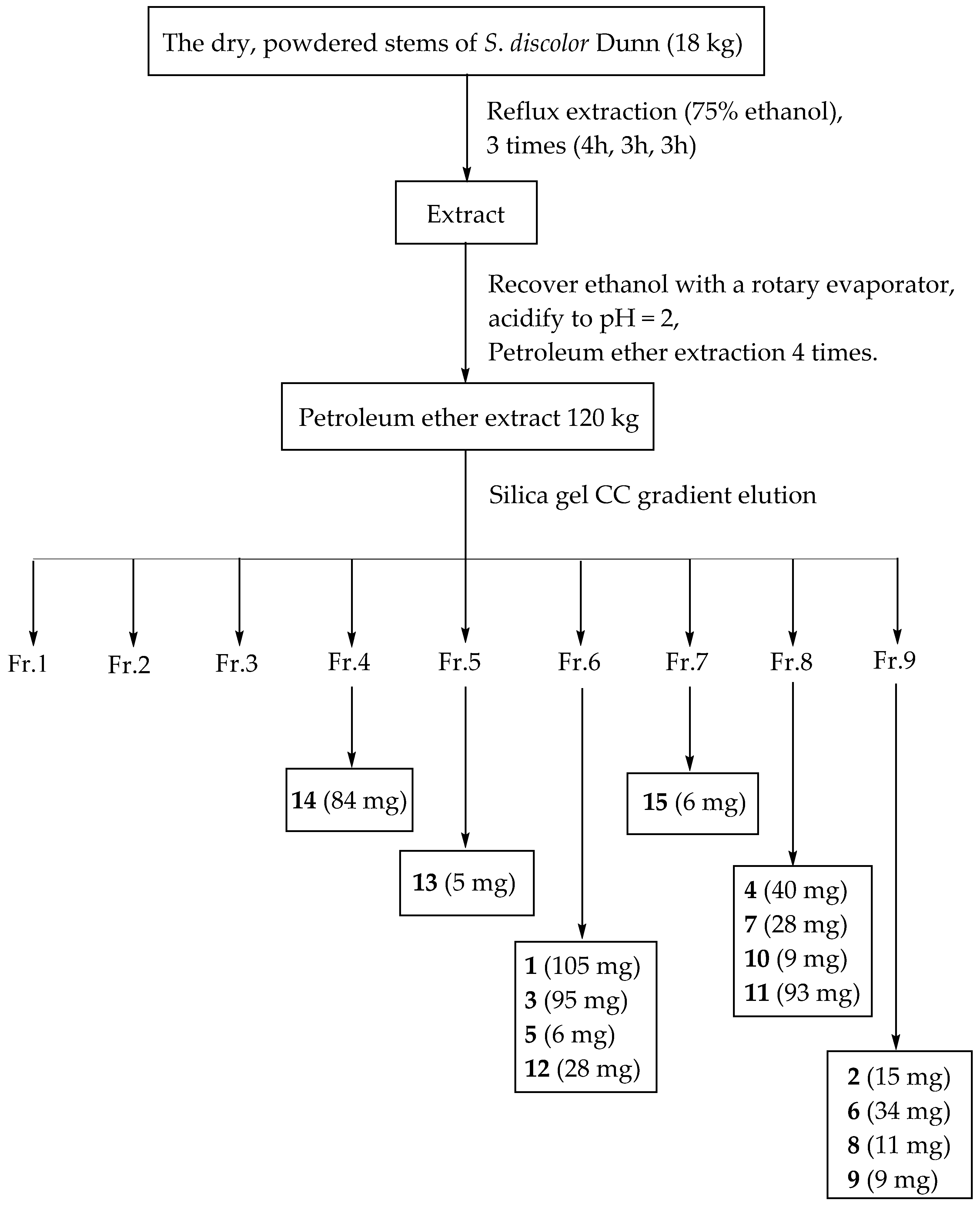
3.3. Extraction and Isolation

The dry, powdered stems of S. discolor Dunn (18 kg) were refluxed three times with 75% ethanol for 4, 3, and 3 h successively. The ethanol in the extract was fully recovered and then the alkaline substances in the extract were removed by 10% bitartrate acidification to pH = 2. The above extracts were extracted and recovered with petroleum ether to obtain 120 g of extract. The petroleum ether extract was subjected to a petroleum ether/ethyl acetate solvent gradient (80:1–1:1) by silica gel CC to obtain nine fractions (Fr. 1–Fr. 9). The separation process is shown in Figure 6.
Fr. 4 was subjected to silica gel CC (petroleum ether/methylene dichloride, 8:1) to obtain compound 14 (84 mg). Fr. 5 was subjected to Sephadex LH-20 CC (eluted with CHCl3/MeOH, 1:1) to obtain compound 13 (5 mg). Fr. 6 was subjected to Sephadex LH-20 CC (eluted with CHCl3/MeOH, 1:1) to obtain four subfractions (Fr. 6.1–Fr. 6.4) and Fr. 6.2 was subjected to silica gel column (petroleum ether/ethyl acetate, 10:1) to obtain five fractions (Fr. 6.2.1–Fr. 6.2.5). Compound 5 (6 mg) was obtained by Sephadex LH-20 CC (eluted with CHCl3/MeOH, 1:1) from Fr. 6.2.1. Compounds 3 (95 mg) and 1 (105 mg) were obtained by silica gel CC (petroleum ether/diethylamine, 15:1) from Fr. 6.2.2. Compound 12 (28 mg) was obtained by Sephadex LH-20 CC (eluted with MeOH) from Fr. 6.2.5. Fr. 7 was subjected to Sephadex LH-20 CC (eluted with CHCl3/MeOH, 1:1) to obtain eight subfractions (Fr. 7.1–Fr. 7.8) and Fr. 7.4 was purified by both Sephadex LH-20 CC (eluted with MeOH) and silica gel CC (petroleum ether/ethyl acetate, 6:1) to obtain compound 15 (16 mg). Fr. 8 was filtered and subjected to silica gel CC (petroleum ether/diethylamine, 15:1) to afford compound 7 (28 mg). The residue was subjected to Sephadex LH-20 CC (eluted with MeOH) to obtain five fractions (Fr. 8.1–Fr. 8.5). Fr. 8.3 was subjected to silica gel CC (petroleum ether/ethyl acetate, 3:1) to obtain compound 4 (40 mg). Compound 11 (93 mg) was obtained by Sephadex LH-20 CC (eluted with CHCl3/MeOH, 1:1) from Fr. 4. Compound 10 (9 mg) was obtained by silica gel CC (petroleum ether/chloroform, 100:1) from Fr. 8.6. Fr. 9 was subjected to Sephadex LH-20 CC (eluted with CHCl3/MeOH, 1:1) to obtain four subfractions (Fr. 9.1–Fr. 9.4) and Fr. 9.1 was subjected to Sephadex LH-20 CC (eluted with MeOH) to afford compounds 6 (34 mg) and 9 (9 mg). Fr. 9.4 was subjected to a decompression silica gel CC (petroleum ether/chloroform, 50:1) to afford compounds 8 (11 mg) and 2 (15 mg).

3.4. Assay of α-glycosidase Inhibition

The inhibitory activity of α-glucosidase was determined by the PNPG method [24]. PBS, different concentrations of samples or positive drugs, α-glucosidase, and 10% DMSO solution (100 μL) were mixed by shaking and incubated for 15 min in a constant temperature incubator at 37 °C. Then, 20 μL of PNPG (2.5 mmol/L) was added, mixed by shock, and incubated in a 37 °C constant-temperature incubator for 15 min. Then, 80 μL of Na2CO3 (0.8 mmol/L) solution was added to stop the reaction and the absorbance was measured by a microplate reader at 405 nm. Five groups were established, including the blank group (90 μL PBS + 10 μL 10% DMSO + 20 μL PNPG (2.5 mmol/L) + 80 μL Na2CO3 (0.8 mmol/L)), background group (90 μL PBS + 10 μL compounds + 20 μL PNPG (2.5 mmol/L) + 80 μL Na2CO3 (0.8 mmol/L)), negative control group (70 μL PBS + 20 μL α-glucosidase + 10 μL 10% DMSO + 20 μL PNPG (2.5 mmol/L) + 80 μL Na2CO3 (0.8 mmol/L)), positive control group (70 μL PBS + 10 μL acarbose + 20 μL α-glucosidase + 20 μL PNPG (2.5 mmol/L) + 80 μL Na2CO3 (0.8 mmol/L)), and drug administration group (70 μL PBS + 20 μL α-glucosidase + 10 μL compounds + 20 μL PNPG (2.5 mmol/L) + 80 μL Na2CO3 (0.8 mmol/L)), with 3 parallel replicates in each group.
Inhibition rate = [(OD negative − OD blank) − (OD sample − OD background)]/(OD negative − OD blank) × 100%.
If the inhibitory rate was close to or higher than that of acarbose, the compound was considered to have α-glycosidase inhibitory activity. The IC50 of the potential compound was measured and calculated by the same method after 5-fold dilution.

4. Conclusions

To isolate α-glycosidase inhibitors from natural products, almost all fifteen pentacyclic triterpenoids were isolated for the first time from the branches and leaves of S. discolor Dunn, which is a kind of ethnic medicinal plant. These triterpenoids included six oleanane-type pentacyclic triterpenoids, seven ursane-type triterpenoids, and two lupanane-type triterpenoids. Among them, four compounds (1 and 79) were new pentacyclic triterpenoids. By further evaluating the α-glycosidase inhibitory activities of these compounds, compounds 1, 3, 8, 9, 13, and 15 showed remarkable activities with IC50 values of 0.27 ± 0.0499, 0.11 ± 0.0222, 0.23 ± 0.0331, 0.23 ± 0.0307, 0.26 ± 0.0383, and 0.09 ± 0.0045 μM, respectively. The results revealed that pentacyclic triterpenoids could be a class of potential α-glycosidase inhibitors.

Supplementary Materials

The following supporting information can be downloaded at: https://www.mdpi.com/article/10.3390/molecules27072161/s1. Figures S1–S40: 1H NMR, 13C NMR, HSQC, HMBC, 1H-1H COSY, NOESY, HRMS, and IR spectra data for compounds (1 and 79), and NOESY spectra data for compound 4.

Author Contributions

S.-Z.M. conceived, designed, and offered guidance for the experiments, and revised the manuscript; J.-H.M. and D.H. performed the experiments, isolated and analyzed monomeric compounds, performed the biology experiments, and wrote the manuscript; J.L. and L.-L.D. offered some data; and X.-J.H. contributed as a supervisor. All authors have read and agreed to the published version of the manuscript.

Funding

This research was funded by the financial support of the Science and Technology Program of Guizhou Province (QKHPTRC (2019) 5659), the National Natural Science Foundation of China (U1812403-3), and the Growth Project of Young Scientific and Technological Talents in Colleges and Universities of Guizhou Province (QJH KY (2021) 181).

Institutional Review Board Statement

Not applicable.

Informed Consent Statement

Not applicable.

Data Availability Statement

The data presented in this study are available in the supplementary materials.

Acknowledgments

The authors are grateful to the Key Laboratory of Chemistry for Natural Products of Guizhou Province and the Chinese Academy of Sciences for their help in the HPLC analysis and NMR measurements; to Yang-Ming Jiang for the NMR measurements; and to the teacher Li-Ping Wang for generous assistance in the bioactivity tests.

Conflicts of Interest

The authors declare no conflict of interest.

Sample Availability

Isolated compounds are not available from the authors.

References

  1. Mahomoodally, M.F.; Lobine, D.; Picot-Allain, M.C.N.; Sadeer, N.; Jugreet, S.; Zengin, G. Conventional and non-conventional targets of natural products in the management of diabetes mellitus and associated complications. Curr. Med. Chem. 2021, 28, 4638–4669. [Google Scholar] [CrossRef] [PubMed]
  2. Alomari, A.S.; Al-Harithy, R.N. Intergenic lnc-LEP-2:6 and lnc-LEP-2:7 as novel biomarkers associated with type 2 diabetes mellitus. Arch. Phy. Biochem. 2021, 6, 1–6. [Google Scholar] [CrossRef]
  3. Simos, Y.V.; Spyrou, K.; Patila, M.; Karouta, N.; Stamatis, H.; Gournis, D. Trends of nanotechnology in type 2 diabetes mellitus treatment. Asian. J. Phar. Sci. 2021, 16, 15–30. [Google Scholar] [CrossRef]
  4. Wu, X.F.; Xu, Y.; Li, Y.P. Research progress of screening models for α-glucosidase inhibitors. Int. J. Phar. 2008, 35, 9–12. [Google Scholar]
  5. Cai, Y.H.; Guo, Y.Q.; Li, Z.; Wu, D.Y.; Li, X.L.; Zhang, H.; Yang, J.J.; Lu, H.; Sun, Z.W.; Luo, H.B.; et al. Discovery and modelling studies of natural ingredients from Gaultheria yunnanensis (FRANCH) against phosphodiesterase-4. Eur. J. Med. Chem. 2016, 114, 134–140. [Google Scholar] [CrossRef]
  6. Li, W.; Lou, L.L.; Zhu, J.Y.; Zhang, J.S.; Liang, A.A.; Bao, J.M.; Tang, G.H.; Yin, S. New lanostane-type triterpenoids from the fruiting body of Ganoderma hainanense. Fitoterapia 2016, 115, 24–30. [Google Scholar] [CrossRef]
  7. Luo, S.Y.; Pu, R.; Tang, Y.Q.; Fan, R.Z.; Yin, S.; Tang, G.H. Euphane- and 19 (10→9) abeo-euphane-type triterpenoids from Jatropha gossypiifolia. Fitoterapia 2020, 143, 104582–104586. [Google Scholar] [CrossRef]
  8. Liu, J.C.; Yang, Q.M.; Zhu, Z.F. Studies on the chemical constttuents of Coriaria sinica maxim. Nat. Sci. 1996, 30, 196–198. [Google Scholar]
  9. Deng, Y.; Li, X.; Wu, F.E. Study on the chemical constituents of Sabia yunnanensis Franch. Chin. Tradit. Herb. Med. 2006, 37, 183–185. [Google Scholar]
  10. Kagawa, M.; Minami, H.; Mai, N.; Takahashi, H.; Takaoka, S.; Fukuyama, Y. Oleanane-type triterpenes from Viburnum awabuki. Phytochemistry 1998, 47, 1337–1341. [Google Scholar] [CrossRef]
  11. Chatterjee, P.; Pezzuto, J.M.; Kouzi, S.A. Glucosidation of betulinic acid by Cunninghamella species. J. Nat. Prod. 1999, 62, 761–763. [Google Scholar] [CrossRef] [PubMed]
  12. Chiang, Y.M.; Kuo, Y.H. Taraxastane-type triterpenes from the aerial roots of Ficus macrocarpa. J. Nat. Prod. 2000, 63, 898–901. [Google Scholar] [CrossRef] [PubMed]
  13. Mashitah, Y.; Hazrulrizawati, H.; Peter, H. Anticholinesterase inhibitory activity of quaternary alkaloids from Tinospora crispa. Molecules 2014, 19, 1201–1211. [Google Scholar]
  14. Li, L.L.; Sun, Z.; Shang, X.Y.; Li, J.J.; Wang, R.; Zhu, J. Studies on triterpenes from Cirsium setosum. J. Chin. Mater. Med. 2012, 37, 951–955. [Google Scholar]
  15. Wang, P.; Song, Q.S.; Xu, W.; Li, S.Z. Study on chemical Constituents of Leaves and branches of Beaumontia grandi flova. Chin. Tradit. Herb. Med. 2009, 40, 1549–1551. [Google Scholar]
  16. Zheng, H.T.; Zhang, G.G.; Zheng, Y.F.; Jin, Y.P.; Du, S.S.; Deng, Z.W. Study on lipid soluble Chemical Constituents of Tibetan medicine Dracocephalum tanguticum Maxim. Chin. J. Med. Chem. 2007, 17, 314–315. [Google Scholar]
  17. Huang, Y.; Zhang, C.L.; Lang, W.W.; Chen, M.S.; Liu, Y.; Liu, B.M. A new oxoaporphine from Sabia limoniacea var. ardisioides. Acta Pharm. Sin. 2018, 5, 778–781. [Google Scholar]
  18. Chen, J.; Chen, B. Two New Pentacyclic Triterpenes from Sabia parviflora. Chin. Chem. Lett. 2002, 13, 345–348. [Google Scholar]
  19. Zhang, Y.W.; Lin, H.; Bao, Y.L.; Wu, Y.; Yu, C.L.; Huang, Y.X.; Li, Y.X. A new triterpenoid and other constituents from the stem bark of Juglans mandshurica. Biochem. Syst. Ecol. 2012, 44, 136–140. [Google Scholar] [CrossRef]
  20. Mahato, S.B.; Kundu, A.P. 13 C NMR Spectra of pentacyclic triterpenoids—a compilation and some salient features. Phytochemistry 1994, 37, 1517–1575. [Google Scholar] [CrossRef]
  21. Wang, Y.Y.; Wu, D.G. Two new triterpenoids from Tripterygium wilfordii. Acta Botanica. Yunnanica 1993, 15, 309–310. [Google Scholar]
  22. Zhang, X.Q.; Li, M.S.; Li, Y.L.; Zhang, X.X.; Lu, S.G.; Wen, X.F.; Hu, J.Z.; Zou, K. An overview of the research on medicinal plants of the genus Sabia in China. Biol. Resources. 2018, 40, 4–17. [Google Scholar]
  23. Wen, D.; Sun, Q.W.; Pan, G.J.; Chen, J.Z.; Lu, X. Research progress of medicinal plants of Pterygophyllum. Guizhou Sci. 2016, 34, 25–31. [Google Scholar]
  24. Chen, Z.B.; Hao, J.J.; Wang, L.P.; Wang, Y.; Kong, F.D.; Zhu, W.M. New α-glucosidase inhibitors from marine algae-derived Streptomycessp.OUCMDZ-3434. Sci. Rep. 2016, 6, 20004–20012. [Google Scholar] [CrossRef]
Figure 1. Structures of compounds 115.
Molecules 27 02161 g001
Figure 2. Key HMBC (plain arrow; (A)), NOESY (dash arrow) correlations of compound 1 (B), and NOESY (dash arrow) correlations of compound 4 (C).
Molecules 27 02161 g002
Figure 3. Key H-H COSY (bold), HMBC (plain arrow; (A)), and NOESY (dash arrow; (B)) correlations of compound 7.
Molecules 27 02161 g003
Figure 4. Key HMBC (plain arrow; (A)) and NOESY (dash arrow; (B)) correlations of compound 8.
Molecules 27 02161 g004
Figure 5. Key HMBC (plain arrow; (A)) and NOESY (dash arrow; (B)) correlations of compound 9.
Molecules 27 02161 g005
Figure 6. Separation process of chemical constituents from S. discolor Dunn.
Molecules 27 02161 g006
Table 1. 1H NMR (600 MHz) data for 1, 4, 7, 8, and 9 (δ in ppm and J in Hz).
Position1 a4 a7 b8 a9 b
1 3.80 (dd, 11.1, 4.2)1.85 (m)
1.63 (t, 1.7)
1.22 (s)
1.42 (s)
23.23 (d, 10.9)
3.30 (d, 10.9)
3.57 (d, 10.9)
3.23 (d, 10.9)
2.34 (q, 12.0)
2.42 (dt, 12.6)
2.04 (m)
1.74 (m)
1.01 (s)
1.75 (d, 4.6)
33.86 (dd, 12.0, 4.6)3.87 (dd, 12.0, 4.6)3.63 (dd, 12.0, 4.4)3.48 (dd, 12.4, 4.3)3.49 (dd, 10.6, 5.6)
51.59 (d, 2.8)2.0 (d, 4.3)1.80 (d, 3.2)0.59 (dd, 11.8, 2.2)0.84 (m)
61.51 (d, 4.0)
1.59 (d, 2.9)
2.25 (d, 5.6)
2.0 (d, 4.3)
1.76 (d, 3.0)
1.68 (d, 1.8)
3.26 (dd, 12.4, 4.3)1.60 (dt, 10.4, 2.6)
1.19 (s)
71.35 (d, 3.2)
1.53 (d, 4.2)
1.35 (d, 3.2)
1.53 (d, 4.2)
1.76 (d, 3.0)
1.68 (d, 1.8)
1.52 (d, 3.2)
1.57 (s)
1.30 (d, 3.8)
1.42 (s)
8 1.23 (s)
92.30 (dd, 11.3, 5.6)2.25 (dd, 11.3, 5.6)2.42 (dd, 12.6, 4.5)1.52 (d, 3.2)
112.40 (dd, 12.0, 4.8)
2.25 (dd, 11.3, 5.6)
2.40 (dd, 12.0, 4.8)
2.25 (dd, 11.3, 5.6)
1.68 (t, 1.8)
1.80 (d, 3.2)
1.43 (d, 2.9) 2.35 (m)
125.20 (dd, 4.5, 2.9)5.22 (dd, 4.4, 2.7)1.80 (d, 3.2)
1.75 (d, 3.0)
1.34 (s)
1.43 (d, 2.9)
1.71 (m)
1.90 (m)
13 1.62 (s)1.57 (s)
150.98 (s)
1.70 (d, 4.6)
1.01 (s)
1.32 (d, 4.6)
1.68 (t, 1.8)
1.28 (s)
1.34 (s)
1.63 (d, 1.7)
1.23 (s)
1.90 (m)
161.19 (s)
1.90 (d, 4.5)
1.21 (s)
2.0 (d, 4.5)
1.68 (t, 1.8)
1.28 (s)
1.26 (s)1.81 (t, 3.3)
2.15 (d, 3.8)
181.97 (dd, 13.5, 4.2)2.10 (dd, 13.6, 4.3)1.28 (s)1.06 (s)1.33 (s)
191.14 (s)
1.74 (s)
0.91 (s)
1.32 (s)
1.96 (s)1.65 (s)1.41(s)
20 2.44 (td, 7.3, 3.9)
211.17 (d, 2.2)
1.31 (s)
1.17 (d, 2.2)
1.02 (s)
5.33 (dd, 6.7, 2.0)5.30 (s)1.79 (d, 3.2)
2.11 (m)
221.37 (d, 3.7)
1.53 (d, 4.2)
1.35 (d, 3.7)
1.51 (d, 4.3)
1.62 (s)
1.28 (s)
1.57 (s)
1.74 (m)
1.40 (s)
1.19 (s)
23β 1.10 (s)β 1.08 (s)β 1.24 (s)α 0.93 (s)β 1.27 (s)
24α 1.03 (s)α 1.04 (s)α 1.09 (s)β 0.96 (s)α 0.90 (s)
25β 1.32 (s)β 1.32 (s)β 1.26 (s)β 0.96 (s)β 1.08 (s)
26β 1.00 (s)β 1.02 (s)β 1.17 (s)β 1.06 (s)β 1.33 (s)
27α 1.21 (s)α 1.21 (s)α 1.01 (s)α 0.76 (s)α 1.42 (s)
283.30 (d, 3.2)
3.21 (d, 10.9)
3.57 (d, 3.2)
3.23 (d, 10.9)
β 0.80 (s)β 0.74 (s)β 1.01 (s)
29α 0.90 (s)α 0.91 (s)α 1.65 (s)α 0.99 (d, 6.4)β 1.03 (d, 2.8)
30β 0.88 (s)β 0.89 (s)β 0.99 (s)β 1.57 (s)α 1.03 (d, 2.8)
a Data measured in CDCl3. b Data measured in C5D5N.
Table 2. 13C NMR (150 MHz) data for 1, 4, 7, 8, and 9 (δ in ppm).
Position1 a4 a7 b8 a9 b
1214.6212.479.738.138.5
242.844.139.527.126.7
379.378.675.579.377.9
438.039.339.738.839.2
551.35452.153.055.5
618.417.818.575.018.6
732.332.534.517.940.5
841.94242.541.647.9
938.939.153.851.442.2
1051.952.344.143.437.0
1125.325.324.824.421.3
12122.912328.434.038.9
13143.1143.139.336.373.8
1439.939.742.742.438.9
1525.425.527.527.728.1
1621.922.037.036.738.3
1736.93734.834.335.5
1842.542.548.948.649.6
1946.146.136.538.343.0
2030.930.9140.0139.941.3
2134.134.1119.3118.828.6
2231.031.042.042.234.5
2322.31628.712.028.4
242728.516.227.816.2
25151513.214.616.2
2617.517.516.816.321.6
2725.825.714.915.017.8
2869.769.918.017.718.4
2933.233.222.622.415.9
3023.523.521.821.614.7
a Data measured in CDCl3. b Data measured in C5D5N.
Table 3. α-glucosidase inhibitory activity of compounds 1, 3, 7, 8, 9, 13 and 15. (n = 3) a.
CompoundIC50 (μM)CompoundIC50 (μM)
10.27 ± 0.049990.23 ± 0.0307
30.11 ± 0.0222130.26 ± 0.0383
70.56 ± 0.0331150.09 ± 0.0045
80.23 ± 0.0135Acarbose b0.35 ± 0.0006
a Data of inactive compounds are not listed. b Positive control.
Publisher’s Note: MDPI stays neutral with regard to jurisdictional claims in published maps and institutional affiliations.

