Article

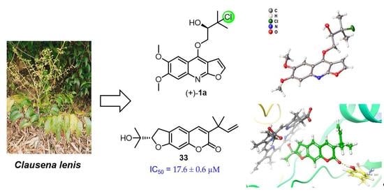
Anti-Neuroinflammatory Components from Clausena lenis Drake

State Key Laboratory of Natural and Biomimetic Drugs, School of Pharmaceutical Sciences, Peking University, No. 38 Xueyuan Road, Haidian District, Beijing 100191, China
*
Author to whom correspondence should be addressed.
Molecules 2022, 27(6), 1971; https://doi.org/10.3390/molecules27061971
Submission received: 15 February 2022 / Revised: 11 March 2022 / Accepted: 16 March 2022 / Published: 18 March 2022

Abstract

:
Clausena lenis Drake (C. lenis) is a folk medicinal herb to treat influenza, colds, bronchitis, and malaria. The 95% and 50% ethanol extract of C. lenis showed significant nitric oxide (NO) inhibition activity in BV-2 microglial cells stimulated by lipopolysaccharide (LPS). Bio-guided isolation of the active extract afforded five new compounds, including a chlorine-containing furoquinoline racemate, (±)-claulenine A (1), an amide alkaloid, claulenine B (2), a prenylated coumarin, claulenin A (3), a furocoumarin glucoside, clauleside A (4), and a multi-prenylated p-hydroxybenzaldehyde, claulenin B (5), along with 33 known ones. Their structures were determined via spectroscopic methods, and the absolute configurations of new compounds were assigned via the electronic circular dichroism (ECD) calculations and single-crystal X-ray diffraction analysis. Compounds 2, 23, 27, 28, 33, and 34 showed potent anti-neuroinflammatory effects on LPS-induced NO production in BV-2 microglial cells, with IC50 values in the range of 17.6–40.9 μM. The possible mechanism was deduced to interact with iNOS through molecular docking.

Graphical Abstract

1. Introduction

Neuroinflammation generally refers to an inflammatory response in the brain or spinal cord and has a pivotal role in the pathogenesis of neurodegenerative diseases, such as Alzheimer’s disease (AD) [1] and Parkinson’s disease (PD) [2,3]. This inflammation is mediated by cytokines, chemokines, reactive oxygen species, and secondary messengers produced by resident central glial cells (microglia and astrocytes), endothelial cells, and immune cells of peripheral origin [4]. Among them, microglia, as innate immune cells of the central nervous system, are major participants in neuroinflammation [1] and have various neuroimmunological functions in the central nervous system under normal and pathological conditions [5].
Phytochemicals, as the main components of natural products, have a variety of pharmacological effects, including anti-inflammation, neuroprotection, anti-cancer, and metabolism regulation [6,7,8,9]. In recent years, some compounds derived from Clausena species have been revealed to present potential anti-neuroinflammatory activities by inhibiting the nitric oxide (NO) production in lipopolysaccharide (LPS)-induced BV-2 microglial cells [10,11,12].
Clausena lenis Drake (C. lenis) belongs to the genus Clausena of Rutaceae family, distributed mainly in the Hainan, southern Guangxi, and Yunnan provinces of China [13]. It has been used as a folk medicine for the treatment of influenza, colds, bronchitis, and malaria. Alkaloids and coumarins are preliminarily verified to be its main constituents [14,15,16,17,18,19]. During our search for anti-neuroinflammatory components from Clausena and its closely related Murraya species [20,21,22], the 95% and 50% ethanol extract of C. lenis showed significant NO inhibition activity in BV-2 microglial cells stimulated by LPS with 73.5% inhibition rate at 80 ug/mL. To trace the potential anti-neuroinflammatory compounds, the ethanol extract of C. lenis was chemically investigated to afford 38 compounds, including five new compounds, namely (±)-claulenine A (1), claulenine B (2), claulenin A (3), clauleside A (4), and claulenin B (5) (Figure 1). (±)-Claulenine A (1) is a racemate of furoquinoline containing chlorine, which is rare in phytochemicals. Herein, the isolation and structure elucidation of the new compounds and the inhibitory effects on LPS-induced NO of the isolates were described. Moreover, the interactions between the bioactive compounds and iNOS via molecular docking were also reported.

2. Results

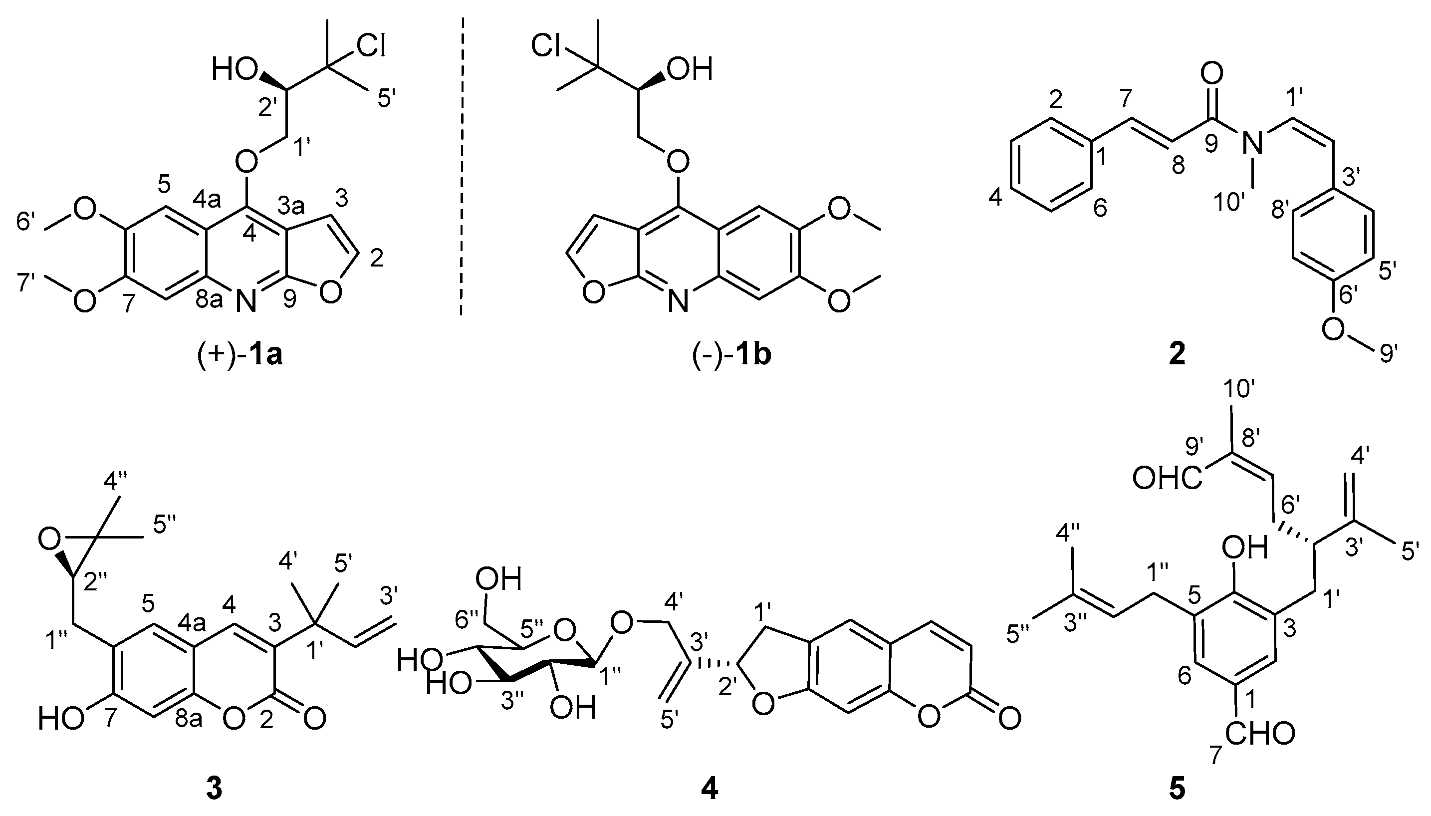
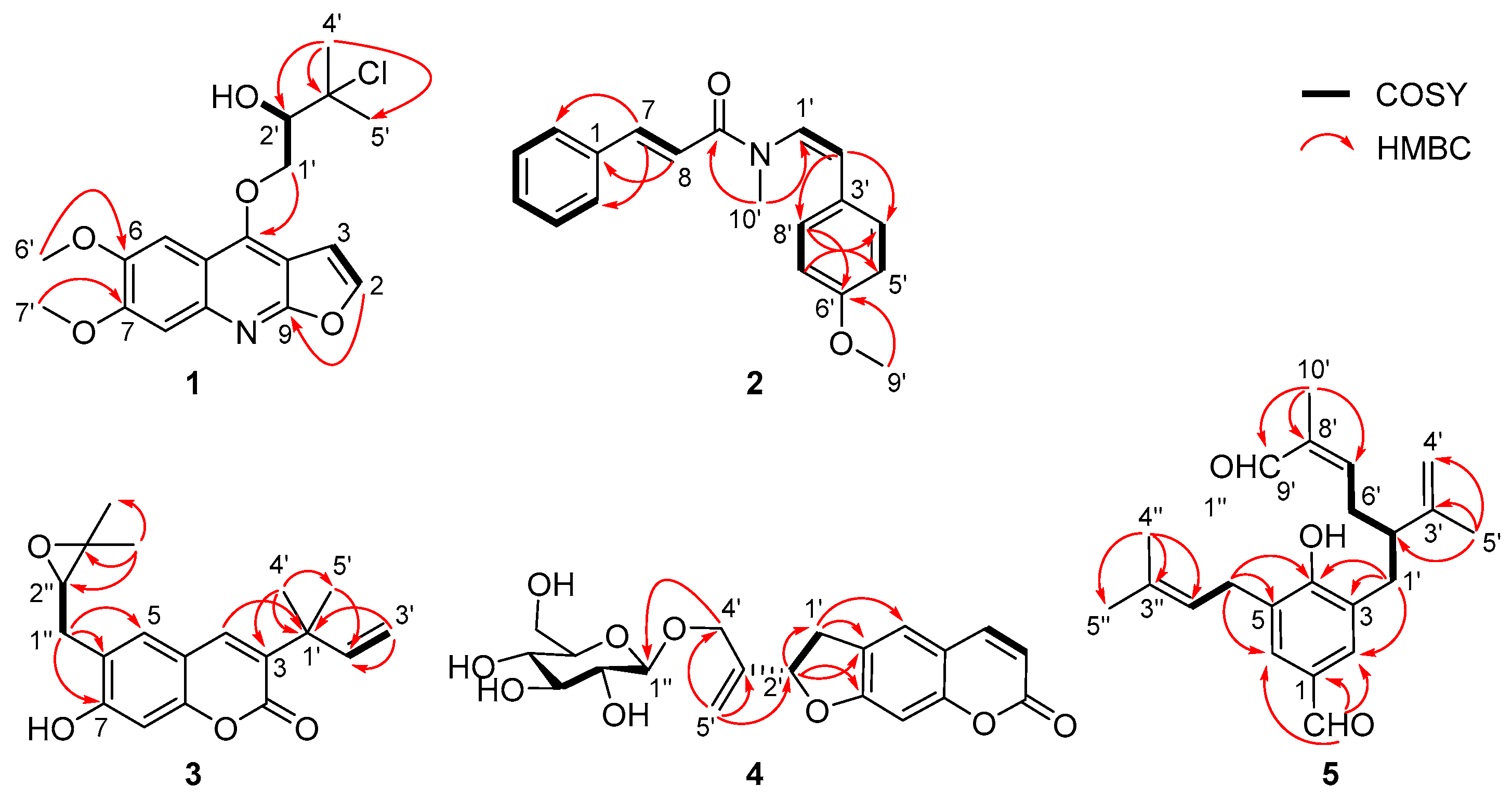
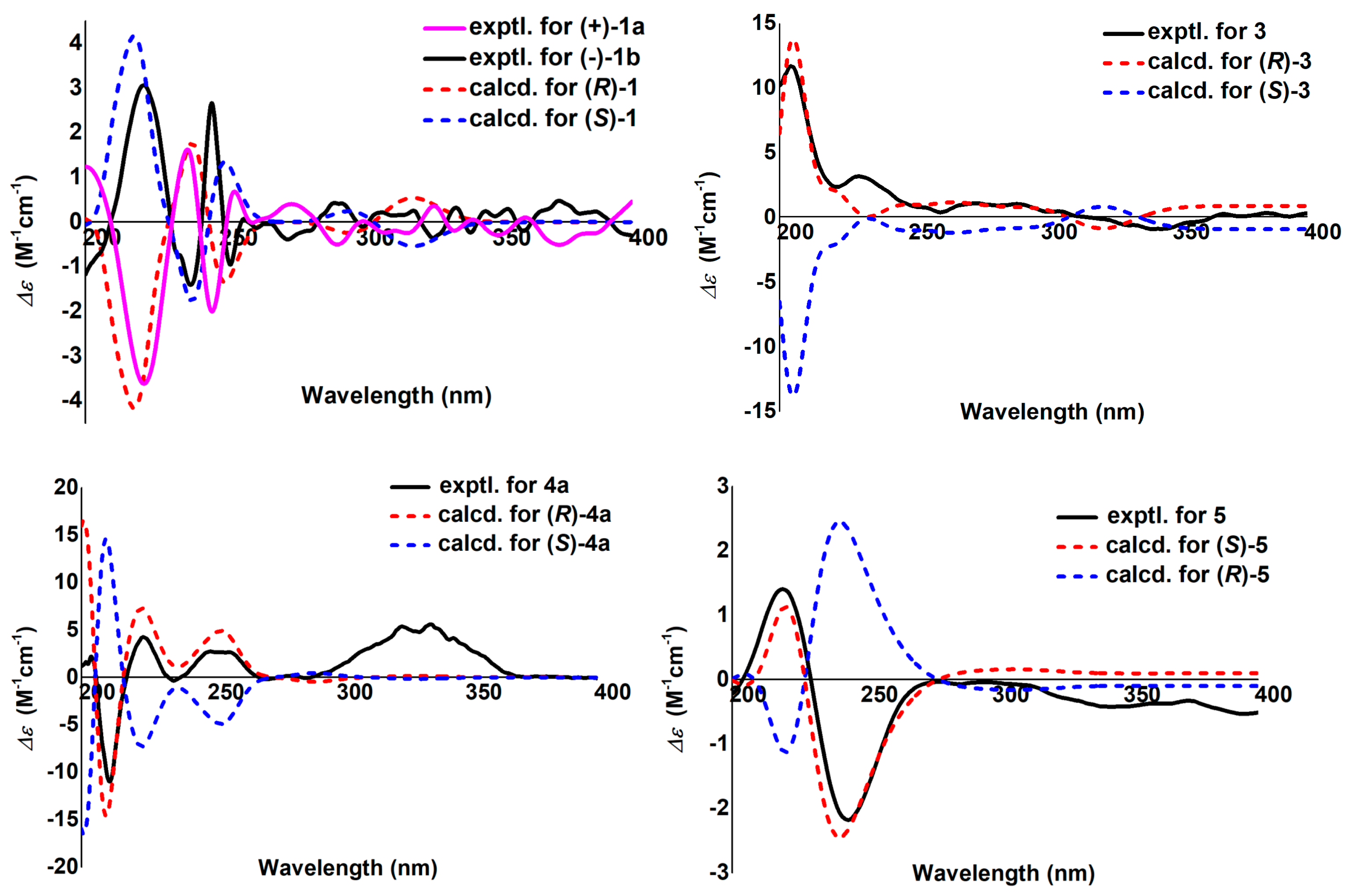
2.1. Structural Elucidation

(±)-Claulenine A (1) was obtained as a white solid, [ α ] D 25 0 (c 0.12, MeOH). The HR-ESI-MS data indicated the presence of one chlorine in the molecule from the relative abundance (∼1/3) of the isotope peaks observed at m/z 366.1101 and 368.1077, corresponding to a molecular formula of C18H20NO5Cl (calcd for C18H21NO5Cl, 366.1108; mass error −1.9 ppm), with nine degrees of unsaturation. Analysis of the 1H-NMR data (Table 1) revealed the presence of a trisubstituted furoquinoline, which was indicated by the typical adjacent protons of a furan moiety at δH 7.59 (1H, d, J = 2.7 Hz, H-2) and 7.00 (1H, d, J = 2.7 Hz, H-3) [23] and two aromatic singlet signals at δH 7.45 and 7.34. Meanwhile, two methoxys (δH 4.03 and 4.00), two methyls (both δH 1.77), an oxymethylene [δH 4.93 (1H, dd, J = 9.8, 3.7 Hz) and 4.75 (1H, dd, J = 9.8, 6.8 Hz)], and an oxymethine [δH 4.18 (1H, dd, J = 6.8, 3.7 Hz)] signals were also observed in the 1H NMR data (Table 1). In the 13C-NMR data of 1, there were 18 carbon resonances, comprising two methyls, two methoxyls, one methylene, five methines (four olefinic and one oxygenated aliphatic), and eight quaternary carbons, which were very similar to those of (2S)-1-[(6,7-dimethoxyfuro[2,3-b]quinolin-4-yl)oxy]-3-methyl-butane-2,3-diol [24]. Considering the molecular formula of 1, one of the two hydroxy groups of (2S)-1-[(6,7-dimethoxyfuro[2,3-b]quinolin-4-yl)oxy]-3-methyl-butane-2,3-diol was deduced to be substituted by a chlorine atom in 1. The above deductions were further confirmed by 2D NMR experiments (Figure 2). The two methoxy groups showed HMBC cross-peaks with the carbon at δC 148.1 and 152.8, respectively, locating them at positions C-6 and C-7; H2-1′ [δH 4.93 (dd, J = 9.8, 3.7 Hz) and 4.75 (dd, J = 9.8, 6.8 Hz)] presented an HMBC correlation to C-4 (δC 154.4), suggesting that the oxygenated prenyl unit was linked to C-4 of furoquinoline via an oxygen bridge. In addition, the chlorine at C-3′ and the hydroxy group at C-2′ were indicated by the key COSY correlations (Figure S11, Supporting Information) of –OH [δH 8.12 (1H, d, J = 6.2 Hz)], H-2′ [δH 4.58 (1H, ddd, J = 7.4, 6.2, 2.8 Hz)], and H-1′ [δH 5.35 (1H, dd, J = 9.6, 2.8 Hz), 5.11 (1H, dd, J = 9.6, 7.4 Hz)] in C5D5N. Thus, the planar structure of 1 was established as shown (Figure 1) and denominated as claulenine A.
However, 1 was isolated as a raceme indicated by its zero specific rotation value and no Cotton effects in the electronic circular dichroism (ECD) spectrum. Further HPLC separation on chiral phase afforded the enantiomers of (+)-1a and (–)-1b (Figure S14, Supporting Information). To clarify their absolute configurations, ECD calculations were utilized, and the results showed that the ECD spectrum of (R)-1 matched well with the experimental curve of (+)-1a (Figure 3). This conclusion was finally confirmed by the single-crystal X-ray diffraction for (+)-1a using Cu Kα radiation (Figure 4). Considering that there are fewer phytochemicals containing chlorine atoms, a freshly prepared methanol extract of the title plant was detected by using the multiple reaction monitoring (MRM) mode of UPLC/Qtrap-MS/MS, and the results suggested that 1 comes from natural source (Figure S15, Supporting Information).
Claulenine B (2) was obtained as a yellow oil, and its molecular formula was determined as C19H19NO2 from the HR-ESI-MS ion at m/z 294.1490 [M + H]+ (calcd for C19H20NO2, 294.1494; mass error −1.4 ppm) and 13C-NMR data. In the 1H NMR spectrum, except for the up-field shifts at δH 3.78 (–OCH3) and 3.10 (–NCH3), the remaining signals were in the aromatic region (δH 6.2–7.7). The 13C NMR data displayed 19 carbons including a signal of amide (δC 166.7). Careful analysis of 1H and 13C NMR data revealed that the signals of 2 resembled those of anhydromarmeline [25], except that the prenyl moiety was replaced by a methoxy group and an additional methyl positioned at the nitrogen atom in 2. The HMBC correlations (Figure 2) of –OCH3/C-6′ (δC 159.5) and –NCH3/C-1′ (δC 127.1), C-9 (δC 166.7) supported the deduction. Besides, the coupling constant of olefinic protons (J = 15.5 Hz for H-7 and H-8; J = 8.6 Hz for H-1′ and H-2′) pointed out that they were E- and Z-oriented, respectively. Hence, the gross structure of claulenine B (2) was depicted as given (Figure 1).
Claulenin A (3), a yellow oil, gave a molecular formula of C19H22O4 (nine degrees of unsaturation), as established by the [M + H]+ ion at m/z 315.1594 (calcd for C19H23O4, 315.1596; mass error −0.6 ppm) in the HR-ESI-MS. The UV spectrum appeared maximum absorptions at 335, 298, 250, and 227 nm, which is typical of the coumarin nucleus [26]. The 1H NMR data of 3 (Table 1) exhibited four methyl singlets (δH 1.22, 1.35, 1.46, and 1.46) and three aromatic/olefinic protons [δH 7.47 (s), 7.19 (s), and 6.70 (s)]. The 13C-NMR and HSQC spectra displayed 19 carbons classified into four methyls, two methylenes (one aliphatic and one olefinic), five methines (one oxygenated aliphatic and four olefinic), and eight quaternary (one ester carbonyl, two aliphatic, and five olefinic, including two oxygenated) carbons. The 1H and 13C-NMR data of 3 bore a close resemblance to those of gravelliferone A [19], except that the methoxyl at C-7 in gravelliferone A was replaced by a hydroxy in 3, which was supported by the molecular formula of 3 and the chemical shift of C-7 (δC 162.4). Therefore, the planar structure of 3 was deduced.
To clarify the absolute configuration of the only chiral center in 3, ECD calculation was performed. The ECD experimental curve was in good agreement with the calculated one of (R)-3 (Figure 3). Taken together, the structure of 3 was finally assigned and named claulenin A.
Clauleside A (4) exhibited an [M − H] ion at m/z 405.1187 in the HR-ESI-MS, corresponding to a molecular formula of C20H22O9 (calcd for C20H21O9, 405.1186; mass error 0.2 ppm). The UV spectrum is similar to that of 3, indicating that 4 is also a coumarin derivative. Inspection of 1H-NMR and 13C-NMR data (Table 1) found a set of characteristic glucosyl signals [δH 4.23 (1H, d, J = 7.8 Hz), 3.81 (1H, d, J = 11.9 Hz), 3.60 (1H, dd, J = 11.9, 4.9 Hz), 3.16–3.38 (4H, m); δC 103.6, 78.0, 78.0, 75.0, 71.6, 62.7], which identified 4 as a coumarin glucoside. Further analysis of NMR data of the aglycone moiety showed that they were basically consistent with those of scataccanol [27]. The HMBC correlations (Figure 2) of H2-4′ [δH 4.44 (1H, d, J = 12.8 Hz), 4.19 (1H, d, J = 12.8 Hz)] to C-1″ (δC 103.6) deduced the linkage of the glucosyl moiety at the C-4′.
The β-d-glucose was demonstrated via analysis of the coupling constant (7.8 Hz) of the anomeric proton [28] and the aryl thiocarbamate derivate of the hydrolyte by HPLC (Figure S40, Supporting Information). The absolute configuration of aglycone moiety (4a) was assigned as (2′R) by comparison of the experimental and calculated ECD curves (Figure 3). Finally, 4 was established as scataccanol 4′-O-β-d-glucopyranoside and referred to as clauleside A.
Claulenin B (5) was isolated as a yellow oil. Its positive-ion HR-ESI-MS data at m/z 363.1933 [M + Na]+ (calcd for C22H28O3Na, 363.1936; mass error −0.8 ppm) established a molecular formula of C22H28O3, with nine indices of hydrogen deficiency. The 1H NMR data (Table 1) of 5 showed signals of two aromatic protons [δH 7.52 (2H, br s)] and four methyls [δH 1.70, 1.71, 1.82, and 1.84 (each 3H, s)]. Moreover, the 13C-NMR (Table 1) and HSQC (Figure S47, Supporting Information) spectra revealed 22 carbon signals, including two aldehyde-carbonyls (δC 195.4 and 191.4), seven aromatic/olefinic quaternary carbons, five methines (one aliphatic and four olefinic), four methylenes (three aliphatic and one olefinic), and four methyls. The presence of three prenyl derivative units in 5 was illustrated through the HMBC correlations (Figure 2) of H3-5′ to C-2′ (δC 46.4), C-3′ (δC 146.2), C-4′ (δC 112.8), H3-10′ to C-7′ (δC 153.2), C-8′ (δC 139.7), C-9′ (δC 195.4), H3-4″ to C-2″ (δC 120.5), C-3″ (δC 137.7), C-5″ (δC 18.2), and the COSY correlations (Figure 2) of H2-1′/H-2′, H2-6′/H-7′, and H2-1″/H-2″ (δH 5.30). In addition, the COSY correlation of H2-6′/H-2′ indicated that the C-6′ of one prenyl unit was connected to another prenyl unit at C-2′, forming a geranyl unit. An aldehyde group at C-9′ and a terminal double bond at C-3′ and C-4′ were further demonstrated by the HMBC correlations of –CHO (δH 9.33, s) to C-8′, C-10′ (δC 9.5) and H2-4′ (δH 4.78 and 4.70, both s) to C-2′, C-5′ (δC 19.4). The geranyl and the prenyl units were determined to be located at the C-3 and C-5 of p-hydroxybenzaldehyde, respectively, from the HMBC correlations of H2-1′ to C-2 (δC 131.4), C-3 (δC 127.9), C-4 (δC 158.9), H2-1″ to C-4, C-5 (δC 126.6), C-6 (δC 130.7), and –CHO (δH 9.82, s) to C-1 (δC 129.5), C-2, C-6. In summary, the planar structure of 5 was depicted as shown (Figure 1).
The absolute configuration of C-2′ was determined by the ECD calculation. The results (Figure 3) shown that the calculated ECD spectrum of (2′S)-5 agrees well with the experimental one, which allowed the absolute configuration of 5 to be specified as 2′S. As a result, the structure of claulenin B (5) was clarified.
Thirty-two known compounds were identified as 4-methoxy-N-methyl-2-quinolone (6) [29], (–)-(S)-edulinine (7) [30], dictamine (8) [31], pteleine (9) [32], cinnamamide (10) [33], N-methylcinnamamide (11) [34], (Z)-N-methyl-3-phenylacrylamide (12) [35], N-2-phenylethylcinnamamide (13) [36], lansiumamide A (14) [37], lansiumamide B (15) [37], (2E)-3-phenyl-N-[(E)-2-phenylvinyl]acrylamide (16) [38], lansamide I (17) [39], 2-benzothiazolol (18) [40], indole (19) [41], N,N′-bis[2(1H-indol-3-yl)ethyl]urea (20) [42], demethylsuberosin (21) [43], swietenocoumarin I (22) [44], xanthyletin (23) [45], 3’-hydroxyxanthyletn (24) [46], (–)-3-(R)-decursinol (25) [47], dimethyl allyl xanthyletin (26) [48], 3-(1,1-dimethylallyl)decursinol (27) [49,50], imperatorin (28) [51], phellopterin (29) [52], (–)-heraclenol (30) [53,54], (+)-isoangenomalin (31) [55], nodakenetin (32) [56], chalepin (33) [57], 6-methoxymicrominutinin (34) [58], marmesinin (35) [59], marmesin glycoside (36) [60], (2R)-2′-hydroxymarmesin (37) [61], and (4′-methyl-[1,1′-biphenyl]-2-yl)(p-tolyl)methanone (38) [62], by comparing their spectroscopic data with those reported in the literature. The absolute configuration of 22 was first defined as S by comparing the calculated and experimental ECD spectra (Figure S51, Supporting Information). In addition, 20 and 38 were reported as natural products for the first time, and 710, 12, 16, 1821, 24, 25, 27, 29, 32, and 3438 and all of the isolates except 23, 26, 28, 31, and 33 were obtained for the first time from Clausena species and C. lenis, respectively.

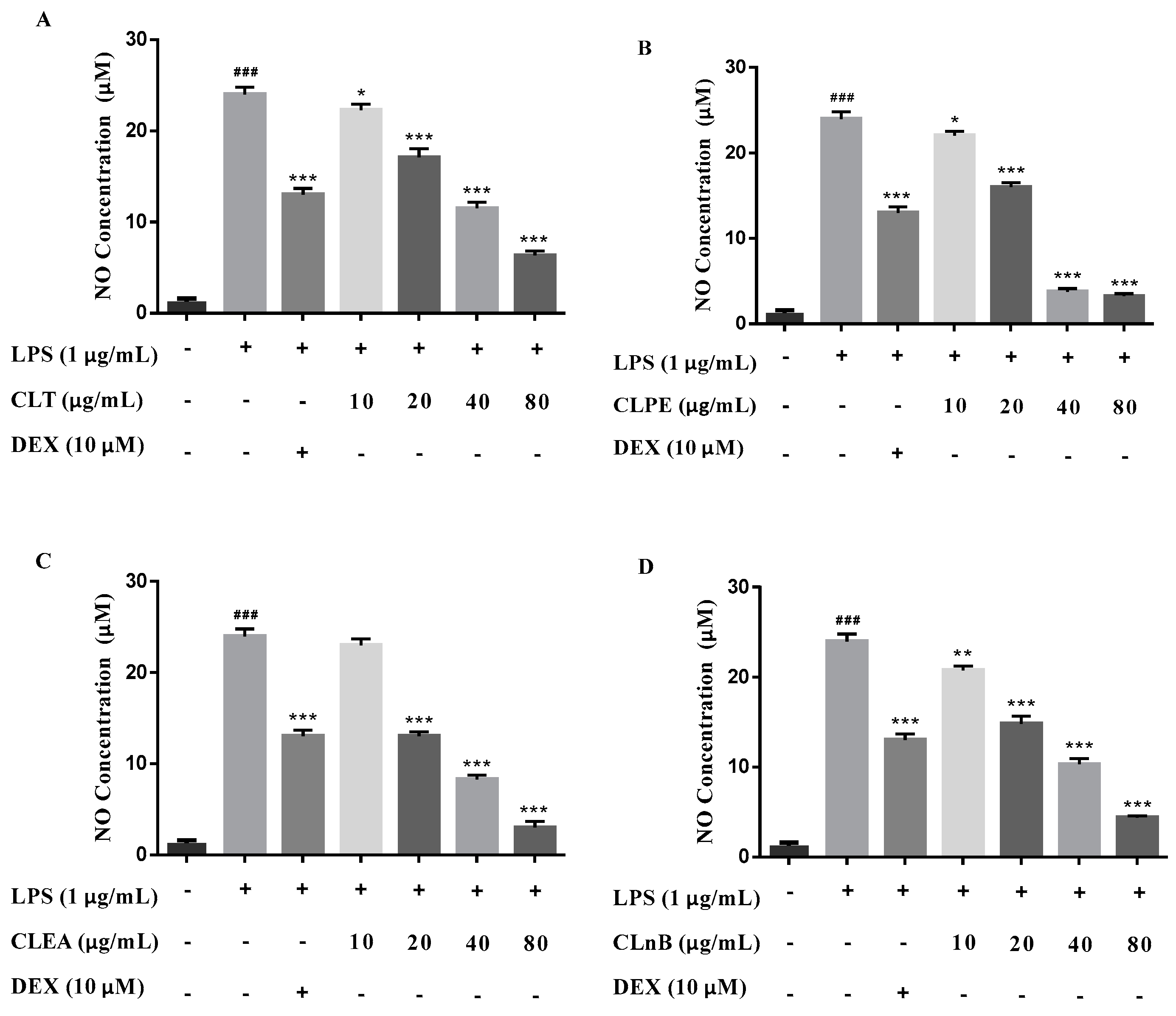
2.2. Anti-Neuroinflammatory Activities

The 95% and 50% ethanol extract (CLT) of C. lenis, along with its three partitioned extracts with different polarity solvents, i.e., petroleum ether extract (CLPE), ethyl acetate extract (CLEA), and n-BuOH extract (CLnB) were evaluated for anti-neuroinflammatory activities based on reduction of NO production stimulated by LPS in BV-2 microglial cells. The results exhibited that NO production can be dose-dependently inhibited in the range of 10–80 μg/mL (Figure 5). To further investigate which compounds are responsible for the effect, all of these isolates (138) were subjected to an evaluation of their anti-neuroinflammatory activities using the same method. Six compounds (2, 23, 27, 28, 33, and 34) inhibited NO production by more than 50% at 50 μM and their IC50 values were finally determined to be from 17.6 to 40.9 μM (Table 2). Dexamethasone (DEX) was used as a positive control. Alongside this, no cytotoxicity was observed in BV-2 microglial cells treated by these test subjects at 50 μM (cell viability > 95%).

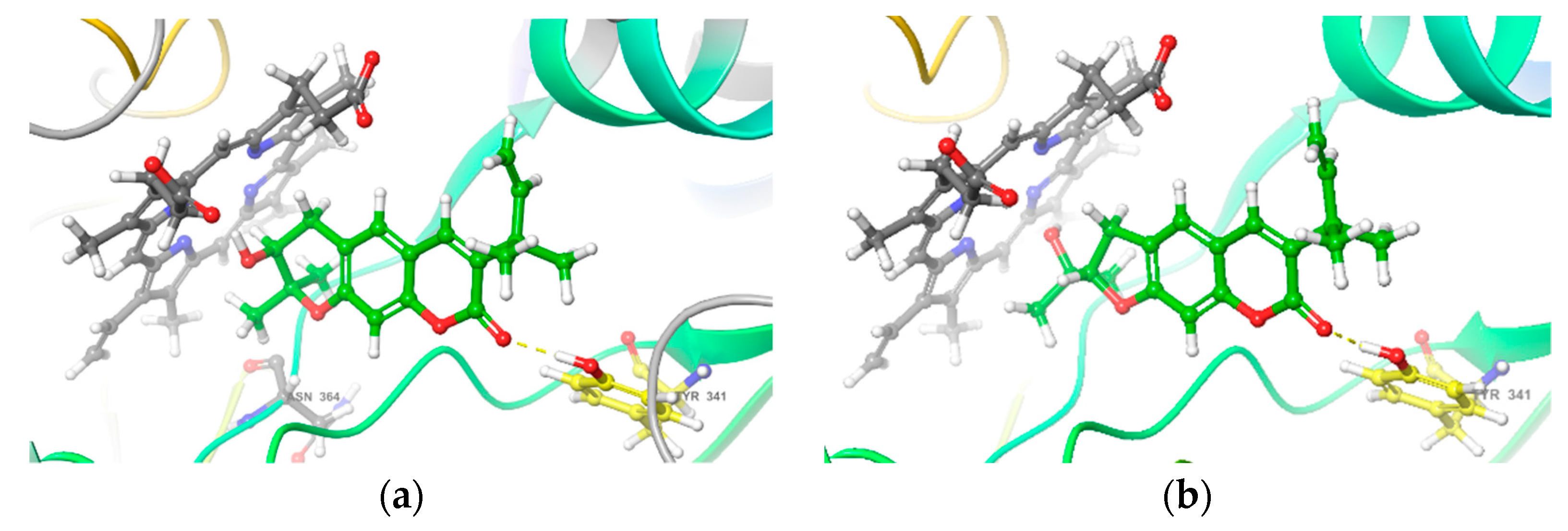
2.3. Interactions of Bioactive Compounds with iNOS

Nitric oxide synthases (NOSs) catalyze the NO production using oxygen and nitrogen derived from arginine. Inducible NOS (iNOS) is one of the isoenzymes of NOSs and plays a major part in NO production during inflammation [63]. In recent years, structure-based calculations have been widely used to predict the pharmacological mechanisms of active compounds, among which molecular docking is a commonly used method [64]. To explore the possible mechanism of those bioactive compounds against NO production, we investigated the interaction between iNOS and compounds 2, 23, 27, 28, 33, and 34 by molecular docking [65,66]. The results showed that compounds 27 and 33 had good affinities with iNOS (Glide Score < −5) (Table 3), and both of them have a hydrogen bond with the residues TYR341 of iNOS (Figure 6). Therefore, the possible mechanism of NO inhibition of 27 and 33 is through interaction with iNOS by targeting the residues in the active pocket of iNOS.

3. Discussion

Thirty-seven compounds were isolated and identified from C. lenis and five of them are new ones (15). The structures were elucidated based on MS, UV, IR, and NMR spectroscopic data and comparison with the data reported in literature. The absolute configurations of new compounds were characterized by using the ECD calculations and single-crystal X-ray diffraction analysis. (±)-Claulenine A (1) is a chlorine-containing furoquinoline racemate. Chlorinated natural products are rare in terrestrial plants, but common in bacteria, marine animals, and macroalgae [67,68]. 1 could be an artifact produced in the isolation procedure, but could also be generated by Fe(II)- and 2-oxoglutarate-dependent halogenases (2ODHs) [69]. The LC/MS analysis of the fresh C. lenis proved 1 to be originated from a natural source, but the real chlorine source is not clear. Claulenine B (2) is a high conjugated amide alkaloid. Claulenin A (3) is a prenylated coumarin with an oxirane ring and clauleside A (4) is a furocoumarin glucoside. Claulenin B (5) is a p-hydroxybenzaldehyde with multi-prenyl substituents. Of the known compounds, 20 and 38 are the first reported natural products, and 20 and 28 compounds are obtained from Clausena species and C. lenis, respectively, for the first time. Most of these compounds are prenylated, so the prenyltransferases play an important role in their biosyntheses.
The ethanol extract and its partitioned extracts with different polarities from C. lenis have significant inhibitory effects on NO production in LPS-induced BV-2 microglial cells. In the subsequent bioactivity-guided fractionation, compounds 2, 23, 27, 28, 33, and 34 were disclosed to be the potentially active compounds with inhibition effects. Most of the bioactive compounds are coumarins, suggesting that coumarins might be the main bioactive substances for the anti-neuroinflammatory properties of the extract of C. lenis. Furthermore, the molecular docking results revealed that 27 and 33 had a good interaction with iNOS, which could be one of the mechanisms for their anti-inflammation effects.

4. Materials and Methods

4.1. General Experimental Procedures

Optical rotations were measured on a Rudolph Autopol IV automatic polarimeter (Rudolph Research Analytical, Hackettstown, NJ, USA). The electronic circular dichroism (ECD) and UV data were acquired on a JASCO, J-1500 CD spectrophotometer (JASCO, Tokyo, Japan). IR spectra were recorded on a Thermo Scientific Nicolet iS50 FT-IR spectrometer (Thermo Fisher Scientific, Waltham, MA, USA). HR-ESI-MS data were measured on a Waters Xevo G2 Q-TOF mass spectrometer (Waters Co., Milford, MA, USA). The nuclear magnetic resonance (NMR) spectra were obtained through a Varian INOVA-500 NMR spectrometer (Varian Co., Palo Alto, CA, USA) with TMS as an internal standard. The column chromatography (CC) was undertaken on silica gel (100−200 and 200−300 mesh, Qingdao Marine Chemical Co., Ltd., Qingdao, China), ODS-A-HG (50 µm; YMC Co., Ltd., Kyoto, Japan), and Sephadex LH-20 (Amersham Pharmacia, Uppsala, Sweden). Preparative TLC and TLC analyses were carried out on the pre-coated silica gel GF254 plates (Qingdao Marine Chemical Co., Ltd., Qingdao, China). Semi-preparative HPLC was carried out using an Agilent Eclipse XDB-C18 column (9.4 mm × 250 mm, i.d., 5 μm) on an Agilent 1260 series LC instrument with a DAD detector (Agilent Technologies, Santa Clara, CA, USA).

4.2. Plant Material

In November 2019, dry leaves and stems of Clausena lenis Drake were collected from Baoting County, Hainan Province, People’s Republic of China. Botanical identification was made by Prof. Pengfei Tu, one of the authors, and a voucher specimen (No. CL201911) was kept in the herbarium of Modern Research Center for Traditional Chinese Medicine, Peking University.

4.3. Extraction and Isolation

The air-dried leaves and stems of C. lenis (20.0 kg) were refluxed with 95% and 50% aqueous ethanol (160 L × 2 h × 2), respectively, and concentrated under reduced pressure to obtain 1.8 kg dry total extract (CLT). The extract was suspended in H2O and extracted with petroleum ether (CLPE), ethyl acetate (CLEA), and n-BuOH (CLnB), successively.
The petroleum ether extract (179.1 g) was eluted by gradient elution of petroleum ether-ethyl acetate (1:0, 50:1, 30:1, 20:1, 10:1, 5:1, 3:1, 1:1, and 0:1, v/v) on a silica gel column to obtain 14 fractions (A−N). Fraction J (1.83 g) was subjected to Sephadex LH-20 (MeOH–CH2Cl2, 1:1, v/v) and produced five subfractions (J.1−J.5). Subfractions J.3 (0.70 g) and J.4 (0.27 g) were purified by semi-preparative HPLC (3.0 mL/min) to yield 2 (4.2 mg, 47% aqueous acetonitrile, tR 29.2 min) and 9 (3.8 mg, 40% aqueous acetonitrile, tR 15.6 min), respectively. Fraction K (14.2 g) was divided into seven fractions (K.1–K.7) by a ODS-A-HG column (aqueous MeOH, 30%–100%). Compounds 33 (26.6 mg, tR 36.7 min) and 27 (4.5 mg, tR 53.8 min) were obtained from subfraction K.4 (780 mg) after purification by semi-preparative HPLC (3.0 mL/min, 42% aqueous acetonitrile). Similarly, 34 (2.8 mg, tR 16.9 min) was purified from subfraction K.2 (50 mg) by semi-preparative HPLC (3.0 mL/min, 35% aqueous acetonitrile). Fraction L (3.42 g) was fractionated into four parts (L.1–L.4) by gel filtration on Sephadex LH-20 (MeOH–CH2Cl2, 1:1, v/v). Subfraction L.2 (1.85 g) was subjected to an ODS-A-HG column (aqueous MeOH, 25%–100%) to give eight fractions (L.2.1–L.2.8). Subfractions L.2.1 (121.2 mg) and L.2.5 (83.4 mg) were further purified by semi-preparative HPLC (3.0 mL/min) to yield 12 (3.2 mg, 15% aqueous acetonitrile, tR 21.8 min) and 22 (6.0 mg, 35% aqueous acetonitrile, tR 39.9 min), respectively. Likewise, subfraction L.2.3 (131.4 mg) was purified to yield 32 (27.2 mg, 25% aqueous acetonitrile, tR 15.7 min) and 25 (3.8 mg, 25% aqueous acetonitrile, tR 18.4 min).
The ethyl acetate extract (160.9 g) was chromatographed on silica gel eluted with petroleum ether-ethyl acetate (1:0, 50:1, 30:1, 20:1, 10:1, 5:1, 3:1, 1:1, and 0:1, v/v) to afford 15 fractions (A−O). Fraction C (0.29 g) was fractionated on a Sephadex LH-20 column (MeOH–CH2Cl2, 1:1, v/v) to yield five parts (C.1–C.5). Subfraction C.3 (89.4 mg) was purified by semi-preparative HPLC (3.0 mL/min, 60% aqueous acetonitrile) to obtain 38 (1.1 mg, tR 44.8 min) and 26 (5.1 mg, tR 48.5 min). Fraction F (9.63 g) was divided into five parts (F.1–F.5) by an MCI GEL CHP20 column (aqueous MeOH, 50%–100%). Subfraction F.1 (129 mg) was purified by semi-preparative HPLC (3.0 mL/min, 28% aqueous acetonitrile) to yield 10 (2.7 mg, tR 12.7 min) and 11 (17.0 mg, tR 14.9 min). Subfraction F.3 (7.09 g) was applied to an ODS-A-HG column (aqueous MeOH, 10%–100%) and further purified by preparative HPLC (3.0 mL/min, 60% aqueous acetonitrile) to yield 14 (6.3 mg, tR 14.4 min) and 17 (143.5 mg, tR 21.4 min). Fraction G (7.6 g) was submitted to Sephadex LH-20 (MeOH) to acquire seven portions (G.1–G.7). Subfraction G.4 (92.3 mg) was purified by semi-preparative HPLC (50% aqueous acetonitrile, 3 mL/min) to yield 23 (2.2 mg, tR 15.8 mind). Fraction H (4.27 g) was subjected to Sephadex LH-20 (MeOH–CH2Cl2, 1:1, v/v) to afford three fractions. Subfraction H.3 (130 mg) was purified by semi-preparative HPLC (3.0 mL/min, 50% aqueous acetonitrile) to yield 15 (2.0 mg, tR 24.9 min). Fraction I (2.88 g) was separated into five fractions (I.1−I.5) using Sephadex LH-20 (MeOH). Subfraction I.3 (318 mg) was purified by semi-preparative HPLC (3.0 mL/min, 50% aqueous acetonitrile) to yield 31 (3.3 mg, tR 13.8 min), 28 (3.4 mg, tR 21.2 min), 29 (6.7 mg, tR 25.2 min), and 5 (7.8 mg, tR 50.5 min). In the same way, 8 (5.5 mg, 60% aqueous acetonitrile, tR 7.1 min) and 16 (6.1 mg, 60% aqueous acetonitrile, tR 10.0 min) were obtained from subfraction I.4 (325 mg). Subfraction I.5 (0.93 g) was applied to an ODS-A-HG column (aqueous MeOH, 50%–100%) and further purified by preparative HPLC (3.0 mL/min, 25% aqueous acetonitrile) to yield 18 (8.5 mg, tR 17.8 min). Fraction K (16.5 g) was fractionated by Sephadex LH-20 (MeOH–CH2Cl2, 1:1, v/v) to give five fractions. Subfraction K.3 (0.75 g) was separated into five fractions by an ODS-A-HG column (aqueous MeOH, 20%–100%). Subfraction K.3.2 (140 mg) was subjected to preparative TLC (petroleum ether-ethyl acetate, 3:1, v/v) and further purified by semi-preparative HPLC (3 mL/min, 22% aqueous acetonitrile) to afford 21 (1.9 mg, tR = 46.2 min). Fraction L (2.86 g) was subjected to Sephadex LH-20 (MeOH–CH2Cl2, 1:1, v/v) to afford five fractions. Subfraction L.3 (0.56 g) and L.4 (0.30 g) was submitted to an ODS-A-HG column (aqueous MeOH, 30%–100%) to yield eight and six fractions, respectively. And subfractions L.3.4 (107.6 mg) and L.4.3 (71.3 mg) were purified by semi-preparative HPLC (3.0 mL/min) to yield 13 (3.1 mg, tR 13.5 min, 50% aqueous acetonitrile) and 20 (0.5 mg, tR 30.6 min, 35% aqueous acetonitrile), respectively. Fraction M (17.1 g) was applied to a silica gel column, eluting with petroleum ether-ethyl acetate (10:1, 8:1, 5:1, 3:1, and 1:1, v/v) to obtain fractions from M.1 to M.8. Subfraction M.5 (1.7 g) was subjected to Sephadex LH-20 (MeOH–CH2Cl2, 1:1, v/v) to afford three fractions. Subfraction M.5.2 (1.26 g) was submitted to an ODS-A-HG column (aqueous MeOH, 10%–100%) to yield three fractions. Among them, subfraction M.5.2.2 (135.1 mg) was purified by semi-preparative HPLC (3.0 mL/min, 45% aqueous acetonitrile) to yield 3 (3.8 mg, tR 28.5 min).
The n-BuOH extract (222.8 g) was subjected to macroporous resin HP-20 (aqueous ethanol, 0%, 20%, 50%, 70%, and 95%) to provide five portions (A–E). Fraction C (27.3 g) was divided into nine parts (C.1–C.9) by an ODS-A-HG column (aqueous MeOH, 5%–100%). Subfraction C.4 (2.50 g) was separated into three fractions (C.4.1–C.4.3) using a Sephadex LH-20 column (MeOH–CH2Cl2, 1:1, v/v). Subfraction C.4.2 (1.44 g) was subjected to a silica gel column with a stepwise gradient of CH2Cl2–MeOH (20:1, 15:1, 10:1, 8:1, 5:1, 3:1, and 1:1) to produce nine fractions (C.4.2.1−C.4.2.9). Subfractions C.4.2.6 (210 mg) were purified by semi-preparative HPLC (3.0 mL/min) to yield 37 (2.8 mg, 15% aqueous acetonitrile, tR 12.8 min). Subfraction C.5 (2.77 g) was separated by a Sephadex LH-20 column (MeOH–CH2Cl2, 1:1, v/v) to obtain three fractions (C.5.1–C.5.3). Subfraction C.5.2 (1.64 g) was further purified by a silica gel column with a gradient of CH2Cl2–MeOH system (20:1, 10:1, 8:1, 5:1, 1:1, and 0:1) to get 35 (9.8 mg). In addition, subfraction C.5.3 (180 mg) was further purified by preparative TLC (CH2Cl2–MeOH, 8:1, v/v) and semi-preparative HPLC (3.0 mL/min, 20% aqueous acetonitrile) to yield 36 (1.8 mg, tR 27.3 min). Subfraction C.7 (2.36 g) was fractionated into four parts (C.7.1–C.7.4) by using Sephadex LH-20 (MeOH–CH2Cl2, 1:1, v/v). Subfraction C.7.2 (1.29 g) was eluted by gradient elution of CH2Cl2–MeOH (1:0, 100:1, 50:1, 20:1, 10:1, 8:1, 5:1, and 1:1, v/v) on a silica gel column to obtain nine fractions (C.7.2.1− C.7.2.9). Subfractions C.7.2.3 (13.5 mg) and C.7.2.5 (83.2 mg) were purified by semi-preparative HPLC (3.0 mL/min) to yield 24 (0.6 mg, 25% aqueous acetonitrile, tR 23.4 min) and 7 (3.2 mg, 20% aqueous acetonitrile, tR 37.8 min). Fraction D (30.5 g) was divided into eight parts (D.1–D.8) by an ODS-A-HG column (aqueous MeOH, 10%–100%). Subfraction D.4 (1.9 g) was fractionated into three parts (D.4.1–D.4.3) by using Sephadex LH-20 (MeOH–CH2Cl2, 1:1, v/v). Subfraction D.4.2 (1.03 g) was applied to a silica gel column (CH2Cl2–MeOH, 10:1, 5:1, 1:1, and 0:1, v/v) to produce seven subfractions (D.4.2.1−D.4.2.7). Purification of subfraction D.4.2.4 (130 mg) by semi-preparative HPLC with 16% aqueous acetonitrile (3.0 mL/min) afforded 4 (22.7 mg, tR 22.8 min). Subfraction D.5 (2.6 g) was subjected to a Sephadex LH-20 column (MeOH–CH2Cl2, 1:1, v/v) to afford three fractions (D.5.1−D.5.3). Subfraction D.5.2 (1.07 g) was applied to a silica gel column, eluting with CH2Cl2–MeOH (100:1, 50:1, 20:1, 10:1, 5:1, 1:1, and 0:1, v/v), to give seven fractions (D5.2.1−D.5.2.7). Subfraction D.5.2.1 (90 mg) was further purified by semi-preparative HPLC (3.0 mL/min, 18% aqueous acetonitrile) to afford 30 (1.0 mg, tR 45.4 min). Fraction E (3.3 g) was fractionated into five parts (E.1–E.5) by gel filtration on Sephadex LH-20 (MeOH–CH2Cl2, 1:1, v/v). Subfraction E.2 (1.72 g) was subjected to a silica gel column (petroleum ether–ethyl acetate, 5:1, 3:1, 2:1, 1:1, and 0:1, v/v) to give nine fractions (E.2.1–E.2.9). Subfraction E.2.8 (245.1 mg) was further purified by semi-preparative HPLC (3.0 mL/min, 50% aqueous acetonitrile) to yield 6 (1.6 mg, tR 9.0 min) and 1 (4.1 mg, tR 17.1 min). The chiral HPLC separation of racemic 1 was performed using a Chiralpak IA column (10 × 250 mm, 5 mm, Daicel, Nanning, China), eluting with n-hexane-isopropanol (85:15, v/v). The detection wavelength was 254 nm, the column temperature was 30 °C, and the flow rate was 3 mL/min. Finally, compounds (+)-1a (1.9 mg, tR 18.7 min) and (−)-1b (1.8 mg, tR 22.7 min) were obtained. Subfraction E.3 (0.49 g) was subjected to a silica gel column (petroleum ether–ethyl acetate, 1:0, 50:1, 30:1, 10:1, 5:1, 3:1, and 1:1, v/v) to give 19 (5.5 mg).

4.3.1. (±)-Claulenine A (1)

White solid; [ α ] D 25 0 (c 0.12, MeOH); UV (MeOH) λmax (log ε) 337 (3.54), 321 (3.82), 309 (3.68), 247 (4.46) nm; IR (KBr) νmax 2932, 1622, 1589, 1507, 1482, 1433, 1368, 1260, 1215, 1089, 1011 cm−1; 1H and 13C-NMR data, see Table 1; HR-ESI-MS m/z 366.1101 [M + H]+ (calcd for C18H21NO5Cl, 366.1108).
(+)-Claulenine A [(+)-1a]: White solid, [ α ] D 25 + 38 (c 0.12, MeOH); ECD (MeOH) λmaxε) 246 (−2.00), 238 (+1.62), 222 (−3.61) nm.
(−)-Claulenine A [(−)-1b]: White solid, [ α ] D 25 − 42 (c 0.10, MeOH); ECD (MeOH) λmaxε) 247 (+2.44), 238 (−1.41), 223 (+2.99) nm.

4.3.2. Claulenine B (2)

Yellow oil; UV (MeOH) λmax (log ε) 290 (3.52), 267 (3.57), 219 (3.54), 200 (3.72) nm; IR (KBr) νmax 2932, 1653, 1607, 1510, 1451, 1371, 1253, 1172, 1086, 1032, 765 cm−1; 1H and 13C-NMR data, see Table 1; HR-ESI-MS m/z 294.1490 [M + H]+ (calcd for C19H20NO2, 294.1494).

4.3.3. Claulenin A (3)

Yellow oil; [ α ] D 25 − 25 (c 0.24, MeOH); ECD (MeOH) λmaxε) 232 (+3.12), 222 (+2.37), 206 (+11.31) nm; UV (MeOH) λmax (log ε) 335 (3.20), 298 (2.89), 250 (3.34), 227 (3.26), 202 (3.68) nm; IR (KBr) νmax 2967, 1715, 1626, 1581, 1490, 1268, 1164, 1131, 986, 784 cm−1; 1H and 13C-NMR data, see Table 1; HR-ESI-MS m/z 315.1594 [M + H]+ (calcd for C19H23O4, 315.1596).

4.3.4. Clauleside A (4)

Yellow solid; [ α ] D 25 + 27 (c 0.32, MeOH); ECD (MeOH) λmaxε) 336 (+10.20), 270 (+1.55), 252 (+8.75), 236 (+4.69), 229 (+9.56), 211 (−3.48) nm; UV (MeOH) λmax (log ε) 334 (3.93), 250 (3.79), 226 (3.94), 206 (4.20) nm; IR (KBr) νmax 3381, 2932, 2882, 1712, 1626, 1568, 1486, 1446, 1363, 1124, 1076, 1038, 823, 610 cm−1; 1H and 13C-NMR data, see Table 1; HR-ESI-MS m/z 405.1187 [M − H] (calcd for C20H21O9, 405.1186).

4.3.5. Claulenin B (5)

Yellow oil; [ α ] D 25 − 10 (c 0.20, MeOH); ECD (MeOH) λmaxε) 244 (−2.18), 219 (+1.40) nm; UV (MeOH) λmax (log ε) 282 (3.24), 229 (3.61), 211 (3.52) nm; IR (KBr) νmax 3421, 2935, 1687, 1602, 1442, 1378, 1291, 1125, 977, 896 cm−1; 1H and 13C NMR data, see Table 1; HR-ESI-MS m/z 363.1933 [M + Na]+ (calcd for C22H28O3Na, 363.1936).

4.3.6. (S)-Swietenocoumarin I (22)

Colorless oil; [ α ] D 25 + 30 (c 0.20, MeOH); ECD (MeOH) λmaxε) 226 (+5.80), 204 (−12.02) nm; ESI-MS m/z 347 [M + H]+.

4.4. ECD Calculations of 1, 3, 4a, 5, and 22

Conformation searches of 1, 3, 4a, 5, and 22 using molecular mechanics calculations were performed in Spartan 14 (Wavefunction Ind.) with MMFF force field with an energy window for acceptable conformers (ewindow) of 5 kcal/mol above the ground state. Then the predominant conformers were optimized by using the TDDFT method at B3LYP/6-31G(d) level in Gaussian 16 [70]. The optimized conformers were used for the ECD calculations, which were performed with Gaussian 16 at B3LYP/6-31+G(d) level. The solvent effects were taken into account by the polarizable-conductor calculation model (PCM, methanol as the solvent). The SpecDis v1.71 program was used to generate ECD calculation curves [71].

4.5. X-ray Crystallography of (+)-1a

The single crystals of (+)-1a were collected from methanol solution at room temperature. An Agilent Gemini E X-ray single-crystal diffractometer with an Oxford Cryostream cooler was used to collect the single crystal data with Cu Kα radiation at T = 174.7 K. The structure was solved with the direct method using SHELXS-97 and refined anisotropically by full-matrix least-squares on F2 using SHELXL-97. The H atoms were placed in calculated positions and refined using a riding model. The absolute configuration was determined by refinement of the Flack parameter based on resonant scattering of the light atoms.
Crystal data for (+)-1a: C18H20ClNO5 (M = 365.80 g/mol), monoclinic, space group P21, size 0.19 × 0.17 × 0.02 mm3, a = 11.1628(2) Å, b = 6.63600(10) Å, c = 11.6688(2) Å, α = 90°, β = 97.049(2)°, γ = 90°, V = 857.85(3) Å3, Z = 2, T = 100.00(10) K, μ(Cu Kα) = 2.230 mm−1, 3400 unique (Rint = 0.0736, Rsigma = 0.0267), which were used in all calculations. The final R1 was 0.0384 [I > 2σ(I)] and the final wR2 was 0.1060 (all data). Flack parameter −0.003(9). Crystallographic data for 1 have been filed with Cambridge Crystallographic Data Centre (CCDC, deposition number: CCDC 2129405). These data can be obtained free of charge from the CCDC via https://www.ccdc.cam.ac.uk/structures/ (accessed on 17 December 2021).

4.6. Absolute Configurations Determination of Sugar Moiety for 4

Compound 4 (1.0 mg) was hydrolyzed with 2 mol/L HCl (5 mL) for 5 h at 85 °C. The reaction product was extracted three times with CH2Cl2. After the aqueous layer was concentrated to dryness, 2 mL anhydrous pyridine containing 2.0 mg L-cysteine methyl ester hydrochloride was added and heated at 60 °C for 1 h. Subsequently, o-tolylisothiocyanate (10 μL) was added and heated at 60 °C for 1 h. D-glucose standard (2 mg) and L-glucose standard (2 mg) were respectively reacted in the same procedure. Then, each reaction mixture was filtered by a 0.22 μm membrane and analyzed directly by an Agilent Extended C18 column (250 mm × 4.6 mm, 5 μm) on an Agilent 1260 HPLC with a gradient elution of MeCN–H2O (5:95–30:70, v/v, 0–30 min, 1.0 mL/min) at 30 °C, and UV detection wavelength was 210 nm (Figure S40, Supporting Information). The sugar moiety of 4 was detected as D-glucose by the same tR value with that of the D-glucose standard derivative [72].

4.7. Cell Culture

Mouse BV-2 microglial cells (Peking Union Medical College Cell Bank, Beijing, China) were cultured in Dulbecco’s Modified Eagle’s Medium (Macgene, Beijing, China) and supplemented with 10% fetal bovine serum (Gibico, Waltham, MA, USA), penicillin (Macgene, 100 U/mL, Beijing, China), and streptomycin (Macgene, 100 μg/mL) in a humidified incubator containing 95% air and 5% CO2 at 37 °C.

4.8. Nitric Oxide (NO) Production Measurement and Cell Viability Assay

BV-2 cells (1 × 10⁵ cells/well) were cultured in 48-well plates and stimulated with 1.0 μg/mL LPS (Escherichia coli 0111:B4, Sigma, St. Louis, MO, USA) with or without test extracts or compounds at 37 °C for 24 h. The production of NO was tested using a commercial assay kit (Nanjing Jiancheng Bioengineering Institute, Nanjing, Jiangsu, China), according to the manufacturer’s instructions. Cell culture supernatants (160 μL) were reacted with 80 μL of the Griess reagent (1% sulfanilamide, 0.1% naphthylethylene diaminedihydrochloride, and 2% phosphoric acid) for 10 min in the dark at room temperature. The absorbance was measured at 540 nm using a microplate reader (Tecan Trading AG, Basel, Switzerland). The experiments were performed in triplicate, and the results are presented as the mean ± SD of three independent experiments. The cell viability was evaluated according to MTT assay. Dexamethasone was used as the positive control.

4.9. Molecular Docking

The crystal structure of iNOS (PDB ID: 3E6T) was obtained from the Protein Data Bank of RCSB (Research Collaboratory for Structural Bioinformatics). Docking simulations between bioactive compounds and iNOS were performed using the Maestro software suite 2015 (Schrodinger, New York, NY, USA). The ligand molecules were drawn with Chem3D Pro 14.0 (CambridgeSoft, Waltham, MA, USA) and optimized by the Ligprep module of Maestro. The protein receptor was prepared by deleting the ligand and water molecules and then was adopted for molecular docking with ligands. The reported inhibitor binding sites of iNOS was chosen as the binding pocket [73].

Supplementary Materials

The following supporting information can be downloaded at: https://www.mdpi.com/article/10.3390/molecules27061971/s1. HR-ESI-MS, UV, IR, 1D and 2D NMR spectra of 15. Chiral HPLC separation of racemic 1. LC/MS chromatograms of detection of 1 in the crude extract of C. lenis. HPLC-UV chromatograms of the derivatives of D-glucose, L-glucose, and the hydrolysate of 4. Structures of 638. ESI-MS and experimental and calculated ECD spectra of 22. These materials are available online.

Author Contributions

Conceptualization, K.-W.Z., P.-F.T. and Y.J.; methodology, S.-S.Z. and Y.J.; validation, S.-S.Z., Y.-F.Z. and M.D.; investigation, S.-S.Z.; writing, review, and editing, S.-S.Z. and Y.J.; supervision, project administration and funding acquisition, Y.J. All authors have read and agreed to the published version of the manuscript.

Funding

This work was financially supported by National Natural Science Foundation of China (NSFC; Nos. 81973199, 81473106, and 81773864).

Institutional Review Board Statement

Not applicable.

Informed Consent Statement

Not applicable.

Data Availability Statement

Not applicable.

Conflicts of Interest

The authors declare no conflict of interest.

Sample Availability

Samples of the compounds are not available from the authors.

References

  1. Leng, F.; Edison, P. Neuroinflammation and microglial activation in Alzheimer disease: Where do we go from here. Nat. Rev. Neurol. 2021, 17, 157–172. [Google Scholar] [CrossRef] [PubMed]
  2. Rasheed, M.; Liang, J.; Wang, C.; Deng, Y.; Chen, Z. Epigenetic regulation of neuroinflammation in Parkinson’s disease. Int. J. Mol. Sci. 2021, 22, 4956. [Google Scholar] [CrossRef] [PubMed]
  3. Yao, L.; Wu, J.; Koc, S.; Lu, G. Genetic imaging of neuroinflammation in Parkinson’s disease: Recent advancements. Front. Cell Dev. Biol. 2021, 9, 655819. [Google Scholar] [CrossRef] [PubMed]
  4. DiSabato, D.J.; Quan, N.; Godbout, J.P. Neuroinflammation: The devil is in the details. J. Neurochem. 2016, 139, 136–153. [Google Scholar] [CrossRef] [PubMed] [Green Version]
  5. Shields, D.C.; Haque, A.; Banik, N.L. Neuroinflammatory responses of microglia in central nervous system trauma. J. Cereb. Blood Flow Metab. 2020, 40, S25–S33. [Google Scholar] [CrossRef] [PubMed]
  6. Shin, S.A.; Joo, B.J.; Lee, J.S.; Ryu, G.; Han, M.; Kim, W.Y.; Park, H.H.; Lee, J.H.; Lee, C.S. Phytochemicals as anti-inflammatory agents in animal models of prevalent inflammatory diseases. Molecules 2020, 25, 5932. [Google Scholar] [CrossRef] [PubMed]
  7. Borah, A.; Paul, R.; Mazumder, M.K.; Bhattacharjee, N. Contribution of β-phenethylamine, a component of chocolate and wine, to dopaminergic neurodegeneration: Implications for the pathogenesis of Parkinson’s disease. Neurosci. Bull. 2013, 29, 655–660. [Google Scholar] [CrossRef] [Green Version]
  8. Kim, C.; Kim, B. Anti-cancer natural products and their bioactive compounds inducing ER stress-mediated apoptosis: A review. Nutrients 2018, 10, 1021. [Google Scholar] [CrossRef] [PubMed] [Green Version]
  9. Rathaur, P.; SR, K.J. Metabolism and pharmacokinetics of phytochemicals in the human body. Curr. Drug Metab. 2020, 20, 1085–1102. [Google Scholar] [CrossRef] [PubMed]
  10. Xia, H.M.; Li, C.J.; Yang, J.Z.; Ma, J.; Chen, X.G.; Zhang, D.; Li, L.; Zhang, D.M. A,D-seco-Limonoids from the stems of Clausena emarginata. J. Nat. Prod. 2014, 77, 784–791. [Google Scholar] [CrossRef] [PubMed]
  11. Xia, H.M.; Li, C.J.; Yang, J.Z.; Ma, J.; Li, L.; Zhang, D.; Bao, X.Q.; Zhang, D.M. Anti-inflammatory amide alkaloids from the stems of Clausena emarginata. J. Asian Nat. Prod. Res. 2014, 16, 971–975. [Google Scholar] [CrossRef] [PubMed]
  12. Liu, J.; Li, C.J.; Ni, L.; Yang, J.Z.; Li, L.; Zang, C.X.; Bao, X.Q.; Zhang, D.; Zhang, D.M. Anti-inflammatory alkaloid glycoside and quinoline alkaloid derivates from the stems of Clausena lansium. RSC Adv. 2015, 5, 80553. [Google Scholar] [CrossRef]
  13. Editorial Committee of Flora of China. Flora of China; Science Press: Beijing, China, 1997; Volume 43, p. 138. [Google Scholar]
  14. He, H.P.; Chen, S.T.; Shen, Y.M.; Chen, C.X.; Zhao, Y.B.; Hao, X.J. A novel dimeric coumarin from Clausena lenis. Chin. Chem. Lett. 2003, 14, 1150–1153. [Google Scholar]
  15. He, H.P.; Shen, Y.M.; Chen, S.T.; He, Y.N.; Hao, X.J. Dimeric coumarin and phenylpropanoids from Clausena lenis. Helv. Chim. Acta 2006, 89, 2836–2840. [Google Scholar] [CrossRef]
  16. Liu, Y.P.; Wen, Q.; Hu, S.; Ma, Y.L.; Jiang, Z.H.; Tang, J.Y.; Fu, Y.H.; Qiu, S.X. Furanocoumarins with potential antiproliferative activities from Clausena lenis. Nat. Prod. Res. 2019, 33, 2631–2637. [Google Scholar] [CrossRef] [PubMed]
  17. Liu, Y.P.; Yan, G.; Xie, Y.T.; Lin, T.C.; Zhang, W.; Li, J.; Wu, Y.J.; Zhou, J.Y.; Fu, Y.H. Bioactive prenylated coumarins as potential anti-inflammatory and anti-HIV agents from Clausena lenis. Bioorg. Chem. 2020, 97, 103699. [Google Scholar] [CrossRef] [PubMed]
  18. Yan, G.; Li, Y.J.; Zhao, Y.Y.; Guo, J.M.; Zhang, W.H.; Zhang, M.M.; Fu, Y.H.; Liu, Y.P. Neuroprotective carbazole alkaloids from the stems and leaves of Clausena lenis. Nat. Prod. Res. 2021, 35, 2002–2009. [Google Scholar] [CrossRef]
  19. Wongthet, N.; Sanevas, N.; Schinnerl, J.; Brecker, L.; Santimaleeworagun, W.; Rosenau, T.; Bacher, M.; Vajrodaya, S. Chemical constituents of Clausena lenis. Nat. Prod. Res. 2021, 35, 3873–3879. [Google Scholar] [CrossRef] [Green Version]
  20. Cao, N.K.; Chen, Y.M.; Ma, X.L.; Zeng, K.W.; Zhao, M.B.; Tu, P.F.; Li, J.; Jiang, Y. Bioactive carbazole and quinoline alkaloids from Clausena dunniana. Phytochemistry 2018, 151, 1–8. [Google Scholar] [CrossRef] [PubMed]
  21. Cao, N.K.; Zhu, S.S.; Li, J.; Tu, P.F.; Jiang, Y. Coumarins from the stems and leaves of Clausena dunniana H. Lév. Biochem. Syst. Ecol. 2020, 90, 104048. [Google Scholar] [CrossRef]
  22. Cao, N.K.; Zhu, S.S.; Chen, Y.M.; Ma, X.L.; Zhao, M.B.; Li, J.; Tu, P.F.; Jiang, Y. A new prenylated coumarin diglycoside with insulin-release promoting activity from Clausena dunniana. J. Asian Nat. Prod. Res. 2021, 23, 385–391. [Google Scholar] [CrossRef] [PubMed]
  23. Ulubelen, A. Alkaloids from Haplophyllum buxbaumii. Phytochemistry 1985, 24, 372–374. [Google Scholar] [CrossRef]
  24. He, H.P.; Shen, Y.M.; Zuo, G.Y.; Yang, X.S.; Hao, X.J. Dinorditerpene, diterpenes, alkaloids, and coumarins from Clausena dunniana. Helv. Chim. Acta 2003, 86, 3187–3193. [Google Scholar] [CrossRef]
  25. Phuwapraisirisan, P.; Puksasook, T.; Jong-aramruang, J.; Kokpol, U. Phenylethyl cinnamides: A new series of a-glucosidase inhibitors from the leaves of Aegle marmelos. Bioorg. Med. Chem. Lett. 2008, 18, 4956–4958. [Google Scholar] [CrossRef] [PubMed]
  26. Ito, C.; Fujiwara, K.; Kajita, M.; Ju-Ichi, M.; Takemura, Y.; Suzuki, Y.; Tanaka, K.; Omura, M.; Furukawa, H. New coumarins from Citrus plants. Chem. Pharm. Bull. 1991, 39, 2509–2513. [Google Scholar] [CrossRef] [Green Version]
  27. Suthiwonga, J.; Thongsriband, Y.; Yenjai, C. A new furanocoumarin from the fruits of Scaevola taccada and antifungal activity against Pythium insidiosum. Nat. Prod. Res. 2017, 31, 453–459. [Google Scholar] [CrossRef]
  28. Chang, H.T.; Okada, T.; Okuyama, T.; Tu, P.F. 1H and 13C NMR assignments for two new angular furanocoumarin glycosides from Peucedanum praeruptorum. Magn. Reson. Chem. 2007, 45, 611–614. [Google Scholar] [CrossRef] [PubMed]
  29. Min, Y.D.; Kwon, H.C.; Yang, M.C.; Lee, K.H.; Choi, S.U.; Lee, K.R. Isolation of limonoids and alkaloids from Phellodendron amurense and their multidrug resistance (MDR) reversal activity. Arch. Pharm. Res. 2007, 30, 58–63. [Google Scholar] [CrossRef] [PubMed]
  30. Boyd, D.R.; Sharma, N.D.; Barr, S.A.; Carroll, J.G.; Mackerracher, D.; Malone, J.F. Synthesis and absolute stereochemistry assignment of enantiopure dihydrofuro- and dihydropyrano-quinoline alkaloids. J. Chem. Soc. Perkin Trans. 2000, 1, 3397–3405. [Google Scholar] [CrossRef]
  31. Sriphana, U.; Thongsri, Y.; Prariyachatigul, C.; Pakawatchai, C.; Yenjai, C. Clauraila E from the roots of Clausena harmandiana and antifungal activity against Pythium insidiosum. Arch. Pharm. Res. 2013, 36, 1078–1083. [Google Scholar] [CrossRef] [PubMed]
  32. Pusset, J.; Lopez, J.L.; Pais, M.; Neirabeyeh, M.A.; Veillon, J.M. Isolation and 2D NMR studies of alkaloids from Comptonella sessilfoliola. Planta Med. 1991, 57, 153–155. [Google Scholar] [CrossRef] [PubMed]
  33. Gómez-Calvario, V.; Rios, M.Y. 1H and 13C NMR data, occurrence, biosynthesis, and biological activity of Piper amides. Magn. Reson. Chem. 2019, 57, 994–1070. [Google Scholar] [CrossRef] [PubMed]
  34. Shen, W.W.; Li, W.; Wang, G.C.; Wu, X.; Ye, W.C.; Li, Y.L. Chemical constituents of Clausena lansium. J. Jinan Univ. (Nat. Sci.) 2012, 33, 506–509. [Google Scholar]
  35. Ottenbacher, R.V.; Kurganskiy, V.I.; Talsi, E.P.; Bryliakov, K.P. Manganese catalyzed enantioselective epoxidation of α,β-unsaturated amides with H2O2. Adv. Synth. Catal. 2021, 363, 2778–2782. [Google Scholar] [CrossRef]
  36. Borges-del-Castillo, J.; Vazquez-Bueno, P.; Secundino-Lucas, M.; Martinez-Martir, A.I.; Joseph-Nathan, P. The N-2-phenylethylcinnamamide from Spilanthes ocymifolia. Phytochemistry 1984, 23, 2671–2672. [Google Scholar] [CrossRef]
  37. Xu, H.Y.; Chen, T.; Huang, L.B.; Shen, Q.J.; Lian, Z.W.; Shi, Y.; Ouyang, M.A.; Song, L.Y. Synthesis and fungicidal activity of lansiumamide A and B and their derivatives. Molecules 2018, 23, 1499. [Google Scholar] [CrossRef] [PubMed] [Green Version]
  38. Bayer, A.; Maier, M.E. Synthesis of enamides from aldenydes and amides. Tetrahedron 2004, 60, 6665–6677. [Google Scholar] [CrossRef]
  39. Goossen, L.J.; Blanchot, M.; Arndt, M.; Salih, K.S.M. Synthesis of botryllamides and lansiumamides via ruthenium-catalyzed hydroamidation of alkynes. Synlett 2010, 11, 1685–1687. [Google Scholar] [CrossRef]
  40. Zhou, Y.; Liu, X.Y.; Yu, H.B.; Lu, X.L.; Jiao, B.H. Research of the secondary metabolites from Antarctic-derived fungus Penicillium sp. S-2-10. Chin. J. Mar. Drugs 2017, 36, 18–22. [Google Scholar]
  41. Devi, P.; Wahidullah, S.; Rodrigues, C.; Souza, L.D. The sponge-associated bacterium Bacillus licheniformis SAB1: A source of antimicrobial compounds. Mar. Drugs 2010, 8, 1203–1212. [Google Scholar] [CrossRef] [PubMed]
  42. Bigi, F.; Frullanti, B.; Maggi, R.; Sartori, G.; Zambonin, E. Reaction of aliphatic amines with acetoacetanilide in the presence of zeolite catalyst. Solvent-free synthesis of symmetric N,N′-dialkylureas. J. Org. Chem. 1999, 64, 1004–1006. [Google Scholar] [CrossRef] [PubMed]
  43. Patre, R.E.; Shet, J.B.; Parameswaran, P.S.; Tilve, S.G. Cascade Wittig reaction-double Claisen and Cope rearrangements: One-pot synthesis of diprenylated coumarins gravelliferone, balsamiferone, and 6,8-diprenylumbelliferone. Tetrahedron Lett. 2009, 50, 6488–6490. [Google Scholar] [CrossRef]
  44. Rao, A.V.R.; Bhide, K.S.; Mujumdar, R.B. Phenolics from the bark of Chloroxylon swietenia DC.: Part II. Isolation of swietenocoumarins G, H and I. Indian J. Chem. 1980, 19B, 1046–1048. [Google Scholar]
  45. Xiong, Y.T.; Huang, G.; Yao, Z.L.; Zhao, C.; Zhu, X.; Wu, Q.L.; Zhou, X.D.; Li, J.K. Screening effective antifungal substances from the bark and leaves of Zanthoxylum avicennae by the bioactivity-guided isolation method. Molecules 2019, 24, 4207. [Google Scholar] [CrossRef] [Green Version]
  46. Park, H.Y.; Kwon, S.B.; Heo, N.K.; Chun, W.J.; Kim, M.J.; Kwon, Y.S. Constituents of the stem of Angelica gigas with Rat lens aldose reductase inhibitory activity. J. Korean Soc. Appl. Biol. Chem. 2011, 54, 194–199. [Google Scholar] [CrossRef]
  47. Malikov, V.M.; Saidkhodzhaev, A.I. Coumarins. Plants, structure, properties. Chem. Nat. Compd. 1998, 34, 345–409. [Google Scholar] [CrossRef]
  48. Carmo, G.D.; Fernandes, T.S.; Pedroso, M.; Ferraz, A.; Neto, A.T.; Silva, U.F.; Mostardeiro, M.A.; Back, D.F.; Dalcol, I.I.; Morel, A.F. Phytochemical and antimicrobial study of Pilocarpus pennatifolius Lemaire. Fitoterapia 2018, 131, 1–8. [Google Scholar] [CrossRef]
  49. Macias, F.A.; Massanet, G.M.; Rodriguez-Luis, F.; Salva, J. Carbon-13 NMR of coumarins I: 3-(1,1-dimethylallyl) derivatives. Magn. Reson. Chem. 1989, 27, 705–707. [Google Scholar] [CrossRef]
  50. De Moura, N.F.; Simionatto, E.; Porto, C.; Hoelzel, S.C.S.; Dessoy, E.C.S.; Zanatta, N.; Morel, A.F. Quinoline alkaloids, coumarins, and volatile constituents of Helietta longifoliata. Planta Med. 2002, 68, 631–634. [Google Scholar] [CrossRef] [PubMed]
  51. Wang, X.B.; Li, G.H.; Li, L.; Zheng, L.J.; Huang, R.; Zhang, K.Q. Nematicidal coumarins from Heracleum candicans Wall. Nat. Prod. Res. 2008, 22, 666–671. [Google Scholar] [CrossRef] [PubMed]
  52. Bergendorff, O.; Dekermendjian, K.; Nielsen, M.; Shan, R.; Witt, R.; Ai, J.; Sterner, O. Furanocoumarins with affinity to brain benzodiazepine receptors in vitro. Phytochemistry 1997, 44, 1121–1124. [Google Scholar] [CrossRef]
  53. Kviesis, J.; Kļimenkovs, I.; Arbidans, L.; Podjava, A.; Kļaviņš, M.; Liepiņš, E. Evaluation of furanocoumarins from seeds of the wild parsnip (Pastinaca sativa L. s.l.). J. Chromatogr. B 2019, 1105, 54–66. [Google Scholar] [CrossRef] [PubMed]
  54. Gowri, P.M.; Haribabu, K.; Kishore, H.; Manjusha, O.; Biswas, S.; Murty, U.S.N. Microbial transformation of (+)-heraclenin by Aspergillus niger and evaluation of its antiplasmodial and antimicrobial activities. Curr. Sci. 2011, 100, 1706–1711. [Google Scholar]
  55. Yamaguchi, S.; Muro, S.; Kobayashi, M.; Miyazawa, M.; Hirai, Y. Absolute structures of some naturally occurring isopropenyldihydrobenzofurans, remirol, remiridiol, angenomalin, and isoangenomalin. J. Org. Chem. 2003, 68, 6274–6278. [Google Scholar] [CrossRef]
  56. Saeed, M.A.; Sabir, A.W. Irritant and cytotoxic coumarins from Angelica glauca Edgew roots. J. Asian Nat. Prod. Res. 2008, 10, 49–58. [Google Scholar] [CrossRef] [PubMed]
  57. Oliveira, F.M.; Sant’ana, A.E.G.; Conserva, L.M.; Maia, J.G.S.; Guilhon, G.M.P. Alkaloids and coumarins from Esenbeckia species. Phytochemistry 1996, 41, 647–649. [Google Scholar] [CrossRef]
  58. Kamperdick, C.; Phuong, N.M.; Sung, T.V.; Schmidt, J.; Adama, G. Coumarins and dihydrocinnamic acid derivatives from Micromelum falcatum. Phytochemistry 1999, 52, 1671–1676. [Google Scholar] [CrossRef]
  59. Liu, J.H.; Xu, S.X.; Meng, Z.Y.; Yao, X.S.; Wu, Y.Q. Further isolation of coumarin from Angelica pubescence Maxim f. biserrata Shan et Yuan. J. Chin. Pharm. Sci. 1997, 6, 221–224. [Google Scholar]
  60. Kassim, N.K.; Lim, P.C.; Ismail, A.; Awang, K. Isolation of antioxidative compounds from Micromelum minutum guided by preparative thin layer chromatography-2,2-diphenyl-1-picrylhydrazyl (PTLC-DPPH) bioautography method. Food Chem. 2019, 272, 185–191. [Google Scholar] [CrossRef]
  61. Lemmich, J. Monoterpene, chromone and coumarin glucosides of Diplolophium buchananii. Phytochemistry 1995, 38, 427–432. [Google Scholar] [CrossRef]
  62. Zhang, F.M.; Shi, Z.Z.; Chen, F.; Yuan, Y. An efficient MnCl2-catalyzed tandem acylation-cross-coupling reaction of o-halobenzoyl chloride with diorganyl magnesium compounds. Appl. Organometal. Chem. 2010, 24, 57–63. [Google Scholar] [CrossRef]
  63. Cinelli, M.A.; Do, H.T.; Miley, G.P.; Silverman, R.B. Inducible nitric oxide synthase: Regulation, structure, and inhibition. Med. Res. Rev. 2020, 40, 158–189. [Google Scholar] [CrossRef]
  64. Tasleem, M.; Alrehaily, A.; Almeleebia, T.M.; Alshahrani, M.Y.; Ahmad, I.; Asiri, M.; Alabdallah, N.M.; Saeed, M. Investigation of antidepressant properties of yohimbine by employing structure-based computational assessments. Curr. Issues Mol. Biol. 2021, 43, 1805–1827. [Google Scholar] [CrossRef] [PubMed]
  65. Zhang, Y.; Liu, J.Z.; Wang, M.Y.; Sun, C.J.; Li, X.B. Five new compounds from Hosta plantaginea flowers and their anti-inflammatory activities. Bioorg. Chem. 2020, 95, 103494. [Google Scholar] [CrossRef] [PubMed]
  66. Liang, H.; Shi, Y.; Zeng, K.; Zhao, M.; Tu, P.; Jiang, Y. Coumarin derivatives from the leaves and twigs of Murraya exotica L. and their anti-inflammatory activities. Phytochemistry 2020, 177, 112416. [Google Scholar] [CrossRef] [PubMed]
  67. Chung, W.J.; Vanderwal, C.D. Stereoselective halogenation in natural product synthesis. Angew. Chem. Int. Ed. 2016, 55, 4396–4434. [Google Scholar] [CrossRef]
  68. Li, C.; Shi, D. Structural and bioactive studies of halogenated constituents from sponges. Curr. Med. Chem. 2020, 27, 2335–2360. [Google Scholar] [CrossRef] [PubMed]
  69. Kim, C.Y.; Mitchell, A.J.; Glinkerman, C.M.; Li, F.S.; Pluskal, T.; Weng, J.K. The chloroalkaloid (−)-acutumine is biosynthesized via a Fe(II)- and 2-oxoglutarate-dependent halogenase in Menispermaceae plants. Nat. Commun. 2020, 11, 1867. [Google Scholar] [CrossRef] [Green Version]
  70. Bruhn, T.; Schaumlöffel, A.; Hemberger, Y.; Pecitelli, G. SpecDis, Version 1.71; Berlin, Germany, 2017. Available online: https:/specdis-software.jimdo.com (accessed on 24 February 2020).
  71. Frisch, M.J.; Trucks, G.W.; Schlegel, H.B.; Scuseria, G.E.; Robb, M.A.; Cheeseman, J.R.; Scalmani, G.; Barone, V.; Petersson, G.A.; Nakatsuji, H.; et al. Gaussian 16; Revision C.01; Gaussian, Inc.: Wallingford, CT, USA, 2016. [Google Scholar]
  72. Tanaka, T.; Nakashima, T.; Ueda, T.; Tomii, K.; Kouno, I. Facile discrimination of aldose enantiomers by reversed-phase HPLC. Chem. Pharm. Bull. 2007, 55, 899–901. [Google Scholar] [CrossRef] [Green Version]
  73. Garcin, E.D.; Arvai, A.S.; Rosenfeld, R.J.; Kroeger, M.D.; Crane, B.R.; Andersson, G.; Andrews, G.; Hamley, P.J.; Mallinder, P.R.; Nicholls, D.J.; et al. Anchored plasticity opens doors for selective inhibitor design in nitric oxide synthase. Nat. Chem. Biol. 2008, 4, 700–707. [Google Scholar] [CrossRef] [Green Version]
Figure 1. Chemical structures of compounds 15.
Molecules 27 01971 g001
Figure 2. Key HMBC correlations of compounds 15.
Molecules 27 01971 g002
Figure 3. Experimental and calculated ECD spectra of 1, 3, 4a, and 5.
Molecules 27 01971 g003
Figure 4. Plot of X-ray crystallographic data for (+)-1a.
Molecules 27 01971 g004
Figure 5. The production of NO stimulated by LPS in BV-2 cells. (A) Inhibitory effects of CLT on LPS-induced NO production in BV-2 cells. (B) Inhibitory effects of CLPE on LPS-induced NO production in BV-2 cells. (C) Inhibitory effects of CLEA on LPS-induced NO production in BV-2 cells. (D) Inhibitory effects of CLnB on LPS-induced NO production in BV-2 cells. Data were presented as the mean ± SD of three independent experiments. ### p < 0.001 versus control group, * p < 0.05, ** p < 0.01, *** p < 0.001 versus LPS group. (CLT: the 95% and 50% ethanol total extract of C. lenis; CLPE: petroleum ether extract of C. lenis; CLEA: ethyl acetate extract of C. lenis; CLnB: n-BuOH extract of C. lenis; DEX: dexamethasone, as the positive control).
Molecules 27 01971 g005
Figure 6. The docking view of interaction between iNOS and compounds 27 (a) or 33 (b).
Molecules 27 01971 g006
Table 1. 1H-NMR (500 MHz) and 13C (125 MHz) NMR Data of 15 (δ in ppm).
No.1 a2 a3 a4 b5 a
δH (J in Hz)δC, TypeδH (J in Hz)δC, TypeδH (J in Hz)δC, TypeδH (J in Hz)δC, TypeδH (J in Hz)δC, Type
1 135.4, C 129.5, C
27.59, d (2.7)143.0, CH7.33, overlap128.8, CH 160.4, C 163.6, C7.52, br s131.4, CH
37.00, d (2.7)104.5, CH7.45, m128.1, CH 131.0, C6.13, d (9.4)112.4, CH 127.9, C
3a 103.0, C
4 154.4, C7.33, overlap129.8, CH7.47, s138.2, CH7.77, d (9.4)146.2, CH 158.9, C
4a 113.2, C 113.3, C 114.3, C
57.45, s100.1, CH7.45, m128.1, CH7.19, s123.4, CH7.33, s125.3, CH 126.6, C
6 148.1, C7.33, overlap128.8, CH 124.7, C 126.7, C7.52, br s130.7, CH
7 152.8, C7.64, d (15.5)142.7, CH 162.4, C 164.7, C9.82, s191.4, CH
87.34, s106.8, CH6.95, d (15.5)118.5, CH6.70, s97.3, CH6.67, s98.3, CH
8a 142.7, C 154.8, C 156.9, C
9 162.9, C 166.7, C
1′4.93, dd (9.8, 3.7)72.5, CH26.38, d (8.6)127.1, CH 40.4, C3.43, dd (15.9, 9.5)34.7, CH22.78, d (7.4, 3.8)34.7, CH2
4.75, dd (9.8, 6.8) 3.16, overlap
2′4.18, dd (6.8, 3.7)77.1, CH6.20, d (8.6)125.6, CH6.16, dd (17.4, 10.7)145.7, CH5.47, t-like (8.7)86.3, CH2.68, m46.4, CH
3′ 71.9, C 127.0, C5.08, d (10.7)112.2, CH2 145.6, C 146.2, C
5.07, d (17.4)
4′1.77, s29.5, CH37.28, d (8.7)130.2, CH1.46, s24.2, CH34.44, d (12.8)70.0, CH24.78, s112.8, CH2
4.19, d (12.8) 4.70, s
5′1.77, s29.0, CH36.83, d (8.7)114.2, CH1.35, s24.2, CH35.26, d (13.9)114.1, CH21.71, s19.4, CH3
6′4.00, s56.1, CH3 159.5, C 2.44, m32.3, CH2
7′4.03, s56.2, CH36.83, d (8.7)114.2, CH 6.35, t (7.2)153.2, CH
8′ 7.28, d (8.7)130.2, CH 139.7, C
9′ 3.78, s55.4, CH3 9.33, s195.4, CH
10′ 3.10, s34.6, CH3 1.70, s9.5, CH3
1″ 3.19, m29.7, CH24.23, d (7.8)103.6, CH3.43, d (7.2)30.7, CH2
2″ 4.71, t (8.8)91.0, CH3.16, overlap75.0, CH5.30, m120.5, CH
3″ 71.8, C3.38, m78.0, CH 137.7, C
4″ 1.46, s26.2, CH33.21, overlap71.6, CH1.82, s26.0, CH3
5″ 1.22, s24.4, CH33.21, overlap78.0, CH1.84, s18.2, CH3
6″ 3.81, d (11.9)62.7, CH2
3.60, dd (11.9, 4.9)
a measured in CDCl3. b measured in MeOD.
Table 2. NO inhibition toward LPS-induced BV-2 cells.
CompoundIC50 (μM)
232.0 ± 1.4
2340.9 ± 0.3
2723.1 ± 0.4
2838.1 ± 2.0
3317.6 ± 0.6
3419.9 ± 0.7
DEX a9.6 ± 0.3
a Dexamethasone, as a positive control. Values are expressed as mean ± SD (n = 3).
Table 3. The glide scores of bioactive compounds with iNOS.
CompoundGlide Score
27−5.616
33−5.228
Publisher’s Note: MDPI stays neutral with regard to jurisdictional claims in published maps and institutional affiliations.

