Article

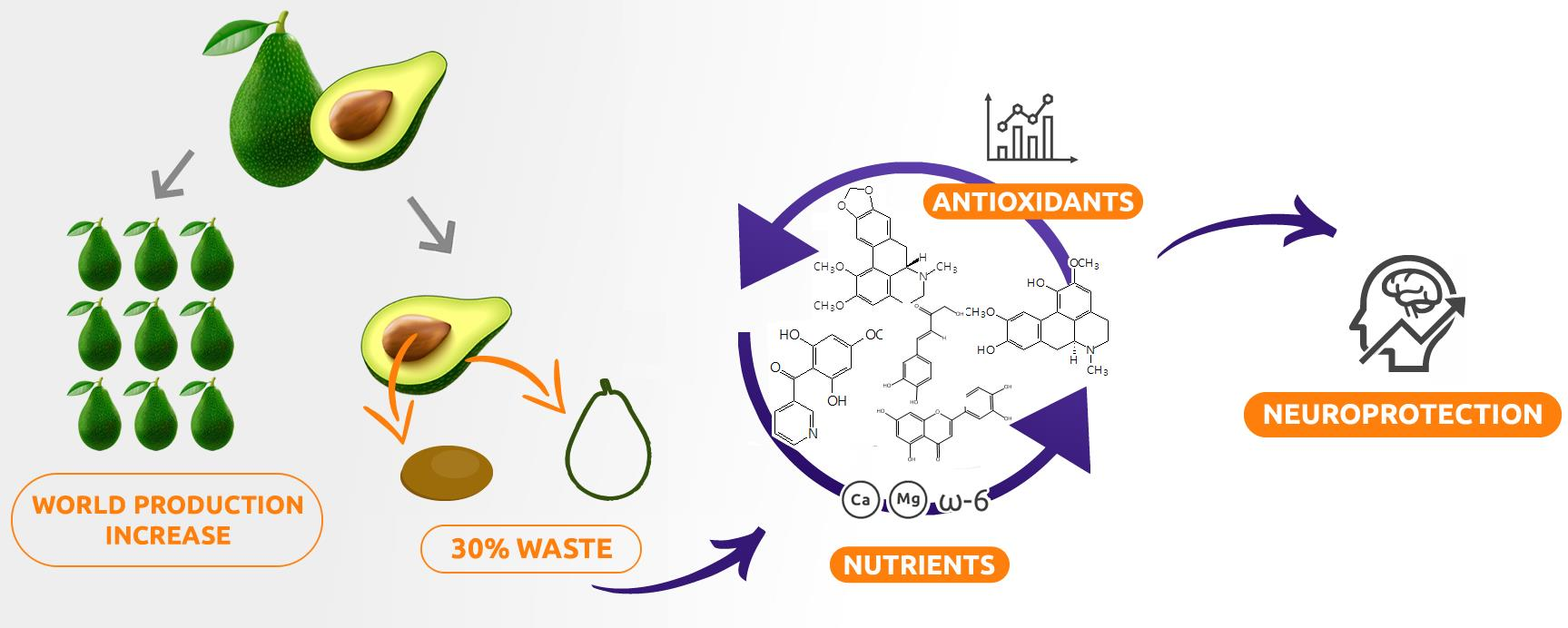
Phytochemicals of Avocado Residues as Potential Acetylcholinesterase Inhibitors, Antioxidants, and Neuroprotective Agents

by
Geisa Gabriela da Silva
1,
Lúcia Pinheiro Santos Pimenta
2,
Júlio Onésio Ferreira Melo
3,
Henrique de Oliveira Prata Mendonça
3,
Rodinei Augusti
2 and
Jacqueline Aparecida Takahashi
2,*
1
Department of Food Science, Faculty of Pharmacy, Universidade Federal de Minas Gerais, Av. Antônio Carlos, 6627, Belo Horizonte 31270-901, Brazil
2
Chemistry Department, Universidade Federal de Minas Gerais, Av. Antônio Carlos, 6627, Belo Horizonte 31270-901, Brazil
3
Exact and Biological Sciences Department, Campus Sete Lagoas, Universidade Federal de São João del-Rei, Sete Lagoas 36307-352, Brazil
*
Author to whom correspondence should be addressed.
Molecules 2022, 27(6), 1892; https://doi.org/10.3390/molecules27061892
Submission received: 28 January 2022 / Revised: 3 March 2022 / Accepted: 11 March 2022 / Published: 15 March 2022

Abstract

:
Avocado (Persea americana) is a widely consumed fruit and a rich source of nutrients and phytochemicals. Its industrial processing generates peels and seeds which represent 30% of the fruit. Environmental issues related to these wastes are rapidly increasing and likely to double, according to expected avocado production. Therefore, this work aimed to evaluate the potential of hexane and ethanolic peel (PEL-H, PEL-ET) and seed (SED-H, SED-ET) extracts from avocado as sources of neuroprotective compounds. Minerals, total phenol (TPC), total flavonoid (TF), and lipid contents were determined by absorption spectroscopy and gas chromatography. In addition, phytochemicals were putatively identified by paper spray mass spectrometry (PSMS). The extracts were good sources of Ca, Mg, Fe, Zn, ω-6 linoleic acid, and flavonoids. Moreover, fifty-five metabolites were detected in the extracts, consisting mainly of phenolic acids, flavonoids, and alkaloids. The in vitro antioxidant capacity (FRAP and DPPH), acetylcholinesterase inhibition, and in vivo neuroprotective capacity were evaluated. PEL-ET was the best acetylcholinesterase inhibitor, with no significant difference (p > 0.05) compared to the control eserine, and it showed neither preventive nor regenerative effect in the neuroprotection assay. SED-ET demonstrated a significant protective effect compared to the control, suggesting neuroprotection against rotenone-induced neurological damage.

Graphical Abstract

1. Introduction

Originated from Central America, the avocado is the fruit of the Persea americana Mill species from the Lauraceae family. It is rich in minerals, vitamins, proteins, fibers, and unsaturated lipids, the latter known to prevent cardiovascular diseases [1,2]. Moreover, avocado seeds display significant antioxidant, anti-inflammatory, and anti-cancer effects which have been associated with elevated percentages of hydrocarbons, sterols, and unsaturated fatty acids. Pulp has the same effects, although at a lower intensity [3]. Finally, the peel is rich in phenolic compounds and minerals, and has significant antioxidant activity, comparable to the seeds and far superior to the pulp [4,5].
In 2020, Mexico was the largest avocado producer, accounting for 2393 thousand tons per year, followed by the Dominican Republic, Peru, Colombia, Indonesia, and Brazil [6]. Since the inedible parts of avocado, mainly constituting peels and seeds, are discarded by the industry, restaurants, or at home, with the growth of avocado production, the amount of residues is also increasing, causing environmental problems [7]. These parts can reach up to 30% of the fruit. Furthermore, it is predicted that avocado production will double shortly [8]. In this sense, changes in the management of this fruit are necessary to accompany the dynamics of economic demands, with minimal environmental impacts [8,9,10]. Because avocado solid waste retains useful bioactive natural compounds, this residue has been targeted by several interesting studies to recover phytochemicals of industrial interest [11,12,13,14].
The antioxidant activity of avocado inedible parts can be explored in the development of neuroprotective agents against oxidative-related diseases like Alzheimer’s [4,5,11,14]. Alzheimer’s disease is one of the most concerning neurodegenerative disorders, affecting over 55 million people, mainly the elderly [15]. This disease can disrupt the synapses between neurons by the action of acetylcholinesterase (AChE), an enzyme that prevents acetylcholine from establishing communication between neurons. There is still no cure for this pathology, but palliative drugs have been very useful in decreasing symptoms and improve the quality of life of patients with Alzheimer’s disease. However, these drugs are usually expensive. The monthly cost of galantamine (30 pills) almost reaches USD 200, while drugs for the treatment of other diseases such as diabetes and hypertension cost less than USD 20 [16]. Therefore, the development of new drugs to treat neurodegenerative diseases from avocado biowaste could widen access to treatment while generating economic gains for the industry.
In this context, this work aimed to validate the nutritional and functional value of avocado residues by carrying out chemical and biological assays on seeds and peels of the Fortuna variety. The mineral, fatty acid, phenolic, and flavonoid contents were determined using spectroscopic and chromatographic methodologies. The phytochemical profile was determined by Paper Spray Mass Spectrometry (PSMS). Biological assays targeted antioxidant potential, AChE inhibition, and neuroprotective efficacy.

2. Results and Discussion

2.1. Residues Weight and Extracts Yields

Avocado fruits were separated into parts and weighted, resulting in 69.9% pulp, 10.2% peel, and 19.9% seed. According to the FAO [17], the world production of avocado is estimated to reach 9.2 tons by 2028. In this scenario, the volume of residues can reach approximately two tons. The peels were extracted by maceration with hexane and ethanol, providing two extracts, PEL-H (1.41 g/100 g of sample) and PEL-ET (16.94 g/100 g of sample). The same procedure was repeated with the seeds to furnish hexane (SED-H, 0.94 g/100 g of sample) and ethanol (SED-ET, 28.58 g/100 g of sample) extracts. The high yields of ethanol extracts reflect greater extraction power than that of hexane. The substitution of organic solvents with an environmentally favorable solvent, such as ethanol, makes the recycling of avocado waste important in terms of green chemistry and sustainability.

2.2. Mineral Contents in Avocado Residues

The mineral contents were quantified by atomic absorption spectroscopy, and the results are presented in Table 1.
According to the results, both avocado peels and seeds are good sources of minerals, especially calcium and magnesium, along with minor amounts of copper, iron, manganese, and zinc. Calcium, together with iron and zinc, are among the most difficult nutrients to obtain using local foods, according to research targeting women and young children from Southeast Asia [18]. In the U.S., mineral supplementation is advised during pregnancy [19]. Women are likely to be more affected by mineral deficiency, especially during childhood, although in many countries like Brazil, the prevalence of mineral deficiency is underreported [20]. Additionally, each ton of an agro-industrial residue containing avocado peels and seeds would correspond to the FAO/WHO’s daily recommended intake of calcium (1024 mg) for over two thousand individuals for one month, which is very considerable given calcium deficiency in some groups. The mineral content of agro-industrial residues is variable. Peels and seeds of avocado have lower mineral content than cocoa honey, a cocoa byproduct [21], but higher than pea peels [22]. Minerals and other nutrient and bioactive compounds present in agro-industrial residues can be processed in different ways, such as snack crackers and dry soup [22].

2.3. Fatty Acid Composition

After chemical derivatization to fatty acid methyl esters (FAMEs), the extracts were evaluated by gas chromatography, allowing identification of the fatty acids presented in Table 2.
Linoleic acid, a ω-6 fatty acid, was the component predominant in the seed extracts (27.4 and 34.8%) and palmitic acid, the major component (42.5–47.9%), in the seed extracts (22.2–23%). Oleic acid was distributed in all samples, but its content was lower in the peel ethanolic extract. Stearic acid was detected in all samples, but mainly in the ethanol extracts (14.7% in the seeds/22.2% in the peels). The composition of the different extracts revealed that seeds had a higher content of unsaturated fatty acids, such as oleic and linoleic, and small amounts of saturated fatty acids, such as palmitic and stearic.
The human body cannot synthesize linoleic acid, an essential fatty acid; therefore, it must be acquired through feeding. Linoleic acid plays a fundamental role in fetal neurological function and infant growth because it is the precursor of arachidonic acid. The uptake of this fatty acid decreases the risk of developing heart disease and it is beneficial for inflammatory processes [23]. Furthermore, palmitic and stearic acids are undesirable in food because saturated fatty acids usually contribute to increased blood cholesterol. However, stearic acid is considered an exception, because a rapid in vivo enzymatic dehydrogenation reaction catalyzed by stearoyl-CoA Δ9-desaturase converts stearic acid to oleic acid (18:1 Δ9) in the body [24]. The fatty acid composition found in avocado residue extracts suggests that they could be better used in combination with other oils, to balance the final composition. For instance, a sample of olive oil studied by Orsavova et al. [25] presented 16.4% of linoleic acid and 16.5% of palmitic acid (~1:1 proportion). A 1:1 mixture of this olive oil and the oil present in the SED-H would increase the proportional quantity of linoleic acid to 26% and the amount of palmitic acid to 19.75%. In the end, such mixed oil, composed of 50% oil recovered from waste biomass, would benefit from an improved content of linoleic acid.
The health claims for avocado fruits are closely related to the high content of fatty acids that are essential for human health [26]. Therefore, the essential fatty acids detected in the residues already consist of valuable metabolites that could be used as food additives or might be extracted, adding value to avocado waste biomass, which is a promising and inexpensive alternative source of linoleic acid.

2.4. Determination of the Total Phenolic and Flavonoid Contents

Avocado is a rich source of polyphenols, including hydroxybenzoic acid, caffeoylquinic acid derivatives, and cinnamic acids [5,27]. Flavonoids, such as quercetin, quercetin glycosides, naringenin, catechin, and epigallocatechin, were described in avocado seeds of the “Hass” variety [5]. In the current study, the total phenolic content (TPC) was estimated using the Folin–Ciocalteu method for each extract and expressed in mg of gallic acid equivalents (GAE)/g of extract. The total flavonoid content (TFC) was determined using the aluminum chloride colorimetric method and expressed as miligram of quercetin per gram of extract. Table 3 presents the results of the polyphenol and flavonoid contents according to the previous methodologies mentioned.
The TPC values was found in the range of 26.33–35.40 mg of gallic acid/g of extract, and the TFC values ranged from 640.72 to 1199.04 mg of quercetin/g of extract. The peel ethanolic extract (PEL-ET) presented the highest content of total phenolic compounds (35.40 ± 0.599 mg of gallic acid/g of ethanol extract), which was significantly different from all other extracts. Similarly, the TPC reported for peels from another variety of avocados using the same methodology (Folin-Ciocalteau) was 47.9 ± 2.7 mg of gallic acid/g of ethanol extract [28]. Conversely, peel ethanolic extracts from different varieties of P. Americana, including the Fortuna variety, presented lower phenolic compounds contents [29].
The peel extracts presented significantly higher flavonoid content than the extracts of the corresponding seeds. Higher percentages of flavonoid compounds were expected in the ethanol extracts, given the polar nature of the most representative flavonoids. However, our results showed that the higher percentages were present in the hexane extracts. This discrepancy can be partially explained by the presence of vitamin E in avocado residues [2]. Vitamin E is a phenolic non-polar compound, known to be present in substantial levels in avocado. Probably, its aliphatic long side chain may have contributed to the results. In addition, the percentages were determined by a colorimetric method that, although being a widely used methodology, is not a specific analysis. Flavonoid compounds have also been identified in the peels and seeds of the Hass and Fuerte varieties using HPLC-DAD [30].

2.5. Total Antioxidant Capacity and Antioxidant Activity Determined by DPPH Radical Scavenging and Ferric Reducing Power

The results regarding the antioxidant functional properties are shown in Table 4. In the total antioxidant activity assay (expressed as mmol of ascorbic acid/g extract) (R² = 0.9339), the values ranged from 630.23 to 770.01 and all extracts from seeds (SED-H and SED-ET) and peel hexane extracts (PEL-H) showed no significant differences.
Peel and seed hexane extracts presented similar behavior in promoting the reduction of ferricyanide ion to ferrocyanide, forming Prussia’s blue. The reducing power of both seed and peel ethanolic extracts was lower. The extract capacities of scavenging DPPH radicals were, in general, low. Only the PEL-ET showed moderate activity presenting over 50% of inhibition, with an IC50 92.557 μg/mL, being, therefore, the most promising extract in this assay. The high antioxidant activity may be explained by the high content of phenolic compounds, which was observed for peel and seed ethanolic extracts.

2.6. Acetylcholinesterase Inhibition

The hexane (SED-H and PEL-H) and ethanolic (SED-ET and PEL-ET) extracts were submitted to acetylcholinesterase inhibition assay. All extracts inhibited acetylcholinesterase activity up to 65%, with the ethanolic extracts more active than the hexane ones (Table 4). This range of extract activities is promising when compared with the activity presented by the control, a pure compound. The extract is a complex mixture and the active constituents are usually minor constituents in the extracts. In this way, the inhibitory capacity of the peel ethanolic extract from avocado (85.6 ± 11.1%) over acetylcholinesterase is outstanding, presenting no significant difference (p > 0.05) compared to the activity of the pure standard, eserine (91.5 ± 1.6%). Comparing the whole picture, the extract PEL-ET demonstrated a direct relationship between the amount of total phenolic compounds and AChE inhibition, corroborating the influence of antioxidant phenolic compounds in the acetylcholinesterase activity. By contrast, an inverse relationship was perceived for the hexane and ethanol extracts of the seeds. The principal component analysis (PCA) score biplot allowed a general qualitative visualization of the results obtained in the biological screening (Figure 1).
The PCA biplot shows the close relationship of the ethanolic extracts with the presence of phenolic compounds, DPPH assay, and acetylcholinesterase inhibition. The peel ethanolic extract (PEL-ET) demonstrated the closest association with the acetylcholinesterase enzyme inhibition assay. Concerning the hexane extracts, the seed extract (SED-H) was mainly related to the total antioxidant activity, while the peel extract (PEL-H) is associated with both ferric reduction power and total flavonoid contents. The hexane extracts are more distant from the AChE assay and present lower activity for acetylcholinesterase inhibition, as observed in the study. Therefore, only the ethanolic extracts were submitted to the in vivo D. melanogaster assay to evaluate the neuroprotective response.

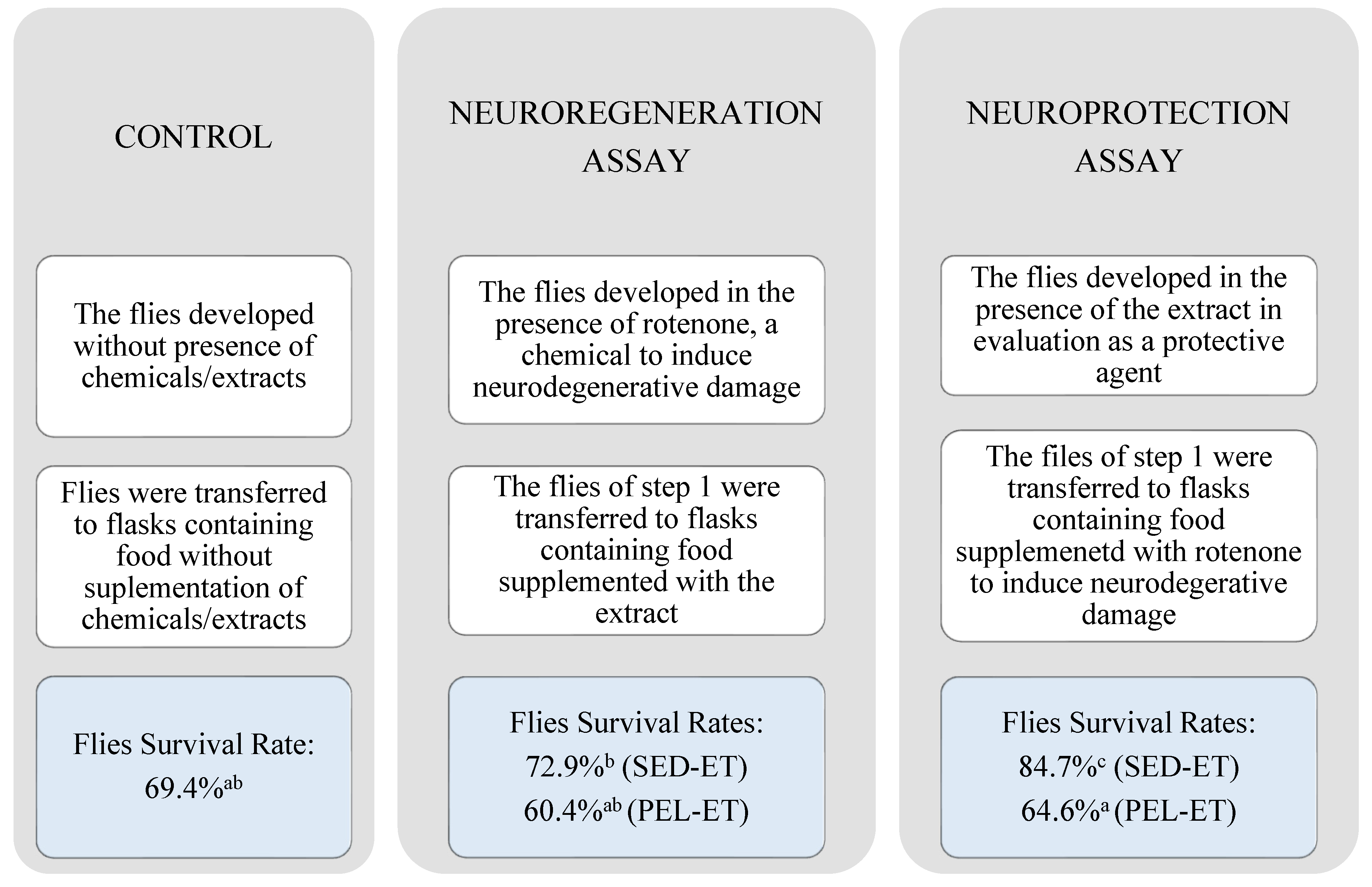
2.7. Activity of P. americana Extracts in Neuroprotection Using D. melanogaster Model

D. melanogaster flies have been used as an in vivo model for the evaluation of neurological damage related to neurodegenerative conditions. In this model, neurological damage can be induced in the flies by in vitro feeding with chemicals such as rotenone, preventing these insects from flying. The D. melanogaster model was used by Jiménez et al. [31] to demonstrate the neuroprotective effect of methanol extract prepared from Solanum ovalifolium, and by Siima et al. [32] to show the effect of flavonoids and polyketides in rescuing locomotor capacity in a Parkinson’s disease-related study [33]. Test compounds can be administered before or after flies’ exposure to rotenone to evaluate protective and regenerative effects, respectively. In the first alternative, the test compound avoids some of the damages that rotenone would cause (preventive action). In the second, damages due to rotenone exposure are established in the flies before test compound administration, and the capacity of the target compound to revert the neurodegenerative effects is evaluated (regenerative action).
In the present work, test compounds were administered before and after exposure to rotenone, to evaluate both effects (Figure 2). The ethanol extracts (PEL-ET and SED-ET) were evaluated by this model and the results were submitted to the Shapiro–Wilk statistical normality test. The p-value found (>0.05) indicated that the distribution was not suitable. Therefore, the Kruskal–Wallis nonparametric test was conducted, identifying a significant difference; subsequently, the Dunn’s test was used, which identified a significant difference p < 0.05 between the samples. Overall, a high mortality rate was observed during the trial in which flies were first exposed to rotenone (Figure 2). In this condition, rotenone caused 60.4–72.9% of flies to die, showing high acute toxicity. Under this condition, peel extract (PEL-ET) did not statistically differ (p > 0.05) from the control, in which flies were not exposed either to the extracts or to rotenone. Therefore PEL-ET showed neither preventive nor regenerative effect in this model.
SED-ET showed a more significant result (Figure 2). Comparing the seed and peel results in this assay, only SED-ET demonstrated a protective effect in this treatment. In addition, the performance of SED-ET was significant compared to the control, presenting a higher number of flies capable of flying above the limit established, which suggests that the seed extract provided a neuroprotective effect against rotenone-induced neurological damage (Figure 2). This flies model has been leading to the identification of new therapeutic targets for research in Parkinson’s [34], Alzheimer’s [35], and other neurodegenerative diseases. The regenerative activity has been the main target of drugs to individuals in the advanced neurodegeneration stage. However, protection against degeneration can be helpful to patients in the early stages of neurodegenerative diseases.

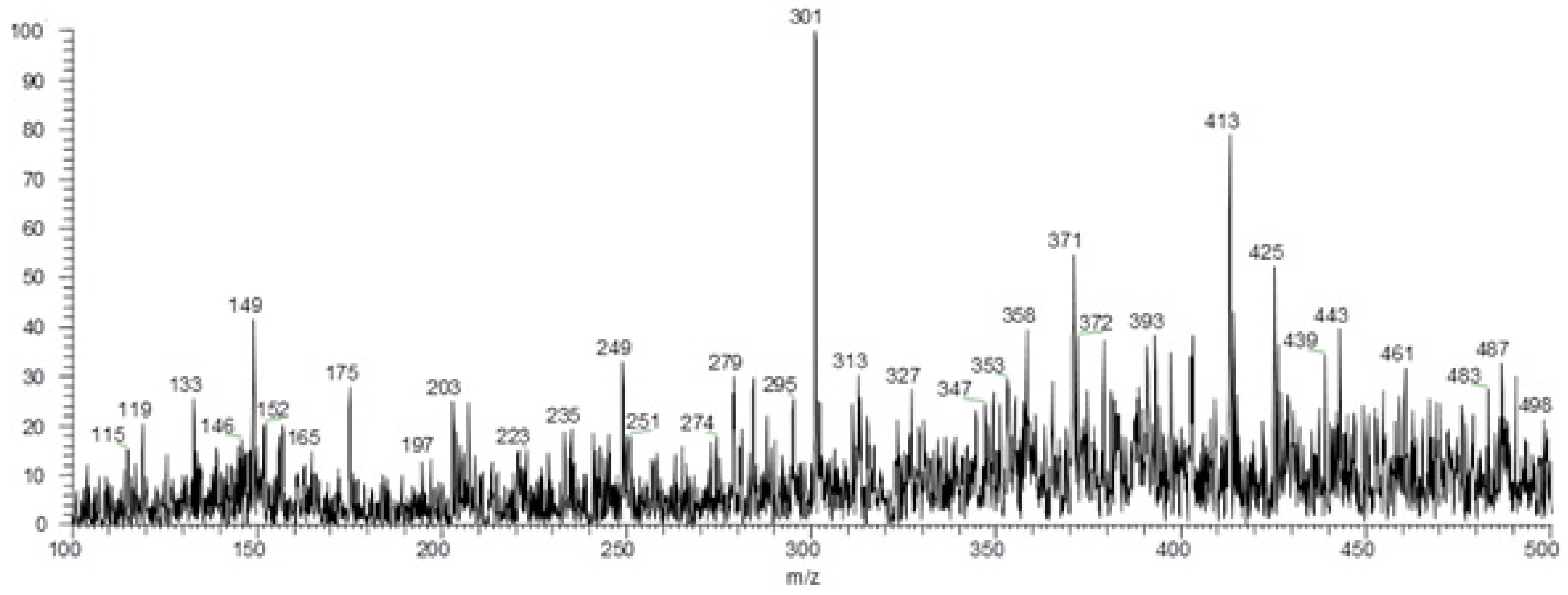
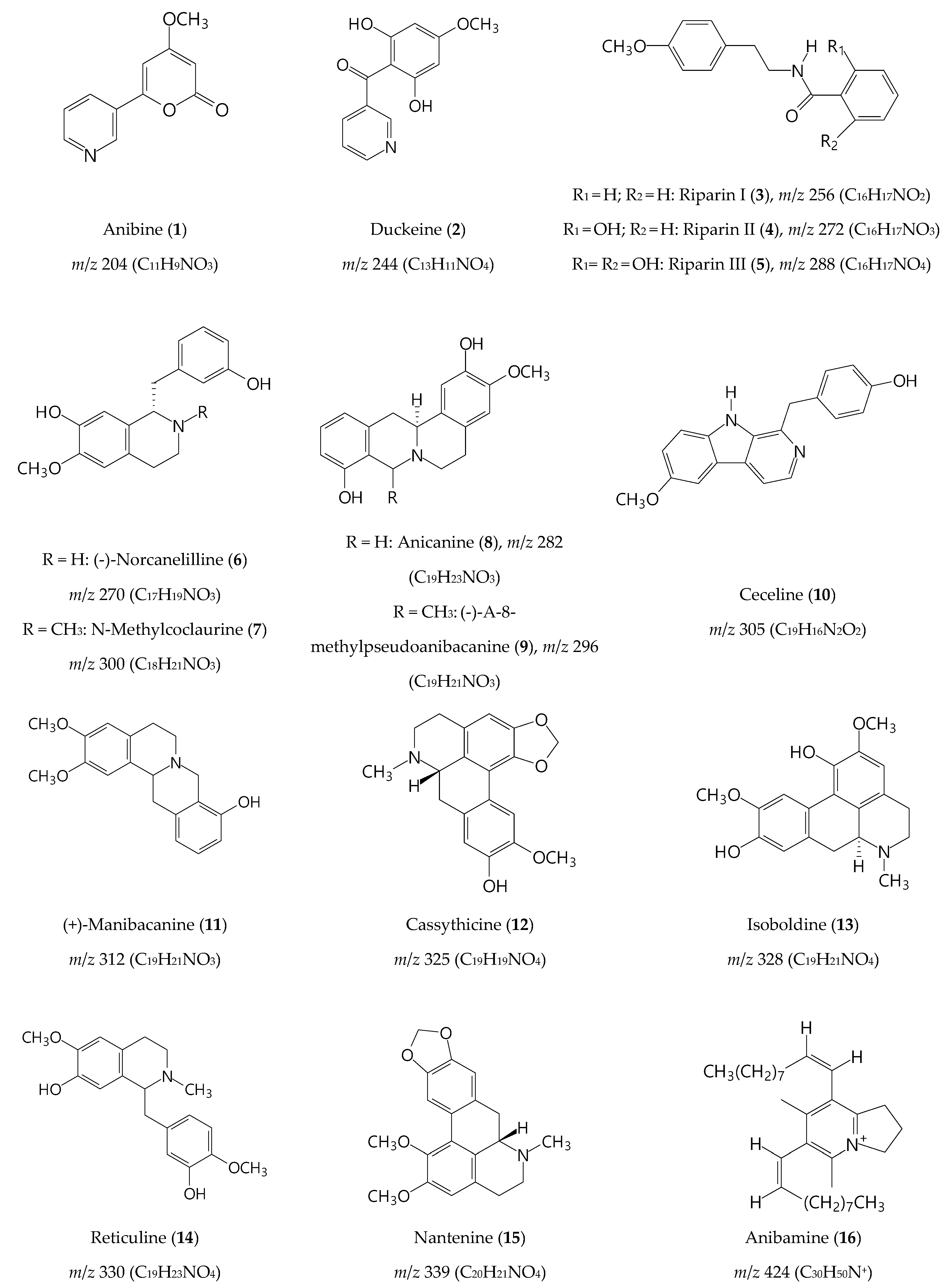
2.8. Identification of Metabolites by Paper Spray Mass Spectrometry

Paper spray mass spectrometry (PSMS) was applied to identify the natural products present in the ethanol extracts. This quick and efficient technique for chemical profile determination is commonly used in the clinic and in forensic research to identify specific ions through direct sample analysis [36,37,38,39]. This study showed no difference in the chemical profile of peel and seed samples. PS (−) MS spectrum of SED-ET is presented in Figure 3. Fifty-five compounds were tentatively identified in the ethanol extracts by PSMS analyzing their MS and MS2 spectra, together with information previously reported in the literature. In the negative mode, 41 metabolites were identified, mainly flavonoids and organic acids, most of them with biological activities already reported [12].
The presence of compounds such as caffeic acid (m/z 179 [M − H]), catechin (m/z 289 [M − H]), rutin (m/z 609 [M − H]), procyanidin isomers, and flavonoids validated the antioxidant activity of the seed extract and were in accordance with previous reports [39,40]. Corroborating the experiments, some of the metabolites detected in the current work were already reported in the seeds and peels of avocado (vanillin, caffeic acid, ferulic acid, synaptic acid, kaempferol, catechin, quercetin-3-glucoside, quercetin-O-arabinosyl-glucoside, rutin, dimers A and B, and trimer A of procyanidin) [12]. Based on the antioxidant effect, Segovia et al. [41] described the role of avocado seed extract as an additive to protect oil mixtures from oxidation. The presence of quinic acid derivatives in the extract also contributed to validating the antioxidant activity and, therefore, highlighted the beneficial properties of this residue as food ingredients for industrial use [42]. Table 5 presents the compounds tentatively identified in the extracts in negative mode.
In the positive mode, fifteen alkaloids were identified: anibine (1), duckeine (2), riparin I, II and III (35), norcanelilline (6), N-methylcoclaurine (7), anicanine (8), (-)-α-8-methylpseudoanibacanine (9), ceceline (10), (+)-manibacanine (11), cassythicine (12), isoboldine (13), reticuline (14), nantenine (15), and anibamine (16) (Figure 4 and Figure 5, Table 6). PS(+)MS full scan of ethanol extract from peel of avocado can be found in Supplementary Materials, Figure S1. It is reported that, as the avocado fruit develops, the carbohydrate amount decreases and the fatty acids and secondary metabolites content increases [46,47]. In ripe avocado fruits, the alkaloids constitute a remarkable percentage of the natural compounds present in the pulp [46,47]. From the alkaloids identified, anibine (1), duckeine (2), and anicanine (8) were not detected in PEL-ET. All other alkaloids were detected in both extracts.
Alkaloids are used as active substances in prescriptions for the treatment of neurodegenerative diseases, e.g., galantamine. Therefore, the alkaloids detected in the extracts may contribute to the acetylcholinesterase inhibition and neuroprotective effect. More specifically, the alkaloids riparin (35) show antioxidant, antinociceptive, anti-inflammatory, and neuroprotective effects [49,50,51]. The aporphine alkaloid nantenine (14) has a potential role in acetylcholinesterase inhibition, since nantenine is present in Unopsis stipitate, a plant traditionally used for cognitive disorders [52]. Furthermore, nantenine possesses pharmaceutical interest as anticonvulsant, due to its direct inhibitory effect on calcium influx [53].

3. Materials and Methods

3.1. Materials and Reagents

Ripe avocado (cultivar Persea americana var. Fortuna) fruits were purchased in Ibirité city (MG, Brazil) in the winter of 2018. P.A. grade hexane (Nox Lab Solutions, Mauá, SP, Brazil) and hydrated ethanol 96% (Emfal, Betim, MG, Brazil) were used. Ascorbic acid (Neon, Suzano, SP, Brazil), gallic acid (Neon, Suzano, SP, Brazil), quercetin (Sigma-Aldrich, St. Louis, MO, USA), and eserine (Sigma-Aldrich, St. Louis, MO, USA) were used as positive controls in the biological assays. Acetylcholinesterase iodide, acid 5′,5′-dithio-bis-(2-nitrobenzoate), bovine serum albumin, and AChE (Sigma-Aldrich, St. Louis, MO, USA) were used in the AChE inhibition assay. For cultivation of Drosophila melanogaster flies, bacteriological agar (Vetec, Duque de Caxias, SP, Brazil), powdered dry yeast (Fleischmann, Sorocaba, SP, Brazil), and nystatin (oral suspension from Germed, Campinas, SP, Brazil) were used. Rotenone (Sigma-Aldrich, At. Louis, MO, USA) was employed in the negative geotaxis assay to induce toxicity. The experiments were carried out in quintuplicate unless otherwise specified.

3.2. Biomass Processing and Extract Preparation

The peels (53.233 g) and seeds (103.431 g) of avocado fruits were separately removed from the pulp, sanitized under running water, and dried separately with paper towel sheets. The peels were sliced, the seeds were grated, and 3 g of each was utilized for atomic absorption spectroscopy analysis. The remaining biomasses (sliced peels and grated seeds) were separately soaked in hexane (600 mL). After 24 h, the hexane fraction was separated by filtration and another aliquot of hexane (600 mL) was added to the vegetal material. The procedure was repeated three times. The hexane fractions were combined; the solvent was removed in a rotary evaporator (60 °C), obtaining the respective hexane extracts of peels (PEL-H) and seeds (SED-H). After hexane extraction, the defatted plant material was further extracted with 96% ethanol, following the same procedure previously described, to furnish the peel (PEL-ET) and seed (SED -ET) ethanolic extracts (Figure 5) [54].

3.3. Mineral Element Analysis by Atomic Absorption

The separated aliquots of fresh peels and seeds (3 g each) were placed in a porcelain crucible and heated until complete carbonization. Subsequently, the crucibles with the carbonized plant material were placed in a muffle at 550 °C, obtaining the organic matter-free ashes. Nitric acid (2 mL) was added to an aliquot of the ashes (0.05 mg) and the volume was completed with distilled water to 5 mL. The resulting suspension was analyzed using Atomic Absorption Spectrometer AA 240 FS (Varian, Australia) to quantify the minerals Ca, Cu, Fe, Mg, Mn, and Zn [4].

3.4. Fatty Acid Gas Chromatography Analysis

Aliquots (10 mg) of the extracts were derivatized to fatty acid methyl esters (FAMEs) [55], solubilized in methanol, and injected into an HP5890 Gas Chromatograph. A Supelcowax-10 analytical column (30 m × 0.2 mm × 0.2 μm) (Supelco) was used for analysis under the following chromatographic conditions (split 1/50): 150 °C, 0 min, 10 °C /min up to 240 °C and detector at 250 °C. Hydrogen was used as carrier gas (4 mL/min) and the injection volume was 1 μL. A standard of fatty acid methyl esters (FAME C14-C22) were injected, and their retention times were used to comparatively identify the fatty acids present in the avocado residues.

3.5. Total Content of Phenol and Flavonoids Assay

The determination of total phenolic content was performed spectrophotometrically with a Folin–Ciocalteau reagent method previously described [56]. Briefly, the ethanolic extract solutions (0.5 mL) were mixed with Folin–Ciocalteau reagent (2.5 mL). The solutions were incubated for 3 min at room temperature, and 0.3 mL of saturated sodium carbonate solution was added to each one. After a further incubation period (20 min at 25 °C), absorbance was read at 760 nm using a Biospectro SP-22 Spectrophotometer. The same procedure was carried out for gallic acid (standard), which was used for constructing the calibration curve (10–100 µg/mL; R² = 0.9291). In the equation of the calibration curve established using gallic acid, “y” was replaced by the absorbance and divided by the mass of the extract/standard. The results were expressed in mg gallic acid/g extract.
The total flavonoid contents were determined by aluminum chloride spectrophotometric method as described by Yang et al. [27]. Briefly, 2.5 mL of the ethanolic extract or standard solutions were mixed with 0.3 mL sodium nitrate solution (5% w/v) with subsequent addition of aluminum chloride solution (10% w/v) (3.0 mL). The mixture was vortexed, and 2.0 mL of sodium hydroxide solution (1 M) was added and well vortexed. After 10 min, the absorbances were determined at 510 nm using a Biospectro SP-22 Spectrophotometer. Quercetin standard solutions (350–600 μg/mL) were used for constructing the calibration curve (R² = 0.9887). The result was expressed as mg of quercetin equivalent per g of extract.

3.6. Antioxidant Activity

Ethanol and hexane extracts (SED-ET, PEL-ET, SED-H, and PEL-H) and the standards (ascorbic acid and gallic acid) were dissolved in ethanol (500 μg/mL), except quercetin (1000 μg/mL). All assays were performed in quintuplicate. Microsoft Excel 2013 software was used for data analysis.

3.6.1. Total Antioxidant Capacity

A reagent solution (100 mL) was prepared mixing sulfuric acid (3.22 mL), bibasic phosphate (0.39 g), and ammonium molybdate (0.49 g). An aliquot (0.3 mL) of ethanolic extract solutions (500 µg/mL) was mixed with 3.0 mL of the reagent solution. The resulting test samples were incubated in an oven at 95 °C for 90 min. After the samples had cooled to room temperature, the absorbances were read at 695 nm on a Biospectro SP 22 Spectrophotometer. The same procedure was carried out with ascorbic acid (positive control) and a negative control was carried out with the solvent. The results were expressed in mmol of ascorbic acid per gram of extract (adapted from [57]).

3.6.2. Ferric Reducing Power Assay

An aliquot (1.0 mL) of the ethanolic extract solutions was mixed with 2.5 mL of phosphate buffer (pH 6.6, 0.2 M) and 2.5 mL of potassium ferricyanide aqueous solution (1% w/v). The mixture was homogenized and incubated in a laboratory water bath at 50 °C for 20 min. Subsequently, 2.5 mL of trichloroacetic acid solution (10% w/v) was added and the mixture was centrifuged (300 rpm, 10 min). An aliquot (2.5 mL) of the superior layer was mixed with 2.5 mL distilled water, and 0.5 mL ferric chloride solution (0.1% w/v) was added. The same procedure was conducted with the positive control. The absorbances were read at 700 nm on a Biospectro SP-22 Spectrofotometer. The results were converted into a percentage of ferric reducing power in relation to the positive control ascorbic acid [57].

3.6.3. Free Radical Capture Assay (DPPH)

Solutions of extracts (100 μL) were added to 96-well plates in quadruplicate, followed by the addition of 2,2-Diphenyl-1-picrylhydrazyl (DPPH) solution (175 μL) prepared in ethanol (0.1 mM). The materials were incubated for 30 min in the dark, after which reading was performed at 490 nm in a microplate reader. Solutions of known concentration of ascorbic acid, the positive control, were used to construct a graph of percent inhibition versus concentration (μg/mL) [58].

3.7. Acetylcholinesterase Inhibition Assay

The extracts were solubilized in dimethylsulfoxide (DMSO) (10 mg/mL) and an aliquot of each one (25 μL) was mixed with aqueous solutions of acetylcholine iodide (15 mM) (25 μL) and 125 μL of a solution containing 5,5′-dithio-bis (2-nitrobenzoic acid) (3 mM), NaCl solution (0.1 M) and MgCl2·6H2O (20 mM) prepared in Tris/HCl buffer (50 mM, pH 8.0, 50 μL) in wells of 96-well plates. The first set of wavelength readings was then performed at 405 nm every 1 min (8 readings). Subsequently, a solution of acetylcholinesterase was prepared (0.22 U/mL) in buffer and 0.1% (w/v) of bovine serum albumin and added to the wells (25 μL each). Eserine (10 mg/mL) was used as positive control. Next, new readings were performed at the same wavelength every 1 min (10 readings) [20]. The following equation was used to calculate the percentage of inhibition (%I), where Ab: Negative control (DMSO) and Aa: Sample absorbance.
%   I = ( A b A a ) × 100 A b

3.8. In Vivo Neuroprotection Activity Using Drosophila melanogaster Model

A sterile mixture containing bananas (400 g), bacteriological agar (4 g), commercial yeast (3.5 g), nystatin (2.5 mL), and propionic acid (0.6 mL) in distilled water (350 mL) was prepared and used to grow D. melanogaster flies for the whole experiment. The cultivations were performed in glass vessels in triplicate. Twelve 6–8-day-old male flies were grown in vessels containing (A) rotenone (neurotoxic inductor, 10 mg/mL in ethanol; 0.4 mL) or (B) extracts (5 mg/mL in water; 0.6 mL). Rotenone and extracts were mixed with the nutritive substrate. A control flask (C) was utilized. Neither rotenone nor extract were added to the control flasks. After 7 days, the flies that were in vessels A and B were anesthetized with ether and interchanged. After 15 days the flies were transferred to falcon tubes that were horizontally placed on a support. A photographic camera was positioned approximately 30 cm away. Then, the support was subjected to three hits against the bench surface, and ten photographs of the tubes containing the flies were recorded. Picture number 6 was used to account for the number of flies that were able to fly above half the tube height after hitting [31].

3.9. Paper Spray Mass Spectrometry

Pulps were analyzed using a Thermo Fisher LCQ FLEET ion-trap mass spectrometer (Thermo Scientific, San Jose, CA, USA) equipped with an ambient paper spray ionization source. Analyses were performed in triplicate in positive and negative ionization modes. For PSMS analyses, a 2 µL sample volume and 40 µL of methanol were added to a triangular chromatographic paper (equilateral, 1.5 cm lengthwise) positioned 10 mm away from the mass spectrometer inlet [37,38,39]. A high voltage was then applied to the paper held by a copper clamp and attached to a three-dimensional moving platform for data acquisition. Instrumental conditions of operation were: PSMS source voltage equal to +4 kV (positive mode) and −3 kV (negative mode); capillary voltage of 40 V; transfer tube temperature of 275 °C; tube lens voltage of 120 V; mass scanning range of 100 to 1000 m/z in positive and negative modes. Ions were fragmented using collision energies from 15 to 45 eV. Some representative MS can be found in Figures S2–S48. Tentative identification of the compounds was carried out using a comparison of the m/z ratios of the data obtained from literature associated with instrumental readings and subsequent fragmentation employing sequential mass spectrometry.

3.10. Statistical Analysis

GraphPad Prism 5.0 software was used for statistical analysis, applying variance analysis (ANOVA) [20]. To evaluate the differences between the means, the Tukey test with significance level of 5% was used. Kruskal–Wallis (nonparametric) and Dunn’s post-test, with significance of 5%, were utilized to evaluate data from neuroprotection assay. SensoMaker 1.61 software was used for main components analyses.

4. Conclusions

The avocado residues produced during industrial processing retain nutrients and bioactive compounds independent of the extraction solvent. Concerning the minerals, avocado peels and seeds displayed significant levels of Ca, Mg, Mn, and Zn. The seeds presented high essential fatty acids content, such as linoleic, palmitic, and oleic acids. Seed and peel ethanolic extracts showed phenolic compounds correlated to their antioxidant and AChE inhibitory activities. Additionally, the seed ethanolic extract exhibited in vivo neuroprotective effect. In vivo studies are an important step to determine the real potential of phytochemicals since they overcome many problems inherent to in vitro models. Therefore, the positive in vivo results encourage research of this extract as a potential candidate to develop drugs to treat patients in the early stages of neurodegenerative illnesses such as Alzheimer’s and Parkinson’s diseases. The PSMS chemical profile of the residues led to the identification of fifty-five metabolites, including phenolic, hydroxycinnamic acids, flavonoids, and alkaloids, most of them already reported as pharmacologically active compounds. Quantification of the phytochemicals will be the object of future work. In addition, toxicity, bioavailability, and suitable formulations should be further investigated before using avocado residues in pharmacotherapy, but the current results pave the way to deeper studies on the in vivo potential of avocado residues as a bioresource in the development of low-cost drugs and functional foods with neuroprotective effects.

Supplementary Materials

The following supporting information can be downloaded at: https://www.mdpi.com/article/10.3390/molecules27061892/s1, Section S1: PSMS of the seed and ethanolic extracts and of the compounds identified, Figure S1: PS(+)MS full scan of ethanol extract from peel of avocado, Figure S2: Anibine PS(+)MS fragmentation and chemical structure, Figure S3: Duckeine PS(+)MS fragmentation and chemical structure, Figure S4: Riparin I PS(+)MS fragmentation and chemical structure, Figure S5: Norcanelilline PS(+)MS fragmentation and chemical structure, Figure S6: Riparin II PS(+)MS fragmentation and chemical structure, Figure S7: Anicanine PS(+)MS fragmentation and chemical structure, Figure S8: Riparin III PS(+)MS fragmentation and chemical structure, Figure S9: (-)-α-methylpseudoanibacanine PS(+)MS fragmentation and chemical structure, Figure S10: N- methylcoclaurine PS(+)MS fragmentation and chemical structure, Figure S11: Ceceline PS(+)MS fragmentation and chemical structure, Figure S12: (+)-manibacanine PS(+)MS fragmentation and chemical structure, Figure S13: Cassythicine PS(+)MS fragmentation and chemical structure, Figure S14: Isoboldine PS(+)MS fragmentation and chemical structure, Figure S15: Reticuline PS(+)MS fragmentation and chemical structure, Figure S16: Anibamine PS(+)MS fragmentation and chemical structure, Figure S17: PS(−)MS full scan of peel ethanolic extract of avocado, Figure S18: Vanillin PS(−)MS fragmentation and chemical structure, Figure S19: Caffeic acid PS(−)MS fragmentation and chemical structure, Figure S20: Quinic acid PS(−)MS fragmentation and chemical structure, Figure S21: Syrinc acid PS(−)MS fragmentation and chemical structure, Figure S22: 5-hydroxyferulic acid PS(−)MS fragmentation and chemical structure, Figure S23: Sinapic acid PS(−)MS fragmentation and chemical structure, Figure S24: Apigenin PS(−)MS fragmentation and chemical structure, Figure S25: Kaempferol PS(−)MS fragmentation and chemical structure, Figure S26: Catechin PS(−)MS fragmentation and chemical structure, Figure S27: Quercetin PS(−)MS fragmentation and chemical structure, Figure S28: Hydroxytyrosol glucoside PS(−)MS fragmentation and chemical structure, Figure S29: p-coumaroyl hexose PS(−)MS fragmentation and chemical structure, Figure S30: 3-O-p-coumaroylquinic acid PS(−)MS fragmentation and chemical structure, Figure S31: Caffeic acid hexoside PS(−)MS fragmentation and chemical structure, Figure S32: 5-O-caffeoylquinic acid PS(−)MS fragmentation and chemical structure, Figure S33: Kaempferol-O-pentoside PS(−)MS fragmentation and chemical structure, Figure S34: Cyanidin-3-O-arabinoside PS(−)MS fragmentation and chemical structure, Figure S35: Vitexin PS(−)MS fragmentation and chemical structure, Figure S36: Peonidin-3-O-pentoside PS(−)MS fragmentation and chemical structure, Figure S37: Phloridzin PS(−)MS fragmentation and chemical structure, Figure S38: Kaempferol-O-hexoside PS(−)MS fragmentation and chemical structure, Figure S39: Dihydroquercetin-3,5-rhamnoside PS(−)MS fragmentation and chemical structure, Figure S40: Isorhamnetin-O-coumaroyl PS(−)MS fragmentation and chemical structure, Figure S41: Quercetin-3-glucoside PS(−)MS fragmentation and chemical structure, Figure S42: Quercetin glucuronide PS(−)MS fragmentation and chemical structure, Figure S43: Caffeoyl hexose-deohyhexoside PS(−)MS fragmentation and chemical structure, Figure S44: Dimethyl ellagic acid hexoside PS(−)MS fragmentation and chemical structure, Figure S45: Apigenin-C-hexoside-C-pentoside PS(−)MS fragmentation and chemical structure, Figure S46: Luteolin-7-O-(2”-O-pentosyl)-hexoside PS(−)MS fragmentation and chemical structure, Figure S47: Catechin diglucopyranoside PS(−)MS fragmentation and chemical structure, Figure S48: Quercetin-3,4′-O-diglucoside PS(−)MS fragmentation and chemical structure.

Author Contributions

Conceptual idea, methodology design, writing, and editing: J.A.T.; Data collection, antioxidant assays, data analysis and interpretation, writing, and editing: G.G.d.S. and L.P.S.P.; Paper Spray analysis and data interpretation: J.O.F.M., H.d.O.P.M. and R.A. All authors have read and agreed to the published version of the manuscript.

Funding

This research was funded by CAPES (Code 001), Fundação de Amparo à Pesquisa do Estado de Minas Gerais (FAPEMIG PPM-00255-18), and Conselho Nacional de Desenvolvimento Científico e Tecnológico (CNPq Grant 304922/2018-8).

Institutional Review Board Statement

Not applicable.

Informed Consent Statement

Not applicable.

Acknowledgments

We acknowledge Lucas Xavier da Silva for drawing the graphical abstracts.

Conflicts of Interest

The authors declare no conflict of interest.

Sample Availability

Samples of the compounds are not available.

References

  1. Ochoa-Zarzosa, A.; Báez-Magaña, M.; Guzmán-Rodríguez, J.J.; Flores-Alvarez, L.J.; Lara-Márquez, M.; Zavala-Guerrero, B.; Salgado-Garciglia, R.; López-Gómez, R.; López-Meza, J.E. Bioactive Molecules From Native Mexican Avocado Fruit (Persea americana var. drymifolia): A Review. Plant Foods Hum. Nutr. 2021, 76, 133–142. [Google Scholar] [CrossRef] [PubMed]
  2. Tesfaye, T.; Ayele, M.; Gibril, M.; Ferede, E.; Limeneh, D.Y.; Kong, F. Beneficiation of avocado processing industry by-product: A review on future prospect. Curr. Res. Green Sustain. Chem. 2022, 5, 100253. [Google Scholar] [CrossRef]
  3. Alkhalaf, M.I.; Alansari, W.S.; Ibrahim, E.A.; ELhalwagy, M.E.A. Anti-oxidant, anti-inflammatory and anti-cancer activities of avocado (Persea americana) fruit and seed extract. J. King Saud Univ. Sci. 2019, 31, 1358–1362. [Google Scholar] [CrossRef]
  4. Borges, C.V.; Maraschin, M.; Coelho, D.S.; Leonel, M.; Gomez, H.A.G.; Belin, M.A.F.; Diamante, M.S.; Amorim, E.P.; Gianeti, T.; Castro, G.R.; et al. Nutritional value and antioxidant compounds during the ripening and after domestic cooking of bananas and plantains. Food Res. Int. 2020, 132, 109061. [Google Scholar] [CrossRef] [PubMed]
  5. Araujo, R.G.; Rodríguez-Jasso, R.M.; Ruíz, H.A.; Govea-Salas, M.; Pintado, M.; Aguilar, C.N. Recovery of bioactive components from avocado peels using microwave-assisted extraction. Food Bioprod. Process. 2021, 127, 152–161. [Google Scholar] [CrossRef]
  6. Baysal, S.S.; Ülkü, M.A. Food Loss and Waste: A Sustainable Supply Chain Perspective. ICGI-Global: Istanbul, Turkey, 2021; ISBN 9789251317549. [Google Scholar]
  7. Yap, K.M.; Sekar, M.; Seow, L.J.; Gan, S.H.; Bonam, S.R.; Mat Rani, N.N.I.; Lum, P.T.; Subramaniyan, V.; Wu, Y.S.; Fuloria, N.K.; et al. Mangifera indica (Mango): A promising medicinal plant for breast cancer therapy and understanding its potential mechanisms of action. Breast Cancer Targets Ther. 2021, 13, 471–503. [Google Scholar] [CrossRef]
  8. Béné, C.; Oosterveer, P.; Lamotte, L.; Brouwer, I.D.; de Haan, S.; Prager, S.D.; Talsma, E.F.; Khoury, C.K. When food systems meet sustainability—Current narratives and implications for actions. World Dev. 2019, 113, 116–130. [Google Scholar] [CrossRef]
  9. González-Chang, M.; Wratten, S.D.; Shields, M.W.; Costanza, R.; Dainese, M.; Gurr, G.M.; Johnson, J.; Karp, D.S.; Ketelaar, J.W.; Nboyine, J.; et al. Understanding the pathways from biodiversity to agro-ecological outcomes: A new, interactive approach. Agric. Ecosyst. Environ. 2020, 301, 107053. [Google Scholar] [CrossRef]
  10. Tumwesigye, K.S.; O’Brien, E.; Oliveira, J.C.; Crean, A.; Sousa-Gallagher, M.J. Engineered food supplement excipients from bitter cassava for minimisation of cassava processing waste in environment. Future Foods 2020, 1–2, 100003. [Google Scholar] [CrossRef]
  11. Ortega-Arellano, H.F.; Jimenez-Del-Rio, M.; Velez-Pardo, C. Neuroprotective Effects of Methanolic Extract of Avocado Persea americana (var. Colinred) Peel on Paraquat-Induced Locomotor Impairment, Lipid Peroxidation and Shortage of Life Span in Transgenic knockdown Parkin Drosophila melanogaster. Neurochem. Res. 2019, 44, 1986–1998. [Google Scholar] [CrossRef]
  12. Salazar-López, N.J.; Domínguez-Avila, J.A.; Yahia, E.M.; Belmonte-Herrera, B.H.; Wall-Medrano, A.; Montalvo-González, E.; González-Aguilar, G.A. Avocado fruit and by-products as potential sources of bioactive compounds. Food Res. Int. 2020, 138, 109774. [Google Scholar] [CrossRef] [PubMed]
  13. Del Castillo-Llamosas, A.; del Río, P.G.; Pérez-Pérez, A.; Yáñez, R.; Garrote, G.; Gullón, B. Recent advances to recover value-added compounds from avocado by-products following a biorefinery approach. Curr. Opin. Green Sustain. Chem. 2021, 28, 100433. [Google Scholar] [CrossRef]
  14. Fuloria, S.; Yusri, M.A.A.; Sekar, M.; Gan, S.H.; Rani, N.N.I.M.; Lum, P.T.; Ravi, S.; Subramaniyan, V.; Azad, A.K.; Jeyabalan, S.; et al. Genistein: A Potential Natural Lead Molecule for New Drug Design and Development for Treating Memory Impairment. Molecules 2022, 27, 265. [Google Scholar] [CrossRef] [PubMed]
  15. World Health Organization (WHO). Global Action Plan on the Public Health Response to Dementia 2017–2025; World Health Organization: Geneva, Switzerland, 2017; p. 27. [Google Scholar]
  16. Bernstein, J.J.J.; Holt, G.B.; Bernstein, J. Price dispersion of generic medications. PLoS ONE 2019, 14, e0225280. [Google Scholar] [CrossRef] [PubMed]
  17. FAO; IFAD; UNICEF; WHO. The State of Food Security and Nutrition in the World 2020; FAO: Rome, Italy, 2020; ISBN 9789251329016. [Google Scholar] [CrossRef]
  18. Ferguson, E.L.; Watson, L.; Berger, J.; Chea, M.; Chittchang, U.; Fahmida, U.; Khov, K.; Kounnavong, S.; Le, B.M.; Rojroongwasinkul, N.; et al. Realistic Food-Based Approaches Alone May Not Ensure Dietary Adequacy for Women and Young Children in South-East Asia. Matern. Child Health J. 2019, 23, 55–66. [Google Scholar] [CrossRef]
  19. Adams, J.B.; Sorenson, J.C.; Pollard, E.L.; Kirby, J.K.; Audhya, T. Evidence-based recommendations for an optimal prenatal supplement for women in the U.S., part two: Minerals. Nutrients 2021, 13, 1849. [Google Scholar] [CrossRef] [PubMed]
  20. Lourenção, L.F.d.P.; de Paula, N.C.; Cardoso, M.A.; Santos, P.R.; de Oliveira, I.R.C.; Fonseca, F.L.A.; da Veiga, G.L.; Alves, B.d.C.A.; Graciano, M.M.d.C.; Pereira-Dourado, S.M. Biochemical markers and anthropometric profile of children enrolled in public daycare centers. J. Pediatr. 2021; in press. [Google Scholar] [CrossRef]
  21. Guirlanda, C.P.; da Silva, G.G.; Takahashi, J.A. Cocoa honey: Agro-industrial waste or underutilized cocoa by-product? Future Foods 2021, 4, 100061. [Google Scholar] [CrossRef]
  22. Mousa, M.M.H.; El-Magd, M.A.; Ghamry, H.I.; Alshahrani, M.Y.; El-Wakeil, N.H.M.; Hammad, E.M.; Asker, G.A.H. Pea peels as a value-added food ingredient for snack crackers and dry soup. Sci. Rep. 2021, 11, 22747. [Google Scholar] [CrossRef]
  23. Marangoni, F.; Agostoni, C.; Borghi, C.; Catapano, A.L.; Cena, H.; Ghiselli, A.; La Vecchia, C.; Lercker, G.; Manzato, E.; Pirillo, A.; et al. Dietary linoleic acid and human health: Focus on cardiovascular and cardiometabolic effects. Atherosclerosis 2020, 292, 90–98. [Google Scholar] [CrossRef] [Green Version]
  24. Park, W.J. The Biochemistry and Regulation of Fatty Acid Desaturases in Animals; Elsevier Inc.: Amsterdam, The Netherlands, 2018; ISBN 9780128112304. [Google Scholar]
  25. Orsavova, J.; Misurcova, L.; Vavra Ambrozova, J.; Vicha, R.; Mlcek, J. Fatty acids composition of vegetable oils and its contribution to dietary energy intake and dependence of cardiovascular mortality on dietary intake of fatty acids. Int. J. Mol. Sci. 2015, 16, 2871. [Google Scholar] [CrossRef]
  26. Guillén-Sánchez, J.; Paucar-Menacho, L.M. Oxidative stability and shelf life of avocado oil extracted cold and hot using discard avocado (Persea americana). Sci. Agropecu. 2020, 11, 127–133. [Google Scholar] [CrossRef] [Green Version]
  27. Velderrain-Rodríguez, G.R.; Quero, J.; Osada, J.; Martín-Belloso, O.; Rodríguez-Yoldi, M.J. Phenolic-rich extracts from avocado fruit residues as functional food ingredients with antioxidant and antiproliferative properties. Biomolecules 2021, 11, 997. [Google Scholar] [CrossRef] [PubMed]
  28. Vinha, A.F.; Moreira, J.; Barreira, S.V.P. Physicochemical Parameters, Phytochemical Composition and Antioxidant Activity of the Algarvian Avocado (Persea americana Mill.). J. Agric. Sci. 2013, 5, 100–109. [Google Scholar] [CrossRef] [Green Version]
  29. Amado, D.A.V.; Helmann, G.A.B.; Detoni, A.M.; de Carvalho, S.L.C.; de Aguiar, C.M.; Martin, C.A.; Tiuman, T.S.; Cottica, S.M. Antioxidant and antibacterial activity and preliminary toxicity analysis of four varieties of avocado (Persea americana Mill.). Braz. J. Food Technol. 2019, 22, 04418. [Google Scholar] [CrossRef]
  30. Tremocoldi, M.A.; Rosalen, P.L.; Franchin, M.; Massarioli, A.P.; Denny, C.; Daiuto, É.R.; Paschoal, J.A.R.; Melo, P.S.; De Alencar, S.M. Exploration of avocado by-products as natural sources of bioactive compounds. PLoS ONE 2018, 13, e0192577. [Google Scholar] [CrossRef] [PubMed] [Green Version]
  31. Jimenez, E.V.; Tovar, J.; Mosquera, O.M.; Cardozo, F. Actividad Neuroprotectora de Solanum ovalifolium (SOlanaceae) contra la toxicidad Inducida por rotenona em Drosophila melanogaster. Rev Fac Cienc. Basicas 2017, 13, 27–35. [Google Scholar] [CrossRef]
  32. Siima, A.A.; Stephano, F.; Munissi, J.J.E.; Nyandoro, S.S. Ameliorative effects of flavonoids and polyketides on the rotenone induced Drosophila model of Parkinson’s disease. NeuroToxicology 2020, 81, 209–215. [Google Scholar] [CrossRef]
  33. Kumar, S.; Behl, T.; Sehgal, A.; Chigurupati, S.; Singh, S.; Mani, V.; Aldubayan, M.; Alhowail, A.; Kaur, S.; Bhatia, S.; et al. Exploring the focal role of LRRK2 kinase in Parkinson’s disease. Environ. Sci. Pollut. Res. 2022, 1–15. [Google Scholar] [CrossRef] [PubMed]
  34. Vos, M.; Klein, C. UnCover Cellular Pathways Underlying Parkinson’s Disease. Cells 2021, 10, 579. [Google Scholar] [CrossRef]
  35. Bolus, H.; Crocker, K.; Boekhoff-Falk, G.; Chtarbanova, S. Modeling neurodegenerative disorders in drosophila melanogaster. Int. J. Mol. Sci. 2020, 21, 3055. [Google Scholar] [CrossRef]
  36. McBride, E.M.; Mach, P.M.; Dhummakupt, E.S.; Dowling, S.; Carmany, D.O.; Demond, P.S.; Rizzo, G.; Manicke, N.E.; Glaros, T. Paper spray ionization: Applications and perspectives. TrAC Trends Anal. Chem. 2019, 118, 722–730. [Google Scholar] [CrossRef]
  37. Silva, V.D.M.; Macedo, M.C.C.; dos Santos, A.N.; Silva, M.R.; Augusti, R.; Lacerda, I.C.A.; Melo, J.O.F.; Fante, C.A. Bioactive activities and chemical profile characterization using paper spray mass spectrometry of extracts of Eriobotrya japonica Lindl. leaves. Rapid Commun. Mass Spectrom. 2020, 34, e8883. [Google Scholar] [CrossRef] [PubMed]
  38. García, Y.M.; Ramos, A.L.C.C.; de Oliveira Júnior, A.H.; de Paula, A.C.C.F.F.; de Melo, A.C.; Andrino, M.A.; Silva, M.R.; Augusti, R.; de Araújo, R.L.B.; de Lemos, E.E.P.; et al. Physicochemical Characterization and Paper Spray Mass Spectrometry Analysis of Myrciaria Floribunda (H. West ex Willd.) O. Berg Accessions. Molecules 2021, 26, 7206. [Google Scholar] [CrossRef] [PubMed]
  39. Do Nascimento, C.D.; de Paula, A.C.C.F.F.; de Oliveira Júnior, A.H.; Mendonça, H.d.O.P.; Reina, L.D.C.B.; Augusti, R.; Figueiredo-Ribeiro, R.d.C.L.; Melo, J.O.F. Paper spray mass spectrometry on the analysis of phenolic compounds in rhynchelytrum repens: A tropical grass with hypoglycemic activity. Plants 2021, 10, 1617. [Google Scholar] [CrossRef] [PubMed]
  40. Weremfo, A.; Adulley, F.; Adarkwah-Yiadom, M. Simultaneous Optimization of Microwave-Assisted Extraction of Phenolic Compounds and Antioxidant Activity of Avocado (Persea americana Mill.) Seeds Using Response Surface Methodology. J. Anal. Methods Chem. 2020, 2020, 7541927. [Google Scholar] [CrossRef] [PubMed]
  41. Segovia, F.J.; Hidalgo, G.I.; Villasante, J.; Ramis, X.; Almajano, M.P. Avocado seed: A comparative study of antioxidant content and capacity in protecting oil models from oxidation. Molecules 2018, 23, 2421. [Google Scholar] [CrossRef] [Green Version]
  42. Mijangos-Ramos, I.F.; Zapata-Estrella, H.E.; Ruiz-Vargas, J.A.; Escalante-Erosa, F.; Gómez-Ojeda, N.; García-Sosa, K.; Cechinel-Filho, V.; Meira-Quintão, N.L.; Peña-Rodríguez, L.M. Bioactive dicaffeoylquinic acid derivatives from the root extract of Calea urticifolia. Rev. Bras. Farmacogn. 2018, 28, 339–343. [Google Scholar] [CrossRef]
  43. Rosero, J.C.; Cruz, S.; Osorio, C.; Hurtado, N. Analysis of Phenolic Composition of Byproducts (Seeds and Peels) of Avocado (Persea americana Mill.) Cultivated in Colombia. Molecules 2019, 24, 3209. [Google Scholar] [CrossRef] [Green Version]
  44. Castro-López, C.; Bautista-Hernández, I.; González-Hernández, M.D.; Martínez-Ávila, G.C.G.; Rojas, R.; Gutiérrez-Díez, A.; Medina-Herrera, N.; Aguirre-Arzola, V.E. Polyphenolic Profile and Antioxidant Activity of Leaf Purified Hydroalcoholic Extracts from Seven Mexican Persea americana Cultivars. Molecules 2019, 24, 173. [Google Scholar] [CrossRef] [Green Version]
  45. Moita, I.S.; Yamaguchi, K.K.L.; Alcântara, J.M.; Silva, Y.C.; Fernandes, N.S.; Nakamura, C.V.; Veiga Junior, V.F. Phytochemical and Biological Studies on Ocotea ceanothifolia (Nees) Mez. Rev. Virtual Quim. 2019, 11, 1267–1276. [Google Scholar] [CrossRef]
  46. Alagbaoso, C.A.; Osakwe, O.S.; Tokunbo, I.I. Changes in proximate and phytochemical compositions of Persea americana mill. (avocado pear) seeds associated with ripening. J. Med. Biomed. Res. 2017, 16, 28–34. [Google Scholar]
  47. Setyawan, H.Y.; Sukardi, S.; Puriwangi, C.A. Phytochemicals properties of avocado seed: A review. IOP Conf. Ser. Earth Environ. Sci. 2021, 733, 012090. [Google Scholar] [CrossRef]
  48. Silva, Y.C.D. Estudo de Marcadores em Espécies de Aniba (Lauraceae) Bioativas da Amazônia. 2018. Available online: https://tede.ufam.edu.br/handle/tede/6494 (accessed on 23 January 2022).
  49. Nunes, G.B.L.; Costa, L.M.; Gutierrez, S.J.C.; Satyal, P.; De Freitas, R.M. Behavioral tests and oxidative stress evaluation in mitochondria isolated from the brain and liver of mice treated with riparin A. Life Sci. 2015, 121, 57–64. [Google Scholar] [CrossRef] [PubMed] [Green Version]
  50. Rodrigues de Carvalho, A.M.; Vasconcelos, L.F.; Moura Rocha, N.F.; Vasconcelos Rios, E.R.; Dias, M.L.; Maria de França Fonteles, M.; Gaspar, D.M.; Barbosa Filho, J.M.; Chavez Gutierrez, S.J.; Florenço de Sousa, F.C. Antinociceptive activity of Riparin II from Aniba riparia: Further elucidation of the possible mechanisms. Chem.-Biol. Interact. 2018, 287, 49–56. [Google Scholar] [CrossRef] [PubMed]
  51. Chaves, R.d.C.; Mallmann, A.S.V.; de Oliveira, N.F.; Capibaribe, V.C.C.; da Silva, D.M.A.; Lopes, I.S.; Valentim, J.T.; Barbosa, G.R.; de Carvalho, A.M.R.; de Fonteles, M.M.F.; et al. The neuroprotective effect of Riparin IV on oxidative stress and neuroinflammation related to chronic stress-induced cognitive impairment. Horm. Behav. 2020, 122, 104758. [Google Scholar] [CrossRef] [PubMed]
  52. Singh, S.; Pathak, N.; Fatima, E.; Negi, A.S. Plant isoquinoline alkaloids: Advances in the chemistry and biology of berberine. Eur. J. Med. Chem. 2021, 226, 113839. [Google Scholar] [CrossRef] [PubMed]
  53. Kaur, J.; Famta, P.; Famta, M.; Mehta, M.; Satija, S. Potential anti-epileptic phytoconstituents: An updated review. J. Ethnopharmacol. 2021, 268, 113565. [Google Scholar] [CrossRef] [PubMed]
  54. Martins, B.d.A.; Sande, D.; Solares, M.D.; Takahashi, J.A. Antioxidant role of morusin and mulberrofuran B in ethanol extract of Morus alba roots. Nat. Prod. Res. 2021, 35, 5993–5996. [Google Scholar] [CrossRef]
  55. Sande, D.; Colen, G.; dos Santos, G.F.; Ferraz, V.P.; Takahashi, J.A. Production of omega 3, 6, and 9 fatty acids from hydrolysis of vegetable oils and animal fat with Colletotrichum gloeosporioides lipase. Food Sci. Biotechnol. 2018, 27, 537–545. [Google Scholar] [CrossRef] [PubMed]
  56. Santos, B.O.; Labanca, R.A. Development and Chemical Characterization of Pequi Pericarp Flour. J. Braz. Chem. Soc. 2022, 00, 1–11, (in press). [Google Scholar]
  57. Bashmil, Y.M.; Ali, A.; Bk, A.; Dunshea, F.R.; Suleria, H.A.R. Screening and characterization of phenolic compounds from Australian grown bananas and their antioxidant capacity. Antioxidants 2021, 10, 1521. [Google Scholar] [CrossRef] [PubMed]
  58. Seyrekoglu, F.; Temiz, H.; Eser, F.; Yildirim, C. Comparison of the antioxidant activities and major constituents of three Hypericum species (H. perforatum, H. scabrum and H. origanifolium) from Turkey. S. Afr. J. Bot. 2022, 146, 723–727. [Google Scholar] [CrossRef]
Figure 1. Principal component analysis biplot, PC1 versus PC2, correlating antioxidant responses, total phenolic content, total flavonoid content, and acetylcholinesterase (AChE) inhibitory activity. DPPH: 2,2-Diphenyl-1-picrylhydrazyl; SED-H: seed hexane extract; SED-ET: seed ethanolic extract; PEL-H: peel hexane extract, and PEL-ET: peel ethanolic extract.
Molecules 27 01892 g001
Figure 2. Outline and results of negative geotaxis assay of peels and seeds ethanol extracts in comparison with the control. Different letters indicate statistical difference by Dunn’s test (p < 0.05). SED-H: seed hexane extract; SED-ET: seed ethanolic extract; PEL-H: peel hexane extract, and PEL-ET: peel ethanolic extract.
Molecules 27 01892 g002
Figure 3. PS (−) MS full scan of ethanol extracts from seed of avocado.
Molecules 27 01892 g003
Figure 4. PS (+) MS full scan of ethanol extract from seed of avocado.
Molecules 27 01892 g004
Figure 5. Chemical structures, m/z and chemical formulas of alkaloids putatively identified by PSMS in seed and peel of avocado.
Molecules 27 01892 g005
Table 1. Mineral content in avocado fresh peels and seeds.
Minerals
(mg/100 g of Sample)
Peels 1Seeds 1
Ca26.78 ± 2.0641.14 ± 8.50
Cu0.20 ± 0.740.48 ± 0.01
Fe0.72 ± 2.061.04 ± 7.40
Mg23.87 ± 3.0931.41 ± 1.82
Mn4.23 ± 10.341.80 ± 6.48
Zn0.67 ± 7.021.11 ± 0.57
1 Values represent mean standard deviations (n = 5).
Table 2. Fatty acids composition of hexane and ethanolic extracts of avocado peels and seeds.
Fatty Acids Contents (%)
Fatty AcidsPeelsSeeds
PEL-HPEL-ETSED-HSED-ET
Miristic 14:00.71.70.71.5
Palmitic 16:042.547.923.022.2
Palmitoleic 16:12.71.82.93.2
Stearic 18:07.022.24.114.7
Oleic 18:118.22.517.316.2
Linoleic 18:24.50.734.827.4
Linolenic 18:31.00.43.01.6
Total saturated fatty acids50.271.827.838.4
Total unsaturated fatty acids26.45.45848.4
Total76.677.285.886.8
SED-H: seed hexane extract; SED-ET: seed ethanolic extract; PEL-H: peel hexane extract, PEL-ET: peel ethanolic extract.
Table 3. Concentrations of phenols and flavonoids in different extracts of avocado peels and seeds.
AssaysExtracts
PEL-HPEL-ETSED-HSED-ET
Total phenolic content (TPC) *26.33 ± 0.48 g35.40 ± 0.60 d32.48 ± 2.00 e32.15 ± 0.39 fe
Total flavonoid content (TFC) **1243.78 ± 32.33 j694.058 ±1.490 l1199.04 ± 49.39 k640.72 ± 9.30 l
SED-H: seed hexane extract; SED-ET: seed ethanolic extract; PEL-H: peel hexane extract, PEL-ET: peel ethanolic extract. * (TPC) = expressed as mg gallic acid/g extract; ** (TFC) = expressed in mg quercetin/g of extract. Data are expressed as the mean ± SD of five replicates. Different letters in the columns indicate statistical difference according to Tukey’s test (p < 0.05).
Table 4. Total antioxidant capacity, antioxidant activity determined by DPPH radical scavenging and ferric reducing power, and acetylcholinesterase inhibition of avocado peels and seeds.
AssaysExtracts
PEL-HPEL-ETSED-HSED-ET
Total antioxidant capacity *26.33 ± 0.48 g35.40 ± 0.60 d32.48 ± 2.00 e32.15 ± 0.39 fe
DPPH scavenging (%)7.77 ± 1.44 o52.20 ± 1.05 m35.89 ± 1.59 n37.60 ± 1.67 n
Ferric reducing power (%) **4.81 ± 1.37 g1.11 ± 0.25 i4.07 ± 1.21 gh2.38 ± 0.24 hi
AChE inhibition (%)70.8 ± 9.7 rq85.6 ± 11.1 pq65.0 ± 8.9 s78.0 ± 6.8 qr
SED-H: seed hexane extract; SED-ET: seed ethanolic extract; PEL-H: peel hexane extract, PEL-ET: peel ethanolic extract, DPPH: 2,2-Diphenyl-1-picrylhydrazyl. * (TPC) = expressed as mg gallic acid/g extract; ** (TFC) = expressed in mg quercetin/g of extract. Data are expressed as the mean ± SD of five replicates. Different letters in the columns indicate statistical differences according to Tukey’s test (p < 0.05). Controls: Ferric reducing power (ascorbic acid): 98.89 ± 9.36%; DPPH capture assay (ascorbic acid): 79.16 ± 0.37%; AChE inhibition (eserine): 91.5 ± 1.6%.
Table 5. Compounds tentatively identified in avocado seed and peel by (−) PSMS.
m/zCompoundChemical StructureClassMS/MSExtractReference
151VanillinC8H8O3Aldehyde136, 107, 93S, P[43]
179Caffeic acidC9H8O4Phenolic acid151, 135S[43]
191Quinic acidC7H12O6Phenolic acid93, 111S, P[43]
197Syringic acidC9H10O5Phenolic acid153, 141, 125S[43]
2095-Hydroxyferulic acidC10H10O5Phenolic acid191, 165, 118S[44]
223Sinapic acidC11H12O5Phenolic acid179, 151, 85S[43]
269ApigeninC15H10O5Flavonoid252, 223, 197S, P[43]
285KaempferolC15H10O6Flavonoid255, 224, 213S, P[43]
289CatechinC15H13O6Flavonoid274, 245, 217, 199S, P[44]
301QuercetinC15H10O7Flavonoid272, 265, 123S, P[43]
315Hydroxytyrosol hexosideC14H20O8Phenolic glycoside297, 269, 243S, P[44]
325p-coumaroyl hexoseC15H17O8Phenolic glycoside261, 197, 183, 170S, P[44]
3373-O-p-coumaroylquinic acidC16H18O8Phenolic compound293, 237, 183S, P[44]
341Caffeic acid-hexosideC15H18O9Phenolic glycoside280, 185, 183, 179S, P[44]
3535-O-caffeoylquinic acidC16H18O9Phenolic compound309, 211, 191, 183S, P[43]
417Kaempferol-O-pentosideC20H18O10Flavonoid glycoside404, 344, 289S, P[44]
419Cyanidin 3-O-pentatosideC20H19O10Flavonoid glycoside395, 361, 292, 287S, P[44]
431VitexinC21H20O10Flavonoid glycoside362, 351, 311, 196S[44]
433Peonidin 3-O-pentosideC21H21O11Flavonoid glycoside420, 389, 301, 205S, P[44]
435PhloridzinC21H24O10Flavonoid glycoside426, 416, 369S[43]
447Kaempferol-O-hexosideC21H19O11Flavonoid glycoside420, 403, 352, 301S, P[44]
449Dihydroquercetin-3,5-rhamnosideC21H22O11Flavonoid glycoside430, 303, 298, 286S[44]
451CinchonainC24H20O9Flavonoid424, 414, 377S, P[44]
461Isorhamnetin-O-coumaroylC22H22O11Flavonoid461, 417, 216S, P[44]
463Quercetin-3-hexosideC21H20O12Flavonoid glycoside464, 384, 316, 300S[44]
473Quercetin-3-O-hexosideC21H19O12Flavonoid glycoside467, 436, 372S, P[45]
477Quercetin glucuronideC21H18O13Flavonoid431, 262, 231S[44]
487Caffeoyl hexose-deoxyhexosideC22H31O12Flavonoid442, 298, 173S[44]
491Dimethyl ellagic acid hexosideC22H22O13Flavonoid343, 275, 269S, P[44]
563Apigenin-C-hexoside-C-pentosideC26H28O14Flavonoid glycoside531, 446, 298S[44]
575Procyanidin dimer AC30H24O12Flavonoid431, 404, 329S[43]
577Procyanidin dimer BC30H25O12Flavonoid532, 516, 420S[44]
579Luteolin 7-O-(2”-O-pentosyl)hexosideC26H28O15Flavonoid glycoside560, 542, 514S, P[44]
593Catechin dihexosideC27H29O15Flavonoid glycoside574, 495, 347S[44]
595Quercetin-O-pentatosyl-hexosideC26H28O16Flavonoid glycoside562, 558, 497S, P[44]
609RutinC27H30O16Flavonoid glycoside573, 564, 208S, P[44]
625Quercetin-3,4’-O-diglucosideC27H30O17Flavonoid glycoside605, 588, 581S, P[43]
863Procyanidin trimer AC45H36O18Flavonoid845, 826, 555S, P[43]
865Procyanidin trimer B-isomer 1C45H38O18Flavonoid829, 735, 560S[43]
S: Avocado seed; P: Avocado peel.
Table 6. Compounds tentatively identified in avocado seed and peel by (+) PSMS.
m/zCompoundChemical StructureMS/MSExtractReference
204Anibine (1)C11H9NO3183, 188, 192S[48]
244Duckeine (2)C13H11NO4226, 235, 187S[48]
256Riparin I (3)C16H17NO2241, 187, 212S, P[48]
270Norcanelilline (6)C17H19NO3214, 261, 240S, P[48]
272Riparin II (4)C16H17NO3263, 250, 254S, P[48]
282Anicanine (8)C19H23NO3200, 183, 192S[48]
288Riparin III (5)C16H17NO4270, 106, 271S, P[48]
296(-)-α-8-methyl-pseudoanibacanine (9)C19H21NO3279, 287, 239S, P[48]
300N-methylcoclaurine (7)C18H21NO3283, 291, 227S, P[48]
305Ceceline (10)C19H16N2O2263, 273, 287S, P[48]
312(+)-Manibacanine (11)C19H21NO3116, 291, 243S, P[48]
325Cassythicine (12)C19H19NO4287, 316, 307S, P[45]
328Isoboldine (13)C19H21NO4297, 178, 310S, P[48]
330Reticuline (14)C19H23NO4187, 218, 235S, P[48]
339Nantenine (15)C20H21NO4249, 330, 321S, P[45]
424Anibamine (16)C30H50N+334, 379, 418S, P[48]
Publisher’s Note: MDPI stays neutral with regard to jurisdictional claims in published maps and institutional affiliations.

