
Medicine-Food Herbs against Alzheimer’s Disease: A Review of Their Traditional Functional Features, Substance Basis, Clinical Practices and Mechanisms of Action

Abstract
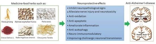
1. Introduction
2. Medicine and Food Herb Extract and Its Ingredients against Alzheimer’s Disease
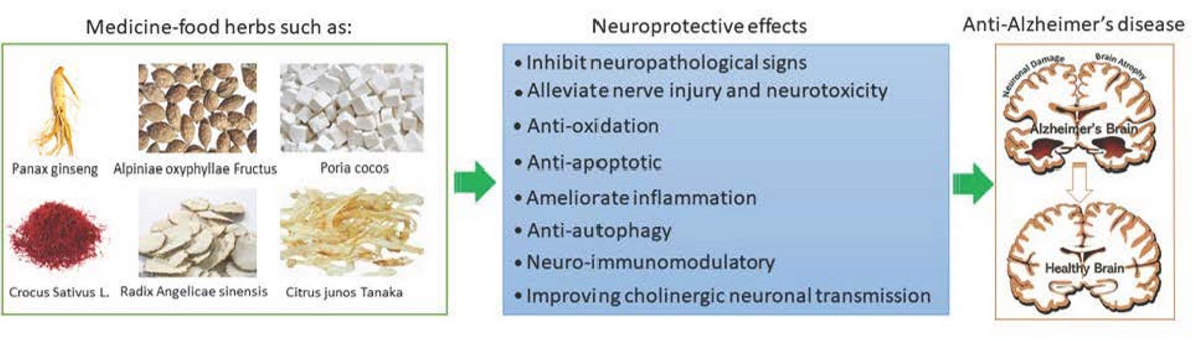
2.1. Panax Ginseng C.A. Mey
2.1.1. Traditional Functional Features
2.1.2. MOAs of the Extract
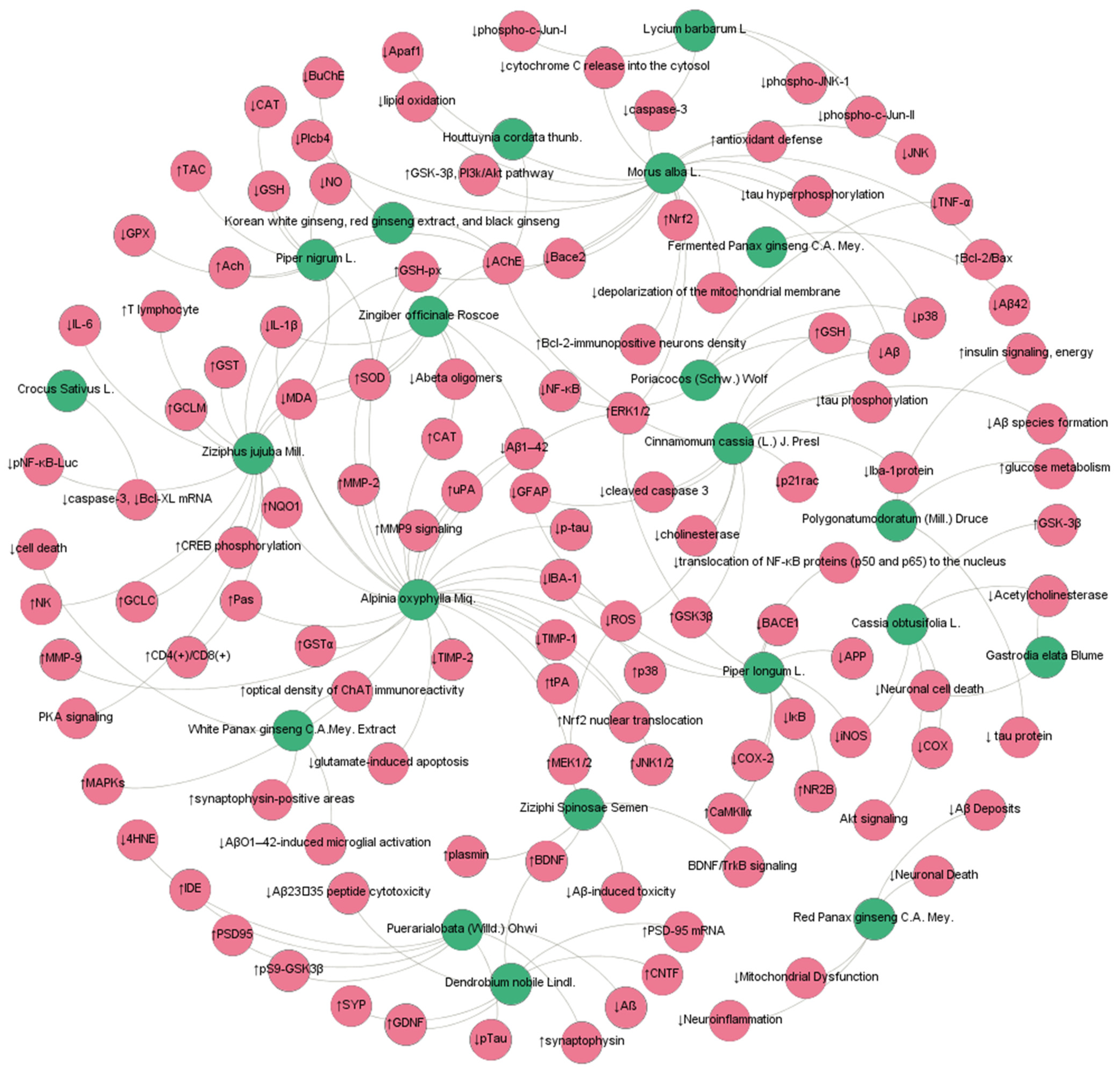
2.1.3. MOAs of Ingredients
2.1.4. Clinical Evidence
2.2. Crocus sativus L.
2.2.1. Traditional Functional Features
2.2.2. MOAs of the Extract
2.2.3. MOAs of Ingredients
2.2.4. Clinical Evidence
2.3. Cistanche afghanica Gilli
2.3.1. Traditional Functional Features
2.3.2. MOAs of the Extract
2.3.3. MOAs of Ingredients
2.3.4. Clinical Evidence
2.4. Angelica acutiloba (Siebold & Zucc.) Kitag.
2.4.1. Traditional Functional Features
2.4.2. MOAs of the Extract
2.4.3. MOAs of Ingredients
2.5. Astragalus aaronii (Eig) Zohary
2.5.1. Traditional Functional Features
2.5.2. MOAs of the Extract
2.5.3. MOAs of Ingredients
2.6. Poriacocos (Schw.) Wolf
2.6.1. Traditional Functional Features
2.6.2. MOAs of the Extract
2.7. Alpinia oxyphylla Miq.
2.7.1. Traditional Functional Features
2.7.2. MOAs of the Extract
2.7.3. MOAs of Ingredients
2.8. Zingiber officinale Roscoe
2.8.1. Traditional Functional Features
2.8.2. MOAs of the Extract
2.8.3. MOAs of Ingredients
2.9. Polygonatumodoratum (Mill.) Druce
2.9.1. Traditional Functional Features
2.9.2. MOAs of the Extract
2.9.3. MOAs of Ingredients
2.10. Piper nigrum L.
2.10.1. Traditional Functional Features
2.10.2. MOAs of the Extract
2.10.3. MOAs of Ingredients
2.11. Ganoderma lucidum (Leyss.ex Fr.) Karst.
2.11.1. Traditional Functional Features
2.11.2. MOAs of the Extract
2.11.3. MOAs of Ingredients
2.12. Puerarialobata (Willd.) Ohwi
2.12.1. Traditional Functional Features
2.12.2. MOAs of the Extract
2.12.3. MOAs of Ingredients
2.13. Ziziphi Spinosae Semen
2.13.1. Traditional Functional Features
2.13.2. MOAs of the Extract
2.13.3. MOAs of Ingredients
2.14. Gastrodia elata Blume
2.14.1. Traditional Functional Features
2.14.2. MOAs of the Extract
2.14.3. MOAs of Ingredients
2.15. Lycium barbarum L.
2.15.1. Traditional Functional Features
2.15.2. MOAs of the Extract
2.15.3. MOAs of Ingredients
2.16. Alpinia officinarum Hance/Mosla chinensis Maxim
2.16.1. Traditional Functional Features
2.16.2. MOAs of Ingredients
2.17. Curcuma longa L.
2.17.1. Traditional Functional Features
2.17.2. MOAs of Ingredients
3. Other MFH with Potential AntiAD Activity
4. Discussion and Conclusions
Author Contributions
Funding
Institutional Review Board Statement
Informed Consent Statement
Data Availability Statement
Conflicts of Interest
References
- Soria Lopez, J.A.; González, H.M.; Léger, G.C. Alzheimer’s disease. Handb. Clin. Neurol. 2019, 167, 231–255. [Google Scholar] [CrossRef] [PubMed]
- Ferrari, C.; Sorbi, S. The complexity of Alzheimer’s disease: An evolving puzzle. Physiol. Rev. 2021, 101, 1047–1081. [Google Scholar] [CrossRef] [PubMed]
- Prince, M.; Bryce, R.; Albanese, E.; Wimo, A.; Ribeiro, W.; Ferri, C.P. The global prevalence of dementia: A systematic review and metaanalysis. Alzheimer’s Dement. 2013, 9, 63–75. [Google Scholar] [CrossRef] [PubMed]
- DeTure, M.A.; Dickson, D.W. The neuropathological diagnosis of Alzheimer’s disease. Mol. Neurodegener. 2019, 14, 32. [Google Scholar] [CrossRef]
- Lashley, T.; Schott, J.M.; Weston, P.; Murray, C.E.; Wellington, H.; Keshavan, A.; Zetterberg, H. Molecular biomarkers of Alzheimer’s disease: Progress and prospects. Dis. Model Mech. 2018, 8, 11. [Google Scholar] [CrossRef]
- Oboudiyat, C.; Glazer, H.; Seifan, A.; Greer, C.; Isaacson, R.S. Alzheimer’s disease. Semin. Neurol. 2013, 33, 313–329. [Google Scholar] [CrossRef]
- Chu, L.W. Alzheimer’s disease: Early diagnosis and treatment. Hong Kong Med. J. 2012, 18, 228–237. [Google Scholar]
- Hou, Y.; Jiang, J.-G. Origin and concept of medicine food homology and its application in modern functional foods. Food Funct. 2013, 4, 1727–1741. [Google Scholar] [CrossRef]
- Owen, L.; Corfe, B. The role of diet and nutrition on mental health and wellbeing. Proc. Nutr. Soc. 2017, 76, 425–426. [Google Scholar] [CrossRef]
- Popa, S.L.; Pop, C.; Dumitrascu, D.L. Diet Advice for Crohn’s Disease: FODMAP and Beyond. Nutrients 2020, 12, 3751. [Google Scholar] [CrossRef]
- Zhang, X.; Wang, X.; Xue, Z.; Zhan, G.; Ito, Y.; Guo, Z. Prevention properties on cerebral ischemia reperfusion of medicine food homologous Dioscorea yam-derived diosgenin based on mediation of potential targets. Food Chem. 2021, 345, 128672. [Google Scholar] [CrossRef] [PubMed]
- Gong, X.; Ji, M.; Xu, J.; Zhang, C.; Li, M. Hypoglycemic effects of bioactive ingredients from medicine food homology and medicinal health food species used in China. Crit. Rev. Food Sci. Nutr. 2019, 60, 2303–2326. [Google Scholar] [CrossRef] [PubMed]
- Song, D.X.; Jiang, J.G. Hypolipidemic Components from Medicine Food Homology Species Used in China: Pharmacological and Health Effects. Arch. Med. Res. 2017, 48, 569–581. [Google Scholar] [CrossRef] [PubMed]
- Lee, S.-T.; Chu, K.; Sim, J.-Y.; Heo, J.-H.; Kim, M. Panax Ginseng Enhances Cognitive Performance in Alzheimer Disease. Alzheimer Dis. Assoc. Disord. 2008, 22, 222–226. [Google Scholar] [CrossRef] [PubMed]
- Heo, J.H.; Lee, S.T.; Chu, K.; Oh, M.J.; Park, H.J.; Shim, J.Y.; Kim, M. An open-label trial of Korean red ginseng as an adjuvant treatment for cognitive impairment in patients with Alzheimer’s disease. Eur. J. Neurol. 2008, 15, 865–868. [Google Scholar] [CrossRef]
- Heo, J.H.; Lee, S.T.; Oh, M.J.; Park, H.J.; Shim, J.Y.; Chu, K.; Kim, M. Improvement of cognitive deficit in Alzheimer’s disease patients by long term treatment with Korean red ginseng. J. Ginseng. Res. 2011, 35, 457–461. [Google Scholar] [CrossRef]
- Akhondzadeh, S.; Shafiee Sabet, M.; Harirchian, M.H.; Togha, M.; Cheraghmakani, H.; Razeghi, S.; Vossoughi, A. A 22-week, multicenter, randomized, double-blind controlled trial of Crocus sativus in the treatment of mild-to-moderate Alzheimer’s disease. Psychopharmacology 2010, 207, 637–643. [Google Scholar] [CrossRef]
- Akhondzadeh, S.; Sabet, M.S.; Harirchian, M.H.; Togha, M.; Cheraghmakani, H.; Razeghi, S.; Moradi, A. Saffron in the treatment of patients with mild to moderate Alzheimer’s disease: A 16-week, randomized and placebo-controlled trial. J. Clin. Pharm. 2010, 35, 581–588. [Google Scholar] [CrossRef]
- Tsolaki, M.; Karathanasi, E.; Lazarou, I.; Dovas, K.; Verykouki, E.; Karakostas, A.; Georgiadis, K.; Tsolaki, A.; Adam, K.; Kompatsiaris, I.; et al. Efficacy and Safety of Crocus sativus L. in Patients with Mild Cognitive Impairment: One Year Single-Blind Randomized, with Parallel Groups, Clinical Trial. J. Alzheimer’s Dis. 2016, 54, 129–133. [Google Scholar] [CrossRef]
- Li, N.; Wang, J.; Ma, J.; Gu, Z.; Jiang, C.; Yu, L.; Fu, X. Neuroprotective Effects of Cistanches Herba Therapy on Patients with Moderate Alzheimer’s Disease. Evid. Based Complement. Altern. Med. 2015, 2015, 103985. [Google Scholar] [CrossRef]
- Fang, S.; Dong, L.; Liu, L.; Guo, J.; Zhao, L.; Zhang, J.; Zhao, Y. HERB: A high-throughput experiment-and refer-ence-guided database of traditional Chinese medicine. Nucleic Acids Res. 2021, 49, D1197–D1206. [Google Scholar] [CrossRef] [PubMed]
- Sun, C.; Liu, J.; Li, N.; Liu, M.; Luo, Z.; Li, H. Traditional Chinese Medicine Shenmayizhi Decoction Ameliorates Memory and Cognitive Impairment Induced by Multiple Cerebral Infarctions. Evid. Based Complement. Altern. Med. 2021, 2021, 6648455. [Google Scholar] [CrossRef] [PubMed]
- Ma, L.; Cao, Y.; Wang, F.; Li, Z.; Wang, Z.; Yang, Y.; Pei, H.; Li, H. Yizhi Qingxin Formula Extract Ameliorates Cognitive Decline in Aged Rats via the Brain-Derived Neurotrophic Factor/Tropomyosin Receptor Kinase B Pathway. Front. Pharmacol. 2020, 11, 510. [Google Scholar] [CrossRef] [PubMed]
- Yang, Y.; Wang, Z.; Cao, Y.; Liu, J.; Li, P.; Li, H.; Liu, M. Yizhiqingxin Formula Alleviates Cognitive Deficits and Enhances Autophagy via mTOR Signaling Pathway Modulation in Early Onset Alzheimer’s Disease Mice. Front. Pharmacol. 2019, 10, 1041. [Google Scholar] [CrossRef] [PubMed]
- Seo, J.S.; Yun, J.H.; Baek, I.S.; Leem, Y.H.; Kang, H.W.; Cho, H.K.; Han, P.L. Oriental medicine Jangwonhwan reduces Abeta(1–42) level and beta-amyloid deposition in the brain of Tg-APPswe/PS1dE9 mouse model of Alzheimer disease. J. Ethnopharmacol. 2010, 128, 206–212. [Google Scholar] [CrossRef]
- Gong, G.; Qi, B.; Liang, Y.T.; Dong, T.T.X.; Wang, H.Y.; Tsim, K.W.K.; Zheng, Y. Danggui Buxue Tang, an ancient Chinese herbal decoction, protects β-amyloid-induced cell death in cultured cortical neurons. BMC Complement. Altern. Med. 2019, 19, 9. [Google Scholar] [CrossRef]
- Huang, Y.; Hu, Z.-Y.; Yuan, H.; Shu, L.; Liu, G.; Qiao, S.-Y.; Sun, L.; Zhou, W.-X.; Zhang, Y.-X. Danggui-Shaoyao-San Improves Learning and Memory in Female SAMP8 via Modulation of Estradiol. Evid. Based Complement. Altern. Med. 2014, 2014, 327294. [Google Scholar] [CrossRef]
- Huang, J.; Wang, X.; Xie, L.; Wu, M.; Zhao, W.; Zhang, Y.; Wang, Q.; Yao, L.; Li, W. Extract of Danggui-Shaoyao-San ameliorates cognition deficits by regulating DHA metabolism in APP/PS1 mice. J. Ethnopharmacol. 2020, 253, 112673. [Google Scholar] [CrossRef]
- Lu, J.; Guo, P.; Liu, X.; Zhang, Y.; Guo, X.; Gao, X.; Chen, Y. Herbal Formula Fo Shou San Attenuates Alzheimer’s Dis-ease-Related Pathologies via the Gut-Liver-Brain Axis in APP/PS1 Mouse Model of Alzheimer’s Disease. Evid. Based Complement. Altern. Med. 2019, 2019, 8302950. [Google Scholar] [CrossRef]
- Hu, Y.R.; Xing, S.L.; Chen, C.; Shen, D.Z.; Chen, J.L. Tiaoxin Recipe, a Chinese herbal formula, inhibits microRNA-34a expression in the APPswe/PS1ΔE9 mouse model of Alzheimer’s disease. J. Integr. Med. 2019, 17, 404–409. [Google Scholar] [CrossRef]
- Wang, F.; Feng, J.; Yang, Y.; Liu, J.; Liu, M.; Wang, Z.; Pei, H.; Wei, Y.; Li, H. The Chinese herbal formula Fuzheng Quxie Decoction attenuates cognitive impairment and protects cerebrovascular function in SAMP8 mice. Neuropsychiatr. Dis. Treat. 2018, 14, 3037–3051. [Google Scholar] [CrossRef] [PubMed]
- Hu, Q.; Yu, B.; Chen, Q.; Wang, Y.; Ling, Y.; Sun, S.; Zhou, C. Effect of Linguizhugan decoction on neuroinflammation and expression disorder of the amyloid β-related transporters RAGE and LRP-1 in a rat model of Alzheimer’s disease. Mol. Med. Rep. 2018, 17, 827–834. [Google Scholar] [CrossRef] [PubMed]
- Shin, S.J.; Jeon, S.G.; Kim, J.I.; Jeong, Y.O.; Kim, S.; Park, Y.H.; Moon, M. Red Ginseng Attenuates Aβ-Induced Mitochondrial Dysfunction and Aβ-mediated Pathology in an Animal Model of Alzheimer’s Disease. Int. J. Mol. Sci. 2019, 20, 3030. [Google Scholar] [CrossRef] [PubMed]
- Kim, J.; Kim, S.H.; Lee, D.S.; Lee, D.J.; Kim, S.H.; Chung, S.; Yang, H.O. Effects of fermented ginseng on memory impairment and β-amyloid reduction in Alzheimer’s disease experimental models. J. Ginseng. Res. 2013, 37, 100–107. [Google Scholar] [CrossRef]
- Choi, J.G.; Kim, N.; Huh, E.; Lee, H.; Oh, M.H.; Park, J.D.; Pyo, M.K.; Oh, M.S. White Ginseng Protects Mouse Hippocampal Cells Against Amyloid-Beta Oligomer Toxicity. Phytother. Res. 2017, 31, 497–506. [Google Scholar] [CrossRef]
- Soeda, S.; Ochiai, T.; Paopong, L.; Tanaka, H.; Shoyama, Y.; Shimeno, H. Crocin suppresses tumor necrosis fac-tor-alpha-induced cell death of neuronally differentiated PC-12 cells. Life Sci. 2001, 69, 2887–2898. [Google Scholar] [CrossRef]
- Sun, Y.; Liu, Z.; Pi, Z.; Song, F.; Wu, J.; Liu, S. Poria cocos could ameliorate cognitive dysfunction in APP / PS1 mice by restoring imbalance of Aβ production and clearance and gut microbiota dysbiosis. Phytother. Res. 2021, 35, 2678–2690. [Google Scholar] [CrossRef]
- Huang, Y.-J.; Hsu, N.-Y.; Lu, K.-H.; Lin, Y.-E.; Lin, S.-H.; Lu, Y.-S.; Liu, W.-T.; Chen, M.-H.; Sheen, L.-Y. Poria cocos water extract ameliorates the behavioral deficits induced by unpredictable chronic mild stress in rats by down-regulating inflammation. J. Ethnopharmacol. 2020, 258, 112566. [Google Scholar] [CrossRef]
- Chang, Y.M.; Ye, C.X.; Ho, T.J.; Tsai, T.N.; Chiu, P.L.; Tsai, C.C.; Huang, C.Y. Alpinia oxyphylla Miquel fruit extract activates MAPK-mediated signaling of PAs and MMP2/9 to induce Schwann cell migration and nerve regeneration. Int. J. Artif. Organs 2014, 37, 402–413. [Google Scholar] [CrossRef]
- Wang, Y.; Wang, M.; Fan, K.; Li, T.; Yan, T.; Wu, B.; Jia, Y. Protective effects of Alpinia oxyphylla Fructus extracts on lipopolysaccharide-induced animal model of Alzheimer’s disease. J. Ethnopharmacol. 2018, 217, 98–106. [Google Scholar] [CrossRef]
- Yu, S.H.; Kim, H.J.; Jeon, S.Y.; Kim, M.R.; Lee, B.S.; Lee, J.J.; Kim, D.S.; Lee, Y.C. Anti-inflammatory and anti-nociceptive activities of Alpinia oxyphylla Miquel extracts in animal models. J Ethnopharmacol. 2020, 260, 112985. [Google Scholar] [CrossRef] [PubMed]
- Yu, X.; An, L.; Wang, Y.; Zhao, H.; Gao, C. Neuroprotective effect of Alpinia oxyphylla Miq. fruits against gluta-mate-induced apoptosis in cortical neurons. Toxicol. Lett. 2003, 144, 205–212. [Google Scholar] [CrossRef]
- Zeng, G.-F.; Zhang, Z.-Y.; Lu, L.; Xiao, D.-Q.; Zong, S.-H.; He, J.-M. Protective Effects of Ginger Root Extract on Alzheimer Disease-Induced Behavioral Dysfunction in Rats. Rejuvenation Res. 2013, 16, 124–133. [Google Scholar] [CrossRef] [PubMed]
- Kongsui, R.; Sriraksa, N.; Thongrong, S. The Neuroprotective Effect of Zingiber cassumunar Roxb. Extract on LPS-Induced Neuronal Cell Loss and Astroglial Activation within the Hippocampus. BioMed Res. Int. 2020, 2020, 4259316. [Google Scholar] [CrossRef] [PubMed]
- Oboh, G.; Ademiluyi, A.O.; Akinyemi, A. Inhibition of acetylcholinesterase activities and some pro-oxidant induced lipid peroxidation in rat brain by two varieties of ginger (Zingiber officinale). Exp. Toxicol. Pathol. 2012, 64, 315–319. [Google Scholar] [CrossRef] [PubMed]
- Mathew, M.; Subramanian, S. In vitro evaluation of anti-Alzheimer effects of dry ginger (Zingiber officinale Roscoe) extract. Indian J. Exp. Boil. 2014, 52, 606–612. [Google Scholar]
- Yang, H.J.; Hwang, J.T.; Kwon, D.Y.; Kim, M.J.; Kang, S.; Moon, N.R.; Park, S. Yuzu Extract Prevents Cognitive Decline and Impaired Glucose Homeostasis in β-Amyloid–Infused Rats. J. Nutr. 2013, 143, 1093–1099. [Google Scholar] [CrossRef]
- Mahdy, K.; Shaker, O.; Wafay, H.; Nassar, Y.; Hassan, H.; Hussein, A. Effect of some medicinal plant extracts on the oxidative stress status in Alzheimer’s disease induced in rats. Eur. Rev. Med. Pharm. Sci. 2012, 16, 31–42. [Google Scholar]
- Hritcu, L.; Noumedem, A.J.; Cioanca, O.; Hancianu, M.; Postu, P.; Mihasan, M. Anxiolytic and antidepressant profile of the methanolic extract of Piper nigrum fruits in beta-amyloid (1–42) rat model of Alzheimer’s disease. Behav. Brain Funct. 2015, 11, 1–13. [Google Scholar] [CrossRef]
- Kim, H.G.; Park, G.; Lim, S.; Park, H.; Choi, J.G.; Jeong, H.U.; Kang, M.S.; Lee, M.K.; Oh, M.S. Mori Fructus improves cognitive and neuronal dysfunction induced by beta-amyloid toxicity through the GSK-3β pathway in vitro and in vivo. J. Ethnopharmacol. 2015, 171, 196–204. [Google Scholar] [CrossRef]
- Kaewkaen, P.; Tong-Un, T.; Wattanathorn, J.; Muchimapura, S.; Kaewrueng, W.; Wongcharoenwanakit, S. Mulberry Fruit Extract Protects against Memory Impairment and Hippocampal Damage in Animal Model of Vascular Dementia. Evid. Based Complement. Altern. Med. 2012, 2012, 263520. [Google Scholar] [CrossRef] [PubMed]
- Shih, P.H.; Chan, Y.C.; Liao, J.W.; Wang, M.F.; Yen, G.C. Antioxidant and cognitive promotion effects of anthocyanin-rich mulberry (Morus atropurpurea L.) on senescence-accelerated mice and prevention of Alzheimer’s disease. J. Nutr. Biochem. 2010, 21, 598–605. [Google Scholar] [CrossRef] [PubMed]
- Song, N.; Yang, H.; Pang, W.; Qie, Z.; Lu, H.; Tan, L.; Li, H.; Sun, S.; Lian, F.; Qin, C.; et al. Mulberry extracts alleviate aβ 25-35-induced injury and change the gene expression profile in PC12 cells. Evid. Based Complement. Altern. Med. 2014, 2014, 150617. [Google Scholar] [CrossRef] [PubMed]
- Huang, H.-J.; Huang, C.-Y.; Lee, M.; Lin, J.-Y.; Hsieh-Li, H.M. Puerariae Radix Prevents Anxiety and Cognitive Deficits in Mice Under Oligomeric Aβ-Induced Stress. Am. J. Chin. Med. 2019, 47, 1459–1481. [Google Scholar] [CrossRef]
- Park, H.J.; Jung, I.H.; Kwon, H.; Yu, J.; Jo, E.; Kim, H.; Ryu, J.H. The ethanol extract of Zizyphus jujuba var. spinosa seeds ameliorates the memory deficits in Alzheimer’s disease model mice. J. Ethnopharmacol. 2019, 233, 73–79. [Google Scholar] [CrossRef]
- Kwon, H.; Jung, I.H.; Yi, J.H.; Kim, J.H.; Park, J.H.; Lee, S.; Kim, D.H. The Seed of Zizyphus jujuba var. spinosa Attenuates Alzheimer’s Disease-Associated Hippocampal Synaptic Deficits through BDNF/TrkB Signaling. Biol. Pharm. Bull. 2017, 40, 2096–2104. [Google Scholar] [CrossRef]
- Yang, T.; Fang, L.; Lin, T.; Li, J.; Zhang, Y.; Zhou, A.; Xie, J. Ultrasonicated sour Jujube seed flavonoids extract exerts ameliorative antioxidant capacity and reduces Aβ-induced toxicity in Caenorhabditis elegans. J. Ethnopharmacol. 2019, 239, 111886. [Google Scholar] [CrossRef]
- Frydman-Marom, A.; Levin, A.; Farfara, D.; Benromano, T.; Scherzer-Attali, R.; Peled, S.; Vassar, R.; Segal, D.; Gazit, E.; Frenkel, D.; et al. Orally administrated cinnamon extract reduces β-amyloid oligomerization and corrects cognitive impairment in Alzheimer’s disease animal models. PLoS ONE 2011, 6, e16564. [Google Scholar] [CrossRef]
- Modi, K.K.; Roy, A.; Brahmachari, S.; Rangasamy, S.B.; Pahan, K. Cinnamon and Its Metabolite Sodium Benzoate Attenuate the Activation of p21rac and Protect Memory and Learning in an Animal Model of Alzheimer’s Disease. PLoS ONE 2015, 10, e0130398. [Google Scholar] [CrossRef]
- Madhavadas, S.; Subramanian, S. Cognition enhancing effect of the aqueous extract of Cinnamomum zeylanicum on non-transgenic Alzheimer’s disease rat model: Biochemical, histological, and behavioural studies. Nutr. Neurosci. 2017, 20, 526–537. [Google Scholar] [CrossRef]
- Park, Y.M.; Lee, B.G.; Park, S.H.; Oh, H.G.; Kang, Y.G.; Kim, O.J.; Lee, H.Y. Prolonged oral administration of Gastrodia elata extract improves spatial learning and memory of scopolamine-treated rats. Lab. Anim. Res. 2015, 31, 69–77. [Google Scholar] [CrossRef][Green Version]
- Huang, G.B.; Zhao, T.; Muna, S.S.; Jin, H.M.; Park, J.I.; Jo, K.S.; Chung, Y.C. Therapeutic potential of Gastrodia elata Blume for the treatment of Alzheimer’s disease. Neural Regen. Res. 2013, 8, 1061–1070. [Google Scholar] [CrossRef] [PubMed]
- Yu, M.S.; Leung, S.K.; Lai, S.W.; Che, C.M.; Zee, S.Y.; So, K.F.; Chang, R.C. Neuroprotective effects of anti-aging oriental medicine Lycium barbarum against beta-amyloid peptide neurotoxicity. Exp. Gerontol. 2005, 40, 716–727. [Google Scholar] [CrossRef]
- Huh, E.; Kim, H.G.; Park, H.; Kang, M.S.; Lee, B.; Oh, M.S. Houttuynia cordata Improves Cognitive Deficits in Cholinergic Dysfunction Alzheimer’s Disease-Like Models. Biomol. Ther. 2014, 22, 176–183. [Google Scholar] [CrossRef] [PubMed]
- Kim, D.H.; Yoon, B.H.; Kim, Y.W.; Lee, S.; Shin, B.Y.; Jung, J.W.; Kim, H.J.; Lee, Y.S.; Choi, J.S.; Kim, S.Y.; et al. The seed extract of Cassia obtusifolia ameliorates learning and memory impairments induced by scopolamine or transient cerebral hypoperfusion in mice. J. Pharmacol. Sci. 2007, 105, 82–93. [Google Scholar] [CrossRef] [PubMed]
- Yi, J.H.; Park, H.J.; Lee, S.; Jung, J.W.; Kim, B.C.; Lee, Y.C.; Ryu, J.H.; Kim, D.H. Cassia obtusifolia seed ameliorates amyloid β-induced synaptic dysfunction through anti-inflammatory and Akt/GSK-3β pathways. J. Ethnopharmacol. 2016, 178, 50–57. [Google Scholar] [CrossRef] [PubMed]
- Chen, J.; Maiwulanjiang, M.; Lam, K.Y.; Zhang, W.L.; Zhan, J.Y.; Lam, C.T.; Xu, S.L.; Zhu, K.Y.; Yao, P.; Lau, D.T.; et al. A standardized extract of the fruit of Ziziphus jujuba (Jujube) induces neuronal differentiation of cultured PC12 cells: A signaling mediated by protein kinase A. J. Agric. Food Chem. 2014, 62, 1890–1897. [Google Scholar] [CrossRef]
- Chen, J.; Yan, A.L.; Lam, K.Y.; Lam, C.T.; Li, N.; Yao, P.; Xiong, A.; Dong, T.T.; Tsim, K.W. A chemically standardized extract of Ziziphus jujuba fruit (Jujube) stimulates expressions of neurotrophic factors and anti-oxidant enzymes in cultured astrocytes. Phytother. Res. 2014, 28, 1727–1730. [Google Scholar] [CrossRef]
- Chen, J.; Du, C.Y.; Lam, K.Y.; Zhang, W.L.; Lam, C.T.; Yan, A.L.; Yao, P.; Lau, D.T.; Dong, T.T.; Tsim, K.W. The standardized extract of Ziziphus jujuba fruit (jujube) regulates pro-inflammatory cytokine expression in cultured murine macrophages: Suppression of lipopolysaccharide-stimulated NF-κB activity. Phytother. Res. 2014, 28, 1527–1532. [Google Scholar] [CrossRef]
- Nie, J.; Tian, Y.; Zhang, Y.; Lu, Y.L.; Li, L.S.; Shi, J.S. Dendrobium alkaloids prevent Aβ(25-35)-induced neuronal and synaptic loss via promoting neurotrophic factors expression in mice. PeerJ 2016, 4, e2739. [Google Scholar] [CrossRef]
- Zhang, W.; Wu, Q.; Lu, Y.L.; Gong, Q.H.; Zhang, F.; Shi, J.S. Protective effects of Dendrobium nobile Lindl. alkaloids on amyloid beta (25-35)-induced neuronal injury. Neural Regen Res. 2017, 12, 1131–1136. [Google Scholar] [CrossRef] [PubMed]
- Li, L.S.; Lu, Y.L.; Nie, J.; Xu, Y.Y.; Zhang, W.; Yang, W.J.; Gong, Q.H.; Lu, Y.F.; Lu, Y.; Shi, J.S. Dendrobium nobile Lindl alkaloid, a novel autophagy inducer, protects against axonal degeneration induced by Aβ(25-35) in hippocampus neurons in vitro. CNS Neurosci. Ther. 2017, 23, 329–340. [Google Scholar] [CrossRef] [PubMed]
- Go, J.; Park, T.S.; Han, G.H.; Park, H.Y.; Ryu, Y.K.; Kim, Y.H.; Hwang, J.H.; Choi, D.H.; Noh, J.R.; Hwang, D.Y.; et al. Piperlongumine decreases cognitive impairment and improves hippocampal function in aged mice. Int. J. Mol. Med. 2018, 42, 1875–1884. [Google Scholar] [CrossRef] [PubMed]
- Gu, S.M.; Lee, H.P.; Ham, Y.W.; Son, D.J.; Kim, H.Y.; Oh, K.W.; Han, S.B.; Yun, J.; Hong, J.T. Piperlongumine Improves Lipopolysaccharide-Induced Amyloidogenesis by Suppressing NF-KappaB Pathway. Neuromol. Med. 2018, 20, 312–327. [Google Scholar] [CrossRef]
- Zhu, J.; Mu, X.; Zeng, J.; Xu, C.; Liu, J.; Zhang, M.; Li, C.; Chen, J.; Li, T.; Wang, Y. Ginsenoside Rg1 Prevents Cognitive Impairment and Hippocampus Senescence in a Rat Model of D-Galactose-Induced Aging. PLoS ONE 2014, 9, e101291. [Google Scholar] [CrossRef]
- Qiu, J.; Li, W.; Feng, S.H.; Wang, M.; He, Z.Y. Ginsenoside Rh2 promotes nonamyloidgenic cleavage of amyloid pre-cursor protein via a cholesterol-dependent pathway. Genet. Mol. Res. 2014, 13, 3586–3598. [Google Scholar] [CrossRef]
- Li, L.; Liu, J.; Yan, X.; Qin, K.; Shi, M.; Lin, T.; Zhu, Y.; Kang, T.; Zhao, G. Protective effects of ginsenoside Rd against okadaic acid-induced neurotoxicity in vivo and in vitro. J. Ethnopharmacol. 2011, 138, 135–141. [Google Scholar] [CrossRef]
- Hwang, S.H.; Shin, T.-J.; Choi, S.-H.; Cho, H.-J.; Lee, B.-H.; Pyo, M.K.; Lee, J.-H.; Kang, J.; Kim, H.-J.; Park, C.-W.; et al. Gintonin, newly identified compounds from ginseng, is novel lysophosphatidic acids-protein complexes and activates G protein-coupled lysophosphatidic acid receptors with high affinity. Mol. Cells 2012, 33, 151–162. [Google Scholar] [CrossRef]
- Guo, J.; Chang, L.; Zhang, X.; Pei, S.; Yu, M.; Gao, J. Ginsenoside compound K promotes β-amyloid peptide clearance in primary astrocytes via autophagy enhancement. Exp. Ther. Med. 2014, 8, 1271–1274. [Google Scholar] [CrossRef]
- Chalatsa, I.; Arvanitis, D.A.; Koulakiotis, N.S.; Giagini, A.; Skaltsounis, A.L.; Papadopoulou-Daifoti, Z.; Tsarbopoulos, A.; Sanoudou, D. The Crocus sativus Compounds trans-Crocin 4 and trans-Crocetin Modulate the Amyloidogenic Pathway and Tau Misprocessing in Alzheimer Disease Neuronal Cell Culture Models. Front. Neurosci. 2019, 13, 249. [Google Scholar] [CrossRef]
- Deng, M.; Zhao, J.Y.; Ju, X.D.; Tu, P.F.; Jiang, Y.; Li, Z.B. Protective effect of tubuloside B on TNFalpha-induced apoptosis in neuronal cells. Acta Pharm. Sin. 2004, 25, 1276–1284. [Google Scholar]
- Li, X.; Gou, C.; Yang, H.; Qiu, J.; Gu, T.; Wen, T. Echinacoside ameliorates D-galactosamine plus lipopolysaccha-ride-induced acute liver injury in mice via inhibition of apoptosis and inflammation. Scand. J. Gastroenterol. 2014, 49, 993–1000. [Google Scholar] [CrossRef] [PubMed]
- Singh, J.C.H.; Kakalij, R.M.; Kshirsagar, R.P.; Kumar, B.H.; Komakula, S.S.B.; Diwan, P. Cognitive effects of vanillic acid against streptozotocin-induced neurodegeneration in mice. Pharm. Biol. 2014, 53, 630–636. [Google Scholar] [CrossRef] [PubMed]
- Yan, J.J.; Kim, D.H.; Moon, Y.S.; Jung, J.S.; Ahn, E.M.; Baek, N.I.; Song, D.K. Protection against beta-amyloid pep-tide-induced memory impairment with long-term administration of extract of Angelica gigas or decursinol in mice. Prog. Neuropsychopharmacol. Biol. Psychiatry 2004, 28, 25–30. [Google Scholar] [CrossRef]
- Li, L.; Du, J.; Zou, L.; Xia, H.; Wu, T.; Kim, Y.; Lee, Y. The Neuroprotective Effects of Decursin Isolated from Angelica gigas Nakai Against Amyloid β-Protein-Induced Apoptosis in PC 12 Cells via a Mitochondria-Related Caspase Pathway. Neurochem. Res. 2015, 40, 1555–1562. [Google Scholar] [CrossRef]
- Kuang, X.; Du, J.R.; Chen, Y.S.; Wang, J.; Wang, Y.N. Protective effect of Z-ligustilide against amyloid beta-induced neurotoxicity is associated with decreased pro-inflammatory markers in rat brains. Pharm. Biochem. Behav. 2009, 92, 635–641. [Google Scholar] [CrossRef]
- Xu, W.; Yang, L.; Li, J. Protection against β-amyloid-induced neurotoxicity by naturally occurring Z-ligustilide through the concurrent regulation of p38 and PI3-K/Akt pathways. Neurochem. Int. 2016, 100, 44–51. [Google Scholar] [CrossRef]
- Wang, X.; Xu, W.; Chen, H.; Li, W.; Li, W.; Zhu, G. Astragaloside IV prevents Aβ1-42 oligomers-induced memory impairment and hippocampal cell apoptosis by promoting PPARγ/BDNF signaling pathway. Brain Res. 2020, 1747, 147041. [Google Scholar] [CrossRef]
- Wang, X.; Wang, Y.; Hu, J.P.; Yu, S.; Li, B.K.; Cui, Y.; Zhang, L.D. Astragaloside IV, a Natural PPARγ Agonist, Reduces Aβ Production in Alzheimer’s Disease Through Inhibition of BACE1. Mol. Neurobiol. 2017, 54, 2939–2949. [Google Scholar] [CrossRef]
- Sun, Q.; Jia, N.; Wang, W.; Jin, H.; Xu, J.; Hu, H. Protective effects of astragaloside IV against amyloid beta1-42 neuro-toxicity by inhibiting the mitochondrial permeability transition pore opening. PLoS ONE 2014, 9, e98866. [Google Scholar] [CrossRef]
- Liu, A.; Zhao, X.; Li, H.; Liu, Z.; Liu, B.; Mao, X.; Jia, Y. 5-Hydroxymethylfurfural, an antioxidant agent from Alpinia oxyphylla Miq. improves cognitive impairment in Aβ 1-42 mouse model of Alzheimer’s disease. Int. Immunopharmacol. 2014, 23, 719–725. [Google Scholar] [CrossRef] [PubMed]
- Shi, S.-H.; Zhao, X.; Liu, A.-J.; Liu, B.; Li, H.; Wu, B.; Bi, K.-S.; Jia, Y. Protective effect of n-butanol extract from Alpinia oxyphylla on learning and memory impairments. Physiol. Behav. 2014, 139, 13–20. [Google Scholar] [CrossRef] [PubMed]
- Zeng, G.-F.; Zong, S.-H.; Zhang, Z.-Y.; Fu, S.-W.; Li, K.-K.; Fang, Y.; Lu, L.; Xiao, D.-Q. The Role of 6-Gingerol on Inhibiting Amyloid β Protein-Induced Apoptosis in PC12 Cells. Rejuvenation Res. 2015, 18, 413–421. [Google Scholar] [CrossRef] [PubMed]
- Gabriel, M.O.; Nikou, M.; Akinola, O.B.; Pollak, D.D.; Sideromenos, S. Western diet-induced fear memory impairment is attenuated by 6-shogaol in C57BL/6N mice. Behav. Brain Res. 2019, 380, 112419. [Google Scholar] [CrossRef] [PubMed]
- Moon, M.; Kim, H.G.; Choi, J.G.; Oh, H.; Lee, P.K.; Ha, S.K.; Kim, S.Y.; Park, Y.; Huh, Y.; Oh, M.S. 6-Shogaol, an active constituent of ginger, attenuates neuroinflammation and cognitive deficits in animal models of dementia. Biochem. Biophys. Res. Commun. 2014, 449, 8–13. [Google Scholar] [CrossRef] [PubMed]
- Wang, D.M.; Yang, Y.J.; Zhang, L.; Zhang, X.; Guan, F.F.; Zhang, L.F. Naringin Enhances CaMKII Activity and Improves Long-Term Memory in a Mouse Model of Alzheimer’s Disease. Int. J. Mol. Sci. 2013, 14, 5576–5586. [Google Scholar] [CrossRef] [PubMed]
- Justin Thenmozhi, A.; William Raja, T.R.; Manivasagam, T.; Janakiraman, U.; Essa, M.M. Hesperidin ameliorates cognitive dysfunction, oxidative stress and apoptosis against aluminium chloride induced rat model of Alzheimer’s disease. Nutr. Neurosci. 2017, 20, 360–368. [Google Scholar] [CrossRef]
- Li, C.; Zug, C.; Qu, H.; Schluesener, H.; Zhang, Z. Hesperidin ameliorates behavioral impairments and neuropathology of transgenic APP/PS1 mice. Behav. Brain Res. 2015, 281, 32–42. [Google Scholar] [CrossRef]
- Hong, Y.; An, Z. Hesperidin attenuates learning and memory deficits in APP/PS1 mice through activation of Akt/Nrf2 signaling and inhibition of RAGE/NF-κB signaling. Arch. Pharm. Res. 2018, 41, 655–663. [Google Scholar] [CrossRef]
- Shin, M.; Liu, Q.F.; Choi, B.; Shin, C.; Lee, B.; Yuan, C.; Song, Y.J.; Yun, H.S.; Lee, I.-S.; Koo, B.-S.; et al. Neuroprotective Effects of Limonene (+) against Aβ42-Induced Neurotoxicity in a Drosophila Model of Alzheimer’s Disease. Biol. Pharm. Bull. 2020, 43, 409–417. [Google Scholar] [CrossRef]
- Zhao, L.; Wang, J.L.; Liu, R.; Li, X.X.; Li, J.F.; Zhang, L. Neuroprotective, anti-amyloidogenic and neurotrophic effects of apigenin in an Alzheimer’s disease mouse model. Molecules 2013, 18, 9949–9965. [Google Scholar] [CrossRef] [PubMed]
- Zhao, L.; Wang, J.L.; Wang, Y.R.; Fa, X.Z. Apigenin attenuates copper-mediated β-amyloid neurotoxicity through antioxidation, mitochondrion protection and MAPK signal inactivation in an AD cell model. Brain Res. 2013, 1492, 33–45. [Google Scholar] [CrossRef] [PubMed]
- Ha, S.K.; Lee, P.; Park, J.A.; Oh, H.R.; Lee, S.Y.; Park, J.-H.; Lee, E.H.; Ryu, J.H.; Lee, K.R.; Kim, S.Y. Apigenin inhibits the production of NO and PGE2 in microglia and inhibits neuronal cell death in a middle cerebral artery occlusion-induced focal ischemia mice model. Neurochem. Int. 2008, 52, 878–886. [Google Scholar] [CrossRef]
- Dourado, N.S.; Souza, C.; De Almeida, M.M.A.; Da Silva, A.B.; dos Santos, B.L.; Silva, V.D.A.; De Assis, A.M.; Da Silva, J.S.; Souza, D.O.; Costa, M.D.F.D.; et al. Neuroimmunomodulatory and Neuroprotective Effects of the Flavonoid Apigenin in in vitro Models of Neuroinflammation Associated with Alzheimer’s Disease. Front. Aging Neurosci. 2020, 12, 119. [Google Scholar] [CrossRef] [PubMed]
- Guo, A.J.; Xie, H.Q.; Choi, R.C.; Zheng, K.Y.; Bi, C.W.; Xu, S.L.; Dong, T.T.; Tsim, K. Galangin, a flavonol derived from Rhizoma Alpiniae Officinarum, inhibits acetylcholinesterase activity in vitro. Chem. Interactions 2010, 187, 246–248. [Google Scholar] [CrossRef] [PubMed]
- Cheng, Y.; Dong, Z.; Liu, S. β-Caryophyllene Ameliorates the Alzheimer-Like Phenotype in APP/PS1 Mice through CB2 Receptor Activation and the PPARγ Pathway. Pharmacology 2014, 94, 1–12. [Google Scholar] [CrossRef] [PubMed]
- Abdul Manap, A.S.; Wei Tan, A.C.; Leong, W.H.; Yin Chia, A.Y. Synergistic Effects of Curcumin and Piperine as Potent Acetylcholine and Amyloidogenic Inhibitors With Significant Neuroprotective Activity in SH-SY5Y Cells via Computational Molecular Modeling and in vitro Assay. Front. Aging Neurosci. 2019, 11, 206. [Google Scholar] [CrossRef]
- Nazifi, M.; Oryan, S.; Esfahani, D.E.; Ashrafpoor, M. The functional effects of piperine and piperine plus donepezil on hippocampal synaptic plasticity impairment in rat model of Alzheimer’s disease. Life Sci. 2021, 265, 118802. [Google Scholar] [CrossRef]
- Chonpathompikunlert, P.; Wattanathorn, J.; Muchimapura, S. Piperine, the main alkaloid of Thai black pepper, protects against neurodegeneration and cognitive impairment in animal model of cognitive deficit like condition of Alzheimer’s disease. Food Chem. Toxicol. 2010, 48, 798–802. [Google Scholar] [CrossRef]
- Liu, Z.J.; Li, Z.H.; Liu, L.; Tang, W.X.; Wang, Y.; Dong, M.R.; Xiao, C. Curcumin Attenuates Beta-Amyloid-Induced Neuroinflammation via Activation of Peroxisome Proliferator-Activated Receptor-Gamma Function in a Rat Model of Alzheimer’s Disease. Front. Pharm. 2016, 7, 261. [Google Scholar] [CrossRef]
- Maiti, P.; Paladugu, L.; Dunbar, G.L. Solid lipid curcumin particles provide greater anti-amyloid, anti-inflammatory and neuroprotective effects than curcumin in the 5xFAD mouse model of Alzheimer’s disease. BMC Neurosci. 2018, 19, 7. [Google Scholar] [CrossRef] [PubMed]
- Park, S.Y.; Jin, M.L.; Kim, Y.H.; Kim, Y.; Lee, S.J. Anti-inflammatory effects of aromatic-turmerone through blocking of NF-κB, JNK, and p38 MAPK signaling pathways in amyloid β-stimulated microglia. Int. Immunopharmacol. 2012, 14, 13–20. [Google Scholar] [CrossRef] [PubMed]
- Chen, M.; Chang, Y.Y.; Huang, S.; Xiao, L.H.; Zhou, W.; Zhang, L.Y.; Zhang, K. Aromatic-Turmerone Attenuates LPS-Induced Neuroinflammation and Consequent Memory Impairment by Targeting TLR4-Dependent Signaling Path-way. Mol. Nutr. Food Res. 2018, 62, 1700281. [Google Scholar] [CrossRef] [PubMed]
- Saga, Y.; Hatakenaka, Y.; Matsumoto, M.; Yoshioka, Y.; Matsumura, S.; Zaima, N.; Konishi, Y. Neuroprotective effects of aromatic turmerone on activity deprivation-induced apoptosis in cerebellar granule neurons. NeuroReport 2020, 31, 1302–1307. [Google Scholar] [CrossRef]
- Hong, X.-P.; Chen, T.; Yin, N.-N.; Han, Y.-M.; Yuan, F.; Duan, Y.-J.; Shen, F.; Zhang, Y.-H.; Chen, Z.-B. Puerarin Ameliorates D-Galactose Induced Enhanced Hippocampal Neurogenesis and Tau Hyperphosphorylation in Rat Brain. J. Alzheimer’s Dis. 2016, 51, 605–617. [Google Scholar] [CrossRef]
- Zhou, Y.-X.; Zhang, H.; Peng, C. Puerarin: A Review of Pharmacological Effects. Phytother. Res. 2013, 28, 961–975. [Google Scholar] [CrossRef]
- Xing, G.; Dong, M.; Li, X.; Zou, Y.; Fan, L.; Wang, X.; Niu, Y. Neuroprotective effects of puerarin against be-ta-amyloid-induced neurotoxicity in PC12 cells via a PI3K-dependent signaling pathway. Brain Res. Bull. 2011, 85, 212–218. [Google Scholar] [CrossRef]
- Lee, Y.H.; Kim, J.H.; Song, C.H.; Jang, K.J.; Kim, C.H.; Kang, J.S.; Choi, Y.H.; Yoon, H.M. Ethanol Extract of Ganoderma lucidum Augments Cellular Anti-oxidant Defense through Activation of Nrf2/HO-1. J Pharmacopuncture 2016, 19, 59–69. [Google Scholar] [CrossRef]
- Zhang, X.; Wang, J.; Gong, G.; Ma, R.; Xu, F.; Yan, T.; Jia, Y. Spinosin Inhibits Aβ(1-42) Production and Aggregation via Activating Nrf2/HO-1 Pathway. Biomol. Ther. 2020, 28, 259–266. [Google Scholar] [CrossRef]
- Xu, F.; He, B.; Xiao, F.; Yan, T.; Bi, K.; Jia, Y.; Wang, Z. Neuroprotective Effects of Spinosin on Recovery of Learning and Memory in a Mouse Model of Alzheimer’s Disease. Biomol. Ther. 2019, 27, 71–77. [Google Scholar] [CrossRef]
- Cai, M.; Jung, I.; Kwon, H.; Cho, E.; Jeon, J.; Yun, J.; Lee, Y.C.; Kim, D.H.; Ryu, J.H. Spinosin Attenuates Alzheimer’s Disease-Associated Synaptic Dysfunction via Regulation of Plasmin Activity. Biomol. Ther. 2020, 28, 131–136. [Google Scholar] [CrossRef] [PubMed]
- Ko, S.Y.; Lee, H.E.; Park, S.J.; Jeon, S.J.; Kim, B.; Gao, Q.; Jang, D.S.; Ryu, J.H. Spinosin, a C-Glucosylflavone, from Zizyphus jujuba var. spinosa Ameliorates Aβ1-42 Oligomer-Induced Memory Impairment in Mice. Biomol. Ther. 2015, 23, 156–164. [Google Scholar] [CrossRef] [PubMed]
- Jia, S.L.; Wu, X.L.; Li, X.X.; Dai, X.L.; Gao, Z.L.; Lu, Z.; Zheng, Q.S.; Sun, Y.X. Neuroprotective effects of liquiritin on cognitive deficits induced by soluble amyloid-β(1-42) oligomers injected into the hippocampus. J. Asian Nat. Prod. Res. 2016, 18, 1186–1199. [Google Scholar] [CrossRef] [PubMed]
- Cui, Y.M.; Ao, M.Z.; Li, W.; Yu, L.J. Effect of glabridin from Glycyrrhiza glabra on learning and memory in mice. Planta Med. 2008, 74, 377–380. [Google Scholar] [CrossRef] [PubMed]
- Du, Y.; Luo, M.; Du, Y.; Xu, M.; Yao, Q.; Wang, K.; He, G. Liquiritigenin Decreases Aβ Levels and Ameliorates Cognitive Decline by Regulating Microglia M1/M2 Transformation in AD Mice. Neurotox Res. 2021, 39, 349–358. [Google Scholar] [CrossRef] [PubMed]
- Yang, W.; Liu, Y.; Xu, Q.Q.; Xian, Y.F.; Lin, Z.X. Sulforaphene Ameliorates Neuroinflammation and Hyperphosphorylated Tau Protein via Regulating the PI3K/Akt/GSK-3β Pathway in Experimental Models of Alzheimer’s Disease. Oxid. Med. Cell Longev. 2020, 2020, 4754195. [Google Scholar] [CrossRef] [PubMed]
- Liu, B.; Gao, J.M.; Li, F.; Gong, Q.H.; Shi, J.S. Gastrodin Attenuates Bilateral Common Carotid Artery Occlu-sion-Induced Cognitive Deficits via Regulating Aβ-Related Proteins and Reducing Autophagy and Apoptosis in Rats. Front. Pharm. 2018, 9, 405. [Google Scholar] [CrossRef]
- Yoon, J.H.; Youn, K.; Ho, C.T.; Karwe, M.V.; Jeong, W.S.; Jun, M. p-Coumaric acid and ursolic acid from Corni fructus attenuated β-amyloid(25-35)-induced toxicity through regulation of the NF-κB signaling pathway in PC12 cells. J. Agric. Food Chem. 2014, 62, 4911–4916. [Google Scholar] [CrossRef]
- Kiefer, D.; Pantuso, T. Panax ginseng. Am. Fam. Physician 2003, 68, 1539–1542. [Google Scholar]
- Ginseng. Drugs and Lactation Database (LactMed); National Library of Medicine: Bethesda, MD, USA, 2006. Available online: https://www.ncbi.nlm.nih.gov/books/NBK501814/ (accessed on 27 November 2021).
- Choi, S.-H.; Hong, M.-K.; Kim, H.-J.; Ryoo, N.; Rhim, H.; Nah, S.-Y.; Kang, L.-W. Structure of ginseng major latex-like protein 151 and its proposed lysophosphatidic acid-binding mechanism. Acta Crystallogr. Sect. D Biol. Crystallogr. 2015, 71, 1039–1050. [Google Scholar] [CrossRef]
- Fernández-Albarral, J.A.; de Hoz, R.; Ramírez, A.I.; López-Cuenca, I.; Salobrar-García, E.; Pinazo-Durán, M.D.; Salazar, J.J. Beneficial effects of saffron (Crocus sativus L.) in ocular pathologies, particularly neurodegenerative retinal diseases. Neural. Regen. Res. 2020, 15, 1408–1416. [Google Scholar] [CrossRef] [PubMed]
- Moshiri, M.; Vahabzadeh, M.; Hosseinzadeh, H. Clinical Applications of Saffron (Crocus sativus) and its Constituents: A Review. Drug Res. 2014, 65, 287–295. [Google Scholar] [CrossRef] [PubMed]
- Hatziagapiou, K.; Kakouri, E.; Lambrou, G.I.; Bethanis, K.; Tarantilis, P.A. Antioxidant Properties of Crocus sativus L. and Its Constituents and Relevance to Neurodegenerative Diseases; Focus on Alzheimer’s and Parkinson’s Disease. Curr. Neuropharmacol. 2019, 17, 377–402. [Google Scholar] [CrossRef] [PubMed]
- Ochiai, T.; Shimeno, H.; Mishima, K.-I.; Iwasaki, K.; Fujiwara, M.; Tanaka, H.; Shoyama, Y.; Toda, A.; Eyanagi, R.; Soeda, S. Protective effects of carotenoids from saffron on neuronal injury in vitro and in vivo. Biochim. Biophys. Acta Gen. Subj. 2007, 1770, 578–584. [Google Scholar] [CrossRef]
- Song, Y.; Zeng, K.; Jiang, Y.; Tu, P. Cistanches Herba, from an endangered species to a big brand of Chinese medicine. Med. Res. Rev. 2021, 41, 1539–1577. [Google Scholar] [CrossRef]
- Lei, H.; Wang, X.; Zhang, Y.; Cheng, T.; Mi, R.; Xu, X.; Zu, X.; Zhang, W. Herba Cistanche (Rou Cong Rong): A Review of Its Phytochemistry and Pharmacology. Chem. Pharm. Bull. 2020, 68, 694–712. [Google Scholar] [CrossRef]
- Fu, Z.; Fan, X.; Wang, X.; Gao, X. Cistanches Herba: An overview of its chemistry, pharmacology, and pharmacokinetics property. J. Ethnopharmacol. 2018, 219, 233–247. [Google Scholar] [CrossRef]
- Gu, C.; Yang, X.; Huang, L. Cistanches Herba: A Neuropharmacology Review. Front. Pharmacol. 2016, 7, 289. [Google Scholar] [CrossRef]
- Wang, N.; Ji, S.; Zhang, H.; Mei, S.; Qiao, L.; Jin, X. Herba Cistanches: Anti-aging. Aging Dis. 2017, 8, 740–759. [Google Scholar] [CrossRef]
- Geng, X.; Song, L.; Pu, X.; Tu, P. Neuroprotective effects of phenylethanoid glycosides from Cistanches salsa against 1-methyl-4-phenyl-1,2,3,6-tetrahydropyridine (MPTP)-induced dopaminergic toxicity in C57 mice. Biol. Pharm. Bull. 2004, 27, 797–801. [Google Scholar] [CrossRef][Green Version]
- Yi, L.; Liang, Y.; Wu, H.; Yuan, D. The analysis of Radix Angelicae Sinensis (Danggui). J. Chromatogr. A 2009, 1216, 1991–2001. [Google Scholar] [CrossRef] [PubMed]
- Gong, G.; Li, N.; Lau, K.-M.; Lee, P.S.-C.; Yan, L.; Xu, M.; Lam, C.T.-W.; Kong, A.Y.-Y.; Lin, H.-Q.; Dong, T.T.-X.; et al. Calycosin orchestrates the functions of Danggui Buxue Tang, a Chinese herbal decoction composing of Astragali Radix and Angelica Sinensis Radix: An evaluation by using calycosin-knock out herbal extract. J. Ethnopharmacol. 2015, 168, 150–157. [Google Scholar] [CrossRef] [PubMed]
- Singh, Y.P.; Rai, H.; Singh, G.; Singh, G.K.; Mishra, S.; Kumar, S.; Srikrishna, S.; Modi, G. A review on ferulic acid and analogs-based scaffolds for the management of Alzheimer’s disease. Eur. J. Med. Chem. 2021, 215, 113278. [Google Scholar] [CrossRef] [PubMed]
- Guo, J.; Shang, E.-X.; Duan, J.-A.; Tang, Y.; Qian, D. Determination of ligustilide in the brains of freely moving rats using microdialysis coupled with ultra-performance liquid chromatography/mass spectrometry. Fitoterapia 2011, 82, 441–445. [Google Scholar] [CrossRef]
- Su, H.; Shaker, S.; Kuang, Y.; Zhang, M.; Ye, M.; Qiao, X. Phytochemistry and cardiovascular protective effects of Huang-Qi (Astragali Radix). Med. Res. Rev. 2021, 41, 1999–2038. [Google Scholar] [CrossRef]
- Baker, J.R.; Vuppusetty, C.; Colley, T.; Papaioannou, A.I.; Fenwick, P.; Donnelly, L.; Ito, K.; Barnes, P.J. Oxidative stress dependent microRNA-34a activation via PI3Kα reduces the expression of sirtuin-1 and sirtuin-6 in epithelial cells. Sci. Rep. 2016, 6, 35871. [Google Scholar] [CrossRef]
- Wang, X.; Gao, F.; Xu, W.; Cao, Y.; Wang, J.; Zhu, G. Depichering the Effects of Astragaloside IV on AD-Like Phenotypes: A Systematic and Experimental Investigation. Oxid. Med. Cell. Longev. 2021, 2021, 1020614. [Google Scholar] [CrossRef]
- Sun, Y. Biological activities and potential health benefits of polysaccharides from Poria cocos and their derivatives. Int. J. Biol. Macromol. 2014, 68, 131–134. [Google Scholar] [CrossRef]
- Ríos, J.-L. Chemical Constituents and Pharmacological Properties of Poria cocos. Planta Med. 2011, 77, 681–691. [Google Scholar] [CrossRef]
- Zhang, Q.; Zheng, Y.; Hu, X.; Hu, X.; Lv, W.; Lv, D.; Chen, J.; Wu, M.; Song, Q.; Shentu, J. Ethnopharmacological uses, phytochemistry, biological activities, and therapeutic applications of Alpinia oxyphylla Miquel: A review. J. Ethnopharmacol. 2018, 224, 149–168. [Google Scholar] [CrossRef]
- Li, J.; Du, Q.; Li, N.; Du, S.; Sun, Z. Alpiniae oxyphyllae Fructus and Alzheimer’s disease: An update and current per-spective on this traditional Chinese medicine. Biomed. Pharm. 2021, 135, 111167. [Google Scholar] [CrossRef] [PubMed]
- He, B.; Xu, F.; Yan, T.; Xiao, F.; Wu, B.; Wang, Y.; Jia, Y. Tectochrysin from Alpinia oxyphylla Miq. alleviates Aβ(1-42) induced learning and memory impairments in mice. Eur. J. Pharm. 2019, 842, 365–372. [Google Scholar] [CrossRef] [PubMed]
- Ginger. Drugs and Lactation Database (LactMed); National Library of Medicine: Bethesda, MD, USA, 2006. Available online: https://www.ncbi.nlm.nih.gov/books/NBK501786/ (accessed on 27 November 2021).
- Mao, Q.-Q.; Xu, X.-Y.; Cao, S.-Y.; Gan, R.-Y.; Corke, H.; Beta, T.; Li, H.-B. Bioactive Compounds and Bioactivities of Ginger (Zingiber officinale Roscoe). Foods 2019, 8, 185. [Google Scholar] [CrossRef]
- Kou, X.; Wang, X.; Ji, R.; Liu, L.; Qiao, Y.; Lou, Z.; Ma, C.; Li, S.; Wang, H.; Ho, C.-T. Occurrence, biological activity, and metabolism of 6-shogaol. Food Funct. 2018, 9, 1310–1327. [Google Scholar] [CrossRef] [PubMed]
- Jiang, J.; Hou, R.; Yang, N.; Li, L.; Deng, J.; Qin, G.; Ding, D. Physiological and TMT-labeled proteomic analyses reveal important roles of sugar and secondary metabolism in Citrus junos under cold stress. J. Proteom. 2021, 237, 104145. [Google Scholar] [CrossRef] [PubMed]
- Habtemariam, S. Rutin as a Natural Therapy for Alzheimer’s Disease: Insights into its Mechanisms of Action. Curr. Med. Chem. 2016, 23, 860–873. [Google Scholar] [CrossRef] [PubMed]
- Aliaga, K.L.J.; Bermejo-Bescós, P.; Benedi, J.; Martín-Aragón, S. Quercetin and rutin exhibit antiamyloidogenic and fibril-disaggregating effects in vitro and potent antioxidant activity in APPswe cells. Life Sci. 2011, 89, 939–945. [Google Scholar] [CrossRef]
- Yu, X.L.; Li, Y.N.; Zhang, H.; Su, Y.J.; Zhou, W.W.; Zhang, Z.P.; Liu, R.T. Rutin inhibits amylin-induced neurocy-totoxicity and oxidative stress. Food Funct. 2015, 6, 3296–3306. [Google Scholar] [CrossRef]
- Chakraborty, S.; Basu, S. Multi-functional activities of citrus flavonoid narirutin in Alzheimer’s disease therapeutics: An integrated screening approach and in vitro validation. Int. J. Biol. Macromol. 2017, 103, 733–743. [Google Scholar] [CrossRef]
- Viswanatha, G.L.; Shylaja, H.; Sandeep Rao, K.S.; Santhosh Kumar, V.R.; Jagadeesh, M. Hesperidin ameliorates im-mobilization-stress-induced behavioral and biochemical alterations and mitochondrial dysfunction in mice by modulating nitrergic pathway. ISRN Pharm. 2012, 2012, 479570. [Google Scholar] [CrossRef]
- Zaplatic, E.; Bule, M.; Shah, S.Z.A.; Uddin, M.S.; Niaz, K. Molecular mechanisms underlying protective role of quercetin in attenuating Alzheimer’s disease. Life Sci. 2019, 224, 109–119. [Google Scholar] [CrossRef] [PubMed]
- Meghwal, M.; Goswami, T.K. Piper nigrum and Piperine: An Update. Phytother. Res. 2013, 27, 1121–1130. [Google Scholar] [CrossRef] [PubMed]
- Salehi, B.; Venditti, A.; Sharifi-Rad, M.; Kręgiel, D.; Sharifi-Rad, J.; Durazzo, A.; Lucarini, M.; Santini, A.; Souto, E.B.; Novellino, E.; et al. The Therapeutic Potential of Apigenin. Int. J. Mol. Sci. 2019, 20, 1305. [Google Scholar] [CrossRef]
- Vinturelle, R.; Mattos, C.; Meloni, J.; Nogueira, J.; Nunes, M.J.; Vaz, I.S.; Rocha, L.; Lione, V.; Castro, H.C.; Das Chagas, E.F. In Vitro Evaluation of Essential Oils Derived from Piper nigrum (Piperaceae) and Citrus limonum (Rutaceae) against the Tick Rhipicephalus (Boophilus) microplus (Acari: Ixodidae). Biochem. Res. Int. 2017, 2017, 5342947. [Google Scholar] [CrossRef]
- Andriana, Y.; Xuan, T.D.; Quy, T.N.; Tran, H.-D.; Le, Q.-T. Biological Activities and Chemical Constituents of Essential Oils from Piper cubeba Bojer and Piper nigrum L. Molecules 2019, 24, 1876. [Google Scholar] [CrossRef] [PubMed]
- Gautam, S.; Karmakar, S.; Bose, A.; Chowdhury, P.K. β-cyclodextrin and curcumin, a potent cocktail for disaggregating and/or inhibiting amyloids: A case study with α-synuclein. Biochemistry 2014, 53, 4081–4083. [Google Scholar] [CrossRef]
- Derosa, G.; Maffioli, P.; Sahebkar, A. Piperine and Its Role in Chronic Diseases. Adv. Exp. Med. Biol. 2016, 928, 173–184. [Google Scholar] [CrossRef]
- Wang, J.; Cao, B.; Zhao, H.; Feng, J. Emerging Roles of Ganoderma Lucidum in Anti-Aging. Aging Dis. 2017, 8, 691–707. [Google Scholar] [CrossRef]
- Ahmad, M.F. Ganoderma lucidum: Persuasive biologically active constituents and their health endorsement. Biomed Pharm. 2018, 107, 507–519. [Google Scholar] [CrossRef]
- Sanodiya, B.S.; Thakur, G.S.; Baghel, R.K.; Prasad, G.B.K.S.; Bisen, P.S. Ganoderma lucidum: A Potent Pharmacological Macrofungus. Curr. Pharm. Biotechnol. 2009, 10, 717–742. [Google Scholar] [CrossRef] [PubMed]
- Qin, C.; Wu, S.; Chen, B.; Wu, X.; Qu, K.; Liu, J.; Lu, Y. Effect of Ganoderma Lucidum Preparation on the Behav-ior, Biochemistry, and Autoimmune Parameters of Mouse Models of APP/PS1 Double Transgenic Alzheimer’s Disease. Zhongguo Yi Xue Ke Xue Yuan Xue Bao. 2017, 39, 330–335. [Google Scholar] [CrossRef]
- Lai, C.S.; Yu, M.S.; Yuen, W.H.; So, K.F.; Zee, S.Y.; Chang, R.C. Antagonizing beta-amyloid peptide neurotoxicity of the anti-aging fungus Ganoderma lucidum. Brain Res. 2008, 1190, 215–224. [Google Scholar] [CrossRef]
- Yoon, H.M.; Jang, K.J.; Han, M.S.; Jeong, J.W.; Kim, G.Y.; Lee, J.H.; Choi, Y.H. Ganoderma lucidum ethanol extract inhibits the inflammatory response by suppressing the NF-κB and toll-like receptor pathways in lipopolysaccha-ride-stimulated BV2 microglial cells. Exp. Med. 2013, 5, 957–963. [Google Scholar] [CrossRef]
- Lu, S.-Y.; Peng, X.-R.; Dong, J.-R.; Yan, H.; Kong, Q.-H.; Shi, Q.-Q.; Li, D.-S.; Zhou, L.; Li, Z.-R.; Qiu, M.-H. Aromatic constituents from Ganoderma lucidum and their neuroprotective and anti-inflammatory activities. Fitoterapia 2019, 134, 58–64. [Google Scholar] [CrossRef]
- Yu, N.; Huang, Y.; Jiang, Y.; Zou, L.; Liu, X.; Liu, S.; Zhu, Y. Ganoderma lucidum Triterpenoids (GLTs) Reduce Neuronal Apoptosis via Inhibition of ROCK Signal Pathway in APP/PS1 Transgenic Alzheimer’s Disease Mice. Oxid. Med. Cell Longev. 2020, 2020, 9894037. [Google Scholar] [CrossRef]
- Zhang, Z.; Lam, T.N.; Zuo, Z. Radix Puerariae: An overview of its chemistry, pharmacology, pharmacokinetics, and clinical use. J. Clin. Pharm. 2013, 53, 787–811. [Google Scholar] [CrossRef]
- Zhou, Y.; Xie, N.; Li, L.; Zou, Y.; Zhang, X.; Dong, M. Puerarin alleviates cognitive impairment and oxidative stress in APP/PS1 transgenic mice. Int. J. Neuropsychopharmacol. 2013, 17, 635–644. [Google Scholar] [CrossRef]
- He, S.-R.; Zhao, C.-B.; Zhang, J.-X.; Wang, J.; Wu, B.; Wu, C.-J. Botanical and Traditional Uses and Phytochemical, Pharmacological, Pharmacokinetic, and Toxicological Characteristics of Ziziphi Spinosae Semen: A Review. Evid. Based Complement. Altern. Med. 2020, 2020, 5861821. [Google Scholar] [CrossRef]
- Villanueva, J.R.; Villanueva, L.R. Experimental and Clinical Pharmacology of Ziziphus jujuba Mills. Phytother. Res. 2017, 31, 347–365. [Google Scholar] [CrossRef]
- Wang, L.E.; Cui, X.Y.; Cui, S.Y.; Cao, J.X.; Zhang, J.; Zhang, Y.H.; Zhao, Y.Y. Potentiating effect of spinosin, a C-glycoside flavonoid of Semen Ziziphi spinosae, on pentobarbital-induced sleep may be related to postsynaptic 5-HT(1A) receptors. Phytomedicine 2010, 17, 404–409. [Google Scholar] [CrossRef]
- Zhan, H.-D.; Zhou, H.-Y.; Sui, Y.-P.; Du, X.-L.; Wang, W.-H.; Dai, L.; Sui, F.; Huo, H.-R.; Jiang, T.-L. The rhizome of Gastrodia elata Blume—An ethnopharmacological review. J. Ethnopharmacol. 2016, 189, 361–385. [Google Scholar] [CrossRef] [PubMed]
- Liu, Y.; Gao, J.; Peng, M.; Meng, H.; Ma, H.; Cai, P.; Xu, Y.; Zhao, Q.; Si, G. A Review on Central Nervous System Effects of Gastrodin. Front. Pharmacol. 2018, 9, 24. [Google Scholar] [CrossRef] [PubMed]
- Li, J.-J.; Liu, S.-J.; Liu, X.-Y.; Ling, E.-A. Herbal Compounds with Special Reference to Gastrodin as Potential Therapeutic Agents for Microglia Mediated Neuroinflammation. Curr. Med. Chem. 2019, 25, 5958–5974. [Google Scholar] [CrossRef] [PubMed]
- Neelam, K.; Dey, S.; Sim, R.; Lee, J.; Eong, K.-G.A. Fructus lycii: A Natural Dietary Supplement for Amelioration of Retinal Diseases. Nutrients 2021, 13, 246. [Google Scholar] [CrossRef]
- Xing, X.; Liu, F.; Xiao, J.; So, K.F. Neuro-protective Mechanisms of Lycium barbarum. Neuromol. Med. 2016, 18, 253–263. [Google Scholar] [CrossRef]
- Gao, Y.; Wei, Y.; Wang, Y.; Gao, F.; Chen, Z. Lycium barbarum: A Traditional Chinese Herb and A Promising Anti-Aging Agent. Aging Dis. 2017, 8, 778–791. [Google Scholar] [CrossRef]
- Finkel, T.; Holbrook, N.J. Oxidants, oxidative stress and the biology of ageing. Nature 2000, 408, 239–247. [Google Scholar] [CrossRef]
- He, M.; Pan, H.; Chang, R.C.; So, K.F.; Brecha, N.C.; Pu, M. Activation of the Nrf2/HO-1 antioxidant pathway con-tributes to the protective effects of Lycium barbarum polysaccharides in the rodent retina after ischemia-reperfusion-induced damage. PLoS ONE 2014, 9, e84800. [Google Scholar] [CrossRef]
- Ma, L.; Dou, H.-L.; Wu, Y.; Huang, Y.-M.; Huang, Y.-B.; Xu, X.-R.; Zou, Z.; Lin, X.-M. Lutein and zeaxanthin intake and the risk of age-related macular degeneration: A systematic review and meta-analysis. Br. J. Nutr. 2011, 107, 350–359. [Google Scholar] [CrossRef]
- Habtemariam, S. Protective Effects of Caffeic Acid, and the Alzheimer’s Brain: An Update. Mini Rev. Med. Chem. 2017, 17, 667–674. [Google Scholar] [CrossRef]
- Basri, A.M.; Taha, H.; Ahmad, N. A Review on the Pharmacological Activities and Phytochemicals of Alpinia officinarum (Galangal) Extracts Derived from Bioassay-Guided Fractionation and Isolation. Pharm. Rev. 2017, 11, 43–56. [Google Scholar] [CrossRef]
- Abubakar, I.B.; Malami, I.; Yahaya, Y.; Sule, S.M. A review on the ethnomedicinal uses, phytochemistry and pharmacology of Alpinia officinarum Hance. J. Ethnopharmacol. 2018, 224, 45–62. [Google Scholar] [CrossRef]
- Tao, L.; Wang, Z.-T.; Zhu, E.-Y.; Lu, Y.-H.; Wei, D.-Z. HPLC analysis of bioactive flavonoids from the rhizome of Alpinia officinarum. S. Afr. J. Bot. 2006, 72, 163–166. [Google Scholar] [CrossRef]
- Liu, R.; Zhang, T.; Yang, H.; Lan, X.; Ying, J.; Du, G. The flavonoid apigenin protects brain neurovascular coupling against amyloid-β25–35-induced toxicity in mice. J. Alzheimers Dis. 2011, 24, 85–100. [Google Scholar] [CrossRef]
- Zhao, L.; Hou, L.; Sun, H.; Yan, X.; Sun, X.; Li, J.; Bian, Y.; Chu, Y.; Liu, Q. Apigenin Isolated from the Medicinal Plant Elsholtzia rugulosa Prevents β-Amyloid 25–35-Induces Toxicity in Rat Cerebral Microvascular Endothelial Cells. Molecules 2011, 16, 4005–4019. [Google Scholar] [CrossRef]
- Soleimani, V.; Sahebkar, A.; Hosseinzadeh, H. Turmeric (Curcuma longa) and its major constituent (curcumin) as nontoxic and safe substances: Review. Phytother. Res. 2018, 32, 985–995. [Google Scholar] [CrossRef]
- Araújo, C.; Leon, L. Biological activities of Curcuma longa L. Mem. Do Inst. Oswaldo Cruz 2001, 96, 723–728. [Google Scholar] [CrossRef]
- Dosoky, N.S.; Setzer, W.N. Chemical Composition and Biological Activities of Essential Oils of Curcuma Species. Nutrients 2018, 10, 1196. [Google Scholar] [CrossRef]
- Tang, M.; Taghibiglou, C. The Mechanisms of Action of Curcumin in Alzheimer’s Disease. J. Alzheimer’s Dis. 2017, 58, 1003–1016. [Google Scholar] [CrossRef]
- Singh, P.K.; Kotia, V.; Ghosh, D.; Mohite, G.M.; Kumar, A.; Maji, S.K. Curcumin Modulates α-Synuclein Aggregation and Toxicity. ACS Chem. Neurosci. 2012, 4, 393–407. [Google Scholar] [CrossRef]
- Da Costa, I.M.; Freire, M.; Cavalcanti, J.R.L.D.P.; De Araújo, D.P.; Norrara, B.; Rosa, I.M.M.M.; De Azevedo, E.P.; Rêgo, A.C.M.D.; Filho, I.A.; Guzen, F.P. Supplementation with Curcuma longa Reverses Neurotoxic and Behavioral Damage in Models of Alzheimer’s Disease: A Systematic Review. Curr. Neuropharmacol. 2019, 17, 406–421. [Google Scholar] [CrossRef]
- Liu, S.-H.; Chuang, W.-C.; Lam, W.; Jiang, Z.; Cheng, Y.-C. Safety Surveillance of Traditional Chinese Medicine: Current and Future. Drug Saf. 2015, 38, 117–128. [Google Scholar] [CrossRef]
- Ji, M.-Y.; Bo, A.; Yang, M.; Xu, J.-F.; Jiang, L.-L.; Zhou, B.-C.; Li, M.-H. The Pharmacological Effects and Health Benefits of Platycodon grandiflorus—A Medicine Food Homology Species. Foods 2020, 9, 142. [Google Scholar] [CrossRef]
- Chapel, J.M.; Ritchey, M.D.; Zhang, D.; Wang, G. Prevalence and Medical Costs of Chronic Diseases Among Adult Medicaid Beneficiaries. Am. J. Prev. Med. 2017, 53, S143–S154. [Google Scholar] [CrossRef]
- Cockerham, W.C.; Hamby, B.W.; Oates, G.R. The Social Determinants of Chronic Disease. Am. J. Prev. Med. 2017, 52, S5–S12. [Google Scholar] [CrossRef]
- Ising, C.; Stanley, M.; Holtzman, D.M. Current thinking on the mechanistic basis of Alzheimer’s and implications for drug development. Clin. Pharm. 2015, 98, 469–471. [Google Scholar] [CrossRef]
- Liu, E.; Wang, D.; Sperling, R.; Salloway, S.; Fox, N.C.; Blennow, K.; Scheltens, P.; Schmidt, M.E.; Streffer, J.; Novak, G.; et al. Biomarker pattern of ARIA-E participants in phase 3 randomized clinical trials with bapineuzumab. Neurology 2018, 90, e877–e886. [Google Scholar] [CrossRef]
- Hügel, H.M. Brain Food for Alzheimer-Free Ageing: Focus on Herbal Medicines. Nat. Compd. Ther. Agents Amyloidogenic Dis. 2015, 863, 95–116. [Google Scholar] [CrossRef]
- Pei, H.; Ma, L.; Cao, Y.; Wang, F.; Li, Z.; Liu, N.; Liu, M.; Wei, Y.; Li, H. Traditional Chinese Medicine for Alzheimer’s Disease and Other Cognitive Impairment: A Review. Am. J. Chin. Med. 2020, 48, 487–511. [Google Scholar] [CrossRef]
- Graziose, R.; Lila, M.A.; Raskin, I. Merging Traditional Chinese Medicine with Modern Drug Discovery Technologies to Find Novel Drugs and Functional Foods. Curr. Drug Discov. Technol. 2010, 7, 2–12. [Google Scholar] [CrossRef]
- Xu, Q.-Q.; Shan, C.-S.; Wang, Y.; Shi, Y.-H.; Zhang, Q.-H.; Zheng, G.-Q. Chinese Herbal Medicine for Vascular Dementia: A Systematic Review and Meta-Analysis of High-Quality Randomized Controlled Trials. J. Alzheimer’s Dis. 2018, 62, 429–456. [Google Scholar] [CrossRef]
- Sancesario, G.M.; Bernardini, S. Alzheimer’s disease in the omics era. Clin. Biochem. 2018, 59, 9–16. [Google Scholar] [CrossRef]
- Wilkins, J.M.; Trushina, E. Application of Metabolomics in Alzheimer’s Disease. Front. Neurol. 2017, 8, 719. [Google Scholar] [CrossRef]


| No. | Registration No | Number of Subjects | Trial Period | Intervention Model | Intervention Group | Controlled Group | Findings/Mechanism | References |
|---|---|---|---|---|---|---|---|---|
| 1 | NCT00391833 | 77 patients | 12 weeks | An open-label study | Panax ginseng C.A. Mey. | Panax ginseng is clinically effective in the cognitive performance of AD patients. | [14] | |
| 2 | 61 patients | 12-week | An open-label trial | Korean red Panax ginseng C.A. Mey. | Either donepezil, galantamine, memantine or rivastigmine | Korean red ginseng showed good efficacy for the treatment of Alzheimer’s disease | [15] | |
| 3 | 91 patients | 24-week | A 24-week randomized open-label study | The effect of KRG on cognitive functions was sustained for 2-year follow-up, indicating feasible efficacies of long-term follow-up for Alzheimer’s disease. | [16] | |||
| 4 | IRCT138711051556N1 | 91 patients | 22 weeks | A multicenter, randomized, double-blind controlled trial | Crocus sativus L. | Donepezil | This phase II study provides preliminary evidence of a possible therapeutic effect of saffron extract in the treatment of patients with mild-to-moderate Alzheimer’s disease. | [17] |
| 5 | 56 patients | 16 weeks | A randomized and placebo-controlled trial | Placebo | Saffron is both safe and effective in mild to moderate AD. | [18] | ||
| 6 | 17 patients on crocus and 18 on a waiting list | 12 months | 1-year single-blind randomized, with parallel groups, clinical trial | Crocus is a good choice for management of aMCImd | [19] | |||
| 7 | 24 participants | Cistanche afghanica Gilli | Donepezil | Cistanches Herba could improve cognitive and independent living ability of moderate AD patients, slow down volume changes of hippocampus, and reduce the levels of T-tau, TNF-α, and IL-1β. | [20] |
| Names | Parts to Be Consumed | Therapeutic Class | Features | |
|---|---|---|---|---|
| Chinese Name | English Name | |||
| Ren shen | Panax ginseng C.A. Mey. | Root | Qi-reinforcing medicinal | To reinforce the vital energy, to remedy collapse and restore the normal pulse, to benefit the spleen and lung, to promote the production of body fluid, and to calm the nerves. |
| Xi hong hua | Crocus sativus L. | Stigma | Blood-activating stasis removing medicinal | To activate blood circulation and eliminate blood stasis, to remove heat from blood and counteract toxicity, and to calm the nerves. |
| Rou cong rong | Cistanche afghanica Gilli | Fleshy stem | Yang-reinforcing medicinal | To reinforce the kidney, replenish vital essence and blood, and induce taxation. |
| Dang gui | Angelica acutiloba (Siebold & Zucc.) Kitag. | Root | Blood-tonifying medicinal | To enrich blood, activate blood circulation, regulate menstruation, relieve pain, and relax bowels. |
| Huang qi | Astragalus aaronii (Eig) Zohary | Root | Qi-reinforcing medicinal | To reinforce qi and invigorate the function of the spleen. |
| Fu ling | Poriacocos (Schw.) Wolf | Sclerotium | Diuretic dampness excreting medicinal | To cause diuresis, to invigorate the spleen function, and to calm the mind. |
| Yi zhi ren | Alpinia oxyphylla Miq. | Fruit | Astringent medicinal | To warm spleen, check diarrhea, warm kidney, reduce urine, and secure essence. |
| Sheng jiang | Zingiber officinale Roscoe | Fresh rhizome | Pungent-warm exterior-releasing medicinal | To induce perspiration and dispel cold, to warm the stomach and arrest vomiting, and to resolve phlegm and relieve cough. |
| Yu zhu | Polygonatumodoratum (Mill.) Druce | Rhizome | Yin-tonifying Medicinal | To nourish yin, promote the production of body fluid, and relieve dryness syndromes. |
| Gao liang jiang | Alpinia officinarum Hance | Rhizome | Interior-warming medicinal | To warm the stomach and relieve vomiting, dispel cold, and relieve pain. |
| Xiang ru | Mosla chinensis Maxim. | Aerial parts | Pungent-warm exterior-releasing medicinal | To promote diaphoresis and release to the exterior, to resolve dampness and harmonize the spleen and stomach, to promote water metabolism and release edema |
| Hei hu jiao | Piper nigrum L. | Fruit | Interior-warming medicinal | To dispel cold from the stomach and to eliminate phlegm. |
| Ling zhi | Ganoderma lucidum (Leyss.ex Fr.) Karst. | Dried sporocarp | Qi-reinforcing medicinal | To invigorate qi and calm the nerves and relieve cough and asthma. |
| Sang shen | Morus alba L. | Ear of fruit | Yin-tonifying medicinal | To promote the production of body fluid. |
| Jiang huang | Curcuma longa L. | Tuberoid | Blood-activating and stasis-dispelling medicinal | To eliminate blood stasis, promote the flow of qi, stimulate the release of menstruation, and relieve pain. |
| Ge gen | Puerarialobata (Willd.) Ohwi | Tuberoid | Pungent-warm exterior-releasing medicinal | To relieve fever, to promote the production of body fluid, to facilitate eruption, and to arrest diarrhea. |
| Suan zao ren | Ziziphi Spinosae Semen | Seed | Heart-nourishing tranquillizing medicinal | To replenish the liver, to cause tranquilizations, to arrest excessive perspiration, and to promote the production of body fluid. |
| Gan cao | Glycyrrhiza uralensis Fisch. | Root, rhizome | Qi-reinforcing drugs | To reinforce the function of the spleen and replenish qi, to remove heat and counteract toxicity, to dispel phlegm and relieve cough, to alleviate spasmodic pain, and to moderate drug actions. |
| Lai fu zi | Raphanus sativus L. | Seed | Digestants | To promote digestion and relieve abdominal distension and to relieve cough and resolve phlegm. |
| Rou gui | Cinnamomum cassia (L.) J.Presl | Bark | Interior-warming medicinal | To supplement body fire, to reinforce yang, and to lead the fire back to the kidney, to dispel cold and relieve pain, and to activate blood circulation and stimulate menstrual discharge. |
| Tian ma | Gastrodia elata Blume | Stem and tuber | Liver-pacifying and wind-extinguishing medicinal | To extinguish wind and check tetany, to calm liver and subdue yang, to dispel wind, and to free network vessels. |
| Gou qi zi | Lycium barbarum L | Fruit | Yin-tonifying medicinal | To benefit the liver and the kidney, to replenish vital essence, and to improve eyesight. |
| Yu xing cao | Houttuynia cordata thunb. | Aerial parts | Heat-clearing and detoxicating medicinal | To remove toxic heat, to promote the drainage of pus, and to relieve dysuria. |
| Jue ming zi | Cassia obtusifolia L. | Ripe seed | Fire-purging medicinal | To remove heat from the liver, to improve eyesight, and to relax bowels. |
| Shan zhu yu | Cornus officinalis Siebold & Zucc. | Fruit | Astringent medicinal | To replenish the liver and kidney, to restrain seminal discharge, and to relieve collapse. |
| Da zao | Ziziphus jujuba Mill. | Ripe fruit | Qi-reinforcing medicinal | To tonify the spleen and replenish qi, to nourish blood, and to ease the mind. |
| Shi hu | Dendrobium nobile Lindl. | Fresh or dried stems | Yin-tonifying medicinal | Treatment of thirst due to impairment to yin or deficiency of body fluid, loss of appetite with nausea, fever in deficiency conditions after a severe disease, and impaired vision. |
| Bi bo | Piper longum L. | Ear of fruit | Interior-warming medicinal | To warm the interior and expel internal cold |
| No. | “Jun Medicine” of MFH | TCH Formulae | Dose Range | Controls | Experimental Models | Mechanism | References | |||
|---|---|---|---|---|---|---|---|---|---|---|
| In Vitro | In Vivo | Positive | Negative | In Vitro | In Vivo | |||||
| 1 | Panax ginseng C.A. Mey. | Shenma Yizhi Decoction (SMYZD) | 2.97, 11.88 g/kg | Wistar rats | ↑NG2, ↑Ang1, ↑PDGFR-β | [22] | ||||
| Yizhi Qingxin Formula (YZQXF) | 0.3, 0.6 mg/kg | Donepezil hydrochloride | Distilled water | Wistar rats | ↑IL-10, ↓amyloid-β peptide, ↓TNFα, ↓IL2, ↓IL-6, ↑BDNF, ↑TrkB, ↑BDNF/TrkB pathway, ↑Erk and Akt signaling | [23] | ||||
| 2.6, 5.2, 10.4 g·kg−1 | Distilled water | APP/PS1 mice | ↑LC3II/LC3I, ↑Beclin1, ↓mTOR, ↑4EBP1, ↓p62, ↑cathepsin D, ↑V-ATPase, ↓Aβ | [24] | ||||||
| Jangwonhwan | 50, 75, 100, 150 µg/mL | 400 mg/kg | SH-SY5Y cells | APPswe/PS1De9 mice | ↓Abeta (1-42), ↓Abeta (1-40) | [25] | ||||
| 2 | Angelica acutiloba (Siebold & Zucc.) Kitag. | Danggui Buxue Tang (DGBXT) | 25, 50, 75, 100 µM | Cortical neurons | ↑Bcl2/Bax, ↓cleaved-caspase 3/9, ↓PARP | [26] | ||||
| Danggui-Shaoyao-San (DGSYS) | 1.6, 3.2, 4.8 g/kg | SAMR1 and SAMP8 mice | ↑E2, ↑NO, ↑glycine | [27] | ||||||
| 1.6, 3.2, 6.4 g/kg | Aricept | Saline | APP/PS1 mice | ↓PEG2, ↓TXB2, ↓LTB4, ↓cPLA2, ↓COX-1, ↓COX-2 | [28] | |||||
| Fo Shou San (FSS) | 1.6, 3.2, 6.4 g/kg | Donepezil | Ultrapure water | APP/PS1 mice | ↓LPS, ↑AP, ↓MDA | [29] | ||||
| 3 | Astragalus aaronii (Eig) Zohary | Tiaoxin Recipe | 0.057 g/d | APPswe/PS1De9 mice | ↓amyloid plaque, ↓Aβ1-42 | [30] | ||||
| Fuzheng Quxie Decoction (FZQXD) | 1.3, 2.6 g/kg | SAMP8 mice | ↓HIF1α | [31] | ||||||
| 4 | Poriacocos (Schw.) Wolf | Lingui Zhugan Decoction (LGZGD) | 4.8, 2.4 or 1.2 g/kg | Donepezil | Saline | Sprague–Dawley rats | ↓TNF-α, ↓IL-1β, ↓Aβ1-42, ↓p-Erk1/2, ↓p-p38, ↓p-NF-κB, ↓p-IκBα, ↓MAPK signaling, ↓NF-κB signaling | [32] | ||
| No. | Extract of MFH | Dose Range | Controls | Experimental Models | Mechanism | References | |||
|---|---|---|---|---|---|---|---|---|---|
| In Vitro | In Vivo | Positive | Negative | In Vitro | In Vivo | ||||
| 1 | Red Panax ginseng C.A. Mey. | 1, 10, 100, 500, and 1000 μg/mL | 100 mg/kg | HT22 cells | 5XFAD mice | ↓Mitochondrial Dysfunction; ↓Aβ Deposits; ↓Neuroinflammation; ↓Neuronal Death | [33] | ||
| Fermented Panax ginseng C.A. Mey. | 200 or 400 μg/mL | 400 and 800 mg/kg | Saline | HeLa cells | ICR mice; APP/PS1 mice | ↓Aβ42 | [34] | ||
| White Panax ginseng C.A. Mey. extract | 100 and 500 mg/kg | Saline | ICR mice | ↓cell death; ↓AβO1–42-induced microglial activation; ↑synaptophysin-positive areas; ↑optical density of ChAT immunoreactivity | [35] | ||||
| Korean white ginseng, red ginseng extract, and black ginseng | 0.5, 2, 4, 6, 8 mg/mL | 2 mg/kg | 0.9% saline | Spectrophotometric method | ICR mice | ↓AChE; ↓BuChE | [16] | ||
| 2 | Crocus Sativus L. | 1, 10 μM | PC-12 cells | ↓caspase-3, ↓Bcl-XL mRNA | [36] | ||||
| 3 | Poriacocos (Schw.) Wolf | 1.2 g kg−1/day | APP/PS1 mice | ↓Aβ | [37] | ||||
| 100, 300, 900 mg/kg | Fluoxetine | 0.5% carboxymethyl cellulose | Sprague–Dawley rats | ↓p38, ↓NF-κB, ↓TNF-α | [38] | ||||
| 4 | Alpinia oxyphylla Miq. | 80, 120, 160, 200, 240 μg/mL | Cerebral cortices of BALB/c mouse | ↓glutamate-induced apoptosis | [39] | ||||
| 20, 40, 60, 80, 100, 150, 200 μg/mL | 30, 60, 100, 150, 200 mg/mL | RSC96 Schwann cells | Sprague–Dawley rats | ↑uPA, ↑tPA, ↑MMP-9, ↑MAPKs, ↑Pas, ↑MMP9 signaling, ↑MEK1/2, ↑ERK1/2, ↑JNK1/2, ↑p38, ↑MMP-2, ↓TIMP-1, ↓TIMP-2 | [40] | ||||
| 360 mg/kg | Donepezil | Saline | ICR mice | ↓IBA-1, ↓IL-1β, ↓Aβ1–42, ↓p-tau | [41] | ||||
| 180, 360 mg/kg | Donepezil | Saline | ICR mice | ↑GSH-px, ↓MDA, ↓Aβ1–42 | [42] | ||||
| 5 | Zingiber officinale Roscoe | 1, 2, 4 g/kg | Saline | Sprague–Dawley rats | ↓MDA, ↑SOD, ↑CAT, ↓NF-Κb, ↓IL-1β | [43] | |||
| 50, 100, 200 mg/kg | Carboxymethyl cellulose | Wistar rats | ↓IL-1β, ↓GFAP | [44] | |||||
| 0.13, 0.19, 0.25, 0.31 mg/mL | Rat brain | ↓AChE, ↓MDA | [45] | ||||||
| 0.02, 0.1, 0.2 mg in 2 Μl of DMSO | Rat hippocampal | ↓Abeta oligomers | [46] | ||||||
| 6 | Polygonatumodoratum (Mill.) Druce | 3% lyophilized 70% ethanol extracts | Hippocampal Slices of Sprague–Dawley rats | Insulin signaling, energy↑, glucose metabolism↑, tau protein↓ | [47] | ||||
| 7 | Piper nigrum L. | 187.5 or 93.75 mg/kg | Aluminum chloride | Rats | ↑Ach, ↑TAC, ↑SOD, ↓AchE, ↓MDA, ↓NO | [48] | |||
| 50 mg/kg, 100 mg/kg | Diazepam | Wistar rats | ↓SOD, ↓GPX, ↓CAT, ↓GSH, ↓MDA | [49] | |||||
| 8 | Morus alba L. | 0.1, 1, 10 μg/mL | 20 mg/kg, 100 mg/kg, and 500 mg/kg | Donepezil | Saline | Hippocampus of Sprague–Dawley rats | Sprague–Dawley rats | ↑GSK-3β, PI3k/Akt pathway, ↓tau hyperphosphorylation, ↑Bcl-2/Bax, ↓depolarization of the mitochondrial membrane, ↓cytochrome C release into the cytosol, ↓caspase-3 | [50] |
| 2, 10, 50 mg/kg | Donepezil | Wistar rats | ↓AChE, ↑Bcl-2-immunopositive neurons density, ↑GSH-Px | [51] | |||||
| 50, 100, 500 mg/kg | SAMR1 and SAMP8 mice | ↓lipid oxidation, ↑antioxidant defense, ↓p38, ↓JNK, ↑ERK1/2, ↑Nrf2, ↓Aβ | [52] | ||||||
| 200 μg/mL | PC12 cells | ↓Apaf1, ↓Bace2, ↓Plcb4 | [53] | ||||||
| 9 | Puerarialobata (Willd.) Ohwi | 340 mg/kg | Saline | C57BL/6J mice | ↓4HNE, ↑pS9-GSK3β, ↓Aß, ↑IDE, ↓pTau, ↑PSD95, ↑synaptophysin | [54] | |||
| 10 | Ziziphi Spinosae Semen | 3, 30 μg/mL | Hippocampal of 5XFAD mice | ↑plasmin | [55] | ||||
| 10, 30, 100 μg/mL | Hippocampal Slices of CD-1 mice | ↑BDNF, BDNF/TrkB signaling, ↑plasmin | [56] | ||||||
| 20, 50, 100, 200, 400 µg/mL | PC12 cells | Caenorhabditis elegans | ↓Aβ-induced toxicity, ↓ROS | [57] | |||||
| 11 | Cinnamomum cassia (L.) J. Presl | 0.75 mg/mL | 100 µg/mL | Water | 5XFAD mice | ↓Aβ species formation | [58] | ||
| 500 μm | 100 mg/kg | BV-2 cells | 5XFAD mice | ↓ROS, ↓p21rac, ↑GSH, ↓GFAP, ↓Iba-1protein, ↓cleaved caspase 3, ↓tau phosphorylation, ↓Aβ | [59] | ||||
| 50 mg/kg | Saline | Sprague–Dawley rat | ↓AChE, ↑GSK3β, ↓cholinesterase | [60] | |||||
| 12 | Gastrodia elata Blume | 0.01, 0.05, 0.5, 1, 5, 10, 20 and 30 mg/mL | 50, 250, 500 mg/kg | Scopolamine | PC12 cells | Sprague–Dawley rats | ↓Neuronal cell death | [61] | |
| 500, 1000 mg/kg | 0.5% cellulose | Rats | ↓Acetylcholinesterase | [62] | |||||
| 13 | Lycium barbarum L | 10−4, 10−3, 10−2, 10−1, 1, 10, 102 μg/mL | Neurons of Sprague–Dawley rats | ↓caspase-3, ↓phospho-JNK-1, ↓phospho-c-Jun-I, ↓phospho-c-Jun-II | [63] | ||||
| 14 | Houttuynia cordata thunb. | 400 mg/kg | Donepezil | Cortical neurons | Male ICR mice | ↓AChE | [64] | ||
| 15 | Cassia obtusifolia L. | 12.5, 25, 50, 100 mg/kg | ICR mice | ↓acetylcholinesterase | [65] | ||||
| 1, 10 μg/mL | 50 mg/kg | Hippocampal | CD-1 mice | ↑GSK-3β, Akt signaling, ↓iNOS, ↓COX | [66] | ||||
| 16 | Ziziphus jujuba Mill. | 0.75, 1.5, 3 mg/mL | Astrocytes from postnatal SD rat | ↑NQO1, ↑GCLC, ↑GCLM, ↑GST | [67] | ||||
| 0.75, 1.5, and 3.0 mg/mL | PC12 cells | PKA signaling, ↑CREB phosphorylation | [68] | ||||||
| 0.375, 0.75, 1.5, and 3.0 mg/mL | RAW 264.7 macrophages | ↓IL-1β, ↓IL-6, ↓pNF-κB-Luc | [69] | ||||||
| 17 | Dendrobium nobile Lindl. | 40 mg/kg | Distilled water | Kunming mice | ↑BDNF, ↑GDNF, ↑CNTF | [70] | |||
| 0.025, 0.25, 2.5 mg/L | Primary neuronal cultures | ↑PSD-95 mRNA, ↑SYP | [71] | ||||||
| 3.5, 35, 350 ng/mL | Hippocampus tissues | ↓Aβ23-35 peptide cytotoxicity, | [72] | ||||||
| 18 | Piper longum L. | 10 μL/g | 0.5% carboxymethyl cellulose | C57BL/6J mice | ↑NR2B, ↑ERK1/2, ↑CaMKIIα | [73] | |||
| 0.5, 1.0, 2.5 μM | 1.5, 3 mg/kg | DMSO + PBS | BV-2 cells | ICR mice | ↓APP, ↓BACE1, ↓COX-2, ↓iNOS, ↓translocation of NF-κB proteins (p50 and p65) to the nucleus, ↓IκB, ↓GFAP, ↓ IBA-1 | [74] | |||
| No. | Ingredients of MFH | Category | Source | Dose Range | Controls | Experimental Models | Mechanism | References | |||
|---|---|---|---|---|---|---|---|---|---|---|---|
| In Vitro | In Vivo | Positive | Negative | In Vitro | In Vivo | ||||||
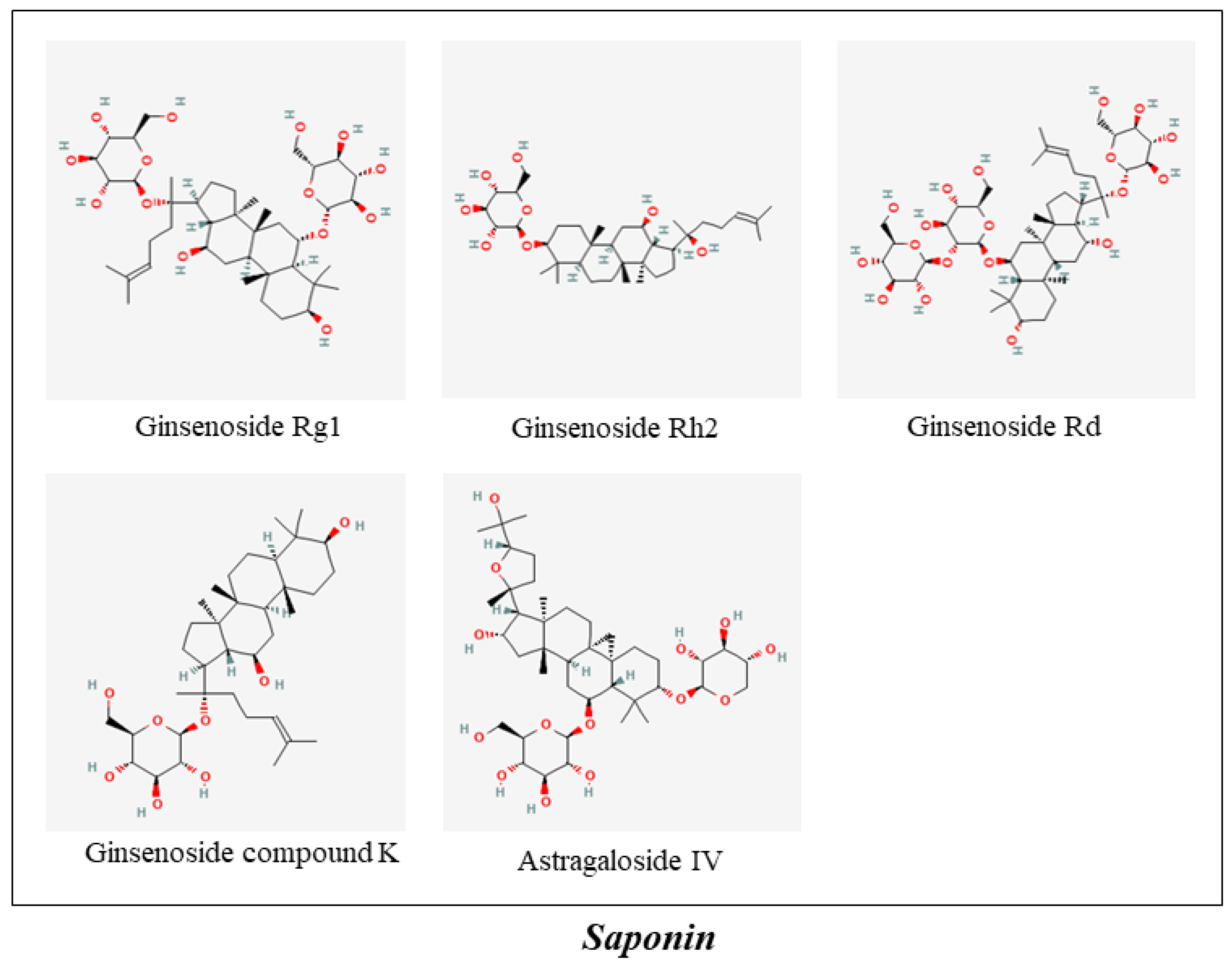
| 1 | GinsenosideRg1 | Saponin | Panax ginseng C.A. Mey. | 20 mg/kg·d | Saline | SD rats | ↑SOX-2, ↓Aeg-1, ↑GSH-Px, ↑SOD, ↓IL-1β, ↓IL-6, ↓TNF-α, p53, ↓p21Cip1/Waf1, ↓p19Arf | [75] | |||
| 2 | Ginsenoside Rh2 | Saponin | 1.5, 2.5, 3.5 μM | 10 mg/kg | Saline | Cortex neurons | tg2576 mice | ↓Abeta 1-40, ↓Abeta 1-42 | [76] | ||
| 3 | Ginsenoside Rd | Saponin | 2.5 or 5 μmol/L | 10mg/kg | Saline | Cortical neurons | Sprague–Dawley rats | ↓tau hyperphosphorylation, ↑PP-2A | [77] | ||
| 4 | Gintonin | Lysophosphatidic Acids-Protein Complexes | 0.1, 0.3, 1.0, 3.0, 10 μg/mL | B103 cells | transient Ca2+ mobilization, Gproteins/PLC/IP3 receptor/Ca2+ pathways | [78] | |||||
| 5 | Ginsenoside compound K | Saponin | 50, 20, 10, 1 and 0 μM | Primary astrocytes | ↓Aβ, ↓mTOR signal pathway | [79] | |||||
| 6 | Trans-Crocin 4/trans-Crocetin | Crocus sativus L. | 0.1, 1, 10, 100, 1000 μM | SH-SY5Y cells; PC12 cells | ↓BACE1, ↓γ-secretases (PSEN1 and PSEN2 complexes), ↓tau, ↓GSK3β, ↓ERK2, ↓pERK1, ↓pERK2 | [80] | |||||
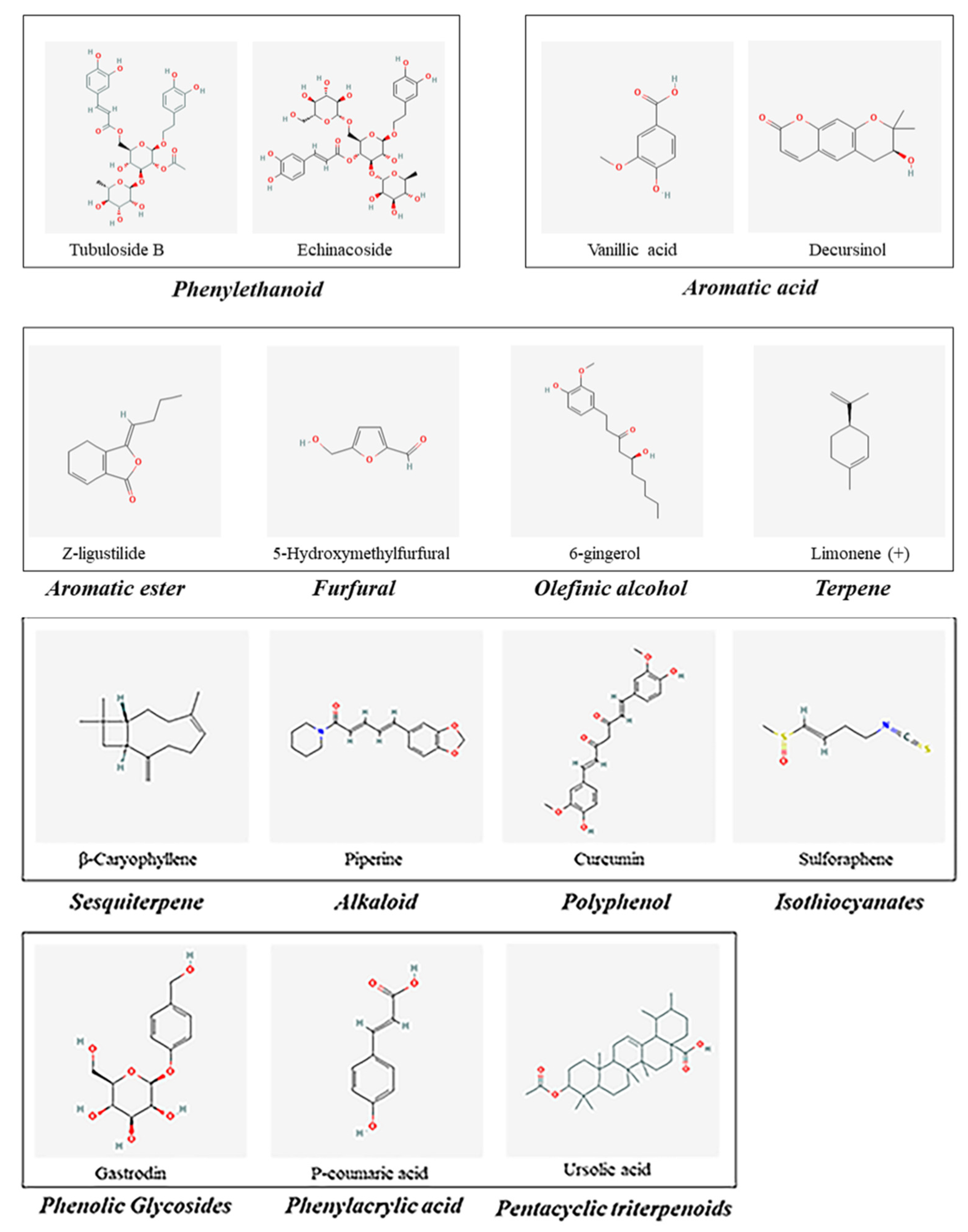
| 7 | Tubuloside B | Phenylethanoid | Cistanche afghanica Gilli | 1, 10, or 100 mg/L | SH-SY5Ycells | ↓ROS, ↓caspase-3, ↓Ca2+ | [81] | ||||
| 8 | Echinacoside | Phenylethanoid | 60 mg/kg | C57BL/6 mice | ↓HMGB1 | [82] | |||||
| 9 | Vanillic acid | Aromatic acid | Angelica acutiloba (Siebold & Zucc.) Kitag. | 25, 50, and 100 mg/kg | Swiss albinomice | ↓AChE, ↓corticosterone, ↓TNF-α | [83] | ||||
| 10 | Decursinol | 2, 4, and 8 mg | ICR mice | ↓Aβ (1-40) induced memory impairment | [84] | ||||||
| 0.01, 0.1, 1.0, 10 μM | PC12 cells | ↓ROS, ↑Bcl-2/Bax, ↑MMP, ↓cytochrome c, ↓Caspase-3 | [85] | ||||||||
| 11 | Z-ligustilide | Aromatic ester | 40 mg/kg | Saline | SPF Wistar rats | ↓Aβ, ↓APP, ↓p-Tau, ↓NF-κB | [86] | ||||
| 0.01, 1, 3, 10, 30 μM | SH-SY5Y cells; PC12 cells | p38/ PI3-K/Akt pathways | [87] | ||||||||
| 12 | Astragaloside IV | Saponin | Astragalus aaronii (Eig) Zohary | 25, 50, 100 μM | HT22 cells | ↑PPARγ/BDNF signaling pathway | [88] | ||||
| 10.0 mg/kg | Saline | APP/PS1 mice | ↑PPARγ, ↓BACE1 | [89] | |||||||
| 10, 25, 50 µM | SK-N-SH cells | ↓Bax, ↓caspase-3, ↑Bcl-2, ↓mPTP, ↓ROS | [90] | ||||||||
| 13 | 5-Hydroxymethylfurfural | Furfural | Alpinia oxyphylla Miq. | 15, 150 μg/kg | Kunming mice | ↓β-secretase, ↓MDA, ↑GPx, ↑SOD | [91] | ||||
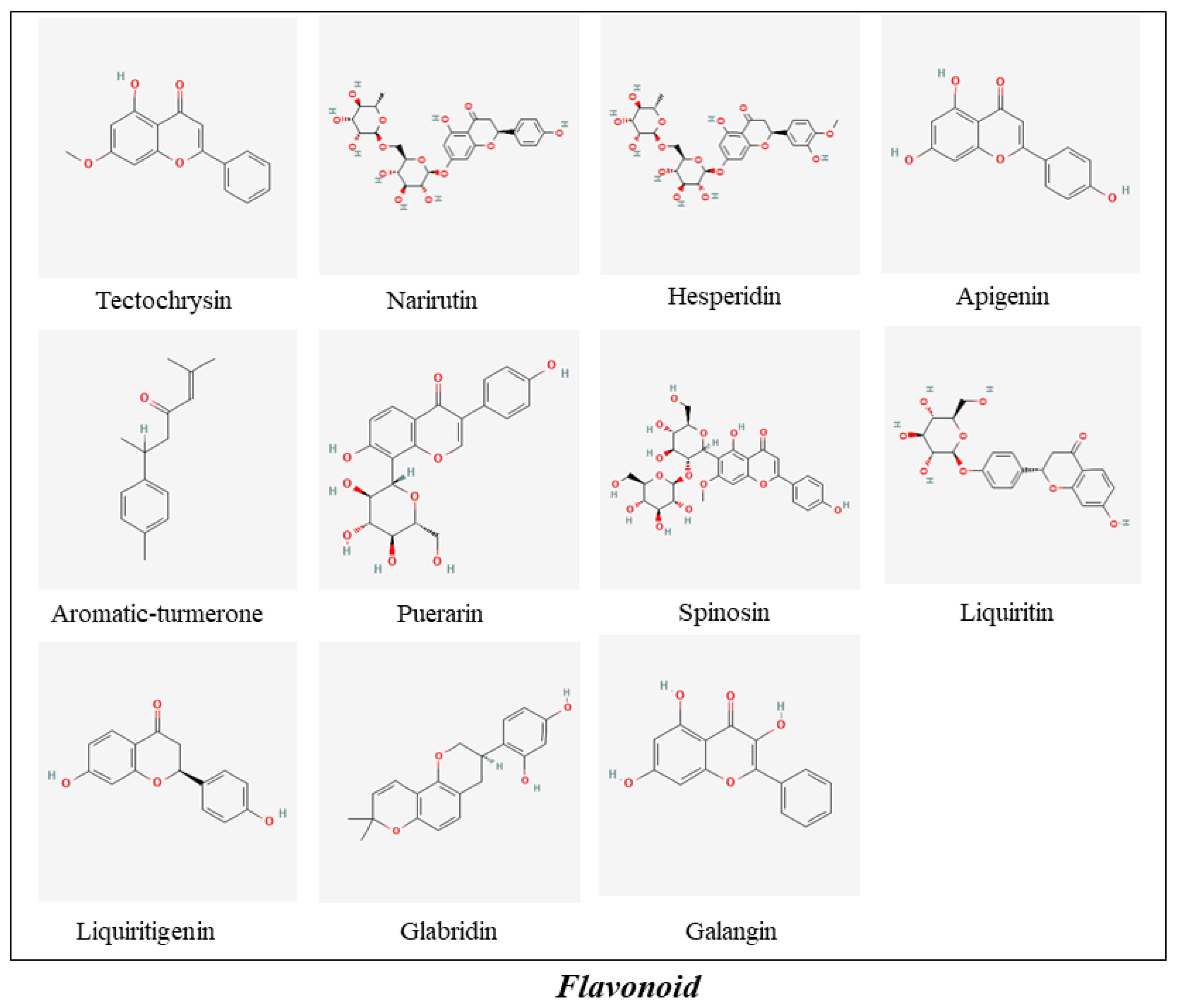
| 14 | Tectochrysin | Flavonoid | 14 µg/kg | Donepezil | Kunming mice | ↑SOD, ↑GSH-px, ↓MDA | [92] | ||||
| 15 | 6-gingerol | Olefinic alcohol | Zingiber officinale Roscoe | 40, 80, 120, 200, 300 μM | PC12 Cells | ↓ROS, ↓NO, ↓LDH, ↑SOD, ↓MDA, ↑p-Akt, ↑p-GSK-3β | [93] | ||||
| 6 mg/kg | DMSO | C57BL/6N mice | ↑emotional memory deficit | [94] | |||||||
| 10 mg/kg | Donepezil | Saline | ICR mice | ↑NGF, ↑PSD-95, ↑synaptophysin | [95] | ||||||
| 16 | Narirutin | Flavonoid | Polygonatumodoratum (Mill.) Druce | 50, 100 mg/kg | APPswe/PS1dE9 mice | ↑CaMKII | [96] | ||||
| 17 | Hesperidin | Flavonoid | 100 mg/kg | Saline | Wistar rats | ↓TBARS, ↑GSH, ↑SOD, ↑catalase, ↑GPx, ↓Bax, ↑Bcl-2 | [97] | ||||
| 1, 3 and 9 μg/mL | 100 mg/kg | 1% CMC | RAW 264.7 cells | APP/PS1 mice | ↓Aβ plaques, ↓iNOS, ↓TNF-α, ↓IL-1β | [98] | |||||
| 20, 40, 80 mg/kg | APP/PS1 mice | ↓TNF-α, ↓C-reactive protein ↓MCP-1, ↓NF-κB, ↑Akt, ↑GSK-3β (Ser 9), ↑Nrf2, ↑HO-1, ↓RAGE, ↓IκBα, ↓NF-κB/p65, ↑Akt/Nrf2 signaling, ↓RAGE/NF-κB signaling | [99] | ||||||||
| 18 | Limonene (+) | Terpene | 50 μg/mL | Fly Strains | ↓Aβ42, ↓NO | [100] | |||||
| 19 | Apigenin | Flavonoid | Mosla chinensis Maxim./Alpinia officinarum Hance | 40 mg/kg | 5% CMC-Na | APP/PS1 Mice | ↓Aβ, ↓BACE1, ERK1/2/CREB/BDNF pathway | [101] | |||
| 0.1, 1.0, 10.0 μM | SH-SY5Y cells | ↓AβPP, ↓ROS, ↑GSH, ↑SOD, ↑GSH-Px, ↓p38 MAPK signal pathway, ↓SAPK/JNK pathway | [102] | ||||||||
| 1, 5 and 10 μM | BV-2 cells | ↓NO, ↓PGE2, ↓JNK, ↓p38 MAPK | [103] | ||||||||
| Neuron/Glial Cells | ↓CD68, ↓OX42, ↓IL-6, ↓gp130, ↑BDNF | [104] | |||||||||
| 20 | Galangin | Flavonoid | Alpinia officinarum Hance | 6.25–400 μM | Rat adult brains | ↓AChE | [105] | ||||
| 21 | β-Caryophyllene | Sesquiterpene | Piper nigrum L. | 16, 48, 144 mg/kg | APP/PS1 mice | ↓COX-2, ↓TNF-α, ↓IL-1β | [106] | ||||
| 22 | Piperine | Alkaloid | 2.5, 5, 10, 25µM | Donepezil | SH-SY5Y cells | ↓AChE | [107] | ||||
| 5 mg/kg | Donepezil | Saline | Wistar rats | ↓MDA, ↓NO | [108] | ||||||
| 5, 10 and 20 mg/kg | Donepezil hydrochloride | Wistar rats | ↓lipid peroxidation, ↓acetylcholinesterase | [109] | |||||||
| 23 | Curcumin | Polyphenol | Curcuma longa L. | 150 mg/kg | APP/PS1 mice | ↓NF-κB pathway, ↑PPAR-γ | [110] | ||||
| 50 mg/kg | PBS | 5xFAD mouse | ↓Aβ, ↓GFAP-IR, ↓Iba-1-IR | [111] | |||||||
| 24 | Aromatic-turmerone | Flavonoid | 5, 10 and 20 μM | HT-22 cells | ↓MMP-9, ↓iNOS, ↓COX-2, ↓TNF-α, ↓IL-1β, ↓IL-6, ↓MCP-1, ↓ROS, ↓IκB-α, ↓JNK, ↓p38 MAPK. | [112] | |||||
| 5, 10 and 20 μM | 50, 100 mg/kg | PBS | BV2 cells | C57 mice | ↓TNF-α, ↓IL-1β, ↓Myd88, ↓MAPK, ↓NF-κB | [113] | |||||
| 50 µg/mL | Cerebellar granule neuron | ↓cleaved caspase-3 | [114] | ||||||||
| 25 | Puerarin | Isoflavone | Puerarialobata (Willd.) Ohwi | 80 mg/kg | Saline | Sprague–Dawley rats | ↓tau hyperphosphorylation, ↓GSK-3β, ↓FGF-2 | [115] | |||
| 30 mg/kg | APP/PS1 transgenic mice | ↑HO-1, GSK-3β/Akt signaling pathways | [116] | ||||||||
| 100 μM | PC12 cells | ↑Bcl-2, ↑p-Bad, ↓caspase-3, ↑Akt | [117] | ||||||||
| 26 | Spinosin | C-glycoside flavonoid | Ziziphi Spinosae Semen | 1.25, 2.5, 5 and 10 mg/kg | 0.5% CMC | ICR mice | ↑ERK–CREB–BDNF signaling | [118] | |||
| 6.25, 12.5, 25 μM | Neuro-2a cells | ↓APP, ↓BACE1, ↑ADAM10, ↓ROS, ↑Nrf2, ↑HO-1, Nrf2/HO-1 signaling pathway | [119] | ||||||||
| 10 and 100 µg/kg | Kunming mice | ↓MDA, ↓Aβ1–42, ↑BDNF, ↑Bcl-2, ↓IL-6 | [120] | ||||||||
| 30 μM | Donepezil | 5XFAD hippocampus | ↑LTP, ↑plasmin | [121] | |||||||
| 0.1, 1, and 10 μg/mL | 4, 20, 100 and 500 μg/mL | Donepezil | Hippocampus | ICR mice | ↑choline acetyltransferase | [122] | |||||
| 27 | Liquiritin | Flavonoids | Glycyrrhiza uralensis Fisch. | 25, 50, 100 mg/kg | Donepezil (3 mg/kg) | SD rats | ↑GSH-Px, ↑SOD, ↓8-OHdG, ↓MDA | [123] | |||
| 28 | Glabridin | Flavonoid | 1, 2 and 4 mg kg–1 | Scopolamine (0.5 mg kg–1) | 0.3 % carboxymethyl cellulose suspension | Kunming mice | ↓cholinesterase | [124] | |||
| 29 | Liquiritigenin | Flavonoid | 2.5, 5, 10, 20, 50, and 100 μM | 30 mg/kg | 0.1% sodium carboxymethyl cellulose | N2A cells; BV2 cells | APP/PS1 double transgenic mice | ↓NLRP3, ↓cleaved caspase-1, ↓caspase-3, ↓IL-1β, ↓TNF-α, ↑IL-4, ↑IL-13, ↓BAX, ↑Bcl-2, ↓Aβ, ↑NEP, ↑IDE | [125] | ||
| 30 | Sulforaphene | Isothiocyanates | Raphanus sativus L. | 0.5, 1, 2, 4, 8, 16, and 32 μM | 25, 50 mg/kg | Donepezil (5 mg/kg) | ddH2O | BV-2 cells | SD rats | ↓TNF-α; IL-6; ↑IL-10, ↑p-Akt (Ser473)/Akt, ↑p-GSK-3β (Ser9)/GSK-3β, ↓NO, ↓NF-κB | [126] |
| 31 | Gastrodin | Phenolic Glycosides | Gastrodia elata Blume | 60 mg/kg | Saline | Sprague–Dawley rats | ↓Aβ1-40/42, ↓APP, ↓β-site APP-cleaving, ↓Beclin-1, ↓LC3-II, ↓p62, ↓Bax, ↑Bcl-2, P38 MAPK signaling pathway | [127] | |||
| 32 | P-coumaric acid | Phenylacrylic acid | Cornus officinalis Siebold & Zucc. | 5, 25, 50 µM | PC12 cells | ↓BACE1, ↓iNOS, ↓p65, ↓ERK1/2, ↓JNK | [128] | ||||
| 33 | Ursolic acid | Pentacyclic triterpenoids | 1,10, 20 µM | PC12 cells | ↓iNOS, ↓COX-2, ↓p65, ↓p-38, ↓JNK, ↓ERK | [128] | |||||
Publisher’s Note: MDPI stays neutral with regard to jurisdictional claims in published maps and institutional affiliations. |
© 2022 by the authors. Licensee MDPI, Basel, Switzerland. This article is an open access article distributed under the terms and conditions of the Creative Commons Attribution (CC BY) license (https://creativecommons.org/licenses/by/4.0/).
Share and Cite
Guo, P.; Zhang, B.; Zhao, J.; Wang, C.; Wang, Z.; Liu, A.; Du, G. Medicine-Food Herbs against Alzheimer’s Disease: A Review of Their Traditional Functional Features, Substance Basis, Clinical Practices and Mechanisms of Action. Molecules 2022, 27, 901. https://doi.org/10.3390/molecules27030901
Guo P, Zhang B, Zhao J, Wang C, Wang Z, Liu A, Du G. Medicine-Food Herbs against Alzheimer’s Disease: A Review of Their Traditional Functional Features, Substance Basis, Clinical Practices and Mechanisms of Action. Molecules. 2022; 27(3):901. https://doi.org/10.3390/molecules27030901
Chicago/Turabian StyleGuo, Pengfei, Baoyue Zhang, Jun Zhao, Chao Wang, Zhe Wang, Ailin Liu, and Guanhua Du. 2022. "Medicine-Food Herbs against Alzheimer’s Disease: A Review of Their Traditional Functional Features, Substance Basis, Clinical Practices and Mechanisms of Action" Molecules 27, no. 3: 901. https://doi.org/10.3390/molecules27030901
APA StyleGuo, P., Zhang, B., Zhao, J., Wang, C., Wang, Z., Liu, A., & Du, G. (2022). Medicine-Food Herbs against Alzheimer’s Disease: A Review of Their Traditional Functional Features, Substance Basis, Clinical Practices and Mechanisms of Action. Molecules, 27(3), 901. https://doi.org/10.3390/molecules27030901