Article

Comparative Investigation of Chemical Constituents of Kernels, Leaves, Husk, and Bark of Juglans regia L., Using HPLC-DAD-ESI-MS/MS Analysis and Evaluation of Their Antioxidant, Antidiabetic, and Anti-Inflammatory Activities

1
Laboratory of Human Pathologies Biology, Department of Biology, Faculty of Sciences, Genomic Center of Human Pathologies, Mohammed V University in Rabat, Rabat BP 1014, Morocco
2
Laboratory of Spectroscopy, Molecular Modeling, Materials, Nanomaterials, Water and Environment, Faculty of Sciences, Mohammed V University in Rabat, Rabat BP 1014, Morocco
3
Laboratoire de Biodiversité, Ecologie et Génome, Faculté des Sciences, Université Mohammed V in Rabat, Rabat BP 1014, Morocco
4
Laboratory of Pharmacology and Toxicology, Faculty of Medicine and Pharmacy, Mohammed V University, Rabat BP 10100, Morocco
5
Department of Clinical Laboratory Sciences, Faculty of Applied Medical Sciences, Najran University, 1988, Najran 61441, Saudi Arabia
6
Faculty of Data Science and Information Technology, INTI International University, Nilai 71800, Malaysia
7
School of Pharmacy, KPJ Healthcare University College, Nilai 71800, Malaysia
*
Authors to whom correspondence should be addressed.
Molecules 2022, 27(24), 8989; https://doi.org/10.3390/molecules27248989
Submission received: 11 September 2022 / Revised: 12 November 2022 / Accepted: 14 November 2022 / Published: 16 December 2022

Abstract

:
Leaves, husk, kernels, and bark methanolic extracts of Juglans regia L. were tested for their in vitro antidiabetic, anti-inflammatory, and antioxidant activities. For these purposes, α-amylase and α-glucosidase were used as the main enzymes to evaluate antidiabetic activities. Moreover, lipoxidase and tyrosinase activities were tested to estimate anti-inflammatory properties. Antioxidant properties of Juglans regia L., extracts were determined using three different assays. Leaves extract has an important radical scavenging activity and a-amylase inhibition. Similarly, husk extracts showed high total phenolic content (306.36 ± 4.74 mg gallic acid equivalent/g dry extract) with an important α-amylase inhibition (IC50 = 75.42 ± 0.99 µg/mL). Kernels exhibit significant tyrosinase (IC50 = 51.38 ± 0.81 µg/mL) correlated with antioxidant activities (p < 0.05). Husk and bark extracts also showed strong anti-lipoxidase activities with IC50 equal to 29.48 ± 0.28 and 28.58 ± 0.35 µg/mL, respectively. HPLC-DAD-ESI-MS/MS analysis highlights the phenolic profile of methanolic extracts of Juglans regia L. plant parts. The identified polyphenols were known for their antioxidant, antidiabetic (dicaffeoyl-quinic acid glycoside in kernels), and anti-inflammatory (3,4-dihydroxybenzoic acid in leaves) activities. Further investigations are needed to determine molecular mechanisms involved in these effects as well as to study the properties of the main identified compounds.

1. Introduction

Oxidative stress and inflammation have been considered important factors linked to diabetes mellitus, chronic pulmonary, cardiovascular, and Alzheimer’s diseases, in addition to the physiopathology of cancer. The oxidative stress is known by the overproduction of ROS (reactive oxygen species) leading to the oxidation of macromolecules, notably lipids, proteins, and nucleic acids, which causes the impairment of cellular functions and apoptosis [1,2,3]. Inflammation is a protective process that involves an arsenal of enzymatic reactions and cellular activation. The prolongation of this process causes the loss of cell imbalance and biological system damage [4]. Thus, for better-multifaceted disease management, it is necessary to develop new drugs implying antioxidant and anti-inflammatory activities. In this context, medicinal plants and derivatives have been widely used for therapeutic purposes to heal and protect against several illnesses. Bioactive compounds extracted from natural products can offer specific properties that could act on specific targets and exhibit several biological properties for treating particular diseases [5,6,7]. The use of medicinal plants has increased in recent years. Therefore, screening biological activities remains an important step in therapeutic virtues research. In vitro assays, including enzyme inhibition assays, are commonly used for biological properties, bioactive natural compounds screening, and also for drug synthesis [8,9].
J. regia is a well-known worldwide species of the Juglans genus belonging to the family of Junglandacae. Fruits are consumed for their nutritional value phytotherapy [10,11,12] and have been widely used in folk medicine, especially for diabetes and inflammatory diseases [13] like eczema [14] and gout disease [15], particularly in Morocco [16], Turkey [17], Iran [18], Italy and Romania [19]. Leaves are used to treat digestive disorders like stomatitis, oral ulcer, and diarrhea [14,20,21,22]. Elsewhere, bark’s preparation, also called souak, is used for teeth hygiene and to treat bucco-dentaire sphere problems such as gum diseases, halitosis, and dental stain due to its depurative and antiseptic properties [10,23,24,25].
The literature reported several biological activities exhibited by J. regia extracts, including anti-inflammatory and antioxidant activities [26,27,28]. Each part of the plant showed different properties with regard to the cultivars [29], extraction method ([30], and geographic conditions [31]. Moreover, the J. regia phytochemical composition varied according to those parameters but the main molecules found are polyphenols, including phenolic acids (gallic acid, vanillic acid, syringic acid, ellagic acid, caffeic acid, ferulic acid, sinapic acid, chlorogenic acid), flavonoids, and tannins [32].
J. regia extracts act on different inflammatory mediators; husk extracts can inhibit the nitric oxide (NO) production in macrophages [28], kernel extracts inhibit the activation and the expression of vascular cell adhesion molecule-1 (VCAM-1) and intercellular adhesion molecule-1 (ICAM-1) [33] and are involved in the diminution of edemas [34], and bark extracts can inhibit membrane hemolysis. In addition, leaf extracts inhibit cyclooxygenase-2 (COX-2), one of the most important targets of anti-inflammatory drugs [35]. On the other hand, other researchers have focused on the anti-diabetic properties of J. regia in particular leaves, which showed excellent hypoglycemic effects [36,37].
To the best of our knowledge, this work is the first comparative study realized in kernels, leaves, husk, and bark of J. regia extracts based on the phytochemical composition and antioxidant, anti-inflammatory, and anti-diabetic activities. To date, no study reported the inhibitory effect of J. regia extracts on lipoxygenase (LOX).
In this work, we investigated the chemical screening of methanolic extracts of different J. regia parts: kernels (MWK), leaves (MWL), husk (MWH), and bark (MWB). In addition, enzymatic inhibitory activities of these part extracts were evaluated against enzymes involving inflammation and diabetes mellitus. Moreover, several methods were used to evaluate their antioxidant capacities (DPPH, FRAP, ABTS).

2. Materials and Methods

2.1. Chemicals

Methanol, Folin–Ciocalteau reagent, (7.5%) Na2CO3, gallic acid, (5%) sodium nitrite solution (NaNO2), 2,2′-azino-bis(3-ethylbenzothiazoline-6-sulfonic acid) (ABTS), 2,2-Diphenyl-1-picrylhydrazyl (DPPH), acetonitrile, (10%) aluminum trichloride (AlCl3), colchicine, Dimethyl sulfoxide (DMSO), ferric chloride (FeCl3), (0.1%) formic acid, nitrogen, iodine solution, PBS buffer, p-nitro-phenyl-α-D-glucopyranoside (p-NPG), (1%) Potassium ferricyanide, potassium persulfate, potassium phosphate buffer, quercetin, rutin, sodium hydroxide, sodium phosphate buffer, Kojic acid, (10%) trichloroacetic acid, Trolox, (4%) vanillin, hydrochloric acid (HCl) were analytical grade. L-Dopa, linoleic acid, tyrosinase, α-amylase, and α-glucosidase are purchased from Sigma.

2.2. Plant Material and Extraction

Leaves, husks, kernels, and bark of the plant J. regia were collected from the Taza region, Morocco. After the drying process, walnut parts were powdered and conserved in the dark at 4 °C. The extractions of leaves, husk, kernels, and bark powders (12.5 g) were realized by methanolic maceration (250 mL) for 24 h under agitation. Then, the mixture was filtered, the solvent was eliminated using a rotary evaporator at 60 °C, and the extracts were conserved at 4 °C.

2.3. Determination of Phenolic Contents

2.3.1. Total Phenolic Content

The determination of total phenolic content in J. regia extracts was performed using the Folin–Ciocalteu procedure [38]. 0.5 mL of Folin–Ciocalteau reagent was added to 0.5 mL of each extract and agitated. Then, 4 mL of 7.5 % Na2CO3 (w/v) was added, and the reaction was incubated at 45 °C for 30 min. Calibration curves were established using gallic acid. The absorbance was measured at 765 nm, and the total phenolic contents were expressed as mg gallic acid equivalents per g of the dry weight of extract (mg GAE/g of extract).

2.3.2. Total Flavonoid Content

The determination of the flavonoid content was carried out according to the aluminum trichloride method developed by Brighente et al. [39]. In test tubes, 1 mL of each of the extracts (1mg / mL) and 6.4 mL of distilled water were successively introduced, then 0.3 mL of the sodium nitrite solution (NaNO2 5%) was added. After 5 min, 0.3 mL of aluminum trichloride (AlCl3 10%) was added. After 6 min, 2 mL of sodium hydroxide (1 M) was added, and the solution was agitated and allowed to stand for 30 min. The absorbances were measured at 510 nm. Rutin was used as a standard under the same analytical conditions. The flavonoid content is expressed in mg of rutin equivalent per g of dried extract (mg RE/g of the extract).

2.3.3. Total Tannin Content

The determination of total tannin contents was effectuated using the Julkunen–Tiitto [40] method. 50 μL of each extract was mixed with 1.5 mL of 4% vanillin, then 750 μL of hydrochloric acid HCl was added. After, the mixture was incubated for 20 min at room temperature in the dark. Colchicine was used as standard. The absorbances were measured at 500 nm. The results are expressed in milligrams equivalent to catechin per gram of the extract (mg CE/g of extract).

2.4. HPLC-DAD-ESI-MS/MS

The chemical composition of J. regia methanolic extracts was determined by high-performance liquid chromatography (Hewlett-Packard 1100 Agilent Technologies) equipped with a DAD detector and an electrospray HP 1100 MSD API (Agilent-Technologies, Palo Alto, CA, USA) under analytical conditions reported by Pallaufa et al. [41]. A negative ionization mode, a capillary voltage of 3000 to 3500 V, and a fragmented variable of the order of 80 to 150 V were used. The column was a Poroshell 120 EC-C1, C18 (150 × 2.1) mm × 5 µm. The mobile phase was (A) 0.1% formic acid in the water, (B) acetonitrile. The established elution gradient was isocratic 15% B for 5 min, 15% B to 20% B over 5 min, 20–25% B over 10 min, 25–35% B over 10 min, 35–50% for 10 min. The total analysis time was 47 min, the flow rate was 0.5 mL/min. Double in-line detection was performed in the DAD using 280 nm and 370 nm as wavelengths and in a mass spectrometer (MS) connected to the HPLC system through the output of the DAD cell. MS detection was performed in a Qtrap API 3200 (Applied Biosystems, Darmstadt, Germany) equipped with an ESI source and a triple quadrupole ion trap mass analyzer. Zero-quality air was used as nebulizer gas (30 psi) and turbo gas for solvent drying (400 °C, 40 psi). Nitrogen served as a curtain (20 psi) and collision gas. The resolution of the quadrupoles was adjusted and the ion sputtering voltage was tuned at −4500 V (in negative mode). The MS detector has been set up to operate in two modes: Enhanced MS Analysis (EMS) and Enhanced Product Ion Analysis (EPIA) (EPI). To acquire an overview of all the ions in the sample, the EMS was utilized to capture the entire scan spectra. The defusing potential (DP) was −450 V, the input potential (EP) was −6 V, and the impact energy (CE) was −10 V. The spectra were recorded between m/z 100 and 1000 (in negative ion mode). For detected parent ion (s) discovered, the fragmentation pattern was then determined using EPI analysis under DP −50 V, EP −6 V, CE −25 V, and collision energy propagation (CES) 0 V [42].

2.5. Antioxidant Assay

2.5.1. DPPH Method

A concentration of 1 mg/mL of each extract was prepared and diluted in methanol to obtain a range concentration from 10 to 1000 µg/mL. 1 mL of each sample concentration or standard was mixed with 0.5 mL of 0.2 mM DPPH methanolic solution. Trolox was used as a standard under the same conditions. Absorbances were measured at 517 nm after 30 min of incubation at dark conditions [43]. The radical scavenging ability (RSA) was expressed in % according to the following equation where the Abs control is the absorbance of the solution containing all reagents except sample (or standard). IC50 was calculated from the plot of RSA vs. extract concentration.
RSA %   = Abs   control   Abs   sample Abs   control × 100

2.5.2. ABTS Method

The antioxidant activity was determined by the 2,2′-azino-bis(3-ethylbenzothiazoline-6-sulfonic acid) (ABTS) method [44]. 2 mM of ABTS was mixed with 70 mM of potassium persulfate. After incubation in the dark during 12–16 h, the ABTS+ solution was diluted with methanol to adjust absorbance to 0.700 ± 0.005 at 734 nm. Thus, 1 mL of each extract concentration (1 mg/mL) or standard was added to 2 mL of diluted ABTS solution to obtain a range concentration from 25 to 1000 µg/mL and incubated for 1 min, and the absorbance was measured at 734 nm. Trolox is used as a standard compound. Scavenging activity in this assay was expressed as the concentration of the extract required to inhibit 50% of the free radical scavenging activity.

2.5.3. FRAP Method

The extract’s ferric ion-reducing power was determined by the FRAP method [45] with some modifications. A concentration of 1 mg/mL of each extract was prepared and diluted in methanol to obtain a range concentration from 5 to 50 µg/mL. Trolox was used as standard. 1 mL of each prepared extract or standard was mixed with 1.25 mL of 0.2 M sodium phosphate buffer (pH 6.6) and 1.25 mL of 1% potassium ferricyanide. The mixture was incubated at 50 °C for 20 min. After cooling, 1.25 mL of 10% trichloroacetic acid was added and centrifuged at 3000 rpm for 10 min. Finally, 1.25 mL of the supernatant was mixed with 1.25 mL distilled water and 0.25 mL FeCl3 solution (0.1%, w/v). Absorbances were measured at 700 nm, and results were expressed as the 50% effective concentration (EC50), which is the antioxidant concentration in mg/mL necessary to achieve an absorbance of 0.5.

2.6. Anti-Inflammatory Assay

2.6.1. Lipooxidase Inhibition Assay

The lipooxidase inhibition assay was realized according to the procedure described by Debayo et al. [46]. A concentration of 1 mg/mL of each extract was prepared in methanol and diluted in 2M borate buffer to obtain a range concentration from 12.5 to 50 µg/mL. 12.5 µL of each extract concentration was added to 487.5 µL of 15-LOX (200 Units/mL) and kept at room temperature. After 5 min, 500 µL of linoleic acid dissolved in ethanol and diluted in the borate buffer were added to the enzymatic mixture and incubated for 5 min at room temperature. The absorbance was measured at 234 nm. Quercetin was used as a positive control, and DMSO was used as a negative control. The enzyme inhibition percentages were determined by the following equation.
% Inhibition = Abs   extract     Abs   blank Abs   negative   control     Abs   blank × 100 %

2.6.2. Tyrosinase Inhibition Assay

The anti-tyrosinase activity was determined according to Huang et al. [47] procedure. A concentration of 1 mg/mL of each extract was prepared in methanol and diluted in 0.05 M PBS buffer (pH 6.5) to obtain a range concentration from 25 to 100 µg/mL. Tyrosinase enzymatic solution (333 U/mL) and L-Dopa (5 mM) were prepared in the PBS buffer. 50 µL of each extract concentration was mixed with 200 µL enzyme solution (3 U/mL), and the mixture was incubated at 37 °C. After 10 min, 500 µL of the substrate (L-Dopa) was added. The enzymatic reaction solution was then incubated for 30 min at 37 °C. The absorbance was measured at 510 nm. The percentage of inhibition was determined by the following formula, where Abs represents absorbance. Kojic acid was used as a standard.
%   of   inhibition = 1   Abs   control     Abs   sample   Abs   control   × 100

2.7. Anti-Diabetic Activity

2.7.1. α-Amylase Inhibition Assay

The effect of J. regia extracts on α-amylase activity was assessed according to Kusano et al. [48] method with some modifications. A concentration of 1 mg/mL of each extract was prepared in methanol and diluted in phosphate buffer (pH 6.9) to obtain a range concentration from 25 to 100 µg/mL. 200 µL of starch solution (substrate) was added to 100 µL of the buffer, and 250 µL of α-amylase (30 U/mL) was then incubated at 37 °C for 15 min. For the sample test, the enzyme was incubated with 250 µL of each extract concentration for 15 min. After adding substrate, the enzymatic reaction was conducted for 15 min and then stopped using 400 µL HCl (0.1 M). Total and remaining starch were measured at 630 nm after adding 500 µL of iodine solution (25 mM). For the positive control, acarbose was used. The percentage of inhibition was calculated by the following formula.
%   of   inhibition = 1 Abs   sub   Abs   enz + sub Abs   sample   Abs   control   Abs   sub Abs   enz + sub   × 100

2.7.2. α-Glucosidase Inhibition Assay

The α-Glucosidase inhibition activity was tested according to Li et al. described method with slight modifications [49]. A concentration of 1 mg/mL of each extract was prepared in methanol and diluted in 1 M potassium phosphate buffer (pH 6.8) to obtain a range concentration from 250 to 1000 µg/mL. 100 µL of sample or acarbose (positive control), 380 µL of p-nitro-phenyl-α-D-glucopyranoside (p-NPG) (0.53 mM), and 250 µL of α-Glucosidase solution (0.015 Units/mL) were mixed in the buffer. After incubating at 37 °C for 20 min, 1mL of Na2CO3 (0.1 M) was added to quench the reaction. The IC50 value is determined by the concentration of α-Glucosidase inhibitor necessary to inhibit 50% of activity under assay conditions. The absorbance was measured at 405 nm, and the inhibition percentages were determined using the following equation:
%   of   inhibition = 1   Abs   enz + sub     Abs   sub Abs   sample   Abs   control Abs   enz + sub     Abs   sub   × 100

2.8. Statistical Analysis

Raw or log-transformed measured parameters were tested for normality and homogeneity of variance to meet the assumptions for parametric statistics. As the assumptions were violated for all parameters (extracts and assays), non-parametric analyses of variance, followed by a Dunn pairwise comparison test of means, were performed. The critical level of significance is set at 0.05.

3. Results and Discussion

3.1. Total Phenolic, Flavonoid, and Tocopherol Contents

The total phenolic content (TPC) analysis revealed a similar quantity of polyphenols in all methanolic extracts of J. regia, contrary to the total flavonoid content (TFC) analysis. The methanolic extract of bark and leaves showed the highest flavonoids quantity compared to husk and kernels, while the total tocopherol content (TTC) analysis showed the presence of tocopherols in low levels in bark methanolic extract (Table 1).

3.2. HPLC-DAD-ESI-MS/MS Analysis

Qualitative analysis of J. regia L. methanolic extracts by HPLC-DAD-ESI-MS/MS was based on the retention times (Rt min), chemical formula, pseudo-molecular ions [MH], and MS/MS fragment ions (m/z).
In MWB, nine compounds were detected (Table 2). Kaempferol diglucoside was detected at [M − H] of m/z 447, as reported by Zeeshan et al. [50]. Three compounds were identified as Caffeic acid derivatives, among them caffeoyl hexoside (m/z 439) and caffeoyl hexose deoxyhexose (m/z 487), as reported by Appendino et al. [51].
In MWK, 14 phenolic compounds were identified (Table 3 belonging to various classes of natural compounds, notably fraxetin-8-O-glucoside (m/z 369), myricetin malonylglucoside (m/z 565), dicaffeoylquinic acid glycoside (m/z 677) as reported, respectively, in the studies [50,52,53]. Among the most dropping classes in this sample, the flavones such as Apigenin pentosyl glucoside with Rt 54.10 min and m/z 563 in addition to acacetin-7-O-rutinoside with m/z of 715 as reported by Gika et al. [54].
In the MWH extract, nine compounds were detected (Table 4). Several chemical classes have been identified as flavonols, notably Quercetin pentoside with [M−H]of m/z 435 at 37.25 minas reported by Gika et al. [54]. Caffeic acid derivatives were detected by three m/z peaks 339, 427, and 487 at 11.64, 27.11, and 36.38 min, respectively, as reported by Appendino et al. [51] and Ieri et al. [52]. Moreover, the flavones were presented by acacetin-aglycone (m/z 283) and apigenin-7-O-glucoside (m/z 431).
In the MWL extract, 11 compounds were identified (Table 5). Several molecules belong to flavonols, notably myricetin malonylglucoside (m/z 565) and kaempferol-malonyl glucoside (m/z 533), as reported by Vagiri et al. (2012). The two compounds with [M−H] at m/z 437 and m/z 453 were identified as p-coumaroyl derivatives, as reported by Ieri et al. [52]. The carbohydrates were represented by Trigalloyl-glucose (m/z 633) and digalloyl-glucose (m/z 483), as reported by Gika et al. [54]. Thus, the caffeic acid derivatives were represented with [M−H] at m/z 531 and m/z 487.
J. regia extracts are rich in several bioactive components. The variation of phytochemical composition between J. regia parts may be due to the variation of patterns used in the synthesis, accumulation, and distribution of secondary metabolites, season, and environmental changes [55].

3.3. Antioxidant Activity: DPPH, FRAP, and ABTS Assays

The antioxidant activities of J. regia husk, bark, kernels, and leaves methanolic extracts were investigated using DPPH, FRAP, and ABTS methods. The results showed a concentration-dependent decrease in percentages of radicals. The data are reported as the concentration needed to inhibit 50% of radicals (IC50). Among all parts tested, the MWH extract showed the highest FRAP radicals’ neutralization capacity (p < 0.05), followed by MWL, MWB, and MWK extracts which were 10.45 ± 0.59 µg/mL, 12.21 ± 0.01 µg/mL, 34.51 ± 0.64 µg/mL and 39.36 ± 0.41 µg/mL (Figure 1a). However, Trolox used as standard control showed an EC50 of 5.25 µg/mL.
Contrary to FRAP assay results, the bark and the kernels methanolic extracts had the highest ABTS neutralization activity with IC50 less than 88 µg/mL (Figure 1b), while Trolox showed an important antioxidant capacity (IC50 = 15.2 µg/mL). In this study, the J. regia husk methanolic extract showed a pronounced ABTS neutralization activity (p < 0.05), which was higher than the results reported by Zhang et al. [30] with IC50 of 324.8 µg/mL vs. 145.86 ± 1.61µg/mL. Similarly, Bhatia et al. [56] investigated the ABTS neutralization activity of the methanolic extract of J. regia bark. They found a low neutralizing activity compared to our results with an IC50 of 601 µg/mL and 83.05 ± 0.39 µg/mL, respectively. On the other hand, the MWH extract had the strongest DPPH radical’s scavenging capacity with IC50 of 32.27 ± 0,69 µg/mL, which was significantly lower than that of leaves methanolic extract (p < 0.05) (Figure 1c). However, Trolox used as positive control inhibited DPPH radical at low concentrations (IC50 = 2.04). Ghasemi et al. [31] investigated the antioxidant activity of MWH extract obtained from 11 different regions in Iran. IC50 found in this previous study ranged from 122 ± 4.5 µg/mL to 302 ± 13 µg/mL, higher than IC50 obtained in our study.
The methanolic extract of Tunisian J. regia bark showed a more pronounced radical scavenging activity (DPPH) compared to our results which were respectively IC50 of 36 µg/mL vs. 123.42 ± 3.71 µg/mL [57] contrary to the aqueous extract which showed a low antioxidant capacity with an IC50 of 582 µg/mL [56]. Concerning the methanolic extract of J. regia leaves, we obtained a similar result compared to the Jabli et al. study [58].

3.4. Anti-Inflammatory Activity

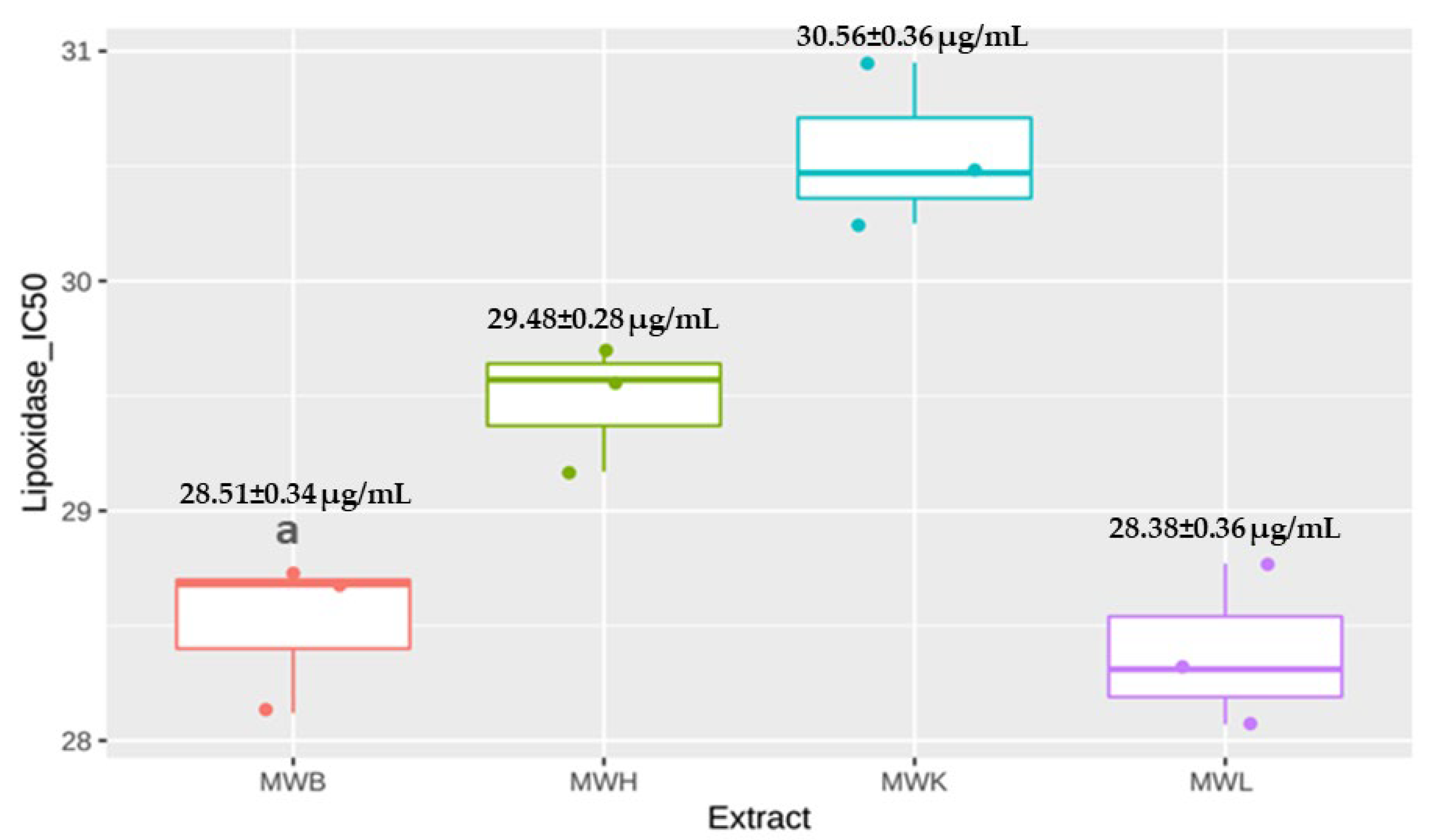
3.4.1. Anti-Lipoxidase Activity

The anti-inflammatory activity of the methanolic extracts of J. regia was performed in vitro by lipoxygenase (LOX) inhibition activity. All methanolic extracts of J. regia leaves, bark, husk, and kernels exhibited high lipoxygenase activity inhibition, respectively, with IC50 of 28.38 ± 0.36, 28.51 ± 0.34, 29.48 ± 0.28, and 30.56 ± 0.36 µg/mL (Figure 2). These inhibitory activities are important compared to the standard (quercetin) (IC50 = 6.72 µg/mL). Lipoxygenases are a group of enzymes implicated in the inflammation process regulation, the production of inflammatory mediators, notably leukotrienes, by catalyzing the oxidation of poly-unsaturated fatty acids, essentially arachidonic acid and linoleic acid, in addition to catalyzing the formation of eicosanoids from arachidonic acid and immune response regulation [59]. The over-expression of lipoxygenase contributes to several pathologies, such as Alzheimer’s disease, diabetes, cancers, and cardiovascular disease, which further motivated research on natural compounds with anti-lipoxygenase activity [60].
Multiple studies have been interested in the anti-LOX activity of other plants. We found that J. regia kernels demonstrated an excellent LOX inhibition activity compared with the methanolic extracts of Cydonia oblonga Mill fruit and Zanthoxylum armatum fruit with IC50 of 99.30 µg/mL and 70.30 µg/mL, respectively [61,62]. Concerning the leaves’ methanolic extracts, some plants have a poor LOX inhibition activity, like Cyclea barbata, Beilschmiedia penangiana, Veronica persica, Cassia alata, and Zanthoxylum armatum. Others have a moderate anti-LOX activity compared with J. regia, notably Artemisia nilagirica, Jatropha gossypifolia, and Ficus curtipes, respectively, with IC50 of 128.20 µg/mL, 162.50 µg/mL, and 200.80 µg/mL. Others have similar LOX inhibition activity compared with J. regia, in particular Memecylon malabaricum, Memecylon talbotianum, and Memecylon umbellatum, respectively, with IC50 of 29.87 µg/mL, 34.60 µg/mL, and 39.19 µg/mL. The same for the bark methanolic extracts, Pterocarpus erinaceus, exhibited poor anti-LOX activity, while Fraxinus rhynchophylla, Zanthoxylum armatum, and Beilschmiedia penangiana had moderate LOX inhibition activity, respectively, with IC50 of 62.60 µg/mL, 90.50 µg/mL, and 176.80 µg/mL [63].
This activity is strongly related to the phytochemical profile of the plants. Flavonoids, as well as other plant secondary metabolites, are well-known inhibitors of pro-inflammatory mediators, the first and most well-known being benzoic acid. In this study, its derivatives were present with relatively high doses in J. regia methanolic leaves extract. 3,4-dihydroxybenzoic acid was investigated in numerous studies, and it was shown that it could directly bind the active site of soybean LOX, the latter sharing significant homologies with mammal’s LOX [64]. Other derivatives of benzoic acid are of significant interest to human and animal nutrition [65]. Quercetin also demonstrated in vitro inhibitory effects on lipoxygenases, particularly 5- and 15-lipoxygenases [66]. Kaempferol also exhibited anti-lipoxygenase-1 activity but with a lesser inhibitory effect compared to quercetin [67].

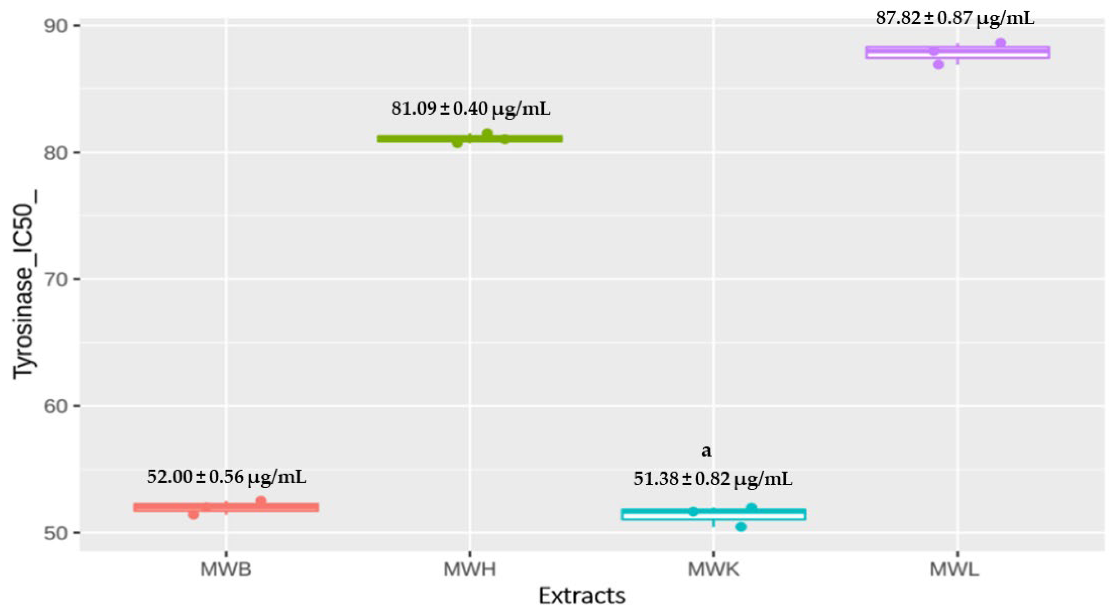
3.4.2. Anti-Tyrosinase Activity

Tyrosinase is a metalloenzyme involved in the browning process of fruits and vegetables [68]. It also alters the melanogenesis process leading to chronic inflammation involved in several diseases. Melanin overproduction is associated with skin cancer and neurodegenerative disorders like Parkinson’s disease. According to the physiopathological role of tyrosinase, several research studies were conducted on tyrosinase inhibitory effects of natural compounds [4,69,70].
J. regia methanolic extracts exhibited strong inhibition of tyrosinase activity, especially the methanolic extracts of kernels and bark, respectively, with IC50 of 51.38 ± 0.82 µg/mL and 52.00 ± 0.56 µg/mL (p < 0.05) followed by the husk (IC50= 81.09 ± 0.40 µg/mL) and the leaves (IC50= 87.82 ± 0.87 µg/mL) (Figure 3). These results can be considered important since the used standard (kojic acid) showed an IC50 value equal to 5.28 µg/mL.
Previous studies on J. regia tyrosinase inhibitory activity reported that the water extract of J. regia leaves, seed, and husk have demonstrated an IC50 of 3.99 mg/mL, 8.83 mg/mL, and 10.13 mg/mL, respectively [71], the hydroethanolic extract of J. regia leaves presented an inhibition up to 50% at 751 ± 0.01 µg of extract/mL [72], in addition to a negligible antityrosinase activity in the extract of leaves collected in Turkey [73]. According to all these data, we note that the methanolic extract of J. regia collected from Morocco exhibits a significant anti-tyrosinase activity. Multiple studies have been focused on the anti-tyrosinase activity of other plant parts, notably the leaves of Ceratonia siliqua, the aerial parts of Cleome arabica and Pituranthos scoparius, whose exhibited moderate tyrosinase inhibition activity, respectively, with IC50 of 200 µg/mL, 124.4 ± 0.69 µg/mL and 125.01 ± 0.72 µg/mL. While other plant extracts had similar anti-tyrosinase activity to J. regia bark, such as Harpephyllum caffrum bark, contrariwise to the bark extracts of Hyaenanche globosa and Cassipourea flanaganii, which showed high activity against the tyrosinase with IC50 of 27.1 ± 042 µg/mL and 22.24 ± 1.32 µg/mL, respectively [74].
The methanolic extracts of kernels and bark phenolic profile showed mainly acacetin-aglycone and apigenin-7-O-glucoside. According to the literature, these flavones are encountered in many medicinal plants and are known for their therapeutic potential, including anti-inflammatory activities [75,76].
Our extracts showed the presence of myricetin, kaempferol, and quercetin which had been identified as tyrosinase inhibitors. Previous studies demonstrated that kaempferol and quercetin could inhibit the oxidation of L-DOPA catalyzed by tyrosinase [70]. Those molecules may be the actors behind the anti-tyrosinase activity of the methanolic extracts of J. regia parts.

3.5. Antidiabetic Activity

The antidiabetic activity of the different extracts of J. regia was assessed using the amylase and glycosidase inhibition assays. MWK and MWH highly inhibit the amylase activity with IC50 values equal to 37.37 ± 1.16 µg/mL and 75.42 ± 0.99 µg/mL, respectively, which are significantly higher (p < 0.05) compared to MWL (IC50 = 327.45 ± 3.13 µg/mL) (Figure 4a). Certainly, J. regia methanolic leaves extract (MWL) has the weakest inhibition effect on the α-amylase activity of all other extracts (p = 0.07), but it remained more effective compared with J. regia aqueous extract, which demonstrated an inhibition of 60% of amylase with 0.4 mg/mL [77]. These results are not significantly important compared with the inhibitory value of α-amylase shown by acarbose (26.11 μg/mL). Likewise, MWB extract released 50% of amylase activity at a concentration less than 210 μg/mL, and this remains more effective compared with bark aqueous extract of J. regia collected in Oued Amlil located in Taza region that showed an IC50 of 5445.33 ± 82.58 μg/mL [78].
Quantitative analysis of J. regia extracts showed that the methanolic kernels extract has more polyphenols than flavonoids, and this may explain its effect on amylase because it has been shown that polyphenols bind with this digestive enzyme affecting starch hydrolysis activity in the small intestine reducing the absorption of glucose and consequently improving glycemic status in patients with diabetes [79,80]. This effect is mediated mainly by condensed and hydrolyzable tannins [79], and indeed, the qualitative analysis by HPLC-DAD-ESI-MS/MS demonstrated the dominance of gallotannin in this extract. In addition to that, the HPLC-DAD-ESI-MS/MS analysis showed that kernels of methanolic extract of J. regia contain a caffeoylquinic acid derivative, including dicaffeoyl-quinic acid glycoside. It was shown that this polyphenol class contributes as the most active principle against diabetes in vitro, and that may explain the excellent effect of this plant extract.
The evaluation of the inhibitory activity of J. regia extracts on α-glycosidase showed moderate results (compared to acarbose IC50 = 0.35 μg/mL). MWL has the lowest IC10 (266 ± 14.54 μg/mL) compared to MWB (922.03 ± 19.72 μg/mL), MWH (789.46 ± 7.19 μg/mL), MWK (978.92 ± 21,58 μg/mL) (Figure 4b). This effect may be due to caffeoylquinic acids, which can strongly inhibit glucosidase [81]. Previous studies had investigated the anti-glycosidase activity of other plants, notably the leaves of Annona senegalensis and Liquidambar formosana, which exhibited high glycosidase inhibition activity, respectively, with IC50 of 97 μg/mL and 59 μg/mL compared to J. regia methanolic extracts [82]. J. regia leaf extracts have previously demonstrated an effective effect on diabetes in vitro and also in vivo by glycemia levels normalization via the inhibition of glucose-6-phosphate translocase and transporter GLUT2 in addition to the reduction of cholesterol synthesis by hydroxymethyl glutaryl-CoA reductase inhibition on rats thanks to caffeoylquinic acid and quercetin [35,37,83] and even in humans [84].

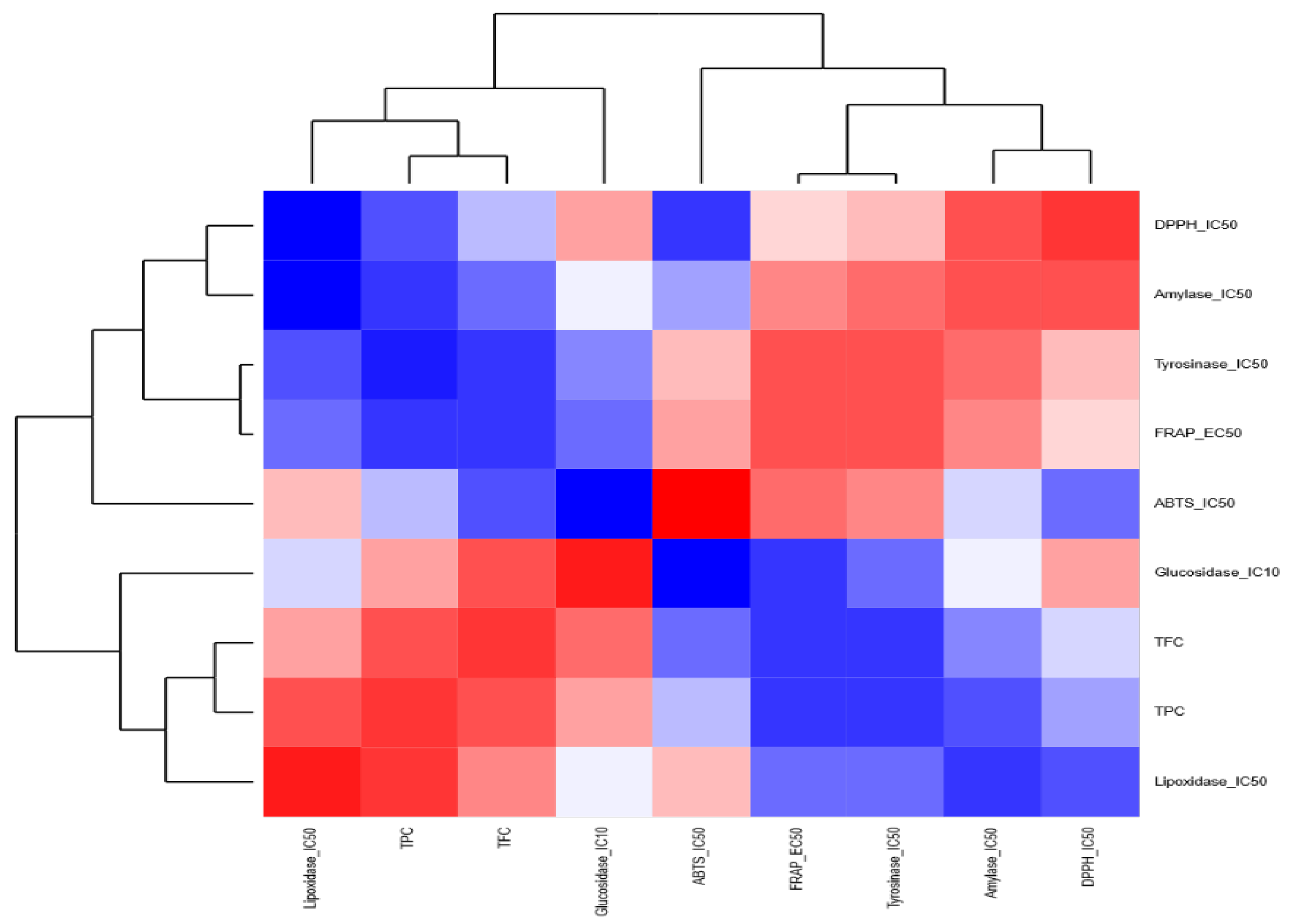
3.6. Correlations among J. regia Biochemical Activities in Different Extracts

To better understand the potential activities of different J. regia part extracts, we investigated the correlations among the biochemical activities, notably antioxidative activity, anti-inflammatory activity, and antidiabetic activity within each J. regia extract (Figure 5). The analysis showed positive correlations between the anti-amylase activity and the antioxidant in the four extracts represented by the FRAP and DPPH assays. On the other hand, the anti-glucosidase activity of MWH and MWK was also positively correlated with the antioxidant activity, while this activity was correlated with TFC and TPC in MWL.
As for the anti-inflammatory properties, the anti-LOX activity was correlated with the TFC and the antioxidant activity in MWB, MWH, and MWK, contrary to MWL, the anti-LOX activity was correlated with TPC and TFC. The anti-tyrosinase activity of MWK and MWL was correlated positively with TFC and FRAP, respectively. When we used a heatmap coupled with a dendrogram to highlight the relationships among different chemical screening within each extract, we found out that for the MWB extract (Figure 6), the chemical screenings are clustered into two distinct groups: (i) the first group contained FRAP, TPC, amylase et lipoxidase and (ii) the second group harbored tyrosinase, TFC, glycosidase, DPPH, and ATBS. Similarly, for the MWK (Figure 7) extract, the heatmap showed two groups: (i) the first one included TFC, ABTS, lipoxidase, tyrosinase, and glycosidase; (ii) the second one contained TPC, DPPH, FRAP, and amylase.
In the case of the MWH extract (Figure 8), (i) the first group is composed of ABTS, TPC, FRAP, and glycosidase, and (ii) the second group comprises amylase, lipoxidase, tyrosinase, TFC, and DPPH. For the MWL extract (Figure 9), there are two groups: (i) the first group encompasses ABTS, FRAP, amylase, tyrosinase, and DPPH, and (ii) the second group consists of glycosidase, lipoxidase, TPC, and TFC.
Definitively the chemical activity profiles varied deeply among the J. regia extracts; this finding should be taken into account when using the different extracts for therapeutic purposes.

4. Conclusions

J. regia is a well-known species widely used for its nutritional benefits and also for its therapeutic properties, as reported in numerous ethnobotanical studies conducted in many countries.
This study is the first to emphasize the variability in antioxidant and biological characteristics across the various parts of the walnut. This variability will be used to guide in vivo studies and, consequently, the uses by herbalists of those parts of plants exhibiting the most relevant activities.
Results highlighted the potential antidiabetic properties of kernels and husk extracts as well as the anti-inflammatory properties of bark extract. In fact, the phenolic profile determined by HPLC-DAD-ESI-MS/MS showed the richness of J. regia in bioactive compounds. However, further investigations concerning the isolation of main chemical compounds, as well as the evaluation of their antioxidant, antidiabetic, and anti-inflammatory effects, are required to determine the molecular mechanisms involved in these biological activities. Moreover, in vivo explorations and toxicological investigations are needed to determine the main pharmaceutical parameters of these compounds as well as to validate their safety.

Author Contributions

Conceptualization, I.B.; data curation, I.B., B.B., A.A.A.A. and A.B.; formal analysis, I.B., S.E., N.S.., M.M.A., A.A.A.A., K.W.G., S.E.H. and N.I.; funding acquisition, K.W.G.; investigation, I.B., S.E., A.B., C.S.T. and N.D.; methodology, I.B., S.E., B.B., M.M.A. and N.D.; project administration, D.T., B.B., Y.M., N.S., A.B. and C.S.T.; resources, K.W.G. and C.S.T.; software, K.W.G.; supervision, I.B. and A.B.; validation, B.B., A.B. and K.W.G.; writing—original draft, I.B., D.T., B.B., Y.M., A.A.A.A., A.B. and S.E.H.; writing—review and editing, S.E., M.M.A., C.S.T. and N.I. All authors have read and agreed to the published version of the manuscript.

Funding

This research did not receive any specific grant from funding agencies in the public, commercial, or not-for-profit sectors.

Institutional Review Board Statement

Not applicable.

Informed Consent Statement

Not applicable.

Data Availability Statement

Not applicable.

Conflicts of Interest

The authors declare no conflict of interest.

Abbreviations

ABTS2,2′-azino-bis(3-ethylbenzothiazoline-6-sulfonic acid)
CatCatechin
CEImpact Energy
CESCollision Energy Propagation
DPDefusing Potential
DPPH2,2-Diphenyl-1-picrylhydrazyl
EMSEnhanced MS Analysis
EPInput Potential
EPIAEnhanced Product Ion Analysis
FRAPFerric Reducing Antioxidant Power
GAGallic acid
GLUT2Glucose transporter 2
HClHydrochloric acid
HPLCHigh-Performance Liquid Chromatography
J. regiaJuglans regia
LOXLipoxygenase
MSMass Spectrometer
MWBMethanolic Extract of Bark
MWHMethanolic extract of Husk
MWKMethanolic Extract of Kernels
MWLMethanolic Extract of Leaves
ROSReactive Oxygen Species
RURutin
TETrolox Equivalent
TFCTotal Flavonoid Content
TPCTotal Phenolic Content
TTCTotal Tocopherol Content

References

  1. Bhatti, J.S.; Sehrawat, A.; Mishra, J.; Sidhu, I.S.; Navik, U.; Khullar, N.; Kumar, S.; Bhatti, G.K.; Reddy, P.H. Oxidative Stress in the Pathophysiology of Type 2 Diabetes and Related Complications: Current Therapeutics Strategies and Future Perspectives. Free Radic. Biol. Med. 2022, 184, 114–134. [Google Scholar] [CrossRef] [PubMed]
  2. Forman, H.J.; Zhang, H. Targeting Oxidative Stress in Disease: Promise and Limitations of Antioxidant Therapy. Nat. Rev. Drug Discov. 2021, 20, 689–709. [Google Scholar] [CrossRef] [PubMed]
  3. Griendling, K.K.; Camargo, L.L.; Rios, F.J.; Alves-Lopes, R.; Montezano, A.C.; Touyz, R.M. Oxidative Stress and Hypertension. Circ. Res. 2021, 128, 993–1020. [Google Scholar] [CrossRef] [PubMed]
  4. Bakhouche, I.; Aliat, T.; Boubellouta, T.; Gali, L.; Şen, A.; Bellik, Y. Phenolic Contents and In Vitro Antioxidant, Anti-Tyrosinase, and Anti-Inflammatory Effects of Leaves and Roots Extracts of the Halophyte Limonium Delicatulum. South Afr. J. Bot. 2021, 139, 42–49. [Google Scholar] [CrossRef]
  5. Bouyahya, A.; Omari, N.E.; El Hachlafi, N.; Jemly, M.E.; Hakkour, M.; Balahbib, A.; El Menyiy, N.; Bakrim, S.; Naceiri Mrabti, H.; Khouchlaa, A. Chemical Compounds of Berry-Derived Polyphenols and Their Effects on Gut Microbiota, Inflammation, and Cancer. Molecules 2022, 27, 3286. [Google Scholar] [CrossRef] [PubMed]
  6. Ji, X.; Shi, S.; Liu, B.; Shan, M.; Tang, D.; Zhang, W.; Zhang, Y.; Zhang, L.; Zhang, H.; Lu, C. Bioactive Compounds from Herbal Medicines to Manage Dyslipidemia. Biomed. Pharmacother. 2019, 118, 109338. [Google Scholar] [CrossRef]
  7. Sasikumar, P.; Aswathy, M.; Prem, P.T.; Radhakrishnan, K.V.; Chakrapani, P.S.B. Plant Derived Bioactive Compounds and Their Potential to Enhance Adult Neurogenesis. Phytomed. Plus 2022, 2, 100191. [Google Scholar]
  8. El Harrad, L.; Bourais, I.; Mohammadi, H.; Amine, A. Recent Advances in Electrochemical Biosensors Based on Enzyme Inhibition for Clinical and Pharmaceutical Applications. Sensors 2018, 18, 164. [Google Scholar] [CrossRef] [Green Version]
  9. Tlili, N.; Sarikurkcu, C. Bioactive Compounds Profile, Enzyme Inhibitory and Antioxidant Activities of Water Extracts from Five Selected Medicinal Plants. Ind. Crops Prod. 2020, 151, 112448. [Google Scholar] [CrossRef]
  10. Bellakhdar, J.; Claisse, R.; Fleurentin, J.; Younos, C. Repertory of Standard Herbal Drugs in the Moroccan Pharmacopoea. J. Ethnopharmacol. 1991, 35, 123–143. [Google Scholar] [CrossRef]
  11. Brown, D. Valerian: Clinical Overview-Phytotherapy Review & Commentary. Townsend Lett. Dr. 1995, 119, 1005–1012. [Google Scholar]
  12. Yeung, H.C. Handbook of Chinese Herbs and Formulas Institute of Chinese Medicine, Los Angeles. Am. Soc. Nutr. Sci. J. Nutr. 2004, 134, 1105–1159. [Google Scholar]
  13. Bouasla, A.; Bouasla, I. Ethnobotanical Survey of Medicinal Plants in Northeastern of Algeria. Phytomedicine 2017, 36, 68–81. [Google Scholar] [CrossRef]
  14. Petran, M.; Dragos, D.; Gilca, M. Historical Ethnobotanical Review of Medicinal Plants Used to Treat Children Diseases in Romania (1860s–1970s). J. Ethnobiol. Ethnomed. 2020, 16, 1–33. [Google Scholar] [CrossRef] [Green Version]
  15. Mardaninejad, S.; Janghorban, M.; Vazirpour, M. Collection and Identification of Medicinal Plants Used by the Indigenous People of Mobarakeh (Isfahan), Southwestern Iran. J. Med. Herbs 2013, 4, 23–32. [Google Scholar]
  16. Ziyyat, A.; Legssyer, A.; Mekhfi, H.; Dassouli, A.; Serhrouchni, M.; Benjelloun, W. Phytotherapy of Hypertension and Diabetes in Oriental Morocco. J. Ethnopharmacol. 1997, 58, 45–54. [Google Scholar] [CrossRef] [PubMed]
  17. Güzel, Y.; Güzelşemme, M.; Miski, M. Ethnobotany of Medicinal Plants Used in Antakya: A Multicultural District in Hatay Province of Turkey. J. Ethnopharmacol. 2015, 174, 118–152. [Google Scholar] [CrossRef]
  18. Nasab, F.K.; Khosravi, A.R. Ethnobotanical Study of Medicinal Plants of Sirjan in Kerman Province, Iran. J. Ethnopharmacol. 2014, 154, 190–197. [Google Scholar] [CrossRef] [PubMed]
  19. Sadgrove, N.J. The New Paradigm for Androgenetic Alopecia and Plant-Based Folk Remedies: 5α-Reductase Inhibition, Reversal of Secondary Microinflammation and Improving Insulin Resistance. J. Ethnopharmacol. 2018, 227, 206–236. [Google Scholar] [CrossRef]
  20. Jamila, F.; Mostafa, E. Ethnobotanical Survey of Medicinal Plants Used by People in Oriental Morocco to Manage Various Ailments. J. Ethnopharmacol. 2014, 154, 76–87. [Google Scholar] [CrossRef]
  21. Jaradat, N.A.; Ayesh, O.I.; Anderson, C. Ethnopharmacological Survey about Medicinal Plants Utilized by Herbalists and Traditional Practitioner Healers for Treatments of Diarrhea in the West Bank/Palestine. J. Ethnopharmacol. 2016, 182, 57–66. [Google Scholar] [CrossRef] [PubMed]
  22. Najem, M.; Harouak, H.; Ibijbijen, J.; Nassiri, L. Oral Disorders and Ethnobotanical Treatments: A Field Study in the Central Middle Atlas (Morocco). Heliyon 2020, 6, e04707. [Google Scholar] [CrossRef]
  23. El-Hilaly, J.; Hmammouchi, M.; Lyoussi, B. Ethnobotanical Studies and Economic Evaluation of Medicinal Plants in Taounate Province (Northern Morocco). J. Ethnopharmacol. 2003, 86, 149–158. [Google Scholar] [CrossRef] [PubMed]
  24. Teixidor-Toneu, I.; Martin, G.J.; Ouhammou, A.; Puri, R.K.; Hawkins, J.A. An Ethnomedicinal Survey of a Tashelhit-Speaking Community in the High Atlas, Morocco. J. Ethnopharmacol. 2016, 188, 96–110. [Google Scholar] [CrossRef] [PubMed]
  25. Zougagh, S.; Belghiti, A.; Rochd, T.; Zerdani, I.; Mouslim, J. Medicinal and Aromatic Plants Used in Traditional Treatment of the Oral Pathology: The Ethnobotanical Survey in the Economic Capital Casablanca, Morocco (North Africa). Nat. Prod. Bioprospect. 2019, 9, 35–48. [Google Scholar] [CrossRef] [PubMed] [Green Version]
  26. Arranz, S.; Pérez-Jiménez, J.; Saura-Calixto, F. Antioxidant Capacity of Walnut (Juglans regia L.): Contribution of Oil and Defatted Matter. Eur. Food Res. Technol. 2008, 227, 425–431. [Google Scholar] [CrossRef] [Green Version]
  27. Erdemoglu, N.; Küpeli, E.; Yeşilada, E. Anti-Inflammatory and Antinociceptive Activity Assessment of Plants Used as Remedy in Turkish Folk Medicine. J. Ethnopharmacol. 2003, 89, 123–129. [Google Scholar] [CrossRef]
  28. Vieira, V.; Pereira, C.; Pires, T.C.; Calhelha, R.C.; Alves, M.J.; Ferreira, O.; Barros, L.; Ferreira, I.C. Phenolic Profile, Antioxidant and Antibacterial Properties of Juglans regia L. (Walnut) Leaves from the Northeast of Portugal. Ind. Crops Prod. 2019, 134, 347–355. [Google Scholar] [CrossRef]
  29. Pereira, J.A.; Oliveira, I.; Sousa, A.; Valentão, P.; Andrade, P.B.; Ferreira, I.C.; Ferreres, F.; Bento, A.; Seabra, R.; Estevinho, L. Walnut (Juglans regia L.) Leaves: Phenolic Compounds, Antibacterial Activity and Antioxidant Potential of Different Cultivars. Food Chem. Toxicol. 2007, 45, 2287–2295. [Google Scholar] [CrossRef]
  30. Zhang, Z.; Liao, L.; Moore, J.; Wu, T.; Wang, Z. Antioxidant Phenolic Compounds from Walnut Kernels (Juglans regia L.). Food Chem. 2009, 113, 160–165. [Google Scholar] [CrossRef]
  31. Ghasemi, M.; Arzani, K.; Hasani, D.; Ghasemi, S. Fatty Acids Composition of Some Selected Walnut (Juglans regia L.) Genotypes in Markazi Province. J. Food Sci. Technol. 2010, 7, 31–37. [Google Scholar]
  32. Bourais, I.; Elmarrkechy, S.; Taha, D.; Mourabit, Y.; Bouyahya, A.; El Yadini, M.; Machich, O.; El Hajjaji, S.; El Boury, H.; Dakka, N. A Review on Medicinal Uses, Nutritional Value, and Antimicrobial, Antioxidant, Anti-Inflammatory, Antidiabetic, and Anticancer Potential Related to Bioactive Compounds of J. regia. Food Rev. Int. 2022, 1–51. [Google Scholar] [CrossRef]
  33. Papoutsi, Z.; Kassi, E.; Chinou, I.; Halabalaki, M.; Skaltsounis, L.A.; Moutsatsou, P. Walnut Extract (Juglans regia L.) and Its Component Ellagic Acid Exhibit Anti-Inflammatory Activity in Human Aorta Endothelial Cells and Osteoblastic Activity in the Cell Line KS483. Br. J. Nutr. 2008, 99, 715–722. [Google Scholar] [CrossRef] [PubMed] [Green Version]
  34. Qamar, W.; Sultana, S. Kernel Modulate Cigarette Smoke Extract Induced Acute Inflammation, Oxidative Stress and Lung Injury in Wistar Rats. Hum. Exp. Toxicol. 2011, 30, 499–506. [Google Scholar] [CrossRef] [PubMed]
  35. Nasiry, D.; Ahmadvand, H.; Talebpour Amiri, F.; Akbari, E. Protective Effects of Methanolic Extract of Juglans regia L. Leaf on Streptozotocin-Induced Diabetic Peripheral Neuropathy in Rats. BMC Complement. Altern. Med. 2017, 17, 1–11. [Google Scholar] [CrossRef] [PubMed]
  36. Mohammadi, J.; Saadipour, K.; Delaviz, H.; Mohammadi, B. Anti-Diabetic Effects of an Alcoholic Extract of Juglans regia in an Animal Model. Turk. J. Med. Sci. 2011, 41, 685–691. [Google Scholar] [CrossRef]
  37. Mollica, A.; Zengin, G.; Locatelli, M.; Stefanucci, A.; Macedonio, G.; Bellagamba, G.; Onaolapo, O.; Onaolapo, A.; Azeez, F.; Ayileka, A. An Assessment of the Nutraceutical Potential of Juglans regia L. Leaf Powder in Diabetic Rats. Food Chem. Toxicol. 2017, 107, 554–564. [Google Scholar] [CrossRef]
  38. Gutfinger, T. Polyphenols in Olive Oils. J. Am. Oil Chem. Soc. 1981, 58, 966–968. [Google Scholar] [CrossRef]
  39. Brighente, I.M.C.; Dias, M.; Verdi, L.G.; Pizzolatti, M.G. Antioxidant Activity and Total Phenolic Content of Some Brazilian Species. Pharm. Biol. 2007, 45, 156–161. [Google Scholar] [CrossRef]
  40. Julkunen-Tiitto, R.; Sorsa, S. Testing the Effects of Drying Methods on Willow Flavonoids, Tannins, and Salicylates. J. Chem. Ecol. 2001, 27, 779–789. [Google Scholar] [CrossRef]
  41. Pallauf, K.; Rivas-Gonzalo, J.C.; Del Castillo, M.D.; Cano, M.P.; de Pascual-Teresa, S. Characterization of the Antioxidant Composition of Strawberry Tree (Arbutus unedo L.) Fruits. J. Food Compos. Anal. 2008, 21, 273–281. [Google Scholar] [CrossRef] [Green Version]
  42. Mraihi, F.; Fadhil, H.; Trabelsi-Ayadi, M.; Chérif, J.-K. Chemical Characterization by HPLC-DAD-ESI/MS of Flavonoids from Hawthorn Fruits and Their Inhibition of Human Tumor Growth. J. New Sci. 2015, 3, 840–846. [Google Scholar]
  43. Yamasaki, K.; Hashimoto, A.; Kokusenya, Y.; Miyamoto, T.; Sato, T. Electrochemical Method for Estimating the Antioxidative Effects of Methanol Extracts of Crude Drugs. Chem. Pharm. Bull. 1994, 42, 1663–1665. [Google Scholar] [CrossRef] [PubMed] [Green Version]
  44. Re, R.; Pellegrini, N.; Proteggente, A.; Pannala, A.; Yang, M.; Rice-Evans, C. Antioxidant Activity Applying an Improved ABTS Radical Cation Decolorization Assay. Free Radic. Biol. Med. 1999, 26, 1231–1237. [Google Scholar] [CrossRef] [PubMed]
  45. Benzie, I.F.; Strain, J.J. The Ferric Reducing Ability of Plasma (FRAP) as a Measure of “Antioxidant Power”: The FRAP Assay. Anal. Biochem. 1996, 239, 70–76. [Google Scholar] [CrossRef] [PubMed] [Green Version]
  46. Adebayo, S.A.; Dzoyem, J.P.; Shai, L.J.; Eloff, J.N. The Anti-Inflammatory and Antioxidant Activity of 25 Plant Species Used Traditionally to Treat Pain in Southern African. BMC Complement. Altern. Med. 2015, 15, 1–10. [Google Scholar] [CrossRef] [PubMed]
  47. Huang, X.-H.; Chen, Q.-X.; Wang, Q.; Song, K.-K.; Wang, J.; Sha, L.; Guan, X. Inhibition of the Activity of Mushroom Tyrosinase by Alkylbenzoic Acids. Food Chem. 2006, 94, 1–6. [Google Scholar] [CrossRef]
  48. Kusano, R.; Ogawa, S.; Matsuo, Y.; Tanaka, T.; Yazaki, Y.; Kouno, I. α-Amylase and Lipase Inhibitory Activity and Structural Characterization of Acacia Bark Proanthocyanidins. J. Nat. Prod. 2011, 74, 119–128. [Google Scholar] [CrossRef]
  49. Li, C.-W.; Chu, Y.-C.; Huang, C.-Y.; Fu, S.-L.; Chen, J.-J. Evaluation of Antioxidant and Anti-α-Glucosidase Activities of Various Solvent Extracts and Major Bioactive Components from the Seeds of Myristica Fragrans. Molecules 2020, 25, 5198. [Google Scholar] [CrossRef]
  50. Zeeshan, H.M.A.; Lee, G.H.; Kim, H.-R.; Chae, H.-J. Endoplasmic Reticulum Stress and Associated ROS. Int. J. Mol. Sci. 2016, 17, 327. [Google Scholar] [CrossRef] [Green Version]
  51. Appendino, G.; Ottino, M.; Marquez, N.; Bianchi, F.; Giana, A.; Ballero, M.; Sterner, O.; Fiebich, B.L.; Munoz, E. Arzanol, an Anti-Inflammatory and Anti-HIV-1 Phloroglucinol α-Pyrone from Helichrysum italicum ssp. Microphyllum. J. Nat. Prod. 2007, 70, 608–612. [Google Scholar] [CrossRef] [PubMed]
  52. Ieri, F.; Martini, S.; Innocenti, M.; Mulinacci, N. Phenolic Distribution in Liquid Preparations of Vaccinium myrtillus L. and Vaccinium vitis Idaea L. Phytochem. Anal. 2013, 24, 467–475. [Google Scholar] [CrossRef] [PubMed]
  53. Vagiri, M.; Ekholm, A.; Andersson, S.C.; Johansson, E.; Rumpunen, K. An Optimized Method for Analysis of Phenolic Compounds in Buds, Leaves, and Fruits of Black Currant (Ribes nigrum L.). J. Agric. Food Chem. 2012, 60, 10501–10510. [Google Scholar] [CrossRef] [PubMed]
  54. Gika, H.G.; Theodoridis, G.A.; Plumb, R.S.; Wilson, I.D. Current Practice of Liquid Chromatography–Mass Spectrometry in Metabolomics and Metabonomics. J. Pharm. Biomed. Anal. 2014, 87, 12–25. [Google Scholar] [CrossRef]
  55. Li, Y.; Kong, D.; Fu, Y.; Sussman, M.R.; Wu, H. The Effect of Developmental and Environmental Factors on Secondary Metabolites in Medicinal Plants. Plant Physiol. Biochem. 2020, 148, 80–89. [Google Scholar] [CrossRef]
  56. Bhatia, K.; Rahman, S.; Ali, M.; Raisuddin, S. In Vitro Antioxidant Activity of Juglans regia L. Bark Extract and Its Protective Effect on Cyclophosphamide-Induced Urotoxicity in Mice. Redox Rep. 2006, 11, 273–279. [Google Scholar] [CrossRef]
  57. Noumi, E.; Snoussi, M.; Noumi, I.; Valentin, E.; Aouni, M.; Al-sieni, A. Comparative Study on the Antifungal and Antioxydant Properties of Natural and Colored Juglans regia L. Barks: A High Activity against Vaginal Candida Strains. Life Sci. J. 2014, 11, 327–335. [Google Scholar]
  58. Jabli, M.; Sebeia, N.; Boulares, M.; Faidi, K. Chemical Analysis of the Characteristics of Tunisian Juglans regia L. Fractions: Antibacterial Potential, Gas Chromatography–Mass Spectroscopy and a Full Investigation of Their Dyeing Properties. Ind. Crops Prod. 2017, 108, 690–699. [Google Scholar] [CrossRef]
  59. Wisastra, R.; Dekker, F.J. Inflammation, Cancer and Oxidative Lipoxygenase Activity Are Intimately Linked. Cancers 2014, 6, 1500–1521. [Google Scholar] [CrossRef] [Green Version]
  60. Sun, Y.; Li, S.; Zeng, F.; Qi, J.; Qin, W.; Tan, C.; Luo, Q.; Wu, D.; Zhang, Q.; Lin, D. Functional Components, Antioxidant Activity and Hypoglycemic Ability Following Simulated Gastro-Intestinal Digestion of Pigments from Walnut Brown Shell and Green Husk. Antioxidants 2019, 8, 573. [Google Scholar] [CrossRef] [Green Version]
  61. Berkoz, M. Antioxidant and Anti-Lipoxygenase Activities of Cydonia oblonga. Medicine 2020, 9, 251–254. [Google Scholar] [CrossRef]
  62. Alam, F.; Ashraf, M. Gaultheria Trichophylla (Royle): A Source of Minerals and Biologically Active Molecules, Its Antioxidant and Anti-Lipoxygenase Activities. BMC Complement. Altern. Med. 2017, 17, 1–9. [Google Scholar] [CrossRef] [Green Version]
  63. Lončarić, M.; Strelec, I.; Moslavac, T.; Šubarić, D.; Pavić, V.; Molnar, M. Lipoxygenase Inhibition by Plant Extracts. Biomolecules 2021, 11, 152. [Google Scholar] [CrossRef]
  64. Russell, W.R.; Scobbie, L.; Duthie, G.G.; Chesson, A. Inhibition of 15-Lipoxygenase-Catalysed Oxygenation of Arachidonic Acid by Substituted Benzoic Acids. Bioorg. Med. Chem. 2008, 16, 4589–4593. [Google Scholar] [CrossRef]
  65. Jenner, P.M.; Hagan, E.C.; Taylor, J.M.; Cook, E.L.; Fitzhugh, O.G. Food Flavourings and Compounds of Related Structure I. Acute Oral Toxicity. Food Cosmet. Toxicol. 1964, 2, 327–343. [Google Scholar] [CrossRef]
  66. Loke, W.M.; Proudfoot, J.M.; Stewart, S.; McKinley, A.J.; Needs, P.W.; Kroon, P.A.; Hodgson, J.M.; Croft, K.D. Metabolic Transformation Has a Profound Effect on Anti-Inflammatory Activity of Flavonoids Such as Quercetin: Lack of Association between Antioxidant and Lipoxygenase Inhibitory Activity. Biochem. Pharmacol. 2008, 75, 1045–1053. [Google Scholar] [CrossRef]
  67. Ha, T.J.; Shimizu, K.; Ogura, T.; Kubo, I. Inhibition Mode of Soybean Lipoxygenase-1 by Quercetin. Chem. Biodivers. 2010, 7, 1893–1903. [Google Scholar] [CrossRef]
  68. Lai, X.; Wichers, H.J.; Soler-Lopez, M.; Dijkstra, B.W. Structure and Function of Human Tyrosinase and Tyrosinase-Related Proteins. Chem. Eur. J. 2018, 24, 47–55. [Google Scholar] [CrossRef]
  69. Rusu, M.E.; Gheldiu, A.-M.; Mocan, A.; Moldovan, C.; Popa, D.-S.; Tomuta, I.; Vlase, L. Process Optimization for Improved Phenolic Compounds Recovery from Walnut (Juglans regia L.) Septum: Phytochemical Profile and Biological Activities. Molecules 2018, 23, 2814. [Google Scholar] [CrossRef] [Green Version]
  70. Zolghadri, S.; Bahrami, A.; Hassan Khan, M.T.; Munoz-Munoz, J.; Garcia-Molina, F.; Garcia-Canovas, F.; Saboury, A.A. A Comprehensive Review on Tyrosinase Inhibitors. J. Enzyme Inhib. Med. Chem. 2019, 34, 279–309. [Google Scholar] [CrossRef] [Green Version]
  71. Akin, M.; Arabaci, G.; Saki, N. Total Phenols, Antioxidant Potential and Tyrosinase Inhibitory Activity of Walnut (Juglans regia L.) Leaf, Husk and Seed. Asian J. Chem. 2013, 25, 9337. [Google Scholar] [CrossRef]
  72. Besrour, N.; Oludemi, T.; Mandim, F.; Pereira, C.; Dias, M.I.; Soković, M.; Stojković, D.; Ferreira, O.; Ferreira, I.C.; Barros, L. Valorization of Juglans regia Leaves as Cosmeceutical Ingredients: Bioactivity Evaluation and Final Formulation Development. Antioxidants 2022, 11, 677. [Google Scholar] [CrossRef] [PubMed]
  73. Uysal, S.; Zengin, G.; Aktumsek, A.; Karatas, S. Chemical and Biological Approaches on Nine Fruit Tree Leaves Collected from the Mediterranean Region of Turkey. J. Funct. Foods 2016, 22, 518–532. [Google Scholar] [CrossRef]
  74. Opperman, L.; De Kock, M.; Klaasen, J.; Rahiman, F. Tyrosinase and Melanogenesis Inhibition by Indigenous African Plants: A Review. Cosmetics 2020, 7, 60. [Google Scholar] [CrossRef]
  75. Singh, S.; Gupta, P.; Meena, A.; Luqman, S. Acacetin, a Flavone with Diverse Therapeutic Potential in Cancer, Inflammation, Infections and Other Metabolic Disorders. Food Chem. Toxicol. 2020, 145, 111708. [Google Scholar] [CrossRef]
  76. Wang, W.; Yue, R.-F.; Jin, Z.; He, L.-M.; Shen, R.; Du, D.; Tang, Y.-Z. Efficiency Comparison of Apigenin-7-O-Glucoside and Trolox in Antioxidative Stress and Anti-Inflammatory Properties. J. Pharm. Pharmacol. 2020, 72, 1645–1656. [Google Scholar] [CrossRef]
  77. Rahimzadeh, M.; Jahanshahi, S.; Moein, S.; Moein, M.R. Evaluation of Alpha-Amylase Inhibition by Urtica Dioica and Juglans regia Extracts. Iran. J. Basic Med. Sci. 2014, 17, 465. [Google Scholar]
  78. Boulfia, M.; Lamchouri, F.; Toufik, H. Mineral Analysis, in Vitro Evaluation of Alpha-Amylase, Alpha-Glucosidase, and Beta-Galactosidase Inhibition, and Antibacterial Activities of Juglans regia L. Bark Extracts. BioMed Res. Int. 2021, 2021, 1585692. [Google Scholar] [CrossRef]
  79. Sun, Q.-Y.; Zhou, H.-H.; Mao, X.-Y. Emerging Roles of 5-Lipoxygenase Phosphorylation in Inflammation and Cell Death. Oxid. Med. Cell. Longev. 2019, 2019, 2749173. [Google Scholar] [CrossRef]
  80. Yaghobian, F.; Dehghan, F.; Azarbayjani, M.A. Forecast of Ameliorating Effect of Dietary Flavonol Consumption in White Tea with or without Aerobic Training on Type 2 Diabetes (T2D) in Females. Clin. Nutr. ESPEN 2021, 45, 134–140. [Google Scholar]
  81. Spínola, V.; Castilho, P.C. Evaluation of Asteraceae Herbal Extracts in the Management of Diabetes and Obesity. Contribution of Caffeoylquinic Acids on the Inhibition of Digestive Enzymes Activity and Formation of Advanced Glycation End-Products (in Vitro). Phytochemistry 2017, 143, 29–35. [Google Scholar] [CrossRef]
  82. Dirir, A.M.; Daou, M.; Yousef, A.F.; Yousef, L.F. A Review of Alpha-Glucosidase Inhibitors from Plants as Potential Candidates for the Treatment of Type-2 Diabetes. Phytochem. Rev. 2021, 21, 1049–1079. [Google Scholar] [CrossRef]
  83. Nasiry, D.; Khalatbary, A.R.; Ahmadvand, H.; Talebpour Amiri, F.B. Effects of Juglans regia L. Leaf Extract Supplementation on Testicular Functions in Diabetic Rats. Biotech. Histochem. 2021, 96, 41–47. [Google Scholar] [CrossRef]
  84. Hosseini, S.; Jamshidi, L.; Mehrzadi, S.; Mohammad, K.; Najmizadeh, A.R.; Alimoradi, H.; Huseini, H.F. Effects of Juglans regia L. Leaf Extract on Hyperglycemia and Lipid Profiles in Type Two Diabetic Patients: A Randomized Double-Blind, Placebo-Controlled Clinical Trial. J. Ethnopharmacol. 2014, 152, 451–456. [Google Scholar] [CrossRef]
Figure 1. Boxplots showing FRAP (A), ABTS (B), and DPPH (C) radicals scavenging activities of J. regia methanolic extracts. a p < 0.05, Data expressed as mean ± SD. Abbreviations. MWB: Methanolic Extract of bark, MWH: Methanolic extract of Husk, MWK: Methanolic Extract of Kernels, MWL Methanolic Extract of Leaves.
Molecules 27 08989 g001
Figure 2. Effect of J. regia methanolic extracts on Lipoxygenase activity. a p < 0.05, Data expressed as mean ± SD. Abbreviations—MWB: Methanolic Extract of bark, MWH: Methanolic extract of Husk, MWK: Methanolic Extract of Kernels, MWL: Methanolic Extract of Leaves.
Molecules 27 08989 g002
Figure 3. Effect of J. regia methanolic extracts on tyrosinase activity. a p < 0.05, Data expressed as mean ± SD. Abbreviations—MWB: Methanolic Extract of bark, MWH: Methanolic extract of Husk, MWK: Methanolic Extract of Kernels, MWL: Methanolic Extract of Leaves.
Molecules 27 08989 g003
Figure 4. Effect of J. regia methanolic extracts on (A) α-amylase and on (B) α-glucosidase enzymatic activities. a p < 0.05, Data expressed as mean ± SD. Abbreviations—MWB: Methanolic Extract of bark, MWH: Methanolic extract of Husk, MWK: Methanolic Extract of Kernels, MWL: Methanolic Extract of Leaves.
Molecules 27 08989 g004
Figure 5. Correlations among chemical activities (antioxidative, anti-inflammatory, antidiabetic, and anti-tyrosinase) within each J. regia extract (A) Methanolic Extract of bark, (B) Methanolic extract of Husk, (C) Methanolic Extract of Kernels, and (D) Methanolic Extract of Leaves.
Molecules 27 08989 g005
Figure 6. Heatmap highlighting the relationships among the chemical screenings within the methanolic extract of bark.
Molecules 27 08989 g006
Figure 7. Heatmap highlighting the relationships among the chemical screenings within the methanolic extract of kernels.
Molecules 27 08989 g007
Figure 8. Heatmap highlighting the relationships among the chemical screenings within the methanolic extract of husk.
Molecules 27 08989 g008
Figure 9. Heatmap highlighting the relationships among the chemical screenings within the methanolic extract of leaves.
Molecules 27 08989 g009
Table 1. Total penolic, flavonoid, and tocopherol contents of Juglans regia methanolic extracts. a p < 0.05, Data expressed as mean ± SD. Abbreviations; GA: Gallic acid; RU: Rutin; Cat: Catechin, MWB: Methanolic Extract of bark, MWH: Methanolic extract of Husk, MWK: Methanolic Extract of Kernels, MWL Methanolic Extract of Leaves.
TPCTFCTTC
EqGA mg/gEqRU mg/gEqCat mg/g
MWL389.40 ± 2.74306.36 ± 9.73
MWH306.36 ± 4.7466.07 ± 2.68
MWK406.95 ± 7.6018.44 ± 4.75 a
MWB413.71 ± 9.73395.71 ± 39.445.44 ± 1.07
Table 2. Identification of main phenolic compounds of J. regia bark extracts using HPLC-DAD-ESI-MS/MS.
NoRt (min)MS/MS (m/z)FragmentCompoundsChemical Class
14.63439169/183/341Caffeoyl HexosideCaffeic acid derivatives
210.47435177/183/195/331/435Quercetin PentosideFlavonols
317.85369183/195/233/331/369Fraxetin-8-O-glucosideHydroxycoumarin
418.74615233/255/331/447/615Quercetin Galloyl-glucosideFlavonols
519.61545233/255/435/447/545O-methyl-epicatechin-sulfate-O-glucosideFlavanols
620.30447299/447Kaempferol GlucosideFlavonols
721.81331285/331Gallic Acid glucosidePhenolic acid
832.26487375/457/487Caffeoyl hexose deoxyhexoseCaffeic acid derivatives
937.32893297/397/527Di-caffeic acid derivativesCaffeic acid derivatives
Table 3. Identification of main phenolic compounds of J. regia kernel extracts using HPLC-DAD-ESI-MS/MS.
NoRt (min)MS/MS (m/z)FragmentCompoundsChemical Class
14.65369269/293/315/369Fraxetin-8-O-glucosideHydroxycoumarin
210.68565255/345/483/565Myricetin malonyl-glucosideFlavonols
311.50665113/335/407/665GallotanninPhenolic acid
420.11691195/435/545/691Quercetin galloyl-glycosideFlavonols
527.94593117/253/425/593Kaempferol glucosyl-rhamnosideFlavonols
632.20563239/365/427/563Apigenin pentosyl glucosideFlavones
737.25737311/409/435/737Quercetin pentosideFlavonols
842.21445291/309/345/445Apigenin-O-glucuronideFlavones
943.25701311/409/663/701TilirosideFlavonols
1050.90677295/313/452/677Dicaffeoylquinic acid glycosidePhenolic
1151.38505141/387/411/505Quercetin Malonyl Hexoside 1Flavonols
1254.10563255/281/563Apigenin Peptosyl glucosideFlavones
1354.66715141/279/309/715Acacetin-7-O-rutinosideFlavone
1458.04903283/667/751/903Tetragalloyl glucoseGlucide
Table 4. Identification of phenolic compounds of J. regia husk extracts using HPLC-DAD-ESI-MS/MS.
NoRt (min)MS/MS (m/z)FragmentCompoundsChemical Class
14.72197147/183/197Dihydroxybenzoic acid derivativePhenolic acid
210.20283255/283Acacetin aglyconeFlavone
311.64339177/197/339Caffeoyl-D-glucoseCaffeic acid derivatives
417.41507191/207/435/507Quercetin O hexoside 1Flavonols
518.97431207/385/417/431Apigenin-7-O-glucosideFlavones
627.11427297/339/373/427Caffeoyl derivativeCaffeic acid derivatives
732.20529141/293/313/441/529p-Coumaroyl derivativeCoumaric acid
836.38487157/171/267/409/433/487Caffeoyl hexose-deoxyhexosideCaffeic acid derivatives
937.25435359/409/417/435Quercetin pentosideFlavonols
Table 5. Identification of phenolic compounds of J. regia leaves extracts using HPLC-DAD-ESI-MS/MS.
NoRt (min)MS/MS (m/z)FragmentCompoundsChemical Class
14.28437145/171/249/437p-Coumaroyl derivativeCoumaric acid
211.60453159/177/339/383/437/453p-Coumaroyl derivativeCoumaric acid
316.65531317/ 417/ 447 /483/531Caffeoyl derivativeCaffeic acid derivatives
418.80533447/ 463/515/533Kaempferol-malonylglucosideFlavonols
519.92565395/435/533/565Myricetin malonyl-glucosideFlavonols
620.47631171/263/459/533/631Quercetin galloyl-glycosideFlavonols
720.93583183/201/485/583Myricetin acetylglycosideFlavonols
821.61711183/255/389/485/711Quercetin-7-O-hexoside-3-O- (malonyl) hexosideFlavonols
922.04633325/469/595/633TrigalloylglucoseGlucide
1032.28483313/339/483DigalloylglucoseGlucide
1136.43487313/343/355/487Caffeoyl hexose-deoxyhexosideCaffeic acid derivatives
Publisher’s Note: MDPI stays neutral with regard to jurisdictional claims in published maps and institutional affiliations.

