Article

Enzymatic Synthesis of Vancomycin-Modified DNA

by
Chiara Figazzolo
1,2,
Frédéric Bonhomme
3,
Saidbakhrom Saidjalolov
4,
Mélanie Ethève-Quelquejeu
4 and
Marcel Hollenstein
1,*
1
Institut Pasteur, Université de Paris Cité, CNRS UMR3523, Department of Structural Biology and Chemistry, Laboratory for Bioorganic Chemistry of Nucleic Acids, 28, rue du Docteur Roux, CEDEX 15, 75724 Paris, France
2
Learning Planet Institute, 8, Rue Charles V, 75004 Paris, France
3
Institut Pasteur, Université de Paris Cité, Department of Structural Biology and Chemistry, Unité de Chimie Biologique Epigénétique, UMR CNRS 3523, 28 rue du Docteur Roux, CEDEX 15, 75724 Paris, France
4
Université Paris Cité, CNRS, Laboratoire de Chimie et Biochimie Pharmacologiques et Toxicologiques, 75006 Paris, France
*
Author to whom correspondence should be addressed.
Molecules 2022, 27(24), 8927; https://doi.org/10.3390/molecules27248927
Submission received: 10 November 2022 / Revised: 6 December 2022 / Accepted: 9 December 2022 / Published: 15 December 2022

Abstract

:
Many potent antibiotics fail to treat bacterial infections due to emergence of drug-resistant strains. This surge of antimicrobial resistance (AMR) calls in for the development of alternative strategies and methods for the development of drugs with restored bactericidal activities. In this context, we surmised that identifying aptamers using nucleotides connected to antibiotics will lead to chemically modified aptameric species capable of restoring the original binding activity of the drugs and hence produce active antibiotic species that could be used to combat AMR. Here, we report the synthesis of a modified nucleoside triphosphate equipped with a vancomycin moiety on the nucleobase. We demonstrate that this nucleotide analogue is suitable for polymerase-mediated synthesis of modified DNA and, importantly, highlight its compatibility with the SELEX methodology. These results pave the way for bacterial-SELEX for the identification of vancomycin-modified aptamers.

Graphical Abstract

1. Introduction

Bacterial antimicrobial resistance (AMR) is one of the biggest public health challenges of the 21st century and occurs when bacteria develop mechanisms that protect them against the effects of antibiotics [1]. AMR can be intrinsic to bacteria, or can be acquired through horizontal gene transfer or mutations in chromosomal genes [2]. This major threat to human health has caused 4.95 million deaths around the world in 2019, and these numbers are expected to rise steeply in the near future [3]. Consequently, an important first step in combatting AMR consists of refining our understanding of the mechanisms underlying drug failures [4,5]. This knowledge will, in turn, guide the development of alternative antimicrobial drugs as well as that of unconventional methods for drug design [6,7,8,9,10]. In this context, vancomycin is amongst the most clinically relevant and effective glycopeptide antibiotics used for the treatment of infections caused by Gram-positive pathogens [11,12]. This antibiotic is considered as a drug of last resort due to its high efficiency, particularly against methicillin-resistant Staphylococcus aureus (MRSA) and Staphylococcus epidermidis (MRSE) [13]. The mechanistic details of vancomycin-driven bactericidal activity have been resolved and involve tight binding to a terminal section of the precursor of the polymer peptidoglycan on the bacterial cell surface. This binding event further blocks transglycosylation and transpeptidation, which are essential for cell wall synthesis and maintenance (Figure 1) [11,12,14]. The efficiency and popularity of vancomycin has led to a clinical overuse [15] of this glycopeptide antibiotic which explains the emergence of resistance in bacterial pathogens. Vancomycin resistance is quite complex, but can essentially either be acquired by target modification or be intrinsic [12,16]. Acquired vancomycin resistance essentially occurs via a simple change in the amino acid composition of the peptidoglycan from D-Ala-D-Ala to D-Ala-D-Lac (X = NH and O in Figure 1, respectively). This minute alteration dramatically reduces the affinity of vancomycin for its peptidoglycan target which causes a significant loss in antimicrobial activity. Intrinsic resistance mechanisms include biofilm construction and formation of dormant stationary phase subpopulations [12,16,17,18]. In general, strategies implemented to combat vancomycin resistance and AMR strive to develop new, small molecule drugs [19,20,21], modify existing scaffolds to extend their lifetimes [13,22,23,24], or explore alternative routes such as the introduction of the multivalency concept [25,26,27] or prevention of biofilm formation [12,28]. Despite significant success of these approaches, universal and robust strategies to combat AMR are still in dire need. Herein, we present a first step towards the development of a novel strategy that combines aptamer recognition, vancomycin analogues tethered to DNA nucleobases, and bacterial-SELEX. Towards this aim, we have synthesized a nucleoside triphosphate equipped with a vancomycin moiety at the C5 site of the nucleobase. We then demonstrated the compatibility of this modified nucleotide with enzymatic DNA synthesis and with the SELEX methodology.

2. Results and Discussion

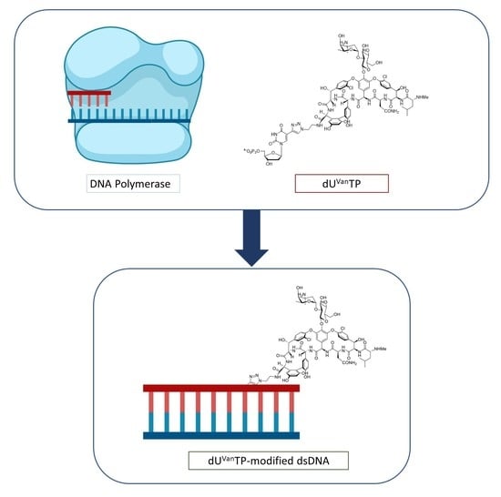
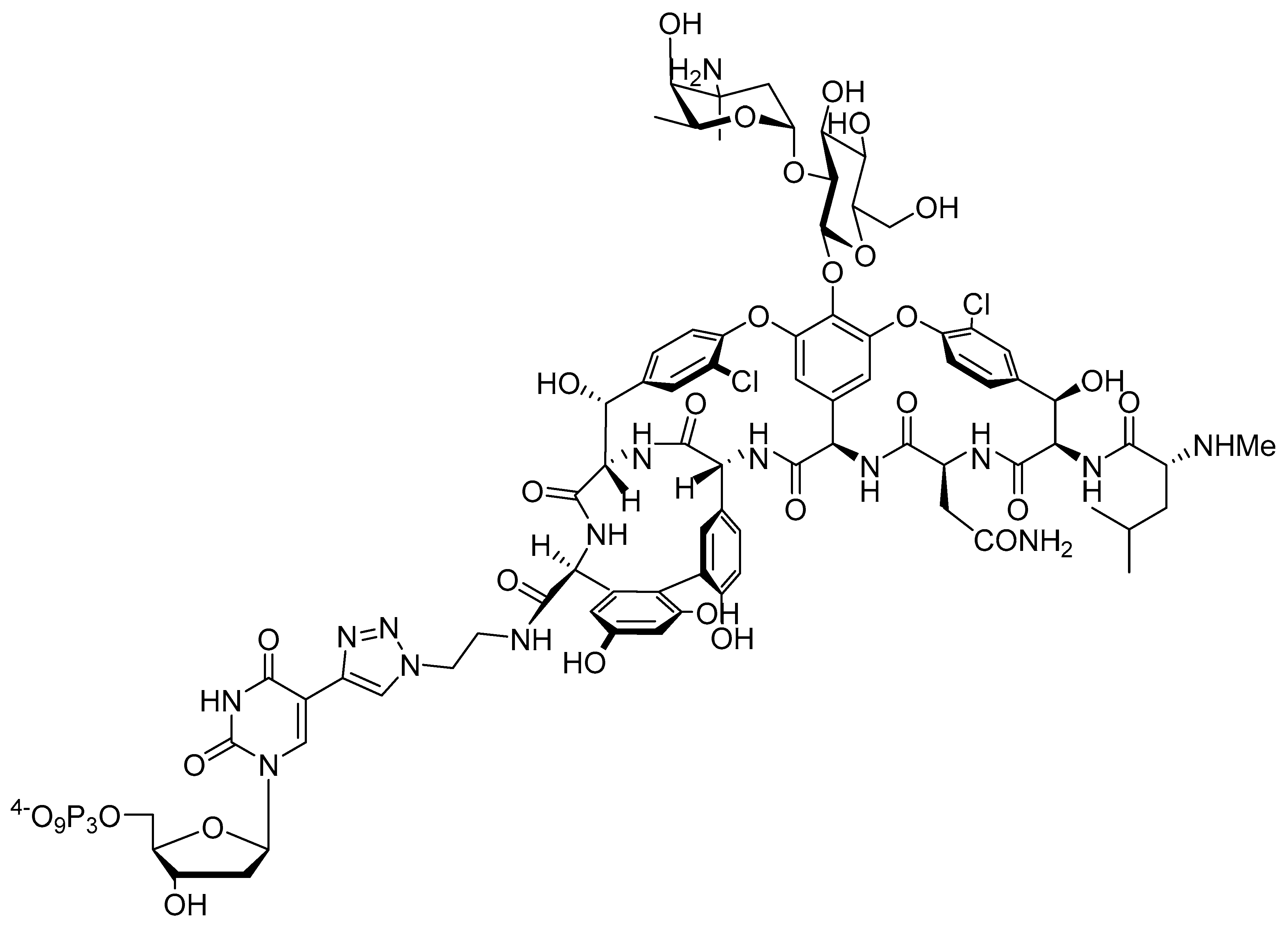
2.1. Design and Chemical Synthesis of Vancomycin-Modified Nucleoside Triphosphate (dUVanTP)

Aptamers are single-stranded nucleic acids capable of binding to targets with high affinity and specificity [31,32]. The first applications of aptamers in the context of microbial infections have previously been reported [33] including inhibition of biofilm formation, delivery of antibiotics conjugated to their scaffold, and participation in specific pathogen destruction [34,35,36]. In contrast, only a few reports have been dedicated to the application of aptamers to combat AMR, mainly as drug delivery systems [37]. For instance, aptamers conjugated to photosensitizers have been proposed as vectors for photodynamic therapy (PDT) to treat infections caused by antibiotic resistant pathogens [38,39,40]. In these examples, previously selected aptamers specific for particular bacterial targets are converted by post-SELEX modification to specific drug delivery agents. While this approach has met some success, post-SELEX modification protocols come with a lot of drawbacks [41,42]. First, the appendage of one or multiple drugs or reporter molecules on the termini of aptamers can lead to a loss in binding affinity (increase in KD values) which is detrimental for such an approach. Secondly, most aptamers are often directed towards diagnostic detection purposes rather than as therapeutic agents and hence do not present a sufficiently developed modification pattern robust enough to resist nuclease-mediated degradation. To combat this, the implementation of a post-SELEX modification strategy to mitigate this limitation and improve their biostability will be required. Third, post-SELEX approaches only allow for the introduction of a limited number of antibiotics (mainly by connection via the 3′- and 5′-termini) which induces high-cost productions and relatively low topical drug concentration. Lastly, upon reaching its intended target, the drug will be released but unless this released compound is a new chemical analogue of the parent drug it is unlikely to elude the defense and resistance mechanisms of the pathogens. In light of these elements, we rationalized that the inclusion of antibiotics on the scaffold of nucleoside triphosphates will yield ways to generate modified aptamers specific for resistant bacterial pathogens and potentially restore the intended antimicrobial activity. By inspection of the vancomycin scaffold, we rationalized that the free carboxylic acid might be a favorable site for connection to a nucleotide. This moiety is not involved in target recognition and previously reported modification at this site usually yielded analogues that retain the original antimicrobial activity [13,27,30]. To form the connection between vancomycin and the aptamer we opted to use the copper(I)-catalyzed azide–alkyne cycloaddition (CuAAC) reaction, since this chemistry has previously been used to generate modified vancomycin derivatives [13]. In addition to this, nucleotides bearing a triazole-connecting linker arm are compatible with enzymatic DNA synthesis [43,44] and aptamer selection [45,46,47]. The synthesis of the vancomycin-bearing deoxyuridine triphosphate dUVanTP (Figure 2) involved synthesis of the previously reported azide-modified vancomycin [13] and CuAAC reaction with 5-ethynyl-dUTP (EdUTP) [48] under standard conditions (see Materials and Methods (Section 4.1) and Supplementary Materials for full synthetic details). It is worth mentioning that the application of standard CuAAC reaction conditions yielded a mixture of species, including the expected nucleotide as well as a product corresponding to dUVanTP coordinated to Cu2+ (Figures S1 and S2). Coordination of the divalent metal cation occurs preferentially through the N-terminal imino NH-CH3 unit, two consecutive nitrogen atoms in the peptide chain, and one oxygen atom from the asparagine amide moiety [49,50]. The binding of vancomycin to Cu2+ is particularly efficient [50] and, hence, required the implementation of a thorough purification step to remove this undesired, cytotoxic transition metal cation from the modified nucleotide (see Materials and Methods (Section 4.1)). Alternatively, copper-free click chemistry may be considered in the future to avoid this rigorous purification step [51].

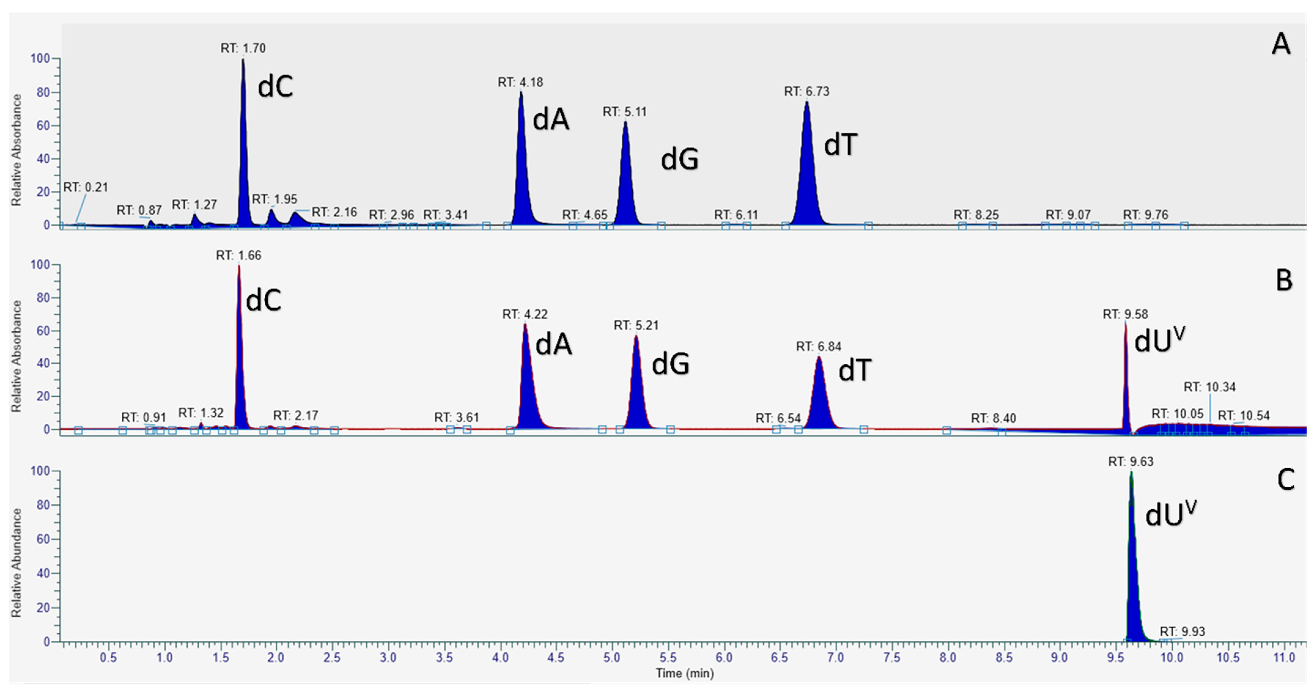
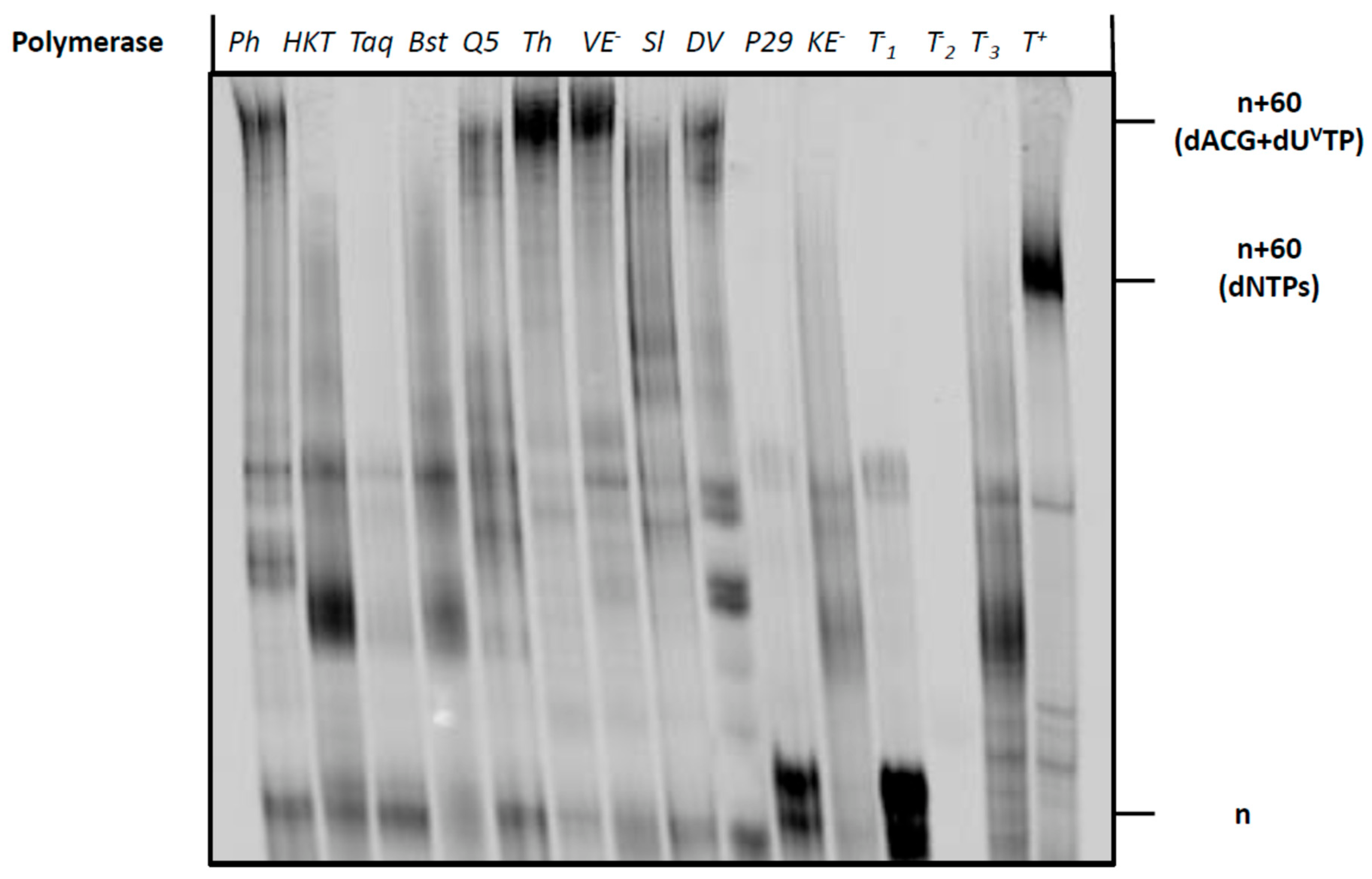
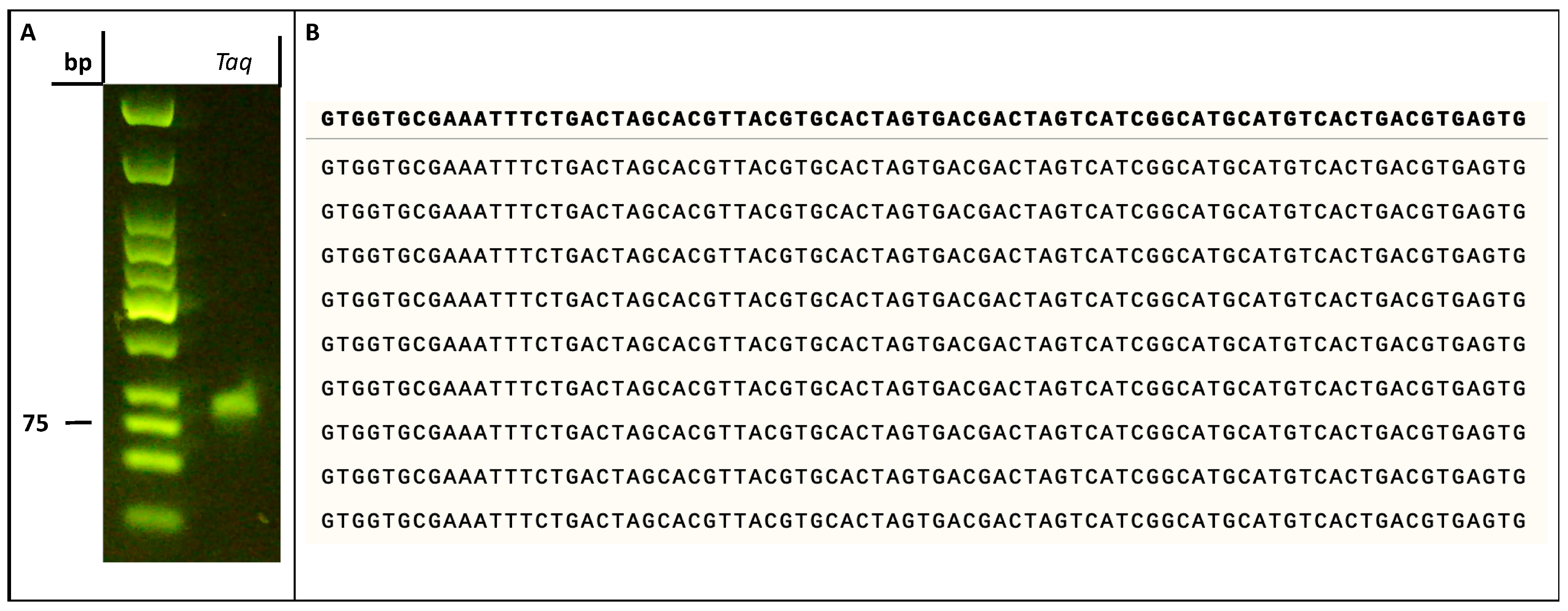
2.2. Biochemical Characterization of the Modified Nucleotide dUVanTP

With dUVanTP in hand, we evaluated the possibility of using this analog to synthesize vancomycin-modified DNA via primer extension (PEX) reactions and PCR. To do so, we first carried PEX reactions using the 20-nucleotide long template T1 and the 5′-FAM-labelled, 19-mer primer P1 (Table 1). This rather simple system allows for the incorporation of a single, modified nucleotide and can be followed by gel electrophoresis, LC-MS analysis, and UV-melting experiments. PEX reactions were performed with Vent (exo) DNA polymerase (vide infra) and the products were analyzed by gel electrophoresis (Figure S9). This analysis revealed the formation of a product with a slower electrophoretic mobility compared to that of the product obtained with dTTP or the unmodified, single-stranded DNA template. In order to confirm the incorporation of a dUVan nucleotide into DNA, we subjected the PEX reaction product to a digestion-LC-MS analysis protocol (see Materials and Methods (Section 4.2)) [52]. For the preparation of a standard for the modified nucleoside which is required for such an analysis, we dephosphorylated dUVanTP with Shrimp Alkaline Phosphatase (rSAP) and analyzed the resulting nucleoside by LC-MS (Figure 3 and Figure S6) and HRMS (Figure S7). PEX reaction products were nuclease-digested, dephosphorylated and the resulting deoxynucleosides were then analyzed by LC-MS (Figure 3).
The LC-MS chromatogram of the digested PEX reaction product using only dA/G/T/CTP displayed the expected four peaks corresponding to each of the four canonical nucleosides (Figure 3A). Similarly, the four canonical nucleosides were also detected in the LC-MS chromatogram of the digested reaction product obtained with dUVanTP, however, an additional peak at longer retention time (Figure 3B) and with a mass corresponding to dUVan was also detected (Figure S8 and Figure 3C). Therefore this analysis univocally confirmed the presence of the modified dUVan nucleotide in the extended primer P1 obtained after PEX reaction.
Next, we sought to evaluate the effect of such a bulky modification on duplex stability. To do so, we performed large-scale PEX reactions on the aforementioned P1/T1 primer/template duplex in the presence of dUVanTP and Vent (exo) DNA polymerase [53,54]. The UV-melting curves of the resulting modified dsDNA were then measured under standard saline buffer conditions and analyzed using a newly written python script for Tm data analysis (see Figure 4 and Supplementary Materials) [55]. The presence of the vancomycin moiety led to a rather significant destabilization of the duplex (ΔTm = −3.9 °C) compared to that of the unmodified dsDNA (62.7 ± 0.3 °C compared to 66.6 ± 0.1 °C). This destabilization might be caused either by the presence of the bulky vancomycin moiety since other large moieties such as polyethylene glycol [56] induced similar decreases in Tm values, or by the thermal penalty imparted by the introduction of a single triazole-containing nucleotide [57,58,59]. Nonetheless, this UV-melting analysis indicates that the modification did not significantly interfere with duplex formation.
Next, we sought to evaluate the possibility of using dUVanTP to synthesize longer vancomycin-containing DNA fragments using polymerase-mediated catalysis. To do so, we first carried out PEX reactions using the 19 nucleotide long 5′-FAM-labelled primer (P2) along with the corresponding 79-mer template (T2) that permits the incorporation of up to nine modified nucleotides (Table 1). A variety of polymerases were employed for PEX reactions, including the Klenow fragment of E. coli (Kf exo) DNA polymerase I, Bst, Hemo Klen Taq, and Taq (family A polymerases), the family B polymerases Phusion, Therminator, Vent (exo), phi29, and Deep Vent, and the Y-family polymerase Sulfolobus DNA Polymerase IV (Dpo4). Analysis by gel electrophoresis of the resulting PEX reaction products (Figure 5), revealed that a number of polymerases (Phusion, Q5, Therminator, Vent (exo) and Deep Vent) readily accepted dUVanTP as a substrate. By contrast, polymerases such as Taq did not tolerate the modified nucleotide and no extended product was formed. The highest conversion yields were observed when Vent (exo) and Therminator were used as polymerases to catalyze the incorporation of the modified nucleotide into DNA. The bands corresponding to full length, modified products possessed a retarded gel electrophoretic mobility compared to that of control reactions performed with canonical nucleotides. This is expected when a bulky residue is attached to the nucleobase [56,60,61]. Despite the rather high substrate tolerance of Therminator for dUVanTP, this polymerase has been shown to misincorporate natural and modified nucleotides [62,63,64,65], and to display an efficient pyrophosphorolysis capacity followed by incorporation of nucleotides [66]. Consequently, we believe that this catalyst might not be suitable for SELEX applications when used in conjunction with dUVanTP and instead we used Vent (exo) for further experiments.
Having established conditions for the enzymatic preparation of long, vancomycin-containing DNA oligonucleotides via PEX reactions, we next set out to evaluate the compatibility of dUVanTP with PCR. To do so, we performed PCR with template T2 along with the forward and reverse primers P3 and P4, respectively. We evaluated the capacity of three thermostable polymerases (i.e., Deep Vent, Vent (exo), and Phusion) to amplify template T2 in the presence of the modified nucleotide, dUVanTP. Surprisingly, the expected amplicons were produced in rather low yields, suggesting that the modified nucleotide is not very well tolerated by polymerases under PCR conditions (Figure S10). Despite the low synthetic yields, PCR represents an alternative to PEX reactions to produce vancomycin-containing dsDNA.

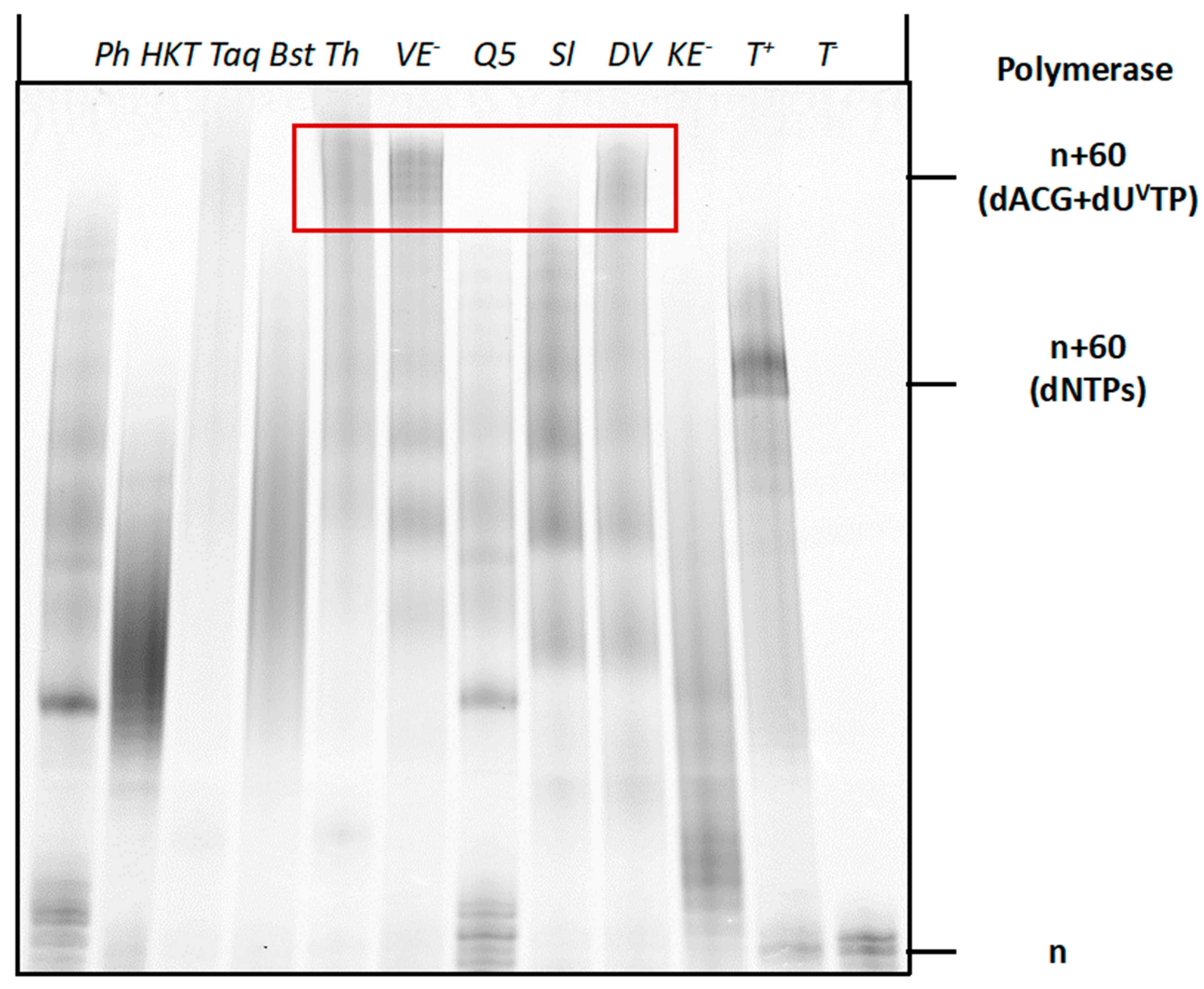
2.3. Compatibility of Vancomycin-Modified Nucleotide with SELEX

After establishing the conditions for the enzymatic synthesis of vancomycin-modified DNA, we next sought to evaluate the compatibility of dUVanTP with the SELEX method for the identification of aptamers [45,67,68,69,70]. SELEX and combinatorial methods of in vitro evolution with modified nucleotides require (1) synthesis of modified, degenerate libraries using PEX reactions or PCR; (2) robust conversion from double-stranded to single-stranded modified libraries [71] and (3) an efficient and high-fidelity regeneration of unmodified DNA from modified templates [54,72,73,74]. In order to assess whether enzymatic synthesis with dUVanTP fulfilled these criteria we first performed PEX reactions with a degenerate library (template T3 in Table 1), a panel of polymerases and dTTP substituted by dUVanTP. As with a defined sequence of the same length, the Therminator, Vent (exo), and Deep Vent DNA polymerases were capable of producing the expected full-length products in high yields (Figure 6). Thus these polymerases could produce fully modified dsDNA libraries and process these more demanding templates in the presence of the bulky modified nucleotide.
Next, we evaluated the possibility of reverse transcribing vancomycin-containing DNA oligonucleotides to unmodified sequences. To do so, we synthesized vancomycin-containing dsDNA by PCR as described above. After a thorough purification step, we subjected the modified product to PCR with canonical nucleotides and Taq (Figure 7A) or Vent (exo) (data not shown) as DNA polymerases. Agarose gel electrophoretic analysis of the resulting reaction products clearly indicated the formation of the expected amplicon (Figure 7A). We next subjected the obtained PCR product with canonical nucleotides and the Taq polymerase to a TA-cloning step followed by Sanger sequencing of nine individual colonies. Sequencing of this small subset of colonies clearly indicated the absence of mutations compared to the expected parent sequence of template T2 (Figure 7B). This analysis suggested a faithful reverse transcription of vancomycin-containing DNA to unmodified oligonucleotides. Lastly, we evaluated the possibility of converting double-stranded, modified sequences to single-stranded oligonucleotides, which is an important prerequisite for SELEX and a notoriously difficult step, especially for heavily modified sequences or bulky modifications such as vancomycin [71]. We first performed PEX reactions with dUVanTP, Vent (exo), and a 5′-phosphorylated analogue of template T1 (template T4, Table 1). The resulting modified dsDNA was then converted to the corresponding ssDNA by λ-exonuclease digestion (Figure S11A). Gel analysis of the resulting products indicated formation of the expected single-stranded sequences (Figure S11B).

3. Conclusions

AMR is a major threat to human health, and alternative treatment modalities and drugs are in dire need. Herein, we have presented the first step towards combining antibiotics and aptamers in order to potentially restore the effectiveness of drugs against resistant bacteria. Towards this aim, we have synthesized a deoxyuridine analogue bearing a vancomycin residue at position C5 of the nucleobase. The resulting nucleotide, dUVanTP, was then shown to be compatible with enzymatic DNA synthesis under PEX reaction conditions and PCR (to a certain extent), despite the presence of such a bulky chemical modification. We have then demonstrated the compatibility of dUVanTP with the SELEX methodology. In particular, we have shown that modified libraries could be generated by enzymatic synthesis and subsequently reverse transcribed into unmodified DNA without introducing mutations. Cell-SELEX experiments against vancomycin resistant strains and subsequent evaluation of the antibacterial activity of modified aptamers are currently underway. We expect the aptamer section of such a construct to bind to membrane and surface proteins, which are the typical targets of cell-SELEX experiments [75]. This binding event will then, in turn, restore binding of vancomycin to the peptidoglycan and, hence, restore its activity. A similar approach using a primer equipped with a hotspot peptide in SELEX was recently employed for the selection of aptamers against the human angiotensin-converting enzyme 2 (hACE2) [76] underscoring the usefulness of such a method. However, the advantage of our approach is that multiple vancomycin-bearing nucleotides will be integrated within the scaffold of the aptamer and will be involved in the binding interactions. Hence, uncertain, labor-intensive post-SELEX structure-activity relationship studies to optimize aptamers will not be required. Lastly, we believe that such an approach could easily be extended to other antibiotics such as β-lactamase inhibitors [7,8] or pentamidine-based scaffolds [77].

4. Materials and Methods

4.1. Chemical Synthesis of dUVanTP

EdUTP [48] (2 mg, 0.004 mmol, 1.5 eq.) and azido-vancomycin [13] (4 mg, 0.0026 mmol, 1 eq.) were respectively dissolved in 100 μL H2O each in two different Eppendorf tubes and degassed with argon. In a separated Eppendorf tube, CuI (3 mg, 0.02 mmol, 0.77 eq.) was suspended in DIPEA (10 μL, 0.06 mmol, 2.3 eq.), 50 μL H2O and 50 μL CH3CN and degassed with Argon. The three fractions were merged and 100 μL DMSO were added. The reaction mixture was degassed with Argon and left to shake at 1000 rpm, at r.t. for 3 h. The reaction mixture was added to 12 mL NaClO4 2% in acetone to precipitate the crude product, centrifuged at 4000 rpm for 15 min, and the surnatant was discarded. The crude was redissolved in 3 mL H2O and purified by anion exchange analytical HPLC (100% B in 25 min; Buffer A, 10 mM TEAB in H2O; Buffer B, 1 M TEAB in H2O) with a DNA Pac PA 100 oligonucleotide column. Prior to each injection, EDTA (0.5 M, pH = 8) was added to the crude solution to coordinate the remaining Cu2+ present in the solution. The fractions corresponding to dUVanTP were merged together and lyophilized overnight to give the product as 2.9 mg (55%) of a white powder. To remove the remaining coordinated Cu2+, dUVanTP was dissolved in 1 mL H2O and stirred with 1 g of Chelex 100 resin for 2 h at rt.
31P NMR (202.4 MHz, D2O): −6.15 (d, J = 20.2 Hz, 1P), −11.17 (d, J = 18.2 Hz, 1P), −22.17 (t, J = 19.2 Hz, 1P); HR-ESI-MS (m/z): calcd for M = C79H94Cl2N15O37CuP3, [M] = 2071.00, found [M-2H]2− = 2068.3518. HR-ESI-MS (m/z): calcd for M = C79H94Cl2N15O37P3, [M] = 2009.48, found [M-2H]2− = 2007.4412.

4.2. Preparation of dUVan Nucleoside Standard

dUVanTP (10 nmol) was incubated with rSAP (10 µL) and rCutSmart buffer (2.2 µL of 10X) in a total volume of 22 µL for 1 h at 37 °C. Afterwards, rSAP was inactivated via incubation for 10 min at 65 °C. The product was purified and characterized via LCMS (Figure S6). HR-ESI-MS (m/z): calcd for M = C79H91Cl2N15O28Cu, [M] = 1833.12, found [M]+ = 1832.4842. HR-ESI-MS (m/z): calcd for M = C79H91Cl2N15O28, [M] = 1769.57, found [M+2H]2+ = 1770.5730.

4.3. Digestion and LC-MS Analysis of PEX Reaction Product

The primer P1 (100 pmol) was annealed to the template T1 (150 pmol) in H2O by heating to 95 °C and then gradually cooling to room temperature (over 1 h). Vent (exo) polymerase (4U), Thermopol 10X buffer (provided by the supplier of the DNA polymerase) (2 μL) and dUVanTP (or dTTP for the positive control) (200 μM) were added for a total volume of 10 μL. The reaction mixture was incubated for 4 h at 60 °C. The formation of the products was verified with an agarose E-GEL (4%) using the E-GEL sample loading buffer (1X). The products were purified via Monarch DNA Cleanup Columns (5 μg) (250 pmol product per column). The products (1 μL), the Nucleoside Digestion Mix buffer (2 μL of 10X) and the Nucleoside Digestion Mix (1 μL) were mixed in a total volume of 20 μL and incubated at 37 °C for 1 h. The digested DNA solution was then injected onto a ThermoFisher Hypersil Gold aQ chromatography column (100 × 2.1 mm, 1.9 μm particle size) heated at 30 °C without any further purification. The flow rate was set at 0.3 mL/min and run with an isocratic elution of 1% ACN in H2O with 0.1% formic acid for 8 min then 100% ACN from 9 to 11 min. Parent ions were fragmented in positive ion mode with 10% normalized collision energy in parallel-reaction monitoring (PRM) mode. MS2 resolution was 17,500 with an AGC target of 2 × 10⁵, a maximum injection time of 50 ms, and an isolation window of 1.0 m/z. The inclusion list contained the following masses: dC (228.1), dA (252.1), dG (268.1), dT (243.1) and 4 (884.8; z = 2). Extracted ion chromatograms of base fragments (±5 ppm) were used for detection (112.0506 Da for dC; 136.0616 for dA; 152.0565 Da for dG; 127.0501 Da for dT and 1625.4652 Da for 4). Synthetic standards were previously injected to confirm the assignment (fragment ion and retention time).

4.4. General Protocol for PEX Reactions

The 5′-FAM-labelled primer (10 pmol) was annealed to the template (15 pmol) in H2O by heating to 95 °C and then gradually cooling to room temperature (1 h). The appropriate polymerase, 5X or 10X enzyme buffer (provided by the supplier of the DNA polymerase), the natural nucleotides (200 μM), and dUVanTP (or dTTP in the positive controls) (200 μM) were added for a total reaction volume of 10 μL. Following incubation for 4 h at the optimal temperature for the enzyme, the reactions were quenched by adding the stop solution (10 μL; formamide (70%), ethylenediaminetetraacetic acid (EDTA; 50 mM), bromophenol (0.1%), xylene cyanol (0.1%)). The reaction mixtures were subjected to gel electrophoresis in denaturing polyacrylamide gel (20%) containing trisborate- EDTA (TBE) 1× buffer (pH 8) and urea (7 M). Products were then analyzed on a phosphorimager.

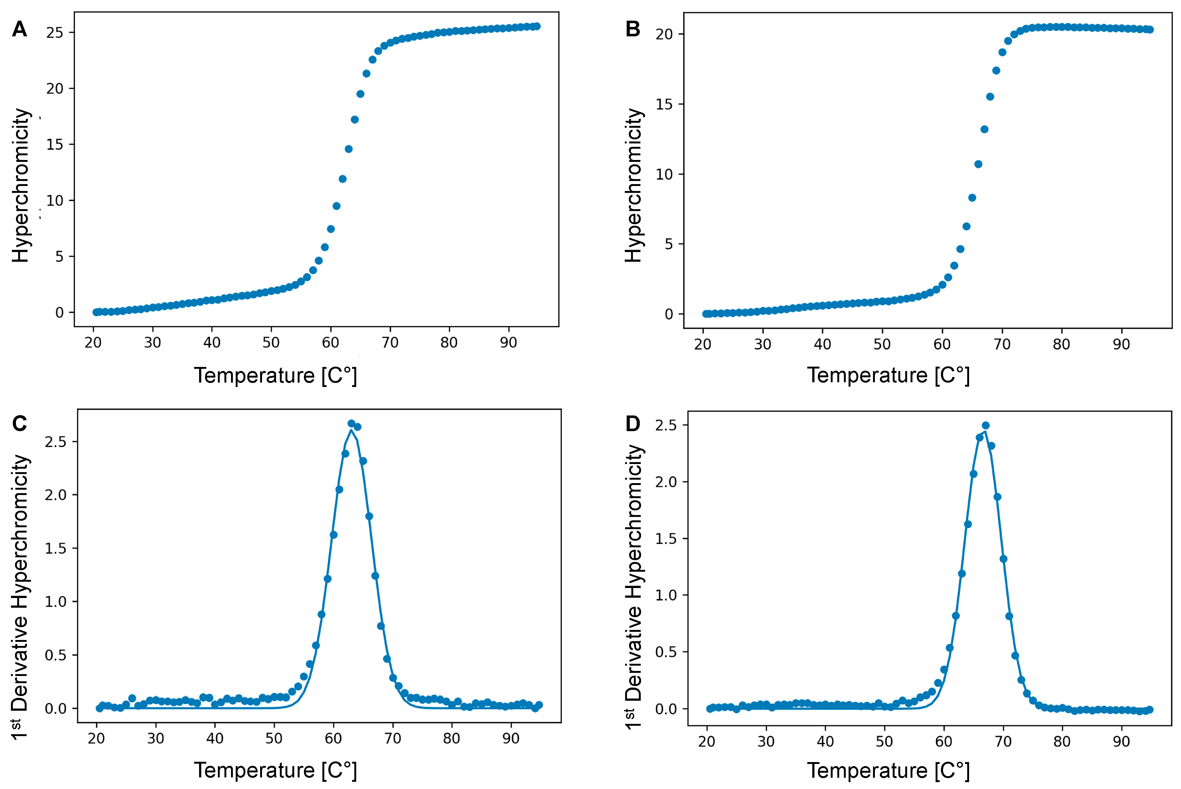
4.5. Thermal Denaturation Experiments

Experiments were recorded in 1X phosphate-buffered saline (PBS) (137 mM NaCl, 2.7 mM KCl, 4.3 mM Na2HPO4, 1.47 mM KH2PO4, pH 7.4) in H2O with 2 μM final duplex concentration. Paraffin oil (150 μL) was added to avoid evaporation. A control cell (1X PBS) was prepared into which the temperature probe was placed. Absorbance was monitored at 260 nm and the heating rate was set to 1 °C/min. A heating-cooling cycle in the temperature range of 20–95 °C was applied and repeated 3 times per sample. The data analysis was performed with a small Python (v3.8.5) script on the heating ramps (see Supplementary Materials). The absorbance melting curves were converted to hyperchromicities and the first derivative of each experimental hyperchromicity point was calculated. Afterwards, the Gaussian function that best fitted the first derivative curve was calculated and the melting temperature was obtained from it. The final Tm value was calculated as the mean of the three obtained values with the relative error.

Supplementary Materials

The following supporting information can be downloaded at: https://www.mdpi.com/article/10.3390/molecules27248927/s1, Figure S1: MALDI-TOF analysis of dUVanTP coordinated with Cu2+; Figure S2: HR-ESI-MS analysis of dUVanTP coordinated with Cu2+; Figure S3: MALDI-TOF analysis of dUVanTP; Figure S4: HR-ESI-MS analysis of dUVanTP; Figure S5: 31P NMR spectrum of dUVanTP; Figure S6: LC-MS analysis of dUVan; Figure S7: HR-ESI-MS analysis of nucleoside dUVan; Figure S8: Full LC-MS chromatogram of the nucleosides stemming from the digested and dephosphorylated PEX reaction product obtained with dUVanTP; Figure S9: Gel image analysis of PEX reaction products obtained with dUVanTP and dTTP; Figure S10: Gel image analysis of PCR reaction products obtained with dUVanTP and dTTP; Figure S11: Schematic representation of the different steps involved in the generation of dUVan-modified ssDNA from the corresponding dsDNA and gel image analysis of the products of the generation of the dUVan-modified ssDNA; Figure S12: Gel image showing the results obtained by performing PCR on 19 different E. Coli colonies; Details of the python script for data analysis of UV-melting experiments.

Author Contributions

Conceptualization, M.H.; methodology, C.F., F.B., M.E.-Q., M.H.; software, C.F.; validation, C.F., F.B., S.S., M.E.-Q., M.H.; formal analysis, C.F., F.B., M.H.; investigation, C.F., F.B. and S.S.; resources, M.E.-Q. and M.H.; writing—original draft preparation, C.F. and M.H.; writing—review and editing, all authors.; visualization, M.H.; supervision, M.E.-Q. and M.H.; project administration, M.H.; funding acquisition, C.F. and M.H. All authors have read and agreed to the published version of the manuscript.

Funding

C.F. gratefully acknowledges the Pasteur-Paris University (PPU) International Ph.D. Program that has received funding from INCEPTION Project (PIA/ANR-16-CONV-0005), the “Ecole Doctorale Frontières de l’Innovation en Recherche et Education—Programme Bettencourt” for financial support. The authors gratefully acknowledge financial support from Institut Pasteur.

Data Availability Statement

The data presented in this study are available on request from the corresponding author.

Acknowledgments

Harley Betts is gratefully acknowledged for proof-reading our manuscript.

Conflicts of Interest

The authors declare no conflict of interest.

Sample Availability

Samples of the compounds are available from the authors.

References

  1. Larsson, D.G.J.; Flach, C.-F. Antibiotic resistance in the environment. Nat. Rev. Microbiol. 2022, 20, 257–269. [Google Scholar] [CrossRef] [PubMed]
  2. Blair, J.M.A.; Webber, M.A.; Baylay, A.J.; Ogbolu, D.O.; Piddock, L.J.V. Molecular mechanisms of antibiotic resistance. Nat. Rev. Microbiol. 2015, 13, 42–51. [Google Scholar] [CrossRef] [PubMed]
  3. Murray, C.J.L.; Ikuta, K.S.; Sharara, F.; Swetschinski, L.; Robles Aguilar, G.; Gray, A.; Han, C.; Bisignano, C.; Rao, P.; Wool, E.; et al. Global burden of bacterial antimicrobial resistance in 2019: A systematic analysis. Lancet 2022, 399, 629–655. [Google Scholar] [CrossRef] [PubMed]
  4. Saidjalolov, S.; Braud, E.; Edoo, Z.; Iannazzo, L.; Rusconi, F.; Riomet, M.; Sallustrau, A.; Taran, F.; Arthur, M.; Fonvielle, M.; et al. Click and Release Chemistry for Activity-Based Purification of β-Lactam Targets. Chem. Eur. J. 2021, 27, 7687–7695. [Google Scholar] [CrossRef] [PubMed]
  5. Shinu, P.; Mouslem, A.K.A.; Nair, A.B.; Venugopala, K.N.; Attimarad, M.; Singh, V.A.; Nagaraja, S.; Alotaibi, G.; Deb, P.K. Progress Report: Antimicrobial Drug Discovery in the Resistance Era. Pharmaceuticals 2022, 15, 413. [Google Scholar] [CrossRef] [PubMed]
  6. Namivandi-Zangeneh, R.; Sadrearhami, Z.; Dutta, D.; Willcox, M.; Wong, E.H.H.; Boyer, C. Synergy between Synthetic Antimicrobial Polymer and Antibiotics: A Promising Platform To Combat Multidrug-Resistant Bacteria. ACS Infect. Dis. 2019, 5, 1357–1365. [Google Scholar] [CrossRef] [Green Version]
  7. Bouchet, F.; Atze, H.; Fonvielle, M.; Edoo, Z.; Arthur, M.; Ethève-Quelquejeu, M.; Iannazzo, L. Diazabicyclooctane Functionalization for Inhibition of β-Lactamases from Enterobacteria. J. Med. Chem. 2020, 63, 5257–5273. [Google Scholar] [CrossRef]
  8. Iqbal, Z.; Sun, J.; Yang, H.; Ji, J.; He, L.; Zhai, L.; Ji, J.; Zhou, P.; Tang, D.; Mu, Y.; et al. Recent Developments to Cope the Antibacterial Resistance via β-Lactamase Inhibition. Molecules 2022, 27, 3832. [Google Scholar] [CrossRef]
  9. Romero, E.; Oueslati, S.; Benchekroun, M.; D’Hollander, A.C.A.; Ventre, S.; Vijayakumar, K.; Minard, C.; Exilie, C.; Tlili, L.; Retailleau, P.; et al. Azetidinimines as a novel series of non-covalent broad-spectrum inhibitors of β-lactamases with submicromolar activities against carbapenemases KPC-2 (class A), NDM-1 (class B) and OXA-48 (class D). Eur. J. Med. Chem. 2021, 219, 113418. [Google Scholar] [CrossRef]
  10. Fu, X.; Yu, J.; Dai, N.; Huang, Y.; Lv, F.; Liu, L.; Wang, S. Optical Tuning of Antibacterial Activity of Photoresponsive Antibiotics. ACS Appl. Bio Mater. 2020, 3, 4751–4755. [Google Scholar] [CrossRef]
  11. Okano, A.; Isley, N.A.; Boger, D.L. Total Syntheses of Vancomycin-Related Glycopeptide Antibiotics and Key Analogues. Chem. Rev. 2017, 117, 11952–11993. [Google Scholar] [CrossRef]
  12. Acharya, Y.; Dhanda, G.; Sarkar, P.; Haldar, J. Pursuit of next-generation glycopeptides: A journey with vancomycin. Chem. Commun. 2022, 58, 1881–1897. [Google Scholar] [CrossRef]
  13. Silverman, S.M.; Moses, J.E.; Sharpless, K.B. Reengineering Antibiotics to Combat Bacterial Resistance: Click Chemistry [1,2,3]-Triazole Vancomycin Dimers with Potent Activity against MRSA and VRE. Chem. Eur. J. 2017, 23, 79–83. [Google Scholar] [CrossRef]
  14. Bugg, T.D.H.; Wright, G.D.; Dutka-Malen, S.; Arthur, M.; Courvalin, P.; Walsh, C.T. Molecular basis for vancomycin resistance in Enterococcus faecium BM4147: Biosynthesis of a depsipeptide peptidoglycan precursor by vancomycin resistance proteins VanH and VanA. Biochemistry 1991, 30, 10408–10415. [Google Scholar] [CrossRef]
  15. Walsh; Wright, Introduction:  Antibiotic Resistance. Chem. Rev. 2005, 105, 391–394. [CrossRef] [Green Version]
  16. Petchiappan, A.; Chatterji, D. Antibiotic Resistance: Current Perspectives. ACS Omega 2017, 2, 7400–7409. [Google Scholar] [CrossRef]
  17. Müller, A.; Klöckner, A.; Schneider, T. Targeting a cell wall biosynthesis hot spot. Nat. Prod. Rep. 2017, 34, 909–932. [Google Scholar] [CrossRef]
  18. Liu, Y.; Jia, Y.; Yang, K.; Li, R.; Xiao, X.; Wang, Z. Antagonizing Vancomycin Resistance in Enterococcus by Surface Localized Antimicrobial Display-Derived Peptides. ACS Infect. Dis. 2020, 6, 761–767. [Google Scholar] [CrossRef]
  19. Melander, R.J.; Melander, C. The Challenge of Overcoming Antibiotic Resistance: An Adjuvant Approach? ACS Infect. Dis. 2017, 3, 559–563. [Google Scholar] [CrossRef]
  20. Chang, M.; Mahasenan, K.V.; Hermoso, J.A.; Mobashery, S. Unconventional Antibacterials and Adjuvants. Acc. Chem. Res. 2021, 54, 917–929. [Google Scholar] [CrossRef]
  21. Chiosis, G.; Boneca, I.G. Selective Cleavage of D-Ala-D-Lac by Small Molecules: Re-Sensitizing Resistant Bacteria to Vancomycin. Science 2001, 293, 1484–1487. [Google Scholar] [CrossRef] [PubMed] [Green Version]
  22. Yarlagadda, V.; Sarkar, P.; Samaddar, S.; Haldar, J. A Vancomycin Derivative with a Pyrophosphate-Binding Group: A Strategy to Combat Vancomycin-Resistant Bacteria. Angew. Chem. Int. Ed. 2016, 55, 7836–7840. [Google Scholar] [CrossRef] [PubMed]
  23. Ma, C.; He, N.; Ou, Y.; Feng, W. Design and Synthesis of New Vancomycin Derivatives. ChemistrySelect 2020, 5, 6670–6673. [Google Scholar] [CrossRef]
  24. Okano, A.; Isley, N.A.; Boger, D.L. Peripheral modifications of [Ψ[CH2NH]Tpg4]vancomycin with added synergistic mechanisms of action provide durable and potent antibiotics. Proc. Natl. Acad. Sci. USA 2017, 114, E5052–E5061. [Google Scholar] [CrossRef] [PubMed] [Green Version]
  25. Xing, B.; Ho, P.L.; Yu, C.-W.; Chow, K.-H.; Gu, H.; Xu, B. Self-assembled multivalent vancomycin on cell surfaces against vancomycin-resistant enterococci (VRE). Chem. Commun. 2003, 2224–2225. [Google Scholar] [CrossRef]
  26. Yang, C.; Ren, C.; Zhou, J.; Liu, J.; Zhang, Y.; Huang, F.; Ding, D.; Xu, B.; Liu, J. Dual Fluorescent- and Isotopic-Labelled Self-Assembling Vancomycin for in vivo Imaging of Bacterial Infections. Angew. Chem. Int. Ed. 2017, 56, 2356–2360. [Google Scholar] [CrossRef]
  27. Griffin, J.H.; Linsell, M.S.; Nodwell, M.B.; Chen, Q.; Pace, J.L.; Quast, K.L.; Krause, K.M.; Farrington, L.; Wu, T.X.; Higgins, D.L.; et al. Multivalent Drug Design. Synthesis and In Vitro Analysis of an Array of Vancomycin Dimers. J. Am. Chem. Soc. 2003, 125, 6517–6531. [Google Scholar] [CrossRef]
  28. Sarkar, P.; Basak, D.; Mukherjee, R.; Bandow, J.E.; Haldar, J. Alkyl-Aryl-Vancomycins: Multimodal Glycopeptides with Weak Dependence on the Bacterial Metabolic State. J. Med. Chem. 2021, 64, 10185–10202. [Google Scholar] [CrossRef]
  29. McComas, C.C.; Crowley, B.M.; Boger, D.L. Partitioning the Loss in Vancomycin Binding Affinity for d-Ala-d-Lac into Lost H-Bond and Repulsive Lone Pair Contributions. J. Am. Chem. Soc. 2003, 125, 9314–9315. [Google Scholar] [CrossRef]
  30. Flint, A.J.; Davis, A.P. Vancomycin mimicry: Towards new supramolecular antibiotics. Org. Biomol. Chem. 2022, 20, 7694–7712. [Google Scholar] [CrossRef]
  31. Ellington, A.D.; Szostak, J.W. In vitro selection of RNA molecules that bind specific ligands. Nature 1990, 346, 818–822. [Google Scholar] [CrossRef]
  32. Tuerk, C.; Gold, L. Systematic Evolution of Ligands by Exponential Enrichment: RNA Ligands to Bacteriophage T4 DNA Polymerase. Science 1990, 249, 505–510. [Google Scholar] [CrossRef]
  33. Afrasiabi, S.; Pourhajibagher, M.; Raoofian, R.; Tabarzad, M.; Bahador, A. Therapeutic applications of nucleic acid aptamers in microbial infections. J. Biomed. Sci. 2020, 27, 6. [Google Scholar] [CrossRef]
  34. Zhang, X.; Soontornworajit, B.; Zhang, Z.; Chen, N.; Wang, Y. Enhanced Loading and Controlled Release of Antibiotics Using Nucleic Acids As an Antibiotic-Binding Effector in Hydrogels. Biomacromolecules 2012, 13, 2202–2210. [Google Scholar] [CrossRef]
  35. Catuogno, S.; Esposito, C.L.; De Franciscis, V. Aptamer-Mediated Targeted Delivery of Therapeutics: An Update. Pharmaceuticals 2016, 9, 69. [Google Scholar] [CrossRef] [Green Version]
  36. Lijuan, C.; Xing, Y.; Minxi, W.; Wenkai, L.; Le, D. Development of an aptamer-ampicillin conjugate for treating biofilms. Biochem. Biophys. Res. Commun. 2017, 483, 847–854. [Google Scholar] [CrossRef]
  37. Chen, X.-F.; Zhao, X.; Yang, Z. Aptamer-Based Antibacterial and Antiviral Therapy against Infectious Diseases. J. Med. Chem. 2021, 64, 17601–17626. [Google Scholar] [CrossRef]
  38. Ocsoy, I.; Yusufbeyoglu, S.; Yılmaz, V.; McLamore, E.S.; Ildız, N.; Ülgen, A. DNA aptamer functionalized gold nanostructures for molecular recognition and photothermal inactivation of methicillin-Resistant Staphylococcus aureus. Colloids Surf. B Biointerfaces 2017, 159, 16–22. [Google Scholar] [CrossRef]
  39. McKenzie, L.K.; Flamme, M.; Felder, P.S.; Karges, J.; Bonhomme, F.; Gandioso, A.; Malosse, C.; Gasser, G.; Hollenstein, M. A ruthenium–oligonucleotide bioconjugated photosensitizing aptamer for cancer cell specific photodynamic therapy. RSC Chem. Biol. 2022, 3, 85–95. [Google Scholar] [CrossRef]
  40. Mallikaratchy, P.; Tang, Z.; Tan, W. Cell Specific Aptamer–Photosensitizer Conjugates as a Molecular Tool in Photodynamic Therapy. ChemMedChem 2008, 3, 425–428. [Google Scholar] [CrossRef]
  41. Röthlisberger, P.; Hollenstein, M. Aptamer chemistry. Adv. Drug Deliv. Rev. 2018, 134, 3–21. [Google Scholar] [CrossRef] [PubMed]
  42. McKenzie, L.K.; El-Khoury, R.; Thorpe, J.D.; Damha, M.J.; Hollenstein, M. Recent progress in non-native nucleic acid modifications. Chem. Soc. Rev. 2021, 50, 5126–5164. [Google Scholar] [CrossRef] [PubMed]
  43. Figazzolo, C.; Ma, Y.; Tucker, J.H.R.; Hollenstein, M. Ferrocene as a potential electrochemical reporting surrogate of abasic sites in DNA. Org. Biomol. Chem. 2022, 20, 8125–8135. [Google Scholar] [CrossRef] [PubMed]
  44. Leone, D.L.; Hubalek, M.; Pohl, R.; Sykorova, V.; Hocek, M. 1,3-Diketone-Modified Nucleotides and DNA for Cross-Linking with Arginine-Containing Peptides and Proteins. Angew. Chem. Int. Ed. 2021, 60, 17383–17387. [Google Scholar] [CrossRef] [PubMed]
  45. Cheung, Y.W.; Röthlisberger, P.; Mechaly, A.E.; Weber, P.; Levi-Acobas, F.; Lo, Y.; Wong, A.W.C.; Kinghorn, A.B.; Haouz, A.; Savage, G.P.; et al. Evolution of abiotic cubane chemistries in a nucleic acid aptamer allows selective recognition of a malaria biomarker. Proc. Natl. Acad. Sci. USA 2020, 117, 16790–16798. [Google Scholar] [CrossRef]
  46. Siegl, J.; Plückthun, O.; Mayer, G. Dependence of click-SELEX performance on the nature and average number of modified nucleotides. RSC Chem. Biol. 2022, 3, 288–294. [Google Scholar] [CrossRef]
  47. Siegl, J.; Nikolin, C.; Phung, N.L.; Thoms, S.; Blume, C.; Mayer, G. Split–Combine Click-SELEX Reveals Ligands Recognizing the Transplant Rejection Biomarker CXCL9. ACS Chem. Biol. 2022, 17, 129–137. [Google Scholar] [CrossRef]
  48. Röthlisberger, P.; Levi-Acobas, F.; Hollenstein, M. New synthetic route to ethynyl-dUTP: A means to avoid formation of acetyl and chloro vinyl base-modified triphosphates that could poison SELEX experiments. Bioorg. Med. Chem. Lett. 2017, 27, 897–900. [Google Scholar] [CrossRef]
  49. Kucharczyk, M.; Brzezowska, M.; Maciąg, A.; Lis, T.; Jeżowska-Bojczuk, M. Structural features of the Cu2+–vancomycin complex. J. Inorg. Biochem. 2008, 102, 936–942. [Google Scholar] [CrossRef]
  50. Świątek, M.; Valensin, D.; Migliorini, C.; Gaggelli, E.; Valensin, G.; Jeżowska-Bojczuk, M. Unusual binding ability of vancomycin towards Cu2+ ions. Dalton Trans. 2005, 3808–3813. [Google Scholar] [CrossRef]
  51. Baskin, J.M.; Prescher, J.A.; Laughlin, S.T.; Agard, N.J.; Chang, P.V.; Miller, I.A.; Lo, A.; Codelli, J.A.; Bertozzi, C.R. Copper-free click chemistry for dynamic in vivo imaging. Proc. Natl. Acad. Sci. USA 2007, 104, 16793–16797. [Google Scholar] [CrossRef] [Green Version]
  52. Nguyen, H.; Abramov, M.; Rozenski, J.; Eremeeva, E.; Herdewijn, P. In Vivo Assembly and Expression of DNA Containing Non-canonical Bases in the Yeast Saccharomyces cerevisiae. ChemBioChem 2022, 23, e202200060. [Google Scholar] [CrossRef]
  53. Cahová, H.; Pohl, R.; Bednárová, L.; Nováková, K.; Cvačka, J.; Hocek, M. Synthesis of 8-bromo-, 8-methyl- and 8-phenyl-dATP and their polymerase incorporation into DNA. Org. Biomol. Chem. 2008, 6, 3657–3660. [Google Scholar] [CrossRef]
  54. Hollenstein, M. Synthesis of Deoxynucleoside Triphosphates that Include Proline, Urea, or Sulfonamide Groups and Their Polymerase Incorporation into DNA. Chem. Eur. J. 2012, 18, 13320–13330. [Google Scholar] [CrossRef]
  55. Zaboikin, M.; Freter, C.; Srinivasakumar, N. Gaussian decomposition of high-resolution melt curve derivatives for measuring genome-editing efficiency. PLoS ONE 2018, 13, e0190192. [Google Scholar]
  56. Baccaro, A.; Marx, A. Enzymatic Synthesis of Organic-Polymer-Grafted DNA. Chem. Eur. J. 2010, 16, 218–226. [Google Scholar] [CrossRef] [Green Version]
  57. Kočalka, P.; Andersen, N.K.; Jensen, F.; Nielsen, P. Synthesis of 5-(1,2,3-Triazol-4-yl)-2′-deoxyuridines by a Click Chemistry Approach: Stacking of Triazoles in the Major Groove Gives Increased Nucleic Acid Duplex Stability. ChemBioChem 2007, 8, 2106–2116. [Google Scholar] [CrossRef]
  58. Gimenez Molina, A.; Raguraman, P.; Delcomyn, L.; Veedu, R.N.; Nielsen, P. Oligonucleotides containing 2′-O-methyl-5-(1-phenyl-1,2,3-triazol-4-yl)uridines demonstrate increased affinity for RNA and induce exon-skipping in vitro. Bioorg. Med. Chem. 2022, 55, 116559. [Google Scholar] [CrossRef]
  59. Fan, H.; Sun, H.; Haque, M.M.; Peng, X. Effect of Triazole-Modified Thymidines on DNA and RNA Duplex Stability. ACS Omega 2019, 4, 5107–5116. [Google Scholar] [CrossRef]
  60. Balintová, J.; Welter, M.; Marx, A. Antibody–nucleotide conjugate as a substrate for DNA polymerases. Chem. Sci. 2018, 9, 7122–7125. [Google Scholar] [CrossRef] [Green Version]
  61. Hollenstein, M. Deoxynucleoside triphosphates bearing histamine, carboxylic acid, and hydroxyl residues—Synthesis and biochemical characterization. Org. Biomol. Chem. 2013, 11, 5162–5172. [Google Scholar] [CrossRef] [PubMed]
  62. Gardner, A.F.; Jackson, K.M.; Boyle, M.M.; Buss, J.A.; Potapov, V.; Gehring, A.M.; Zatopek, K.M.; Corrêa, I.R., Jr.; Ong, J.L.; Jack, W.E. Therminator DNA Polymerase: Modified Nucleotides and Unnatural Substrates. Front. Mol. Biosci. 2019, 6, 28. [Google Scholar] [CrossRef] [Green Version]
  63. Dunn, M.R.; Larsen, A.C.; Zahurancik, W.J.; Fahmi, N.E.; Meyers, M.; Suo, Z.; Chaput, J.C. DNA Polymerase-Mediated Synthesis of Unbiased Threose Nucleic Acid (TNA) Polymers Requires 7-Deazaguanine To Suppress G:G Mispairing during TNA Transcription. J. Am. Chem. Soc. 2015, 137, 4014–4017. [Google Scholar] [CrossRef] [PubMed]
  64. Gardner, A.F.; Wang, J.; Wu, W.; Karouby, J.; Li, H.; Stupi, B.P.; Jack, W.E.; Hersh, M.N.; Metzker, M.L. Rapid incorporation kinetics and improved fidelity of a novel class of 3′-OH unblocked reversible terminators. Nucleic Acids Res. 2012, 40, 7404–7415. [Google Scholar] [CrossRef] [PubMed] [Green Version]
  65. Diafa, S.; Evequoz, D.; Leumann, C.J.; Hollenstein, M. Enzymatic Synthesis of 7,5-Bicyclo-DNA Oligonucleotides. Chem. Asian J. 2017, 12, 1347–1352. [Google Scholar] [CrossRef]
  66. Jang, M.-Y.; Song, X.-P.; Froeyen, M.; Marlière, P.; Lescrinier, E.; Rozenski, J.; Herdewijn, P. A Synthetic Substrate of DNA Polymerase Deviating from the Bases, Sugar, and Leaving Group of Canonical Deoxynucleoside Triphosphates. Chem. Biol. 2013, 20, 416–423. [Google Scholar] [CrossRef] [Green Version]
  67. Renders, M.; Miller, E.; Lam, C.H.; Perrin, D.M. Whole cell-SELEX of aptamers with a tyrosine-like side chain against live bacteria. Org. Biomol. Chem. 2017, 15, 1980–1989. [Google Scholar] [CrossRef]
  68. Chan, K.Y.; Kinghorn, A.B.; Hollenstein, M.; Tanner, J.A. Chemical Modifications for a Next Generation of Nucleic Acid Aptamers. ChemBioChem 2022, 23, e202200006. [Google Scholar] [CrossRef]
  69. Gawande, B.N.; Rohloff, J.C.; Carter, J.D.; von Carlowitz, I.; Zhang, C.; Schneider, D.J.; Janjic, N. Selection of DNA aptamers with two modified bases. Proc. Natl. Acad. Sci. USA 2017, 114, 2898–2903. [Google Scholar] [CrossRef] [Green Version]
  70. Minagawa, H.; Sawa, H.; Fujita, T.; Kato, S.; Inaguma, A.; Hirose, M.; Orba, Y.; Sasaki, M.; Tabata, K.; Nomura, N.; et al. A high-affinity aptamer with base-appended base-modified DNA bound to isolated authentic SARS-CoV-2 strains wild-type and B.1.617.2 (delta variant). Biochem. Biophys. Res. Commun. 2022, 614, 207–212. [Google Scholar] [CrossRef]
  71. Ondruš, M.; Sýkorová, V.; Hocek, M. Traceless enzymatic synthesis of monodispersed hypermodified oligodeoxyribonucleotide polymers from RNA templates. Chem. Commun. 2022, 58, 11248–11251. [Google Scholar] [CrossRef]
  72. Mei, H.; Liao, J.-Y.; Jimenez, R.M.; Wang, Y.; Bala, S.; McCloskey, C.; Switzer, C.; Chaput, J.C. Synthesis and Evolution of a Threose Nucleic Acid Aptamer Bearing 7-Deaza-7-Substituted Guanosine Residues. J. Am. Chem. Soc. 2018, 140, 5706–5713. [Google Scholar] [CrossRef]
  73. Freund, N.; Taylor, A.I.; Arangundy-Franklin, S.; Subramanian, N.; Peak-Chew, S.-Y.; Whitaker, A.M.; Freudenthal, B.D.; Abramov, M.; Herdewijn, P.; Holliger, P. A two-residue nascent-strand steric gate controls synthesis of 2′-O-methyl- and 2′-O-(2-methoxyethyl)-RNA. Nat. Chem. 2022. [Google Scholar] [CrossRef]
  74. Hervey, J.R.D.; Freund, N.; Houlihan, G.; Dhaliwal, G.; Holliger, P.; Taylor, A.I. Efficient synthesis and replication of diverse sequence libraries composed of biostable nucleic acid analogues. RSC Chem. Biol. 2022, 3, 1209–1215. [Google Scholar] [CrossRef]
  75. Sola, M.; Menon, A.P.; Moreno, B.; Meraviglia-Crivelli, D.; Soldevilla, M.M.; Cartón-García, F.; Pastor, F. Aptamers Against Live Targets: Is In Vivo SELEX Finally Coming to the Edge? Mol. Ther. Nucleic Acids 2020, 21, 192–204. [Google Scholar] [CrossRef]
  76. Lee, M.; Kang, B.; Lee, J.; Lee, J.; Jung, S.T.; Son, C.Y.; Oh, S.S. De novo selected hACE2 mimics that integrate hotspot peptides with aptameric scaffolds for binding tolerance of SARS-CoV-2 variants. Sci. Adv. 2022, 8, eabq6207. [Google Scholar] [CrossRef]
  77. Stokes, J.M.; MacNair, C.R.; Ilyas, B.; French, S.; Côté, J.-P.; Bouwman, C.; Farha, M.A.; Sieron, A.O.; Whitfield, C.; Coombes, B.K.; et al. Pentamidine sensitizes Gram-negative pathogens to antibiotics and overcomes acquired colistin resistance. Nat. Microbiol. 2017, 2, 17028. [Google Scholar] [CrossRef]
Figure 1. Chemical structure of vancomycin and description of the acquired resistance to vancomycin. The antibacterial activity of vancomycin stems from its high binding affinity(4.4 × 10⁵ M−1) for the terminal D-Ala-D-Ala sequence of the precursor peptidoglycan pentapeptide [29]. This binding creates a steric blockade which prevents the enzyme-mediated transpeptidation of the free amine of D-Lys or diamino pimelic acid (DAP) on the second last D-Ala residue from occurring. This in turn induces inhibition of bacterial cell wall biosynthesis. The origin of VanA and VanB bacterial resistance stems from a change in amino acid composition from D-Ala-D-Ala (X = NH) to D-Ala-D-Lac (X = O) [14]. The presence of an ester instead of an amide moiety suppresses a hydrogen bond and introduces a repulsive interaction between oxygen-centered lone pairs. This in turn is responsible for the 1000-fold reduction of binding affinity of vancomycin for the peptidoglycan [12,29,30].
Molecules 27 08927 g001
Figure 2. Chemical structure of modified nucleotide dUVanTP.
Molecules 27 08927 g002
Figure 3. LC-MS chromatograms of deoxynucleosides from digested PEX reaction products obtained with dUVanTP and dTTP. (A) LC-MS chromatogram of the digested product obtained with dTTP; (B) LC-MS chromatogram of the digested product obtained with dUVan; (C) LC-MS chromatogram of the synthesized dUVan standard.
Molecules 27 08927 g003
Figure 4. Illustrative UV melting curves (λ = 260 nm) of: (A) the vancomycin-modified duplex and (B) the corresponding natural DNA duplex. The corresponding first derivatives are shown in panels (C) and (D), respectively. The total duplex concentration was 2 µM in 137 mM NaCl, 2.7 mM KCl, 4.3 mM Na2HPO4, 1.47 mM KH2PO4, and at pH 7.4.
Molecules 27 08927 g004
Figure 5. Gel analysis (PAGE 20%) of PEX reaction products carried out with the Phusion (Ph) (2U), Hemo Klem Taq (HKT) (8U), Taq (5U), Bst (8U), Q5 (2U), Therminator (Th) (2U), Vent (exo) (VE) (2U), Dpo4 (Sl) (2U), Deep Vent (DV) (2U), Phi29 (P29) (10U) and Kf (exo) (KE) (5U) DNA polymerases with primer P2 and template T2. Each reaction mixture contained T2 (15 pmol), P2 (10 pmol), dATP, dCTP, dGTP (200 μM), dUVanTP (200 μM), polymerase buffer (1 μL for 10X and 2 μL for 5X) and the respective polymerase and was incubated for 4 h at the appropriate temperature. The negative controls were performed without polymerase (T1), primer P2 (T2) and dUVanTP (T3). The positive control (T+) contained dTTP instead of dUVanTP. n corresponds to unreacted primer P2, n+60 (dNTPs) corresponds to the expected full-length product in presence of dNTPs and n+60 (dATP, dCTP, dGTP + dUVanTP) corresponds to the expected full-length product in presence of dUVanTP instead of dTTP.
Molecules 27 08927 g005
Figure 6. Gel analysis (PAGE 20%) of PEX reaction products carried out with the Phusion (Ph) (2U), Hemo Klem Taq (HKT) (8U), Taq (5U), Bst (8U), Q5 (2U), Therminator (Th) (2U), Vent (exo) (VE) (2U), Dpo4 (Sl) (2U), Deep Vent (DV) (2U), Phi29 (P29) (10U) and Kf (exo) (KE) (5U) DNA polymerases with primer P2 and template T3. Each reaction mixture contained T3 (15 pmol), P2 (10 pmol), dATP, dCTP, dGTP (200 μM), dUVanTP (200 μM), polymerase buffer (1 μL for 10X and 2 μL for 5X) and the respective polymerase and was incubated for 4 h at the appropriate temperature. The negative controls were performed without polymerase (T1), primer P2 (T2) and dUVanTP (T3). The positive control (T+) contained dTTP instead of dUVanTP. n corresponds to unreacted primer P2, n+60 (dNTPs) corresponds to the expected full-length product in presence of dNTPs and n+60 (dATP, dCTP, dGTP + dUVanTP) corresponds to the expected full-length product in presence of dUVanTP instead of dTTP.
Molecules 27 08927 g006
Figure 7. (A) Gel image (agarose 2%) analysis showing the product of the conversion of dUVan-modified dsDNA into natural dsDNA by means of PCR in presence of Taq as a polymerase. The reaction was performed in 25 μL and contained purified dUVan-modified dsDNA (10 nM), primers P3/P4 (500 nM), dNTPs (200 μM), Mg2+ (2 mM), Thermopol buffer (2.5 μL of 10X) and Taq (5U, 1 μL). The PCR program was 95 °C for 5 min, (95 °C 30′’, 52 °C 30′’, 75 °C 30′’) X 10 cycles, and a final elongation at 75 °C for 5 min. (B) Sequence alignment of nine different plasmid colonies obtained by PCR conversion of dUVan-modified dsDNA to natural DNA followed by cloning and sequencing. The first line in bold corresponds to the sequence of template T2, while the following nine lines correspond to the sequenced products.
Molecules 27 08927 g007
Table 1. DNA primer and templates used for primer extension reactions and PCR to evaluate the compatibility of dUVanTP with enzymatic DNA synthesis a.
P15′-TAC GAC TCA CTA TAG CCT C’
T15′-AGA GGC TAT AGT GAG TCG TA
T25′-CAC TCA CGT CAG TGA CAT GCA TGC CGA TGA CTA GTC GTC ACT AGT GCA CGT AAC GTG CTA GTC AGA AAT TTC GCA CCA C
T35′-CAC TCA CGT CAG TGA CAT GC N40 GTC AGA AAT TTC GCA CCA C
T45′-Phos-AGA GGC TAT AGT GAG TCG TA
P25′-FAM-GTG GTG CGA AAT TTC TGA C
P35′-GTG GTG CGA AAT TTC TGA C
P45′-CAC TCA CGT CAG TGA CAT GC
a italicized letters represent primer binding sites and bold, red letters indicate sites where modified nucleotides can be incorporated. N40 represents the randomized region of the degenerate library.
Publisher’s Note: MDPI stays neutral with regard to jurisdictional claims in published maps and institutional affiliations.

