Article

Preparation, Characterization and In Vitro Biological Activities of New Diphenylsulphone Derived Schiff Base Ligands and Their Co(II) Complexes

by
Kundalkesha D. Gaikwad
1,2,
Panchsheela Ubale
3,
Rahul Khobragade
4,
Sachin Deodware
2,
Pratibha Dhale
2,
Mahadev R. Asabe
5,
Rekha M. Ovhal
5,
Pranav Singh
6,
Prashant Vishwanath
7,
Chandan Shivamallu
8,
Raghu Ram Achar
9,
Ekaterina Silina
10,11,
Victor Stupin
10,
Natalia Manturova
10,
Ali A. Shati
12,
Mohammad Y. Alfaifi
12,
Serag Eldin I. Elbehairi
12,13,
Shashikant H. Gaikwad
2,* and
Shiva Prasad Kollur
14,*
1
Department of Chemistry, Sangameshawar College, Solapur 413 001, India
2
Chemistry Research Laboratory, Department of Chemistry, Shri Shivaji Mahavidyalaya, Solapur 413 411, India
3
Department of Chemistry, N. K. Orchid College of Engineering and Technology, Solapur 413 002, India
4
Department of Microbiology, Dr. Babasaheb Ambedkar Marathwada University, Sub Campus, Osmanabad 413 501, India
5
Department of Chemistry, Walchand College of Art and Science, Solapur 413 006, India
6
Department of Medicine, Kasturba Medical College, Manipal Academy of Higher Education, Udupi 576 104, India
7
Centre for Excellence in Molecular Biology and Regenerative Medicine, Department of Biochemistry, JSS Medical College, JSS Academy of Higher Education and Research, Mysuru 570 015, India
8
Department of Biotechnology and Bioinformatics, School of Life Sciences, JSS Academy of Higher Education and Research, Mysuru 570 015, India
9
Division of Biochemistry, School of Life Sciences, JSS Academy of Higher Education and Research, Mysuru 570 015, India
10
Department of Hospital Surgery, N.I. Pirogov Russian National Research Medical University, Moscow 117997, Russia
11
Institute of Biodesign and Modeling of Complex Systems, I.M. Sechenov First Moscow State Medical University (Sechenov University), Moscow 119991, Russia
12
Biology Department, Faculty of Science, King Khalid University, Abha 9004, Saudi Arabia
13
Cell Culture Lab, Egyptian Organization for Biological Products and Vaccines (VACSERA Holding Company), 51 Wezaret El-Zeraa St., Giza 22311, Egypt
14
School of Physical Sciences, Amrita Vishwa Vidyapeetham, Mysuru Campus, Mysuru 570 026, India
*
Authors to whom correspondence should be addressed.
Molecules 2022, 27(23), 8576; https://doi.org/10.3390/molecules27238576
Submission received: 26 October 2022 / Revised: 24 November 2022 / Accepted: 25 November 2022 / Published: 5 December 2022
(This article belongs to the Special Issue Metal Complexes in Drug Research)

Abstract

:
The present work describes the chemical preparation of Schiff bases derived from 4,4′-diaminodiphenyl sulfone (L1–L5) and their Co(II) metal complexes. The evaluation of antimicrobial and anticancer activities against MCF-7 cell line and human lung cancer cell line A-549 was performed. The aforementioned synthesized compounds are characterized by spectroscopic techniques and elemental analysis confirms successful synthesis. The results from the above analytical techniques revealed that the complexes are in an octahedral geometry. The antimicrobial activity of the synthesized Schiff base ligands and their metal complexes under study was carried out by using the agar well diffusion method. The ligand and complex interactions for biological targets were predicted using molecular docking and high binding affinities. Further, the anticancer properties of the synthesized compounds are performed against the MCF-7 cell line and human lung cancer cell line A-549 using adriamycin as the standard drug.

1. Introduction

Coordination compounds play an important role in our daily lives, with applications ranging from biology to industry. Because of their high selectivity and target specificity in treating a variety of life-threatening diseases, coordination complexes are now replacing traditional organic drugs in biology. Metals, such as copper, calcium, iron, zinc, and cobalt, are important elements that have enormous biological activity when combined with certain metal proteins that help transport oxygen and are also useful in electronic transfer reactions and ion storage. Over the years, the chemistry of Schiff base complexes has progressed quickly, finding solutions in coordination and stereochemistry [1,2]. Cobalt 59 is the naturally occurring isotope. It is a highly essential trace element to all humans and animals [3,4,5].
Cobalt has several significant uses in a variety of fields. For instance, in vitamin B12 (cobalamin) form, it is important for a variety of biological functions. Cobalamin is essential for red blood cell formation, synthesis of DNA, and child growth and development. Co is also used as a catalyst in several reactions, in addition to both uses. Cobalt is also involved in the production of neurotransmitters, which are essential for the proper functioning of the nervous system, and thus the entire body. Furthermore, cobalt is needed for the formation of amino acids and few proteins in the myelin sheath in nerve cells. Glutamatase, dialdehydase, methionine synthease, mutase, and dipeptidase all contain cobalt. Further, Cobalt increases ATP turnover, which is essential for red blood cell development and animal growth [6,7].
Schiff base ligands and their coordination metal complexes are widely studied, owing to their easy and simple synthesis and good solubility in various solvents. Currently, such complexes are considered as successful models of biological compounds. One example is cobalt, which, despite its medicinal potential, is largely overlooked by pharmaceutical chemistry. Although there are some exceptional reviews of cobalt-based therapeutic research [8,9,10], the biological properties of cobalt complexes differ significantly based on the chelation strategy. Antimicrobials, anticancer agents, and protein aggregation inhibitors are only a few of the possible therapeutic activities of cobalt-Schiff base complexes. In addition, cobalt complexes are widely used as catalysts [11,12,13] in asymmetric hetero Diels Alder reactions [14] and in the asymmetric addition of organometallic reagents to aldehydes [15,16]. A lot of research has suggested that Co complexes have the potential to target cancer proteins. Schiff base Co(II) complexes, in particular, have better anticancer activity than cis-platin against cancer cells, e.g., HeLa and MCF-7 [17,18,19,20,21,22,23].
The present study deals with the preparation of Schiff bases and their Co(II) complexes using the known procedures. Further, the biological activities, viz. antimicrobial and anticancer activities, were performed to know the biological efficacy of the synthesized compounds.

2. Experimental

2.1. Materials and Methods

2-Hydroxy-1-naphathaldehyde, 5-bromo-2-hydroxybenzaldehyde, 4,4′-diaminodiphenylsulphone, 2-hydroxy-3-methoxy benzaldehyde, 5-chloro-2-hydroxybenzaldehyde, and 2-hydroxy-benzaldehyde were of AR grade from Sigma Aldrich. CoCl2.6H2O was purchased from Merck Chemical Ltd. (Mumbai, India). All chemicals/reagents were used as received. Euro EA CHNS elemental analyzer is used to determine the elemental composition of synthesized compounds. The functional groups in the molecules were determined by FT-IR spectroscopy in the range of 400–4000 cm−1 in KBr disc using Perkin-Elmer 1200 FT-IR spectrometer. The thermal response of the investigated complexes were taken using the Shimadzu DT-50 thermal analyzer with a heating rate of 10 °C/min in an atmosphere saturated with nitrogen. Electronic absorption of the compounds was investigated using a UV-Visible spectrophotometer (Shimadzu, UV-1800). 1H-NMR of the Schiff base ligands was recorded using Bruker Avance (400 MHz) 1H-NMR spectrometer.

2.2. Chemical Synthesis

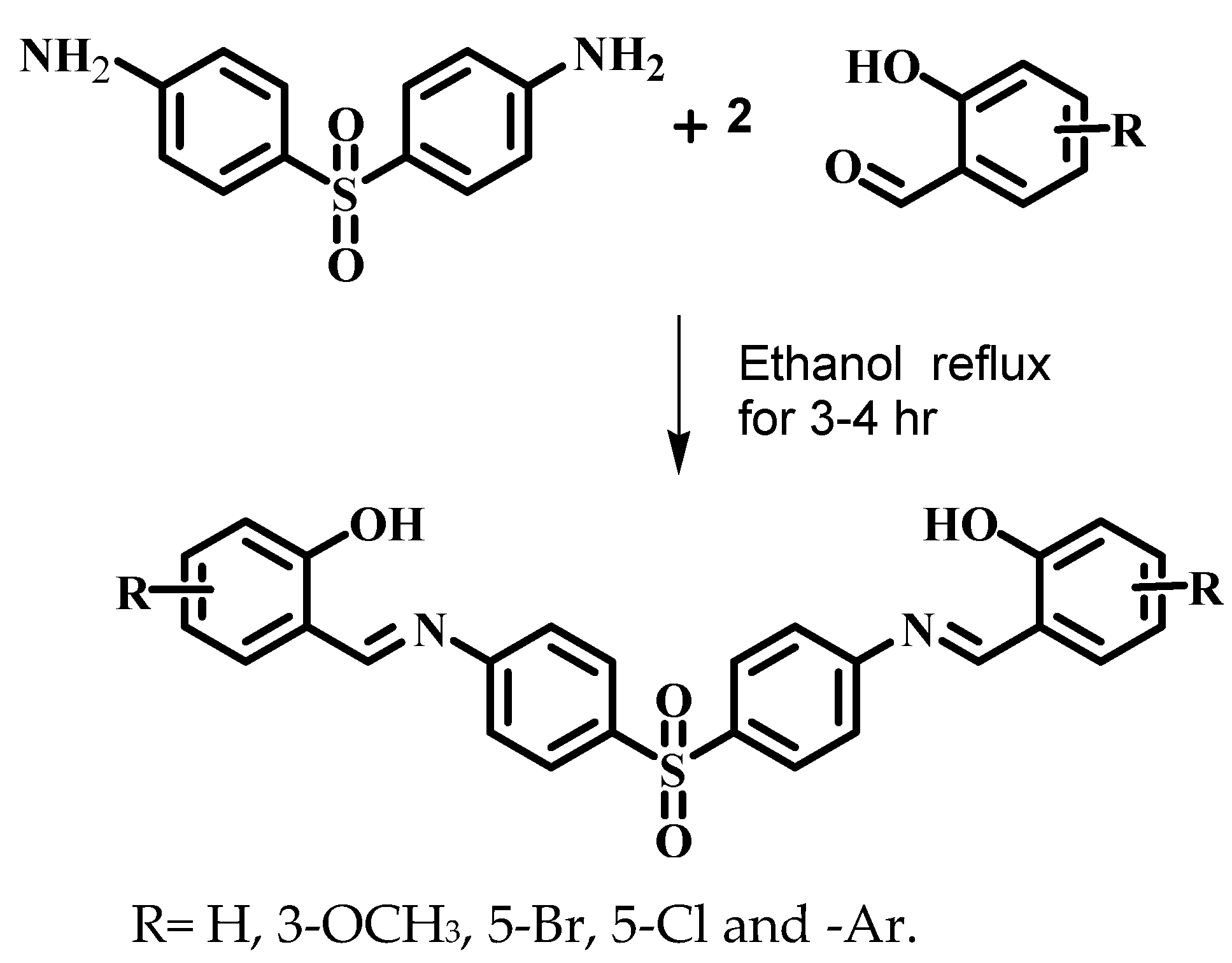
2.2.1. Preparation of Schiff Base Ligands (L1–L5)

The synthesis of series of Schiff bases is schematically represented in Scheme 1 and reported previously [23]. It is planned to synthesize tetradentate ligands consisting of ONNO, four donor sequence thus, 4,4′-diaminodiphenyl Sulfone (Dapsone) is made to react with 2,2-hydroxy-benzaldehyde, 2-hydroxy-1-naphathaldehyde, 2-hydroxy-3-methoxy benzaldehyde, 5-bromo-2-hydroxybenzaldehyde, and 5-chloro-2-hydroxybenzaldehyde in 1:1 ratio in ethanolic solution and the reaction mixture is refluxed for 4 h to yield Schiff bases (L1–L5). The physical and chemical characterization data of all the five Schiff base ligands are presented below:

2,2′-(4,4′-Sulfonylbis(4,1-phenylene)bis(azan-1-yl-1-ylidene))bis(methan-1-yl-1-ylidene)-diphenol (L1)

Yield: 77%; mp: 231 °C; FT-IR (KBr, ν/cm−1): 3459 (OH), 3376 (Ar-C-H), 1615 (C=N), 1566 (C=C) 1274 (asymmetric -SO2-stretch), 1185 (symmetric -SO2- stretch); 1H-NMR (400 MHz, DMSO-d6) δ: 12.56 (1H, s, Ar-OH), 8.80 (1H, s, Azomethine), 6.55–8.05 (8H, m, Ar-H); Mass (m/z): 456 [M+]; Elemental analysis(%) for C26H20N2O4S: Expt.(calcd), C 68.43 (68.41), H 4.40 (4.39), N 6.21 (6.14).

1,1′-(4,4′-Sulfonylbis(4,1-phenylene)bis(azan-1-yl-1-ylidene))bis(methan-1-yl-1-ylidene)-dinapthalen-2-ol (L2)

Yield: 82%; m.p.: 239 °C; FT-IR (KBr, ν/cm−1): 3434 (OH), 3222 (Ar-C-H), 1619 (C=N), 1544 (C=C), 1283 (asymmetric -SO2-stretch), 1186 (symmetric -SO2- stretch); 1H-NMR (400 MHz, DMSO-d6) δ: 12.83 (1H, s, Ar-OH), 8.72 (1H, s, Azomethine), 6.49–8.01 (10H, m, Ar-H); Mass (m/z): 556 [M+ + K]; Elemental analysis(%) for C34H24N2O4S: Expt.(calcd), C 73.42 (73.26), H 4.65 (4.48), N 5.13 (5.17).

2,2′-(4,4′-Sulfonylbis(4,1-phenylene)bis(azan-1-yl-1-ylidene))bis(methan-1-yl-1-ylidene)bis-(4-bromophenol) (L3)

Yield: 68%; m.p.: 233 °C; FT-IR (KBr, ν/cm−1): 3427 (OH), 3230 (Ar-C-H), 1621 (C=N), 1547 (C=C), 1275 (asymmetric -SO2-stretch), 1182 (symmetric -SO2- stretch); 1H-NMR (400 MHz, DMSO-d6) δ: 12.89 (1H, s, Ar-OH), 8.68 (1H, s, Azomethine), 6.67–8.23 (7H, m, Ar-H); Mass (m/z): 614 and 616 [M+ and M+ + 2]; Elemental analysis(%) for C26H18Br2N2O4S: Expt.(calcd), C 50.18 (50.07), H 2.93 (2.86), N 4.56 (4.48).

6,6′-(4,4′-Sulfonylbis(4,1-phenylene)bis(azan-1-yl-1-ylidene))bis(methan-1-yl-1-ylidene)bis-(2-methoxyphenol) (L4)

Yield: 80%; m.p.: 249 °C; FT-IR (KBr, ν/cm−1): 3432 (OH), 3236 (Ar-C-H), 1614 (C=N), 1579 (C=C), 1273 (asymmetric -SO2-stretch), 1187 (symmetric -SO2- stretch); 1H-NMR (400 MHz, DMSO-d6) δ: 12.89 (1H, s, Ar-OH), 8.70 (1H, s, Azomethine), 6.50–8.11 (7H, m, Ar-H), 3.81 (3H, s,-OCH3); Mass (m/z): 516 and 518 [M+ and M+ + 2]; Elemental analysis(%) for C28H24N2O6S: Expt.(calcd), C 65.16 (65.12), H 4.80 (4.74), N 5.53 (5.48).

2,2′-(4,4′-Sulfonylbis(4,1-phenylene)bis(azan-1-yl-1-ylidene))bis(methan-1-yl-1-ylidene)bis-(4-chlorophenol) (L5)

Yield: 86%; mp: 260 °C; FT-IR (KBr, ν/cm−1): 3457 (OH), 3241 (Ar-C-H), 1627 (C=N), 1563 (C=C), 1268 (asymmetric -SO2-stretch), 1181 (symmetric -SO2- stretch); 1H-NMR (400 MHz, DMSO-d6) δ: 12.70 (1H, s, Ar-OH), 8.51 (1H, s, Azomethine), 6.67–7.97 (7H, m, Ar-H); Mass (m/z): 524 [M+]; Elemental analysis(%) for C26H18Cl2N2O4S: Expt.(calcd.), C 60.46 (60.33), H 3.48 (3.41), N 5.42 (5.35).

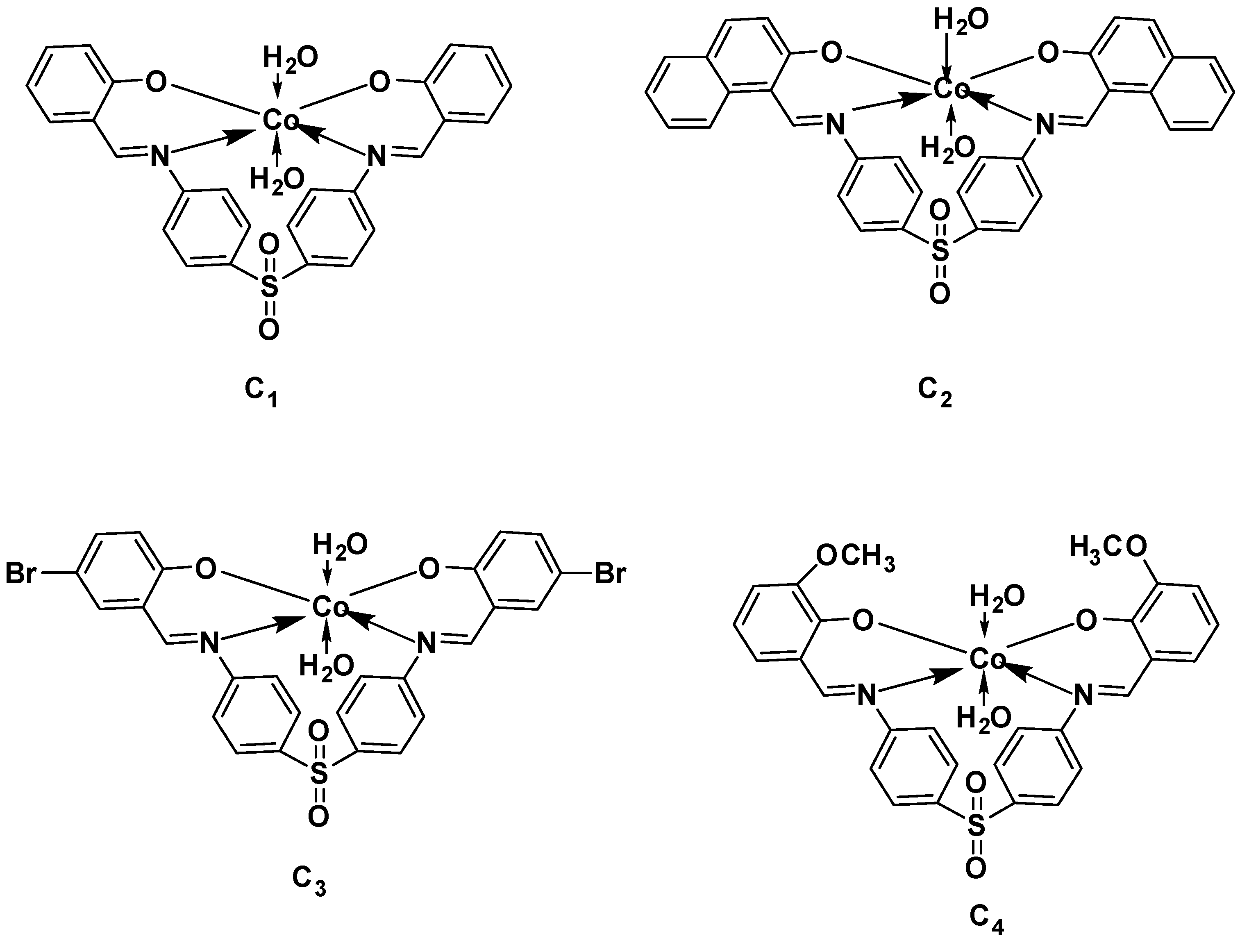
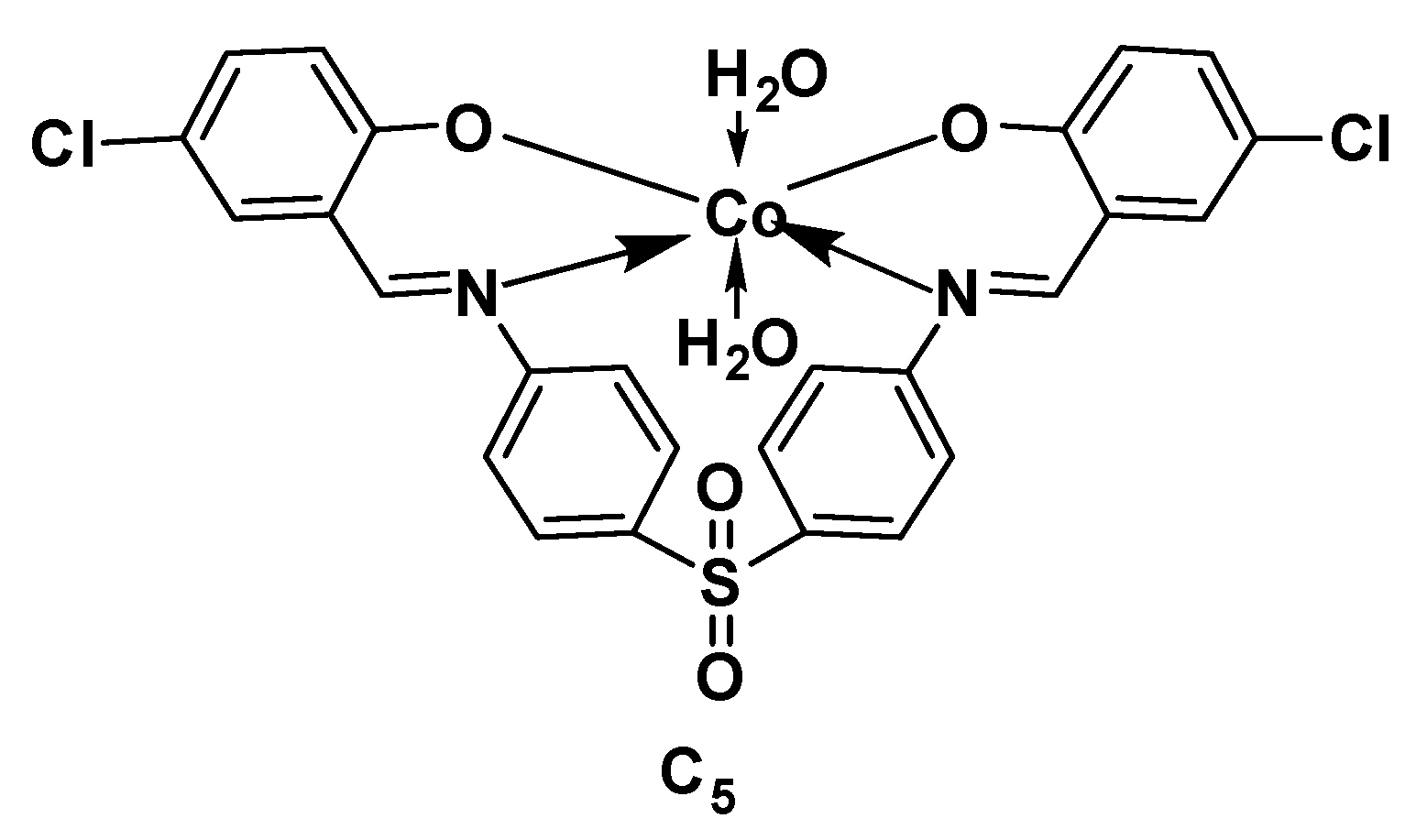
2.2.2. Synthesis of Co(II) Complexes (C1–C5)

An ethanolic solution of 0.1 M (4.56 g in 25 mL of ethanol) of the Schiff base ligand (L1) was added to a Co(II) chloride solution of 0.102 M (2.59 g in 10 mL of ethanol). The above reaction mixture was refluxed under continuous stirring for about 6 h. The progress of reaction was checked by TLC and spots were visualized under UV light. The precipitate obtained was then filtered, washed thoroughly with ethanol, and dried in desiccator over anhydrous CaCl2. Co(II) complexes with other Schiff bases (L2 (5.56 g), L3 (6.14 g), L4 (5.16 g) and L5 (5.24 g)) were synthesized using the above procedure. The synthesized complexes are characterized using physico-chemical techniques. The tentative structures of the prepared complexes are depicted in Figure 1.

2.3. Biological Assay

2.3.1. Antimicrobial Studies

The antimicrobial activities of the synthesized Schiff base ligands and their cobalt metal complexes under study were carried out by using the agar well diffusion method. The Gram-positive pathogens Bacillus subtilis (ATCC 6633) and Staphylococcus aureus (ATCC6538) and Gram-negative pathogens Klebsiella pneumonia (ATCC 13883), Pseudomonas aeruginosa (ATCC9027), and Escherichia coli (ATCC 8739) were used in the biological potency evaluation. The antifungal activity of the compounds was tested against Aspergillus niger (ATCC 16404) and Candida albicans (ATCC 10231). Standard drugs used for the study were Ciprofloxacin and Nystatin for bacterial and fungal pathogens respectively [24].
The Muller Hinton agar plate surface was inoculated by the spread plate method with microbial inoculum over the entire agar surface [25]. Then, a hole with a diameter of 6–8 mm is punched aseptically with a sterile cork borer, and the volume of the desired antimicrobial compound dissolved in DMSO with desired concentration was introduced into the well. Then, agar plates were incubated under suitable conditions depending on bacterial/fungal species requirements. The antimicrobial agents diffuse in the agar medium and inhibit the growth of the microbial strain tested. The inhibition zones exhibit around the antimicrobial compound and are measured as the diameter of the zone of inhibition in millimeters (mm) [26,27].

2.3.2. Molecular Docking Studies

Molecular docking is one of the most essential tools used in drug discovery due to its ability to predict, the conformation of small-molecule ligands within the appropriate target binding site with a substantial degree of accuracy. To find out the possible mode of action of the synthesized Schiff bases (L1–L5) and Co(II) complexes (C1–C5), molecular docking calculations of cysteine protease human cathepsin ki.-e.CDK7 (Cyclin dependent kinase-7) PDB ID: 1au2 were carried out using AutoDock 4.2. Lamarckian genetic algorithm. Auto Grid was used to define the active site and the grid size was set to 46 × 54 × 56 points which covers all the active site residues [20]. The grid spacing of 0.375 Å was centered on the selected flexible residues, which are the active sites of the CDK-7. The step size of 2.0 Å for translation and 10° for rotation were selected. The maximum number of energy evaluations was set to 2,500,000. A total of 10 runs were performed, and for each run, a maximum number of 27,000 genetic algorithms (GA) generations were performed on a single population of 150 individuals. The best-docked conformation among 10 conformations was obtained with the lowest binding energy values [28]. Interaction between cysteine protease human cathepsin ki.-e.CDK7 (Cyclin-dependent kinase-7) PDB ID: 1au2 with various ligands under study was visualized using molecular visualization tools such as Chimera (Pettersen et al., 2004). In addition, by using LigPlot+ tool hydrogen bonding and hydrophobic interactions were predicted for the complex [29].

2.3.3. Anticancer Activities by SRB Assay

The cell lines were cultured and grown in an appropriate medium containing 10% fetal bovine serum (FBS) and 2 mM L-glutamine. In the presentation investigation of the screening experiment, 5000 cells/well were inoculated into 96 well microtiter plates in 100 µL. After cell inoculation, the microtiter plates were incubated at 37 °C, 5% CO2, 95% air, and 100% relative humidity for 24 h before the addition of experimental drugs. Experimental drugs were solubilized in a suitable solvent (100 mg/mL) and diluted to 1 mg/mL using double distilled water and frozen before use. During the time of drug addition, an aliquot of frozen concentrate (1 mg/mL) was thawed and diluted to 100, 200, 400, and 800 μg/mL with a complete medium containing the test sample. Aliquots of 10 µL of these different drug dilutions were added to the appropriate microtiter wells already containing 90 µL of the medium, resulting in the required final drug concentrations, i.e., 10, 20, 40, 80 μg/mL. After the addition of the test compound, the plates were incubated at standard conditions for 48 h, and the assay was terminated by the addition of cold TCA. Cells were fixed in situ by the gentle addition of 50 µL of cold 30% (w/v) TCA (final concentration, 10% TCA) and incubated for 60 min at 4 °C. The supernatant was discarded. The plates were washed five times with tap water and air dried. Sulforhodamine B (SRB) solution (50 µL) at 0.4% (w/v) in 1% acetic acid was added to each of the wells, and plates were incubated for 20 min at room temperature. After staining, the unbound dye was recovered, and the residual dye was removed by washing five times with 1% acetic acid. The plates were air dried. The bound stain was subsequently eluted with 10 mM trizma base, and the absorbance was read on a plate reader at a wavelength of 540 nm with 690 nm reference wavelength. Percent growth was calculated on a plate-by-plate basis for test wells relative to control wells. Percent growth was expressed as the ratio of the average absorbance of the test well to the average absorbance of the control wells × 100. This has been shown in the form of the equation below:
Percentage   growth = Average   absorbance   of   the   cell   test Average   absorbance   of   the   control   well × 100

3. Results and Discussion

The elemental analysis data of synthesized Schiff bases and their complexes were found to be consistent with the expected result. The analytical and physical data of all the synthesized Schiff bases (L1 to L5) and their Co(II) complexes (C1 to C5) are given in Table 1. Theoretical and experimentally observed values of elemental analysis of compounds are in good agreement with the molecular formula.

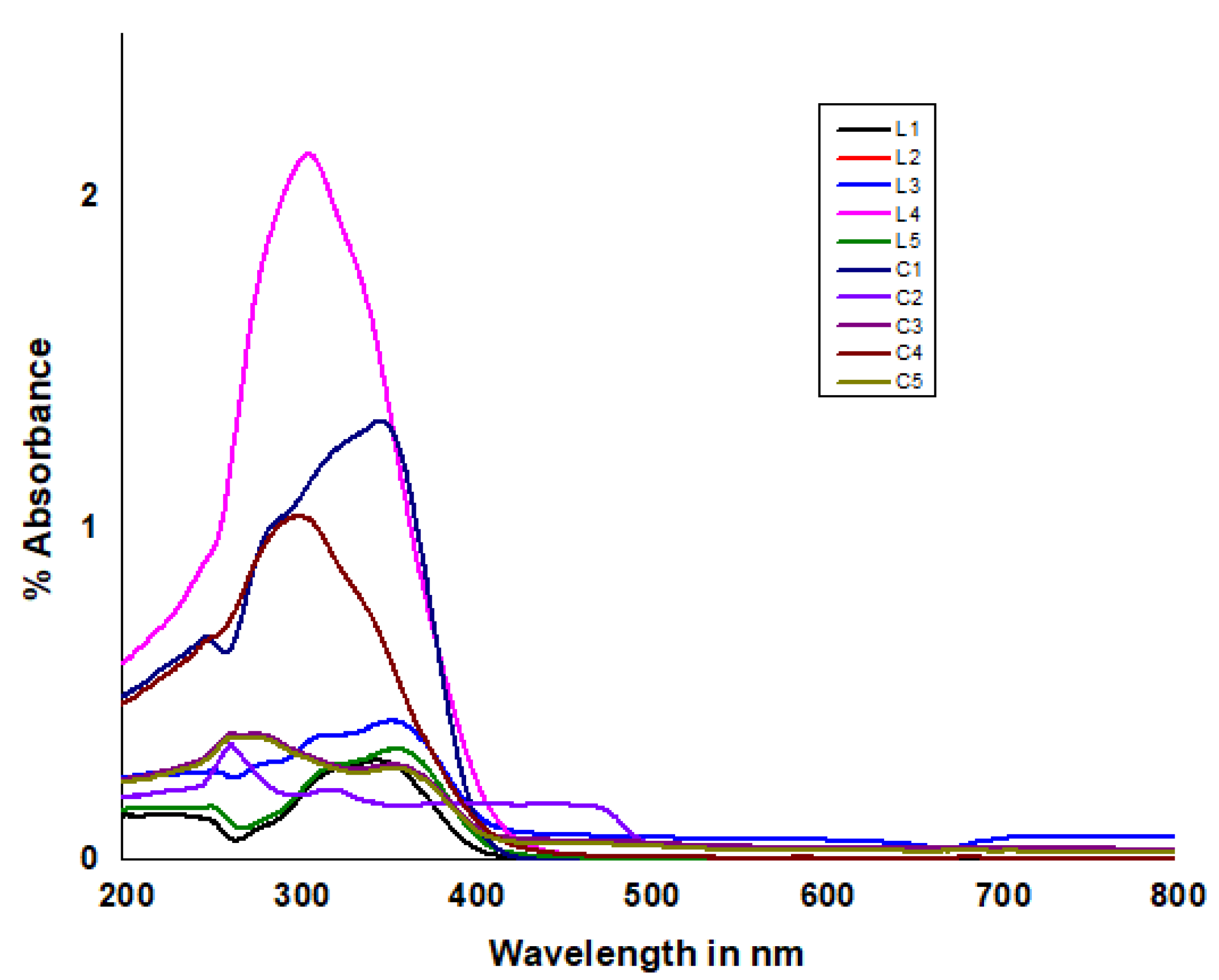
3.1. Electronic Spectral Studies amd Magnetic Moment Studies

Electronic absorption spectra of Co(II) Schiff base complexes were recorded at room temperature in DMSO solution and the results are shown in Table 2 and Figure 2 respectively. The electronic spectra of C1, C2, and C3 showed three absorption bands. The first two were in the range 248–290 nm, which are due to π → π* transition of the aromatic ring and the third one was in the range 345–399 nm, which was assigned to n→ π* transition of azomethine group (C=N). The UV-Visible spectrum of C4 and C5 showed two absorption bands (Figure S24). First, one was at 255 and 245 nm, respectively, which was attributed to π → π* transition of the aromatic ring, and the second one at around 310 and 342 nm, which was assigned to n → π* transition of azomethine group (C=N) of Schiff base. The magnetic moment values of Co(II) complexes were observed in the range of 4.37–5.08 B.M. (Table 2) because of high-spin magnetic moments, corresponding to the unpaired electrons. It appears from the magnetic moment data of Co(II) complexes that they are paramagnetic in nature, and hence are of six-coordinate complexes [30].

3.2. FT- IR Spectral Studies

The formation of Co(II) complexes were confirmed as important shifts in azomethine group (C=N) and phenolic -OH bands by comparing the infrared spectroscopic data of metal complexes and their respective ligands. The IR spectral data of metal complexes are presented in Table 3. The stretching vibrational band observed around 1627–1614 cm−1 is a characteristic of the azomethine (C=N) nitrogen atom present in the free Schiff base ligand. Due to metal coordination, the expected typical imine band in the range 1620–1596 cm−1 was observed in the Co(II) complexes. In addition, the –OH stretching and bending vibrational frequencies of the substituted salicylaldehyde appeared in the region 3459–3427 cm−1. The disappearance of these two peaks in the spectra of all the Co(II) complexes indicates that the coordination takes place via the enolic –OH group. Furthermore, the presence of a broad band at around 3435–3367 cm−1 in the Co(II) complexes suggests the presence of coordinate water molecules to the central metal ion [31,32,33]. Additional evidence for the coordination of the azomethine nitrogen is the presence of ν(M-N) bands in the frequency range of 562–544cm−1 and ν(M-O) bands in the frequency range of 517–505 cm−1 [34,35,36]. It is noteworthy that the unchanged band position of sulphonyl groups suggests that sulphone is not taking part in the coordination (Figures S1–S9) [37,38].

3.3. 1H-NMR Spectral Studies

The 1H-NMR spectra of all the synthesized Schiff base ligands were recorded in DMSO solvent and expressed in ppm. The 1H-NMR spectra of the synthesized compounds exhibit signals due to aromatic protons as a multiplet at 6.49–8.23 ppm. In the 1H-NMR spectrum of the Schiff base ligands, a singlet observed downfield around 12.56–12.92 ppm, integrating for one proton, is assigned to –OH [39]. Similarly, the azomethine proton (attached to the carbon close to the nitrogen atom) appears around 8.68–8.80 ppm as a singlet signal [40]. Representative proton 1H-NMR spectra of L1, L2, L3, and L5 are shown in Figures S10–S13. The proton and carbon NMR of complexes are depicted in Figures S25–S30.

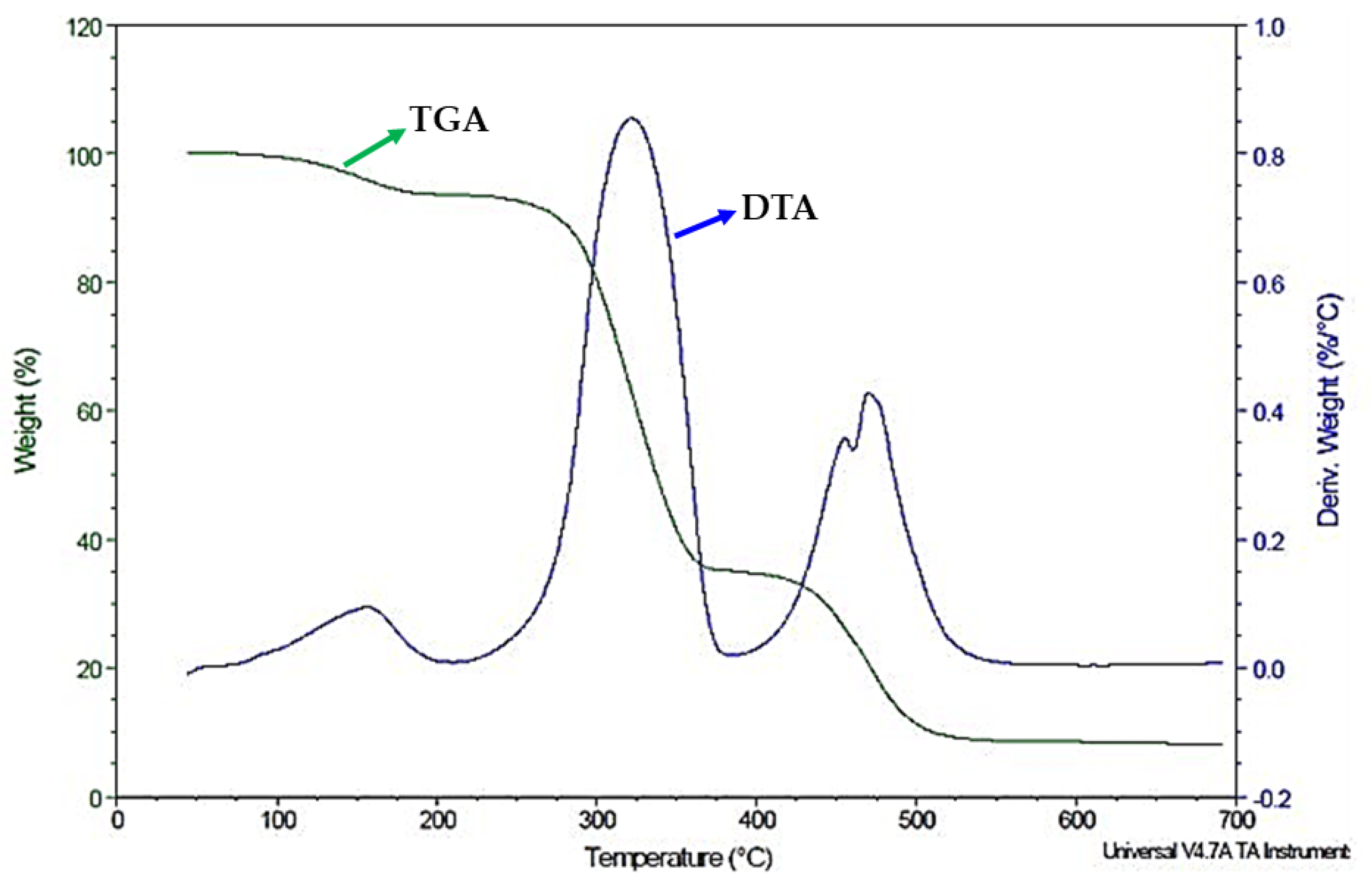
3.4. Thermogravimetric Analysis

The loss of water molecules was observed below 150 °C, which is due to the presence of lattice water molecules [41,42]. This further tells us that the coordinated water molecules occupy some position in the coordination sphere of the central metal ion. These water molecules are more strongly bonded to the metal ions thus eliminated at the higher temperatures. A further rise in the temperature, leading to the loss of mass in a slow gradual manner, was observed, which may be caused due to decomposition of metal complexes by the fragmentation and thermal degradation of organic parts and at the end, the metal oxide was formed as residue [43]. The thermal data are provided in Table 4 and representative thermal graphs of Co(II) complex (C1) is shown in Figure 3.

3.5. Mass Spectral Studies

The mass spectra of all the Schiff base ligands exhibit parent ion peaks, due to their respective molecular ion (M+), corresponding to the molecular weight and confirming their molecular composition. The proposed molecular formula of these compounds was confirmed by comparing their molecular formula weights with the m/z values. The mass spectra of the Schiff base ligands are depicted in Figures S14–S18. In addition, the fragmentation pattern of L1 is depicted in Figure S23.

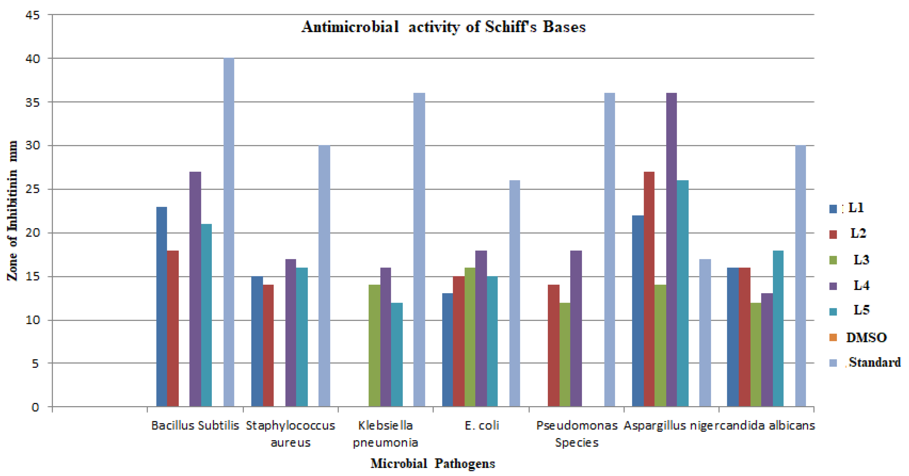
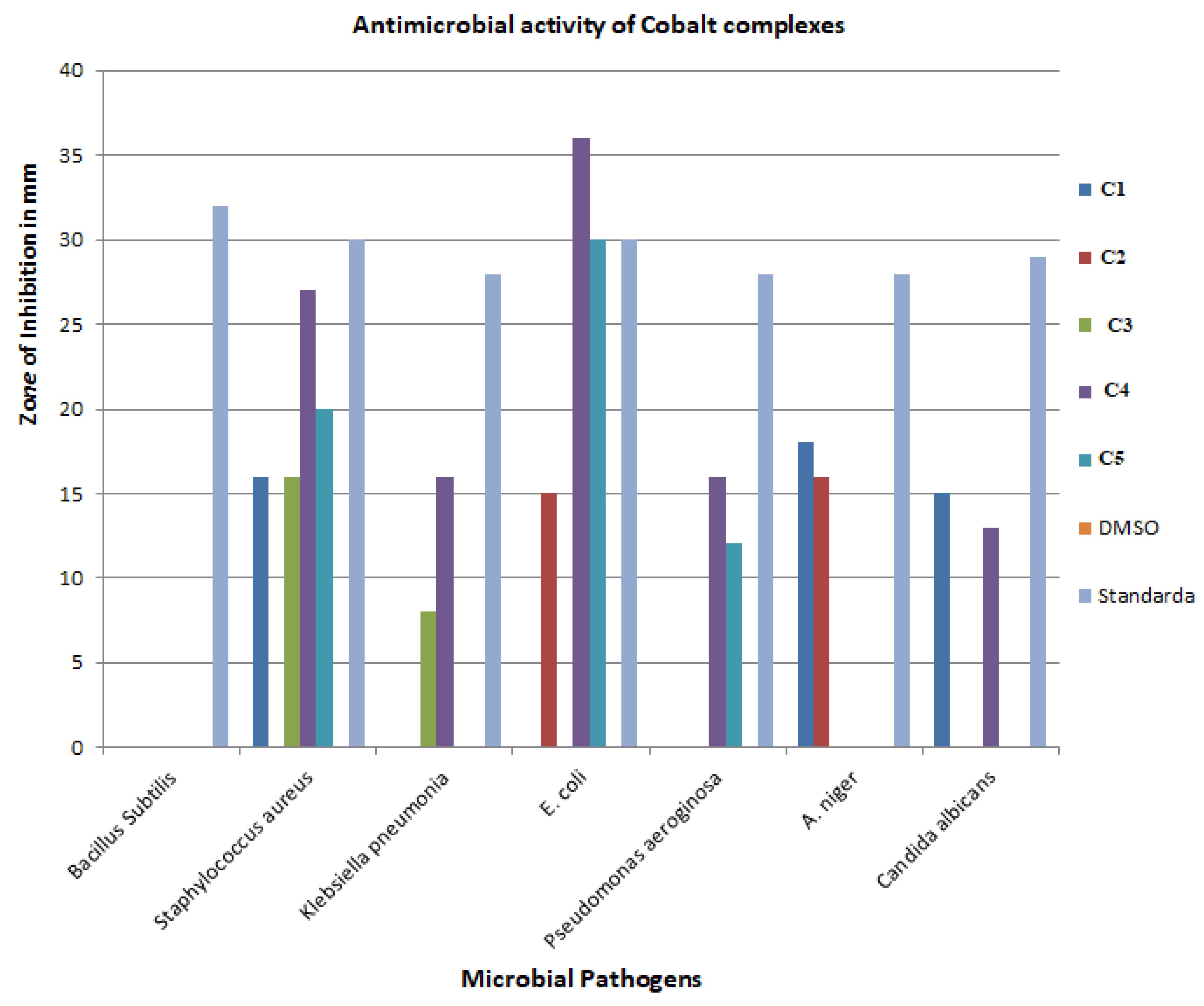
3.6. Antibacterial and Antifungal Activities

The antibacterial activity of synthesized Co(II) complexes was evaluated using different pathogens, including the Gram-positive Bacillus Subtilis and Staphylococcus aureus, Gram-negative Klebsiella Species, E. coli, and Pseudomonas aeruginosac, and fungal pathogens Aspergillus niger and Candida albicans. Antibacterial activity against DMSO and standard drugs Ciprofloxacin and Nystatin were also carried out. Bacterial and fungal pathogens showed the different zone of inhibitions against cobalt complexes (Figures S17–S20).
Considering the case of Bacillus Subtilis, for synthetic complex compounds (C1–C5) and DMSO, there was no zone of inhibition observed. The zone of inhibition for the standard drug Ciprofloxacin was 32 mm. However, in the case of Staphylococcus aureus, for synthetic compounds, C1, C3, C4, and C5, the zone of inhibition was 16, 16, 27, and 20, respectively. There was no zone of inhibition in the case of C2 and DMSO. The zone of inhibition for the standard drug Ciprofloxacin is 30 mm. Similarly, for Klebsiella pneumonia, for synthetic compounds, C3 and C4, the zone of inhibition was 8 and 16 mm, respectively. There was no zone of inhibition in the case of C1, C2, C5, and DMSO. The zone of inhibition for the standard drug Ciprofloxacin is 28 mm. Similarly, with E. coli, for synthetic compounds, C2, C4, and C5, the zone of inhibition was 15, 36, and 30 mm, respectively. There was no zone of inhibition in the case of C1, C3, and DMSO. The zone of inhibition for the standard drug Ciprofloxacin was 30 mm. In the case of Pseudomonas aeroginosa, for synthetic compounds, C4, and C5 the zone of inhibition was 16 and 12 mm, respectively. There was no zone of inhibition in the case of C1, C2, and DMSO. The zone of inhibition for the standard drug Ciprofloxacin was 28 mm (Figures S19–S22).
It is interesting to note that all the Schiff bases and their Co(II) complexes showed antibacterial activity against Gram-positive and Gram-negative bacteria (Table 5 and Table 6). This indicates the broad-spectrum ability of these compounds against the different pathogens. Ciprofloxacin as a standard drug and DMSO as a control was used for all bacterial species.
The Schiff base L4 showed the highest antibacterial activity when compared with the other ligands (Figure 4). These compounds are not only active against bacteria, but also exhibit antifungal activity (Figures S19 and S20). Further, L4 exhibited strong antifungal activity against Aspergillus niger (ATCC 16404). It is noteworthy that antifungal activities were higher than the standard antifungal drug Nystatin.
On the other hand, it was observed from these results that the Co(II) complexes were less effective against both Gram-positive and Gram-negative. Moreover, only a few complexes show antifungal activity. There was no zone of inhibition shown by any cobalt complex against Bacillus subtilis. Out of five cobalt metal complexes, only C4 showed antibacterial activity against both Gram-positive, Gram-negative, and fungal pathogens (Figure 5).

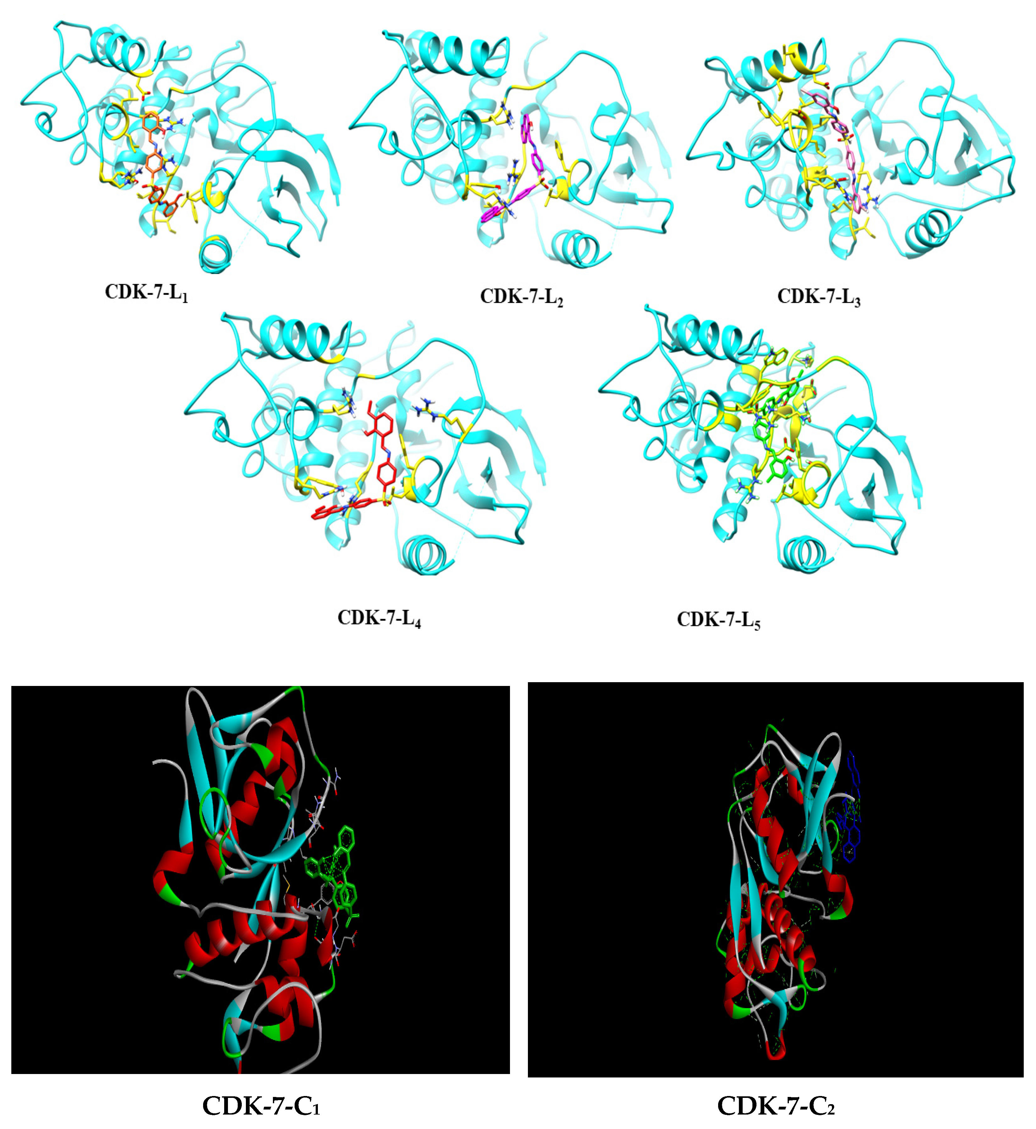
3.7. Molecular Docking Studies

To know the most preferred conformation of all Schiff base ligands with protein PDB ID:1au2, we performed the docking study of each ligand for 10 confirmations. The best-docked conformation among 10 conformations was obtained with the lowest binding energy. The binding values are −9.25 kcal/mol, −9.40 kcal/mol, −9.12 kcal/mol, −7.11 kcal/mol, and +285.63 kcal/mol for ligands CDK-7-L1, CDK-7-L2, CDK-7-L3, CDK-7-L4, and CDK-7-L5, respectively (Table 7). Further, the best-docked complexes were analyzed for hydrogen bonding interactions and hydrophobic interactions [44,45].
The results revealed that ARG179, ARG136, TYR190, and ARG188 residues of cysteine protease human cathepsin are involved in hydrogen bonding interactions. Whereas GLU99, ILE133, LEU134, ARG136, ASP137, LEU138, LYS139, PRO140, PHE156, GLY157, LEU158, PHE162, THR175, ARG176, TRP177, ARG179, LEU183, LEU184, ASP218, ARG188, MET189, and TYR190 residues involved in hydrophobic interactions.
Compound L2 has the highest binding affinity followed by L1, L3, and L4, whereas compound L5 has the lowest affinity among all the docked ligands, which is 285.63 K. Cal/Mol. Further, the L2 molecule showed interaction with amino acids, e.g., ARG188, TYR190, and PHE156, as well as hydrophobic and other interactions with LUE158, LEU134, ARG136, ASP137, THR175, ARG176, ARG179, and PHE162. The highest binding affinity of L2 than the other Schiff bases is mainly due to the involvement of ARG188 in the hydrogen bonding interaction and TYR190, LEU134, ARG136, ASP137, LEU158, THR175, ARG176, ARG179, and PHE162 in hydrophobic and other interactions with amino acid.
Using the data from docking interactions, 2D and 3D images are shown in Figure 6. Ligand L1 has the highest binding affinity followed by L3 L4, and L5 and whereas ligand L5 has the lowest affinity among all the docked Schiff base ligands, which is 285.63 kcal/mol. On the other hand, the docking results of Co(II) complexes revealed that GLN22, PHE23 SER161, ARG136, ARG179, TYR 190, and ARG188 residues of cysteine protease human cathepsin are involved in hydrogen bonding interactions. Whereas ALA198, LEU183, ALA180, VAL194, TYR178, LYS139, THR175, ASP137, LEU138, LYS41, ASN142, and PHE162, residues are involved in hydrophobic interactions, as depicted in Table 8.
The compound has C2 highest binding affinity, followed by C3, C5, and C1, whereas compound C5 has the lowest affinity among all the docked ligands, 2.47 K. Cal/Mol. Further, C2 complex and its interaction with amino acids, e.g., LEU134, PHE156, ILE133, GLU62, LUE158, ILE55, ARG136, ASP137, PHE162, ARG176, and ARG179, are involved in hydrophobic and other interactions.

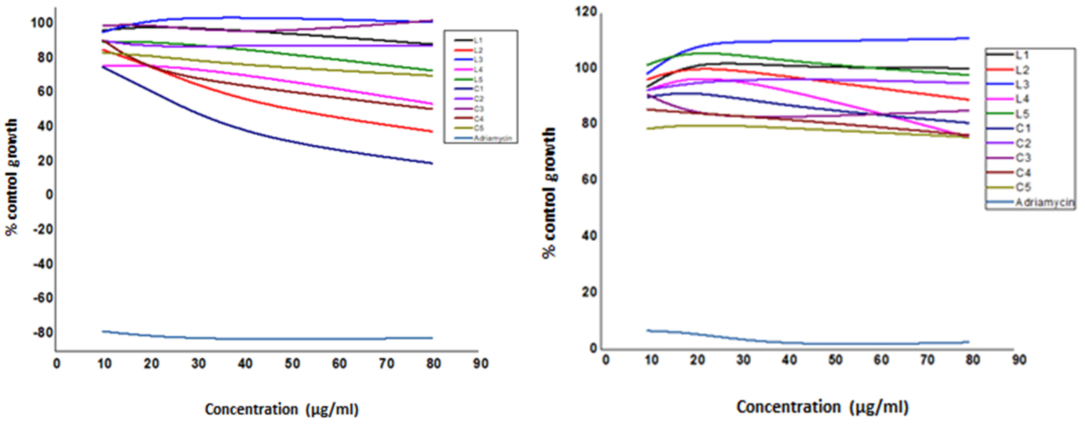
3.8. Effect of Ligands (L1–L5) and Their Complexes (C1–C5) on Antiproliferative Activity

All the synthesized compounds were screened for their anticancer activity against human breast cancer cell line MCF-7 and human lung cancer cell line A-549. The growth curves of human breast cancer cell line MCF-7 and human lung cancer cell line A-549 of Schiff bases (L1–L5) and their Co(II) complexes (C1–C5) are represented in Figure 7. The anticancer activity was measured in vitro for the newly synthesized compounds against breast cancer cell line MCF-7and human lung cancer cell line A-549 using the Sulforhodamine B stain (SRB) assay method [43]. The average values for % control growth for the cell line MCF-7 and human lung cancer cell line A-549 are listed in Table 9 and Table 10.
The anticancer activity results based on average values for % control growth revealed that ligands and their Co(II) complexes are active at 20 and 40 μg/mL concentration. It is interesting to note that the complexes are more potent as compared to their respective ligand, except in the case of L3 and its complex. Moreover, it is noteworthy that the Schiff base ligands and Co(II) complexes are more active as compared to the standard drug adriamycin at all concentrations.

4. Conclusions

To sum up, the synthesized Schiff bases act as a tetradentate ligand and coordinated with the Co(II) ion through imine nitrogen and phenolic oxygen atoms. The binding of ligand to a metal ion was confirmed by elemental analysis, spectral studies (UV-Visible and FT-IR), and TGA measurements. The Co(II) complexes were found to exhibit octahedral geometry. All the Schiff bases (L1–L5) and their Co(II) complexes demonstrated moderate to good antimicrobial activity against the tested microbial species. The anticancer studies of the ligands and their Co(II)complexes showed significant activities against MCF-7 and human lung cancer cell line A-549 cancer cells. Particularly, the ligand L3 showed potential activity compared with the other tested compounds, which in turn was compared with the standard drug, adriamycin. Further, the molecular docking results revealed that ARG188, TYR190, and ARG136 residues of cysteine protease human cathepsin are involved in hydrogen bonding interactions.

Supplementary Materials

The following supporting information can be downloaded at: https://www.mdpi.com/article/10.3390/molecules27238576/s1, Figures S1–S9: FT-IR spectra of ligands and complexes; Figures S10–13: 1H-NMR of Schiff base ligands; Figures S14–S18: Mass spectra of ligands; Figures S19–S22: Antimicrobial activities of ligands and complexes; Figure S23: Mass fragmentation pattern of L1; Figure S24: UV-Vis absorption spectra of complexes; Figures S25–S30: 1H and 13C-NMR od complexes.

Author Contributions

Conceptualization, K.D.G.; P.U.; S.H.G. and S.P.K.; methodology, S.D.; P.D. and R.K.; software, P.S., P.V. and C.S.; validation, M.R.A. and R.M.O.; formal analysis, R.R.A., E.S., V.S. and N.M.; investigation, S.P.K.; resources, A.A.S., M.Y.A. and S.E.I.E.; data curation, K.D.G., P.U. and M.R.A.; writing—original draft preparation, K.D.G.; writing—review and editing, S.H.G. and S.P.K.; visualization, E.S., V.S. and N.M.; supervision, S.H.G.; project administration, P.U.; funding acquisition, A.A.S., M.Y.A. and S.E.I.E. All authors have read and agreed to the published version of the manuscript.

Funding

This research is funded by Deanship of Scientific Research at King Khalid University under the project grant number R.G.P. 2/78/43.

Institutional Review Board Statement

Not Applicable.

Informed Consent Statement

Not Applicable.

Data Availability Statement

Not available.

Acknowledgments

The authors gratefully acknowledge the ACTREC, Tata memorial center, Kharghar, New Mumbai, India for providing anticancer activity. We also acknowledge Shri Shivaji Mahavidyalaya, Barshi for providing research facilities. PU is grateful to the N.K. Orchid college of Engineering and Technology, Solapur, India for infrastructure support. CS and PV thankfully acknowledge JSSAHER for infrastructure facilities. SPK greatly thank the Director, Amrita Vishwa Vidyapeetham, Mysuru Campus for providing infrastructure support.

Conflicts of Interest

The authors have no conflict of interest.

References

  1. Khandar, A.A.; Nejati, K.; Rezvani, Z. Syntheses, Characterization and Study of the Use of Cobalt (II) Schiff–Base Complexes as Catalysts for the Oxidation of Styrene by Molecular Oxygen. Molecules 2005, 10, 302–311. [Google Scholar] [CrossRef] [PubMed] [Green Version]
  2. Martell, A.E.; Sawyer, D.T. Oxygen-Co Complexes and Oxygen Activation by Transition Metals; Plenum Press: New York, NY, USA, 1998. [Google Scholar]
  3. Nagata, T.; Yorozu, K.; Yamada, T.; Mukaiyama, T. Enantioselective Reduction of Ketones with Sodium Borohydride, Catalyzed by Optically Active (β-Oxoaldiminato)cobalt(II) Complexes. Angew. Chem. Int. Ed. 1995, 34, 2145. [Google Scholar] [CrossRef]
  4. Estiú, G.L.; Jubert, A.H.; Costamagna, J.; Vargas, J. Quantum Chemical Calculations of the Structures and Electronic Properties of N,N‘- Bis(3,5-dibromosalicylidene)-1,2-diaminobenzene and Its Cobalt(II) Complex. Origin of the Redox Activity of the Cobalt Complex. Inorg. Chem. 1996, 35, 263–266. [Google Scholar] [CrossRef] [PubMed]
  5. Zhang, Y.-L.; Ruan, W.-J.; Zhao, X.-J.; Wang, H.-G.; Zhu, Z.-A. Synthesis and characterization of axial coordination cobalt(III) complexes containing chiral Salen ligands. Polyhedron 2003, 22, 1535–1545. [Google Scholar] [CrossRef]
  6. Cini, R.; Moore, S.J.; Marzilli, L.G. Strong Trans Influence Methoxymethyl Ligand in B12 Cobaloxime and Imine/Oxime Model Complexes:  Structural, Spectroscopic, and Molecular Mechanics Investigations. Inorg. Chem. 1998, 37, 6890. [Google Scholar] [CrossRef]
  7. Sethi, R.; Ahuja, M. Synthesis, Characterization and Antibacterial Activity of Cobalt Complex of 2-Pyrazoline with Pyridinyl MoietyInt. J. Pharm. Technol. Res. 2016, 9, 35. [Google Scholar]
  8. Ghosh, P.; Chowdhury, A.R.; Saha, S.K.; Ghosh, M.; Pal, M.; Murmu, N.C.; Banerjee, P. Synthesis and characterization of redox non-innocent cobalt(III) complexes of a O,N,O donor ligand: Radical generation, semi-conductivity, antibacterial and anticancer activities. Inorganica Chim. Acta 2015, 429, 99–108. [Google Scholar] [CrossRef]
  9. Ghosh, M.; Layek, M.; Fleck, M.; Saha, R.; Bandyopadhyay, D. Synthesis, crystal structure and antibacterial activities of mixed ligand copper(II) and cobalt(II) complexes of a NNS Schiff base. Polyhedron 2015, 85, 312–319. [Google Scholar] [CrossRef]
  10. Singh, U.; Malla, A.M.; Bhat, I.A.; Ahmad, A.; Bukhari, M.N.; Bhat, S.; Anayutullah, S.; Hashmi, A.A. Synthesis, molecular docking and evaluation of antifungal activity of Ni(II),Co(II) and Cu(II) complexes of porphyrin core macromolecular ligand. Microb. Pathog. 2016, 93, 172–179. [Google Scholar] [CrossRef]
  11. Ubale, P.; Mokale, S.; More, S.; Waghamare, S.; More, V.; Munirathinam, N.; Dilipkumar, S.; Das, R.K.; Reja, S.; Helavi, V.B.; et al. Evaluation of in vitro anticancer, antimicrobial and antioxidant activities of new Cu(II) complexes derived from 4(3H)-quinazolinone: Synthesis, crystal structure and molecular docking studies. J. Mol. Struct. 2021, 1251, 131984. [Google Scholar] [CrossRef]
  12. Ashok, U.P.; Kollur, S.P.; Anil, N.; Arun, B.P.; Jadhav, S.N.; Sarsamkar, S.; Helavi, V.B.; Srinivasan, A.; Kaulage, S.; Veerapur, R.; et al. Preparation, Spectroscopic Characterization, Theoretical Investigations, and In Vitro Anticancer Activity of Cd(II), Ni(II), Zn(II), and Cu(II) Complexes of 4(3H)-Quinazolinone-Derived Schiff Base. Molecules 2020, 25, 5973. [Google Scholar] [CrossRef] [PubMed]
  13. Nesterov, D.S.; Nesterova, O.V. Polynuclear Cobalt Complexes as Catalysts for Light-Driven Water Oxidation: A Review of Recent Advances. Catalysts 2018, 8, 602. [Google Scholar] [CrossRef] [Green Version]
  14. Mil’Grom, A.E.; Chegolya, A.S.; Filippova, A.V.; Vladyko, G.V.; Karako, N.I. Synthesis and antivirus activity of unprotonated complex salts of amino acids with nickel and cobalt. Pharm. Chem. J. 1984, 18, 179–182. [Google Scholar] [CrossRef]
  15. Zhang, H.; Huang, J.; Meng, F. Cobalt-catalyzed diastereo- and enantioselective allyl addition to aldehydes and α-ketoesters through allylic C–H functionalization. Cell Rep. Phys. Sci. 2021, 2, 100406. [Google Scholar] [CrossRef]
  16. Wang, N.; Lin, Q.Y.; Feng, J.; Zhao, Y.L.; Wang, Y.J.; Li, S.K. Crystal structures, DNA interaction and antiproliferative activities of the cobalt(II) and zinc(II) complexes of 2-amino- 1,3,4-thiadiazole with demethylcantharate. Inorg. Chim. Acta 2010, 363, 3399. [Google Scholar] [CrossRef]
  17. Munteanu, C.R.; Suntharalingam, K. Advances in cobalt complexes as anticancer agents. Dalton Trans. 2015, 44, 13796–13808. [Google Scholar] [CrossRef]
  18. Cavicchioli, M.; Zaballa, A.M.L.; de Paula, Q.A.; Prieto, M.B.; Oliveira, C.C.; Civitareale, P.; Ciriolo, M.R.; Ferreira, A.M.D.C. Oxidative Assets Toward Biomolecules and Cytotoxicity of New Oxindolimine-Copper(II) and Zinc(II) Complexes. Inorganics 2019, 7, 12. [Google Scholar] [CrossRef] [Green Version]
  19. King, A.P.; Gellineau, H.A.; Ahn, J.-E.; MacMillan, S.N.; Wilson, J.J. Bis(thiosemicarbazone) Complexes of Cobalt(III). Synthesis, Characterization, and Anticancer Potential. Inorg. Chem. 2017, 56, 6609–6623. [Google Scholar] [CrossRef]
  20. Banerjee, S.; Chakravarty, A.R. Metal Complexes of Curcumin for Cellular Imaging, Targeting, and Photoinduced Anticancer Activity. Accounts Chem. Res. 2015, 48, 2075–2083. [Google Scholar] [CrossRef]
  21. Wasylenko, D.J.; Ganesamoorthy, C.; Borau-Garcia, J.; Berlinguette, C.P. Electrochemical evidence for catalytic water oxidation mediated by a high-valent cobalt complex. Chem. Commun. 2011, 47, 4249–4251. [Google Scholar] [CrossRef]
  22. Cahiez, G.; Moyeux, A. Cobalt-Catalyzed Cross-Coupling Reactions. Chem. Rev. 2010, 110, 1435–1462. [Google Scholar] [CrossRef] [PubMed]
  23. Gaikwad, K.D.; Khobragade, R.M.; Deodware, S.A.; Ubale, P.A.; Dhale, P.C.; Ovhal, R.M.; Shivamallu, C.; Ankegowda, V.M.; Raghavendra, H.; Gaikwad, S.H.; et al. Chemical synthesis, spectral characterization and biological activities of new diphenylsulphone derived Schiff base ligand and their Ni(II) complexes. Results Chem. 2022, 4, 100617. [Google Scholar] [CrossRef]
  24. Waghmare, S.R.; Mulla, M.N.; Marathe, S.R.; Sonawane, K.D. Ecofriendly production of silver nanoparticles using Candida utilis and its mechanistic action against pathogenic microorganisms. 3 Biotech 2015, 5, 33–38. [Google Scholar] [CrossRef] [PubMed] [Green Version]
  25. El-Sonbati, A.; Mahmoud, W.; Mohamed, G.G.; Diab, M.; Morgan, S.; Abbas, S. Synthesis, characterization of Schiff base metal complexes and their biological investigation. Appl. Organomet. Chem. 2019, 33, e5048. [Google Scholar] [CrossRef]
  26. Devi, J.; Yadav, M.; Kumar, A.; Kumar, A. Synthesis, characterization, biological activity, and QSAR studies of transition metal complexes derived from piperonylamine Schiff bases. Chem. Pap. 2018, 72, 2479. [Google Scholar] [CrossRef]
  27. Koçoğlu, S.; Ogutcu, H.; Hayvalı, Z. Photophysical and antimicrobial properties of new double-armed benzo-15-crown-5 ligands and complexes. Res. Chem. Intermed. 2019, 45, 2403–2427. [Google Scholar] [CrossRef]
  28. Mirdya, S.; Drew, M.G.; Chandra, A.K.; Banerjee, A.; Frontera, A.; Chattopadhyay, S. A series of hydrogen bond mediated dinuclear nickel(II) complexes with reduced Schiff base ligands: An insight into the nature of their short intermolecular hydrogen bonds. Polyhedron 2020, 179, 114374. [Google Scholar] [CrossRef]
  29. Bikas, R.; Mirzakhani, P.; Noshiranzadeh, N.; Sanchiz, J.; Krawczyk, M.S.; Kalofolias, D.A.; Lis, T. Synthesis, crystal structure and magnetic properties of a pentanuclear Mn (III) cluster with 1, 2, 4-triazole based Schiff base ligand. Inorg. Chim. Acta 2020, 505, 119461. [Google Scholar] [CrossRef]
  30. Nejo, A.A.; Kolawole, G.A.; Nejo, A.O. Synthesis, characterization, antibacterial, and thermal studies of unsymmetrical Schiff-base complexes of cobalt(II). J. Coord. Chem. 2010, 63, 4398. [Google Scholar] [CrossRef]
  31. Saghatforoush, L.A.; Chalabian, F.; Aminkhani, A.; Karimnezhad, G.; Ershad, S. Synthesis, spectroscopic characterization and antibacterial activity of new cobalt(II) complexes of unsymmetrical tetradentate (OSN2) Schiff base ligands. Eur. J. Med. Chem. 2009, 44, 4490–4495. [Google Scholar] [CrossRef]
  32. Sebastian, M.; Arun, V.; Robinson, P.P.; Leeju, P.; Varghese, D.; Varsha, G.; Yusuff, K.K.M. Synthesis, characterization, and structure of a new cobalt(II) Schiff-base complex with quinoxaline- 2-carboxalidine-2-amino-5-methylphenol. J. Coord. Chem. 2009, 63, 307–314. [Google Scholar] [CrossRef]
  33. Barakat, A.; Soliman, S.M.; Ali, M.; Elmarghany, A.; Al-Majid, A.M.; Yousuf, S.; Ul-Haq, Z.; Choudhary, M.I.; El-Faham, A. Synthesis, crystal structure, evaluation of urease inhibition potential and the docking studies of cobalt(III) complex based on barbituric acid Schiff base ligand. Inorg. Chim. Acta 2019, 503, 119405. [Google Scholar] [CrossRef]
  34. Ghosh, K.; Dutta, T.; Drew, M.G.; Frontera, A.; Chattopadhyay, S. A combined experimental and theoretical study on an ionic cobalt(III/II) complex with a Schiff base ligand. Polyhedron 2020, 182, 114432. [Google Scholar] [CrossRef]
  35. Luo, X.-Q.; Liu, Q.-R.; Han, Y.-J.; Xue, L.-W. Syntheses, X-ray Single Crystal Structures and Biological Activities of Cobalt(III) Complexes with Reduced Schiff Base Ligands. Acta Chim. Slov. 2020, 67, 159–166. [Google Scholar] [CrossRef]
  36. Hricovíniová, Z.; Hricovíni, M.; Kozics, K. New series of quinazolinone derived Schiff’s bases: Synthesis, spectroscopic properties and evaluation of their antioxidant and cytotoxic activity. Chem. Pap. 2017, 72, 1041–1053. [Google Scholar] [CrossRef]
  37. Tang, B.; Yue, T.; Wu, J.; Dong, Y.; Ding, Y.; Wang, H. Rapid and sensitive spectrofluorimetric determination of trace amount of Cr(III) with o-vanillin-8-aminoquinoline. Talanta 2004, 64, 955. [Google Scholar] [CrossRef]
  38. Tas, E.; Kilic, A.; Durgun, M.; Yilmaz, I.; Ozdemir, I.; Gurbuz, N. Mono- and dinuclear Pd(II) complexes of different salicylaldimine ligands as catalysts of transfer hydrogenation of nitrobenzene with cyclohexene and Suzuki–Miyaura coupling reactions. J. Organomet. Chem. 2009, 694, 446–454. [Google Scholar] [CrossRef]
  39. Venkatachalam, G.; Ramesh, R. Ruthenium(III) bis-bidentate Schiff base complexes mediated transfer hydrogenation of imines. Inorg. Chem. Commun. 2006, 9, 703–707. [Google Scholar] [CrossRef]
  40. Kalkhambkar, R.G.; Kulkarni, G.M.; Kamanavalli, C.M.; Premkumar, N.; Asdaq, S.; Sun, C.M. Synthesis and biological activities of some new fluorinated coumarins and 1-aza coumarins. Eur. J. Med. Chem. 2008, 43, 2178–2188. [Google Scholar] [CrossRef]
  41. Ashok, U.P.; Kollur, S.P.; Arun, B.P.; Sanjay, C.; Suresh, K.S.; Anil, N.; Baburao, H.V.; Markad, D.; Castro, J.O.; Frau, J.; et al. In vitro anticancer activity of 4(3H)- Quinazolinone derived Schiff base and its Cu(II), Zn(II) and Cd(II) complexes: Preparation, X-ray structural, spectral characterization and theoretical investigations. Inorg. Chim. Acta 2020, 511, 119846. [Google Scholar] [CrossRef]
  42. Vinusha, H.; Kollur, S.P.; Revanasiddappa, H.; Ramu, R.; Shirahatti, P.S.; Prasad, M.N.; Chandrashekar, S.; Begum, M. Preparation, spectral characterization and biological applications of Schiff base ligand and its transition metal complexes. Results Chem. 2019, 1, 100012. [Google Scholar] [CrossRef]
  43. Razali, N.; Razab, R.; Junit, S.M.; Aziz, A.A. Radical scavenging and reducing properties of extracts of cashew shoots (Anacardium occidentale). Food Chem. 2008, 111, 38–44. [Google Scholar] [CrossRef]
  44. Dundas, J.; Ouyang, Z.; Tseng, J.; Binkowski, A.; Turpaz, Y.; Liang, J. CASTp: Computed atlas of surface topography of proteins with structural and topographical mapping of functionally annotated residues. Nucleic Acids Res. 2006, 34, W116–W118. [Google Scholar] [CrossRef] [PubMed] [Green Version]
  45. Laskowski, R.A.; Swindells, M.B. LigPlot+: Multiple ligand–protein interaction diagrams for drug discovery. J. Chem. Inf. Model. 2011, 51, 2778–2786. [Google Scholar] [CrossRef] [PubMed]
Scheme 1. General synthetic route of Schiff base ligands (L1–L5).
Molecules 27 08576 sch001
Figure 1. Proposed structure of synthesized Co(II) complexes (C1–C5).
Molecules 27 08576 g001aMolecules 27 08576 g001b
Figure 2. The UV-Vis bands (in nm) Schiff bases and complexes.
Molecules 27 08576 g002
Figure 3. TGA-DTA graph of a C1 complex.
Molecules 27 08576 g003
Figure 4. Graphical representation of antibacterial and antifungal activities of Schiff bases against bacterial and fungal pathogens.
Molecules 27 08576 g004
Figure 5. Graphical representation of antibacterial and antifungal activities of Co(II) complexes against bacterial and fungal pathogens.
Molecules 27 08576 g005
Figure 6. Binding interaction of Schiff base ligands (L1-L5) (top) and Co(II) complexes (C1–C2) with CDK-7 protein (bottom).
Molecules 27 08576 g006
Figure 7. The growth curves of human breast cancer cell line MCF-7 (left) and human lung cancer cell line A-549 (right) of Schiff bases and their Co(II) complexes.
Molecules 27 08576 g007
Table 1. Elemental analysis data of Schiff base ligands and their Co(II) complexes.
CompoundMol. FormulaMol. Wt.M.P.
(°C)
Elemental Analysis
C%
Found (calc.)
H%
Found (calc.)
N%
Found (calc.)
L1C26H20N2O4S45623168.43(68.41)4.40 (4.39)6.21 (6.14)
L2C34H24N2O4S55623973.42 (73.26)4.65 (4.48)5.13(5.17)
L3C26H18Br2N2O4S61423350.18 (50.07)2.93 (2.86)4.56 (4.48)
L4C28H24N2O6S51624965.16(65.12)4.80 (4.74)5.53 (5.48)
L5C26H18Cl2N2O4S52426060.46 (60.33)3.48 (3.41)5.42 (5.35)
C1C26H22N2O6SCo54934757.19
(56.97)
4.08
(4.00)
5.37
(5.13)
C2C34H26N2O6SCo64933262.98
(62.89)
4.09
(4.03)
4.37
(4.31)
C3C26H20N2O6SBr2Co70531945.07
(44.56)
2.93
(2.86)
4.12
(3.99)
C4C28H26N2O8SCo60935256.23
(55.34)
4.37
(4.30)
4.77
(4.63)
C5C26H20N2O6SCl2Co61732451.76
(50.77)
3.32
(3.25)
4.66
(4.55)
Table 2. Electronic spectral and magnetic moment values for Co(II)-Schiff base metal complexes.
Complexλmax (nm)Band Assignmentsµeff. (BM)
248π-π*
C1289π-π*4.47
345n-π*
250π-π*
C2283π-π*4.37
399n-π*
277π-π*
C3290π-π*5.08
375n-π*
C4255
310
π-π*
n-π*
4.86
245π-π*
C5342n-π*4.68
Table 3. FT-IR absorption bands (in cm−1) of the Schiffbases and their Co(II) complexes.
Compoundυ (OH/H2O)υ (C=N)υ (M-N)υ (M-O)
L134591615--
L234341619--
L334271621--
L434321614--
L534571627--
C133671614544514
C234331620552511
C334351610562510
C434021596547505
C533691601552517
Table 4. Stepwise thermal decomposition of Co(II) metal complexes.
CompoundThermogravimetry (TG)Mass Loss (%)Decomposition Product Loss
StageTemp (°C)FoundCalculated
C1I120–2507.056.55-2H2O
II250–44080.7482.91Organic moiety
III440–100011.0210.19-CoO
C2I120–2606.115.47-2H2O
II260–41081.0382.98Organic moiety
III410–100010.9111.55-CoO
C3I120–3206.145.11-2H2O
II320–42583.6984.26Organic moiety
III425–100011.1110.63-CoO
C4I120–3057.025.91-2H2O
II305–41082.3181.79Organic moiety
III410–100010.9612.30-CoO
C5I120–2504.965.83-2H2O
II250–45083.0382.03Organic moiety
III450–100012.3512.14-CoO
Table 5. Antibacterial and Antifungal activities of Schiff bases ligands.
Microorganisms.L1L2L3L4L5DMSOStandard a
Zone of growth inhibition in diameter (mm)
Gram Positive
Bacillus subtilis(ATCC 6633)2318-2721-40
Staphylococcus aureus (ATCC 6538)1514-1716-30
Gram negative
Klebsiella pneumonia (ATCC 13883)--141612-36
Escherichia coli (ATCC 8739)1315161815-26
Pseudomonas aeruginosa (ATCC 9027)-141218--36
Fungal pathogens
Aspergillus niger (ATCC 16404)2227143626-17
Candida albicans (ATCC 10231)1616121318-30
a Standard used for antibacterial and antifungal activity was Ciprofloxacin and Nystatin respectively. “-”indicates a negative result.
Table 6. Antibacterial and Antifungal activities of Co(II) complexes.
MicroorganismsC1C2C3C4C5DMSOStandard a
Zone of growth inhibition in diameter (mm)
Gram Positive
Bacillus Subtilis(ATCC 6633)------32
Staphylococcus Aureus (ATCC 6538)16-162720-30
Gram Negative
Klebsiella pneumonia (ATCC 13883)--0816--28
Escherichia coli (ATCC 8739)-15-3630-30
Pseudomonas aeruginosa (ATCC 9027)---1612-28
Fungal Pathogens
Aspergillus niger (ATCC 16404)1816----28
Candida albicans (ATCC 10231)15--13--29
a Standard used for antibacterial and antifungal activity was Ciprofloxacin and Nystatin. “-“ indicates a negative result.
Table 7. The Binding values of Schiff base ligands with CDK-7 protein.
CompoundLowest Binding Affinity (kcal/mol)RMSD from Reference Structure (Å)Hydrogen Bond InteractionHydrogen Bond Length
in Å
Hydrophobic and Other Interactions
CDK7-L1−9.2542.421PHE1362.85LEU158, LEU134, ARG179, LEU183, ILE133, ARG136, LEU184, ASP218
ARG1882.81
2.84
TYR1902.61
CDK7-L2−9.4035.068ARG 1882.59
2.73
3.08
3.13
TYR190, LEU134, ARG136, ASP137, LEU 158, THR175, ARG176, ARG179, PHE162
CDK7-L3−9.1237.191ARG1792.79LEU134, ARG136, LEU 183, LEU184, ASP218
TYR1903.02
CDK7-L4−7.1143.983ARG1362.89
2.95
3.27
ARG179, PHE162, LEU158, ARG188, MET189, LEU134
TYR1903.27
2.6
CDK7-L5+285.6335.985ARG1792.64GLU99, LYS139, PRO140, TRP177, THER175, ARG176, ARG136, LEU183, LEU138, LEU158, GLY157, PHE162
ASN1413.05
Table 8. The Binding values of Co(II) metal complexes with CDK-7 protein.
CompoundsLowest Binding Affinity (kcal/mol)RMSD from Reference Structure(Å)Hydrogen Bond InteractionHydrogen Bond Length
in Å
Hydrophobic and Other Interactions
CDK-7-C1−2.4739.661GLN222.46ALA198, LEU183, ALA180, VAL194, TYR178, LYS139, THR175, ASP137, LEU138, LYS41, ASN142, PHE162
PHE233.22
SER1612.56
2.94
ARG1362.36
ARG1792.64
CDK-7-C2−8.0942.083TYR1902.80LEU134, PHE156, ILE133, GLU62, LEU158, ILE55
ARG136, ASP137, PHE162, ARG176, ARG179
CDK-7-C3−4.1843.033ARG1883.00
2.53
MET189, ILE55, PHE162, LEU134
TYR1903.25
CDK-7-C4−3.0041.939ARG1883.08
2.98
MET189, PHE162, ILE55, GLY163, PRO165
TYR1902.79
2.81
CDK-7-C5−3.9242.169ARG1882.99MET189, LEU134, LEU158, PHE162, ILE55
TYR1902.90
2.56
Table 9. Average values for % control growth for the cell line MCF-7.
Concentration
(μg/mL)
Average Values for % Control Growth
L1L2L3L4L5C1C2C3C4C5Adriamycin
1096.385.095.275.789.774.990.399.190.683.2−78.5
2098.975.7103.176.389.961.486.499.972.581.7−81.6
4096.553.3104.271.085.633.787.493.563.875.8−83.6
8088.237.5101.053.673.019.187.6102.050.570.0−82.5
Table 10. Average values for % control growth for the lung cell line A-549.
Concentration
(μg/mL)
Average Values for % Control Growth
L1L2L3L4L5C1C2C3C4C5Adriamycin
1093.696.298.392.3101.489.992.490.885.578.66.6
20103.4101.5110.198.2107.392.495.383.284.280.35.9
40100.597.3109.693.1102.586.596.882.482.179.01.1
80100.289.0110.875.797.880.794.985.176.475.62.5
Publisher’s Note: MDPI stays neutral with regard to jurisdictional claims in published maps and institutional affiliations.

