Article

Dual Targeting of MDM4 and FTH1 by MMRi71 for Induced Protein Degradation and p53-Independent Apoptosis in Leukemia Cells

1
Department of Pharmacology and Therapeutics, Roswell Park Comprehensive Cancer Center, Buffalo, NY 14263, USA
2
Department of Chemistry, University at Buffalo, State University of New York, Buffalo, NY 14260, USA
*
Authors to whom correspondence should be addressed.
These authors contributed equally to this work.
Molecules 2022, 27(22), 7665; https://doi.org/10.3390/molecules27227665
Submission received: 5 October 2022 / Revised: 31 October 2022 / Accepted: 3 November 2022 / Published: 8 November 2022
(This article belongs to the Special Issue Small Molecules in Targeted Cancer Therapy)

Abstract

:
MDM2 and MDM4 are cancer drug targets validated in multiple models for p53-based cancer therapies. The RING domains of MDM2 and non-p53-binder MDM2 splice isoforms form RING domain heterodimer polyubiquitin E3 ligases with MDM4, which regulate p53 stability in vivo and promote tumorigenesis independent of p53. Despite the importance of the MDM2 RING domain in p53 regulation and cancer development, small molecule inhibitors targeting the E3 ligase activity of MDM2-MDM4 are poorly explored. Here, we describe the synthesis and characterization of quinolinol derivatives for the identification of analogs that are capable of targeting the MDM2-MDM4 heterodimer E3 ligase and inducing apoptosis in cells. The structure-activity-relationship (SAR) study identified structural moieties critical for the inhibitory effects toward MDM2-MDM4 E3 ligase, the targeted degradation of MDM4 and FTH1 in cells, and anti-proliferation activity. Lead optimization led to the development of compound MMRi71 with improved activity. In addition to accumulating p53 proteins in wt-p53 bearing cancer cells as expected of any MDM2 inhibitors, MMRi71 effectively kills p53-null leukemia cells, an activity that conventional MDM2-p53 disrupting inhibitors lack. This study provides a prototype structure for developing MDM4/FTH1 dual-targeting inhibitors as potential cancer therapeutics.

Graphical Abstract

1. Introduction

The tumor suppressor p53 (TP53) is a well-established drug target whose activation has been shown to induce tumor regression in several models [1,2,3]. In normal and cancer cells, p53 activity is inhibited mainly by MDM2 (murine double minute 2) and MDM4, RING domain-containing proteins [4,5]. MDM4 (also known as MDMX) is the only MDM2 homolog that is expressed at levels higher than MDM2 due to its increased expression and protein stability in most cancer types. The N-terminus of both MDM2 and MDM4 binds to the p53 transactivation domain and inhibits the p53-dependent transcriptional transactivation of the downstream genes, while the C-terminal RING domain of MDM2 can bind to E2 ubiquitin-conjugating enzymes for p53 ubiquitination and to the RING domain of MDM4 to form heterodimers [5,6]. The development of small molecule inhibitors of the MDM2-p53 interaction has been a research focus for decades since the discovery of the first-in-class small molecule Nutlin3a [7], and several Nultlin3-like MDM2 inhibitors are in clinical trials [8]. These inhibitors are intended to bind to the p53-binding pocket of MDM2, which prevents MDM2 from binding to p53, thus releasing p53 from MDM2-mediated inhibition and eliciting tumor suppression in a p53-dependent manner. Accordingly, p53 mutation confers intrinsic and acquired resistance to MDM2-p53 disruptor inhibitors [9]. Further, this type of MDM2 inhibitor is challenged with resistance conferred by MDM4 overexpression in cancer cells since MDM4 can bind to and inhibit the p53 transactivation domain in the absence of MDM2 [10,11,12].
The significance of the RING-RING interaction of MDM2-MDM4 in regulating the p53 degradation and p53 function in vivo has been established by in vitro biochemical studies [13,14,15] and mouse genetic modeling [4,16,17,18]. Crystallographic studies on the RING heterodimers of MDM2-MDM4 found that the RING heterodimer forms a new E2—interacting interface in the MDM2 RING domain [19]. We have shown how MDM2 alone only mediates the multi-monoubiquitination of p53, and MDM2-MDM4 heterodimers mediate p53 polyubiquitination for degradation. Both the mono and polyubiquitination activity of MDM2 depend on the same E2—binding interface, and all three components of the MDM2/MDM4/p53 tertiary complex are substrates for polyubiquitination by the RING heterodimer E3 ligase [15]. Further, in a mouse model expressing an intact RING domain but an E3-dead MDM2L466A mutant, we have shown that the E3 ligase activity of MDM2-MDM4 RING heterodimers is not only essential for p53 regulation in vivo but is also required for cell cycle progression independent of p53 [20]. We explored the RING-RING interaction of MDM2-MDM4 as a new targeting interface for drug development and identified several primary hits with a potent inhibitory effect on the MDM2-MDM4-mediated ubiquitination of p53 while lacking inhibitory activity toward the NEDD4-1 E3 ligase and the control enzyme. The hits were designated as MMRi for the MDM2-MDM4 RING inhibitors [21]. In a secondary screen, we identified MMRi62 to be a modifier of the MDM2-MDM4 E3 ligase activity, inducing MDM4 degradation in cells and p53-independent apoptosis in leukemic cells [22], as well as ferroptosis in pancreatic cancers associated with the degradation of FTH1 (Ferritin Heavy Chain 1) [23]. Here, we reported structural activity relationship studies (SAR) leading to the identification of MMRi71 as an inhibitor of the E3 ligase activity of MDM2-MDM4, an inducer of MDM4 and FTH1 dual protein degradation and p53-independent apoptosis in leukemia cells. Interestingly, the anti-cancer potency of MMRi67, the first lead in the development of the structurally optimized prodrug MMRi71, was not readily apparent until benzoic acid was introduced as an ethyl benzoate ester. Conversely, the inhibition of E3 ligase activity is only observed when MMRi67 is in its benzoic acid form.

2. Results and Discussion

2.1. Structural Requirement for the Anti-Proliferative Activity of MMRi62 and MMRi67 Derivatives

Primary hits of MMRi6 are quinolinols identified in an enzyme-based HTS for inhibition toward the MDM2-MDM4 RING-domain mediated E3 ligase reaction [21]. With available MMRi6 analogs from the Hit2Lead library, we performed secondary screens and identified two analogs with unique properties: MMRi62 binds to preformed MDM2-MDM4 RING heterodimers for preferential MDM4 ubiquitination and acts as an MDM4-degrader in cells with potent apoptosis-inducing activity; while MMRi67 was the most potent MDM2-MDM4 E3 ligase inhibitor, it lacked MDM4 degradation and pro-apoptotic activity in cells [22]. To our knowledge, MMRi67 is the only small molecule with demonstrated inhibitory activity against the MDM2-MDM4 E3 ligase. The capability of p53-independent apoptosis induction by MMRi62 promises the potential application of the quinolinol derivatives as novel cancer therapies for killing p53-mutant drug-resistant cancer cells. This potential motivated us to perform SAR studies on MMRi62 and MMRi67 for lead optimization (Figure 1) with the hope of identifying a compound that possessed both MDM2-MDM4 E3 ligase inhibition and cancer cell growth inhibition activities.
Anti-proliferation assays were performed for the compounds shown in Figure 1 using the wtp53-bearing NALM6 (p53-dependent) and shP53NALM6 (p53-independent) leukemia cell lines. The chemical structures of MMRi62 and MMRi67 differed in a few positions. MMRi62 had two chlorines on the phenyl ring and none on the quinolinol ring, while MMRi67 had chlorine on the quinolinol ring and a carboxylic acid on the phenyl ring (Figure 1). To test the impact of a quinolinol chloro-substituent on the MMRi62 series, we synthesized 62-1, which was added to this substituent. This analog showed a somewhat weaker cell growth inhibitory effect in the NALM6 and shP53NALM6 cells than MMRi62 (Table 1). Analog 62-1 lost a comparatively greater extent of activity in the p53-independent cell line. To test the importance of the quinolinol chlorine on the MMRi67 series, we synthesized 67-1, which lacked this substituent. This compound was found to have a similar (weak) leukemic cell growth inhibition to MMRi67. We concluded from these data that the quinolinol chlorine substituent modestly reduced compound cytotoxicity.
We next probed the modification of the carboxylic acid on the phenyl ring of MMRi67. Analog 67-2, which replaced the carboxylic acid in MMRi67 with a similar electron-withdrawing nitro group, and its corresponding 3-nitro analog, 67-3, both showed over 20 times better IC50 against NALM6 and shP53NALM6 cell growth than MMRi67. We hypothesized that if the carboxylic acid in MMRi67 existed substantially in its charged carboxylate form, its cell permeability would be low, thereby reducing its ability to inhibit cancer cell growth. Indeed, the ethyl ester analog 67-4 proved to be a much more potent cell growth inhibitor, with over a 50-fold lower IC50 than the MMRi67 of 0.22 μM and 0.21 μM in the NALM6 and shP53NALM6 cells, respectively. Analog 67-5, an ethyl ester derivative that lacks the chloro functionality on the quinolinol, showed a modestly better antiproliferative activity than 67-4 (Table 1).
The critical role of the quinolinol’s hydroxyl moiety in both MMRi62 and MMRi67 was determined. Analogs lacking the hydroxyl, 62-2, 67-6, and 67-7, all showed poor activity in the cell proliferation assays compared to their lead compounds. Analog 67-6 completely lost its antiproliferative activity in the leukemia cells with IC50 > 100 μM. Analog 67-7, with the ethyl ester group substitution but lacking the hydroxyl, showed an 80-150-fold weaker cell proliferation inhibitory activity compared to 67-5.
The importance of the respective protons on the phenol and amine was additionally tested in the 62 series. Methylated phenol 62-3 was 70-fold and 172-fold less active than MMRi62 in the NALM6 and shP53NALM6 cells, respectively. Methylated amine 62-4, cyclic aminal 62-5, and an esterase-labile ester analog 62-6 also showed a weaker anti-leukemic cell growth activity compared to the parent MMRi62. Interestingly, the amide analog 62-7, lacking the amine proton, showed a modestly better antiproliferative activity. The amide bond was not expected to be very metabolically labile; instead, the intact 62-7 was expected to be responsible for the antiproliferative activity. These data may support some covalent reactivity with biochemical targets (the N-acetyl-2-aminopyridine could be better solvolysis leaving group than 2-aminopyridine,). An MMRi62 analog containing the phenol but lacking quinoline heterocycle 62-8 also showed increased IC50s against cell growth.
The water solubility of these MMRi compounds, estimated by a calculated logP, indicated that they were slightly lipophilic (MMRi62 LogP = 5.1, analog 67-4, LogP = 4.8). We hypothesized that their water solubility, which would aid in formulation, could be enhanced by the addition of heteroatoms to the 2-aminopyridine ring. Therefore, a pyrimidine domain was substituted in MMRi62 to generate 62-9 and in MMRi67 to generate analogs 67-8 and 67-9. The predicted LogP of the pyrimidine analog 62-9 was 4.4 and for the pyrimidino analog 67-9 was 4.0. The analysis of these more water-soluble analogs in the antiproliferation of leukemic cells indicated that their activities were only modestly diminished, suggesting that the pyrimidine modification was tolerated. Analog 62-10, with additional nitrogen in the 2,3-dichlobenzene ring, was 100 times less active than MMRi62, indicating that not all changes were tolerated on this aryl ring. In addition, it should be noted that 62-11 lacked substitution on the benzene ring and was ten times less potent than MMRi62. Analogs 62-10 and 62-11 were likely less active than MMRi62 for different reasons since their impact on the electron density of their respective arene is opposite: 62-10’s pyridine should be more electron-poor than MMRi62’s dichlorobenzene, while 62-11’s unsubstituted phenyl ring should be more electron-rich than MMRi62’s dichlorobenzene. Such electron-density changes can impact the solvolysis potential of their respective benzylic amines.
Although the phenol is critical to the antiproliferative activity of the MMRi compounds, it can be seen as a potential source of molecular aggregation, either via a hydrogen bonding network or transient metal chelation [24]. Thus, to mitigate these potential unwanted reactivities [25], an esterase-labile propionate moiety was introduced to the lead pyrimidinyl analog 67-9 to generate the prodrug MMRi71 (Scheme 1). We noted that MMRi71 could also be converted to 67-8 if both of its esters are hydrolyzed by cellular esterases. MMRi71, an IC50 of 0.23 μM in the NALM6 cells and 0.29 μM in the shP53NALM6 cells, showed improved p53-independent antiproliferative activity in leukemia cells and was selected as a potential candidate for further investigation.

2.2. MMRi67 Is a Bona Fide Inhibitor of MDM2-MDM4 E3 Ligase

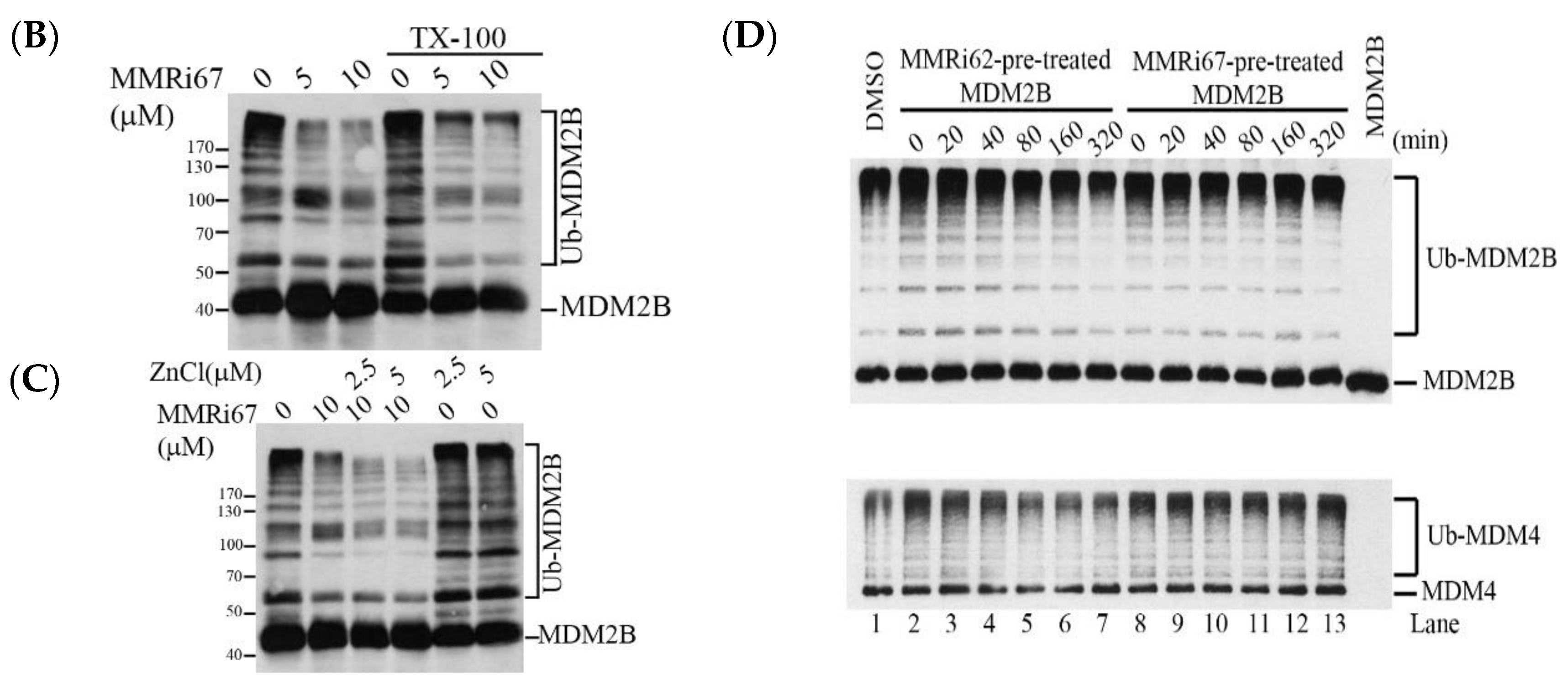
False positive hits in biochemical or cell-based screens with so-called PAINS (pan assay interference compounds) are of concern and hinder lead optimization [26,27]. We reasoned that MMRi62 and MMRi67 are unlikely PAINS since we used NEDD4-1 as a non-specific control E3 ligase in validation assays [21], which was further validated in this study by the selectivity of MMRi67 for Mdm2B versus NEDD4-1 (Figure 2A). Moreover, MMRi62 and MMRi67 have a distinct effect on MDM2-MDM4 E3 ligase activity, MDM4 degradation, and apoptosis induction despite their subtle structural difference [22]. The identification of PAINS from a real hit requires more than simply running a filter screen [28,29]. However, some chemical behaviors of PAINS can be tested, including protein reactivity, colloidal aggregation, and metal chelation [26,27]. To rule out colloidal aggregator activity, we performed an E3 ligase assay in the presence or absence of 0.01% of the non-ionic detergent, Trixton-X-100. Our results indicated that the MMRi67-mediated inhibition of MDM2-MDM4 E3 ligase was not affected in the presence of the detergent (Figure 2B).
The structure of MMRi67 predicts the metal chelation potential [24]. Since Zn is required for maintaining the RING domain structure, it is possible that MMRi67 chelates away Zn and collapses the RING structure, leading to the dissociation of the RING-RING interaction between MDM2-MDM4 and the loss of the E3 ligase activity. We hypothesized that an excessive amount of Zn would neutralize the chelating activity of MMRi67 and cancel its inhibition of MDM2-MDM4 E3 ligase activity. We performed an E3 ligase assay in the presence of Zn, and our results indicated that the presence of Zn did not affect the ability of MMRi67 to inhibit MDM2-MDM4 E3 ligase activity.
The preincubation of MDM4 proteins with either MMRi62 or MMRi67 for a period from 20 to 320 min at 10 μM did not affect the E3 ligase activity of the MDM2-MDM4 E3 complex (data not shown). However, the preincubation of MDM2B proteins with MMRi62 but not MMRi67 inhibited MDM2-MDM4 E3 ligase activity (Figure 2D), suggesting that covalent binding contributes to some extent to MMRi62-mediated effects on the MDM2B-MDM4 E3 ligase reaction (e.g., via the alkylation of a quinone methide formed from 2-aminopyridine loss via the solvolysis of MMRi62. See Supporting Information for further discussion). This covalent binding potential of MMRi62 is similar to that of a macrophage migration inhibitory factor (MIF) inhibitor [30], which shares structural similarities with MMRi62. However, the covalent binding is not involved in the inhibitory mechanism of MMRi67 toward the MDM2-MDM4 E3 ligase in vitro. Taken together, our results suggest that MMRi67 bears a specific inhibitory activity toward the MDM2-MDM4 E3 ligase.

2.3. Structural Requirement of MMRi67 Derivatives for Inhibitory Activity toward MDM2-MDM4 E3 Ligase

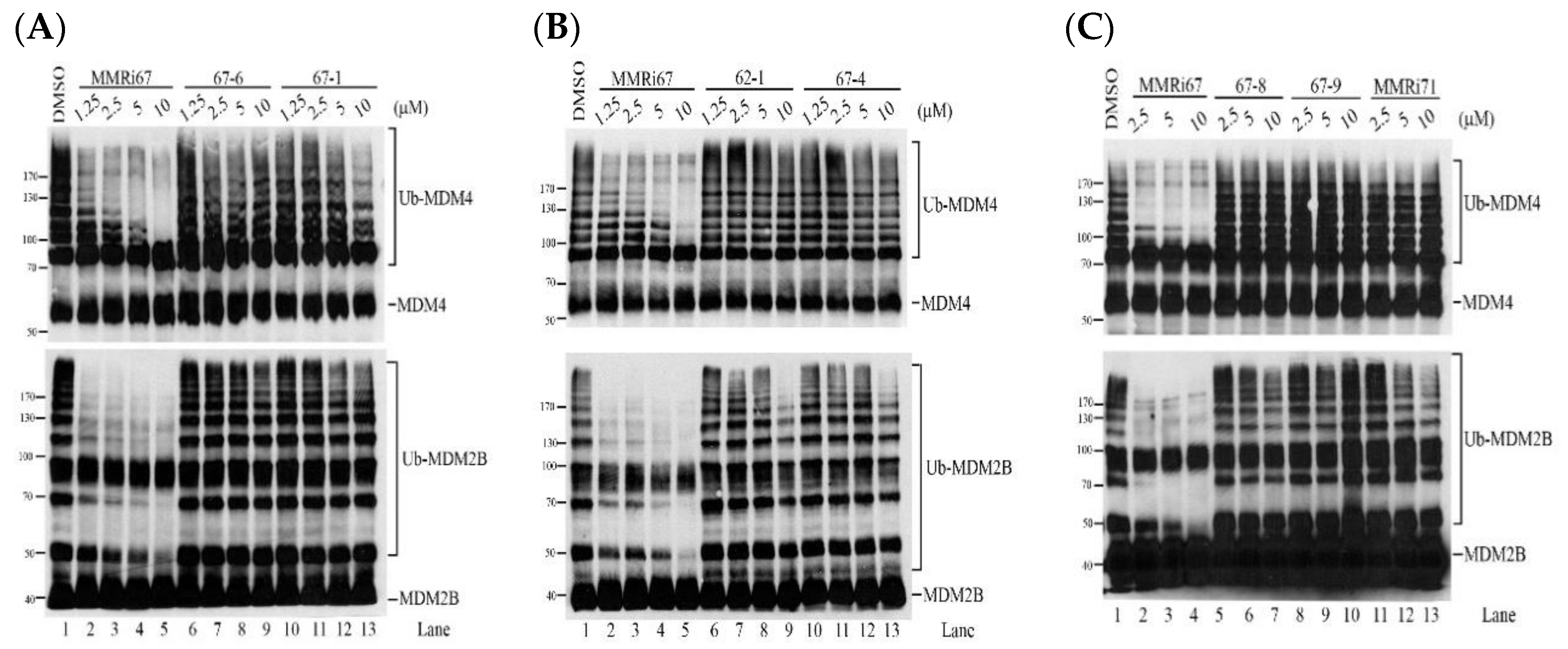
To identify the pharmacophore of MMRi67, it involved the inhibition of the MDM2-MDM4 E3 ligase, and some of its analogs (Figure 1) were evaluated in MDM2-MDM4 E3 ligase assays. Analogs 67-1, lacking the quinolinol chlorine substituent, and 67-6, lacking the phenol, both lost the strong E3 ligase inhibitory activity of MMRi67. However, 67-1 showed a modest inhibition at 10 μM (Figure 3A). It is possible that the phenol of MMRi67 is involved in a critical molecular interaction that can be impacted by the phenolic pKa (which would be lowered by the quinolinol chlorine substituent). To check whether the benzoic acid in MMRi67 plays a critical role in its E3 ligase activity for MDM2 and MDM4, 62-1 with a dichlorophenyl substitution and 67-4 (Figure 1) containing the ethyl benzoate were evaluated with E3 ligase assays. The results showed weak inhibition of MDM2 and MDM4 ubiquitination at concentrations of 10 μM for both the 62-1 and 67-4 derivatives, indicating that the E3 ligase inhibitory action of MMRi67 strongly depends on the carboxylic acid substituent (Figure 3B). It is possible that the carboxylic acid is also involved in a critical molecular interaction with its biochemical target.
A pyridine-to-pyrimidine change in MMRi67 to generate analog 67-8 resulted in the loss of the MDM4 E3 ligase inhibitory effect; however, partial inhibition of the MDM2B E3 ligase at high concentrations was observed. Analog 67-9, containing a pyrimidine and ethyl benzoate, lost both MDM4 and MDM2B E3 ligase effects. MMRi71, containing a pyrimidine and ethyl benzoate as well as a phenolic propionate ester, lost activity to inhibit MDM4 ubiquitination but retained an inhibitory effect on MDM2B ubiquitination in a dose-dependent manner (Figure 3C). Overall, the hydroxyl, chloro, carboxylic acid, and pyridine functionalities of MMRi67 all impacted its MDM4-MDM2B E3 ligase inhibition.

2.4. Induced MDM4 and FTH1 Degradation Is Associated with the Pro-Apoptotic Activity of MMRi67 Derivatives

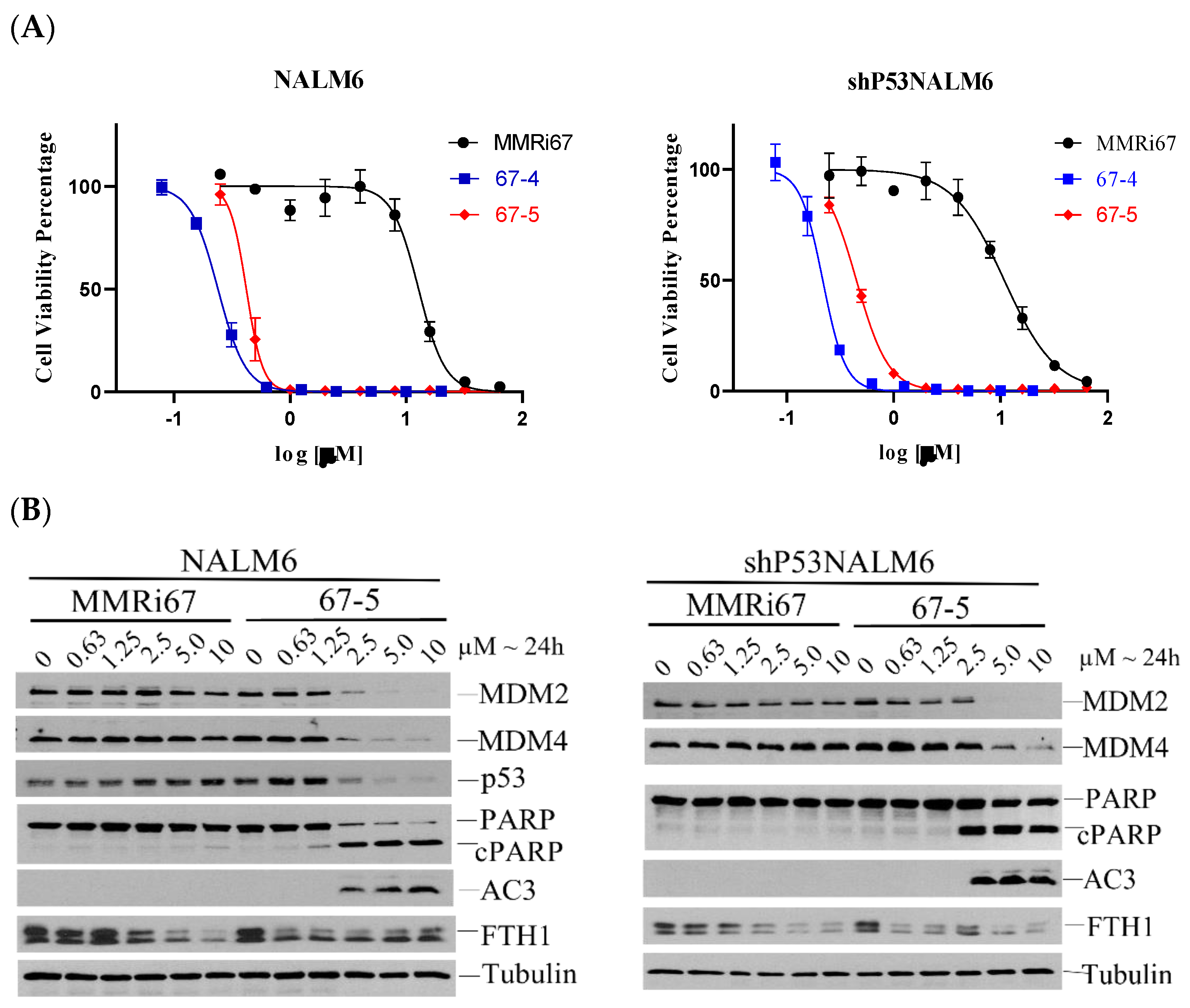
The p53-independent pro-apoptotic activity of the quinolinol compound MMRi62 was associated with an induced MDM4 and FTH1 degradation in cells [22,23]. To determine whether the increased antiproliferative activity of the potent MMRi67 analogs was also associated with p53-independent apoptosis and MDM2/MDM4 and FTH1 protein degradation, we conducted a Western blot analysis for the activated Caspase 3 and caspase-mediated PARP cleavage. Consistent with its improved anti-proliferation activity, analog 67-5, lacking the quinolinol chloro of MMRi67, and bearing the ethyl benzoate, induced the downregulation of MDM4/MDM2, caspase 3 activation, and PARP cleavage in a concentration-dependent manner (Figure 4A,B). Moreover, the pro-apoptotic effect of 67-5 was independent of p53 since the apoptotic response to 67-5 between the NALM6 and shp53NALM6 cells showed no difference. Similar results were obtained with 67-4 (data not shown). These results demonstrate that the improved cell-killing ability of 67 derivatives uses a similar mechanism of action as MMRi62, i.e., MDM4 degradation is a necessary molecular event associated with apoptosis induction [22]. Although the 67-5 effectively induced downregulation of FTH1, as far as the apoptosis of leukemia cells is concerned, it appeared that MDM2/MDM4 degradation, but not FTH1 degradation, was associated with apoptosis induction, since MMRi67 also induced FTH1 degradation at concentrations ≥2.5 μM yet did not elicit any apoptotic response (Figure 4B). Nevertheless, we could not exclude the role of FTH1 degradation in predisposing cells to apoptosis since FTH1 is a ferroptosis target whose depletion increases reactive oxygen species (ROS) and induces ferroptosis in other cell types [23,31].
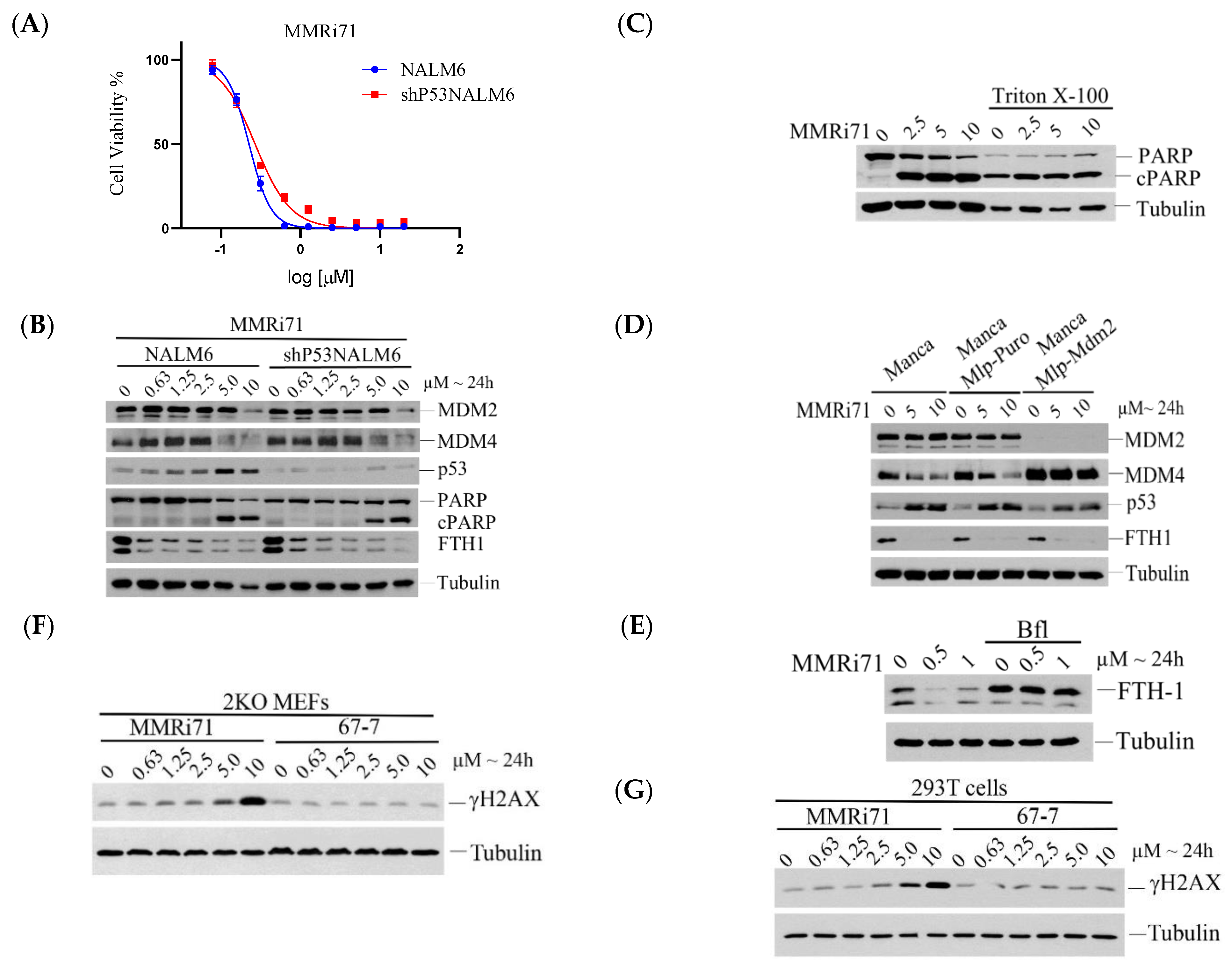
Analog MMRi71 showed strong anti-leukemic activity in the cell proliferation inhibitory assay along with the dose-dependent inhibition of MDM2B E3 ligase activity. We hypothesized that the structural modifications in MMRi71 (compared to MMRi67) would increase water solubility (pyridine changed to pyrimidine) and decrease molecular self-aggregation (via hydrogen bonding or through metals involving the phenol, changed to the propionate ester) while maintaining the cell permeability potential of the candidate (by applying the ethyl benzoate in place of the benzoic acid) and its optimal effect on cellular targets (given that esters can be hydrolyzed in cells). To test this, anti-proliferation assays showed that MMRi71 had the same potency in inhibiting the growth of NALM6 and shP53NALM6 cells with an IC50s of 0.2 μM (Figure 5A), indicating p53-independent cytotoxicity. Interestingly, the phenol ester MMRi71 was substantially more cytotoxic than phenol ester 62-6 even though their parent phenols 67-9 and MMRi62 have similar cell growth IC50′s. The greater aqueous solubility of the pyrimidine lends to MMRi71 may contribute to a more rapid ester hydrolysis and to its active form, phenol 67-9.
MMRi71-treated NALM6 and shP53NALM6 cells were used in the Western blot analysis for changes in the MDM4 and FTH1 proteins. The MMRi71 treatment induced effective downregulation of MDM4 and FTH1 in both NALM6 and shP53NALM6 cells, and the induced apoptosis shown by the PARP cleavage indicated a p53-independent mechanism (Figure 5B). To test whether MMRi71-induced apoptosis was not a result of colloidal aggregation, we performed cellular experiments in the presence of Triton X-10. Since the presence of 0.01%, Trixton-X-100 appeared to induce apoptotic cell death in the NALM6 cells, and the tolerable concentrations of Triton X-100 were between 0.001 and 0.0025% (data not shown). We performed cellular experiments in the presence of 0.0025% Triton X-100, and the results showed that MMRi71 induced a similar extent of apoptotic responses indicated by the cleaved PARP in the presence of the detergent (Figure 5C), suggesting that MMRi71-induced apoptosis was not a result of colloidal aggregation.
We have shown that MMRi62-induced MDM4 degradation was MDM2-dependent [22] and that MMRi62-induced FTH1 degradation was dependent on a lysosomal pathway [23]. To determine if the MMRi71-induced MDM4 degradation occurred via the same mechanism as MMRi62, we performed a WB analysis of MDM4 degradation by MMRi71 using a pair of MDM2-high MANCA lymphoma cells in which MDM2 was either stably knocked down by lentivirus-mediated microRNA (miRNA) or left untouched with a control miRNA. Our results indicated that MDM4 expression levels were elevated in MDM2-knockdown MANCA-mlp-MDM2 cells and that treatment with MMRi71 did not induce MDM4 degradation. The elevated MDM4 expression in the MDM2-knockdown cells was consistent with the report that MDM2 promoted the ubiquitination and degradation of MDM4 [32]. The abolishment of MDM4 degradation by MMRi71 in the absence of MDM2 in MANCA-mlp-MDM2 cells suggests that MMRi71-induced MDM4 degradation is MDM2-dependent (Figure 5D). To determine whether the MMRi71-induced FTH1 degradation was lysosome dependent, we performed a rescue experiment with the lysosome inhibitor Bafilomycin A1 (BAF1). Our results showed that BAF1 fully rescued FTH1 in the NALM6 cells (Figure 5E). These results suggested that MMRi71 induced MDM2-dependent proteasomal degradation of MDM4 and the lysosome-dependent degradation of FTH1, the same mechanisms of action used by MMRi62 [22], in addition to its potential inhibitory activity toward MDM2-MDM4 E3 ligase activity.
The substantially improved activity of MMRi71 over MMRi67 may be due to its increased covalent binding capability or its improved cellular permeability, or both. MMRi62, which has structural similarities to MMRi71, did show some covalent binding effects to MDM2 (Figure 2D). Based on the studies with a quinolinol analog MIF inhibitor, covalent binders in this molecular class can be specific inhibitors [30]. Whether MMRi71 is a covalent inhibitor targeting cellular MDM2-MDM4 for degradation needs further characterization in future studies.
Quinolinol compounds were reported to cause DNA damage via chelating metal ions and the generation of ROS [33]. To test whether MMRi71 caused DNA damage, we used MMRi71 and the inactive compound 67-7, which lacked the metal chelating capability to treat the p53/Mdm2-double knockout (2KO) MEFs and human 293T cell lines, followed by detecting γ-H2AX which is an indicator of DNA damage [34]. The use of 2KO MEFs and 293T cells will exclude the apoptosis-associated γ-H2AX signal due to apoptotic DNA fragmentation since these two cell types are resistant to apoptosis. Our results showed that MMRi71 but not the inactive 67-7 increased γ-H2AX slightly at 5 μM and significantly at 10 μM in both the cell lines (Figure 5F,G). These results suggest that MMRi71 at concentrations of ≥5 μM has the potential to induce DNA damage, possibly by MMRi71-induced ROS. These results are consistent with our observation that MMRi62 induces ROS generation in pancreatic cells [23]. ROS induced by MMRi62-like compounds likely involves both metal chelation and FTH1 degradation, which releases ferrous iron, although metal chelation is not involved in its action on the E3 ligase activity (Figure 2C). Since MMRi71 induced apoptosis at ≥5 μM at 24 h in the NALM6 cells (Figure 5C) and 67-7 did not generate ROS is an inactive compound, these results conclude that ROS generation is required for the antitumor activity of MMRi71. However, we speculate that ROS-induced DNA damage itself is not the major mechanism contributing to cell killing by MMRi62-like compounds since they have a high cancer-selective toxicity. We have shown that MMRi62 inhibits leukemic NALM6 (B-cell precursor leukemia) cell growth at a 125-fold potency compared to its inhibition of normal peripheral blood mononuclear cells (PBMCs) [22]. This 125-fold difference in MMRi62 sensitivity is not likely the result of DNA damage since DNA damage by radiation kills normal human lymphocytes, and B cell lymphoma cells have a comparable capability with the D0 of 1.95Gy for lymphocytes and of 1.38Gy for Burkitt’s lymphoma cell [35], where D0 is the radiation dose required to reduce the fraction of the surviving cells to 37% of its previous value. Accordingly, similar to MMRi62, the mechanisms of action for MMRi71 should involve multiple drug targets that predispose cancer cells to its selective toxicity, which warrants identification in future studies.

3. Materials and Methods

3.1. Representative Chemistry Methods

3.1.1. Ethyl 4-((5-Chloro-8-hydroxyquinolin-7-yl)(pyrimidin ylamino)methyl)benzoate (67-9) Synthesis

To a 250 mL dry round-bottomed flask equipped with a reflux condenser, 2-aminopyrimidine (4.3 g, 45.3 mmol, 1.2 equiv), ethyl 4-formylbenzoate (6.8 g, 38.2 mmol, 1.0 equiv), and 5-chloro-8-hydroxyquinoline (8.2 g, 45.8 mmol, 1.2 equiv) were dissolved in CH3CN (100 mL). Following the addition of formic acid (1.4 mL, 37.1 mmol 1.0 equiv), the solution was stirred at reflux for 16 h. The solution was allowed to cool to rt, was concentrated, and resuspended in acetone. The heterogenous mixture was filtered, and the precipitate was washed with cold acetone and hexanes to give 67-9 as a white solid (5.2 g, 31% yield). mp = 150–151 °C; 1H NMR (300 MHz, CDCl3) δ 8.80 (d, J = 4.2 Hz, 1H), 8.49 (d, J = 8.6 Hz, 1H), 8.30 (d, J = 4.8 Hz, 2H), 7.99 (d, J = 7.9 Hz, 2H), 7.55 (dd, J = 19.6, 10.2 Hz, 4H), 6.79 (d, J = 8.2 Hz, 1H), 6.58 (t, J = 4.9 Hz, 1H), 6.39 (d, J = 8.3 Hz, 1H), 4.34 (q, J = 7.2 Hz, 2H), 1.36 (t, J = 7.2 Hz, 3H); 13C NMR (75 MHz, CDCl3) δ 166.3, 161.5, 158.2, 148.7, 148.5, 146.6, 138.7, 133.3, 129.8, 129.4, 127.0, 126.8, 125.6, 123.6, 122.5, 120.7, 111.5, 60.9, 54.8, 14.3; IR neat film: 3293, 2978, 1716, 1583, 1496 cm−1; HRMS (ESI) calculated for [C23H20ClN4O3] 390.1004, found 390.1011.

3.1.2. Ethyl 4-((8-(Propionyloxy)quinolin-7-yl)(pyrimidin-2-ylamino)methyl)benzoate (MMRi71) Synthesis

In a 5 mL dry round-bottomed flask, analog 67-9 (5.0 g, 11.5 mmol, 1.0 equiv) was dissolved in dry CH2Cl2 (100 mL) under an argon atmosphere. Potassium carbonate (3.18 g, 23.0 mmol, 2.0 equiv) was added, and the solution was cooled to 0 °C. Propionoyl chloride (1.0 mL, 11.5 mmol, 1.0 equiv) was then added to the solution. The mixture was allowed to warm to rt and stirred for 1 h. The reaction mixture was then filtered through Celite and washed with CH2Cl2. The supernatant was then treated with 1 g of DMT-functionalized silica gel and stirred for 15 min. The mixture was filtered and concentrated. The resulting crude solid was resuspended in Et2O and washed with deionized water. The organic layer was dried over Na2SO4 and then concentrated. The crude mixture was purified by flash column chromatography (silica gel, 50% ether:hexanes) to yield MMRi71 as a greenish-white solid (2.53 g, 48% yield). mp = 178–179 °C; 1H NMR (400 MHz, CDCl3) δ 8.91 (d, J = 2.8 Hz, 1H), 8.50 (d, J = 6.8 Hz, 1H), 8.23 (d, J = 4.8 Hz, 2H), 8.00 (d, J = 8.4 Hz, 2H), 7.54 (s, 1H), 7.50 (dd, J = 8.5, 4.2 Hz, 1H), 7.43 (d, J = 8.1 Hz, 2H), 6.83 (d, J = 7.8 Hz, 1H), 6.57 (t, J = 4.8 Hz, 1H), 6.10 (d, J = 7.8 Hz, 1H), 4.36 (q, J = 7.1 Hz, 2H), 2.68 (q, J = 7.6 Hz, 2H), 1.37 (t, J = 7.1 Hz, 3H), 1.20 (t, J = 7.5 Hz, 3H); 13C NMR (101 MHz, CDCl3) δ 172.2, 166.2, 161.3, 158.1, 151.2, 145.5, 144.6, 142.0, 134.3, 133.0, 130.0, 129.8, 129.0, 127.1, 126.7, 125.7, 122.4, 111.8, 61.0, 53.8, 27.3, 14.3, 9.0. IR neat film: 3270, 2982, 1773, 1716, 1578, 1491 cm−1; HRMS (ESI) calculated for [C26H23ClN4NaO4] 513.1300, found 513.1307.

3.2. Biological Assays and Methods

3.2.1. Cell Culture and Small-Molecule Compounds

NALM6 and shP53NALM6 leukemic cell lines were cultured in an RPMI-1640 medium supplemented with 10% fetal bovine serum, 50 U/mL penicillin, and 50 μg/mL streptomycin. The shP53NALM6 cell line was established using pLKO.1-p53 (purchased from Sigma) (Plasmid #19119) [36] followed by puromycin selection at 1 μg/mL for 2 days, then a clonal expansion in the puromycin-free medium. MANCA, MANCA-mlp-puro, and MANCA-mlp-MDM2 were generous gifts from Prof. Jill Bargonetti (Hunter College, CUNY, NY, USA) and were generated as described previously [37] and maintained in a 10% FBS-Pen/Strep- RPMI-1640 medium. Small molecule compounds were synthesized, purified, and characterized in-house and were dissolved in DMSO as 10 mM stocks for cell proliferation assays.

3.2.2. Western Blotting, In Vitro Ubiquitination, and Apoptosis Analysis

The Western blotting procedure and antibodies for the target proteins were described previously [22]. In vitro assays for ubiquitination by MDM2B-MDM4 were performed as described previously with minor modifications [15]. Briefly, the reactions were carried out at 30 °C for 1 h in a volume of 20 μL reaction in the presence of different concentrations of compounds or vehicle solvent DMSO, followed by a WB of p53 with DO-1, MDM2 with rabbit anti-MDM2 (MDM2 (D1V2Z) (#86934, Cell Signaling Technology, Danvers, MA 01923, USA), or MDM4 with a rabbit anti-MDM4 antibody (Proteintech, Rosemont, IL 60018, USA, Cat no: 17914-1-AP). The apoptotic response to compounds was measured by Western blotting using specific antibodies from Cell Signaling Technology, Danvers, MA 01923, USA) for activated caspase 3 (Cleaved Caspase-3 (Asp175) (5A1E) (#9664) and PARP (PARP Antibody #9542).

3.2.3. IC50 Measurement and Analysis

The procedure was described previously [22]. Briefly, cells at 10,000/well were plated in 96-well plates at 100 µL/well, and compounds of different concentrations were added to each well at 100 µL/well. After 70 h treatment, 40 µL of 6x resazurin stock solution was added to each well, followed by a 2 h development of fluorescent metabolites by viable cells before reading OD600 in the BioTek Synergy 2 Microplate Reader. The IC50 values were obtained by the Chou-Median-Effect Equation using CompuSyn software [38], and dose-effect curves were obtained by GraphPad using the affected fractions of compound-treated wells normalized against no-drug control wells with a non-liner regression model.

4. Conclusions

In summary, the antiproliferative activity of these potent MMRi67 derivatives was associated with their effects on the dual degradation of MDM4 and FTH1 compared to MMRi67. The SAR results from MMRi67 derivatives identified the hydroxyl and the chloro in the quinoline ring and the benzoic acid to be the critical structural elements that contribute to MDM2-MDM4 E3 ligase inhibitory effects of MMRi67 in vitro. Whereas MMRi67 is not highly cytotoxic, its ethyl benzoate analogs, including MMRi71, were, and they exhibited a strong MDM2/MDM4 and FTH1 degradation in cells. MMRi71 represents a new class of dual inhibitors targeting the degradation of MDM2/MDM4 and FTH1 for p53-independent apoptosis in leukemia cells. Up to this point, our small molecule structural optimization efforts have focused on discerning and optimizing structural attributes that impact biochemical target interactions, the antiproliferation of cancer cells, and physical features related to compound formulation (optimizing LogP and discouraging metal chelation). Going forward, we will increase our analysis of factors related to in vivo efficacy and toxicity. Currently, we note that the lead compound MMRi71 has a molecular weight of 490.94 g/mol, contains one hydrogen bond donor, seven potential hydrogen-bond acceptor nitrogens and oxygens, and a LogP of 4.6. The calculated total polar surface area of MMRi71 is 101.71 Å2, and it has nine rotatable bonds. All of these properties conform to those recommended for orally available drugs [39,40]. Further lead optimization to improve the potency, pharmacokinetics, and pharmacodynamics profiles is warranted for developing clinically useful derivatives in this chemical domain.

5. Patents

Provisional patent: Antiproliferative Betti Bases and Prodrugs Thereof, pending.

Supplementary Materials

The following supporting information can be downloaded at: https://www.mdpi.com/article/10.3390/molecules27227665/s1, Experimental procedures and data for characterization of all new compounds, including NMR spectra, are provided in the chemistry supporting information [30,41,42,43,44,45,46,47,48,49].

Author Contributions

Conceptualization, S.R.C. and X.W.; methodology, R.L., S.L.G., C.X., and L.W.D.; validation, R.L. and S.L.G.; investigation, R.L. and S.L.G.; chemical synthesis and compound characterization, S.L.G. and L.W.D.; biological experiments, R.L. and. X.W.; writing—original draft preparation, R.L., X.W., and S.R.C.; writing—review and editing, R.L., X.W., and S.R.C.; supervision, S.R.C. and X.W.; project administration, S.R.C. and X.W.; funding acquisition, S.R.C. and X.W. All authors have read and agreed to the published version of the manuscript.

Funding

This research was funded by the National Institutes of Health, R01CA208352 to XW and GM078383 to SRC and Developmental Therapeutic Program Seed Funds from the Roswell Park Alliance Foundation to XW and SRC. We also thank the American Chemical Society for the Cope Mid-Career Award to SRC (unrestricted funds). This work was also supported by P30CA016056 from the National Cancer Institute (NCI) involving the use of the Roswell Park Comprehensive Cancer Center’s shared resources. We acknowledge the Chemistry Instrument Center (CIC), University at Buffalo, SUNY for high resolution mass spectroscopy analysis.

Institutional Review Board Statement

Not applicable.

Informed Consent Statement

Not applicable.

Data Availability Statement

All data are included in the article and/or Supplementary Materials.

Acknowledgments

We thank David Hangauer (UB emeritus professor) for insightful discussions. We also thank Yuping Wang for technical assistance.

Conflicts of Interest

The authors declare no conflict of interest.

References

  1. Martins, C.P.; Brown-Swigart, L.; Evan, G.I. Modeling the Therapeutic Efficacy of p53 Restoration in Tumors. Cell 2006, 127, 1323–1334. [Google Scholar] [CrossRef] [PubMed] [Green Version]
  2. Ventura, A.; Kirsch, D.G.; McLaughlin, M.E.; Tuveson, D.A.; Grimm, J.; Lintault, L.; Newman, J.; Reczek, E.E.; Weissleder, R.; Jacks, T. Restoration of p53 function leads to tumour regression in vivo. Nature 2007, 445, 661–665. [Google Scholar] [CrossRef] [PubMed]
  3. Xue, W.; Zender, L.; Miething, C.; Dickins, R.A.; Hernando, E.; Krizhanovsky, V.; Cordon-Cardo, C.; Lowe, S.W. Senescence and tumour clearance is triggered by p53 restoration in murine liver carcinomas. Nature 2007, 445, 656–660. [Google Scholar] [CrossRef] [Green Version]
  4. Wang, X.; Jiang, X. Mdm2 and MdmX partner to regulate p53. FEBS Lett. 2012, 586, 1390–1396. [Google Scholar] [CrossRef] [PubMed] [Green Version]
  5. Wade, M.; Li, Y.-C.; Wahl, G.M. MDM2, MDMX and p53 in oncogenesis and cancer therapy. Nat. Rev. Cancer 2013, 13, 83–96. [Google Scholar] [CrossRef] [Green Version]
  6. Marine, J.-C.; Francoz, S.; Maetens, M.; Wahl, G.M.; Toledo, F.; Lozano, G. Keeping p53 in check: Essential and synergistic functions of Mdm2 and Mdm4. Cell Death Differ. 2006, 13, 927–934. [Google Scholar] [CrossRef]
  7. Vassilev, L.T.; Vu, B.T.; Graves, B.; Carvajal, D.; Podlaski, F.; Filipovic, Z.; Kong, N.; Kammlott, U.; Lukacs, C.; Klein, C.; et al. In vivo activation of the p53 pathway by small-molecule antagonists of MDM2. Science 2004, 303, 844–848. [Google Scholar] [CrossRef] [Green Version]
  8. Konopleva, M.; Martinelli, G.; Daver, N.; Papayannidis, C.; Wei, A.; Higgins, B.; Ott, M.; Mascarenhas, J.; Andreeff, M. MDM2 inhibition: An important step forward in cancer therapy. Leukemia 2020, 34, 2858–2874. [Google Scholar] [CrossRef]
  9. Wang, S.; Chen, F.-E. Small-molecule MDM2 inhibitors in clinical trials for cancer therapy. Eur. J. Med. Chem. 2022, 236, 114334. [Google Scholar] [CrossRef]
  10. Patton, J.T.; Mayo, L.D.; Singhi, A.D.; Gudkov, A.V.; Stark, G.R.; Jackson, M.W. Levels of HdmX Expression Dictate the Sensitivity of Normal and Transformed Cells to Nutlin-3. Cancer Res. 2006, 66, 3169–3176. [Google Scholar] [CrossRef]
  11. Hu, B.; Gilkes, D.M.; Farooqi, B.; Sebti, S.M.; Chen, J. MDMX Overexpression Prevents p53 Activation by the MDM2 Inhibitor Nutlin. J. Biol. Chem. 2006, 281, 33030–33035. [Google Scholar] [CrossRef] [PubMed] [Green Version]
  12. Gembarska, A.; Luciani, F.; Fedele, C.; Russell, E.A.; Dewaele, M.; Villar, S.; Zwolinska, A.; Haupt, S.; de Lange, J.; Yip, D.; et al. MDM4 is a key therapeutic target in cutaneous melanoma. Nat. Med. 2012, 18, 1239–1247. [Google Scholar] [CrossRef] [PubMed] [Green Version]
  13. Linares, L.K.; Hengstermann, A.; Ciechanover, A.; Müller, S.; Scheffner, M. HdmX stimulates Hdm2-mediated ubiquitination and degradation of p53. Proc. Natl. Acad. Sci. USA 2003, 100, 12009–12014. [Google Scholar] [CrossRef] [PubMed] [Green Version]
  14. Kawai, H.; Lopez-Pajares, V.; Kim, M.M.; Wiederschain, D.; Yuan, Z.-M. RING Domain–Mediated Interaction Is a Requirement for MDM2’s E3 Ligase Activity. Cancer Res. 2007, 67, 6026–6030. [Google Scholar] [CrossRef] [PubMed] [Green Version]
  15. Wang, X.; Wang, J.; Jiang, X. MdmX Protein Is Essential for Mdm2 Protein-mediated p53 Polyubiquitination. J. Biol. Chem. 2011, 286, 23725–23734. [Google Scholar] [CrossRef] [PubMed] [Green Version]
  16. Itahana, K.; Mao, H.; Jin, A.; Itahana, Y.; Clegg, H.V.; Lindström, M.S.; Bhat, K.P.; Godfrey, V.L.; Evan, G.I.; Zhang, Y. Targeted Inactivation of Mdm2 RING Finger E3 Ubiquitin Ligase Activity in the Mouse Reveals Mechanistic Insights into p53 Regulation. Cancer Cell 2007, 12, 355–366. [Google Scholar] [CrossRef] [Green Version]
  17. Huang, L.; Yan, Z.; Liao, X.; Li, Y.; Yang, J.; Wang, Z.; Zuo, Y.; Kawai, H.; Shadfan, M.; Ganapathy, S.; et al. The p53 inhibitors MDM2/MDMX complex is required for control of p53 activity in vivo. Proc. Natl. Acad. Sci. USA 2011, 108, 12001–12006. [Google Scholar] [CrossRef] [Green Version]
  18. Pant, V.; Xiong, S.; Iwakuma, T.; Quintás-Cardama, A.; Lozano, G. Heterodimerization of Mdm2 and Mdm4 is critical for regulating p53 activity during embryogenesis but dispensable for p53 and Mdm2 stability. Proc. Natl. Acad. Sci. USA 2011, 108, 11995–12000. [Google Scholar] [CrossRef] [Green Version]
  19. Linke, K.; Mace, P.D.; Smith, C.A.; Vaux, D.L.; Silke, J.; Day, C.L. Structure of the MDM2/MDMX RING domain heterodimer reveals dimerization is required for their ubiquitylation in trans. Cell Death Differ. 2008, 15, 841–848. [Google Scholar] [CrossRef] [Green Version]
  20. Chinnam, M.; Xu, C.; Lama, R.; Zhang, X.; Cedeno, C.D.; Wang, Y.; Stablewski, A.B.; Goodrich, D.W.; Wang, X. MDM2 E3 ligase activity is essential for p53 regulation and cell cycle integrity. PLoS Genet. 2022, 18, e1010171. [Google Scholar] [CrossRef]
  21. Wu, W.; Xu, C.; Ling, X.; Fan, C.; Buckley, B.P.; Chernov, M.V.; Ellis, L.; Li, F.; Muñoz, I.G.; Wang, X. Targeting RING domains of Mdm2–MdmX E3 complex activates apoptotic arm of the p53 pathway in leukemia/lymphoma cells. Cell Death Dis. 2015, 6, e2035. [Google Scholar] [CrossRef] [PubMed] [Green Version]
  22. Lama, R.; Xu, C.; Galster, S.L.; Querol-García, J.; Portwood, S.; Mavis, C.K.; Ruiz, F.M.; Martin, D.; Wu, J.; Giorgi, M.C.; et al. Small molecule MMRi62 targets MDM4 for degradation and induces leukemic cell apoptosis regardless of p53 status. Front. Oncol. 2022, 12, 933446. [Google Scholar] [CrossRef] [PubMed]
  23. Li, J.; Lama, R.; Galster, S.L.; Inigo, J.R.; Wu, J.; Chandra, D.; Chemler, S.R.; Wang, X. Small-Molecule MMRi62 Induces Ferroptosis and Inhibits Metastasis in Pancreatic Cancer via Degradation of Ferritin Heavy Chain and Mutant p53. Mol. Cancer Ther. 2022, 21, 535–545. [Google Scholar] [CrossRef] [PubMed]
  24. Krawczyk, M.; Pastuch-Gawołek, G.; Pluta, A.; Erfurt, K.; Domiński, A.; Kurcok, P. 8-Hydroxyquinoline Glycoconjugates: Modifications in the Linker Structure and Their Effect on the Cytotoxicity of the Obtained Compounds. Molecules 2019, 24, 4181. [Google Scholar] [CrossRef] [PubMed] [Green Version]
  25. Oliveri, V.; Giuffrida, M.L.; Vecchio, G.; Aiello, C.; Viale, M. Gluconjugates of 8-hydroxyquinolines as potential anti-cancer prodrugs. Dalton Trans. 2012, 41, 4530–4535. [Google Scholar] [CrossRef] [PubMed]
  26. Baell, J.B.; Holloway, G.A. New Substructure Filters for Removal of Pan Assay Interference Compounds (PAINS) from Screening Libraries and for Their Exclusion in Bioassays. J. Med. Chem. 2010, 53, 2719–2740. [Google Scholar] [CrossRef] [Green Version]
  27. Aldrich, C.; Bertozzi, C.; Georg, G.I.; Kiessling, L.; Lindsley, C.; Liotta, D.; Merz, J.K.M.; Schepartz, A.; Wang, S. The Ecstasy and Agony of Assay Interference Compounds. ACS Med. Chem. Lett. 2017, 8, 379–382. [Google Scholar] [CrossRef] [Green Version]
  28. Kenny, P.W. Comment on The Ecstasy and Agony of Assay Interference Compounds. J. Chem. Inf. Model. 2017, 57, 2640–2645. [Google Scholar] [CrossRef]
  29. Baell, J.B.; Nissink, J.W.M. Seven Year Itch: Pan-Assay Interference Compounds (PAINS) in 2017—Utility and Limitations. ACS Chem. Biol. 2017, 13, 36–44. [Google Scholar] [CrossRef] [Green Version]
  30. McLean, L.R.; Zhang, Y.; Li, H.; Li, Z.; Lukasczyk, U.; Choi, Y.-M.; Han, Z.; Prisco, J.; Fordham, J.; Tsay, J.T.; et al. Discovery of covalent inhibitors for MIF tautomerase via cocrystal structures with phantom hits from virtual screening. Bioorganic Med. Chem. Lett. 2009, 19, 6717–6720. [Google Scholar] [CrossRef]
  31. Mumbauer, S.; Pascual, J.; Kolotuev, I.; Hamaratoglu, F. Ferritin heavy chain protects the developing wing from reactive oxygen species and ferroptosis. PLoS Genet. 2019, 15, e1008396. [Google Scholar] [CrossRef] [PubMed] [Green Version]
  32. Pan, Y.; Chen, J. MDM2 Promotes Ubiquitination and Degradation of MDMX. Mol. Cell Biol. 2003, 23, 5113–5121. [Google Scholar] [CrossRef] [PubMed] [Green Version]
  33. Prachayasittikul, V.; Prachayasittikul, V.; Prachayasittikul, S.; Ruchirawat, S. 8-Hydroxyquinolines: A review of their metal chelating properties and medicinal applications. Drug Des. Dev. Ther. 2013, 7, 1157–1178. [Google Scholar] [CrossRef] [PubMed] [Green Version]
  34. Mah, L.J.; El-Osta, A.; Karagiannis, T.C. gammaH2AX: A sensitive molecular marker of DNA damage and repair. Leukemia 2010, 24, 679–686. [Google Scholar] [CrossRef] [Green Version]
  35. Heylmann, D.; Rödel, F.; Kindler, T.; Kaina, B. Radiation sensitivity of human and murine peripheral blood lymphocytes, stem and progenitor cells. Biochim. Et Biophys. Acta 2014, 1846, 121–129. [Google Scholar] [CrossRef]
  36. Godar, S.; Ince, T.A.; Bell, G.W.; Feldser, D.; Donaher, J.L.; Bergh, J.; Liu, A.; Miu, K.; Watnick, R.S.; Reinhardt, F.; et al. Growth-Inhibitory and Tumor- Suppressive Functions of p53 Depend on Its Repression of CD44 Expression. Cell 2008, 134, 62–73. [Google Scholar] [CrossRef] [Green Version]
  37. Rosso, M.; Polotskaia, A.; Bargonetti, J. Homozygous mdm2 SNP309 cancer cells with compromised transcriptional elongation at p53 target genes are sensitive to induction of p53-independent cell death. Oncotarget 2015, 6, 34573–34591. [Google Scholar] [CrossRef]
  38. Chou, T.-C. Theoretical Basis, Experimental Design, and Computerized Simulation of Synergism and Antagonism in Drug Combination Studies. Pharmacol. Rev. 2006, 58, 621–681. [Google Scholar] [CrossRef]
  39. Lipinski, C.A.; Lombardo, F.; Dominy, B.W.; Feeney, P.J. Experimental and computational approaches to estimate solubility and permeability in drug discovery and development setting. Adv. Drug Deliv. Rev. 2001, 46, 3–26. [Google Scholar] [CrossRef]
  40. Veber, D.F.; Johnson, S.R.; Cheng, H.Y.; Smith, B.R.; Ward, K.W.; Kopple, K.D. Molecular Properties That Influence the Oral Bioavailability of Drug Candidates. J. Med. Chem. 2002, 45, 2615–2623. [Google Scholar] [CrossRef]
  41. Shergalis, A.; Xue, D.; Gharbia, F.Z.; Driks, H.; Shrestha, B.; Tanweer, A.; Cromer, K.; Ljungman, M.; Neamati, N. Characterization of Aminobenzylphenols as Protein Disulfide Isomerase Inhibitors in Glioblastoma Cell Lines. J. Med. Chem. 2020, 63, 10263–10286. [Google Scholar] [CrossRef] [PubMed]
  42. Weinert, E.E.; Dondi, R.; Colloredo-Melz, S.; Frankenfield, K.N.; Mitchell, C.H.; Freccero, M.; Rokita, S.E. Substituents on Quinone Methides Strongly Modulate Formation and Stability of Their Nucleophilic Adducts. J. Am. Chem. Soc. 2006, 128, 11940–11947. [Google Scholar] [CrossRef] [PubMed] [Green Version]
  43. Kanizsai, I.; Madácsi, R.; Hackler, L., Jr.; Gyuris, M.; Szebeni, G.J.; Huzián, O.; Puskás, L.G. Synthesis and Cytoprotective Characterization of 8-Hydroxyquinoline Betti Products. Molecules 2018, 23, 1934. [Google Scholar] [CrossRef] [Green Version]
  44. Pobbati, A.V.; Mejuch, T.; Chakraborty, S.; Karatas, H.; Bharath, S.R.; Guéret, S.M.; Goy, P.-A.; Hahne, G.; Pahl, A.; Sievers, S.; et al. Identification of Quinolinols as Activators of TEAD-Dependent Transcription. ACS Chem. Biol. 2019, 14, 2909–2921. [Google Scholar] [CrossRef] [PubMed]
  45. Olyaei, A.; Sadeghpour, M. Recent advances in the synthesis and synthetic applications of Betti base (aminoalkylnaphthol) and bis-Betti base derivatives. RSC Adv. 2019, 9, 18467–18497. [Google Scholar] [CrossRef] [PubMed] [Green Version]
  46. Lim, C.; Popov, A.A.; Tselikov, G.; Heo, J.; Pliss, A.; Kim, S.; Kabashin, A.V.; Prasad, P.N. Organic Solvent and Surfactant Free Fluorescent Organic Nanoparticles by Laser Ablation of Aggregation-Induced Enhanced Emission Dyes. Adv. Opt. Mater. 2018, 6, 1800164. [Google Scholar] [CrossRef]
  47. Pauli, G.F.; Chen, S.-N.; Simmler, C.; Lankin, D.C.; Gödecke, T.; Jaki, B.U.; Friesen, J.B.; McAlpine, J.B.; Napolitano, J.G. Importance of Purity Evaluation and the Potential of Quantitative 1H NMR as a Purity Assay. J. Med. Chem. 2014, 57, 9220–9231. [Google Scholar] [CrossRef] [PubMed] [Green Version]
  48. Chen, Q.-Y.; He, Y.-B.; Yang, Z.-Y. A new method for reduction of phenyl fluoroalkanesulphonates to arenes catalysed by palladium. J. Chem. Soc. Chem. Commun. 1986, 1452–1453. [Google Scholar] [CrossRef]
  49. Andreu, R.; Ronda, J.C. Synthesis of 3,4-Dihydro-2H-1,3-benzoxazines by Condensation of 2-Hydroxyaldehydes and Primary Amines: Application to the Synthesis of Hydroxy-Substituted and Deuterium-Labeled Compounds. Synth. Commun. 2008, 38, 2316–2329. [Google Scholar] [CrossRef]
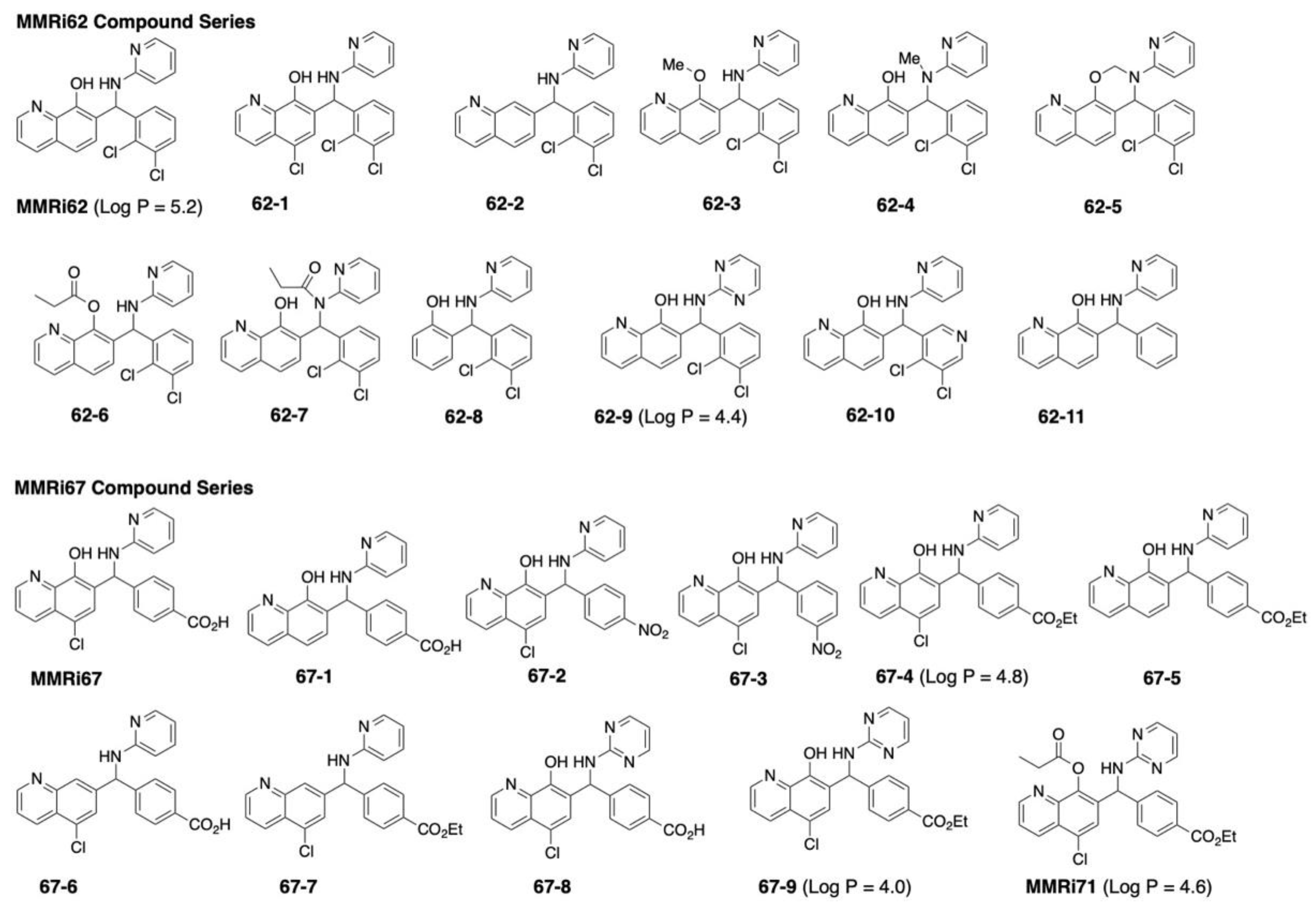
Figure 1. Structures of MMRi62 and MMRi67 compound series.
Molecules 27 07665 g001
Scheme 1. Synthesis of MMRi71. A representative synthesis is shown for MMRi71, the lead compound our study concludes with. For synthesis details of MMRi71, MMRi62, and MMRi67 series see the M and M and Supporting Information.
Molecules 27 07665 sch001
Figure 2. MMRi67 is a small molecule inhibitor of MDM2-MDM4 E3 ligase activity. (A) MMRi67 inhibits the ubiquitination of MDM2B (left) and MDM4 (middle) by MDM2B-MDM4 E3 complex but not NEDD4-1 (right) in the in vitro E3 ligase assays. WB analysis of MDM4, MDM2B, and NEDD4-1 using specific antibodies after in vitro ubiquitination reaction in the presence of solvents or indicated concentrations of MMRi67. The smearing bands of ubiquitinated MDM4 (Ub-MDM4), ubiquitinated MDM2B (Ub-MDM2B), and ubiquitinated NEDD4-1 (Ub-NEDD4-1) are indicated. (B) The E3 ligase inhibitory activity of MMRi67 is not abolished by the presence of 0.01% Triton X-100. Same procedure as in (A) except the ubiquitination reaction was performed in the presence of 0.01% Triton X-100. Only MDM2B data are shown. (C) The E3 ligase inhibitory activity of MMRi67 is not abolished by the presence of increasing concentrations of Zn to neutralize the potential chelating capability of MMRi67. The same procedure as in (A) except for the presence of indicated concentrations of Zn. Only MDM2B data are shown. (D) Preincubation of MDM2B proteins with MMRi67 does not abolish MDM2B E3 ligase activity. Same procedure as in (A) except MDM2B was preincubated with either 10 μM MMRi62 or MMRi67 for the indicated min before 10-times dilution and was used for ubiquitination reaction; the preincubation did not affect the ubiquitination of MDM2B or MDM4 by the MDM2B-MDM4 E3 complex.
Molecules 27 07665 g002aMolecules 27 07665 g002b
Figure 3. Assessment of the inhibitory effect of MMRi67 derivatives toward MDM2B-MDM4 E3 ligase in the in vitro E3 ligase assays. (A) Comparison of MMRi67 with hydroxyl to hydrogen substituted (67-6) and chloro to hydrogen substituted (67-1) analogs in inhibition of the MDM4 and MDM2B ubiquitination in vitro. WB analysis of MDM4 or MDM2B using specific antibodies after in vitro ubiquitination reaction in the presence of a solvent or indicated compound at the indicated concentrations. The smearing bands of ubiquitinated MDM4 (Ub-MDM4) and MDM2B (Ub-MDM2B) are indicated. (B) Same analysis as in (A) except for the comparison of MMRi67 with carboxylic acid substituted analogs 62-1 and 67-4 in the inhibition of MDM4 and MDM2B ubiquitination in vitro. (C) Same analysis as in (A) except for the comparison of MMRi67 with pyridine substituted analogs 67-8 and pyridine, hydroxyl, and carboxylic acid substituted analogs 67-9 and MMRi71 in the inhibition of MDM4 and MDM2B ubiquitination in vitro.
Molecules 27 07665 g003
Figure 4. Anti-proliferation activity of 67-5 is associated with the downregulation of MDM2, MDM4, and FTH1 protein expression and caspase 3 activations. (A) Improved anti-proliferation activity of 67-4 and 67-5 compared with MMRi67. Antiproliferation assays in NALM6 and shp53NALM6 cells. The IC50 in NAML6 cells was 12.8 μM for MMRi67 and 0.38 μM for 67-5. The IC50 in shP53NAML6 cells was 10.9 μM for MMRi67 and 0.45 μM for 67-5. (B) Improved antiproliferation activity of 67-5 is associated with the downregulation of MDM2, MDM4, and FTH1, as well as caspase activation and PARP cleavage. WB analysis of indicated proteins and apoptotic effect induced by 67-5 compared to MMRi67 in both NALM6 and shP53NALM6 cells.
Molecules 27 07665 g004
Figure 5. Characterization of MMRi71 in cells. (A) The anti-proliferation activity of MMRi71 is p53-independent with IC50 of 0.23 μM in NALM6 and 0.29 μM in shp53NALM6 cells. (B) WB analysis shows how MMRi71 induces MDM4/MDM2 and FTH1 protein degradation and apoptotic effect in NALM6 and shP53NALM6 cells. (C) The pro-apoptotic effect of MMRi71 is not affected by the presence of 0.0025% Triton X-100 in NALM-6 cells, as shown by the WB analysis of apoptotic PARP cleavage. (D) MMRi71-induced MDM4 degradation is MDM2 dependent. WB analysis showing Mdm2 knockdown in Manca-mlp-Mdm2 cells and abolished MMRi71-induced MDM4 degradation. (E) MMRi71-induced FTH1 degradation is lysosome dependent. WB showed the rescue of MMRi71-induced FTH1 degradation by 50 nM lysosome inhibitor Bafilomycin A1 (Bfl) in NALM6 cells. (F) (G) DNA damage induction by MMRi71 and 67-7. Mouse p53/Mdm2 double knockout MEFs (2KO) (F) or human 293T cells (G) were treated with indicated concentrations of MMRi71 and 67-7 for 24 h at the indicated concentrations, followed by WB analysis for gammaH2AX and Tubulin with specific antibodies. Tubulin served as a protein loading control.
Molecules 27 07665 g005
Table 1. MMRi62 and MMRi67 compound series IC50.
EntryNALM6 IC50 (μM)shP53NALM6 IC50 (μM)
MMRi620.12 ± 0.0010.14 ± 0.001
62-10.29 ± 0.010.46 ± 0.04
62-232.4 ± 2.2535.5 ± 2.62
62-38.58 ± 0.8324.2 ± 4.38
62-44.84 ± 0.484.68 ± 0.52
62-52.62 ± 0.361.51 ± 0.12
62-61.89 ± 0.531.08 ± 0.08
62-70.35 ± 0.010.26 ± 0.02
62-828.6 ± 2.8728.5 ± 3.82
62-90.30 ± 0.020.36 ± 0.03
62-10
62-11
MMRi67
67-1
67-2
67-3
67-4
67-5
67-6
67-7
67-8
67-9
MMRi71
12.0 ± 1.79
2.10 ± 0.22
12.80 ± 1.57
8.93 ± 0.41
0.40 ± 0.01
0.38 ± 0.03
0.53 ± 0.03
0.38 ± 0.03
>100
18.2 ± 6.54
2.13 ± 0.18
0.27 ± 0.02
0.23 ± 0.01
27.1 ± 5.58
2.66 ± 0.17
10.89 ± 1.56
10.1 ± 0.51
0.47 ± 0.01
0.48 ± 0.02
0.57 ± 0.04
0.45 ± 0.01
>100
31.7 ± 3.52
2.36 ± 0.15
0.37 ± 0.003
0.29 ± 0.02
The treatment was tri replicated and repeated two times. IC50 ± SD were performed using non-linear regression analysis.
Publisher’s Note: MDPI stays neutral with regard to jurisdictional claims in published maps and institutional affiliations.