Article

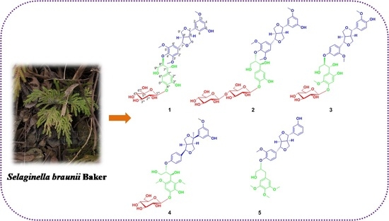
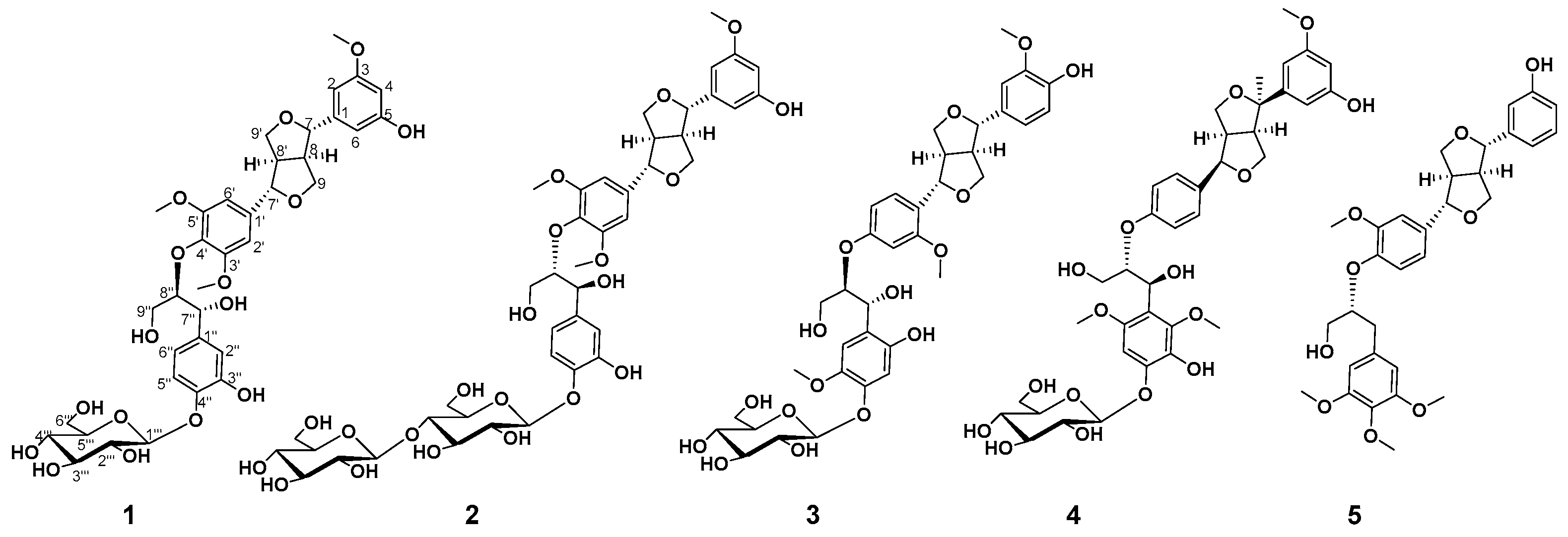
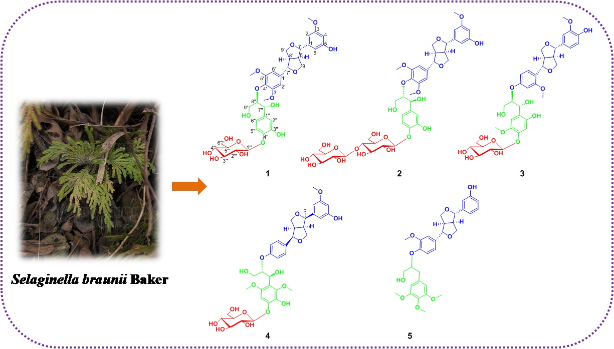
Brasesquilignan A–E, Five New Furofurans Lignans from Selaginella braunii Baker

1
Xiangya School of Pharmaceutical Sciences, Central South University, Changsha 410013, China
2
Institute of Chinese MateriaMedica, Hunan Academy of Chinese Medicine, Changsha 410013, China
3
Hunan QingYa Health Service Limited Company, Changsha 410083, China
4
Haikou People’s Hospital and Central South University, Xiangya School of Medicine Affiliated Haikou Hospital, Haikou 570208, China
5
The First Hospital of Hunan University of Chinese Medicine, Changsha 410007, China
6
Hunan Key Laboratory of Diagnostic and Therapeutic Drug Research for Chronic Diseases, Central South University, Changsha 410013, China
*
Author to whom correspondence should be addressed.
Molecules 2022, 27(19), 6349; https://doi.org/10.3390/molecules27196349
Submission received: 19 August 2022 / Revised: 16 September 2022 / Accepted: 21 September 2022 / Published: 26 September 2022

Abstract

:
Five new furofurans lignans, Brasesquilignan A–E (1–5), were isolated from the aqueous ethanol extract of Selaginella braunii Baker. Their structures were elucidated by extensive analysis of NMR and HRESIMS data. Their absolute configurations were determined by CD spectra, enzymatic hydrolysis, and GCMS analysis. Furthermore, all compounds were evaluated for anti-proliferative activities against various human cancer cellsin vitro. Compounds 2 and 3 exhibited weak inhibitorypotency against five human cancer cells.

Graphical Abstract

1. Introduction

Selaginella braunii Baker is a perennial herb belonging to the genus Selaginella and mainly distributed south of the Yangtze River [1]. The whole plant is commonly used in traditional Chinese medicine forantiphlogistic, detoxicating, heat-clearing, and cough-relieving purposes. In previous phytochemical investigations of the genus Selaginella, lignans were a class of abundant chemical components, and diverse structural types of lignans have been isolated [2,3,4,5,6,7,8]. Lignans from Selaginella mainly consisted of sinapyl or piniol alcohol derivatives and mainly included neolignans, dibenzyltyrolactones, furofurans, norlignans, dibenzylbutanes, and oxyneolignans. Among them, the type of furofurans lignans was one of the most important. Modern pharmacological studies indicated its diverse bioactivities, such as antitumor [2], neuroprotective [3], and antioxidant [4] properties. As part of continuing research on the discovery of novel bioactive secondary metabolites from Selaginella, five new furofurans lignans, brasesquilignans A-E (15) (Figure 1), were obtained from the 75% EtOH extract of S.braunii Baker. Their structures, including absolute configuration, were elucidated by spectroscopic methods and enzymatic hydrolysis. Moreover, all compounds were evaluated for their anti-proliferative activities against various human cancer cells in vitro.

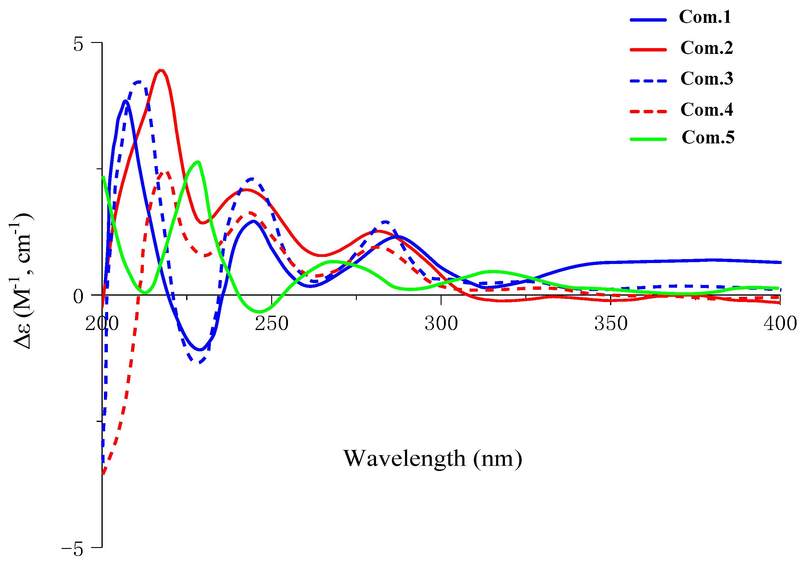
2. Results and Discussion

Compound 1 was obtained as white amorphous powder, and its molecular formula was confirmed asC36H44O16 by HRESIMS 755.2681 [M + Na]+ (calcd. for C36H44NaO16, 755.2527). The 1H NMR spectrum of 1 exhibited signals for eight aromatic protons, indicating the existence of one set of 1, 3, 5-trisubstituted benzene system(δH 7.03 (1H, brs, H-6), 6.87 (1H, brs, H-2), and 6.78 (1H, m, H-4)), one set of 1, 3, 4-trisubstituted aromatic proton signals (δH 6.97 (1H, d, J = 1.6 Hz, H-2″), 6.78 (1H, m, H-6″), and 6.73 (1H, m, H-5″)), and one set of the 1, 3, 4, 5-tetra-substituted benzene system (δH 6.89 (1H, d, J = 1.4 Hz, H-6′) and 6.75 (1H, m, H-2′)). Moreover, there was one anomeric proton signal at δH 4.25 (1H, d, J = 7.8 Hz, H-1‴), as well as three methoxyl proton signals at δH 3.79 (3H, brs, 3-OCH3), 3.76 (3H, brs, 3′-OCH3), and 3.76 (3H, brs, 5′-OCH3). The 13C NMR spectrum of 1 showed 30 carbon signals, of which δC 147.3, 119.2, 115.6, 111.0, 70.6, and 56.1 were overlapping signals. The 1H and 13C NMR (Table 1 and Table 2) indicated a furofuran lignan glycoside for 1, which shared high similarity with those of erythro-syringylglycerol-β-O-4′-(+)-isoeucommin A 4‴-O-β-D-glucopyranoside [9], except for the different substitution of aryl groups with the C-4/C-5″replaced by hydrogen and C-5/C-3″ replaced by hydroxyl. The location of the substitution of aryl groups of 1 wasfurther determined by HMBC spectroscopic analysis (Figure 2).The small coupling constants of JH-7, H-8 (4.1Hz)/JH-7′, H-8′ (4.0 Hz) and the chemical shift differences of ΔδH–9(0.4) and ΔδH–9′(0.4) (ΔδH–9 = δH–9aδH–9b andΔδH–9′ = δH–9′aδH–9′b) showed that the relative configuration was erythro [10,11,12]. In addition, the coupling constant of JH-7″, H-8″, (7.4 Hz) confirmed the relative configuration as threo [13,14,15]. According to the CD spectrum of 1 (Figure 2), the positive Cotton effect at 285 nm and negative Cotton effect at 228 nm indicated that the absolute configuration was determined as 7S, 7′S, 8R, 8′R, 7″R and 8″R [16,17,18,19].The coupling constant of the anomeric proton (δH4.89, d, J = 7.4, H-1‴) prompted the existence of β-configuration. The presence of D-glucose was confirmed by enzymatic hydrolysis and GC-MS analysis compared with authentic material. Thus, the structure of 1 was identified as (-) (7S, 7′S,7″R, 8R, 8′R, 8″R)-5, 3″-dihydroxy-3, 3′, 5′-trimethoxy-4″-O-β-D-glucopyranosyl-7, 9′: 7′, 9-diepoxy-4, 8″-oxy-8, 8′- sesquineolignan-7″, 9″-diol, named brasesquilignan A (Supplementary Materials).
Compound 2 was obtained as a white amorphous powder, and its molecular formula was confirmed asC42H54O21, determined by HRESIMS 895.8730 [M + H]+ (calcd. for C42H55O21895.8730). Careful comparison of the NMR data of 2 (Table 1 and Table 2) with those of 1 revealed that the 2 was a glycoside of 1 located at C-4‴, which was confirmed by HMBC (Figure 3) correlations between anomeric proton H-1″″(δH4.25, d, J = 7.7)and C-4‴ (δC 70.4). The two sugars were confirmed as β-D-configuration by the anomeric protons coupling constant (δH4.24, d, J = 7.7, H-1‴and δH4.25, d, J = 7.7, H-1″″), further enzymatic hydrolysis, and GC-MS analysis. The similar coupling constants (JH-7, H-8, JH-7′, H-8′, and JH-7″, H-8″)of 2 showed the relative configuration was consistent with 1. However, the CD spectrum of 2 (Figure 2) showed a positive Cotton effect at 228 nm and 285nm, illustrating that the absolute configuration of 2 was 7S, 7′S,7″S, 8R, 8′R, 8″S. Therefore, the structure of 2 was identified as (+) (7S, 7′S,7″S, 8R, 8′R, 8″S)-5, 3″-dihydroxy-3, 3′, 5′-trimethoxy-4″-O-β-D-glucopyranosyl-(1→4)-O-β-D-glucopyranosyl-7, 9′: 7′, 9- diepoxy-4, 8″-oxy-8, 8′-sesquineolignan-7″, 9″-diol, named brasesquilignan B.
Compound 3 was obtained as a white amorphous powder, and its molecular formula was confirmed asC36H44O16by HRESIMS 755.2473 [M + Na]+(calcd. for C36H44NaO16, 755.2527). The 1H NMR and 13C NMR data (Table 1 and Table 2) of 3 were quite similar to those of 1, except for the different substitution of aryl groups. The 1H NMR of 3 indicated the existence of a set of 1, 3, 4-trisubstituted aromatic proton signals, δH 6.88 (1H, d, J = 1.6 Hz, H-2), 6.75 (1H, d, J = 1.6 Hz, H-6), and 6.72 (1H, d, J = 8.1 Hz, H-5); a set of 1, 2, 4-trisubstituted aromatic proton signals, δH 7.06 (1H, d, J = 8.5 Hz, H-6′), 6.97 (1H, d, J = 1.9 Hz, H-3′), and 6.85 (1H, dd, J = 8.6, 1.8 Hz, H-5′); and a set of 1, 3, 4, 6-tetra-substituted aromatic proton signals, δH 6.87 (1H, brs, H-5″) and 6.86 (1H, brs, H-2″). The location of the functional groups and NMR data assignments of 3 were determined by HMBC and HSQC spectroscopic analysis (Figure 2). The sugar of 3 was confirmed as β-D-configuration by the anomeric proton coupling constant (δH4.89, d, J = 7.4, H-1‴), enzymatic hydrolysis, and GC-MS analysis. Comparing coupling constants (JH-7, H-8, JH-7′, H-8′, and JH-7″, H-8″) and CD spectra of 3 with 1 (Figure 2), the absolute configuration of 3 was determined as7S, 7′S,7″R, 8R, 8′R, 8″R. Therefore, the structure of 3 was identified as (-) (7S, 7′S, 7″R, 8R, 8′R, 8″R)-4, 2″- dihydroxy-3, 2′, 5″-trimethoxy-4″-O-β-D-glucopyranosyl-7, 9′: 7′, 9-diepoxy-4, 8″-oxy-8, 8′- sesquineolignan-7″, 9″-diol, named brasesquilignan C.
Compound 4 was obtained as a white amorphous powder, which had a molecular formula of C37H46O14 based on a protonated molecular ion peak at m/z 769.2638 [M + Na]+ (calcd. for C37H46NaO14 769.2684) in the HRESIMS data. The 1H NMR and 13C NMR data (Table 1 and Table 2) suggested that 4 and 1 are the same type of compound. However, the 1H NMR of compound 4 showed eight aromatic proton signals, including a set of 1, 4-disubstituted aromatic proton signals, δH 6.97 (1H, d, J = 1.5 Hz, H-5′), 6.82 (2H, brs, H-2′), 6.78 (1H, dd, J = 8.0, 1.6 Hz, H-3′), and 6.70 (3H, m, H-6′); a set of 1, 3, 5-trisubstituted aromatic proton signals, δH 6.82 (2H, brs, H-6) and 6.70 (3H, m, H-2/4); and a 1, 2, 3, 4, 6-penta-substituted aromatic proton signal, δH 6.74 (1H, brs, H-5″). In addition, compound 4 had one more methyl proton signal at δH 1.07 (3H, brs, 7-CH3). In the 13C NMR spectrum of 4, the carbon signals δC 67.5, 52.9, 79.6,and 22.0 were significantly different from 1, and δC 79.6 is a quaternary carbon, suggesting that there is a methyl on the furan ring of 4. Further, the correlation between δH6.82 (2H, brs) and δH6.78 (1H, dd, J =1.6, 8.0 Hz) in the 1H-1H COSY spectrum and the HMBC (Figure 3) correlations from H-2′ (δH6.82, 1H, brs) to C-7′ (δC 82.3), H-3′ (δH6.78, 1H, dd, J = 1.6, 8.0 Hz) to C-8″ (δC 87.3),and 7-CH3 (δH 1.07, 3H,) to C-8 (δC 67.5) indicated that the methyl was located at C-7 (Figure 3). The HMBC (Figure 3) correlations between H-1‴(δH4.23, 1H, d, J = 7.8 Hz) and C-4″ (δC132.4) indicated that the glycosyl was located at C-4″. The coupling constant of the anomeric protons (J = 7.8 Hz) prompted the existence of β-configuration. Moreover, the presence of D-glucose was confirmed by enzymatic hydrolysis and GC-MS analysiscompared with standard material. The coupling constants (JH-7′, H-8′ = 6.4 Hz and JH-7″, H-8″= 7.2 Hz) showed that the relative configurations were both threo. The CD spectrum of 4 (Figure 2) showed a positive Cotton effect at both 285nm and 240nm, illustrating that the absolute configuration was 7S, 7′S,7″S, 8R, 8′R, 8″S. Therefore, the structure of 4 was identified as (-) (7S, 7′S,7″S, 8R, 8′R, 8″S)-5, 3″-dihydroxy-3, 2″, 6″-trimethoxy-4″-O-β-D-glucopyranosyl-7, 9′: 7′, 9-diepoxy-7-methyl-4, 8″-oxy-8, 8′-sesquineolignan-7″, 9″-diol, named brasesquilignan D.
Compound 5 was obtained as a white amorphous powder. Its molecular formula was determined to be C31H36O9by HRESIMS at m/z 553.2416 [M + H]+ (calcd. for C31H37O9 553.2438). Compound 5 had a similar 1H and 13C spectrum (Table 1 and Table 2) to compound 1, except that there were no sugar-related signals and different substituent positions. The 1H NMR spectrum showed nine aromatic protons, including a group of 1, 3-disubstituted aromatic proton signals, δH 6.89 (1H, d, J = 1.4 Hz, H-2), 6.76 (1H, d, J = 1.6 Hz, H-6), 6.74 (1H, d, J = 1.6 Hz, H-4), and 6.59 (1H, m, H-5); one set of 1, 3, 4-trisubstituted aromatic proton signals, δH 6.65 (1H, d, J = 8.0 Hz, H-5′) and 6.60 (2H, brs, H-2′ / 6′); and a set of 1, 3, 4, 5-tetrasubstituted aromatic proton signals, δH 6.73 (1H, brs, H -2″) and 6.71 (1H, brs, H-6″). The 13C NMR spectrum showed18 aromatic carbon signals, δC 104.1~153.6; 4 methoxy carbon signals, δC 56.5 (5″-OCH3), 56.4 (4″-OCH3), 56.1 (3″-OCH3), and 56.0 (3′-OCH3); and 9 aliphatic carbon signals. In the HMBC spectrum, the correlation between H-5′ (δH 6.65, 1H, d, J = 8.0 Hz,) with C-8″ (δC 84.0) revealed that the connection of the two fragments is C-4′-O-C-8″. The location of the other functional groups and NMR data assignments of 5 were determined by HMBC and HSQC spectroscopic analysis (Figure 3). The relative configuration of 5 was determined by the chemical shift differences ofthe two pairs of diastereotopic methylene protons of H-9 and H-9′. The approximately equal values of ΔδH–9(0.4) and ΔδH–9′(0.4) suggested thatH-7/H-8 and H-7′/H-8′ wereTrans [10,12]. The CD spectrum of 5 (Figure 2) showed the positive Cotton effect at both 280 nm and 230 nm, illustrating that the absolute configuration was 7S, 7′S,7″S, 8R, 8′R, 8″S. Therefore, the structure of 5 was identified as (+)(7S, 7′S,7″S, 8R, 8′R, 8″S) 5, 3″-dihydroxy -3, 3′, 5′-trimethoxy-7, 9′: 7′, 9-diepoxy-4, 8″-oxy-8, 8′-sesquineolignan-9″-alcohol, named brasesquilignan E.
All compounds were examined for their anti-proliferative activity on A375, A549, MCF-7, MDA-MB-231,and SK-MEL-28 cells by the MTT assay using standard staurosporine (STS) as a positive control. All compounds exhibited weak inhibitory potency againstA549 and MCF-7cells (Table 3).

3. Materials and Methods

3.1. General Experimental Procedures

HRESIMS data were measured on an Agilent Technologies liquid chromatograph connected to Q-TOF mass spectra (Thermo Fisher, Massachusetts, MA, USA). NMR spectra were recorded on a Bruker AV-500 MHz spectrometer (Bruker, Karlsruhe, Germany) using DMSO-d6 as solvent and tetramethylsilane (TMS).GCMS was measured on GCMS-QP2010 Ultra (Shimadzu Corporation, Kyoto, Japan). Column chromatography (CC) was performed on HW-40C (TOYOPEARL TOSOH, Tokyo, Japan). Optical rotations were measured on an INESA SGW-3 polarimeter. Analytical and Semi-preparative HPLC was performed on an Agilent 1200 equipped with a DAD detector and a siligreen C18 column (5/10 μm, 250 × 10 mm, siligreen, Beijing, China). All solvents were of analytical grade.

3.2. Plant Material

The whole herbs of S.braunii Baker were collected from Hunan province in People’s Republic of China, in August 2015, and identified by Prof. Kangping Xu (Xiangya School of Pharmaceutical Sciences, Central South University). A specimen (no. 20150816) was deposited at the Xiangya School of Pharmaceutical Sciences, Central South University.

3.3. Extraction and Isolation

Whole herbs of S.braunii Baker (13.0 kg) were exhaustively extracted with 75% aqueous ethanol under reflux (2 times, 104 L × 2 h).After vacuum concentration, the extract was suspended in H2O and partitioned with petroleum ether, ethyl acetate. The water fraction (200 g) was fractionated by Macroporous resin HPD-100column chromatography, successively eluting with H2O, 30, 70, and 95% EtOH-H2O to obtain four fractions (FrA-D). FrC was performed on HW-40C (MeOH/H2O in gradient) to obtain seven fractions (FrC1-C7). FrC3 was subjected to gel column chromatography and semi-preparative liquid chromatography (3.0 mL/min, 280 nm, ACN-H2O, 3.0:7.0, V/V) repeatedly to obtain compounds1 (2.0 mg), 3 (2.6 mg), and 4 (1.0 mg). FrC2 was subjected to gel column chromatography and semi-preparative HPLC (3.0 mL/min, 280 nm, ACN-H2O, 3.0:7.0, V/V) repeatedly to obtain compound 2 (1.5 mg). FrC4was further purified by repeated chromatography (gel column and semi-preparative RP-HPLC) to yield compound 5 (2.9 mg).
Brasesquilignan A (1): white amorphous powder, [ α ] D 25 -30.2 (c 0.06, MeOH), HPLC-UV (ACN-H2O) λmaxnm: 205, 225, 280, HRESIMS, m/z 755.2681 [M + Na]+ (calcd. for C36H44NaO16755.2527),1H NMR (500 MHz in DMSO-d6) and 13C NMR (125 MHz in DMSO-d6). For data, see Table 1 and Table 2.
Brasesquilignan B (2): white amorphous powder, [ α ] D 25 -2.3 (c 0.05, MeOH), HPLC-UV (ACN-H2O) λmax nm: 205, 225, 280, HRESIMS, m/z: 895.8730 [M+H]+ (calcd. for C42H55O21895.8730),1H NMR (500 MHz in DMSO-d6) and 13C NMR (125 MHz in DMSO-d6).For data, see Table 1 and Table 2.
Brasesquilignan C (3): white amorphous powder, [ α ] D   25 -10.4 (c 0.2, MeOH), HPLC-UV (ACN-H2O) λmax nm: 205, 225, 280, HRESIMS, m/z: 755.2473 [M+Na]+(calcd. for C36H44NaO16755.2527),1H NMR (500 MHz in DMSO-d6) and 13C NMR (125 MHz in DMSO-d6).For data, see Table 1 and Table 2.
Brasesquilignan D (4): white amorphous powder, [ α ] D   25 -15.4 (c 0.05, MeOH), HPLC-UV (ACN-H2O) λmax nm: 205, 225, 280, HRESIMS, m/z: 769.2638 [M+Na]+ (calcd. for C37H46NaO14, 769.2684),1H NMR (500 MHz in DMSO-d6) and 13C NMR (125 MHz in DMSO-d6). For data, see Table 1 and Table 2.
Brasesquilignan E (5): white amorphous powder, [ α ] D   25 +28.2 (c 0.05, MeOH), HPLC-UV (ACN-H2O) λmax nm: 205, 225, 280, HRESIMS, m/z: 553.2416 [M+H]+ (calcd. for C31H37O9, 553.2438), 1H NMR (500 MHz in DMSO-d6) and 13C NMR (125 MHz in DMSO-d6). For data, see Table 1 and Table 2.

3.4. Enzymatic Hydrolysis

Compounds 15 (each 0.5 mg), cellulase (400 u/mg), and buffered saline solution (acetic acid/sodium acetate, PH = 6, 1 mL) were added in centrifuge tube (5 mL, sample: cellulose= 1:30), and incubated for 96 h in 37 °C. After extraction with CHCl3, the aqueous layer of reaction mixture was concentrated and dried to obtain the monosaccharide fraction. The residue was dissolved in pyridine (0.6 mL) with 1.0 mg of L-cysteine methyl ester hydrochloride and heated (60 °C, 1 h). Then, trimethylsilylimidazole (0.6 mL) was added and heated (60 °C, 1 h). The reaction mixture was analyzed by GC-MS under the following conditions: Column, Rtx-5MS (0.5 μm × 30.0 mm, 0.32 mm); front inlet 300 °C, column 150–300 °C at 15 °C/min. The monosaccharides of compounds were confirmed by comparing the retention times with those of standard sugar (subjected to the same method).

3.5. Anti-Proliferative Evaluation

All compounds were tested with positive control (staurosporine, STS). MDA-MB-231, SK-MEL-28, and A375 (Shanghai Institutes for Biological Sciences, Chinese Academy of Sciences) were cultured in DMEM/F12 (1:1) medium (Hyclone) supplemented with 10% FBS and 100 U/mL penicillin/streptomycin at 37 °C and 5% CO2. MCF-7 and A549 (Shanghai Institutes for Biological Sciences, Chinese Academy of Sciences) were cultured by DMEM/HIGH GLUCOSE medium (Hyclone) supplemented with 10% FBS and 100 U/mL penicillin/streptomycin at 37 °C and 5% CO2. All human cancer cells were plated in 96-well plates (25,000 cells/mL). Then, each well was supplemented at various concentrations of test compounds in triplicate for 24 h, and MTT (20 μL 5 mg/mL) was added. After incubation for 4 h, the medium was removed, and DMSO (150 μL) was added before further incubation (30 min, 37 °C). The OD (570 nm) value of each well was measuredwitha microplate reader MD5 (Molecular devices, San Jose, CA, USA). The half-inhibitory concentration (IC50) values were calculated by SPSS 25. software.

Supplementary Materials

The following supporting information can be downloaded at: https://www.mdpi.com/article/10.3390/molecules27196349/s1.

Author Contributions

Original draft preparation, F.C.; performing he experiments, F.C., J.W., Y.Z., H.Z., and H.L.; data analysis, Y.W., G.L., J.T., and Z.C.; review and editing, F.C., X.H., G.D., Y.L., and G.Z.; supervision, P.Z. and K.X. All authors have read and agreed to the published version of the manuscript.

Funding

This research was funded by the Construction Program of Hunan’s innovative Province (CN)-High-tech Industry Science and Technology Innovation Leading Project (no. 2020SK2002),the Natural Science Foundation of Hunan Province (no. 2021JJ40309), the Hunan Province Scientific Research Program of TCM (no. 202023 and 2021171), the Hainan Key Research Project (no. ZDYF2022SHFZ081 and ZDYF2020213), the Key Project of Changsha Science and Technology Plan (no. kq1801072 and kh2201060) and the Changsha Natural Science Foundation (no. Kq2014143 and Kq2014178).

Institutional Review Board Statement

Not applicable.

Informed Consent Statement

Not applicable.

Data Availability Statement

The data presented in this study are available in the Supplementary Materials.

Conflicts of Interest

The authors declare no conflict of interest.

Sample Availability

Samples of the compounds are available from the authors.

References

  1. Editorial Committee of the Flora of China. Flora of China; Science Press: Beijing, China, 2004; p. 108. [Google Scholar]
  2. Wang, Y.H.; Sun, Q.Y.; Yang, F.M.; Long, C.L.; Zhao, F.W.; Tang, G.H.; Niu, H.M.; Wang, H.; Huang, Q.Q.; Xu, J.J. Neolignans and caffeoyl derivatives from Selaginella moellendorffii. Helv. Chim. Acta 2010, 93, 2467–2477. [Google Scholar] [CrossRef]
  3. Cheng, F.; Xu, K.P.; Liu, L.F.; Yao, C.P.; Xu, P.S.; Zhou, G.; Li, D.; Li, X.M.; Chen, K.; Zou, Z.X.; et al. New neolignans from Selaginella picta and their protective effect on HT-22 cells. Fitoterapia 2018, 127, 69–73. [Google Scholar] [CrossRef] [PubMed]
  4. Long, H.P.; Li, F.S.; Xu, K.P.; Yang, Z.B.; Li, J.; Peng, J.; Tan, G.S. Bioactive compounds from Selaginella involven Spring that protect PC-12 cells. Chin. Chem. Lett. 2014, 25, 805–808. [Google Scholar] [CrossRef]
  5. Lin, R.C.; Skaltsounis, A.L.; Seguin, E.; Tillequin, F.; Koch, M. Phenolic constituents of Selaginella doederleinii. Planta Med. 1994, 60, 168–170. [Google Scholar] [CrossRef]
  6. Feng, W.S.; Chen, H.; Zheng, X.K.; Wang, Y.Z.; Gao, L.; Li, H.W. Two new secolignans from Selaginella sinensis (Desv.) Spring. J. Asian Nat. Prod. Res. 2009, 11, 658–662. [Google Scholar] [CrossRef] [PubMed]
  7. Zhuo, J.X.; Wang, Y.H.; Su, X.L.; Mei, R.Q.; Yang, J.; Kong, Y.; Long, C.L. Neolignans from Selaginella moellendorffii. Nat. Prod. Bioprospect. 2016, 6, 161–166. [Google Scholar] [CrossRef] [PubMed]
  8. Wu, B.; Wang, J. Phenolic compounds from Selaginella moellendorffii. Chem.Biodivers. 2011, 8, 1735–1747. [Google Scholar] [CrossRef] [PubMed]
  9. Katagiri, S.; Watanabe, Y.; Yaoita, Y.; Kikuchi, M.; Machida, K. Two new phenolic glycosides from Viburnum plicatum var. plicatum f. plicatum. Nat. Prod. Commun. 2011, 6, 1901–1904. [Google Scholar] [CrossRef] [PubMed]
  10. Shao, S.Y.; Yang, Y.N.; Feng, Z.M.; Jiang, J.S.; Zhang, P.C. An efficient method for determining the relative configuration of furofuran lignans by 1H NMR Spectroscopy. J. Nat. Prod. 2018, 81, 1023–1028. [Google Scholar] [CrossRef] [PubMed]
  11. Greger, H.; Hofer, O. New unsymmetrically substituted tetrahydrofurofuran lignans from artemisia absinthium: Assignment of the relative stereochemistry by lanthanide induced chemical shifts. Tetrahedron 1980, 36, 3551–3558. [Google Scholar] [CrossRef]
  12. Zhao, M.Z.; Shen, Y.; Xu, W.; Chen, Y.Z.; Jiang, B. A new lignan glycoside from Astragalus yunnanensis. J. Asian Nat. Prod. Res. 2019, 22, 1–7. [Google Scholar] [CrossRef] [PubMed]
  13. Park, S.Y.; Hong, S.S.; Han, X.H.; Hwang, J.S.; Lee, D.H.; Ro, J.S.; Hwang, B.Y. Lignans from Arctium lappa and their inhibition ofLPS-induced nitric oxide production. Chem. Pharm. Bull. 2007, 55, 150–152. [Google Scholar] [CrossRef] [PubMed]
  14. Kim, K.H.; Moon, E.; Kim, S.Y.; Lee, K.R. Lignans from the tuber-barks of Colocasis antiquorum var. esculenta and theirantimelanogenic activity. J. Agric. Food Chem. 2010, 58, 4779–4785. [Google Scholar] [CrossRef] [PubMed]
  15. Wang, W.; Ali, Z.; Li, X.C.; Khan, I.A. Neolignans from the leaves of Casearia sylvestris Swartz. Helv. Chim. Acta 2010, 93, 139–146. [Google Scholar] [CrossRef]
  16. Xiao, H.H.; Dai, Y.; Wong, M.S.; Yao, X.S. New lignans from the bioactive fraction of Sambucus williamsii Hance and proliferation activities on osteoblastic-like UMR106 cells. Fitoterapia 2014, 94, 29–35. [Google Scholar] [CrossRef] [PubMed]
  17. Liang, X.; Zhu, C.G.; Li, Y.R.; Tian, Y.; Lin, S.; Yuan, S.P.; Hu, J.F.; Hou, Q.; Chen, N.H.; Yang, Y.C.; et al. Lignans and neolignans from Sinocalamus affinis and their absolute configurations. J. Nat. Prod. 2011, 74, 1188–1200. [Google Scholar] [CrossRef]
  18. Muhit, M.A.; Umehara, K.; Mori-Yasumoto, K.; Noguchi, H. Furofuran Lignan Glucosides with Estrogen-Inhibitory Properties from the Bangladeshi Medicinal Plant Terminalia citrina. J. Nat. Prod. 2016, 79, 1298–1307. [Google Scholar] [CrossRef] [PubMed]
  19. Greca, M.D.; Molinaro, A.; Monaco, P.; Previtera, L. Lignans from Arum italicum. Phytochemistry 1994, 35, 777–779. [Google Scholar] [CrossRef]
Figure 1. Structures of compounds 15.
Molecules 27 06349 g001
Figure 2. The CD spectra of compounds 15 (MeOH).
Molecules 27 06349 g002
Figure 3. Key 1H-1H COSY and HMBC correlations of compounds 15.
Molecules 27 06349 g003
Table 1. 1H NMR (500 MHz) data of 15 in DMSO-d6.
Position12345
1
26.87, brs6.86, brs6.88, d (1.6)6.70, m6.89, d (1.4)
3
46.78, m6.78, m 6.70, m6.74, d (1.6)
5 6.72, d (8.1) 6.59, m
67.03, brs7.03, brs6.75, d (1.6)6.82, brs6.76, d (1.6)
74.63, d (4.1)4.62, d (4.0)4.63, t (4.2) 4.60, m
83.06, m3.05, m3.06, m3.47 overlapped3.03, m
9a4.13, m4.15, m4.13, m3.88, m4.13, m
9b3.73, m3.72, m3.73, m3.57, m3.73, m
1′
2′6.75, m6.73, m 6.82, brs6.60, brs
3′ 6.97, d (1.9)6.78, dd (1.6, 8.0)
4′
5′ 6.85, dd (8.6, 1.8)6.97, d (1.5)6.65, d (8.0)
6′6.89, d (1.4)6.88, brs7.06, d (8.5)6.70, m6.60, brs
7′4.61, d (4.1)4.64, d (4.0)4.63, t (4.2)4.63, d (6.4)4.60, m
8′3.06, m3.05, m3.06, m2.19, m3.03, m
9′a4.13, m4.15, m4.13, m3.68, m4.13, m
9′b3.73, m3.72, m3.73, m3.46 overlapped3.73, m
1″
2″6.97, d (1.6)6.96, brs 6.73, brs
3″ 6.87, brs
4″
5″6.73, m6.75, m 6.74, brs
6″6.78, m6.78, m6.86, brs 6.71, brs
7″3.61, m3.58, m3.43, m3.59, m4.11, m3.40 overlapped
8″5.47, d (7.4)5.45, d (7.4)5.51, d (6.5)5.46, d (7.2)4.12, m
9″a3.97, m3.72, m3.72, m3.95, m4.11, m
9″b3.72, m3.04, m3.63, m3.06, m3.40 overlapped
1‴4.25, d (7.8)4.24, d (7.7)4.89, d (7.4)4.23, d (7.8)
2‴3.00, m2.97, m3.25, m3.00, m
3‴3.08, m3.05, m3.25, m3.11, m
4‴3.06, m3.06, m3.14, m3.06, m
5‴3.18, m3.16, m3.26, m3.10, m
6‴3.65, m
3.42, overlapped
3.65, m
3.42, overlapped
3.64, m
3.43, m
3.66, m
3.43 overlapped
–OCH3(3-) 3.79, brs(3-) 3.78, brs(4-) 3.77, brs(3-) 3.76, brs(3″-) 3.71, brs
(3′-) 3.76, brs(3′-) 3.76, brs(2′-) 3.75, brs(2″-) 3.74, brs(3′-) 3.74, brs
(5′-) 3.76, brs(5′-) 3.75, brs(3″-) 3.80, brs(6″-) 3.74, brs(4″-) 3.75, brs
(5″-) 3.76, brs
7-CH3 1.07, d (6.7)
1″″ 4.25, d (7.7)
2″″ 2.98, m
3″″ 3.05, m
4″″ 3.05, m
5″″ 3.33, m
6″″ 3.50 overlapped
Table 2. 13C NMR (125 MHz) data of 15 in DMSO-d6.
Position12345
1135.2135.2132.6134.5145.1
2111.0110.9110.9113.2110.9
3144.0144.0148.0143.9146.4
4119.2119.2146.4115.5119.1
5147.3147.3115.6146.0122.1
6115.6115.6119.0117.6114.1
785.985.985.579.685.6
854.354.254.067.554.0
971.571.471.572.371.4
1′146.6146.4146.6135.1135.3
2′115.6115.7149.5110.4104.1
3′148.0148.0110.9119.1148.0
4′132.0132.1135.7146.1129.8
5′148.1148.1118.3110.9115.6
6′111.0110.9115.6118.7104.1
7′85.685.685.982.385.8
8′54.054.054.352.954.1
9′71.371.471.559.071.5
1″129.5129.5129.4129.2129.1
2″111.1110.9147.3147.8115.6
3″147.3147.3115.6146.8148.4
4″132.5132.6135.0132.4153.6
5″115.7115.6144.0115.6148.4
6″119.2119.1110.9148.0104.1
7″51.051.053.950.961.9
8″87.487.287.287.384.0
9″70.670.663.470.562.2
1‴103.3103.2100.5103.2
2‴73.973.973.673.9
3‴77.377.277.377.2
4‴70.670.470.170.5
5‴77.477.377.577.4
6‴61.561.561.061.5
-OCH3(3-) 56.2(3-) 56.2(4-) 56.2(3-) 56.1(3″-) 56.1
(3′-) 56.1(3′-) 56.1(2′-) 56.2(2″-) 56.0(3′-) 56.0
(5′-) 56.1(5′-) 56.1(3″-) 56.1(6″-) 56.0(4″-) 56.4
(5″-) 56.5
7-CH3 22.0
1″″ 103.9
2″″ 73.8
3″″ 77.2
4″″ 68.9
5″″ 76.3
6″″ 67.5
Table 3. Anti-proliferative activity ofall compoundsagainst five human cancer cell linesin vitro.
CompoundIC50 (μM)
SK-MEL-28A375A549MCF-7MDA-MB-231
1N/AN/AN/A93.69 ± 5.54N/A
248.30 ± 5.2935.12 ± 2.5427.82 ± 2.3822.09 ± 2.3944.02 ± 2.32
356.82 ± 4.8363.57 ± 1.4938.88 ± 2.8531.26 ± 1.1453.56 ± 1.44
4N/AN/AN/AN/AN/A
5N/AN/AN/AN/AN/A
STS0.04 ± 0.0080.06 ± 0.0060.4 ± 0.110.2 ± 0.040.03 ± 0.005
N/A: Not active; STS: Staurosporine used as a positive control.
Publisher’s Note: MDPI stays neutral with regard to jurisdictional claims in published maps and institutional affiliations.

