Article

The Steric Effect in Preparations of Vanadium(II)/(III) Dinitrogen Complexes of Triamidoamine Ligands Bearing Bulky Substituents

1
Faculty of Engineering, Aichi Institute of Technology, 1247 Yachigusa, Yakusa-cho, Toyota 470-0392, Japan
2
Graduate School of Engineering, Nagoya Institute of Technology, Gokiso-cho, Showa-ku, Nagoya 466-8555, Japan
*
Author to whom correspondence should be addressed.
Molecules 2022, 27(18), 5864; https://doi.org/10.3390/molecules27185864
Submission received: 24 August 2022 / Revised: 6 September 2022 / Accepted: 7 September 2022 / Published: 9 September 2022

Abstract

:
The reactions of newly designed lithiated triamidoamines Li3LR (R = iPr, Pen, and Cy2) with VCl3(THF)3 under N2 yielded dinitrogen–divanadium complexes with a μ-N2 between vanadium atoms [{V(LR)}2(μ-N2)] (R = iPr (1) and Pen (2)) for the former two, while not dinitrogen–divanadium complexes but a mononuclear vanadium complex with a vacant site, [V(LCy2)] (R = Cy2 (3)), were obtained for the third ligand. The V–NN2 and N–N distances were 1.7655(18) and 1.219(4) Å for 1 and 1.7935(14) and 1.226(3) Å for 2, respectively. The ν(14N–14N) stretching vibrations of 1 and 2, as measured using resonance Raman spectroscopy, were detected at 1436 and 1412 cm–1, respectively. Complex 3 reacted with potassium metal in the presence of 18-crown-6-ether under N2 to give a hetero-dinuclear vanadium complex with μ-N2 between vanadium and potassium, [VK(LCy2)(μ-N2)(18-crown-6)] (4). The N–N distance and ν(14N–14N) stretching for 4 were 1.152(3) Å and 1818 cm−1, respectively, suggesting that 4 is more activated than complexes 1 and 2. The complexes 1, 2, 3, and 4 reacted with HOTf and K[C10H8] to give NH3 and N2H4. The yields of NH3 and N2H4 (per V atom) were 47 and 11% for 1, 38 and 16% for 2, 77 and 7% for 3, and 80 and 5% for 4, respectively, and 3 and 4, which have a ligand LCy2, showed higher reactivity than 1 and 2.

1. Introduction

Dinitrogen activation using vanadium ions has been intensely investigated by bioinorganic chemists and coordination chemists in order to understand the role of vanadium ions as an important factor in vanadium nitrogenase enzymes [1,2,3,4,5,6,7,8,9]. Most of the dinitrogen–vanadium complexes studied are dinuclear complexes with a μ-N2 ligand in the end-on mode [10]. On the other hand, mononuclear vanadium–dinitrogen complexes are scarce [11,12,13,14]. It has also been previously reported that some dinitrogen–vanadium complexes produce ammonia and hydrazine [15,16,17,18,19,20,21,22]. Recently, dinitrogen–vanadium complexes, which are supported with anionic pyrrole-based PNP-type pincer and aryloxy ligands, have been successfully used for catalytic dinitrogen reduction by a group of Nishibayashi [20].
Triamidoamine (tris(2-amidoethyl)amine) ligands are very useful ligands that bind as multidentate ligands when forming metal complexes, creating binding sites for external ligands on the axis. Therefore, many complexes with the ligands have been investigated [14,21,22,23,24,25,26,27,28,29,30,31,32,33,34,35,36,37,38,39,40,41,42,43]. Schrock and co-workers reported the molybdenum complex with the triamidoamine ligand with a very bulky substituent group, HIPT (3,5-(2,4,6-iPr3C6H2)2C6H3) [37]. This molybdenum complex is the first example of catalytic ammonia production [38]. In this complex, the HIPT group functioned to inhibit dimerization of the molybdenum center and provide a pocket for binding an external ligand, such as N2, HNN, H2NN, NH, NH3, N, NO, THF, CO, S, Me3SiN, and so on [14,37,39,40]. Additionally, they also argued that the N2 ligand is protonated in the distal pathway by steric hindrance of the HIPT group [39]. On the other hand, dinuclear dinitrogen–molybdenum complexes with triamidoamine ligands have also been studied, but the substituents of these triamidoamine ligands are smaller than HIPT, such as TMS, aryl, and alkyl groups [21,22,41,42,43,44], and those with larger substituents have not been studied.
We have previously reported the syntheses and crystal structures of the dinitrogen–divanadium complexes bearing a triamidoamine ligand with a secondary C atom on the terminal N atom [{V(LR)}2(μ-N2)] (R = iBu, EtBu, iPr2Bn, Bn, MeBn) and studied the conversion of the bridging N2 ligand to ammonia using these complexes in the presence of proton sources (HOTf, [LutH](OTf)) and reductants (M+[C10H8]- M = K or Na) [21,22]. Furthermore, the crystal structure and protonation reaction of its Na+ adduct ([Na{V(LiBu)}2(μ-N2)]) were also studied (Figure 1) [22]. By introducing a secondary carbon atom on the terminal nitrogen atom of the trimidoamine ligand, these dinitrogen complexes can easily form dimer structures because the steric hindrance around the vanadium ion is smaller than those modified with bulky silyl or aryl groups. Therefore, we considered it would be possible to systematically investigate the structure and reactivity of mononuclear or dinuclear dinitrogen complexes using a series of triamidoamine ligands with different steric hindrances. The space-filling models of previously reported divanadium–dinitrogen complexes [{V(LR)}2(μ-N2)] (R = iBu and MeBn) are shown in Figure 1. In these complexes, there is no space around the secondary carbon atom (red), suggesting that a tertiary carbon atom was introduced on the terminal N atom to form a mononuclear vanadium–dinitrogen complex. If mononuclear and dinuclear complexes can be synthesized using triamidoamine ligands with the same backbone, meaningful comparisons can be made regarding their structures and reactivities in dinitrogen activation.
In this study, three vanadium complexes with a series of triamidoamine ligands bearing bulky substituents were synthesized under N2 atmosphere, and the characterizations, crystal structures, and protonation reactivities of the obtained complexes were investigated, compared, and discussed with those previously reported.

2. Results and Discussion

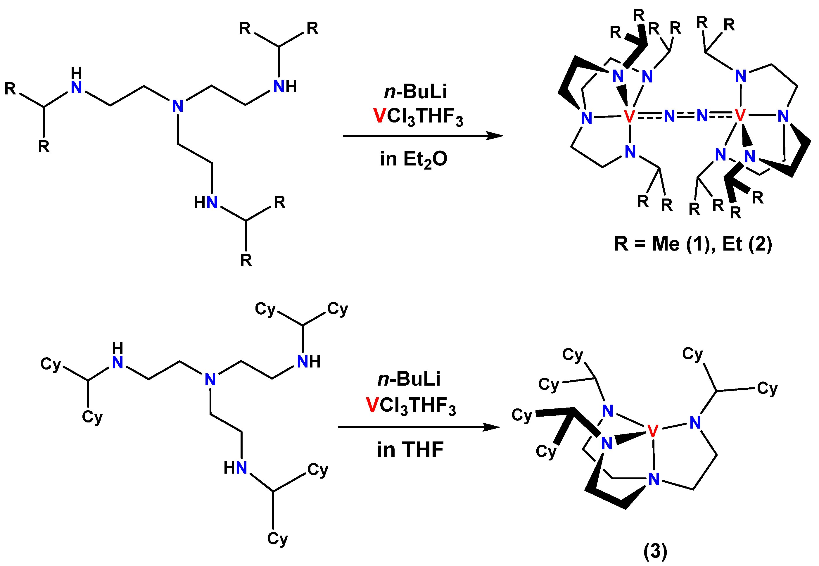
2.1. Syntheses of Ligands and Their Vanadium Complexes 1, 2, and 3

Three types of tren derivatives H3LR (R = iPr (tris(2-isopropylaminoethyl)amine, H3LiPr), Pen (tris(2-(3-pentylamino)ethyl)amine, H3LPen), and Cy2 (tris(2-dicyclohexylmethylaminoethyl)amine, H3LCy2)) were prepared using previously reported methods [21,22]. H3LiPr and H3LPen were obtained as light-yellow oil and H3LCy2 as colorless crystals, which were characterized using 1H NMR, 13C NMR, and IR spectroscopic methods. The H3LR was deprotonated and used as a triamidoamine ligand for the synthesis of dinitrogen complexes.
The reactions of lithiated triamidoamines Li3LR (R = iPr, Pen, and Cy2) with VCl3(THF)3 at room temperature under N2 produced dinitrogen–divanadium complexes [{V(LR)}2(μ-N2)] (R = iPr (1) and Pen (2)) for the former two. On the other hand, for the third ligand, a mononuclear vanadium complex [V(LCy2)] (3) was obtained instead of a dinitrogen–divanadium complex (Scheme 1). When all complex solutions were left at room temperature for several days, single crystals of complexes 1 and 3 were obtained as dark green crystals, and that of complex 2 was observed as dark purple crystals. These complexes were stable at low temperature under N2 atmosphere but decomposed under air atmosphere.

2.2. Crystal Structures of 1 and 2

The crystal structures of 1 and 2 are shown in Figure 2, and the crystal parameters are listed together in Table 1 and Table S1. Complexes 1 and 2 were expected to be mononuclear vanadium complexes because of their bulky substituents, but they turned out to be dinuclear vanadium complexes with bridging dinitrogen in the end-on mode. The coordination geometries around the vanadium centers in 1 and 2 are a nearly undistorted triangular bipyramid (τ = 1.0 and 1.0, respectively) (Figure 2), where 1 is a perfect trigonal bipyramidal geometry, 0 is a perfect square pyramidal geometry [45]. The N–N bond lengths for 1 and 2 are 1.219(4) and 1.226(3) Å, respectively, and that of complex 2 are slightly longer than those of 1 and the previously reported divanadium–dinitrogen complexes [21,22]. The bond lengths around the vanadium center in 2 (V–NN2 (1.7935(14) Å), V–Namido (1.9276(16), 1.9284(16), 1.9234(16) Å), and V–Namine (2.1854(16) Å)) are also more elongated than those of 1 (V–NN2 (1.7647(18) Å), V–Namido (1.896(2), 1.9123(14), 1.9123(14) Å), and V–Namine (2.173(2) Å)). This may also be due to the greater steric repulsion between the alkyl substituents on the N atoms of the triamidoamine ligand in 2 than in 1. Comparing the space-filling model of 2 with that of 1, it is obvious that the pentyl group in 2 surrounds the vanadium center more than the isopropyl group in 1 (Figure 3). Thus, it appears that all bond lengths are extended to maintain the dimeric structure, overcoming the pull away to the mononuclear vanadium complex. In fact, the V•••V distances and the distances of the vanadium ion from the plane decided by three Namido atoms are 4.7482(8) and 0.3055(12) Å for 1 and 4.8128(7) and 0.3495(10) Å for 2, respectively, making them longer for complex 2 than for complex 1. This finding appears to be due to the strong attraction of vanadium ions in complex 2 to the μ-N2 ligand.
The μ-N2 ligands in 1 and 2 are stabilized by hydrogen bonding interactions between the N2 ligand and the methine hydrogen atoms on the N atoms (CH•••NN2 = av. 2.488 Å for 1, av. 2.611 Å for 2). This interaction may also contribute to the formation of dimer structures.

2.3. Crystal Structure of 3

The crystal structure of complex 3 is shown in Figure 4, and the crystal parameters are listed in Table 2 and Table S1, respectively. Complex 3, unlike 1 and 2, was a mononuclear vanadium complex with no dinitrogen coordination. The geometry around the vanadium center is a trigonal pyramid, with a vacancy on the opposite side of Namine (N5) in [LCy2]3-. The V1–N3 (1.9433(12) Å) and V1–N4 bonds (1.9593(12) Å) are longer than the V1–N2 bond (1.9281(13) Å) because the steric repulsions between the cyclohexane rings of the dicyclohexylmethyl group on the N2 atom and those on the N3 and N4 atoms are weakened by the overhang of those on the N2 atom attached to the V center. The averaged V–Namido bond length in 3 (1.9436 Å) is longer than that of the related triamidoamine–vanadium complex, [V(tBuMe2SiN)3N] (V–Namido = 1.930(av.) Å) [26]. This fact indicates that the dicyclohexylmethyl group of 3 leads greater steric repulsion than the tBuMe2Si group.

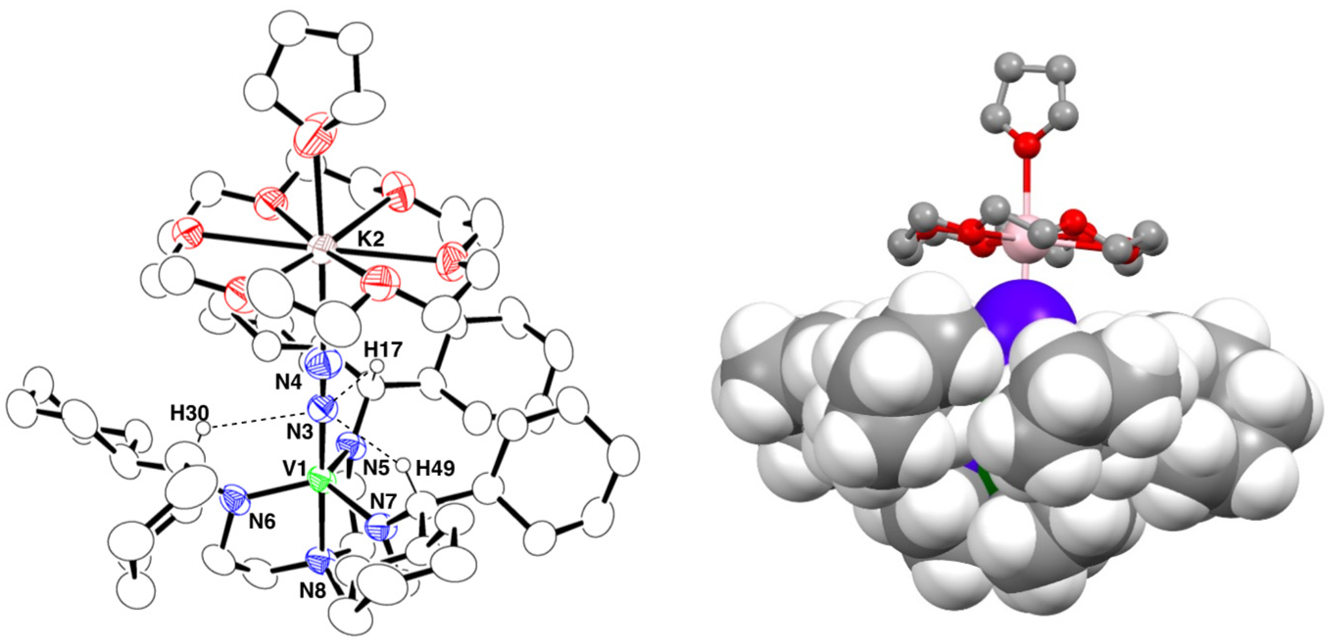
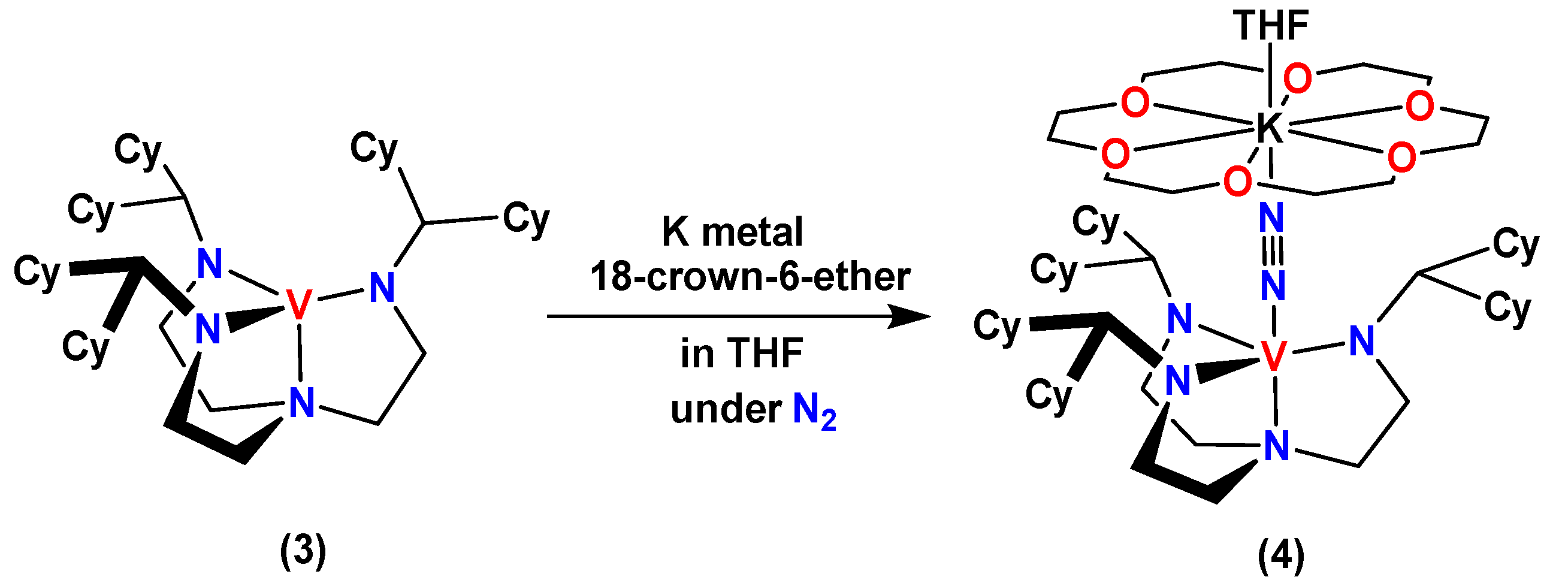
2.4. Synthesis and Crystal Structure of 4

The crystal structure of 3 shows that there is an open site on the vanadium ion. Therefore, we attempted to synthesize the N2 adduct by reacting 3 with potassium metal under N2 in the presence of 18-crown-6-ether (Scheme 2). Fortunately, a single crystal of the N2 adduct (4) was obtained as green crystals using recrystallization from THF/hexane at −35 °C. Complex 4 gradually decomposed at room temperature even under inert gas (N2 or Ar).
The crystal structure of 4 is shown in Figure 5, and the crystal parameters are listed in Table 2 and Table S1, respectively. Complex 4 had a bridging dinitrogen ligand between the vanadium(II) ion and potassium ion in the end-on mode, the coordination geometry around the vanadium center was trigonal bipyramidal, and the THF molecule coordinated to the potassium ion from the opposite side of the N2 ligand. The N–N and V–NN2 bond lengths of 4 are 1.152 (3) and 1.853 (3) Å, respectively. The average of three V–Namido bond lengths was found to be 1.960 Å, which is more elongated than those of 3 (1.9436 (av.) Å). This is thought to be due to the increased steric repulsion between the dicyclohexylmethyl groups as a result of the increased ionic radius of the vanadium ion due to the reduction in V(III) to V(II) and the incorporation of the N2 ligand in the axial position. The N2 ligand of 4 was also stabilized by hydrogen bonding interactions between the methine proton and the N3 atom of dinitrogen (CH•••N3(N2) = av. 2.596 Å). The distance between the mean plane decided by three Namido atoms and the vanadium ion (V1 atom) was 0.2888(14) Å, which is smaller than those of 1 and 2. The space-filling model of 4 is shown in Figure 5 (right). It is clear from this figure that the dicyclohexylmethyl group surrounds not only the α-nitrogen but also the β-nitrogen, suggesting that the dinitrogen–vanadium complex with [LCy2]3- ligand is too large to form a dimer structure.

2.5. Raman and Infrared Spectra of 1, 2, and 4

The ν(14N–14N) stretching vibrations of 1 and 2 were detected at 1436 and 1412 cm–1 using resonance Raman spectroscopic measurements, respectively (Figures S1 and S2). 15N-labeled 1 (1′) and 2 (2′) were both split by a Fermi doublet to show peaks at 1399, 1337 cm–1 and 1380, 1335 cm–1, respectively. The ν(V–14N) stretching vibrations of 1 and 2 were observed at 796 and 728 cm–1 from the IR spectra, respectively (Figures S3 and S4). The ν(14N–14N) and ν(V–14N) values of 1 are larger than those of 2, which are in good agreement with the N–N and V–NN2 bond lengths trends for 1 and 2. However, these ν(14N–14N) values are larger than those of previously reported divanadium–dinitrogen complexes (1394–1402 cm−1), even though 1 and 2 have longer N–N bonds than those of previously reported divanadium–dinitrogen complexes (1.200 (5)–1.226 (3) Å) [21,22]. Such inversions in bond lengths and vibrational spectra are sometimes observed in the activation chemistry of dinitrogen with transition metals [46].
In the IR spectral measurements, the ν(14N–14N) peaks for 4 were observed as two bands at 1830 and 1818 cm–1, which were shifted to 1768 and 1759 cm–1 when 15N2 was used in the place of 14N2 (Figure S5). The IR bands at 1830 and 1768 cm−1 were assigned as overtones of the 910 and 885 cm−1, respectively. When 15N-labeled 4 (4′) was dissolved in THF at room temperature and recrystallized under 14N2, the ν(14N–14N) stretching vibration was observed. This means that the N2 ligand of 4 is easily exchanged in THF because of the weak V–NN2 bond.

2.6. 1H-, 15N-, and 51V-NMR Spectra

1H NMR spectra of 1 and 2 exhibited sharp peaks in the diamagnetic region, as shown in Figures S6 and S7, respectively. The methine peak of 2 (4.77 ppm) was observed in a higher magnetic field region than that of 1 (5.27 ppm). 15N NMR spectra of 1 and 2 with 15N-labeled N2 were detected at 25.2 and 41.6 ppm (Figures S8 and S9), and 51V NMR spectra of 1 and 2 were observed at 211 and 47.6 ppm, respectively (Figures S10 and S11). The peaks of 2 were both observed in a lower magnetic field region than those in 1 and our previously reported divanadium–dinitrogen complexes (15N NMR: 25.2–33.4 ppm, 51V NMR: −240.2−143.8 ppm) [21,22]. These findings indicate that the electron densities on the N atoms of dinitrogen and the V atom in 2 are lower than those of complex 1 and the previously reported divanadium–dinitrogen complexes because the electron donation from Namido atoms to the V atom in 2 was smaller than those complexes. These facts correspond well with the result that the V–Namide bond length is the longest among the dinuclear vanadium–dinitrogen complexes with triamidoamine ligands reported so far [21,22], due to the large steric repulsion between the pentyl groups. On the other hand, the 1H NMR spectrum of 3 gave a broadened paramagnetic peak at 915 ppm in the lower magnetic field region (Figure S12) and that of 4 showed broad peaks at 10.2, 3.28, 0.32, 0.08, −0.28, −0.75, −0.95, −1.63, −15.2, and −30.5 ppm in the diamagnetic to higher magnetic field region (Figure S13). The spectrum of 4 includes not only 4 but also solvents (n-hexane, THF) and a free ligand, H3LCy2 (Figure S14). It is thought that the free ligand was probably produced by the decomposition of 4 due to its low thermal stability at room temperature. Unfortunately, we were unable to characterize the products containing vanadium(II) ions produced in the decomposition of 4. The effective magnetic moment (μeff) of 3 was 2.73μB at 298 K as determined by Evans’s NMR solution method [47,48], which indicates that the spin state of 3 is S = 1. Similarly, μeff of 4 was estimated to be 1.76μB at 298 K, indicating that the spin state of 4 is S = 1/2. These spin states correspond well with the structural findings that complex 3 is a mononuclear vanadium(III) complex with a vacant site, [V(LCy2)], and complex 4 is a hetero-dinuclear vanadium(II) complex with μ-N2 between vanadium and potassium, [VK(LCy2)(μ-N2)(18-crown-6)].

2.7. Protonation of 14 in the Presence of Reductants

Protonation of 1, 2, 3, and 4 was carried out with a reductant (M[C10H8] (M = Na+ or K+)) and an acid (HOTf) in THF at −78 °C. The previously reported protonation of the dinuclear vanadium–dinitrogen complexes produced only ammonia, while 1, 2, 3, and 4 produced both ammonia and hydrazine [21,22]. The yields of NH3 and N2H4 were estimated from the peak intensities of NH4+ using 1H NMR (7.04 ppm) and using the p-dimethylaminobenzaldehyde method using UV-vis spectra (458 nm), respectively (Figures S15 and S16). When using K[C10H8] as a reductant, the yields of NH3 and N2H4 were 47 and 11% for 1, 38 and 16% for 2, 78 and 7% for 3, and 80 and 5% for 4 (per V atom), respectively (Table 3 and Table S2). Their yields were higher than when using Na[C10H8]. Thus, the results suggest that the yield of products is dependent on the kind of alkali metal ions. The yield of NH3 for 2 was less than that of 1, and the yield of N2H4 was higher than that of 1. The result that the V–NN2 bond length is longer in 2 than in 1 suggests that mononuclear vanadium species are more likely to form from the vanadium–dinitrogen complex because the steric repulsion of substituents is greater in 2 than in 1. Therefore, the intermediates of the protonation reaction were estimated to be a hetero-dinuclear Na+-/K+-V(μ-N2) complex, as in 4. The yield with K[C10H8] as the reducing agent was higher than that with Na[C10H8], suggesting that potassium ions bound to N2 ligands are more readily exchanged to protons than sodium ions. We have recently found and reported similar behavior in the triamidoamine–chromium–dinitrogen system [49].
On the other hand, the yields of the protonation products of 3 and 4 were higher than those of 1 and 2, and the yield of N2H4 was less than one tenth of that of NH3. This result suggests that protonation of mononuclear dinitrogen complexes gives higher yields of protonated products than dinuclear dinitrogen complexes and that the dicyclohexylmethyl groups in 3 or 4 surround the Nα atom of the N2 ligand, protecting it from proton attack and preventing the formation of N2H4 (Figure 5 right).
These findings suggest that the alkyl substituents on the secondary carbon atoms adjacent to the terminal N atom of triamidoamine stabilize the dimer structure, while the substituents on the tertiary carbon atoms destabilize the dimer structure or stabilize the monomer structure. These results also suggest that hetero-dinuclear dinitrogen complexes consisting of vanadium(II), alkali metal ions, and bridging N2 ligands are formed as intermediates in the protonation reactions of vanadium complexes 1, 2, and 3. It was also found that hetero-dinuclear complexes produce both NH3 and N2H4, while the divanadium complexes produce only NH3.

3. Materials and Methods

3.1. General Procedures

All manipulations were carried out under an inert N2 or Ar atmosphere using either a vacuum and N2/Ar gas manifold, or a MBraun MB 150B-G glovebox (N2/Ar). Reagents and solvents employed were commercially available. All anhydrous solvents were purchased from Wako Ltd. and were bubbled with argon to degas. The ligand tris(2-isopropylaminoethyl)amine (H3LiPr) was synthesized according to the literature methods [28].

3.2. Physical Measurements

1H-, 13C-, 15N-, and 51V-NMR spectra were recorded on a JEOL JNM-ECA500, or a JNM-ECA600 FT NMR spectrometer operating at 500 MHz (1H), at 125.77 MHz (13C) in C6D6 or DMSO-d6 at 298 K. 1H and 13C chemical shifts were referenced using residual protonated solvent resonance (C6D6: 7.16 ppm (1H) and 128.06 ppm (13C), DMSO-d6: 2.50 ppm (1H)). 15N and 51V NMR chemical shifts were externally referenced using HCONH2 (−266.712 ppm (15N)) and VOCl3 (0.00 ppm (51V)). Electronic absorption spectra were recorded on a JASCO V-770 spectrophotometer. FT-IR spectra were taken on an Agilent Cary 630 FTIR spectrophotometer. A resonance Raman spectroscopy was performed using a JASCO NRS-3300 spectrometer with 532 nm-wavelength Nd:YAG excitation source.

3.3. X-ray Crystallography Procedures

The data for 1, 2, and 3 were measured on Rigaku R-AXIS RAPID diffractometer using multi-layer mirror monochromated Mo Kα (λ = 0.71073 Å) radiation. The data for 4 were measured on Rigaku R-AXIS RAPID II diffractometer using multi-layer mirror monochromated Cu Kα (λ = 1.54178 Å) radiation. Crystal data and experimental details are listed in Table S1. The calculations were performed with the Olex2 software package [50]. All structures were solved using ShelXT [51] structure solution program using the intrinsic phasing method, and the other atoms were found in subsequent Fourier maps. The structures were refined with ShelXL [52] using least squares minimization. All non-hydrogen atoms were anisotropically refined, unless otherwise stated. The hydrogen atoms were placed at their idealized positions, and the riding model was assumed, unless otherwise stated. CCDC-1970392 (1), 2167309 (2), 2167311 (3), 2167310 (4) contain the supplementary crystallographic data for this paper. These data can be obtained free of charge from The Cambridge Crystallographic Data Centre via www.ccdc.cam.ac.uk/data_request/cif, accessed on 22 August 2022.

3.4. Synthesis of Tris(2-(3-pentylamino)ethyl)amine (H3LPen)

Tris(2-aminoethyl)amine (14.6 g, 0.10 mol) was added to excess 3-pentanone (100 mL) and refluxed with a Dean–Stark trap overnight. The excess 3-pentanone was removed by evaporation. The mixture was cooled to 0 °C, and MeOH (50.0 mL) was added. Sodium borohydride (11.7 g, 0.31 mol) was added, and the mixture was stirred overnight at R.T. Sodium hydroxide (12.3 g, 0.31 mol) in water (100 mL) was added, and the aqueous layer was extracted 3 times by Et2O (100 mL). The organic layer was dried over Na2SO4, and the solvent was removed by evaporation. The mixture was distilled to give a light-yellow oil (yield 33.1 g, 93%). 1H NMR (500 MHz, C6D6, 298 K): δ (ppm) 0.950 (t, 18H, –CH3), 1.45 (quin, 12H, CH–CH2–CH3), 2.37 (quin, 3H, CH–(CH2CH3)2), 2.50 (t, 6H, NH–CH2–CH2), 2.62 (t, 6H, NH–CH2–CH2). 13C{1H} NMR (125.77 MHz, C6D6, 298 K): δ (ppm) 10.18, 26.53, 45.34, 55.13, 60.64.

3.5. Synthesis of Tris(2-dicyclohexylmethylaminoethyl)amine (H3LCy2)

Tris(2-aminoethyl)amine (14.6 g, 0.10 mol) was added to dicyclohexyl ketone (77.6 g, 0.40 mol) in toluene (100 mL) and refluxed with a Dean–Stark trap for 2 days. The excess dicyclohexyl ketone was removed in vacuo. The mixture was cooled to 0 °C, and MeOH (50.0 mL) was added. Sodium borohydride (11.7 g, 0.31 mol) was added, and the mixture was stirred overnight at R.T. Sodium hydroxide (12.3 g, 0.31 mol) in water (100 mL) was added, and the aqueous layer was extracted 3 times by Et2O (100 mL). The organic layer was dried over Na2SO4, and the solvent was removed by evaporation. The mixture MeOH (100 mL) and hexane (10 mL) was added and stored at −30 °C. The white solid was filtered and washed using solvent (2-propanol:hexane = 10:1). The white solid was resolved using CHCl3. Recrystallization was done by standing still in R.T. and white crystals were obtained (yield 43.5 g, 64%). 1H NMR (500 MHz, C6D6, 298 K): δ (ppm) 1.12–2.02 (m, 69H, Cy2–CH–), 2.62 (t, 6H, NH–CH2–CH2), 2.82 (t, 6H, NH–CH2–CH2). 13C{1H} NMR (125.77 MHz, C6D6, 298 K): δ (ppm) 27.23, 27.32, 27.38, 29.11, 31.81, 41.42, 50.68, 56.46, 68.90.

3.6. Synthesis of [{V(LiPr)}2(μ-14N2)] (1)

A 20 mL Schlenk flask was charged with H3LiPr (1.0 g, 3.7 mmol) and diethyl ether (4.00 mL) and cooled to −78 °C under nitrogen (14N2). n-butyllithium (4.2 mL, 11.0 mmol, 2.6 M in hexane) was added using a syringe. After 15 min, the reaction mixture was slowly warmed to 25 °C and was stirred for 1 h at room temperature. The reaction mixture was then cooled to −78 °C and VCl3THF3 (1.4 g, 3.7 mmol) was added via cannula to another Schlenk flask. The reaction mixture was again slowly warmed to 25 °C and was stirred overnight. The solvent was removed in vacuo, and the residue was extracted with diethyl ether (6 mL). The extract was filtered through Celite. The diethyl ether extract was transferred to a 20 mL Schlenk flask and stored in a fridge at −35 °C. [{VLiPr}2(μ-N2)] was obtained as dark green crystals (yield 0.57 g, 46%). 1H NMR (500 MHz, C6D6, 298 K): δ (ppm) 1.44 (d, 36H, –CH3), 2.40 (t, 12H, NH–CH2–CH2), 3.29 (t, 12H, N–CH2–CH2), 5.27 (sep, 6H, CH–(CH3)2), 13C{1H} NMR (125.77 MHz, C6D6, 298 K): δ (ppm) 23.46, 48.22, 53.11, 60.84, 51V NMR (131.56 MHz, C6D6, 298 K): δ (ppm) –211.1. FT IR (ATR, cm–1): 2957, 2916, 2892, 2851, 2788, 2652, 1457, 1438, 1380, 1364, 1347, 1332, 1328, 1282, 1259, 1239, 1202, 1155, 1129, 1099, 1077, 1056, 1028, 1002, 967, 939, 905, 862, 840, 818, 796, 789, 747, 618, 592, 572, 549, 495, 469.

3.7. [{V(LiPr)}2(μ-15N2)] (1)

Complex 1′ was synthesized using the same method as complex 1 using 15N2 instead of 14N2. Complex 1′ was obtained as dark green crystals (yield 0.29 g, 24%). 15N-NMR (60.815 MHz, C6D6, 298 K): δ (ppm) 25.22. FT-IR (KBr, cm–1): 779 (ν(V–15N)).

3.8. Synthesis of [{V(LPen)}2(μ-14N2)] (2)

Complex 2 was synthesized using the same method as complex 1 using H3LPen instead of H3LiPr. Complex 2 was obtained as purple crystals (yield 1.1 g, 89%). 1H NMR (500 MHz, C6D6, 298 K): δ (ppm) 1.15 (t, 36H, –CH3), 1.74 (quin, 24H, CH–CH2–CH3), 2.37 (t, 12H, N–CH2–CH2), 3.20 (t, 6H, N–CH2–CH2), 4.77 (quin, 6H, CH–(CH2CH3)2). 13C{1H} NMR (125.77 MHz, C6D6, 298 K): δ (ppm) 12.51, 27.71, 48.57, 53.05, 71.52. 51V NMR (131.56 MHz, C6D6, 298 K): δ (ppm) –47.55. FT IR (ATR, cm–1): 2955, 2920, 2892, 2870, 2845, 2788, 1444, 1371, 1341, 1330, 1280, 1259, 1237, 1205, 1144, 1127, 1107, 1047, 1026, 993, 952, 905, 898, 866, 857, 834, 827, 812, 758, 728, 663, 642, 590, 559, 549, 572, 549, 527, 519, 486.

3.9. Synthesis of [{V(LPen)}2(μ-15N2)] (2′)

Complex 2′ was synthesized using the same method as complex 2 using 15N2 instead of 14N2. Complex 2′ was obtained as purple crystals (yield 0.49 g, 41%). 15N NMR (60.815 MHz, C6D6, 298 K): δ (ppm) 41.59. FT IR (ATR, cm–1): 715 (ν(V–15N)).

3.10. Synthesis of [V(LCy2)] (3)

The complex 3 was synthesized using the same method as complex 1 using H3LCy2 in place of H3LiPr. THF for reaction solvent and hexane for recrystallization were used instead of diethyl ether (yield 0.79 g, 74%). FT IR (ATR, cm–1): 2955, 2920, 2892, 2870, 2845, 2788, 1444, 1371, 1341, 1330, 1280, 1259, 1237, 1205, 1144, 1127, 1107, 1047, 1026, 993, 952, 905, 898, 866, 857, 834, 827, 812, 758, 728, 663, 642, 590, 559, 549, 572, 549, 527, 519, 486.

3.11. Synthesis of [VK(LCy2)(μ-14N2)(18-crown-6)] (4)

A 15 mL vial was inserted with complex 3 (0.20 g, 0.27 mmol) and THF (6 mL) under nitrogen gas. A quantity of K metals (0.11 g, 2.7 mmol, 10 eq.) was added to the solution, and the solution was stirred at R.T. overnight. The mixture was filtered through Celite, and the filtrate was added 18-crown-6-ether (79.8 mg, 0.30 mmol) in THF (1 mL). The mixture was stirred at R.T. for 1 min. Hexane (6 mL) was slowly added to the reaction mixture. Recrystallization of the complex 4 at −35 °C yielded a green crystal (0.13 mg, 39%). FT IR (ATR, cm–1): 1830, 1818 (ν(14N–14N)).

3.12. Synthesis of [VK(LCy2)(μ-15N2)(18-crown-6)] (4′)

Complex 4′ was synthesized using the same method as complex 4 using 15N2 instead of 14N2. Complex 4′ was obtained as green crystals (60.2 mg, 18%). FT IR (ATR, cm–1): 1768, 1759 (ν(15N–15N)).

3.13. Protonation of 1, 2, 3, and 4 with Reductant and Proton Source

A 6 mL THF solution of the reductant (M[C10H8] (M = Na, K)), freshly prepared from alkali metal (1.1 mmol, 80 eq.) and naphthalene (0.15 mg, 1.1 mmol, 80 eq.) in a thick-walled glass bomb, was added to a 5 mL THF solution of 1 (10.0 mg, 1.5 × 10⁻² mmol) at –78 °C, and the mixture was stirred for 1 h under N2. The reaction mixture turned from deep green to greenish brown. HOTf (0.17 g, 1.1 mmol) was added to the vigorously stirred reaction mixture of 1, and the resultant solution was slowly warmed to 25 °C. After the solution was attired for 1 h at room temperature, the solvents were removed under reduced pressure to give a white solid containing ammonium and hydrazinium salts. The residue in the Schlenk tube was washed using diethyl ether and then, extracted using H2O (10 mL), and solvents of an aliquot (5 mL) of this solution were evaporated. The residue was analyzed using 1H NMR methods (for NH3). The other aliquot reacted with p-dimethylaminobenzaldehyde (for N2H4).

3.14. NH3 Quantification Procedure

Quantification of ammonium salts was estimated using 1H NMR spectroscopy. The quantification of NH4+ was carried out using the method reported by Ashley and co-workers [53]. 14NH4+ was integrated relative to the vinylic protons of 2,5-dimethylfuran, contained within a DMSO-d6 capillary insert (δ:5.83, s, 2H), which was calibrated using a standard 5.2 × 10⁻² M solution of NH4+ in DMSO-d6 (Figure S15 and Table S2).

3.15. N2H4 Quantification Procedure

The quantification of N2H5+ was carried out using the method reported by Ashley and co-workers [53]. The aliquots were analyzed for N2H4 via UV-vis spectroscopy using a standard spectrophotometric method, which reacted with an acidic p-dimethylaminobenzaldehyde solution and generated a yellow azine dye with a characteristic electronic absorption feature at 458 nm. The N2H4 in aliquot was quantified by comparison to the calibration curve (Figure S16).

4. Conclusions

In this study, we prepared vanadium(III) complexes with newly designed triamidoamine ligands ([LR]3- R = iPr, Pen, and Cy2) (1, 2, and 3, respectively) under N2, which have tertiary C atoms adjacent to the terminal N atoms of the ligand. The X-ray structure analyses for 1, 2, and 3 revealed that complexes 1 and 2 were divanadium–dinitrogen complexes with a μ-N2 ligand between two vanadium ions in the end-on mode ([{V(LR)}2(N2)] R = iPr and Pen), while complex 3 was a mononuclear vanadium complex without N2 coordination, [V(LCy2)]. These results indicate that complexes 1 and 2 with isopropyl and pentyl groups, respectively, can form dimeric structures, but due to steric repulsion between the dicyclohexylmethyl groups of the ligands, such a dimeric structure was not formed in complex 3. The evidence of this is that all V–Namido bond lengths around the vanadium(III) ion are the longest among complex 2 and the divanadium(III)–dinitrogen complexes with triamidoamine ligands reported so far. Furthermore, complex 3, which has an even larger dicyclohexylmethyl group, did not form a dimer structure due to steric repulsion of the dicyclohexylmethyl group and formed a mononuclear complex. Reduction in complex 3 with potassium metal in the presence of 18-crown-6-ether produced the thermally unstable vanadium(II)–dinitrogen complex (4). The crystal structure of 4 was revealed to be a hetero-dinuclear complex with a bridging N2 ligand between V(II) and the K+ ion surrounded with 18-crown-6-ether.
Reactions of complexes 1, 2, 3, and 4 with M[C10H8] (M = Na+ or K+) and HOTf yielded NH3 and N2H4, which are significantly different from our previous results using divanadium–dinitrogen complexes with similar triamidoamine ligands that we reported as releasing only ammonia [21,22]. This may be explained due to the following facts. In the case of the previously reported divanadium–dinitrogen complexes with a secondary C atom on the terminal N atom of the triamidoamine ligand, the addition of alkali metal ions resulted in the formation of a divanadium–dinitrogen complex as a reaction intermediate. On the other hand, in the cases of 1, 2, 3, and 4 with tertiary C atoms instead of secondary C atoms, the addition of alkali metal ions resulted in the formation of a hetero-dinuclear nitrogen complex. Surrounding the Nα atom with large substituents increases the yield of NH3 and conversely, decreases the yield of N2H4. These results indicate that the large steric repulsion around the Nα atom prevents the protonation reaction into the Nα atom.
Here, we have found that changing the carbon atom adjacent to the terminal N atom of a triamidoamine ligand from a secondary carbon to a tertiary carbon can have a significant effect on structure and reactivity. These results provide an important finding to dinitrogen fixation catalysis and the model study of nitrogenase.

Supplementary Materials

The following supporting information can be downloaded at: https://www.mdpi.com/article/10.3390/molecules27185864/s1, Figure S1: Raman spectra of 1; Figure S2: Raman spectra of 2; Figure S3: IR spectra of 1; Figure S4: IR spectra of 2; Figure S5: IR spectra of 4; Figure S6: 1H NMR spectrum of 1; Figure S7: 1H NMR spectrum of 2; Figure S8: 15N NMR spectrum of 1′; Figure S9: 15N NMR spectrum of 2′; Figure S10: 51V NMR spectrum of 1; Figure S11: 51V NMR spectrum of 2; Figure S12: 1H NMR spectra of 3; Figure S13: 1H NMR spectrum of 4; Figure S14: 1H NMR spectra of H3LCy2 and 4; Figure S15: 1H NMR spectrum of 14NH4+; Figure S16: Calibration curves for hydrazine quantification; Table S1: Experimental Data for X-ray Diffraction studies on Crystalline Complexes 1, 2, 3, and 4; Table S2: Yields of NH3 and N2H4. The CCDC deposition numbers 1970392 (1), 2167309 (2), 2167311 (3), and 2167310 (4) contain the supplementary crystallographic data for this paper, which can obtained free of charge via emailing data [email protected], or by contacting The Cambridge Crystallographic Data Centre at 12 Union Road, Cambridge CB2 1EZ, UK; Fax: +44-1223-336033.

Author Contributions

Conceptualization, Y.K. (Yoshiaki Kokubo) and Y.K. (Yuji Kajita); methodology, Y.K. (Yoshiaki Kokubo) and Y.K. (Yuji Kajita); validation, Y.K. (Yoshiaki Kokubo) and Y.K. (Yuji Kajita); formal analysis, Y.K. (Yoshiaki Kokubo), Y.K. (Yuji Kajita), I.I., K.N., W.H. and T.O.; investigation, Y.K. (Yoshiaki Kokubo) and Y.K. (Yuji Kajita); data curation, Y.K. (Yoshiaki Kokubo); writing—original draft preparation, Y.K. (Yoshiaki Kokubo) and Y.K. (Yuji Kajita); writing—review and editing, Y.K. (Yuji Kajita), S.K. and H.M.; supervision, Y.K. (Yuji Kajita), S.K. and H.M.; project administration, Y.K. (Yuji Kajita); funding acquisition, Y.K. (Yuji Kajita) and H.M. All authors have read and agreed to the published version of the manuscript.

Funding

This research was funded by the Japan Society for the Promotion of Science (JSPS), grant number (B) 20H027520 and (C) 20K05545.

Institutional Review Board Statement

Not applicable.

Informed Consent Statement

Not applicable.

Data Availability Statement

The data presented in this study are contained within this article and are supported by the data in the Supplementary Materials.

Acknowledgments

We thank Haruyo Nagao (Institute for Molecular Science) for 15N NMR spectroscopy measurement. This work was supported by the Nanotechnology Platform Program (Institute for Molecular Science) (JPMXP09S21MS1012) and the Advanced Research Infrastructure for Materials and Nanotechnology (JPMXP1222MS1018) of the Ministry of Education, Culture, Sports, Science and Technology (MEXT), Japan. We also acknowledge the Japan Society for the Promotion of Science (JSPS) for a grant in aid of scientific research (B and C) (20H027520, 20K05545).

Conflicts of Interest

The authors declare no conflict of interest.

Sample Availability

Samples are not available from the corresponding authors.

References

  1. Hales, B.J.; Case, E.E.; Morningstar, J.E.; Dzeda, M.F.; Mauterer, L.A. Isolation of a New Vanadium-Containing Nitrogenase from Azotobacter vinelandii. Biochemistry 1986, 25, 7251–7255. [Google Scholar] [CrossRef] [PubMed]
  2. Arber, J.M.; Dobson, B.R.; Eady, R.R.; Stevens, P.; Hasnain, S.S.; Garner, C.D.; Smith, B.E. Vanadium K-edge X-ray absorption spectrum of the VFe protein of the vanadium nitrogenase of Azotobacter chroococcum. Nature 1986, 325, 372–374. [Google Scholar] [CrossRef]
  3. Eady, R.R. Structure-Function Relationships of Alternative Nitrogenases. Chem. Rev. 1996, 96, 3013–3030. [Google Scholar] [CrossRef]
  4. Rehder, D. The coordination chemistry of vanadium as related to its biological functions. Coord. Chem. Rev. 1999, 182, 297–322. [Google Scholar] [CrossRef]
  5. Smith, B.E. Structure, function, and biosynthesis of the metallosulfur clusters in nitrogenases. Adv. Inorg. Chem. 1999, 47, 159–218. [Google Scholar]
  6. Eady, R.R. Current status of structure function relationships of vanadium nitrogenase. Coord. Chem. Rev. 2003, 237, 23–30. [Google Scholar] [CrossRef]
  7. Janas, Z.; Sobota, P. Aryloxo and thiolato vanadium complexes as chemical models of the active site of vanadium nitrogenase. Coord. Chem. Rev. 2005, 249, 2144–2155. [Google Scholar] [CrossRef]
  8. Sippel, D.; Einsle, O. The structure of vanadium nitrogenase reveals an unusual bridging ligand. Nat. Chem. Biol. 2017, 13, 956–960. [Google Scholar] [CrossRef]
  9. Sippel, D.; Rohde, M.; Netzer, J.; Trncik, C.; Gies, J.; Grunau, K.; Djurdjevic, I.; Decamps, L.; Einsle, O. A bound reaction intermediate sheds light on the mechanism of nitrogenase. Science 2018, 359, 1484–1489. [Google Scholar] [CrossRef]
  10. Duman, L.M.; Sita, L.R. Group 5 Transition Metal-Dinitrogen Complexes. In Transition Metal-Dinitrogen Complexes, 1st ed.; Nishibayashi, Y., Ed.; Wiley-VCH Verlag GmbH & Co. KGaA: Weinheim, Germany, 2019; pp. 159–220. [Google Scholar]
  11. Rehder, D.; Woitha, C.; Priebsch, W.; Gailus, H. Trans-[Na(thf)][V(N2)2(Ph2PCH2CH2PPh2)2]: Structural characterization of a dinitrogenvanadium complex, a functional model for vanadiumnitrogenase. J. Chem. Soc. Chem. Commun. 1992, 28, 364–365. [Google Scholar] [CrossRef]
  12. Woitha, C.; Rehder, D. Vanadium(-I) Dinitrogen Complexes with N2 Coordinated End-on: Functional Models for the “Alternative Nitrogenase”. Angew. Chem. Int. Ed. Engl. 1990, 29, 1438–1440. [Google Scholar] [CrossRef]
  13. Gailus, H.; Woitha, C.; Rehder, D. Dinitrogenvanadates(-I): Synthesis, reactions and conditions for their stability. J. Chem. Soc. Dalton Trans. 1994, 23, 3471–3477. [Google Scholar] [CrossRef]
  14. Smythe, N.C.; Schrock, R.R.; Müller, P.; Weare, W.W. Synthesis of [(HIPTNCH2CH2)3N]V Compounds (HIPT) 3,5-(2,4,6-i-Pr3C6H2)2C6H3) and an Evaluation of Vanadium for the Reduction of Dinitrogen to Ammonia. Inorg. Chem. 2006, 45, 9197–9205. [Google Scholar] [CrossRef] [PubMed]
  15. Nishibayashi, Y.; Tanabe, Y. Recent advances in nitrogen fixation upon vanadium complexes. Coord. Chem. Rev. 2019, 381, 135–150. [Google Scholar]
  16. Ferguson, R.; Solari, E.; Floriani, C.; Chiesi-Villa, A.; Rizzoli, C. Fixation and Reduction of Dinitrogen by Vanadium(II) and Vanadium(III): Synthesis and Structure of Dinitrogenmesitylvanadium Complexes. Angew. Chem. Int. Ed. Engl. 1993, 32, 396–397. [Google Scholar] [CrossRef]
  17. Ferguson, R.; Solari, E.; Floriani, C.; Osella, D.; Ravera, M.; Re, N.; Chiesi-Villa, A.; Rizzoli, C. Stepwise reduction of dinitrogen occurring on a divanadium model compound: A synthetic, structural, magnetic, electrochemical, and theoretical investigation on the [V=N=N=N](n+) [n = 4-6] based complexes. J. Am. Chem. Soc. 1997, 119, 10104–10115. [Google Scholar] [CrossRef]
  18. Liu, G.; Liang, X.; Meetsma, A.; Hessen, B. Synthesis and structure of an aminoethyl-functionalized cyclopentadienyl vanadium(i) dinitrogen complex. Dalton Trans. 2010, 39, 7891–7893. [Google Scholar] [CrossRef]
  19. Vidyaratne, I.; Crewdson, P.; Lefebvre, E.; Gambarotta, S. Dinitrogen Coordination and Cleavage Promoted by a Vanadium Complex of a σ,π,σ-Donor Ligand. Inorg. Chem. 2007, 46, 8836–8842. [Google Scholar] [CrossRef]
  20. Sekiguchi, Y.; Arashiba, K.; Tanaka, H.; Eizawa, A.; Nakajima, K.; Yoshizawa, K.; Nishibayashi, Y. Catalytic Reduction of Molecular Dinitrogen to Ammonia and Hydrazine Using Vanadium Complexes. Angew. Chem. Int. Ed. 2018, 57, 9064–9068. [Google Scholar] [CrossRef]
  21. Kokubo, Y.; Yamamoto, C.; Tsuzuki, K.; Nagai, T.; Katayama, A.; Ohta, T.; Ogura, T.; Wasada-Tsutsui, Y.; Kugimiya, S.; Masuda, H. Dinitrogen Fixation by Vanadium Complexes with a Triamidoamine Ligand. Inorg. Chem. 2018, 57, 11884–11894. [Google Scholar] [CrossRef]
  22. Kokubo, Y.; Wasada-Tsutsui, Y.; Yomura, S.; Yanagisawa, S.; Kubo, M.; Kugimiya, S.; Kajita, Y.; Ozawa, T.; Masuda, H. Syntheses, Characterizations, and Crystal Structures of Dinitrogen-Divanadium Complexes Bearing Triamidoamine Ligands. Eur. J. Inorg. Chem. 2020, 2020, 1456–1464. [Google Scholar] [CrossRef]
  23. Schrock, R.R. Transition Metal Complexes That Contain a Triamidoamine Ligand. Acc. Chem. Res. 1997, 30, 9–16. [Google Scholar] [CrossRef]
  24. Cummins, C.C.; Lee, J.; Schrock, R.R.; Davis, W.M. Trigonalmonopyramidal M(III) complexes of the type M(N3N) [M = titanium, vanadium, chromium, manganese, iron; N3N = [(tert-BuMe2Si)NCH2CH2]3N]. Angew. Chem. Int. Ed. Engl. 1992, 31, 1501–1503. [Google Scholar] [CrossRef]
  25. Cummins, C.C.; Lee, J.; Schrock, R.R. Phosphinidenetantalum(V) complexes [(N3N)Ta = PR] as phospha-Wittig reagents (R = Ph, Cy, tert-Bu; N3N = (Me3SiNCH2CH2)3N). Angew. Chem. Int. Ed. Engl. 1993, 32, 756–759. [Google Scholar] [CrossRef]
  26. Cummins, C.C.; Schrock, R.R. Synthesis of an iron(IV) cyanide complex that contains the triamidoamine ligand [(tert-BuMe2SiNCH2CH2)3N]3. Inorg. Chem. 1994, 33, 395–396. [Google Scholar] [CrossRef]
  27. Cummins, C.C.; Schrock, R.R.; Davis, W.M. Synthesis of Terminal Vanadium(V) Imido, Oxo, Sulfido, Selenido, and Tellurido Complexes by Imido Group or Chalcogen Atom Transfer to Trigonal Monopyramidal V[N3N] (N3N = [(Me3SiNCH2CH2)3N]3-). Inorg. Chem. 1994, 33, 1448–1457. [Google Scholar] [CrossRef]
  28. Duan, Z.; Verkade, J.G. Synthesis and Characterization of a Novel Azatitanatrane. Inorg. Chem. 1995, 34, 4311–4316. [Google Scholar] [CrossRef]
  29. Naiini, A.A.; Menge, W.M.P.B.; Verkade, J.G. Titanatranes and azatitanatranes: Nucleophilic substitution reactions on the axial position. Inorg. Chem. 1991, 30, 5009–5012. [Google Scholar] [CrossRef]
  30. Nomura, K.; Schrock, R.R.; Davis, W.M. Synthesis of Vanadium(III), -(IV), and -(V) Complexes That Contain the Pentafluorophenyl-Substituted Triamidoamine Ligand [(C6F5NCH2CH2)3N]3-. Inorg. Chem. 1996, 35, 3695–3701. [Google Scholar] [CrossRef]
  31. Freundlich, J.S.; Schrock, R.R. Synthesis of Triamidoamine Complexes of Niobium. Inorg. Chem. 1996, 35, 7459–7461. [Google Scholar] [CrossRef]
  32. Rosenberger, C.; Schrock, R.R.; Davis, W.M. Synthesis and Structure of a Trigonal Monopyramidal Vanadium(III) Complex, [(C6F5NCH2)3N]V, and the Vanadium(IV) Product of Its Oxidation, {[(C6F5NCH2CH2)2N(CH2CH2NHC6F5)]V(O)}2. Inorg. Chem. 1997, 36, 123–125. [Google Scholar] [CrossRef]
  33. Pinkas, J.; Gaul, B.; Verkade, J.G. Group 13 azatranes: Synthetic, conformational, and configurational features. J. Am. Chem. Soc. 1993, 115, 3925–3931. [Google Scholar] [CrossRef]
  34. Plass, W.; Verkade, J.G. A novel transmetalation reaction: A route to transition metallatranes. J. Am. Chem. Soc. 1992, 114, 2275–2276. [Google Scholar] [CrossRef]
  35. Cummins, C.C.; Schrock, R.R.; Davis, W.M. Synthesis of Vanadium and Titanium Complexes of the Type RM[(Me3SiNCH2CH2)3N] (R = Cl, Alkyl) and the Structure of ClV[(Me3SiNCH2CH2)3N]. Organometallics 1992, 11, 1452–1454. [Google Scholar] [CrossRef]
  36. Christou, V.; Arnold, J. Synthesis of monomeric terminal chalcogenides via template-induced disilylchalcogenide elimination: Structure of [ETa-{Me3SiNCH2CH2}3N]] (E = selenium, tellurium). Angew. Chem. Int. Ed. Engl. 1993, 32, 1450–1452. [Google Scholar] [CrossRef]
  37. Yandulov, D.V.; Schrock, R.R.; Rheingold, A.L.; Ceccarelli, C.; Davis, W.M. Synthesis and Reactions of Molybdenum Triamidoamine Complexes Containing Hexaisopropylterphenyl Substituents. Inorg. Chem. 2003, 42, 796–813. [Google Scholar] [CrossRef]
  38. Yandulov, D.V.; Schrock, R.R. Catalytic Reduction of Dinitrogen to Ammonia at a Single Molybdenum Center. Science 2003, 301, 76–78. [Google Scholar] [CrossRef]
  39. Yandulov, D.V.; Schrock, R.R. Studies Relevant to Catalytic Reduction of Dinitrogen to Ammonia by Molybdenum Triamidoamine Complexes. Inorg. Chem. 2005, 44, 1103–1117. [Google Scholar] [CrossRef]
  40. Smythe, N.C.; Schrock, R.R.; Müller, P.; Weare, W.W. Synthesis of [(HIPTNCH2CH2)3N]Cr Compounds (HIPT = 3,5-(2,4,6-i-Pr3C6H2)2C6H3 and an Evaluation of Chromium for the Reduction of Dinitrogen to Ammonia. Inorg. Chem. 2006, 45, 7111–7118. [Google Scholar] [CrossRef]
  41. Doyle, L.R.; Wooles, A.J.; Liddle, S.T. Liddle Bimetallic Cooperative Cleavage of Dinitrogen to Nitride and Tandem Frustrated Lewis Pair Hydrogenation to Ammonia. Angew. Chem. Int. Ed. 2019, 58, 6674–6677. [Google Scholar] [CrossRef]
  42. Ghana, P.; van Krüchten, F.D.; Spaniol, T.P.; van Leusen, J.; Kögerler, P.; Okuda, J. Conversion of dinitrogen to tris(trimethylsilyl)amine catalyzed by titanium triamido-amine complexes. Chem. Commun. 2019, 55, 3231–3234. [Google Scholar] [CrossRef] [PubMed]
  43. Doyle, L.R.; Wooles, A.J.; Jenkins, L.C.; Tuna, F.; McInnes, E.J.L.; Liddle, S.T. Catalytic Dinitrogen Reduction to Ammonia at a Triamidoamine–Titanium Complex. Angew. Chem. Int. Ed. 2018, 57, 6314–6318. [Google Scholar] [CrossRef] [PubMed] [Green Version]
  44. Shih, K.-Y.; Schrock, R.R.; Kempe, R. Synthesis of Molybdenum Complexes That Contain Silylated Triamidoamine Ligands. A p-Dinitrogen Complex, Methyl and Acetylide Complexes, and Coupling of Acetylides. J. Am. Chem. Soc. 1994, 116, 8804–8805. [Google Scholar] [CrossRef]
  45. Addison, A.W.; Rao, T.N.; Reedijk, J.; van Rijn, J.; Verschoor, G.C. Synthesis, structure, and spectroscopic properties of copper(II) compounds containing nitrogen–sulphur donor ligands; the crystal and molecular structure of aqua [1,7-bis(N-methylbenzimidazol-2′-yl)-2,6-dithiaheptane]copper(II) perchlorate. J. Chem. Soc. Dalton Trans. 1984, 7, 1349–1356. [Google Scholar] [CrossRef]
  46. Singh, D.; Buratto, W.R.; Torres, J.F.; Murray, L.J. Activation of Dinitrogen by Polynuclear Metal Complexes. Chem. Rev. 2020, 120, 5517–5581. [Google Scholar] [CrossRef]
  47. Evans, D.F. The determination of the paramagnetic susceptibility of substances in solution by nuclear magnetic resonance. J. Chem. Soc. 1959, 2003–2005. [Google Scholar] [CrossRef]
  48. Sur, S.K. Measurement of magnetic susceptibility and magnetic moment of paramagnetic molecules in solution by high-field Fourier transform NMR spectroscopy. J. Magn. Reson. 1989, 82, 169–173. [Google Scholar] [CrossRef]
  49. Kokubo, Y.; Tsuzuki, K.; Sugiura, H.; Yomura, S.; Wasada-Tsutsui, Y.; Ozawa, T.; Yanagisawa, S.; Kubi, M.; Kugmiya, S.; Masuda, H.; et al. Syntheses, Characterizations, Crystal Structures, and Protonation Reactions of Dinitrogen Chromium Complexes Supported with Triamidoamine Ligands. Inorg. Chem. 2022; submitted for publication. [Google Scholar]
  50. Dolomanov, O.V.; Bourhis, L.J.; Gildea, R.J.; Howard, J.A.K.; Puschmann, H. OLEX2: A complete structure solution, refinement and analysis program. J. Appl. Crystallogr. 2009, 42, 339–341. [Google Scholar] [CrossRef]
  51. Sheldrick, G.M. SHELXT–Integrated space-group and crystal-structure determination. Acta Crystallogr. Sect. A Found. Adv. 2015, 71, 3–8. [Google Scholar] [CrossRef]
  52. Sheldrick, G.M. A short history of SHELX. Acta Crystallogr. Sect. A Found. Crystallogr. 2008, 64, 112–122. [Google Scholar] [CrossRef] [PubMed]
  53. Hill, P.J.; Doyle, L.R.; Crawford, A.D.; Myers, W.K.; Ashley, A.E. Selective Catalytic Reduction of N2 to N2H4 by a Simple Fe Complex. J. Am. Chem. Soc. 2016, 138, 13521–13524. [Google Scholar] [CrossRef] [PubMed] [Green Version]
Figure 1. The structures of our previously reported divanadium–dinitrogen complexes [[{V(LR)}2(μ-N2)] (top) and space-filling models of two dinitrogen complexes [{V(LR)}2(μ-N2)] (R = iBu and MeBn) (bottom). The hydrogen, nitrogen, and vanadium atoms are shown in white, blue, and green, respectively. Secondary carbon atoms on the terminal N atoms are red, and the other carbon atoms are gray.
Molecules 27 05864 g001
Scheme 1. Syntheses of 1, 2, and 3.
Molecules 27 05864 sch001
Figure 2. X-ray structures of 1 (left) and 2 (right) with the atom numbering scheme (50% probability thermal ellipsoids). Hydrogen atoms except for the hydrogen on the methine carbons and disordered atoms are omitted for clarity. The atoms with superscripts i, ii, and iii in structure of 1 are related to the atoms without them by symmetry operations (x, 1 − y, 1 − z), (1 − x, 1 − y, 1 − z), and (x, 1 − y, − z), respectively. The atoms with and without superscript i in the structure of 2 are related to each other by symmetry operation (1/2 − x, 3/2 − y, 1 − z).
Molecules 27 05864 g002
Figure 3. Ball and stick (top) and space-filling models (bottom) of 1 (left side) and 2 (right side).
Molecules 27 05864 g003
Figure 4. Side (left) and top (right) views of X-ray structure of 3 with the numbering scheme (50% probability thermal ellipsoids). Hydrogen atoms were omitted for clarity.
Molecules 27 05864 g004
Scheme 2. Synthesis of 4.
Molecules 27 05864 sch002
Figure 5. (Left) X-ray structure of 4 with the atom numbering scheme (50% probability thermal ellipsoids). The hydrogen atoms, except for the hydrogen on the methine carbons neighboring on the terminal N atoms of the triamidoamine ligand, disordered atoms, and crystal solvates are omitted for clarity. (Right) Side view of space-filling model of 4. The hydrogen atoms of THF and 18-crown-6-ether are omitted, and the potassium ion, 18-crown-6-ether, and THF are shown in ball and stick model for clarity (C, H, N, O, K, and V are shown in gray, white, blue, red, pink, and green colors, respectively).
Molecules 27 05864 g005
Table 1. Selected bond lengths (Å) and angles (deg) for 1 and 2.
1 [a]
V1–N21.7647(18)V1–N31.896(2)V1–N41.9123(14)
V1–N4i1.9123(14)V1–N52.173(2)N2–N2i1.219(4)
V•••Vi4.7482(8)----
N2–V1–N5178.98(9)N2i–N2–V1178.1(3)N2–V1–N398.08(9)
N2–V1–N499.78(5)N2–V1–N4i99.78(5)N3–V1–N4116.68(5)
N3–V1–N4i116.68(5)N4–V1–N4i119.08(10)--
2 [b]
V1–N21.7935(14)V1–N31.9276(16)V1–N41.9284(16)
V1–N51.9234(16)V1–N62.1854(16)N2–N2i1.226(3)
V•••Vi4.8128 (7)----
N2–V1–N3101.91(7)N2–V1–N4100.01(7)N2–V1–N599.43(6)
N2–V1–N6177.75(7)N3–V1–N4116.88(7)N3–V1–N5116.43(7)
N3–V1–N680.34(6)N4–V1–N5117.04(7)N4–V1–N678.74(6)
N5–V1–N679.57(6)N2i–N2–V1177.80(19)--
[a] The atoms with and without superscript i in structure of 1 are related to each other by symmetry operation (x, 1 − y, 1 − z). [b] The atoms with and without superscript i in structure of 2 are related to each other by symmetry operation (1/2 − x, 3/2 − y, 1 − z).
Table 2. Selected bond lengths (Å) and angles (deg) for 3 and 4.
3
V1–N21.9281 (13)V1–N31.9433 (12)V1–N41.9593 (12)
V1–N52.0687 (13)----
N2–V1–N3119.11 (6)N2–V1–N4118.23 (5)N2–V1–N584.13 (5)
N3–V1–N4119.29 (5)N3–V1–N583.35 (5)N4–V1–N584.17 (5)
4
V1–N31.853 (3)V1–N51.965 (2)V1–N61.960 (2)
V1–N71.955 (2)V1–N82.172 (2)N3–N41.152 (3)
N4–K22.648 (3)----
N8–V1–N3179.07 (10)V1–N3–N4179.08 (3)N3–N4–K2173.3 (2)
N5–V1–N6118.34 (10)N6–V1–N7118.80 (10)N7–V1–N5116.46 (10)
N5–V1–N881.59 (9)N6–V1–N881.40 (10)N7–V1–N881.59 (9)
Table 3. Quantification of ammonia and hydrazine produced from the reactions of 14 with reductants and proton sources [a].
EntryComplexReductantProton SourceYield [b] /%
NH3 [c]N2H4 [c]
11Na[C10H8]HOTf80.4
2K[C10H8]4711
32Na[C10H8]HOTf511
4K[C10H8]3816
53Na[C10H8]HOTf7n.d.
6K[C10H8]777
74K[C10H8]HOTf805
[a] All reactions were carried out in THF at −78 °C under N2. [b] Yields are given for a vanadium ion, and these values are an average of three trials. [c] Yields of NH3 and N2H4 were determined by 1H NMR measurement and p-dimethylaminobenzaldehyde method. Concentration: 6.7 × 10⁻³ M for this work.
Publisher’s Note: MDPI stays neutral with regard to jurisdictional claims in published maps and institutional affiliations.

