
Environmentally Safe Photodynamic Control of Aedes aegypti Using Sunlight-Activated Synthetic Curcumin: Photodegradation, Aquatic Ecotoxicity, and Field Trial

 , , , , , ,

Abstract
:1. Introduction
2. Results and Discussion
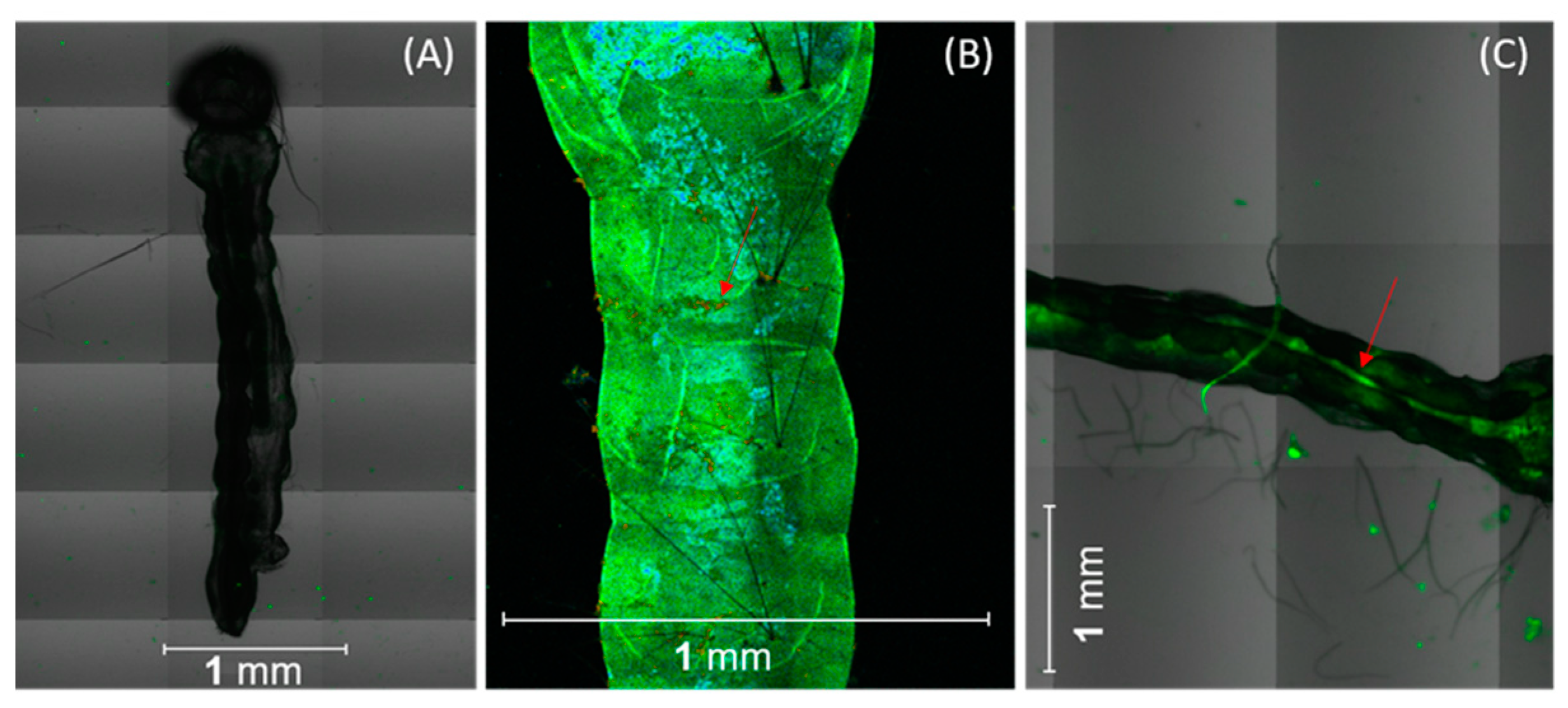
2.1. Curcumin Localization in Aedes aegypti Tissues
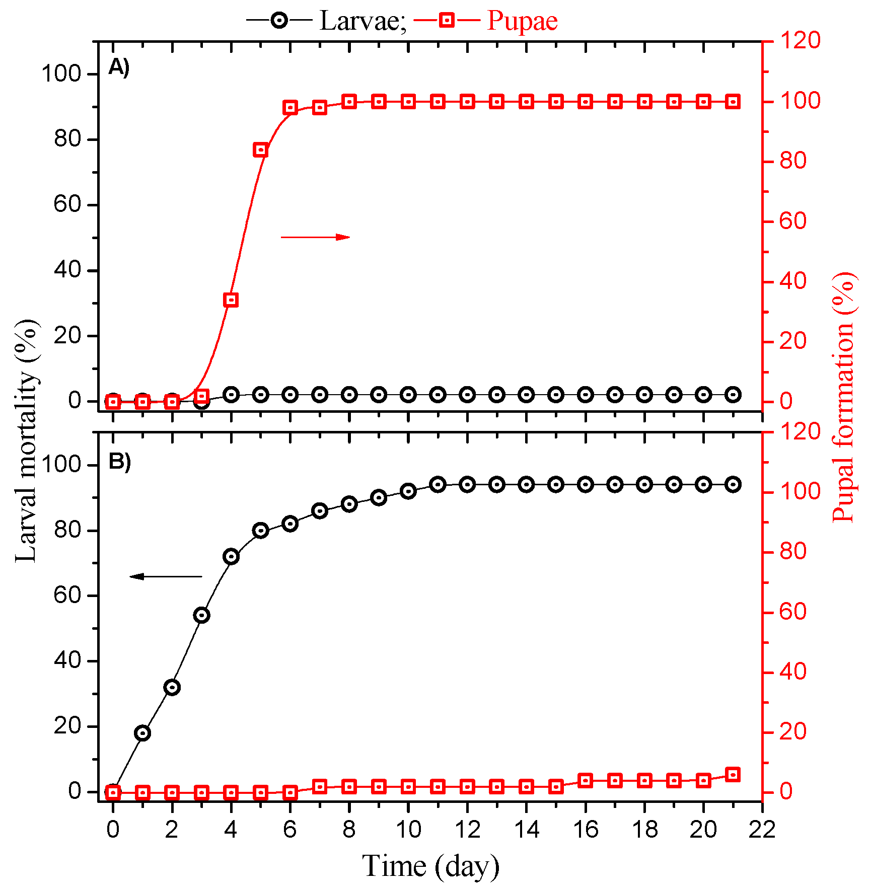
2.2. Photodynamic Control
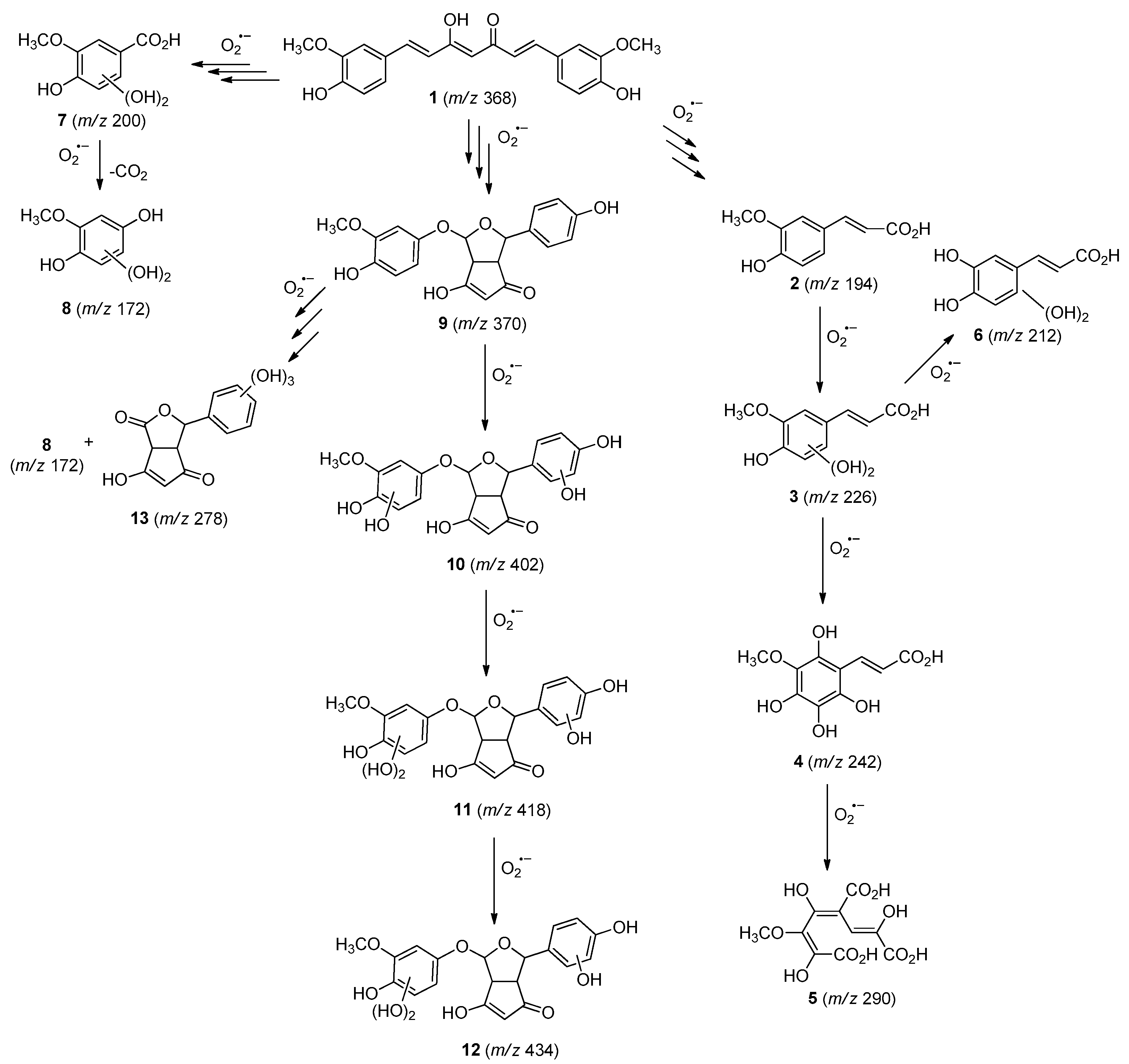
2.3. Curcumin Photodegradation Byproducts
2.4. Prediction of Ecotoxicity of Intermediates from Curcumin Photodegradation
3. Materials and Methods
3.1. Photosensitizer
3.2. Photodynamic Control Bioassays
3.3. Photodegradation
3.4. Liquid Chromatography with Mass Spectrometer (LC-MS)
3.5. Prediction of Acute Toxicity of Intermediates for Aquatic Organisms
4. Conclusions
Supplementary Materials
Author Contributions
Funding
Data Availability Statement
Acknowledgments
Conflicts of Interest
References
- Powell, J.R. Mosquito-Borne Human Viral Diseases: Why Aedes Aegypti? Am. J. Trop. Med. Hyg. 2018, 98, 1563–1565. [Google Scholar] [CrossRef]
- Souza-Neto, J.A.; Powell, J.R.; Bonizzoni, M. Aedes Aegypti Vector Competence Studies: A Review. Infect. Genet. Evol. 2019, 67, 191–209. [Google Scholar] [CrossRef]
- Amelia-Yap, Z.H.; Chen, C.D.; Sofian-Azirun, M.; Low, V.L. Pyrethroid Resistance in the Dengue Vector Aedes Aegypti in Southeast Asia: Present Situation and Prospects for Management. Parasit. Vectors 2018, 11, 332. [Google Scholar] [CrossRef]
- Santos, V.S.V.; Silva, C.E.; Oliveira, C.M.; de Morais, C.R.; Limongi, J.E.; Pereira, B.B. Evaluation of Toxicity and Environmental Safety in Use of Spinosad to Rationalize Control Strategies against Aedes Aegypti. Chemosphere 2019, 226, 166–172. [Google Scholar] [CrossRef]
- Santos, V.S.V.; Limongi, J.E.; Pereira, B.B. Association of Low Concentrations of Pyriproxyfen and Spinosad as an Environment-Friendly Strategy to Rationalize Aedes Aegypti Control Programs. Chemosphere 2020, 247, 125795. [Google Scholar] [CrossRef]
- World Health Organization (WHO). Vector-Borne Disease Control Fourth Edition, 4th ed.; World Health Organization: Geneva, Switzerland, 2009; ISBN 9789241598781. [Google Scholar]
- World Health Organization (WHO). Global Insecticide Use for Vector-Borne Disease Control: A 10-Year Asessment (2010–2019), 6th ed.; World Health Organization: Geneva, Switzerland, 2021; ISBN 9789240032033. [Google Scholar]
- Das, B.; Ghosal, S.; Mohanty, S. Aedes: What Do We Know about Them and What Can They Transmit. In Vectors and Vector-Borne Zoonotic Diseases; IntechOpen: London, UK, 2019. [Google Scholar]
- World Health Organization (WHO). Global Insecticide Use for Vector-Borne Disease Control Fifth Edition Fifth Edition WHO Pesticide Evaluation Scheme (WHOPES) Departement of Control of Neglected Tropical Diseases (NTD), 5th ed.; World Health Organization: Geneva, Switzerland, 2011; ISBN 9789241502153. [Google Scholar]
- Laura de Sene Amâncio Zara, A.; Maria dos Santos, S.; Synthia Fernandes-Oliveira, E.; Gomes Carvalho, R.; Evelim Coelho, G. Estratégias de Controle Do Aedes Aegypti: Uma Revisão. Epidemiol. E Serviços Saúde 2016, 25, 1–2. [Google Scholar] [CrossRef]
- Boyce, R.; Lenhart, A.; Kroeger, A.; Velayudhan, R.; Roberts, B.; Horstick, O. Bacillus Thuringiensis Israelensis (Bti) for the Control of Dengue Vectors: Systematic Literature Review. Trop. Med. Int. Health 2013, 18, 564–577. [Google Scholar] [CrossRef]
- Mitchell-Foster, K.; Ma, B.O.; Warsame-Ali, S.; Logan, C.; Rau, M.E.; Lowenberger, C. The Influence of Larval Density, Food Stress, and Parasitism on the Bionomics of the Dengue Vector Aedes Aegypti (Diptera: Culicidae): Implications for Integrated Vector Management. J. Vector Ecol. 2012, 37, 221–229. [Google Scholar] [CrossRef]
- Rose, R. Pesticides and Public Health: Integrated Methods of Mosquito Management. Emerg. Infect. Dis. 2001, 7, 17–23. [Google Scholar] [CrossRef]
- Parra-Henao, G.; Coelho, G.; Escobar, J.P.; Gonzalvez, G.; Bezerra, H. Beyond Traditional Vector Control and the Need for Strengthening Integrated Vector Management in Latin America. Ther. Adv. Infect. Dis. 2021, 8, 1–4. [Google Scholar] [CrossRef]
- De Souza, L.M.; Inada, N.M.; Venturini, F.P.; Carmona-Vargas, C.C.; Pratavieira, S.; de Oliveira, K.T.; Kurachi, C.; Bagnato, V.S. Photolarvicidal Effect of Curcuminoids from Curcuma Longa Linn. against Aedes Aegypti Larvae. J. Asia. Pac. Entomol. 2019, 22, 151–158. [Google Scholar] [CrossRef]
- De Souza, L.M.; Venturini, F.P.; Inada, N.M.; Iermak, I.; Garbuio, M.; Mezzacappo, N.F.; de Oliveira, K.T.; Bagnato, V.S. Curcumin in Formulations against Aedes Aegypti: Mode of Action, Photolarvicidal and Ovicidal Activity. Photodiagnosis Photodyn. Ther. 2020, 31, 101840. [Google Scholar] [CrossRef]
- Lima, A.; Silva, C.; Caires, C.; Prado, E.; Rocha, L.; Cabrini, I.; Arruda, E.; Oliveira, S.; Caires, A. Evaluation of Eosin-Methylene Blue as a Photosensitizer for Larval Control of Aedes Aegypti by a Photodynamic Process. Insects 2018, 9, 109. [Google Scholar] [CrossRef]
- Lucantoni, L.; Magaraggia, M.; Lupidi, G.; Ouedraogo, R.K.; Coppellotti, O.; Esposito, F.; Fabris, C.; Jori, G.; Habluetzel, A. Novel, Meso-Substituted Cationic Porphyrin Molecule for Photo-Mediated Larval Control of the Dengue Vector Aedes Aegypti. PLoS Negl. Trop. Dis. 2011, 5, e1434. [Google Scholar] [CrossRef]
- Dąbrowski, J.M. Reactive Oxygen Species in Photodynamic Therapy: Mechanisms of Their Generation and Potentiation. Adv. Inorg. Chem. 2017, 70, 343–394. [Google Scholar]
- Villén, L.; Manjón, F.; García-Fresnadillo, D.; Orellana, G. Solar Water Disinfection by Photocatalytic Singlet Oxygen Production in Heterogeneous Medium. Appl. Catal. B Environ. 2006, 69, 1–9. [Google Scholar] [CrossRef]
- Wang, R.; Zhang, B.; Liang, Z.; He, Y.; Wang, Z.; Ma, X.; Yao, X.; Sun, J.; Wang, J. Insights into Rapid Photodynamic Inactivation Mechanism of Staphylococcus Aureus via Rational Design of Multifunctional Nitrogen-Rich Carbon-Coated Bismuth/Cobalt Nanoparticles. Appl. Catal. B Environ. 2019, 241, 167–177. [Google Scholar] [CrossRef]
- Kwiatkowski, S.; Knap, B.; Przystupski, D.; Saczko, J.; Kędzierska, E.; Knap-Czop, K.; Kotlińska, J.; Michel, O.; Kotowski, K.; Kulbacka, J. Photodynamic Therapy–Mechanisms, Photosensitizers and Combinations. Biomed. Pharmacother. 2018, 106, 1098–1107. [Google Scholar] [CrossRef]
- Ali, A.; Wang, Y.-H.; Khan, I.A. Larvicidal and Biting Deterrent Activity of Essential Oils of Curcuma Longa, Ar -Turmerone, and Curcuminoids Against Aedes Aegypti and Anopheles Quadrimaculatus (Culicidae: Diptera). J. Med. Entomol. 2015, 52, 979–986. [Google Scholar] [CrossRef]
- Madhu, S.K.; Shaukath, A.K.; Vijayan, V.A. Efficacy of Bioactive Compounds from Curcuma Aromatica against Mosquito Larvae. Acta Trop. 2010, 113, 7–11. [Google Scholar] [CrossRef]
- Sagnou, M.; Mitsopoulou, K.P.; Koliopoulos, G.; Pelecanou, M.; Couladouros, E.A.; Michaelakis, A. Evaluation of Naturally Occurring Curcuminoids and Related Compounds against Mosquito Larvae. Acta Trop. 2012, 123, 190–195. [Google Scholar] [CrossRef]
- Venturini, F.P.; de Souza, L.M.; Garbuio, M.; Inada, N.M.; de Souza, J.P.; Kurachi, C.; de Oliveira, K.T.; Bagnato, V.S. Environmental Safety and Mode of Action of a Novel Curcumin-Based Photolarvicide. Environ. Sci. Pollut. Res. 2020, 27, 29204–29217. [Google Scholar] [CrossRef]
- Gordon, O.N.; Schneider, C. Vanillin and Ferulic Acid: Not the Major Degradation Products of Curcumin. Trends Mol. Med. 2012, 18, 361–363. [Google Scholar] [CrossRef]
- Kharat, M.; Du, Z.; Zhang, G.; McClements, D.J. Physical and Chemical Stability of Curcumin in Aqueous Solutions and Emulsions: Impact of PH, Temperature, and Molecular Environment. J. Agric. Food Chem. 2017, 65, 1525–1532. [Google Scholar] [CrossRef]
- Schneider, C.; Gordon, O.N.; Edwards, R.L.; Luis, P.B. Degradation of Curcumin: From Mechanism to Biological Implications. J. Agric. Food Chem. 2015, 63, 7606–7614. [Google Scholar] [CrossRef]
- Gordon, O.N.; Luis, P.B.; Sintim, H.O.; Schneider, C. Unraveling Curcumin Degradation. J. Biol. Chem. 2015, 290, 4817–4828. [Google Scholar] [CrossRef]
- Priyadarsini, K.I. Photophysics, Photochemistry and Photobiology of Curcumin: Studies from Organic Solutions, Bio-Mimetics and Living Cells. J. Photochem. Photobiol. C Photochem. Rev. 2009, 10, 81–95. [Google Scholar] [CrossRef]
- Rekha, R.; Vaseeharan, B.; Vijayakumar, S.; Abinaya, M.; Govindarajan, M.; Alharbi, N.S.; Kadaikunnan, S.; Khaled, J.M.; Al-anbr, M.N. Crustin-Capped Selenium Nanowires against Microbial Pathogens and Japanese Encephalitis Mosquito Vectors–Insights on Their Toxicity and Internalization. J. Trace Elem. Med. Biol. 2019, 51, 191–203. [Google Scholar] [CrossRef]
- Consoli, R.A.G.B.; Oliveira, R.L.d. Principais Mosquitos de Importância Sanitária No Brasil; Editora FIOCRUZ: Rio de Janeiro, Brazil, 1994; ISBN 9788575412909. [Google Scholar]
- Tauil, P.L. Aspectos Críticos Do Controle Do Dengue No Brasil. Cad. Saude Publica 2002, 18, 867–871. [Google Scholar] [CrossRef]
- Mohammed, A.; Chadee, D.D. Effects of Different Temperature Regimens on the Development of Aedes Aegypti (L.) (Diptera: Culicidae) Mosquitoes. Acta Trop. 2011, 119, 38–43. [Google Scholar] [CrossRef]
- Reiskind, M.H.; Janairo, M.S. Tracking Aedes Aegypti (Diptera: Culicidae) Larval Behavior Across Development: Effects of Temperature and Nutrients on Individuals’ Foraging Behavior. J. Med. Entomol. 2018, 55, 1086–1092. [Google Scholar] [CrossRef]
- Lopes, T.F.; Holcman, M.M.; Barbosa, G.L.; Domingos, M.d.F.; Barreiros, R.M.O.V. Laboratory Evaluation of the Development of Aedes Aegypti in Two Seasons: Influence of Different Places and Different Densities. Rev. Inst. Med. Trop. Sao Paulo 2014, 56, 369–374. [Google Scholar] [CrossRef]
- Mezzacappo, N.F.; Souza, L.M.; Inada, N.M.; Dias, L.D.; Garbuio, M.; Venturini, F.P.; Corrêa, T.Q.; Moura, L.; Blanco, K.C.; de Oliveira, K.T.; et al. Curcumin/D-mannitol as Photolarvicide: Induced Delay in Larval Development Time, Changes in Sex Ratio and Reduced Longevity of Aedes aegypti. Pest Manag. Sci. 2021, 77, 2530–2538. [Google Scholar] [CrossRef]
- Kalaivani, K.; Senthil-Nathan, S.; Murugesan, A.G. Biological Activity of Selected Lamiaceae and Zingiberaceae Plant Essential Oils against the Dengue Vector Aedes aegypti L. (Diptera: Culicidae). Parasitol. Res. 2012, 110, 1261–1268. [Google Scholar] [CrossRef]
- Legrini, O.; Oliveros, E.; Braun, A.M. Photochemical Processes for Water Treatment. Chem. Rev. 1993, 93, 671–698. [Google Scholar] [CrossRef]
- Boule, P.; Meunier, L.; Bonnemoy, F.; Boulkamh, A.; Zertal, A.; Lavedrine, B. Direct Phototransformation of Aromatic Pesticides in Aqueous Solution. Int. J. Photoenergy 2002, 4, 69–78. [Google Scholar] [CrossRef]
- De Melo da Silva, L.; Cavalcante, R.P.; Cunha, R.F.; Gozzi, F.; Dantas, R.F.; de Oliveira, S.C.; Machulek, A. Tolfenamic Acid Degradation by Direct Photolysis and the UV-ABC/H2O2 Process: Factorial Design, Kinetics, Identification of Intermediates, and Toxicity Evaluation. Sci. Total Environ. 2016, 573, 518–531. [Google Scholar] [CrossRef]
- Mazellier, P.; Leverd, J. Transformation of 4-Tert-Octylphenol by UV Irradiation and by an H2O2/UV Process in Aqueous Solution. Photochem. Photobiol. Sci. 2003, 2, 946. [Google Scholar] [CrossRef]
- Vogna, D.; Marotta, R.; Napolitano, A.; Andreozzi, R.; D’Ischia, M. Advanced Oxidation of the Pharmaceutical Drug Diclofenac with UV/H2O2 and Ozone. Water Res. 2004, 38, 414–422. [Google Scholar] [CrossRef]
- Wang, Y.-J.; Pan, M.-H.; Cheng, A.-L.; Lin, L.-I.; Ho, Y.-S.; Hsieh, C.-Y.; Lin, J.-K. Stability of Curcumin in Buffer Solutions and Characterization of Its Degradation Products. J. Pharm. Biomed. Anal. 1997, 15, 1867–1876. [Google Scholar] [CrossRef]
- Cavalcante, R.P.; Dantas, R.F.; Bayarri, B.; González, O.; Giménez, J.; Esplugas, S.; Machulek, A. Photocatalytic Mechanism of Metoprolol Oxidation by Photocatalysts TiO 2 and TiO 2 Doped with 5% B: Primary Active Species and Intermediates. Appl. Catal. B Environ. 2016, 194, 111–122. [Google Scholar] [CrossRef]
- Chen, P.; Lv, W.; Chen, Z.; Ma, J.; Li, R.; Yao, K.; Liu, G.; Li, F. Phototransformation of Mefenamic Acid Induced by Nitrite Ions in Water: Mechanism, Toxicity, and Degradation Pathways. Environ. Sci. Pollut. Res. 2015, 22, 12585–12596. [Google Scholar] [CrossRef] [PubMed]
- Vialaton, D.; Richard, C.; Baglio, D.; Paya-Perez, A.-B. Phototransformation of 4-Chloro-2-Methylphenol in Water: Influence of Humic Substances on the Reaction. J. Photochem. Photobiol. A Chem. 1998, 119, 39–45. [Google Scholar] [CrossRef]
- Vialaton, D.; Richard, C. Phototransformation of Aromatic Pollutants in Solar Light: Photolysis versus Photosensitized Reactions under Natural Water Conditions. Aquat. Sci. 2002, 64, 207–215. [Google Scholar] [CrossRef]
- Cavalcante, R.P.; Dantas, R.F.; Wender, H.; Bayarri, B.; González, O.; Giménez, J.; Esplugas, S.; Machulek, A. Photocatalytic Treatment of Metoprolol with B-Doped TiO2: Effect of Water Matrix, Toxicological Evaluation and Identification of Intermediates. Appl. Catal. B Environ. 2015, 176–177, 173–182. [Google Scholar] [CrossRef]
- Marković, M.; Jović, M.; Stanković, D.; Kovačević, V.; Roglić, G.; Gojgić-Cvijović, G.; Manojlović, D. Application of Non-Thermal Plasma Reactor and Fenton Reaction for Degradation of Ibuprofen. Sci. Total Environ. 2015, 505, 1148–1155. [Google Scholar] [CrossRef]
- Department of Health and Human Services, U.S. Guidance for Industry Environmental Assessment of Human Drug and Biologics Applications; Guidance: Rockville, MD, USA, 1998.
- Persoone, G.; Marsalek, B.; Blinova, I.; Törökne, A.; Zarina, D.; Manusadzianas, L.; Nalecz-Jawecki, G.; Tofan, L.; Stepanova, N.; Tothova, L.; et al. A Practical and User-Friendly Toxicity Classification System with Microbiotests for Natural Waters and Wastewaters. Environ. Toxicol. 2003, 18, 395–402. [Google Scholar] [CrossRef]
- Rafiee, Z.; Nejatian, M.; Daeihamed, M.; Jafari, S.M. Application of Curcumin-Loaded Nanocarriers for Food, Drug and Cosmetic Purposes. Trends Food Sci. Technol. 2019, 88, 445–458. [Google Scholar] [CrossRef]
- Goel, A.; Kunnumakkara, A.B.; Aggarwal, B.B. Curcumin as “Curecumin”: From Kitchen to Clinic. Biochem. Pharmacol. 2008, 75, 787–809. [Google Scholar] [CrossRef]
- Lao, C.D.; Ruffin, M.T.; Normolle, D.; Heath, D.D.; Murray, S.I.; Bailey, J.M.; Boggs, M.E.; Crowell, J.; Rock, C.L.; Brenner, D.E. Dose Escalation of a Curcuminoid Formulation. BMC Complement. Altern. Med. 2006, 6, 10. [Google Scholar] [CrossRef]
- Sunagawa, Y.; Katanasaka, Y.; Hasegawa, K.; Morimoto, T. Clinical Applications of Curcumin. PharmaNutrition 2015, 3, 131–135. [Google Scholar] [CrossRef]
- Carmona-Vargas, C.C.; de Carvalho Alves, L.; Brocksom, T.J.; de Oliveira, K.T. Combining Batch and Continuous Flow Setups in the End-to-End Synthesis of Naturally Occurring Curcuminoids. React. Chem. Eng. 2017, 2, 366–374. [Google Scholar] [CrossRef]
- Tang, S.B.; Lee, J.C.; Jung, J.K.; Choi, M.-Y. Effect of Erythritol Formulation on the Mortality, Fecundity and Physiological Excretion in Drosophila Suzukii. J. Insect Physiol. 2017, 101, 178–184. [Google Scholar] [CrossRef] [PubMed]
- van der Sman, R.G.M. Sugar and Polyol Solutions as Effective Solvent for Biopolymers. Food Hydrocoll. 2016, 56, 144–149. [Google Scholar] [CrossRef]
- World Health Organization. Guidelines for Laboratory and Field Testing of Mosquito Larvicides; World Health Organization: Geneva, Switzerland, 2005; pp. 1–41. [Google Scholar]
- Vilarinhos, P.; Dias, D.G.S.; Monnerat, R.G. Persistência Larvicida de Formulações de Bacillus Thuringiensis Subsp. Israelensis Para o Controle de Larvas de Aedes asegypti; INFOTECA-E: Brasilia, Brazil, 2003; p. 18. [Google Scholar]
- Castro, D.C.; Cavalcante, R.P.; Jorge, J.; Martines, M.A.U.; Oliveira, L.C.S.; Casagrande, G.A.; Machulek Jr., A. Synthesis and Characterization of Mesoporous Nb 2 O 5 and Its Application for Photocatalytic Degradation of the Herbicide Methylviologen. J. Braz. Chem. Soc. 2015, 2, 303–313. [Google Scholar] [CrossRef]
- Ramos, D.D.; Bezerra, P.C.S.; Quina, F.H.; Dantas, R.F.; Casagrande, G.A.; Oliveira, S.C.; Oliveira, M.R.S.; Oliveira, L.C.S.; Ferreira, V.S.; Oliveira, S.L.; et al. Synthesis and Characterization of TiO2 and TiO2/Ag for Use in Photodegradation of Methylviologen, with Kinetic Study by Laser Flash Photolysis. Environ. Sci. Pollut. Res. 2015, 22, 774–783. [Google Scholar] [CrossRef] [PubMed]
- Da Rosa, A.P.P.; Cavalcante, R.P.; da Silva, D.A.; da Silva, L.d.M.; da Silva, T.F.; Gozzi, F.; McGlynn, E.; Brady-Boyd, A.; Casagrande, G.A.; Wender, H.; et al. H2O2-Assisted Photoelectrocatalytic Degradation of Mitoxantrone Using CuO Nanostructured Films: Identification of by-Products and Toxicity. Sci. Total Environ. 2019, 651, 2845–2856. [Google Scholar] [CrossRef]
- De Melo da Silva, L.; Gozzi, F.; Sirés, I.; Brillas, E.; de Oliveira, S.C.; Machulek, A. Degradation of 4-Aminoantipyrine by Electro-Oxidation with a Boron-Doped Diamond Anode: Optimization by Central Composite Design, Oxidation Products and Toxicity. Sci. Total Environ. 2018, 631–632, 1079–1088. [Google Scholar] [CrossRef]
- Tay, K.S.; Madehi, N. Ozonation of Ofloxacin in Water: By-Products, Degradation Pathway and Ecotoxicity Assessment. Sci. Total Environ. 2015, 520, 23–31. [Google Scholar] [CrossRef]
- Wright, R.T.; Fay, K.; Kennedy, A.; Mayo-Bean, K.; Moran-Bruce, K.; Meylan, W.; Ranslow, P.; Lock, M.; Nabholz, J.V.; Runnen, J.V.; et al. Operation Manual for the Ecolological Structure-Activity Relationship Model (ECOSAR) Class Program. U.S. Environmental Protection Agency. 2022; v2.2; pp. 1–38. Available online: https://www.epa.gov/system/files/documents/2022-03/operation-manual-v.2.2_1.pdf (accessed on 10 July 2022).
- Wright, R.T.; Fay, K.; Kennedy, A.; Mayo-Bean, K.; Moran-Bruce, K.; Meylan, W.; Ranslow, P.; Lock, M.; Nabholz, J.V.; Runnen, J.V.; et al. Methodology Document for the Ecological Structure-Activity Relationship Model (ECOSAR) Class Program. U.S. Environmental Protection Agency. 2022; v2.2; pp. 1–40. Available online: https://www.epa.gov/system/files/documents/2022-03/methodology-document-v.2.2.pdf (accessed on 10 July 2022).


| Compound | Accurate Mass [M+H]+ | Retention Time (min) | Molecular Formula | Proposed Structure |
|---|---|---|---|---|
| 172 m/z | 173.0103 | 1 | C7H8O5 | 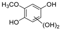 |
| 194 m/z | 194.9671 | 1.1 | C10H10O4 |  |
| 200 m/z | 201.0466 | 1 | C8H8O6 | 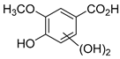 |
| 212 m/z | 212.9753 | 1.1 | C10H8O6 | 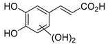 |
| 226 m/z | 226.9489 | 1 | C10H10O6 | 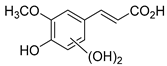 |
| 242 m/z | 242.9826 | 1.1 | C10H10O7 |  |
| 278 m/z | 279.0721 | 1 | C13H10O7 | 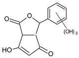 |
| 290 m/z | 290.9712 | 1 | C10H10O10 | 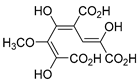 |
| 370 m/z | 370.9319 | 1.1 | C20H18O7 | 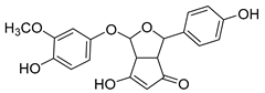 |
| 402 m/z | 402.8989 | 1.1 | C20H18O9 | 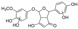 |
| 418 m/z | 418.8622 | 1.1 | C20H18O10 | 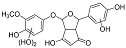 |
| 434 m/z | 434.8924 | 1.1 | C20H18O11 | 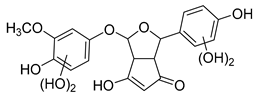 |
| Compound | 2 (194 m/z) | 13 (278 m/z) | 9 (370 m/z) | |
|---|---|---|---|---|
| Organism | Structure | 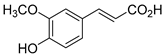 | 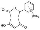 | 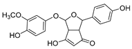 |
| Daphnia | (mg L−1) | 287.5 | 2.3 × 10⁴ | 988.16 |
| TU | 3.5 × 10⁻³ | 4.3 × 10⁻⁵ | 1.2 × 10⁻³ | |
| Fish | (mg L−1) | 534.4 | 7.3 × 10³ | 1.9 × 10³ |
| TU | 1.9 × 10⁻³ | 1.4 × 10⁻⁴ | 5.3 × 10⁻⁴ | |
| Green algae | (mg L−1) | 171.3 | 1.8 × 10⁴ | 525.5 |
| TU | 5.8 × 10⁻³ | 5.4 × 10⁻⁵ | 1.9 × 10⁻³ | |
| Toxicity | Non-toxic | Non-toxic | Non-toxic | |
| 1.42 | −1.93 | 1.12 |
Publisher’s Note: MDPI stays neutral with regard to jurisdictional claims in published maps and institutional affiliations. |
© 2022 by the authors. Licensee MDPI, Basel, Switzerland. This article is an open access article distributed under the terms and conditions of the Creative Commons Attribution (CC BY) license (https://creativecommons.org/licenses/by/4.0/).
Share and Cite
Lima, A.R.; Silva, C.M.; da Silva, L.M.; Machulek, A., Jr.; de Souza, A.P.; de Oliveira, K.T.; Souza, L.M.; Inada, N.M.; Bagnato, V.S.; Oliveira, S.L.; et al. Environmentally Safe Photodynamic Control of Aedes aegypti Using Sunlight-Activated Synthetic Curcumin: Photodegradation, Aquatic Ecotoxicity, and Field Trial. Molecules 2022, 27, 5699. https://doi.org/10.3390/molecules27175699
Lima AR, Silva CM, da Silva LM, Machulek A Jr., de Souza AP, de Oliveira KT, Souza LM, Inada NM, Bagnato VS, Oliveira SL, et al. Environmentally Safe Photodynamic Control of Aedes aegypti Using Sunlight-Activated Synthetic Curcumin: Photodegradation, Aquatic Ecotoxicity, and Field Trial. Molecules. 2022; 27(17):5699. https://doi.org/10.3390/molecules27175699
Chicago/Turabian StyleLima, Alessandra R., Cicera M. Silva, Lucas M. da Silva, Amilcar Machulek, Jr., Antônio P. de Souza, Kleber T. de Oliveira, Larissa M. Souza, Natalia M. Inada, Vanderlei S. Bagnato, Samuel L. Oliveira, and et al. 2022. "Environmentally Safe Photodynamic Control of Aedes aegypti Using Sunlight-Activated Synthetic Curcumin: Photodegradation, Aquatic Ecotoxicity, and Field Trial" Molecules 27, no. 17: 5699. https://doi.org/10.3390/molecules27175699
APA StyleLima, A. R., Silva, C. M., da Silva, L. M., Machulek, A., Jr., de Souza, A. P., de Oliveira, K. T., Souza, L. M., Inada, N. M., Bagnato, V. S., Oliveira, S. L., & Caires, A. R. L. (2022). Environmentally Safe Photodynamic Control of Aedes aegypti Using Sunlight-Activated Synthetic Curcumin: Photodegradation, Aquatic Ecotoxicity, and Field Trial. Molecules, 27(17), 5699. https://doi.org/10.3390/molecules27175699