Article

Underestimation about the Contribution of Nitrate Reducers to Iron Cycling Indicated by Enterobacter Strain

1
School of Resources and Environmental Engineering, Anhui University, Hefei 230601, China
2
Key Lab of Urban Environment and Health, Institute of Urban Environment, Chinese Academy of Sciences, Xiamen 361021, China
*
Author to whom correspondence should be addressed.
Molecules 2022, 27(17), 5581; https://doi.org/10.3390/molecules27175581
Submission received: 29 June 2022 / Revised: 19 August 2022 / Accepted: 25 August 2022 / Published: 30 August 2022

Abstract

:
Nitrate-reducing iron(II) oxidation (NRFO) has been intensively reported in various bacteria. Iron(II) oxidation is found to be involved in both enzymatic and chemical reactions in nitrate-reducing Fe(II)-oxidizing microorganisms (NRFOMs). However, little is known about the relative contribution of biotic and abiotic reactions to iron(II) oxidation for the common nitrate reducers during the NRFO process. In this study, the typical nitrate reducers, four Enterobacter strains E. hormaechei, E. tabaci, E. mori and E. asburiae, were utilized as the model microorganisms. The comparison of the kinetics of nitrate, iron(II) and nitrite and N2O production in setups with and without iron(II) indicates a mixture of enzymatic and abiotic oxidation of iron(II) in all four Enterobacter strains. It was estimated that 22−29% of total oxidized iron(II) was coupled to microbial nitrate reduction by E. hormaechei, E. tabaci, E. mori, and E. asburiae. Enterobacter strains displayed an metabolic inactivity with heavy iron(III) encrustation on the cell surface in the NRFOmedium during days of incubation. Moreover, both respiratory and periplasmic nitrate-reducing genes are encoded by genomes of Enterobacter strains, suggesting that cell encrustation may occur with periplasmic iron(III) oxide precipitation as well as the surface iron(II) mineral coating for nitrate reducers. Overall, this study clarified the potential role of nitrate reducers in the biochemical cycling of iron under anoxic conditions, in turn, re-shaping their activity during denitrification because of cell encrustation with iron(III) minerals.

1. Introduction

Nitrate-reducing Fe(II) oxidation (NRFO) with the production of iron(III), NO2, NO, N2O and N2 have been found in soil, fresh water and brackish water [1]. NRFO is of great importance in driving iron biogeochemical cycling and removing nitrogen, metal(loid)s and radionuclides from the active sledge system and groundwater, respectively [2,3].
There are a lot of bacteria isolated with the activity of NRFO, such as Acidovorax, Microbacterium, Pseudomonas, Citrobacter, Enterobacteriaceae spp., etc. [4,5,6,7]. Nevertheless, the mechanism around the Fe(II) oxidoreductase involved in nitrate reduction coupled to iron(II) oxidation is still needed to be deciphered. Moreover, chemodenitrification (4Fe(II) + 2NO2 + 5H2O → 10FeO(OH) + N2O + 6H+) complicates our understanding of the NRFO process, which always leads to a combination of biotic with abiotic iron(II) oxidation during NRFO process [2]. Up to date, all isolated neutrophilic nitrate-reducing Fe(II)-oxidizing microorganisms (NRFOMs) are mixotrophic, requiring an organic co-substrate for their continuous cultivation and oxidation of Fe(II) to Fe(III) [2,4,5,6]. It provides a cue that respiratory nitrate reduction is essential for the capability of NRFO by these NRFOMs. As a result, nitrite is an unavoidable by-product followed by a chemical reaction with iron(II) in the system, which is a challenge to the existence of enzymatic NRFO. Moreover, NRFO displays an intrinsic capability in all nitrate reducers, which directly implicates catalysis by nitrogen oxide reductases in cells and does not require a specialized oxidoreductase. Ishii et al. have indicated non-enzymatic iron(II) oxidation from 67 members affiliated with Pseudogulbenkiania, which are characterized as denitrifier [8,9]. However, this conclusion is arbitrary since there is no comparison of nitrate-reducing extent between setups amended with or without iron(II). Inconsistently, Jamieson et al. estimated that 60–70% of overall iron(II) oxidation is caused by an enzymatic pathway for NRFOMs including Acidovorax strain BoFeN1 and 2AN, A. ebreus strain TPSY, Paracoccus denitrificans Pd 1222 and Pesudogulbenkiania sp. strain 2002, which is depended on compilation and model-based interpretation from published experimental data [2]. It has been estimated that only 30–40% of oxidized iron(III) is owing to the chemodenitrification process for these strains [2]. However, it is still unknown about this discrepancy between NRFOMs and common nitrate reducers. Therefore, more research is still necessary to unveil the relative contribution of enzymatic reaction and chemodenitrification to iron(III) oxidation, especially for the common nitrate reducers.
Cell encrustation is observed on the surface or in the periplasmic space of NRFOMs and nitrate reducers, consequently, causing physical inactivity including termination of nutrition uptake and further cell metabolism [1,10,11,12]. This is likely to result in the re-estimation of their contribution to iron and nitrogen cycling. The position of cell encrustation actually reflects the types of nitrate reductases expressed by these microorganisms, including the respiratory nitrate reductases and periplasmic nitrate reductases [13]. According to the previous reports, substantial numbers and high diversity of nitrate reducers are widely distributed in various environments under anoxic or aerobic conditions [14], whereas, the atlas for the composition of nitrate reductases for these common nitrate reducers needs to be established.
The genus Enterobacter, which belongs to the family Enterbacteriaceae, is abundant in various environments such as soil, water and gut of soil animals [15]. Enterobacter spp. are typical nitrate reducers and can be used as model nitrate reducers [16]. In this study, four Enterobacter strains were employed, aiming to explore the style of their roles in iron(II) oxidation during the NRFO process. Moreover, the published documentation and public database were referred to construct a heatmap of nitrate-reducing genes possessed in Enterbacteriaceae bacteria, further investigating the potential position of cell encrustation formed for the bacterial cells after NRFO.

2. Results

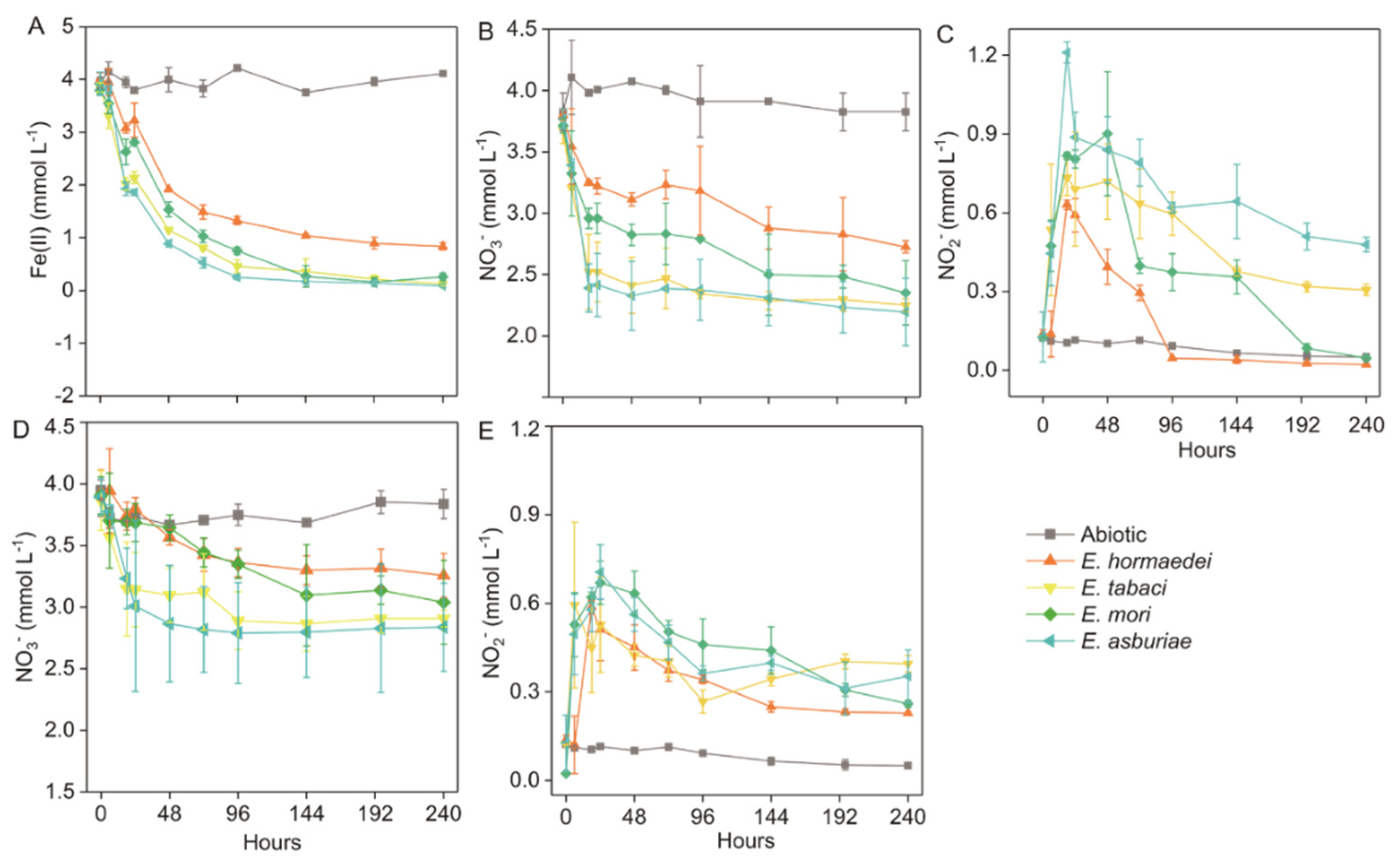
2.1. Nitrate Reduction and Iron(II) Oxidation by Enterobacter Strains

There were four Enterobacter strains, including hormaechei, E. tabaci, and E. asburiae, used in this study (Table 1 and Supplementary Figure S1). Compared to abiotic setups, Fe(II) and NO3 concentrations decreased with incubation for all groups inoculated with Enterobacter strains in the NRFO medium. The concentration of Fe(II) decreased sharply within 48 h and was then kept stable after 96-h incubation for all strains in the NRFO medium during the incubation (Figure 1A). Similarly, the time course of nitrate concentrations shared a similar trend with that of Fe(II) concentration for all biotic setups in the NRFO medium during the incubation (Figure 1B). For the nitrite concentrations, all biotic setups were observed to be rapidly elevated in the first 18 h and then decreased until exhaustion in addition to the amendment inoculated with E. tabaci and in the NRFO medium during the incubation (Figure 1C). For the NRFO medium without iron(II) addition, the decrease in nitrate and nitrite was also identified for all biotic setups amended with Enterobacter strains during the incubation, however, which displayed a lower extent than those in setups amended with iron(II) (Figure 1D,E). There was a hundred micromolar of nitrite remaining in the medium after 240-h incubation (Figure 1E).
The cell copy numbers of Enterobacter strains ranged from 8.68 × 10⁹ to 1.81 × 10¹⁰ at the beginning of the incubation, and they slightly increased after 240 h in addition to setups amended with E. hormaechei (Figure 2A). In comparison, strain E. tabaci and E. asburiae displayed higher extents of iron(II) oxidation and nitrate reduction than E. hormaechei and E. mori (Figure 2B). The strain E. tabaci, E. mori and E. asburiae possessed the ability to almost completely oxidize iron(II), while there was around 0.84 mM iron(II) remaining in the medium for E. hormaechei after 240-h incubation (Figure 2B). Moreover, all the strains showed incomplete nitrate reduction after 240 h (Figure 2C). Additionally, the nitrite content of E. hormaechei and E. mori approached 0 mM, while 0.31 mM and 0.48 mM of nitrite were still contained in the medium for biotic setups inoculated with strain E. tabaci and E. asburiae after 240 h, respectively (Figure 1C). The production of N2O was detected in all biotic treatments containing with and without iron(II) (Figure 2D). In comparison, the concentration of headspace N2O was higher in the iron(II)-containing setups than that in biotic treatments without iron(II) (Figure 2D).

2.2. Morphological Characteristics of Enterobacter Strains after Iron(II) Oxidation and Nitrate Reduction

It was observed that cells of these four strains were smooth on the surface when they were cultivated in the R2A medium (Figure 3A–D). Whereas, cells of them were identified to be covered by heavy encrustation after incubation in the NRFO medium (Figure 3E–H). In order to further characterize the coat on the surface of cells, Raman spectroscopy was employed. Spectra of all cells from these four strains were consistent with that of the standard iron oxide (Figure 3C).
In order to investigate the metabolic activity of Enterobacter strains, C-D bond (2040 and 2300 cm−1) was detected for cells of Enterobacter strains after incubation in the NRFO medium. For the cells coated with encrustation, there were no detectable peaks in the region between 2040 and 2300 cm−1 (Supplementary Figure S2).

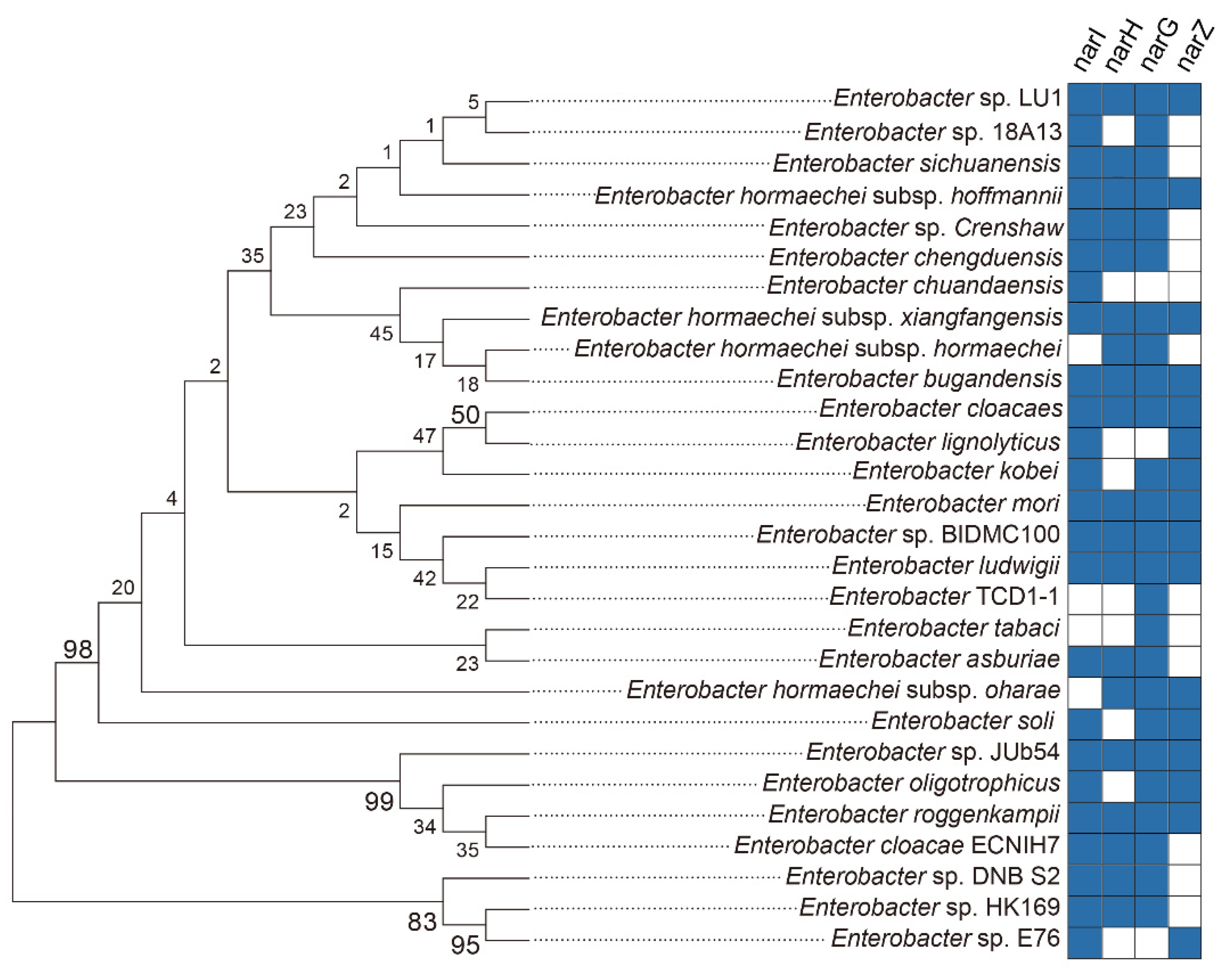
2.3. Nitrate Reductase Contained in the Enterobacteriaceae Strains

Nitrate reduction-relevant genes, including narG, nasA, nirK, nirS, norB and nosZ, were detected in genomes of these four Enterobacter strains (Table 2). In order to investigate the potential capability of nitrate reduction for the genus Enterobacter, four nitrate reductase-encoding genes, including narI, narH, n,arG and narZ, were investigated in the genomes of 28 Enterobacter strains (Figure 4 and Supplementary Table S2). For the gene narI, 24 strains were found to include this gene in their genomes in addition to E. Hormaechei subsp. hormaechei, E. Hormaechei subsp. oharae and E. tabaci (Figure 4 and Supplementary Table S2). In comparison, there were 19, 25, and 16 strains encoding narH, narG and narZ genes in their genomes, respectively (Figure 4 and Supplementary Table S2).
To further explore the possibility of nitrate reduction by Enterobacteriaceae bacteria in environments, three types of nitrate reductases, including respiratory nitrate reductase 1, 2 and periplasmic nitrate reductase, were searched depended on the NCBI database. For the respiratory nitrate reductase 1, consisting of four subunits such as NarG, NarH, NarI and NarJ, it was more abundant in Enterobacteriaceae spp. than the respiratory nitrate reductase 2 and periplasmic nitrate reductase (Supplementary Figure S3 and Supplementary Table S3). Three subunits (e.g., NarV, N,arY and NarZ) make up the respiratory nitrate reductase 2, which were widely identified in genomes of Enterobacteriaceae bacteria (Supplementary Figure S3 and Supplementary Table S3). In comparison, around 7 genera, e.g., Shigella, Escherichia, Salmonella, Citrobacter, etc., possess genes encoding the subunits of periplasmic nitrate reductase containing NapA, NapB, NapC, NapD and NapF (Supplementary Figure S3 and Supplementary Table S3).

3. Discussion

3.1. Microbial-Mediated Nitrate-Dependent Fe(II) Oxidation by Enterobacter Strains

Aiming to unveil the ability of NRFO by Enterobacter spp., four strains were chosen to perform the incubation. No organic electron donor was added in the NRFO medium. In comparison with setups amended with both Fe(II) and nitrate, reduced nitrate and nitrite were also detected in biotic setups without the addition of iron(II) (Figure 1D,E). However, contents of consumed nitrate and nitrite in iron(II)-absent treatments were markedly lower than those in setups amended with Fe(II) (Figure 1D,E). For one thing, it meant that these strains probably employed the stored endogenous organic carbon within cells, which may be generated during the pre-cultivation of Enterobacter strains in the R2A medium. It has been documented that endogenous carbon could perform an as energy source for denitrification when the exogenous organic substrates are exhausted [17,18,19,20,21,22,23,24,25,26,27,28,29,30,31,32,33,34,35,36,37,38,39,40,41,42]. Also, the remaining NO2 (0.23−0.39 mM) in the incubation (Figure 1E) indicated that limited endogenous organic carbon was not sufficient enough to support the complete reduction of nitrite. For the other, part of reduced nitrate was considered to be coupled to microbial iron(II) oxidation by Enterobacter strains in setups amended with both nitrate and iron(II) because the abiotic reaction between iron(II) and nitrate is slow [9]. The chemical equation is as follows [43].
NO3 + 2Fe2+ + 2H+ → 2Fe3+ + NO2 + H2O
2NO2 + 4Fe2+ + 6H+ → N2O + 4 Fe3+ + 3H2O
During the incubation, a total of 1.08−1.61 mM nitrate was consumed in setups amended with iron(II), whereas the amount of reduced nitrate was 0.67−1.06 mM in iron(II)-absent setups (Figure 1). It was roughly estimated that a small part of nitrate (0.41−0.55 mM) reduction (34–38%) was microbially driven by the exogenous donor-iron(II). In the term of enzymatic NRFO, nitrate is assumed to be exclusively reduced to nitrite [2]. Based on the ratio of nitrate to iron(II) [stoichiometric equations of NRFO (Equation (1))], 0.82−1.10 mM iron(II) was thought to be coupled to biotic nitrate reduction, which accounted for 22−29% of total oxidized iron(II) during the incubation for the strains E. hormaechei, E. tabaci, E. mori and E. asburiae, respectively (Figure 1).
Nitrite concentration fluctuated in all biotic setups without iron(II) during the incubation was likely to be attributed to the microbial reduction of nitrite by Enterobacter strains, which was agreed with the relevant function genes-nirK and nirS were found from their genomes (Table 2). However, the extent of nitrite reduction was stopped after around 96 h of incubation (Figure 1F). It may be due to limited endogenous organic electron donors reserved with the in cells of microorganisms. In contrast, more nitrite was utilized during the incubation added with iron(II) (Figure 1C), which indicated that decrease of nitrite could result from the mixture of microbial catalyzation and chemodenitrification during the incubation. The amount of N2O, which was higher in the setups with iron(II) than in the iron(II)-absent ones (Figure 2D), was consistent with the consumed nitrite in biotic setups with and without iron(II). It further suggested a mixed microbial nitrite reduction and chemodenitrification (Equation (2)) during the NRFO incubation. Correspondingly, 2.70−2.98 mM Fe(II) would be chemically oxidized by NO2 in the setups amended with Fe(II) and nitrate, contributing 71−78% of total iron(II) depleted during the incubation. This result presented a different relative contribution of biotic iron(II) oxidation and chemdenitrification to the iron(II) oxidation from the incubation with several mixotrophic NRFO bacteria including Acidovorax strain BoFeN1, 2AN, A. ebreus strain TPSY, Paracoccus denitrificans Pd 1222, and Pseudogulbenkiania sp. strain 2002. They have been demonstrated that about 60–75% of overall iron(II) oxidation was owing to the biotic process, and the section of organic ligands and exopolymeric substances by these bacteria can enhance abiotic oxidation of iron(II).

3.2. Cell Encrustation and Physical Inactivity after NRFO Process by Enterobacter Strains

It can be found that more than 2 mM nitrate was still kept in the medium for all the biotic setups after 240 h of incubation, indicating that an insufficient electron donor was included in the medium. However, there was around 0.12−0.88 molar of iron(II) remaining in the incubation for all the setups with four Enterobacter strains. Moreover, nitrate reduction almost ceased after 144 h for the biotic setups with E. hormaechei, E. tabaci and E. asburiae and 192 h for the biotic setups with E. mori (Figure 1B), which was almost at the same time with iron(II) oxidation (Figure 1A,B). In addition, the kinetic of nitrate exhibited the same trend as that of iron(II) during the incubation (Figure 1). Moreover, the was no large increase in the cell numbers of Enterobacter strains during 240-h incubation (Supplementary Table S2). All these Enterobacter strains seemed to enter a state of physical inactivity after hundred hours of NRFO reaction. Heavy cell encrustation by iron(III) oxides (Figure 3) can explain the loss of metabolic activity of Enterobacter strains. Furthermore, in order to verify the physical dormancy of Enterobacter, (H2)D2O-based Raman microscopy had been employed and no C-D bonds (ranging from 2040 to 2300 cm−1) were detected in all cells of iron(II)-amended setups after 240-h incubation (Supplementary Figure S2). This phenomenon agreed with previous reports about Bacillus ferroxidans, Dechloromonas sp. UWNR4, Acidovorax sp. 2AN [3,4,44]. It can speculate that the heavy encrustation on the surface of cells was the culprit for preventing the utilization of water and other substrates including iron(II), nitrate, and nitrite for cells of Enterobacter strains. The potential product NO generated from chemodenitrification is also toxic and can react with metalloproteins in the components of the electron transport chain [9,45], which may also contribute to the inactivity of Enterobacter strains. The cell encrustation of iron(III) oxides is identified as the artifacts of an abiotic reaction between sorbed Fe2+ and biogenic NO2 in batch experiments [44]. The increase in the Fe(II) concentration (higher than 800 mg/L) can enhance the extent of cell encrustation in a continuous up-flow biofilter, whereas Fe(II)EDTA would prevent cells from encrustation and the nitrogen removal efficiency is as high as 90% [44]. Although numerous literatures indicated that microbial NRFO may result in cell crustation [46,47,48,49], Paracoccus versutus LYM is found with no cell encrustation formed when amended with organic co-substrate and Fe(II)EDTA was amended as electron donor [50]. Enterobacter strains in this study were encrusted by the mixed abiotic and biotic NRFO processes under the condition of no addition of exogenous organic matter. Our previous results demonstrated that encrusted and metabolic inactive cells of mixotrophic NRFO bacteria-Bacillus ferroxidans can re-awake and take off the iron(III) encrustation after re-incubation of these cells in the organics-abundant medium without the addition of iron(II) [51]. These suggested that the metabolic state of denitrifers or nitrate reducers might shift between inactive cells and active ones without encrustation inhibition in natural environments, such as the flooding and drainage of paddy soils.

3.3. The Potential Contribution of Nitrate Reducers to Iron Cycling Implicated by Enterobacter Strains

In this study, Enterobacter strains mediated NRFO is indicated to result in a combination of microbial iron(II) oxidation coupled to nitrate reduction with chemodenitrification by nitrite and iron(II), together contributing to iron(II) consumption during the incubation. It provides an insight into the potential of nitrate reducers or denitrifiers to markedly drive iron cycling via chemodenitrification, which is triggered by nitrite catalyzed by nitrate reductase Nar in iron(II)-rich environments such as flooded paddy soils. Most of Enterobacteriaceae strains were found to possess both respiratory nitrate reductase 1 or 2 (Supplementary Figure S3), locating the intracellular membrane of microorganisms. Respiratory nitrate reductases 1 and 2 are responsible for nitrate reduction activity when cells are grown anaerobically and aerobically in nitrate-containing environments, respectively [52]. It suggested that most of the Enterobacter strains could mediate nitrate reduction whether under anaerobic or aerobic conditions. The iron(III) oxides encrusted on cell surfaces of Enterobacter strains in this study suggested that microbial NRFO might depend on the a dedicated Fe(II) oxidoreductase according to Clark’s study, which would be followed by proton motive force generation and ATP production [9]. Although cells of Enterobacter strain entered into a state of metabolic inactivity, the NRFO process could be an alternative strategy to obtain energy for the nitrate reducers in the short term under an environment contained limited organic substrates [42]. In comparison, intracellular reactions between Fe(II) and periplasmic nitrate reductase (Nap) are essential to initiate extensive NRFO, which consumes periplasmic protons to reduce nitrate without energetic benefit [9,53]. Several genera harboring genes (napA, napB, napC, napD and napF) encoding periplasmic nitrate reductase (Supplementary Figure S3 and Supplementary Table S3), similarly, it could pose periplasmic iron(III) precipitation and limitation of carbon corporation [53]. Hence, these highly diverse and abundant nitrate reducers ubiquitously distributed in soils and aquatic environments, may contribute extremely to iron cycling.

4. Materials and Methods

4.1. Isolation of Nitrate-Dependent Fe(II)-Oxidizing Bacterium

Four strains are widely distributed in diverse environments such as soils and waters [17,18,19,20], including Enterobacter hormaechei (CGMCC 1.10608T), E. tabaci (CGMCC 1.15707T), E. mori (CGMCC 1.10322T) and E. asburiae (JCM 6501T) were purchased from Japan Collection of Microorganisms (JCM) and China General Microbiological Culture Collection Center (CGMCC). R2A medium [21] was used for routine cultivation under the oxic condition of these four strains affiliated with Enterobacter.

4.2. Experimental Setup

Nitrate-reducing Fe(II)-oxidization (NRFO) medium was employed for investigating the ability to oxidize Fe(II) by Enterobacter strains in this study. NRFO medium was constituted of mineral medium (pH 6.8–7.2), 10 mM NaNO3, 10 mM FeCl2, vitamin solution (1 mL L−1) [22], trace element solution SL10 (1 mL L−1) [22], selenite-tungstate solution (1 mL L−1) [10] and bicarbonate buffer (22 mM), which were prepared according to previous description [4,11]. The mineral medium included MgSO4·7H2O (0.5 g L−1), CaCl2·2H2O (0.1 g L−1), NH4Cl (0.3 g L−1) and KH2PO4 (0.6 g L−1) [23,24,25]. FeCl2 was added to the mineral medium in the anaerobic chamber after the mineral medium autoclaved (120 °C for 20 min) and cooled to room temperature under N2/CO2 (80/20%) [4,26,27], and the formed Fe(II) carbonates and Fe(II) phosphates were removed using sterile 0.22 µm filters after 3 days of precipitation in the anaerobic glove box [27]. The final concentration of Fe(II) in the NRFO medium was around 5–8 mM.
The cultures were initialed by inoculating of bacterial suspensions into 20 mL of NRFO medium and then incubated in the dark at 30 °C in the anaerobic chamber (N2:CO2:H2 = 90:5:5; Shel Lab Bactron IV, Portland, OR, USA). Meanwhile, the NRFO medium without the addition of bacterial suspension was set as the control. All the setups were prepared in three replicates. Before inoculation, the bacterial suspension was obtained by cultivating Enterobacter strains in the R2A agar for 3 days at 30 °C under anoxic conditions. The bacterial colonies on R2A plates were removed using anoxic sterile 0.9% NaCl (w/v), harvested at 8000 g for 10 min, washed three times with anoxic mineral medium and resuspended in 60 mL mineral medium. The cell numbers of bacterial suspension for four Enterobacter strains were quantified via 16S rRNA-based qPCR (Roche 480, Roche, Indianapolis, IN, USA). The information on primers and thermal cycling conditions for qPCR as described in Supplementary Table S1. The amplification was carried out in triplicates using the primer set of 515F-907R [28]. The reaction mixture contained 1 µL DNA as a template, 10 µL of SYBR 2 Premix EX Taq, 0.8 µL of each primer and 7.4 µL of ddH2O, and the reaction contained no DNA template in the negative control. Genomic DNA was extracted from cells of Enterobacter strains using FastDNA Spin Kit (MP Biomedical, France). DNA of Enterobacter hormaechei was used to clone these genes to prepare standard plasmids. Standard curves were produced using serial dilutions of the standard plasmids [24]. Only one peak was detected at the melting temperature (Tm) of 82.5 °C, which indicated the specificity of amplicons. We only accept the reactions with efficiencies ranging from 90% to 110% [24].

4.3. PCR

For the Enterobacter strains, the nitrogen cycling genes, including napA (periplasmic-bound nitrate reductase) [29], narG (nitrate reductase) [30], nasA (assimilatory nitrate reductase) [31], nirK (copper-containing nitrite reductase) [14], nirS (cytochrome cd1-containing nitrite reductase) [32], norB (nitric oxide reductase) [33] and nosZ (nitrous oxide reductase) [34] were determined using PCR. The information about primer sets and thermal cycles were detailed in Supplementary Table S1.

4.4. Chemical Analyses

Ferrous iron concentrations were analyzed using the modified ferrozine assay by Klueglein and Kappler [27], which, as a result, prevented oxidation of Fe(II) by the nitrite at acidic pH through ferrous iron with sulfamic acid but not HCl. In brief, 100 µL of culture suspension was mixed with 900 µL of 40 mM sulfamic acid for 1 h at room temperature [27]. The culture suspension was collected using syringes in the anaerobic chamber. An aliquot of 20 µL of ferrous iron extract was added with 180 µL of ferrozine solution (1 g ferrozine in 50 mM HEPES buffer, Ph = 7), which was followed by the formation of ferrous complex quantified at 562 nm using UV/Vis spectrometer (Thermo Scientific Varioskan LUX, ThermoFisher, Waltham, MA, USA). The concentrations of NO3 and NO2 were analyzed with ion chromatography (Dionex ICS-3000 system, Diones, Sunnyvales, CA, USA). Gas chromatography (Agilent-7890, Agilent Technologies, Santa Clara, CA, USA) was employed to determine headspace N2O.

4.5. Phenotypic Analysis

Cell morphology of Enterobacter spp. was characterized after growth of Enterobacter spp. on R2A media or NRFO medium using scanning electron microscopy (SEM, S-4800, Hitachi). Cell samples for SEM analysis were prepared in the anaerobic chamber as described before [11]. In brief, the cell samples were centrifuged for 5 min at 2000 rpm and supernatants were then discarded. After being fixed in 3% (v/v) glutaraldehyde buffered with 0.1 M sodium phosphate buffer (pH 7.2) for 1 h at room temperature, cells of Enterobacter strains were then washed three times in sodium phosphate buffer through centrifuged for 5 min at 2000 rpm, dehydrated in a graded alcohol series and dried using Critical Point Dryers (Leica EM CPD300, Weztlar, Germany) for 6 h. The dried cells were mounted onto a stub using double-sided carbon tape and then coated with a thin layer of gold. The samples were examined using a scanning electron microscope (Merlin compact, Zeiss, Germany).

4.6. Raman Spectroscopy

The cell morphology and iron(III) oxides coated on cell surfaces of Enterobacter strains were characterized by a confocal Raman system (Horiba Jobin Yvon S. A. S, Paris, France) set up with an integrated Olympus BXFM microscope equipped with 600 g/mm grating [35]. In brief, 2 mL of cell suspension was taken from cultures inoculated with Enterobacter strains after 240 h of incubation in the anaerobic chamber. After mixing an equal volume of cell suspension and anoxic D2O (D-99.9% atom%; Sigma-Aldrich), all the setups were anaerobically incubated for 48 h in the dark at 30 °C. Cells sampled from NRFO medium were washed with sterile ddH2O three times by centrifugation at 2000× g for 3 min. Then, cells were suspended in the ddH2O again, and 10 μL of them was transferred to the test slide and dried for 30 min at room temperature under anoxic conditions. The provided excitation was a 532 nm laser with the power of 50 μW on the cell samples. A 100× objective lens (Olympus) was employed to collect the Raman signal. The axial and lateral resolutions were ca. 2 μm and ca. 1 μm, respectively. The acquisition time of 1 s and a Raman spectrum ranging from 500 to 2500 cm−1 was used during the operation. The wavelength was calibrated by focusing the laser (532 nm) beam on a silicon wafer with a 100× objective, which showed the first-order phonon band of silicon at 520.6 cm−1. Generally, a total of twenty Raman spectra were collected from different areas selected randomly on each cell sample.

4.7. Phylogenetic Analysis

In order to investigate the potential ability of nitrate reduction in Enterobacter strains, nitrate reductases were checked through the NCBI database based on their genome information and published literature. A total of 28 strains were identified to possess the relevant genes such as narI, narH, narG, and narZ, and the 16S rRNA gene sequences of Enterobacter strains containing these genes were retrieved from the NCBI database. Alignment of 16S rRNA genes was performed using Silva (https://www.arb-silva.de/aligner/, accessed on 23 March 2022) and a phylogenetic tree was calculated with MEGA version 5.0s based on neighbor-joining methods (bootstrap values, 1000 replications) [36,37,38].

5. Conclusions

Typical nitrate reducers, four Enterobacter strains including E. hormaechei, E. tabaci, E. mori and E. asburiae, displayed a mixed biotic and chemical oxidation of iron(II) coupled to nitrate reduction. A total of 22~29% of iron(II) oxidation was roughly calculated to be linked to nitrate reduction through enzymatic NRFO by Enterobacter strains. Cell encrustation with iron(III) oxides and metabolic inactivity were observed for these four Enterobacter strains during the NRFO process after 96-h incubation. Genes encoding respiratory and periplasmic nitrate reductases were ubiquitously possessed by genomes of Enterobacteriaceae bacteria. They may result in the chemical reaction between nitrite and iron(II), which was one of the culprits for the cell iron(III) mineral coating and metabolic inactivity during the NRFO process. Therefore, this study indicated the interplay between microbial nitrate reduction and iron(II) oxidation by nitrate reducers, leading to the inhibition of metabolic activity for cells. Overall, this study suggests an underestimated contribution of ubiquitous nitrate reducers to biochemical iron cycling in environments.

Supplementary Materials

The following supporting information can be downloaded at: https://www.mdpi.com/article/10.3390/molecules27175581/s1, Supplementary Table S1. Primers and qPCR processes used in this study. Supplementary Table S2. Information about 16S rRNA of Enterobacter strains containing nitrate-reducing genes such as narI, narH, narG and narZ. Supplementary Table S3. Information about bacteria possessing nitrate-reducing genes based on NCBI database. Supplementary Figure S1. The culture of Enterobacter strains in the NRFO medium after 10-day incubation. 0# represented the abiotic setup, and 2#, 3#, 4# and 5# represented the biotic setups amended with E. hormaechei, E. tabaci, E. mori and E. asburiae, respectively. Supplementary Figure S2. Raman spectra of Enterobacter strains cultured in the NRFO medium after 240-h incubation. Supplementary Figure S3. The abundance of Enterobacteriaceae bacteria possessing nitrate-reducing genes based on NCBI database.

Author Contributions

M.-J.L. and G.-W.Z. designed the research; M.-J.L., M.-Y.W., X.-T.F. and G.-W.Z. performed the experiments and analyzed data; M.-J.L. and G.-W.Z. wrote the paper. All authors have read and agreed to the published version of the manuscript.

Funding

This work was supported by the National Natural Science Foundation of China (42107143) and the Science Foundation for Young Scholars of Anhui University.

Institutional Review Board Statement

Not applicable.

Informed Consent Statement

Not applicable.

Acknowledgments

We acknowledge editors and reviewers for polishing the language and in-depth discussion.

Conflicts of Interest

The authors declare that the research was conducted in the absence of any commercial or financial relationships that could be construed as a potential conflict of interest.

Sample Availability

Samples of the compounds are not available from the authors.

References

  1. Nordhoff, M.; Tominski, C.; Halama, M.; Byrne, J.; Obst, M.; Kleindienst, S.; Behrens, S.; Kappler, A. Insights into nitrate-reducing Fe (II) oxidation mechanisms through analysis of cell-mineral associations, cell encrustation, and mineralogy in the chemolithoautotrophic enrichment culture KS. Appl. Environ. Microbiol. 2017, 83, e00752-17. [Google Scholar] [CrossRef]
  2. Jamieson, J.; Prommer, H.; Kaksonen, A.H.; Sun, J.; Siade, A.J.; Yusov, A.; Bostick, B. Identifying and quantifying the intermediate processes during nitrate-dependent iron (II) oxidation. Environ. Sci. Technol. 2018, 52, 5771–5781. [Google Scholar] [CrossRef]
  3. Zhang, M.; Zhangzhu, G.; Wen, S.; Lu, H.; Wang, R.; Li, W.; Ding, S.; Ghulam, A.; Zheng, P. Chemolithotrophic denitrification by nitrate-dependent anaerobic iron oxidizing (NAIO) process: Insights into the evaluation of seeding sludge. Chem. Eng. J. 2018, 345, 345–352. [Google Scholar] [CrossRef]
  4. Klueglein, N.; Picardal, F.; Zedda, M.; Zwiener, C.; Kappler, A. Oxidation of Fe(II)-EDTA by nitrite and by two nitrate-reducing Fe(II)-oxidizing Acidovorax strains. Geobiology 2015, 13, 198–207. [Google Scholar] [CrossRef] [PubMed]
  5. Chakraborty, A.; Roden, E.E.; Schieber, J.; Picardal, F. Enhanced growth of Acidovorax sp. strain 2AN during nitrate-dependent Fe(II) oxidation in batch and continuous-flow systems. Appl. Environ. Microbiol. 2011, 77, 8548–8556. [Google Scholar] [CrossRef] [PubMed]
  6. Benz, M.; Brune, A.; Schink, B. Anaerobic and aerobic oxidation of ferrous iron at neutral pH by chemoheterotrophic nitrate-reducing bacteria. Arch. Microbiol. 1998, 169, 159–165. [Google Scholar] [CrossRef] [PubMed]
  7. Brons, H.J.; Hagen, W.R.; Zehnder, A.J.B. Ferrous iron dependent nitric oxide production in nitrate reducing cultures of Escherichia coli. Arch. Microbiol. 1991, 155, 341–347. [Google Scholar] [CrossRef]
  8. Ishii, S.; Joikai, K.; Otsuka, S.; Senoo, K.; Okabe, S. Denitrification and nitrate-dependent Fe(II) oxidation in various Pseudogulbenkiania strains. Microbes Environ. 2016, 31, 293–298. [Google Scholar] [CrossRef]
  9. Carlson, H.K.; Clark, I.C.; Melnyk, R.A.; Coates, J.D. Toward a mechanistic understanding of anaerobic nitrate-dependent iron oxidation: Balancing electron uptake and detoxification. Front. Microbiol. 2012, 3, 57. [Google Scholar] [CrossRef]
  10. Kappler, A.; Schink, B.; Newman, D.K. Fe(III) mineral formation and cell encrustation by nitrate dependent Fe(II) oxidizers the nitrate dependent Fe(II) oxidizer strain BoFeN1. Geobiology 2005, 3, 235–245. [Google Scholar] [CrossRef]
  11. Zhou, G.W.; Yang, X.R.; Ronn, R.; Su, J.Q.; Cui, L.; Zheng, B.X.; Zhu, Y.G. Metabolic Inactivity and Re-awakening of a Nitrate Reduction Dependent Iron(II)-Oxidizing Bacterium Bacillus ferrooxidans. Front. Microbiol. 2019, 10, 1494. [Google Scholar] [CrossRef]
  12. Klueglein, N.; Zeitvogel, F.; Stierhof, Y.D.; Floetenmeyer, M.; Konhauser, K.O.; Kappler, A. Potential role of nitrite for abiotic Fe(II) oxidation and cell encrustation during nitrate reduction by denitrifying bacteria. Appl. Environ. Microbiol. 2014, 80, 1051–1061. [Google Scholar] [CrossRef]
  13. Miot, J.; Remusat, L.; Duprat, E.; Gonzalez, A.; Pont, S.; Poinsot, M. Fe biomineralization mirrors individual metabolic activity in a nitrate-dependent Fe (II)-oxidizer. Front. Microbiol. 2015, 6, 879. [Google Scholar] [CrossRef]
  14. Philippot, L. Tracking nitrate reducers and denitrifiers in the environment. Biochem. Soc. Trans. 2005, 33, 200–204. [Google Scholar] [CrossRef]
  15. Brenner, D.J.; Farmer, J.J., III. Enterobacteriaceae. In Bergey’s Manual of Systematics of Archaea and Bacteria; John Wiley & Sons, Inc.: Hoboken, NJ, USA, 2015; pp. 1–24. [Google Scholar]
  16. Grimont, P.A.; Grimont, F. Enterobacter. In Bergey’s Manual of Systematics of Archaea and Bacteria; John Wiley & Sons, Inc.: Hoboken, NJ, USA, 2015; pp. 1–17. [Google Scholar]
  17. Mojarad, M.; Alemzadeh, A.; Ghoreishi, G.; Javaheri, M. Kerosene biodegradation ability and characterization of bacteria isolated from oil-polluted soil and water. J. Environ. Chem. Eng. 2016, 4, 4323–4329. [Google Scholar] [CrossRef]
  18. Luang-In, V.; Saengha, W.; Deeseenthum, S.; Maneewan, K.; Udomwong, P. Identification of soil bacteria isolated from Nasinuan community forest with potential application in agriculture. J. Sustain. Sci. Manag. 2021, 16, 153–165. [Google Scholar] [CrossRef]
  19. Abdullahi, S.; Haris, H.; Zarkasi, K.Z.; Amir, H.G. Complete genome sequence of plant growth-promoting and heavy metal-tolerant Enterobacter tabaci 4M9 (CCB-MBL 5004). J. Basic Microbiol. 2021, 61, 293–304. [Google Scholar] [CrossRef]
  20. Gyaneshwar, P.; Parekh, L.; Archana, G.; Poole, P.; Collins, M.; Hutson, R.; Kumar, G.N. Involvement of a phosphate starvation inducible glucose dehydrogenase in soil phosphate solubilization by Enterobacter asburiae. FEMS Microbiol. Lett. 1999, 171, 223–229. [Google Scholar] [CrossRef]
  21. Reasoner, D.J.; Geldreich, E.E. A new medium for the enumeration and subculture of bacteria from potable water. Appl. Environ. Microbiol. 1985, 49, 1–7. [Google Scholar] [CrossRef]
  22. Lovley, D.R.; Phillips, E.J. Novel mode of microbial energy metabolism: Organic carbon oxidation coupled to dissimilatory reduction of iron or manganese. Appl. Environ. Microbiol. 1988, 54, 1472–1480. [Google Scholar] [CrossRef] [Green Version]
  23. Amstaetter, K.; Borch, T.; Kappler, A. Influence of humic acid imposed changes of ferrihydrite aggregation on microbial Fe(III) reduction. Geochim. Cosmochim. Acta 2012, 85, 326–341. [Google Scholar] [CrossRef]
  24. Yang, X.R.; Li, H.; Nie, S.A.; Su, J.Q.; Weng, B.S.; Zhu, G.B.; Yao, H.Y.; Gilbert, J.A.; Zhu, Y.G. Potential contribution of anammox to nitrogen loss from paddy soils in Southern China. Appl. Environ. Microbiol. 2015, 81, 938–947. [Google Scholar] [CrossRef] [PubMed]
  25. Ratering, S.; Schnell, S. Nitrate-dependent iron (II) oxidation in paddy soil. Environ. Microbiol. 2001, 3, 100–109. [Google Scholar] [CrossRef] [PubMed]
  26. Hegler, F.; Posth, N.R.; Jiang, J.; Kappler, A. Physiology of phototrophic iron(II)-oxidizing bacteria: Implications for modern and ancient environments. FEMS Microbiol. Ecol. 2008, 66, 250–260. [Google Scholar] [CrossRef] [PubMed]
  27. Klueglein, N.; Kappler, A. Abiotic oxidation of Fe(II) by reactive nitrogen species in cultures of the nitrate-reducing Fe(II) oxidizer Acidovorax sp. BoFeN1-questioning the existence of enzymatic Fe(II) oxidation. Geobiology 2013, 11, 180–190. [Google Scholar] [CrossRef]
  28. Stubner, S. Enumeration of 16S rDNA of Desulfotomaculum lineage 1 in rice field soil by real-time PCR with SybrGreen™ detection. J. Microbiol. Methods 2002, 50, 155. [Google Scholar] [CrossRef]
  29. Christopher, M.D.; Paul, B.; Blackall, L.L.; Mcewan, A.G. Aerobic nitrate respiration in a nitrite-oxidising bioreactor. FEMS Microbiol. Lett. 2000, 184, 113–118. [Google Scholar]
  30. Laurent, P.; Séverine, P.; Fabrice, M.L.; Stéphanie, H.; Jean Claude, G. Molecular analysis of the nitrate-reducing community from unplanted and maize-planted soils. Appl. Environ. Microbiol. 2002, 68, 6121–6128. [Google Scholar]
  31. Allen, A.E.; Booth, M.G.; Frischer, M.E.; Verity, P.G.; Zehr, J.P.; Zani, S. Diversity and Detection of Nitrate Assimilation Genes in Marine Bacteria. Appl. Environ. Microbiol. 2001, 67, 5343. [Google Scholar] [CrossRef]
  32. Braker, G.; Fesefeldt, A.; Witzel, K.P. Witzel Development of PCR primer systems for amplification of nitrite reductase genes (nirK and nirS) to detect denitrifying bacteria in environmental samples. Appl. Environ. Microbiol. 1998, 64, 3769–3775. [Google Scholar] [CrossRef]
  33. Paolina, G.; Baggs, E.M.; Prosser, J.I. Phylogeny of nitrite reductase (nirK) and nitric oxide reductase (norB) genes from Nitrosospira species isolated from soil. FEMS Microbiol. Lett. 2010, 266, 83–89. [Google Scholar]
  34. Throbäck, I.N.; Enwall, K.; Jarvis, Å.; Hallin, S. Reassessing PCR primers targeting nirS, nirK and nosZ genes for community surveys of denitrifying bacteria with DGGE. FEMS Microbiol. Ecol. 2004, 49, 401–417. [Google Scholar] [CrossRef] [PubMed]
  35. Cui, L.; Chen, P.; Chen, S.; Yuan, Z.; Yu, C.; Ren, B.; Zhang, K. In situ study of the antibacterial activity and mechanism of action of silver nanoparticles by surface-enhanced Raman spectroscopy. Anal. Chem. 2013, 85, 5436–5443. [Google Scholar] [CrossRef] [PubMed]
  36. Larkin, M.A.; Blackshields, G.; Brown, N.P.; Chenna, R.; McGettigan, P.A.; McWilliam, H.; Valentin, F.; Wallace, L.M.; Wilm, A.; Lopez, R. Clustal W and Clustal X version 2.0. Bioinformatics 2007, 23, 2947–2948. [Google Scholar] [CrossRef]
  37. Felsenstein, J. Phylogenies from Gene Frequencies: A Statistical Problem. Syst. Zool. 1985, 34, 300–311. [Google Scholar] [CrossRef]
  38. Tamura, K.; Stecher, G.; Peterson, D.; Filipski, A.; Kumar, S. MEGA6: Molecular evolutionary genetics analysis version 6.0. Mol. Biol. Evol. 2013, 30, 2725–2729. [Google Scholar] [CrossRef]
  39. Freeman, C.; Lock, M.A. The biofilm polysaccharide matrix: A buffer against changing organic substrate supply? Limnol. Oceanogr. 1995, 40, 273–278. [Google Scholar] [CrossRef]
  40. Ra, C.; Lo, K.; Shin, J.; Oh, J.; Hong, B. Biological nutrient removal with an internal organic carbon source in piggery wastewater treatment. Water Res. 2000, 34, 965–973. [Google Scholar] [CrossRef]
  41. Silverstein, J.; Schroeder, E.D. Performance of SBR activated sludge processes with nitrification/denitrification. J. Water Pollut. Control. Fed. 1983, 55, 377–384. [Google Scholar]
  42. Oh, J.; Silverstein, J. Aceate limitation and nitrite accumulation during denitrification. J. Environ. Eng. 1999, 125, 234–242. [Google Scholar] [CrossRef]
  43. Wang, R.; Xu, S.Y.; Zhang, M.; Ghulam, A.; Dai, C.L.; Zheng, P. Iron as electron donor for denitrification: The efficiency, toxicity and mechanism. Ecotoxicol. Environ. Saf. 2020, 194, 110343. [Google Scholar] [CrossRef] [PubMed]
  44. Chakraborty, A.; Picardal, F. Neutrophilic, nitrate-dependent, Fe(II) oxidation by a Dechloromonas species. World J. Microbiol. Biotechnol. 2013, 29, 617–623. [Google Scholar] [CrossRef] [PubMed]
  45. Beckman, J.S.; Koppenol, W.H. Nitric oxide, superoxide, and peroxynitrite: The good, the bad, and ugly. Am. J. Physiol.-Cell Physiol. 1996, 271, C1424–C1437. [Google Scholar] [CrossRef] [PubMed]
  46. Miot, J.; Benzerara, K.; Morin, G.; Kappler, A.; Bernard, S.; Obst, M.; Férard, C.; Skouri-Panet, F.; Guigner, J.-M.; Posth, N.; et al. Iron biomineralization by anaerobic neutrophilic iron-oxidizing bacteria. Geochim. Cosmochim. Acta 2009, 73, 696–711. [Google Scholar] [CrossRef]
  47. Dippon, U.; Pantke, C.; Porsch, K.; Larese-Casanova, P.; Kappler, A. Potential function of added minerals as nucleation sites and effect of humic substances on mineral formation by the nitrate-reducing Fe(II)-oxidizer Acidovorax sp. BoFeN1. Environ. Sci. Technol. 2012, 46, 6556–6565. [Google Scholar] [CrossRef]
  48. Schädler, S.; Burkhardt, C.; Hegler, F.; Straub, K.L.; Miot, J.; Benzerara, K.; Kappler, A. Formation of Cell-Iron-Mineral Aggregates by Phototrophic and Nitrate-Reducing Anaerobic Fe(II)-Oxidizing Bacteria. Geomicrobiol. J. 2009, 26, 93–103. [Google Scholar] [CrossRef]
  49. Zhou, J.; Wang, H.; Yang, K.; Ji, B.; Chen, D.; Zhang, H.; Sun, Y.; Tian, J. Autotrophic denitrification by nitrate-dependent Fe(II) oxidation in a continuous up-flow biofilter. Bioprocess Biosyst. Eng. 2016, 39, 277–284. [Google Scholar] [CrossRef]
  50. Dong, X.; Zhang, Y.; Zhou, J.; Chen, M.; Wang, X.; Shi, Z. Fe(II)EDTA-NO reduction coupled with Fe(II)EDTA oxidation by a nitrate- and Fe(III)-reducing bacterium. Bioresour. Technol. 2013, 138, 339–344. [Google Scholar] [CrossRef]
  51. Zhou, G.W.; Yang, X.R.; Su, J.Q.; Zheng, B.X.; Zhu, Y.G. Bacillus ferrooxidans sp. nov., an iron(II)-oxidizing bacterium isolated from paddy soil. J. Microbiol. 2018, 56, 472–477. [Google Scholar] [CrossRef]
  52. Sparacino-Watkins, C.; Stolz, J.F.; Basu, P. Nitrate and periplasmic nitrate reductases. Chem. Soc. Rev. 2014, 43, 676–706. [Google Scholar] [CrossRef]
  53. Carlson, H.K.; Clark, I.C.; Blazewicz, S.J.; Iavarone, A.T.; Coates, J.D. Fe(II) oxidation is an innate capability of nitrate-reducing bacteria that involves abiotic and biotic reactions. J. Bacteriol. 2013, 195, 3260–3268. [Google Scholar] [CrossRef]
Figure 1. Time course of Fe(II), NO3 and NO2 for Enterobacter strains in the NRFO medium containing with (AC) and without (D,E) iron(II) during the incubation. The error bars indicated the standard deviations of three replications.
Molecules 27 05581 g001
Figure 2. The ability of iron(II) oxidization and nitrate reduction by Enterobacter strains in the NRFO medium after 240 h of incubation. (A) The number of cells for Enterobacter strains in the NRFO medium. (B) The extent of iron(II) oxidation by Enterobacter strains in the NRFO medium during 10−day incubation. (C) The extent of nitrate reduction by Enterobacter strains in the NRFO medium during 10−day incubation. (D) The concentration of N2O in the NRFO medium after 10−day incubation. “Fe(II)+NO3” and “NO3” indicated the medium containing with and without iron(II) during the incubation.
Molecules 27 05581 g002
Figure 3. Morphological characteristics of Enterobacter strains in the LB (AD) and NRFO (EH) medium and iron(III) oxides (IL) in the surface of the corresponding cells using Raman microspectroscopy.
Molecules 27 05581 g003
Figure 4. Nitrate-reducing genes harbored in the Enterobacter strains according to the NCBI database. Neighbor-joining (bootstrap: 1000 replicates) phylogenetic tree based on 16S rRNA gene sequences indicating the phylogenetic position of the Enterobacter strains. Bar represents 1 substitution per 100 nucleotide positions. Bootstrap values above 50% are given at the node.
Molecules 27 05581 g004
Table 1. Information of Enterobacter strains.
StrainCulture Preservation OrganizationIsolation Source
E. hormaecheiCGMCC 1.10608TPig farm
E. tabaciCGMCC 1.15707TStem of a tobacco plant
E. moriCGMCC 1.10322TDiseased mulberry roots
E. asburiaeJCM 6051Mulberry
Table 2. Genes linked to denitrification and nitrate reduction harbored in the genomes of strains affiliated with Enterobacter.
GenePCR ProductsE. hormaecheiE. tabaciE. moriE. asburiae
napA1040 bp
narG650 bp++++
nasA700 bp++++
nirK526 bp++++
nirS774 bp++++
norB669 bp++++
nosZ300 bp++++
Publisher’s Note: MDPI stays neutral with regard to jurisdictional claims in published maps and institutional affiliations.

