Article

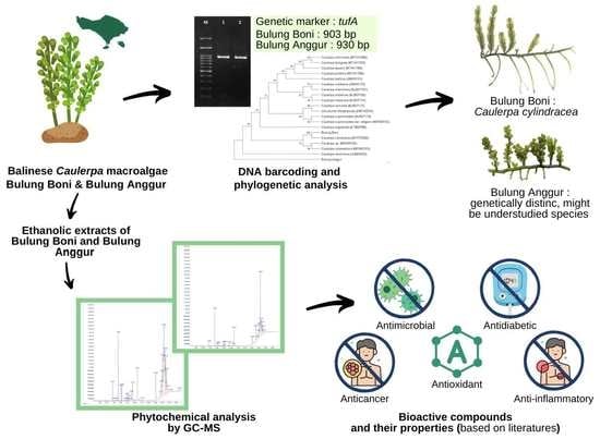
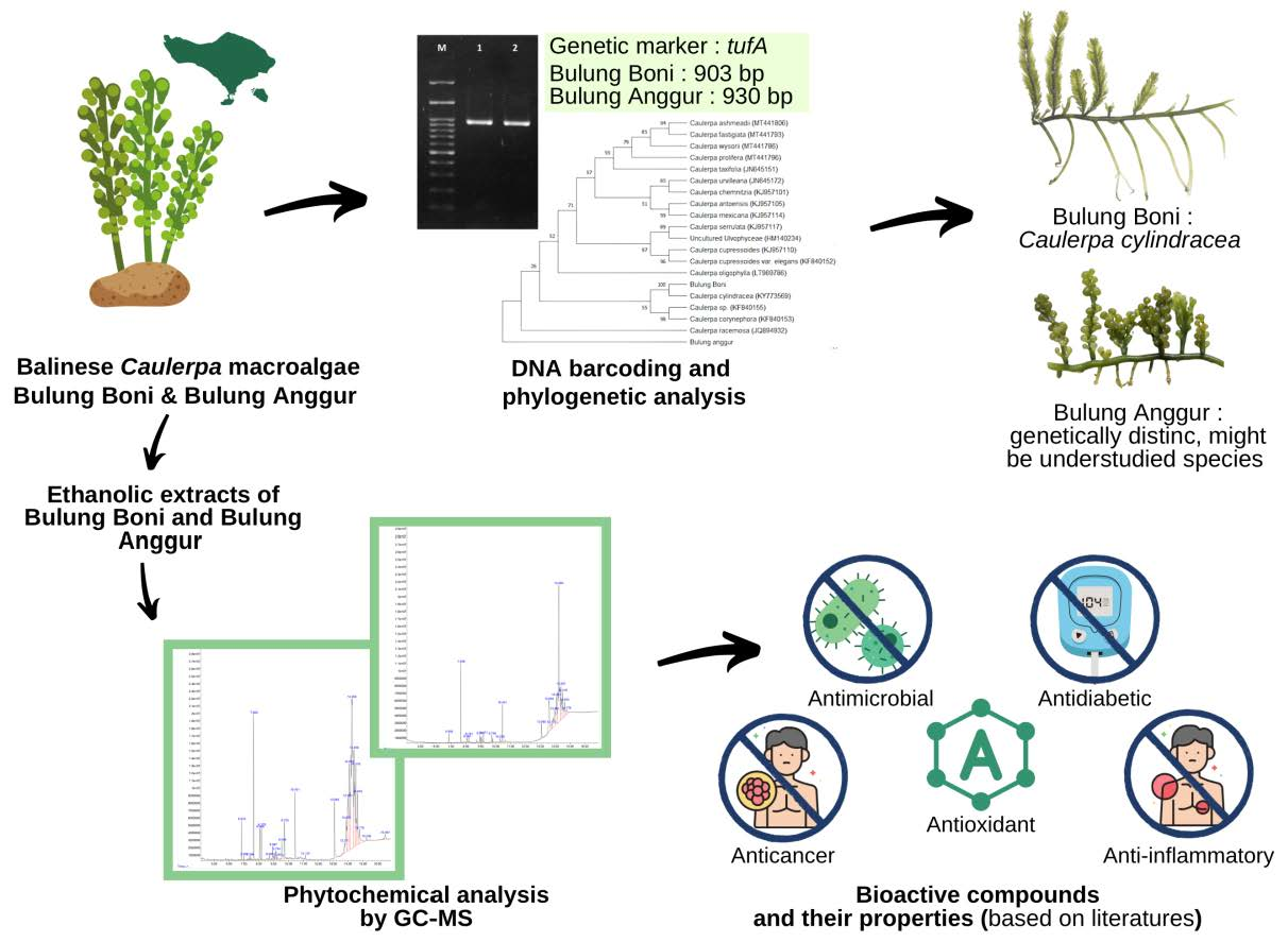
Phytochemical Analysis and Molecular Identification of Green Macroalgae Caulerpa spp. from Bali, Indonesia

by
I Gede Putu Wirawan
1,
Ni Kadek Emi Sintha Dewi
1,
Maria Malida Vernandes Sasadara
2,*,
I Gde Nengah Adhilaksman Sunyamurthi
3,
I Made Jawi
4,
I Nyoman Wijaya
1,
Ida Ayu Putri Darmawati
1,
I Ketut Suada
1 and
Anak Agung Keswari Krisnandika
1
1
Department of Biotechnology, Faculty of Agriculture, Udayana University, Bali 80361, Indonesia
2
Department of Natural Medicine, Universitas Mahasaraswati Denpasar, Denpasar 80233, Indonesia
3
Department of Dermatology and Venereology, Faculty of Medicine, Warmadewa University, Bali 80239, Indonesia
4
Department of Pharmacology, Faculty of Medicine, Udayana University, Bali 80232, Indonesia
*
Author to whom correspondence should be addressed.
Molecules 2022, 27(15), 4879; https://doi.org/10.3390/molecules27154879
Submission received: 7 July 2022 / Revised: 22 July 2022 / Accepted: 25 July 2022 / Published: 30 July 2022
(This article belongs to the Special Issue Bioactive Compounds from Nature: New Research and Prospects)

Abstract

:
The studies of the Bulung Boni and Bulung Anggur (Caulerpa spp.) species and secondary metabolites are still very limited. Proper identification will support various aspects, such as cultivation, utilization, and economic interests. Moreover, understanding the secondary metabolites will assist in developing algae-based products. This study aimed to identify these indigenous Caulerpa algae and analyze their bioactive components. The tufA sequence was employed as a molecular marker in DNA barcoding, and its bioactive components were identified using the GC-MS method. The phylogenetic tree was generated in MEGA 11 using the maximum likelihood method, and the robustness of the tree was evaluated using bootstrapping with 1000 replicates. This study revealed that Bulung Boni is strongly connected to Caulerpa cylindracea. However, Bulung Anggur shows no close relationship to other Caulerpa species. GC-MS analysis of ethanolic extracts of Bulung Boni and Bulung Anggur showed the presence of 11 and 13 compounds, respectively. The majority of the compounds found in these algae have been shown to possess biological properties, such as antioxidant, antibacterial, anticancer, anti-inflammation, and antidiabetic. Further study is necessary to compare the data obtained using different molecular markers in DNA barcoding, and to elucidate other undisclosed compounds in these Caulerpa algae.

Graphical Abstract

1. Introduction

Seaweeds, also known as macroalgae, are eukaryotic and non-flowering plants with no true stem, leaves, or root surrounding their reproductive systems [1,2]. As with terrestrial plants, seaweeds also subsist by photosynthesis. Seaweeds include a variety of pigments and are taxonomically classified into Chlorophyta (green algae that have chlorophyll pigment), Rhodophyta (red algae that have phycocyanin, and phycoerythrin pigments), and Phaeophyta (brown algae that have fucoxanthin pigment) [3].
Seaweed has been consumed for centuries throughout the world, most notably in Asian countries such as Japan, Korea, China, and Indonesia, but on a smaller scale. Seaweeds have also been used for numerous food, fertilizer, and pharmaceutical products [2]. Seaweed species, such as Caulerpa spp., Gracilaria spp., and Euchema spinosum, are utilized and widely consumed in Bali [4,5,6]. Caulerpa seaweeds, locally known as Bulung Boni and Bulung Anggur, have a wide range of biological properties. Julyasih et al. [4] discovered that Caulerpa sp. possesses the highest antioxidant activity compared to Gracilaria spp. and Euchema spinosum. Moreover, Caulerpa extract could help prevent photoaging through its inhibitory effect on MMP-1 and its protective mechanism against oxidative DNA damage, according to Wiraguna et al. [7].
Caulerpa (Bryopsidophyceae) is a multinucleate siphonous coenocyte and its thallus is differentiated into rhizoid, stolon, assimilator, and branchlets (ramuli) [8,9]. The complete tallus of Caulerpa seaweed can reach a length of nearly 1 m (e.g., Caulerpa taxifolia). Stolons, or green prostrate axis, give rise to photosynthetic fronds that are erect (assimilators). These assimilators can be leaf-like or have a central axis (rachis) with lateral branchlets (ramuli) arranged in a variety of patterns and shapes [10].
Although it has been widely taken and used, to date, no research has identified the taxonomic profile of Bulung Boni and Bulung Anggur. According to Farnsworth et al., [11] species identification is essential to documenting, managing, and sustaining organism diversity. The Caulerpa algae are well-known for their great phenotypic plasticity, which refers to the ability of the same species to exhibit a variety of morphological structures in response to changing environmental conditions [12,13]. Accurate identification and characterization of seaweed species are critical for resolving their taxonomic uncertainty, especially for those with commercial interests. Thus, molecular identification is essential to correctly identifying the Caulerpa species. DNA barcoding is a precise and rapid molecular technique for identifying species through the use of short genetic markers [14]. The barcode regions rbcL, matK, and tufA are frequently employed in green macroalgae. tufA is a chloroplast gene that encodes the elongation factor TU. Numerous studies have utilized tufA as a genetic marker for identifying green algae due to its high rate of amplification and sequencing success [9,15,16,17,18,19,20,21,22,23].
Additionally, the chemical compositions of these Caulerpa seaweeds have not been investigated. Identifying the secondary metabolites and their properties in Caulerpa algae will assist in their utilization and the production of algae-based cosmetics and pharmaceutical medications. Therefore, it is necessary to study the bioactive components of these two Caulerpa algae. The objective of this study was to identify two indigenous Caulerpa seaweed species in Bali, Bulung Boni and Bulung Anggur, using tufA sequence as a molecular marker in DNA barcoding, and to analyze their bioactive components using GC-MS analysis.

2. Results and Discussion

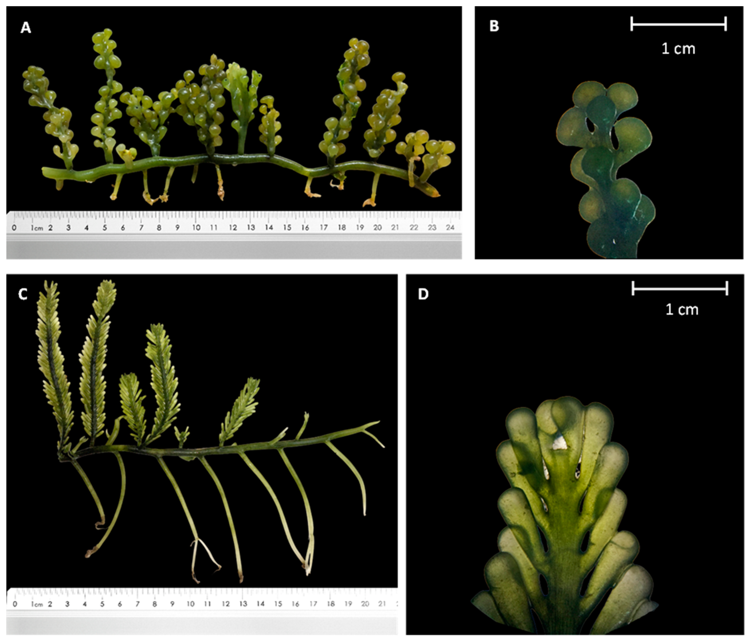
2.1. Morphological Characterization

Bulung Boni and Bulung Anggur, like other Caulerpa algae, have green-colored thalli composed of stolons, rhizoids, and erect fronds or assimilators (Figure 1). Bulung Boni has a distichous assimilator with a maximum height of 10 cm. It also features uncrowded cylindrical or clavate ramuli radially and distichously arranged. On the other hand, Bulung Anggur has an 8 cm irregular assimilator with vesiculated ramuli that have no distinct arrangement.
Caulerpa spp. are known as macroalgae with a high degree of phenotypic plasticity, making precise identification difficult based on morphological observations alone. Morphologically, Bulung Boni resembles Caulerpa cylindracea. C. cylindracea showed a straightforward morphology, with creeping stolons and spherical branchlets with upright shoots and grape-like ramuli, distributed radially or distichously [10,24,25,26]. In contrast, the morphological characteristics of Bulung Anggur are similar to those of Caulerpa macrophysa. Both Bulung Anggur and Caulerpa macrophysa have thallus that can grow up to 3–5 m in length and are found in a lower intertidal and upper subtidal area with strong water movement. Colorless rhizoidal holdfasts hold the prostrate terete, bare branching stolon and erect, terete branches of this seaweed in thick clusters on the sandy–muddy substrate [24].

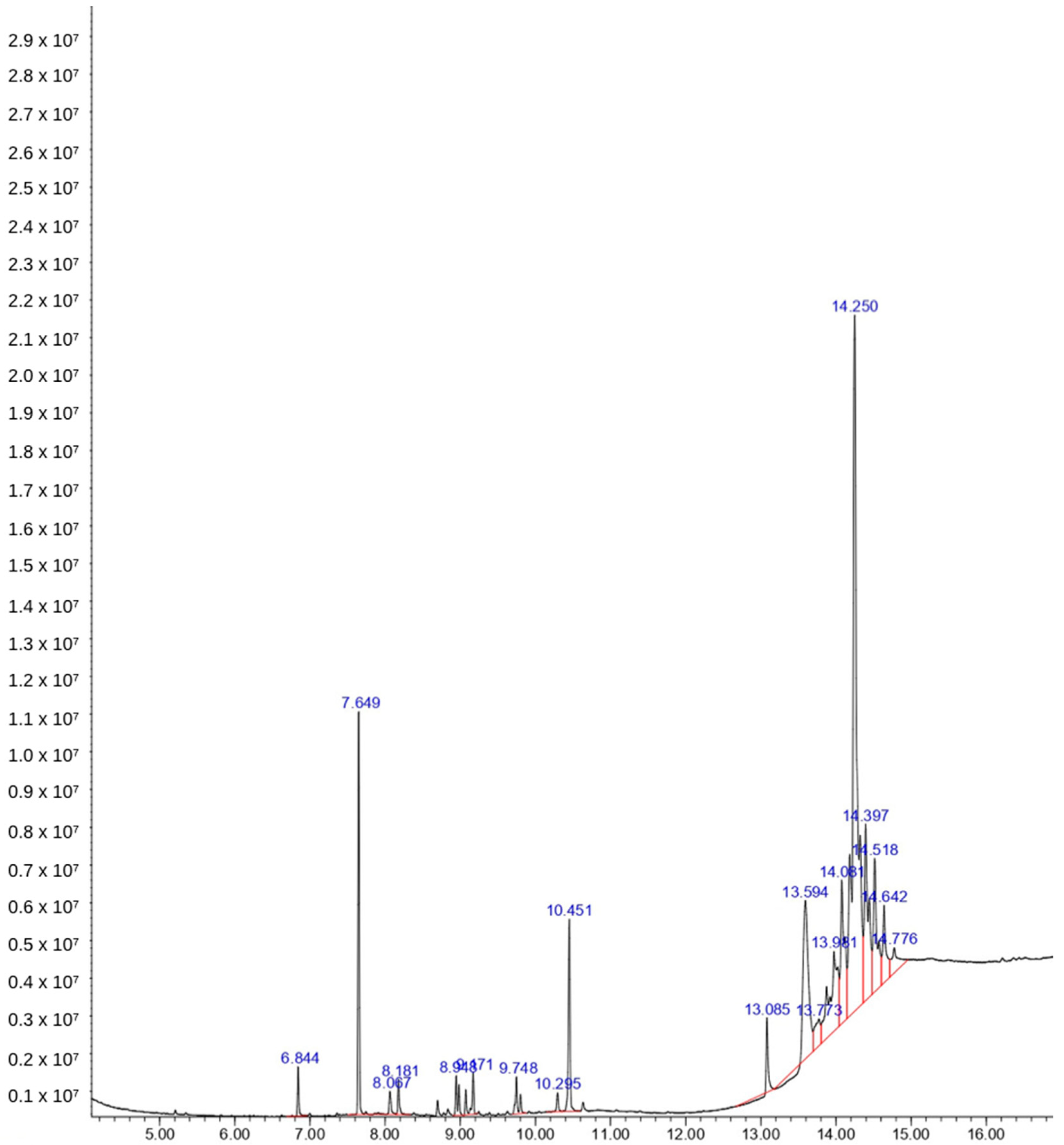
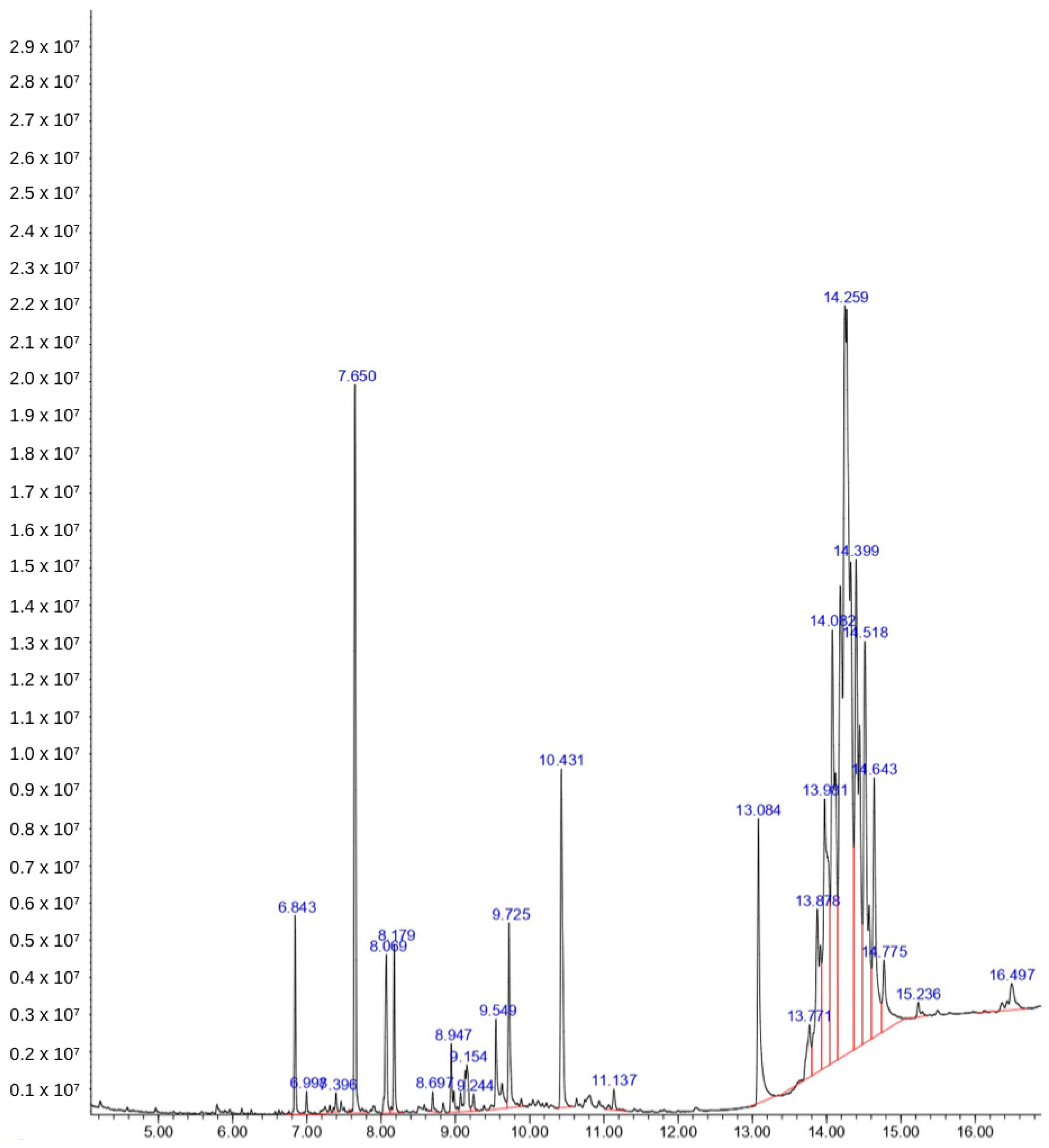
2.2. Phytochemical Analysis

Phytochemical analysis using GC-MS resulted in a chromatogram with several peaks. There are 11 compounds in the ethanol extract of Bulung Boni (Figure 2), and 13 compounds in the ethanol extract of Bulung Anggur (Figure 3). Information on chemical names, retention time (Rt), area under curve (AUC), molecular weight, and formula of Bulung Boni and Bulung Anggur are shown in Table 1 and Table 2, respectively. Cyclohexanamine was the dominant compound in Bulung Boni, as shown by the large area under the curve. Meanwhile, Terephthalic acid is the dominant compound in Bulung Anggur. Based on the phytochemical analysis, it can be observed that the ethanolic extracts of Bulung Boni and Bulung Anggur consist mainly of fatty acid compounds.
The pH (7.74–7.92), BOD5/Five-Day Biological Oxygen Demand (2.8–5.4 mg/L calculated using the Delzer and McKenzie standard method [27]), and temperature (28.9–30.5 °C) of the saltwater in the Serangan island region are within the acceptable range for marine biota; however, salinity (29.9–32.7 ppt) is inadequate [28]. Stress generated by salinity can trigger plants and algae to produce higher secondary metabolites [29]. Hence, this condition might affect the growth of Bulung Boni and Bulung Anggur, resulting in the production of several secondary metabolites.
Several compounds with the highest peak in the chromatogram could not be matched to the database library (they have low quality or probability percentage). These compounds could be quite novel and require additional analysis to clarify their nature. However, various compounds in these algae have been proven to have biological activities. Cyclohexanamine is considered the dominant chemical of Bulung Boni. This chemical can be found in several plants, such as Duddingtonia flagrans and Rhazya stricta [30,31]. D. flagrans showed the dominant presence of cyclohexanamine. This plant possesses a potent nematicidal activity [30]. Cyclohexanamine is also present in Rhazya stricta, which showed antidiabetic activity by inhibiting various hyperglycemic key enzymes [32]. Eicosane, in Bulung Boni, has been identified in several studies. Eicosane, an alkane compound ordinarily present in wax, acts as the plant protector against physical damage and prevents the plant from dehydration [33,34]. Eicosane possesses anti-inflammatory, antibacterial, and antitumor properties [35,36,37]. The compound 1-Dodecanol is also found in the ethanolic extract of Bulung Boni, which has been proven to have insecticide and antibacterial properties [38,39]. Neophytadiene, present in both Bulung Boni and Bulung Anggur, is a terpene compound with potent antibacterial, antifungal, anti-inflammatory, antioxidant, antipyretic, and analgesic properties [40].
Several researchers have discovered that the majority of the compounds in Bulung Anggur have biological properties. Heptadecane and nonadecane are compounds that have been reported for their antimicrobial activity [36]. Hexadecanoic acid, commonly known as palmitic acid, has antioxidant, pesticide, nematicide, hypo-cholesterolemic [41], and antibacterial properties [42]. Phytol is a chlorophyll constituent that possesses antimicrobial, anticancer, anti-inflammatory, antidiuretic, antidiabetic, and immunostimulatory properties [40]. Hexadecanoic acid and phytol are the dominant chemicals of other Balinese seaweed, Bulung Sangu (Gracilaria sp.), which showed an antioxidant and anti-inflammatory activity [5]. Loliolide is a β-carotene derivative that has been extracted from marine algae, such as Undaria pinnatifida, Sargassum crassifolium, Corallina pilulifera, and green alga Enteromorpha compressa [43,44,45,46]. Loliolide has anti-cancer, antimicrobial, antioxidant, anti-inflammatory, and antiaging properties, and is also used in treating diabetes and depression [47,48].
TLC (thin layer chromatography) examination revealed the presence of carotenoids, such as β-carotene, Canthaxanthin, Chlorophyll b, Fucoxanthin, Antheraxanthin, Astaxanthin diester, Astaxanthin monoester, and Neoxanthin in Bulung Boni [49]. However, no carotenoid component was detected in Bulung Boni in this investigation. The absence of carotenoid in Bulung Boni might be due to its infinitesimal peak and consequent under-detection in GC-MS analysis. Furthermore, since carotenoid molecules are heat-labile, the GC (gas chromatography) or GC-MS (gas chromatography-mass spectrometry) methods are ineffective for detecting carotenoids [50].
This research establishes that Bulung Boni and Bulung Anggur are chemically distinct. The difference in the number of compounds could be attributable to the environment wherein they grow (temperature, light intensity, salinity, nutrition, and pH), the biotic factors (pathogen or predator), or their genetic structure. Bulung Boni and Bulung Anggur are presumably different variations or species of Caulerpa that are genetically distinct and produce different secondary compounds. In GC-MS analysis, ethanolic extracts of Caulerpa mexicana and Caulerpa racemosa exhibit different compounds; however, these two Caulerpa algae have simulant biological activities, such as potent anti-inflammatory, antidiabetic, and antioxidant properties [51,52].
Seaweed, or macroalgae, is an excellent source of protein, polysaccharides, lipids, and fiber. Multiple studies have demonstrated the high lipid content of various green macroalgae, especially Caulerpa. In addition, seaweed is also a source of minerals [2]. Further research to investigate protein, amino acids, total sugar, lipid content, and mineral profiles will support the utilization of this Caulerpa seaweed.

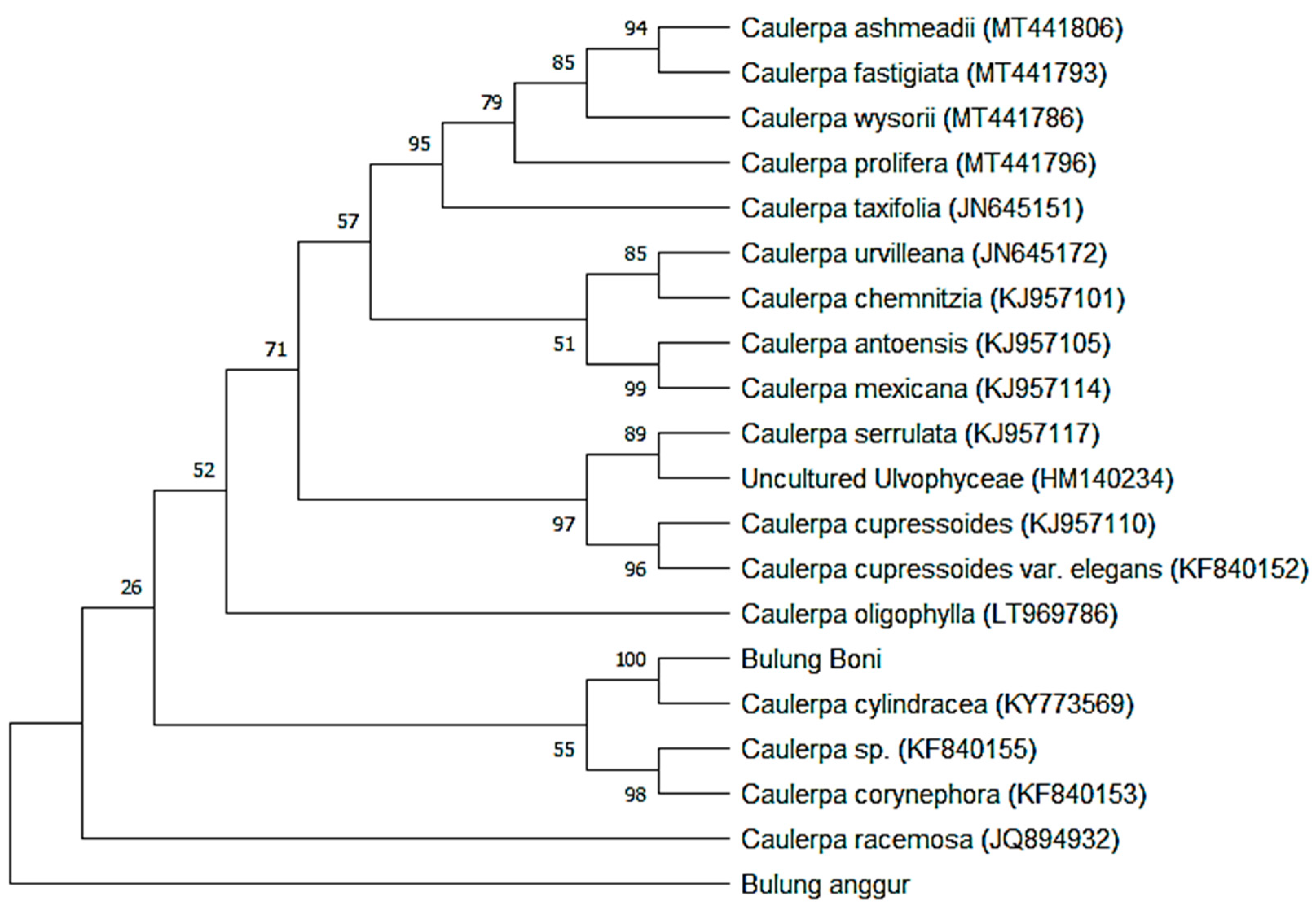
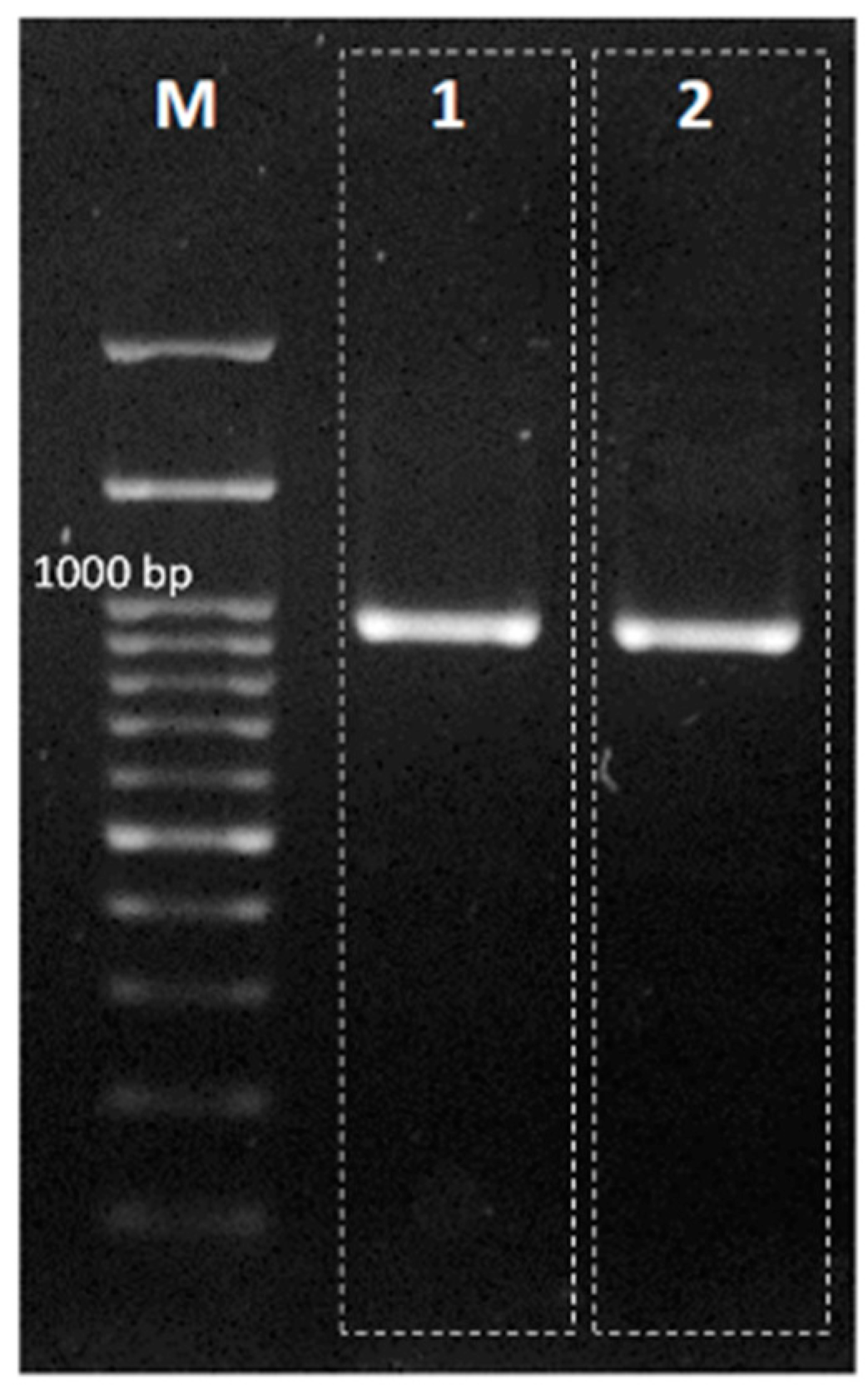
2.3. Molecular Identification

The correct and successful identification of seaweed strains is aided by identification based on comprehensive taxonomy. DNA barcoding is a quick and accurate method. This method is considered the best approach for species identification. DNA barcoding does not require the whole genome sequence but only a short strand of DNA sequences created from the standard marker region of the entire genome [14]. The Bulung Boni and Bulung Anggur were successfully amplified using the tufA primer (Figure 4). The products of PCR amplification of Bulung Boni and Bulung Anggur with tufA primers were 903 and 930 bp, respectively. There were 18 species with an identity percentage of more than 90% (against Bulung Boni and Bulung Anggur) selected from the NCBI-BLAST results and used in the phylogenetic analysis (Table 3).
The amplification of the tufA sequence performed well in our investigation. The tufA gene is an excellent candidate for a molecular marker due to its conserved nature across various taxa [9]. tufA encodes protein synthesis elongation factor Tu. Tu was moved from the chloroplast to the nucleus within the green algal lineage that gave rise to terrestrial plants [53]. tufA, ITS (internal transcribed spacer), and rbcL (rubisco large subunit) molecular markers are frequently used in phylogeny identification. Nevertheless, tufA produced the best results in identifying Chlorophyceae compared to ITS and rbcL [54]. Numerous other studies have also demonstrated that tufA has excellent amplification and sequencing results for identifying green algae compared to other markers [16,17,18].
According to the phylogenetic tree reconstruction results (Figure 5), Bulung Boni is closely related to Caulerpa cylindracea, with a 100 percent bootstrap value. Thus, morphological and molecular identification of Bulung Boni produced similar findings. Additionally, Bulung Boni and Caulerpa cylindracea have a genetic distance of 0.000 (Table 3). Caulerpa cylindracea has been raised from Caulerpa racemosa var. cylindracea because of its genetic independence [12]. C. cylindracea is one of the most invasive algae species that has been reported by several researchers [55,56,57,58]. As a result of its invasive character and high productivity, Bulung Boni has the potential to be utilized and commercialized.
Bulung Anggur, on the other hand, seems to be an outgroup on the phylogenetic tree, indicating that it is genetically distinct from other Caulerpa homologs determined by NCBI-BLAST. Bulung Anggur shares morphological traits with Caulerpa racemosa, particularly with Caulerpa racemosa var. macrophysa (Sonder ex Kützing) [58]. Moreover, Bulung Anggur has the closest genetic relationship distance to Caulerpa racemosa, at 1.200 (Table 3). Thus, it is assumed that Bulung Anggur is a species that is still understudied, given that there are few taxa in the GenBank database that have similar sequences to Bulung Anggur.
The majority of seaweed species growing in Bali’s waters are poorly described taxonomically. These limitations can hinder various aspects, especially in cultivation and nature conservation. In this study, the application of DNA barcodes with the tufA marker has successfully identified one species of Caulerpa seaweed in Bali. This study has been able to locate the proximity of the Bulung Boni species to other Caulerpa species. However, the tufA marker was insufficient to distinguish Bulung Anggur accurately. Before concluding that Bulung Anggur is a novel indigenous strain, it is necessary to conduct additional research to examine its phylogeny using various markers utilized to identify Caulerpa algae, including ITS, rbcl, and rDNA [17].

3. Materials and Methods

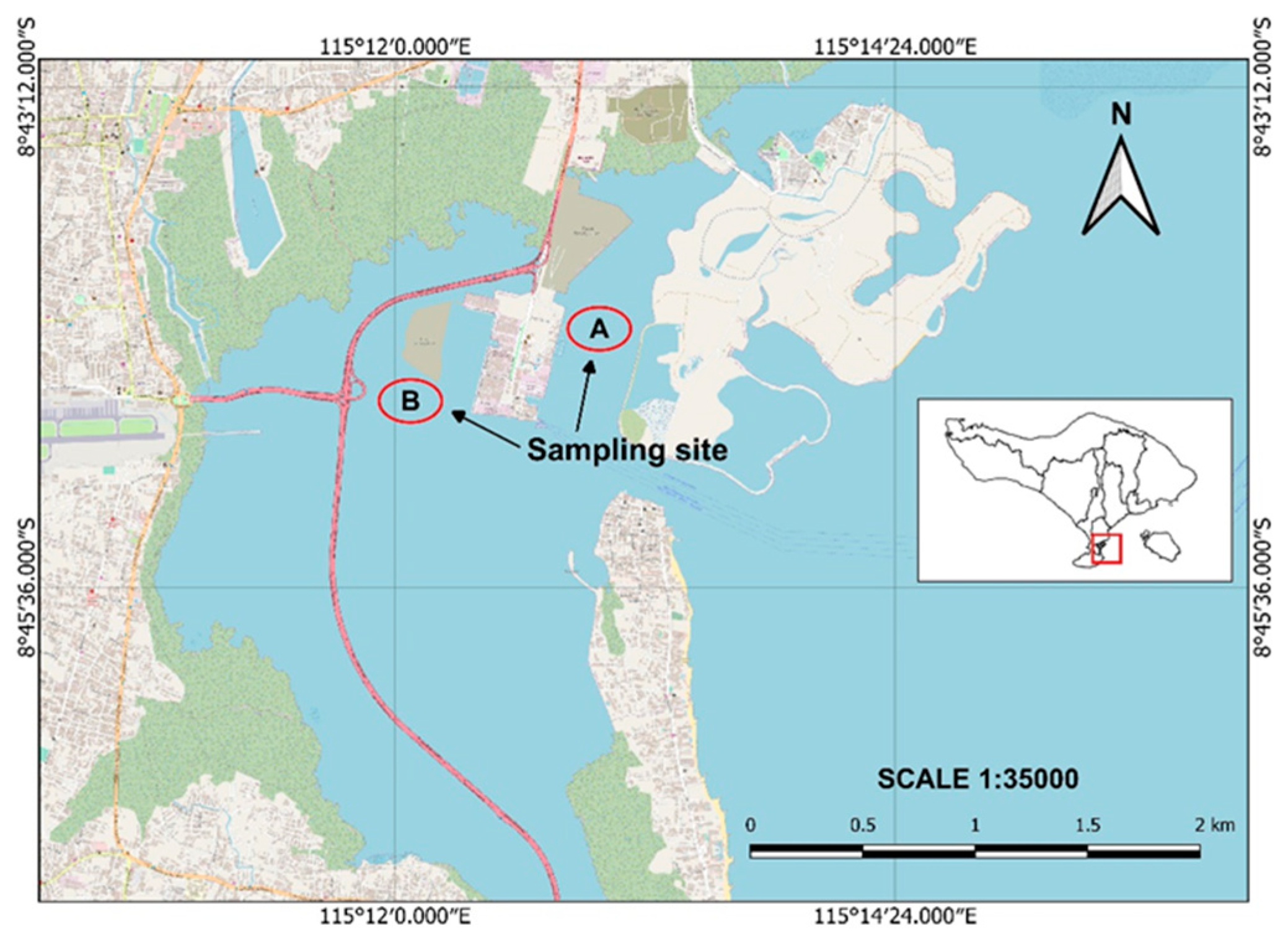
3.1. Sample Collection

The Caulerpa algae, Bulung Boni and Bulung Anggur, were collected from the coastal area around Serangan Island, Bali (Figure 6). The samples were subsequently washed with clean water to remove dirt and epiphytes. Bulung Boni and Bulung Anggur were morphologically identified by referring to the morphological descriptions of several previous studies [10,24,25,59]. Fresh samples were stored in a refrigerator to be used afterward in further analysis.

3.2. Maceration and GC-MS Analysis

Clean Bulung Boni and Bulung Anggur were air-dried in the shade for 2 days and then dried in the oven for 7 days at 45 °C. Dried samples were then powdered using an electric blender. The samples of each alga (15 g) were macerated in ethanol solvent (150 mL). The mixtures were kept for 96 h, then filtered and concentrated using a rotary vacuum evaporator to produce the crude extract.
The crude extract was then subjected to phytochemical analysis using GC-MS. Gas chromatography analysis was carried out on the Agilent 7890B GC (Santa Clara, CA, USA) coupled with a mass detector Agilent 5977B GC/MSD (Santa Clara, CA, USA). A measure of 1.0 µL of the extract was injected into the chromatograph at an injector temperature of 250 °C. The column (Wakosil-II5C18 4.6*200 mm, Richmond, VA, USA) oven temperature was programmed to increase from 70 °C to 290 °C at a rate of 10 °C/min. The run time for GC was 17 min. The identification of chemical compounds and the interpretation of mass spectra of GC-MS were carried out using the Wiley Spectral library.

3.3. DNA Extraction and PCR Amplification

Total genomic DNA of fresh Bulung Boni and Bulung Anggur samples were extracted using the Plant Genomic DNA Mini Kit (GP100, Geneaid, New Taipei City, Taiwan), following the manufacturer’s procedure.
The tufA region was amplified using published primers by Fama et al. [9], i.e., tufA F 5′TGAAACAGAAMAWCGTCATTATGC-3′ and tufA R 5′-CCTTCNCGAATMGCRAAWCGC-3′. PCR amplification was conducted using MyTaq HS Red Mix (Bioline, Bio-25048, London, UK). The PCR reactions were performed with the following profiles: pre-denaturation (94 °C for 2 min), 35 cycles of denaturation (98 °C for 15 s), annealing (50 °C for 30 s), extension (72 °C for 45 s), and followed by termination at 4 °C. Exactly 2 µL of PCR products were visualized using an agarose gel and selected for the sequencing process. The Big Dye Terminator Cycle Sequencing Ready Reaction Kit (Applied Biosystems, Foster City, CA, USA) was used to determine the sequence bi-directionally.

3.4. Computational Analysis

Sequences obtained in this study were compared to sequences in NCBI (National Center for Biotechnology Information) by BLASTN analysis. Accessions with the highest identity percentage were selected, and multiple sequence alignment was carried out with ClustalW (MEGA 11th version, Philadelphia, PA, USA). The phylogenetic tree was constructed using the maximum likelihood method and the Tamura 3-parameter model in MEGA 11 [60]. The robustness of the tree was evaluated using bootstrapping with 1000 replicates.

4. Conclusions

In conclusion, the ethanolic extracts of Bulung Boni and Bulung Anggur may be a source of valuable pharmaceuticals with antioxidant, antibacterial, anticancer, anti-inflammatory, antitumor, and antiaging properties that could be manufactured as cosmetics or healthy processed foods. However, further study is necessary to elucidate other undisclosed compounds in these Caulerpa algae. Furthermore, this study revealed that the Balinese Caulerpa algae, Bulung Boni and Bulung Anggur, are genetically distinct species, with Bulung Boni being strongly connected to Caulerpa cylindracea and Bulung Anggur showing no close relationship to other Caulerpa species. Although the tufA sequence was amplified effectively and generated a robust phylogenetic tree, additional research is required to compare the results obtained using different molecular markers.

Author Contributions

Conceptualization, I.G.P.W. and M.M.V.S.; methodology, I.G.P.W. and N.K.E.S.D.; software, N.K.E.S.D.; validation, M.M.V.S., I.M.J., I.K.S. and I.N.W.; formal analysis, N.K.E.S.D. and I.G.P.W.; investigation, I.G.P.W. and N.K.E.S.D.; resources, N.K.E.S.D. and A.A.K.K.; data curation, I.A.P.D. and I.G.N.A.S.; writing—original draft preparation, N.K.E.S.D.; writing—review and editing, M.M.V.S. and I.G.P.W.; visualization, N.K.E.S.D.; supervision, I.M.J. and I.G.P.W.; project administration, A.A.K.K. and I.G.N.A.S.; funding acquisition, I.G.P.W. All authors have read and agreed to the published version of the manuscript.

Funding

This research was funded by Udayana University, grant number B/78.331/UN14.4.A/PT.01.03/2022.

Institutional Review Board Statement

Not applicable.

Informed Consent Statement

Not applicable.

Data Availability Statement

This study uses data published in GenBank, which can be accessed on https://www.ncbi.nlm.nih.gov/ (accessed on 9 December 2021). These data are used in the BLAST application to compare the sequences in the GenBank database with the sequences obtained in this study.

Acknowledgments

The authors would like to acknowledge the authorities of the Udayana University for the administrative and technical support.

Conflicts of Interest

The authors declare no conflict of interest.

Sample Availability

Samples of the compounds are not available from the authors.

References

  1. Bjerregaard, R.; Valderrama, D.; Sims, N.; Radulovich, R.; Diana, J.; Capron, M.; Forster, J.; Goudey, C.; Yarish, C.; Hopkins, K.; et al. Seaweed Aquaculture for Food Security, Income Generation and Environmental Health in Tropical Developing Countries; World Bank Group: Washington, DC, USA, 2016. [Google Scholar] [CrossRef]
  2. Kolanjinathan, K.; Ganesh, P.; Saranraj, P. Pharmacological Importance of Seaweeds: A Review. World J. Fish Mar. Sci. 2014, 6, 1–15. [Google Scholar] [CrossRef]
  3. Ragunath, C.; Kumar, Y.A.S.; Kanivalan, I.; Radhakrishnan, S. Phytochemical Screening and Gc-Ms Analysis of Bioactive Constituents in the Methanolic Extract of Caulerpa Racemosa (Forssk.) j. Agardh and Padina Boergesenii Allender & Kraft. Curr. Appl. Sci. Technol. 2020, 20, 380–393. [Google Scholar] [CrossRef]
  4. Julyasih, M.K.; Suata, K.; Wirawan, I.; Mantik Astawa, I. Seaweed Extracts Improve Lipid Profile of Wistar Rat. Indones. J. Biomed. Sci. 2011, 5, 224796. [Google Scholar]
  5. Sasadara, M.M.V.; Wirawan, I.G.P.; Jawi, I.M.; Sritamin, M.; Dewi, N.N.A.; Adi, A.A.A.M. Anti-Inflammatory Effect of Red Macroalgae Bulung sangu (Gracilaria Sp.) Extract in UVB-Irradiated Mice. Pakistan J. Biol. Sci. 2021, 24, 80–89. [Google Scholar] [CrossRef]
  6. Wirawan, I.G.P.; Malida, M.; Sasadara, V.; Wijaya, I.N. DNA Barcoding in Molecular Identification and Phylogenetic Relationship of Beneficial Wild Balinese Red Algae, Bulung sangu (Gracilaria Sp.). Bali Med. J. 2021, 10, 82–88. [Google Scholar] [CrossRef]
  7. Wiraguna, G.P.; Anak Agung Pangkahila, W.M.A. Antioxidant Properties of Topical Caulerpa Sp. Extract on UVB-Induced Photoaging in Mice. Dermatol. Rep. 2018, 10, 20–25. [Google Scholar] [CrossRef] [Green Version]
  8. Coneva, V.; Chitwood, D.H. Plant Architecture without Multicellularity: Quandaries over Patterning and the Soma-Germline Divide in Siphonous Algae. Front. Plant Sci. 2015, 6, 287. [Google Scholar] [CrossRef] [Green Version]
  9. Famà, P.; Wysor, B.; Kooistra, W.H.C.F.; Zuccarello, G.C. Molecular Phylogeny of the Genus Caulerpa (Caulerpales, Chlorophyta) Inferred from Chloroplast TufA Gene. J. Phycol. 2002, 38, 1040–1050. [Google Scholar] [CrossRef] [Green Version]
  10. Zubia, M.; Draisma, S.G.A.; Morrissey, K.L.; Varela-álvarez, E.; de Clerck, O. Concise Review of the Genus Caulerpa J.V. Lamouroux. J. Appl. Phycol. 2020, 32, 23–39. [Google Scholar] [CrossRef]
  11. Farnsworth, E.J.; Chu, M.; Kress, W.J.; Neill, A.K.; Best, J.H.; Pickering, J.; Stevenson, R.D.; Courtney, G.W.; Vandyk, J.K.; Ellison, A.M. Next-Generation Field Guides. Bioscience 2013, 63, 891–899. [Google Scholar] [CrossRef] [Green Version]
  12. Belton, G.S.; van Reine, W.F.P.; Huisman, J.M.; Draisma, S.G.A.; Gurgel, C.F.D. Resolving Phenotypic Plasticity and Species Designation in the Morphologically Challenging Caulerpa racemosa-peltata Complex (Chlorophyta, Caulerpaceae). J. Phycol. 2014, 50, 32–54. [Google Scholar] [CrossRef] [PubMed]
  13. Estrada, J.L.; Bautista, N.S.; Dionisio-Sese, M.L. Morphological Variation of Two Common Sea Grapes (Caulerpa Lentillifera and Caulerpa Racemosa) from Selected Regions in the Philippines. Biodiversitas 2020, 21, 1823–1832. [Google Scholar] [CrossRef]
  14. Letchuman, S. Short Introduction of DNA Barcoding. Int. J. Res. 2018, 5, 673–686. [Google Scholar]
  15. Stam, W.T.; Olsen, J.L.; Zaleski, S.F.; Murray, S.N.; Brown, K.R.; Walters, L.J. A Forensic and Phylogenetic Survey of Caulerpa Species (Caulerpales, Chlorophyta) from the Florida Coast, Local Aquarium Shops, and e-Commerce: Establishing a Proactive Baseline for Early Detection. J. Phycol. 2006, 42, 1113–1124. [Google Scholar] [CrossRef]
  16. Saunders, G.W.; Kucera, H. An Evaluation of RbcL, TufA, UPA, LSU and ITS as DNA Barcode Markers for the Marine Green Macroalgae. Cryptogam. Algol. 2010, 31, 487–528. [Google Scholar]
  17. Kazi, M.A.; Reddy, C.R.K.; Jha, B. Molecular Phylogeny and Barcoding of Caulerpa (Bryopsidales) Based on the TufA, RbcL, 18S RDNA and ITS RDNA Genes. PLoS ONE 2013, 8, e82438. [Google Scholar] [CrossRef] [Green Version]
  18. Sauvage, T.; Payri, C.; Draisma, S.G.A.; van Reine, W.F.P.H.; Verbruggen, H.; Belton, G.S.; Frederico, C.; Gurgel, D.; Gabriel, D.; Sherwood, A.R.; et al. Molecular Diversity of the Caulerpa racemosaCaulerpa peltata Complex (Caulerpaceae, Bryopsidales) in New Caledonia, with New Australasian Records for C. racemosa Var. Cylindracea. Phycologia 2013, 52, 222, Erratum in Phycologia 2013, 52, 6–13. [Google Scholar] [CrossRef] [Green Version]
  19. Dumilag, R.V.; Aguinaldo, Z.Z.A.; Alcoriza, V.A.M.; Balucanag, M.P.S.B.; Dulalia, A.R.T.; Sayasa, A.R. DNA Barcodes of Caulerpa Species (Caulerpaceae, Chlorophyta) from the Northern Philippines. Philipp. J. Sci. 2019, 148, 337–347. [Google Scholar]
  20. Karthick, P.; Murthy, K.N.; Ramesh, C.; Narayana, S.; Mohanraju, R. Molecular Authentication of Green Algae Caulerpa (Caulerpales, Chlorophyta) Based on ITS and TufA Genes from Andaman Islands, India. Indian J. Exp. Biol. 2020, 58, 109–114. [Google Scholar]
  21. Darmawan, M.; Zamani, N.P.; Irianto, H.E.; Madduppa, H. Molecular Characterization of Caulerpa Racemosa (Caulerpales, Chlorophyta) from Indonesia Based on the Plastid TufA Gene. Squalen Bull. Mar. Fish. Postharvest Biotechnol. 2021, 16, 101–109. [Google Scholar] [CrossRef]
  22. Vieira, C.; De Ramon N’Yeurt, A.; Rasoamanendrika, F.A.; D’Hondt, S.; Tran, L.A.T.; Van den Spiegel, D.; Kawai, H.; De Clerck, O. Marine Macroalgal Biodiversity of Northern Madagascar: Morpho-Genetic Systematics and Implications of Anthropic Impacts for Conservation; Springer: Dordrecht, The Netherlands, 2021; Volume 30, ISBN 0123456789. [Google Scholar]
  23. Du, G.; Wu, F.; Mao, Y.; Guo, S.; Xue, H.; Bi, G. DNA Barcoding Assessment of Green Macroalgae in Coastal Zone around Qingdao, China. J. Ocean Univ. China 2014, 13, 97–103. [Google Scholar] [CrossRef]
  24. Kumar, J.G.S.; Umamaheswari, S.; Kavimani, S.; Ilavarasan, R. Pharmacological Potential of Green Algae Caulerpa: A Review. Int. J. Pharm. Sci. Res. 2019, 10, 1014. [Google Scholar] [CrossRef]
  25. Belton, G.S.; Draisma, S.G.A.; van Reine, W.F.P.H.; Huisman, J.M.; Gurgel, C.F.D. A Taxonomic Reassessment of Caulerpa (Chlorophyta, Caulerpaceae) in Southern Australia, Based on TufA and RbcL Sequence Data. Phycologia 2019, 58, 234–253. [Google Scholar] [CrossRef]
  26. Santamaría, J.; Golo, R.; Cebrian, E.; García, M.; Vergés, A. Stressful Conditions Give Rise to a Novel and Cryptic Filamentous Form of Caulerpa Cylindracea. Front. Mar. Sci. 2021, 8, 548679. [Google Scholar] [CrossRef]
  27. Delzer, G.C.; McKenzie, S.W. Five-Day Biochemical Oxygen Demand. In U.S. Geological Survey Techniques of Water-Resources Investigations; USGS: Reston, VA, USA, 2003; Volume 7, pp. 1–21. [Google Scholar]
  28. Saraswati, N.L.G.R.A.; Arthana, I.W.; Hendrawan, I.G. Analisis Kualitas Perairan Pada Wilayah Perairan Pulau Serangan Bagian Utara Berdasarkan Baku Mutu Air Laut. J. Mar. Aquat. Sci. 2017, 3, 163. [Google Scholar] [CrossRef]
  29. Gengmao, Z.; Yu, H.; Xing, S.; Shihui, L.; Quanmei, S.; Changhai, W. Salinity Stress Increases Secondary Metabolites and Enzyme Activity in Safflower. Ind. Crops Prod. 2015, 64, 175–181. [Google Scholar] [CrossRef]
  30. Mei, X.; Wang, X.; Li, G. Pathogenicity and Volatile Nematicidal Metabolites from Duddingtonia Flagrans against Meloidogyne Incognita. Microorganisms 2021, 9, 2268. [Google Scholar] [CrossRef]
  31. Albeshri, A.; Baeshen, N.A.; Bouback, T.A.; Aljaddawi, A.A. A Review of Rhazya Stricta Decne Phytochemistry, Bioactivities, Pharmacological Activities, Toxicity, and Folkloric Medicinal Uses. Plants 2021, 10, 2508. [Google Scholar] [CrossRef] [PubMed]
  32. Mahmood, R.; Kayani, W.K.; Ahmed, T.; Malik, F.; Hussain, S.; Ashfaq, M.; Ali, H.; Rubnawaz, S.; Green, B.D.; Calderwood, D.; et al. Assessment of Antidiabetic Potential and Phytochemical Profiling of Rhazya Stricta Root Extracts. BMC Complement. Med. Ther. 2020, 20, 293. [Google Scholar] [CrossRef]
  33. Zhou, G.; Shi, Q.S.; Huang, X.M.; Xie, X.B. The Three Bacterial Lines of Defense against Antimicrobial Agents. Int. J. Mol. Sci. 2015, 16, 21711–21733. [Google Scholar] [CrossRef] [Green Version]
  34. DeLeon, S.; Clinton, A.; Fowler, H.; Everett, J.; Horswill, A.R.; Rumbaugh, K.P. Synergistic Interactions of Pseudomonas Aeruginosa and Staphylococcus Aureus in an In Vitro Wound Model. Infect. Immun. 2014, 82, 4718–4728. [Google Scholar] [CrossRef] [PubMed] [Green Version]
  35. Chuah, X.; Okechukwu, P.; Amini, F.; Teo, S. Eicosane, Pentadecane and Palmitic Acid: The Effects in In Vitro Wound Healing Studies. Asian Pac. J. Trop. Biomed. 2018, 8, 490–499. [Google Scholar] [CrossRef]
  36. Faridha Begum, I.; Mohankumar, R.; Jeevan, M.; Ramani, K. GC–MS Analysis of Bio-Active Molecules Derived from Paracoccus Pantotrophus FMR19 and the Antimicrobial Activity Against Bacterial Pathogens and MDROs. Indian J. Microbiol. 2016, 56, 426–432. [Google Scholar] [CrossRef] [PubMed] [Green Version]
  37. Yu, F.R.; Lian, X.Z.; Guo, H.Y.; McGuire, P.M.; De Li, R.; Wang, R.; Yu, F.H. Isolation and Characterization of Methyl Esters and Derivatives from Euphorbia Kansui (Euphorbiaceae) and Their Inhibitory Effects on the Human SGC-7901 Cells. J. Pharm. Pharm. Sci. 2005, 8, 528–535. [Google Scholar] [PubMed]
  38. Cueto, G.M.; Zerba, E.; Picollo, M.I. Biological Effect of 1-Dodecanol in Teneral and Post-Teneral Rhodnius prolixus and Triatoma infestans (Hemiptera: Reduviidae). Mem. Inst. Oswaldo Cruz 2005, 100, 59–61. [Google Scholar] [CrossRef] [PubMed]
  39. Togashi, N.; Shiraishi, A.; Nishizaka, M.; Matsuoka, K.; Endo, K.; Hamashima, H.; Inoue, Y. Antibacterial Activity of Long-Chain Fatty Alcohols against Staphylococcus aureus. Molecules 2007, 12, 139–148. [Google Scholar] [CrossRef] [PubMed] [Green Version]
  40. Raman, V.; LA, S.; Pardha, S.; Rao, N.; Krishna, N.B.; Sudhakar, M.; Radhakrishnan, T. Antibacterial, Antioxidant Activity and GC-MS Analysis of Eupatorium odoratum. Asian J. Pharm. Clin. Res. 2012, 5, 99–106. [Google Scholar]
  41. Komansilan, A.; Abadi, A.L.; Yanuwiadi, B.; Kaligis, D.A. Isolation and Identification of Biolarvicide from Soursop (Annona muricata Linn) Seeds to Mosquito (Aedes aegypti) Larvae. Int. J. Eng. Technol. 2012, 12, 28–32. [Google Scholar]
  42. Huang, Y.P.; Chang, Y.T.; Hsieh, S.L.; Sandnes, F.E. An Adaptive Knowledge Evolution Strategy for Finding Near-Optimal Solutions of Specific Problems. Expert Syst. Appl. 2011, 38, 3806–3818. [Google Scholar] [CrossRef] [Green Version]
  43. Kimura, J.; Maki, N. New Loliolide Derivatives from the Brown Alga Undaria pinnatifida. J. Nat. Prod. 2002, 65, 57–58. [Google Scholar] [CrossRef]
  44. Kuniyoshi, M. Germination Inhibitors from the Brown Alga Sargassum crassifolium (Phaeophyta, Sargassaceae). Bot. Mar. 1985, 28, 501–504. [Google Scholar] [CrossRef]
  45. Yuan, Z.H.; Han, L.J.; Fan, X.; Li, S.; Shi, D.Y.; Sun, J.; Ma, M.; Yang, Y.C.; Shi, J.G. Chemical Constituents from Red Alga Corallina pilulifera. Zhongguo Zhongyao Zazhi 2006, 31, 1787–1790. [Google Scholar] [PubMed]
  46. Percot, A.; Yalçin, A.; Aysel, V.; Erduǧan, H.; Dural, B.; Güven, K.C. Loliolide in Marine Algae. Nat. Prod. Res. 2009, 23, 460–465. [Google Scholar] [CrossRef]
  47. Grabarczyk, M.; Wińska, K.; Mączka, W.; Potaniec, B.; Anioł, M. Loliolide—The Most Ubiquitous Lactone. Folia Biol. Oecologica 2015, 11, 1–8. [Google Scholar] [CrossRef]
  48. Park, S.H.; Kim, D.S.; Kim, S.; Lorz, L.R.; Choi, E.; Lim, H.Y.; Hossain, M.A.; Jang, S.G.; Choi, Y.I.; Park, K.J.; et al. Loliolide Presents Antiapoptosis and Antiscratching Effects in Human Keratinocytes. Int. J. Mol. Sci. 2019, 20, 651. [Google Scholar] [CrossRef] [PubMed] [Green Version]
  49. Julyasih, K.S.M.; Wirawan, I.G.P. Potential Effect of Macro Alga Caulerpa Sp. and Gracilaria Sp. Extract Lowering Malondialdehyde Level of Wistar Rats Fed High Cholesterol Diet. Int. J. Biosci. Biotechnol. 2017, 5, 71–79. [Google Scholar] [CrossRef]
  50. Rezanka, T.; Olsovska, J.; Sobotka, M.; Sigler, K. The Use of APCI-MS with HPLC and Other Separation Techniques for Identification of Carotenoids and Related Compounds. Curr. Anal. Chem. 2009, 5, 1–25. [Google Scholar] [CrossRef]
  51. Rahul, M.; Suresh, N.; Anil, T.; Arun, M.; Nivedita, G.; Mv, B. GC-MS Analysis of Phytocomponents of Seaweed, Caulerpa Racemosa. Int. J. Pharma Res. Rev. 2014, 3, 39. [Google Scholar]
  52. Sakunthala, M.; Paul, J.J.P. GC-MS Analysis of Ethanolic Extract of Caulerpa Mexicana Sonder Ex Kuetzing from Manapad Coast, Tamil Nadu, India. J. Emerg. Technol. Innov. Res. 2019, 6, 244–247. [Google Scholar]
  53. Baldauf, S.L.; Manhart, J.R.; Palmer, J.D. Different Fates of the Chloroplast TufA Gene Following Its Transfer to the Nucleus in Green Algae. Proc. Natl. Acad. Sci. USA 1990, 87, 5317–5321. [Google Scholar] [CrossRef] [Green Version]
  54. Vieira, H.H.; Bagatini, I.L.; Guinart, C.M.; Vieira, A.A.H. TufA Gene as Molecular Marker for Freshwater Chlorophyceae. Algae 2016, 31, 155–165. [Google Scholar] [CrossRef] [Green Version]
  55. Bentaallah, M.E.A.; Taibi, N.E.; Cantasano, N. Additional New Records of Caulerpa cylindracea Sonder 1845 along the West Algerian Coasts. Indian J. Geo-Mar. Sci. 2021, 50, 122–129. [Google Scholar]
  56. Boumediene, H.K.; Lotfi, B.T. First Record of Invasive Green Algae Caulerpa racemosa Var. Cylindracea in Oran Bay (Western Alegria). Indian J. Geo-Mar. Sci. 2019, 48, 335–342. [Google Scholar]
  57. Cantasano, N.; Pellicone, G.; Di Martino, V. The Spread of Caulerpa cylindracea in Calabria (Italy) and the Effects of Shipping Activities. Ocean Coast. Manag. 2017, 144, 51–58. [Google Scholar] [CrossRef]
  58. Grant, W.M. Molecular Phylogeography and Climate Change Biology of the Invasive Green Marine Macroalgae Caulerpa taxifolia and Caulerpa cylindracea in Australia. Ph.D. Thesis, The University of Adelaide, Adelaide, Australia, 2015; pp. 1–133. [Google Scholar]
  59. Fernández-García, C.; Wysor, B.; Riosmena-Rodríguez, R.; Peña-Salamanca, E.; Verbruggen, H. Species Richness Estimates for the Eastern Tropical Pacific. Phytotaxa 2016, 252, 185–204. [Google Scholar] [CrossRef] [Green Version]
  60. Tamura, K.; Stecher, G.; Kumar, S. MEGA11: Molecular Evolutionary Genetics Analysis Version 11. Mol. Biol. Evol. 2021, 38, 3022–3027. [Google Scholar] [CrossRef]
Figure 1. Morphological images of Bulung Anggur (A,B) and Bulung Boni (C,D).
Molecules 27 04879 g001
Figure 2. Chromatogram of Bulung Boni ethanol extract. The largest AUC showed in 14.250 of retention time, indicating that Cyclohexanamine is the dominant chemical of Bulung Boni.
Molecules 27 04879 g002
Figure 3. Chromatogram of Bulung Anggur ethanol extract. The largest AUC showed in 14.259 of retention time, indicating that Terephthalic acid is the dominant chemical of Bulung Boni.
Molecules 27 04879 g003
Figure 4. Electrophoresis results of PCR amplification of Bulung Anggur (1) and Bulung Boni (2) with tufA as a primer.
Molecules 27 04879 g004
Figure 5. Phylogenetic tree for Bulung Boni and Bulung Anggur based on tufA sequences, constructed with maximum likelihood.
Molecules 27 04879 g005
Figure 6. Map of sampling sites. (A: sampling site of Bulung Anggur; B: sampling site of Bulung Boni).
Molecules 27 04879 g006
Table 1. Identified chemicals in Bulung Boni chromatogram.
ChemicalsRtAUCMolecular WeightFormula
Cyclododecane6.8440.78168.32C12H24
Pentanoic acid7.6496.48102.13C5H10O2
3-Heptadecene8.0670.69238.5C17H34
Eicosane8.1810.65282.5C20H42
Neophytadiene8.9481.08278.5C20H38
11,13-Dimethyl-12-tetradecen-1-ol acetate9.7481.09282.5C18H34O2
1-Dodecanol10.4513.92186.33C12H26O
Androst-5,15-dien-3ol acetate13.5949.03314.5C21H30O2
2-Naphthalene-sulfonic acid13.9817.89924.9C39H31N7NaO13S3+
Cyclohexanamine14.25036.9199.17C7H15N
2-p-Nitrophenyl-1,3,4-Oxadiazol-5-one14.7762.04207.14C8H5N3O4
AUC, Area Under Curve (%); Rt, retention time. Molecular weights are expressed in g/mol. All chemicals are listed from shortest to longest Rt.
Table 2. Identified chemicals in Bulung Anggur chromatogram.
ChemicalsRtAUCMolecular WeightFormula
1-Decene6.8431.24140.27C10H20
Pentadecene6.9980.23210.4C15H30
Dodecane7.3960.35170.33C12H26
8-Heptadecene8.0691.58238.5C17H34
Heptadecane8.1791.10240.5C17H36
Loliolide8.6970.16196.24C11H16O3
Neophytadiene8.9470.65278.5C20H38
1-Nonadecene9.1540.93266.5C19H38
Hexadecanoic acid9.5491.29256.42C16H32O
Phytol10.4313.53296.5C20H40O
1-Hydroxy-1-o-fluorophenyl-4-nitroimidazole-3-oxide13.7710.90100.08C4H5N3O2
Terephthalic acid14.25931.99166.13C6H4(CO2H)2
1,2-Cyclohexanedicarboxylic acid14.39910.69172.18C8H12O4
AUC, Area Under Curve (%); Rt, retention time. Molecular weights are expressed in g/mol All chemicals are listed from shortest to longest Rt.
Table 3. Barcode alignment for Bulung Boni and Bulung Anggur collected from Bali. The best matched identify with NCBI-BLAST are noted with their GenBank accession number, pairwise distance, and similarity percentage (ID).
SpeciesAccession NoBulung BoniBulung Anggur
PairwiseIDPairwiseID
Caulerpa racemoseJQ8949320.01394.10%1.20039.60%
Caulerpa cylindraceaKY7735690.00094.30%1.25040.80%
Caulerpa sp.KF8401550.01097.70%1.25041.00%
Caulerpa corynephoraKF8401530.00996.40%1.22640.20%
Caulerpa serrulateKJ9571170.02695.60%1.25941.10%
Caulerpa cupressoidesKJ9571100.02795.40%1.26241.00%
Caulerpa cupressoides var. elegansKF8401520.02895.20%1.24040.20%
Caulerpa urvilleanaJN6451720.02695.80%1.24641.20%
Ulcultured UnvophyceaeHM1402340.02791.80%1.26940.40%
Caulerpa antoensisKJ9571050.03594.70%1.27541.00%
Caulerpa mexicanaKJ9571140.03494.90%1.25741.20%
Caulerpa chemnitziaKJ9571010.03594.70%1.25041.40%
Caulerpa wysoriiMT4417860.03788.20%1.28940.30%
Caulerpa proliferaMT4417960.03895.00%1.30140.30%
Caulerpa ashmeadiiMT4418060.03994.70%1.24140.60%
Caulerpa oligophyllaLT9697860.02187.90%1.22637.00%
Caulerpa fastigiateMT4417930.04192.30%1.23440.30%
Caulerpa taxifoliaJN6451510.04494.20%1.27840.60%
Bulung boni-- 1.22941.10%
Bulung anggur-1.22941.10%--
Pairwise, pairwise distance; ID, percentage of identity.
Publisher’s Note: MDPI stays neutral with regard to jurisdictional claims in published maps and institutional affiliations.

