Review

Anthocyanins as Key Phytochemicals Acting for the Prevention of Metabolic Diseases: An Overview

1
Department of Agricultural Egineering Sciences, University of Agricultural Sciences and Veterinary Medicine, Manastur Street 3-5, 400372 Cluj-Napoca, Romania
2
Department of Veterinary Medicine, University of Agricultural Sciences and Veterinary Medicine, Manastur Street 3-5, 400372 Cluj-Napoca, Romania
*
Author to whom correspondence should be addressed.
Molecules 2022, 27(13), 4254; https://doi.org/10.3390/molecules27134254
Submission received: 18 May 2022 / Revised: 23 June 2022 / Accepted: 28 June 2022 / Published: 1 July 2022

Abstract

:
Anthocyanins are water-soluble pigments present in fruits and vegetables, which render them an extensive range of colors. They have a wide distribution in the human diet, are innocuous, and, based on numerous studies, have supposed preventive and therapeutical benefits against chronic affections such as inflammatory, neurological, cardiovascular, digestive disorders, diabetes, and cancer, mostly due to their antioxidant action. Despite their great potential as pharmaceutical applications, they have a rather limited use because of their rather low stability to environmental variations. Their absorption was noticed to occur best in the stomach and small intestine, but the pH fluctuation of the digestive system impacts their rapid degradation. Urine excretion and tissue distribution also occur at low rates. The aim of this review is to highlight the chemical characteristics of anthocyanins and emphasize their weaknesses regarding bioavailability. It also targets to deliver an update on the recent advances in the involvement of anthocyanins in different pathologies with a focus on in vivo, in vitro, animal, and human clinical trials.

1. Introduction

Anthocyanins (ANs) are natural components that give beautiful colors of blue, purple, pink and red to leaves, fruits, and flowers, and are responsible for a plethora of health beneficial functions as dietary antioxidants, that can fight free radicals which raise the risk of chronic diseases onset such as: neuronal disorders, inflammatory conditions, diabetes, obesity, cardiovascular diseases and cancer [1]. More than one thousand ANs have been identified in vegetal sources and over 600 are found in natural foods of vegetal sources [2].
ANs are plant water-soluble pigments that are synthesized in the cytoplasm and are stored in high concentrations in the vacuoles. [3,4,5]. In plants, ANs seem to facilitate pollination and seed dispersion, provide foliar protection against biotic and abiotic stress conditions, or act as sunscreen protection [1,6].
Over time, it was accepted that the daily consumption of fruits and vegetables prevents several chronic diseases and prolongs the healthy condition of humans. ANs are one of the main classes of nutraceuticals relevant to well-being due to their antioxidant, anti-inflammatory, antitumoral, and antimicrobial properties [7].
In addition to their importance as human and animal valuable food resources, they have another application in the food industry as natural colorants [8]. One such example is E163, a commercial food additive obtained from grape skin, rich in ANs, and used to color jams, sweets, or beverages. The advantage of consuming ANs is that they are not toxic, even in higher doses. Apart from their health benefits, they have value-added properties due to their antioxidant characteristic or an increasing appealing aspect in food [9,10].
With their vast distribution and low dietary toxicity, ANs are superior compared to other flavonoids in terms of human consumption, with an estimated intake of 200 mg/day [11,12]. However, there is one downfall to them, namely, their color and function stability is highly labile and strongly affected by environmental factors such as pH, temperature, light exposure, oxygen, copigmentation, enzymes, solvents, and chemical structure [13].
This aspect is particularly reflected in their structural fate, once they are ingested and subjected to travel through diverse and extreme changes of environmental conditions, which impacts their final capacity to exert their beneficial functions [7]. The digestion of ANs starts in the oral cavity. The majority of ANs reach the stomach where, due to optimal pH conditions, can partly be absorbed directly in the blood plasma and delivered to the liver, and a higher amount reaches the small intestine, where they suffer structural modifications and produce degradation metabolites, that are also uptaken by the liver and furtherly distributed to other tissues [14].
The purpose of this review is to provide an overview of the chemical properties of ANs and their therapeutical advances, with emphasis on the most recent advances in in vivo and in vitro biological studies with additional clustering of the methods used for their extraction and qualitative/quantitative analysis as well as the physicochemical properties of ANs.

2. ANs Chemistry, Biosynthesis and Stability
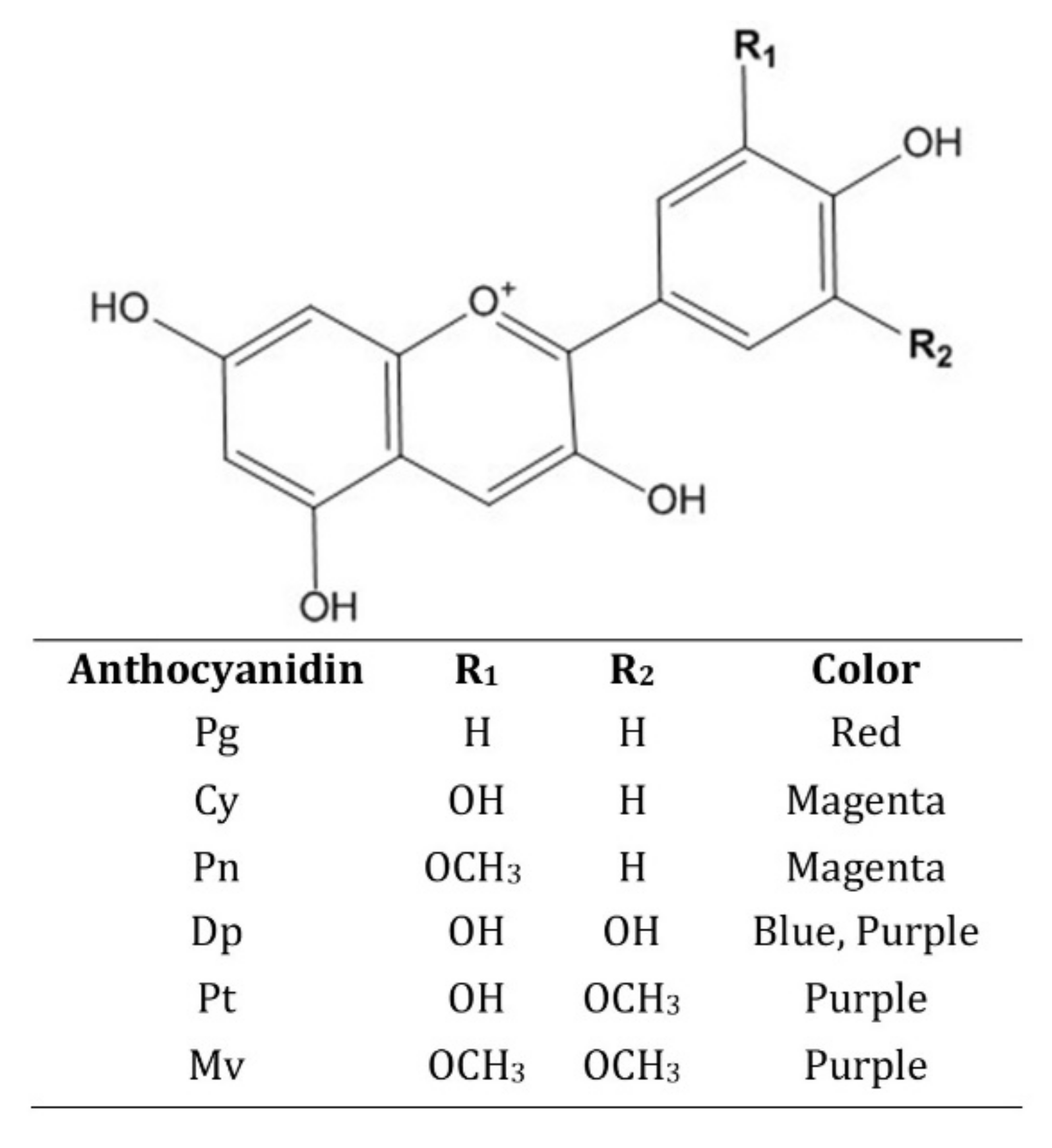
2.1. Chemistry

From a chemical perspective, ANs have a simple carbon chain (C6-C3-C6) structure, which includes them in the subgroup of flavonoids [15]. Specifically, the chemical structure composed of an aromatic ring attached to another heterocyclic ring that contains oxygen, the latter being linked by a carbon–carbon bond to a third aromatic ring is called anthocyanidin, or the aglycon of the anthocyanin [16]. In certain positions on these cyclic structures, hydrogen atoms are replaced by different functional groups [17]. When a moiety of sugar is attached to the aglycon (anthocyanidin), a new chemical compound called anthocyanin is formed [18].
Anthocyanidins are considered to be more stable than ANs. There are around 20 types of such aglycons found in nature, six of them being contained by 90% of all ANs, which are: cyanidin (Cy), delphinidin (Dp), pelargonidin (Pg), malvidin (Mv), petunidin (Pt), and peonidin (Pn) [19]. Their chemical structures and other physico-chemical properties are illustrated in Figure 1. The structural difference between these compounds is the attachment of different radicals in certain key positions on the benzene rings, which is the basis of their different physical properties. For example, Pg is responsible for color variations from orange to dark red, while Cy is responsible for colors from red to magenta and purple [20]. Pn is a compound derived from Cys, thus being responsible for colors such as purple and red-orange [21]. Dp together with its derivatives Pt (a methylated radical) and Mv (two methylated radicals) are the source pigments for the color spectrum from purple to dark blue [22].
Among these six popular anthocyanidins, Cy is the most frequent aglycon found in nature. It is regularly found as glycoside and occurs at most in sources such as chokeberries, blackberries, red cabbage, blood oranges, peaches, and plumbs [23]. Dp is abundant in grapes and, consequently, in red wine, eggplants, pomegranate, and other berries [24]. Pg dominates in beans, strawberries, radish and pomegranate, and flowers such as petunia and roses [25]. Pn was identified in cherries, blueberries, cranberries, mango, or sweet potato [26]. Mv and Pt, the least frequent anthocyanidins, are of no less importance, as they are present in important quantities in red berries, black currant, bilberries, grapes, and red wine [27].
Anthocyanidins can be further modified in versatile ways, an example being their glycosylation by attaching 1 to 3 glycosidic groups such as arabinose, glucose, xylose, galactose, and rhamnose [10]. In turn, these glycosyl moieties can be further glycosylated or acylated with various aliphatic or aromatic chemical groups [28].
The chemical nature of ANs makes them more vulnerable to degradation and structural re-arrangements, which also impact the displayed hue, due to the exposure to various environmental factors such as pH, light, oxygen, enzymes, copigmentation, chemical structure, and temperature. In these conditions, in aqueous solution, ANs can take four different forms: flavylium cation (red), carbinol base (colorless), chalcone (yellow), and anionic quinonoidal base (blue) [29]. The hydroxyl and methoxy groups of anthocyanidins act as auxochromes, electron-donating groups, which cause a bathochromic effect that results in the change of color from red-orange to purple-blue [30].

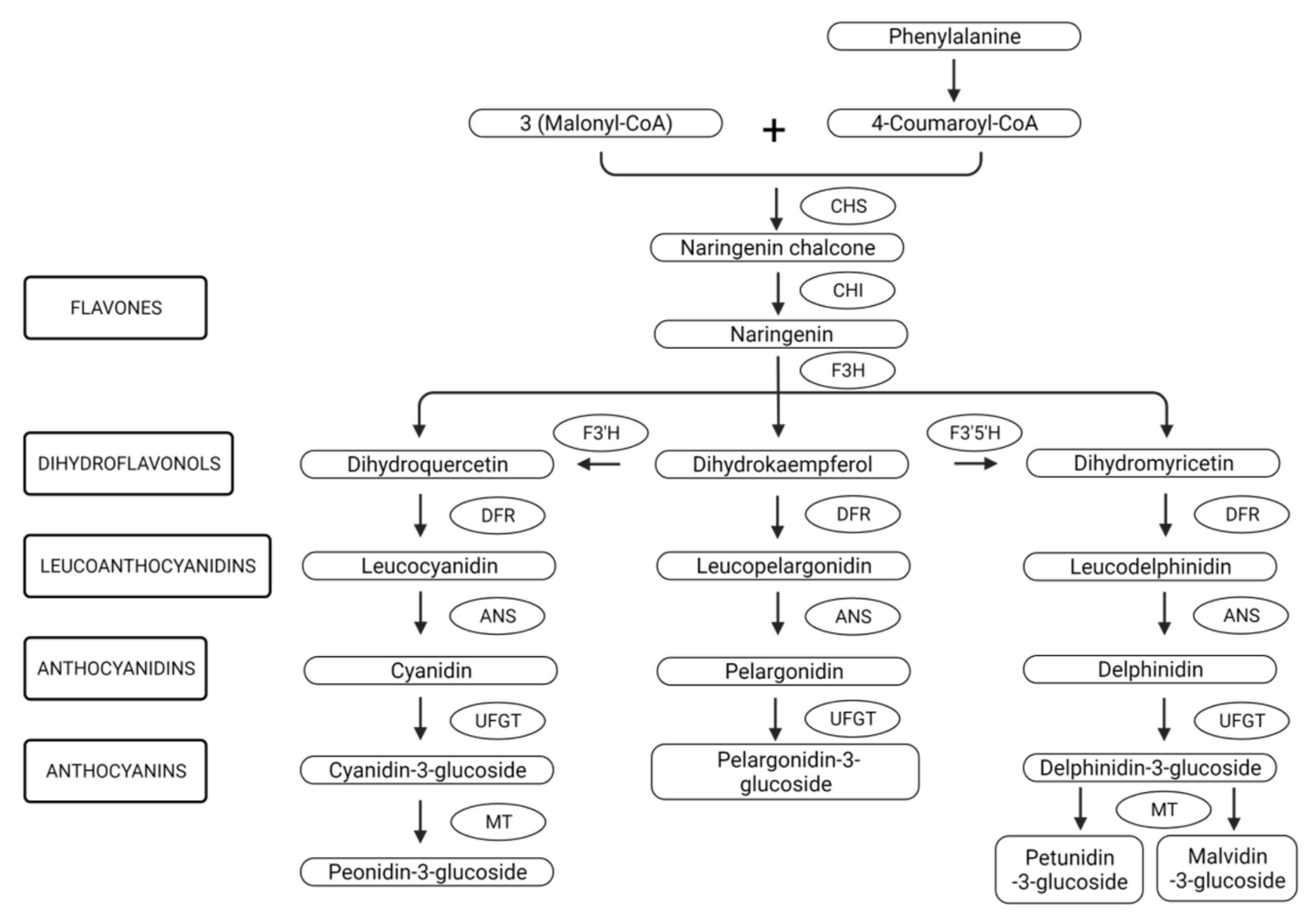
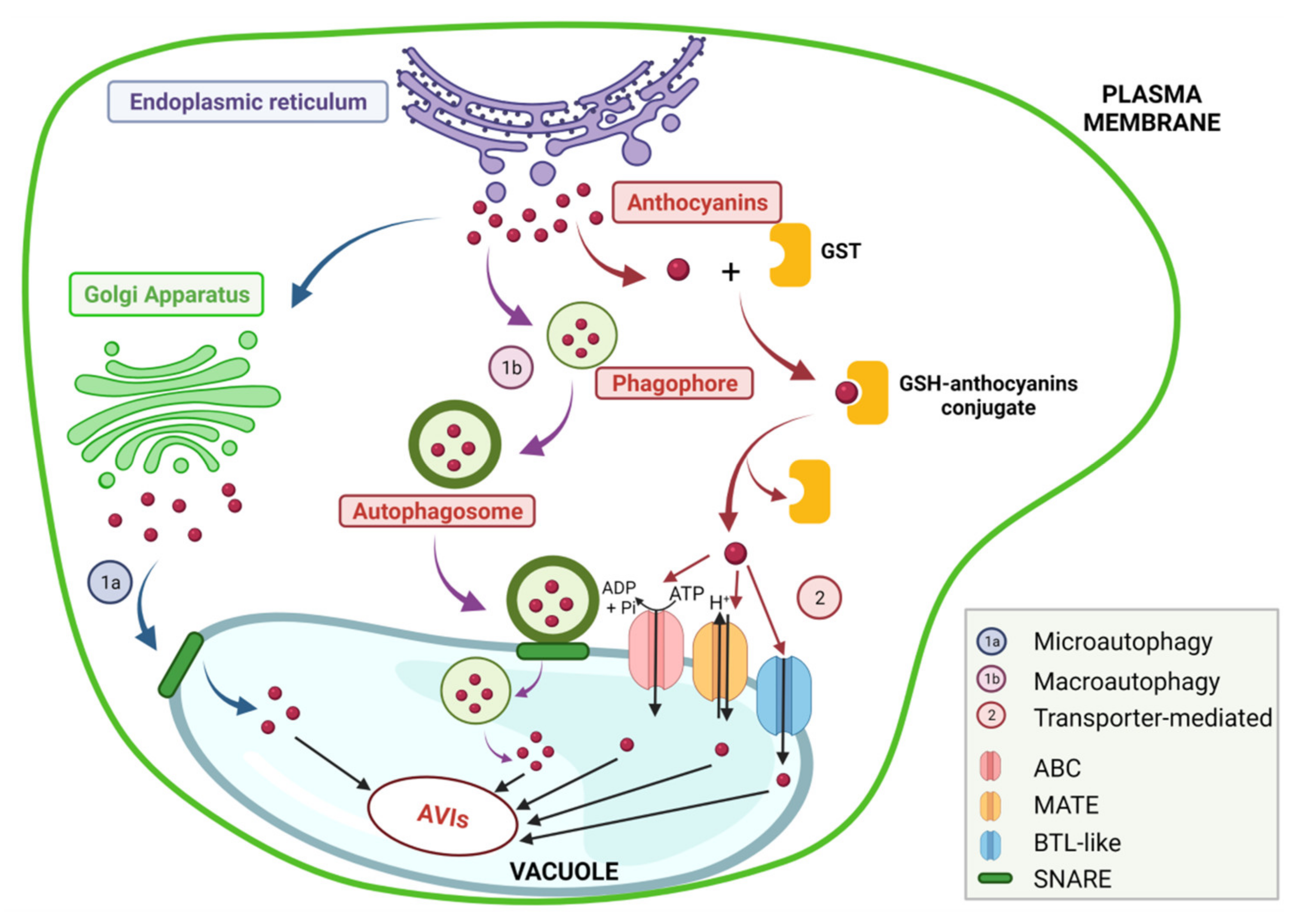
2.2. Biosynthesis, Genetic Regulation and Stability

For many fruits, the occurrence of ANs is a ripening indicator, since the specific color indicates the maturity of the fruit [31]. Depending on the site of production, three groups of fruits have been categorized: fruits that display ANs in their skin, fruits that store ANs both in their skin and flash, and, finally, fruits that produce ANs after an external stimulus, such as light exposure [22]. Regardless of the source, the production of specific ANs varieties is determined by the genetic background, also known as developmental regulation, while their concentration is prone to be influenced by external factors, or environmental regulation [32].
The synthesis of these colored pigments takes place in the cytosolic part of the smooth endoplasmic reticulum [33]. The first step in anthocyanin biosynthesis (Figure 2) is a condensation reaction between three molecules of malonyl-CoA and one molecule of 4-coumaroyl-CoA, the amino acid phenylalanine being the precursor of the last compound [21]. The reaction is catalyzed by the enzyme chalcone synthase (CHS) and the resulting product is naringenin chalcone. This reaction product is further isomerized under the action of chalcone isomerase (CHI) in naringenin [34].
Furthermore, this compound is subjected to a series of enzymatic changes under the action of hydroxylases and reductases with the formation of leuco-ANs. The production of ANs is genetically controlled by a complicated signaling pathway called the MYB–bHLH–WD40 (MBW) complex [22]. The first enzymes involved in the biosynthesis of ANs are regulated by the expression of the early biosynthesis genes (EBGs), namely: CHS, CHI, F3H, F3′H, and FLS). This action is controlled by the activity of the seven R2R3-MYB subgroups of transcription factors (MYB11, MYB12, MYB111), along with MYB75/PAP1 [35]. Furthermore, anthocyanidin synthase (ANS) contributes to the formation of anthocyanidins. Being unstable, various sugars are attached to them via methyltransferases and glucosyltransferases, thus forming anthocyanin molecules [36].
Finally, ANs are transferred and stored in vacuoles by two types of mechanisms (Figure 3): vesicle-mediated transport (micro- or macro-autophagy) or membrane transporter-mediated transport (multidrug and toxic compound extrusion (MATE) transporter, ATP-binding cassette (ABC) transporter, or bilitranslocase (BTL)-like transporters) [37].
The enzymes involved in the final part of the synthesis, also known as late flavonoid biosynthesis genes (LBGs) such as DRF, ANS, and UFGT, as well as the membrane tonopast transporters, ABC and MATE, are activated by the upregulation of the ternary complex MBW (R2R3-MYB, bHLH, and WD40) [38]. The MBW family of transcription factors is considered to be crucial in the biosynthesis of ANs and has been identified in ripened fruits such as woodland strawberries, in large quantities, especially the R2R3-MYB protein [39]. MYB genes that belong to the MBW have also been associated with proanthocyanins production in strawberries, as FvMYB3, FvMYB9, FvMYB11, FvMYB22, FvMYB64, and FvMYB105 genes were expressed especially in the green stage of the strawberries development, which highlights the involvement of the MBW complex in different stages of ripening of the fruits [39]. Moreover, the MBW complex was also observed to participate in the activation of the EBGs in some cases such as corn (Zea mays) [40].
The first motif, R2R3, binds the promoter to the gene, and can both activate or inhibit the expression of a specific gene [41]. The second transcription factor, bHLH, also showed regulatory effects on gene expression and importance in ANs accumulation [42], while the third one, WD40, proved to stabilize the MBW complex [43].
The well-described molecular mechanism of ANs biosynthesis was also associated with the effects of external factors such as light exposure on ANs concentration [32]. For example, UV direct light proved an upregulation of ANs production in apples (cultivars ‘Iwai’, ‘Sansa’, ‘Tsugaru’, ‘Homei-Tsugaru’, and ‘Akane’) and strawberries (Fragaria × ananassa Duchesne var. Elsanta) and a more rapid darkening of the skin color [44,45]. In another study conducted on red apples (Malus domestica), it was observed that the MdMYB1 gene, responsible for the coloration of the apples’ skin, was successfully transcripted when the fruit was exposed to light. However, in the dark, the protein was degraded via the ubiquitin-proteasome pathway, with a direct impact on the color of the apples, which highlights how the light controls the ANs accumulation [46].
Although the genetic regulation mechanism was well described, there are still open questions that require a deeper study. The correlation between the role of the MBW complex transcription factors and their behavior in different environmental conditions and in a wider variety of ANs sources could bring a more specific insight into understanding the developmental and environmental regulations of the biosynthesis process and their intercorrelation [22].
Considering that ANs are extremely unstable and prone to degradation, their stability is another topic of great interest to scientists, especially due to the wide range of applications demonstrated over time for them. Usually, anthocyanins are degraded due to the joint action of internal and external factors [47]. The main factors that influence their stability are: pH, light, oxygen, enzymes, copigmentation, chemical structure, and temperature [48].

2.2.1. Influence of pH

ANs mainly give their source different hues from the color range of glossy orange, red, pink, purple, and blue, as determined genetically [32]. However, there are factors that greatly influence color expression, such as the pH of the environment [7]. The first aspect to consider is that ANs are reactive molecules due to the lack of electrons in the flavylium cation nucleus [20]. Due to their ionic structure, ANs are very sensitive to the pH of the environment. Therefore, at pH 1, the flavylium cation form is predominant and responsible for its red color, and, at the same time, offers the greatest stability of ANs in water [7]. At pH values between 2 and 4, the blue quinonoidal base is predominant, and as the pH increases, the colorless carbinol pseudobase and the colorless chalcone appear [18].
It is worth mentioning that at pH above 7, the degradation of the compound takes place, most ANs being more stable in acidic conditions than in alkaline ones [7]. For example, ANs from Cabernet Sauvignon red wine were investigated in simulated gastrointestinal digestion [49]. The majority of the 22 ANs initially detected in the wine were observed to be stable at acidic pH, during the gastric digestion process. However, the intestinal digestion simulation, which occurs at basic pH values, leads to a dramatic reduction in the ANs content [49]. Another study tested ANs extracted from blueberries (Britewell rabbiteye) that were exposed to 40 °C, in the dark, but at different pH values: 3.0, 4.0, 5.0, 6.0, 7.0, 8.0, and 9.0 [50]. The highest TAC (total anthocyanins content) was measured at pH 3.0 (329.459 ± 1.235 mg/100 g fresh fruit), and a continuous decrement of concentration was observed inversely proportional to the pH value: 110.388 ± 0.409 mg/100 g fruits at pH 7.0, and 49.767 ± 1.079 mg/100 g fruits at pH 9.0 [50].
The color of ANs is affected by the presence or absence of water [51]. When the water content decreases, the hydration process is reduced, being favorable for color stability. As water concentration decreases, few free water molecules are available for proton accommodation; therefore, the flavylium cation is stable [52]. In water, the pKa constant value is 3.8, while in a water-deprived environment, it decreases to 4.8, which gives a red color [53].
As the pH value increases from slightly acidic to neutral, deprotonation of the flavylium cation starts and generates the blue quinonoidal species, highly unstable at low pH. At basic pH, the deprotonation reaction carries on and forms purple quinonoid anions (Figure 4) [54].

2.2.2. Temperature and Oxygen Influence

Temperature is a key factor for better stability and preservation. The temperature at which the ANs are exposed to has great importance in the food industry, because the storage conditions of anthocyanin-rich beverages influence the quality of the product and, additionally, the maintenance or loss of ANs [55] and certain unfavorable conditions can hinder the ANs’ biosynthesis [56]. The main events that occur upon ANs as a result of thermal exposure are deglycosylation, cleavage, water nucleophilic attack, and polymerization [57,58].
A study compared the effect of two storage temperatures: 35 °C and 25 °C, and revealed that the higher temperature reduced the total content of ANs to less than half the content in grapes (Vitis vinifera L. cv. Cabernet Sauvignon) stored at 25 °C [59]. At 40 °C, their color changed from red to orange, despite keeping a constant acidic environment [60]. Heating the ANs shifts the equilibrium structure towards the colorless forms resulting in the loss of the desired red pigmentation.
Marquez and coworkers [38] tested three different temperature options while extracting ANs through controlled dehydration of Tempranillo red grapes: 30 °C, 40 °C, and at alternations between 40 °C and 15 °C, every 12 h. The best results were obtained in the second experiment; the grapes dried at 40 °C had the most intense color, which indicates the highest concentration of ANs. The must obtained at 30 °C still contained significant amounts of ANs but displayed a slight brown color. The least satisfactory result was obtained in the final experiment because the reduction in the temperature value allowed oxygen to diffuse through the grape skin and caused degradation reactions of ANs and loss of the red color [61].
Grape juices of ”Merlot” and ”Ruby” grape cultivars were passed through a series of experiments to evaluate ANs exposure at 2, 25, and 35 °C storage temperatures [62]. After 365 days, the ANs (Mv-3-glucoside, Dp-3-glucoside, Pt-3-glucoside, Pn-3-glucoside, and Cy-3-glucoside) were stored at 25 and 35 °C and degraded, in both cultivars (95–99%), compared to the juice kept at 5 °C (50–60%—“Merlot”; 74–81%—“Ruby”). Cy and Dp aglycones proved to have the highest sensitivity to high temperatures. There was also a strong correlation between high-temperature degradation and the change of color, highlighting that low temperatures are favorable for a longer preservation of ANs.
Moreover, exposure of Cy-3-glucoside and Cy-3-rutinoside from black rice aqueous extract to increasing temperatures from 100 to 165 °C showed a greater negative impact on ANs degradation, compared to increasing the pH value from 2.2 to 6. In addition, Cy-3-rutinoside has been proven to be the most resistant to thermal and pH changes [63].
It was also demonstrated that the degradation process of ANs is amplified by high temperatures with the presence of oxygen [62,64]. Both peel and flesh of Malus profusion fruits were exposed to two temperature regiments, each one divided into two intervals with an exposure time of 12 h:12 h, as follows: (1) room, RT (18 °C:25 °C), and (2) high, HT (33°C:25 °C), both in hypoxic (2%) and normoxic (21%) environments. A methanol:formic acid (70%:2%) ANs extract was obtained from frozen fruits and cy-3-galactoside was measured as the highest anthocyanin compound (≈800 µmol Kg−1 FW). After 1 week of treatment, RT treatments marked an increase in the cy-3-galactoside concentration as a consequence of ANs biosynthesis, regardless of oxygen conditions. However, in HT conditions, a significant concentration decrease was marked for cy-3-galactoside, with higher degradation in hypoxic conditions (67%) than in normoxic (54%).
Furthermore, HT was directly correlated with an upregulation of the MpPOD1, MpPOD8, and MpPOD9 genes transcription, furtherly coupled with enhanced activities of peroxidases and H2O2, next responsible for the enzymatic degradation of ANs. On the other hand, maintaining samples at RT stimulated the ANs synthesis possibly by stabilizing physiological conditions such as pH and H2O2 concentration, which indicates, once more, the important role that temperature plays in the fate of ANs [65].
The presence of oxygen also brings a high contribution to ANs degradation, and it usually accompanies other factors for a more dramatic infliction on ANs stability but also works on its own. To prove this point, fresh-cut strawberries were stored in three different conditions with a focus on bioactive composition modifications [64]. Five packaging conditions were evaluated (2.5 kPa O2 + 7 kPa CO2, 10 kPa O2 + 5 kPa CO2, 21 kPa O2, 60 kPa O2, 80 kPa O2) for storage efficiency, for 21 days, at 4 °C. The ANs were extracted with methanol, and it was observed that their content had different behaviors, with the best antioxidant preservation at low oxygen values (2.5 kPa O2 + 7 kPa CO2). However, oxygen concentrations have mostly been linked to a postponed degradation of thermally handled ANs; namely, in the abovementioned study [64], high temperatures coupled with low oxygen concentrations led to the upregulation of POD genes, which influence other metabolic pathways that inhibit the biosynthesis of ANs [66].
Oxygen was also associated with fluorescent light as an enhancer for ANs decay [67]. Cranberry ANs were extracted with acidified ethanol and exposed for 14 days to fluorescent light, in a low oxygen environment. In oxygen-free conditions, light proved to have an insignificant effect on ANs degradation. However, when traces of oxygen were present, the pigments were oxidized and their degradation was accelerated [67]. This strongly indicates the altogether connection between different factors which act synergistically on the fate of ANs.

2.2.3. Light

It is generally accepted that the production of ANs is stimulated under stress conditions when exposed to light, as was shown in several studies in the literature [68,69,70]. Moreover, ANs have the capacity to absorb light, which enables them to act as protective shields for cells such as retinal pigment epithelium (RPE) cells, against light damage, in vitro. When RPE cells were treated with different concentrations of the methanolic, purified blueberry methanolic extract (0.1, 1, and 10 µg/mL extract) and then exposed to white light irradiation of 2500 lux (420–800 nm) for 12 h, the expression of VEGF was downregulated to normal values and cellular senescence and apoptosis were suppressed [71]. On the other hand, excess light can cause photooxidation of pigments.
One such example was investigated on the fate of an ethanolic blackberry anthocyanin-rich extract with an initial concentration of 106 mg·L−1 Cy-3-glucoside, stored in darkness and also exposed to light [72]. One week of light exposure at 3968.30 lux determined the ANs to change color, and also induced the degradation of 76% of the total monomeric ANs concentration, while darkness storage maintained their chemical and color stability for a longer period of time and also recorded only 29% degradation of monomeric ANs [72].
However, to end on a more positive note, it was demonstrated that different light affects ANs unevenly. In one study, three different light ranges were investigated (white, red, and blue) in vivo, on strawberry fruits, in order to assess their role in ANs accumulation [73]. Strawberries were exposed to three light-emitting diodes (LED) and the ANs content was measured on the 25th day after flowering. It was demonstrated that red and blue light contributed to the high expression of key factors involved in anthocyanins biosynthesis and, therefore, led to the increase in total ANs content (TA), Pg-3-glucoside, and Pg-3-malonylglucoside concentrations, while white light treated strawberries reported the lowest concentrations of ANs [73].

2.2.4. Copigmentation/Glycosylation and Acylation

Copigmentation is a phenomenon that exclusively occurs in the class of ANs among polyphenols, and contributes to their chemical stability [74]. Copigmentation is simply described as a noncovalent hydrophobic π–π interaction between an anthocyanin and a colorless organic molecule, which enhances the color and increases the stability of the molecule. For example, Dp and Pt have free hydroxyl groups on their B ring, which are easily polarizable and great sites for complexing such molecules [75].
The colorless molecules associate with the pyrylium ring of the anthocyanin without any chemical bonding. This proximity creates changes in the distribution and density of electrons, which cause UV–VIS absorption modifications of ANs [76]. These reactions can be identified as elevated λmax maximal absorption wavelengths compared to the uncomplexed ANs, known as the hyperchromic effect, and also, as changes in the λmax shift towards higher values (red-bluish color as bathochromic shift) which means that the complexed molecule will have a longer wavelength absorption [77]. For example, the interaction of one specific anthocyanin with different types of copigments can lead to different colorations of the molecule [75].
Three types of copigmentation have been classified so far: self-association (hydrophobic interactions between different ANs), intramolecular (association between the aromatic anthocyanidin and one or more acyl moieties, that are part of that anthocyanin), and intermolecular (Ans-colorless compounds: flavonoids, phenolic acids, amino acids, organic acids, alkaloids, purines, or Ans-metals: Al, Fe, Cu, Mg) [18,78,79]. Self-association and intramolecular reactions usually occur in flowers, whereas intermolecular copigmentation is mostly present among berries and other fruits [77].
In nature, especially in flowers, copigmentation has been optimized and engineered for optimal coloring and lasting stability. Copigmentation is also of great importance in the food industry since the association between food products and color is so strong [80]. One such example is wine, with around 50% of its color accountable for copigmentation reactions. However, in different storage conditions, a decrease in the concentration of ANs has been observed, which suggests the codependency of copigmentation with other environmental factors, such as pH or temperature [81]. ANs and copigments (total flavonols) contents were measured in Merlot and Syrah wines obtained from Greek grape cultivars, and it was discovered that after one month of storage, the concentration of anthocyanins decreased rapidly, whereas the copigments remained constant, due to their higher stability [82].
In another study, Cy-3-glucoside was copigmented with three phenolic compounds (ferulic acid, dopamine, and (+)-catechin), and their stability was evaluated according to the pigment:copigment molar ratio (1:1, 1:10, 1:100), in a pH range (pH 3–7) and at different temperatures (20, 30, 40, and 50 °C) [83]. The highest hyperchromic shift was observed in Cy-3-glucoside association with (+)-catechin, at molar ratio 1:100, pH 3 and 20 °C, followed by ferulic acid and finally, dopamine. (+)-catechin was also used to complex ANs from mulberries juice and significantly reduced their degradation during pasteurization, at 80 °C, for 15 min. The study showed that copigmentation plays an important role in maintaining the stability of ANs. However, the stability of the copigmented complex is itself dependent on various factors, among which the chemical structure of the copigment ((+)-catechin had the highest number of free hydroxyl groups), and also the temperature and the pH value of the environment, with best stability results at low values of both factors [83].
As already mentioned in the section above, the chemical structure of ANs is linked with their activity including intramolecular copigmentation. Specifically, the number, type, and position of the sugar involved in the glycosylation reaction, as well as the decoration of ANs with different types of glycosyl and acyl units, significantly influence the stability, bioavailability, antioxidant, and anticancer activity of ANs [19].
As mentioned above, anthocyanidins (the aglycones) are rarely found naturally, as they are very unstable. ANs’ chemical and physical properties are influenced and modified by glycosylation and acylation, and they change the molecular size and polarity. Water solubility is increased by glycosylation and decreased by acylation. Moreover, glycosylation increases stability due to its role in the formation of an intramolecular H-bonding network [78].
Furthermore, acylation of ANs can assure a higher stability and this form of intramolecular pigmentation structure offers protection against nucleophilic water attack [79]. Moreover, the number of acyl groups is also relevant. When compared, mono-acylated ANs from red potato showed lower stability than di-acylated ANs from red radish. A higher number of acyl groups have a “sandwich-like” structure that protects the positively charged pyrylium nucleus by positioning themselves on either side of the chromophore, due to hydrophobic interaction [74,79]. Another study that compared extracts of chokeberries, containing monoglycosylated ANs, and black carrot extracts, with 60%:40% acylated:glycosylated ANs, showed that acylated ANs had higher stability, while glycosylated showed the highest antioxidant content when extracted in methanol, possibly because the addition of acyl groups on the sugar blocks the hydrogen transfer from hydroxyl groups to the unpaired electrons. The chemical structure was also correlated with the extracting solvent since glycosylated ANs preferred methanol, whereas acylated ANs were extracted better in ethanol [84].

2.3. Effects of Extraction Methods on ANs

Extraction has a long history of use in the medicine and food industries and dates back to ancient times. Moreover, natural colorants sales have grown in the past 13 years at an annual rate of over 7%, in a more and more urgent attempt to replace synthetic dyes [85]. Alongside the advance in industry and technology, the market demand for a higher production rate of natural bioactive compounds implies the use of more efficient technologies to obtain higher yields of ANs [86].
The extraction of ANs is a fundamental process that refers to the conversion of a natural or raw matrix rich in ANs to a sample exclusively containing ANs [87]. An efficient extraction method for ANs includes high recovery, reduced potential of degradation or alteration, economical affordability, time-consuming optimization, and environmental friendliness [86]. However, the sensitive physico-chemical profile of ANs makes them highly susceptible to degradation and loss of stability and biological activity. In order to overcome these obstacles, vast knowledge about the factors that influence ANs’ stability and function is required [88]. Therefore, great attention should be paid to factors such as the matrix source of ANs (solid/liquid/powder), the collection time of the vegetable or its parts, the solvent of extraction, the size of the sample, the chemical structures of anthocyanins that are expected (glycosylated/acylated), the temperature and the type of extraction process, in order to provide a maximum presence of anthocyanins compounds [86,89].
By far, the most commonly used method is organic solvent extraction. The selection of the extracting solvent is one of the most important steps in achieving a proper extraction and high yield of recovery. Although methanol is the most popular choice with high extraction yield [84], it should be considered toxic and is not recommended for food or nutraceutical purposes. Searching food-friendly solvents with similar or close efficiency, it has been discovered that ethanol also assures high ANs recovery, closely followed by acetone. Water, although the most adequate for the food industry, has a very poor output in the final ANs recovery [90]. Liquid/liquid partition has also been used as a way of ANs purification, using a combination of solvents at different ratios such as acetone:chloroform/ethyl acetate/water [91]. Another crucial aspect is the acidification of the extracting solvent, in order to obtain superior recovery yield and ensure a better preservation of ANs [92]. Hydrochloric, formic, acetic, and trifluoroacetic acids are amongst the most utilized acids [85,93].
ANs are very responsive to high temperatures which affect the structure, stability, color, and composition. For this reason, ANs should be handled at temperatures as low as possible (below 40 °C) [50]. For example, in the process of solvent removal by rotavapor, most studies used 35–40 °C as the optimal temperature range for good extraction and maintenance of ANs stability, depending on the extraction method [89]. For methods that require shorter extracting time, higher temperatures could be used (100 °C), for a short time, e.g., 5 min [94].
To choose the best extraction method, a range of technologies and methods that have been described and optimized recently, sometimes difficult to make the best selection. Numerous extraction techniques approach modern and advanced technology since they represent the founding principle in the current urge to develop more rapid and efficient methods [86]. Some of the most utilized traditional extracting methods comprise maceration, digestion, infusion, percolation, reflux extraction, and Soxhlet [95]. They have been successfully used for decades, being simple without expensive materials or equipment.
Although the conventional techniques are easy to perform and are satisfactory in results, there are some downfalls such as the use of large volumes of toxic solvents for prolonged extracting amounts of time, which demand more efficient and ergonomic methods. Modern techniques have been developed with the means of replacing the tedious traditional methods that cannot ensure a scalable route for mass production [96]. Some of the most utilized modern techniques in the extraction of ANs are supercritical fluid extraction (SFE), pressurized liquid extraction (PLE), microwave-assisted extraction (MAE), ultrasound-assisted extraction (UAE), ultrasound/microwave-assisted extraction (UMAE), pulsed electric field-assisted extraction (PEF), enzyme-assisted extraction (EAE), and moderate electric field (MEF), high hydrostatic pressure extraction (HHPE), ohmic heating, and accelerated solvent extraction (ASE) [92,94,97].

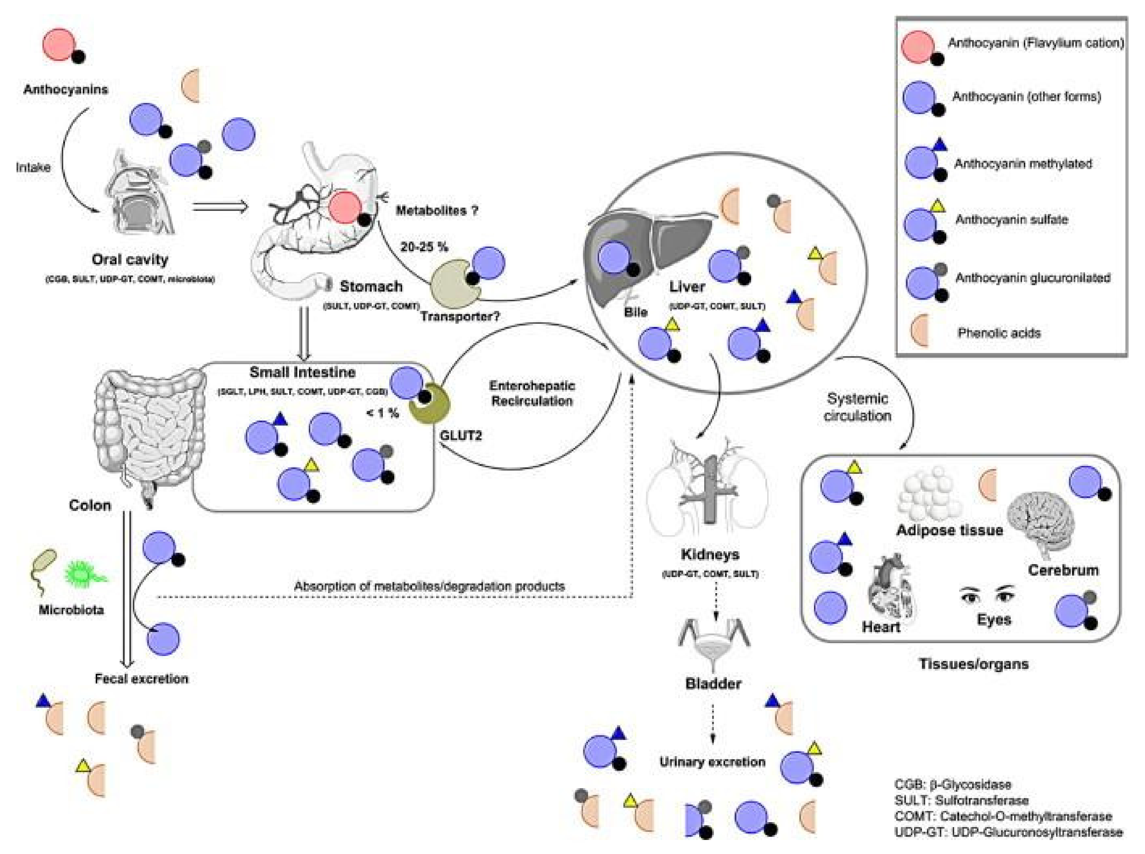
3. ANs Bioavailability

The efficiency of ANs is strictly connected to their bioavailability [29] and specific site of action, after their ingestion and absorption [98]. In this perspective, it is noteworthy that in order to understand the link between ANs and their health-promoting actions, it is of great importance not only to know the amount of ANs from a source/treatment but also the concentration that remains bioactive in the organism.
ANs have the capacity to re-arrange their chemical structure in response to the pH of the environment. However, once they are ingested, their trail encounters several pH jumps, such as 6.7 in saliva, 1–5 in the stomach, 7.4 in the blood, and 7.5–8 in the small intestine [99,100]. Their bioavailability is thus affected, which vexes researchers as to whether their biological properties are the effect of ANs or are due to their degradation metabolites.
In an approach to overcome this underexplored subject, several studies have been conducted on the fate of ANs after ingestion, both dietary (from natural sources) [6] and as supplements [6,101,102]. Their findings revealed that ANs could be identified in native (glycosylated) forms in the plasma, in the stomach and could also be stored in some tissues and organs such as the liver and eyes, and even pass the blood–brain barrier to the cortex and the cerebellum, in pig subjects [103]. In several studies performed in humans using a variety of anthocyanin-rich sources, the concentration of plasma compounds in the range of 6–2400 mg has been reported [97,104,105,106,107]. Similarly, an in vivo study in rats showed the presence of ANs in the stomach at a concentration of 2013.2 ± 280.2 μM at 0.5 h after administration of a dose of 500 mg/kg dose of Cy-3-glucoside [108]. Moreover, the absorption of 23.0 nmol of ANs was recorded 30 min after the injection of a concentration of 70.9 nmol in the gastric lumen of rats [101].
Further, ANs metabolites were observed in other organs such as the liver, heart, jejunum, kidneys, bladder, prostate, testes, and fat tissue, in rats [102]. The rapid breakdown was also observed in human subjects with 56% of the metabolites of Cy-3-glucoside still present in the circulatory system after 48 h from ingestion. The maximum concentration obtained, measured in μmol/L, was: 5.21 ± 3.43 for hippuric acid, 1.51 ± 0.75 for phenylpropenoic and phenylacetic acids, 0.48 ± 0.10 for protocatechuic acid and phloroglucinaldehyde, 1.17 ± 0.52 for phase II conjugates of Cy-3-glucoside and Cy, and 5.54 ± 0.49 for phase II conjugates of protocatechuic acid [109].
It has been thus observed that ANs are absorbed in different manners according to their molecular size, aglycone, and the glycosylation and/or acylation feature, as well as the pH of the environment. Moreover, it has been discussed that the absorption is also influenced by the concentration of ANs administered, this was pointed out in a study of 13 healthy volunteers who consumed 0.8 mg of ANs/kg of body weight from chokeberry juice [110].
Once ingested, ANs follow the continuous path of the gastrointestinal tract of different physico-chemical settings (saliva, stomach, intestine, and colon), and different populations of microorganisms. Following the absorption, they enter further compartments and environments (Figure 5), which all put a toll on the stable flavylium cation and, consequently, on the final concentration of ANs that remains available in the organism [111].

3.1. Oral Cavity Absorption

The interaction of ANs with digestive enzymes or food proteins is the first transformation they undergo in the oral cavity. A study by Walle et al., 2005, demonstrated the hydrolysis of flavonoid glucosides to aglycones under the action of β-glucosidases derived from bacteria and oral epithelial cells of human subjects [112]. In another study, ANs extracted from black peanuts skins exerted an inhibitory effect on the digestive enzymes with IC50 values of 123.4 μg mL−1 for α-amylase and 82.75 μg mL−1 for α-glucosidase [113]. The interaction between different proteins and ANs has been studied in several studies. For example, Mv-3-galactoside in blueberry extract was stabilized by binding to α-casein or β-casein. In turn, these proteins have undergone structural changes in α-helix, β-sheet, turn, and random coil content [114].
Several studies have shown a lower rate of absorption in the body when compared to the amount administered orally, due to the very low percentage measured in plasma, shortly after ingestion [99,115]. Generally, 1% of the total content could be found in the circulatory system, after 15–30 min post-ingestion [116]. However, parts of ANs can be subjected to degradation to metabolites, or could bind to plasma proteins, or could be uptaken by cells from mouth tissue [99].
Although the intestinal lumen and the liver are considered to be the main absorption and digestion sites of ANs, small quantities could be recovered in the mouth too. Most of the ANs are degraded as a result of enzymatic and microbiota actions. The process was mostly dependent on the chemical structure of ANs. For example, a clinical trial showed that ANs from chokeberry juice had different fates, after 5 min of incubation in the oral cavity. Cy-3-xyloside had the lowest recovery yield, hence the highest deterioration, while Cy-3-glucoside could be identified in the epithelium cells [117]. However, there are possible oral components that contribute to the intraoral metabolism of ANs such as: saliva, oral microflora, and oral tissues [99,118]. Such effects have been observed in humans who were administered black raspberries ANs [118]. Native forms of ANs as well as degraded forms were identified in the saliva and in the oral cavity.
The mechanism behind their uptake in the mouth tissue is believed to be attributed to enzymes such as sodium-dependent glucose co-transporter (SGLT1), β-glycosidase (derived from oral bacteria and epithelial cells), UDP-glucuronosyl-transferase, catechol-O-methyltransferase (COMT), which contribute to the absorption of ANs, increasing water solubility or their hydrolyzation to simple aglycones [118].
Another study suggests that ANs degradation in the saliva is mostly mediated by the resident microbiota, a network of over 700 kinds of microorganisms, mostly composed of bacteria, fungi, and viruses [119,120].
Among the metabolites found in the saliva, several studies could identify low amounts of Cy, Dp, Pn, Mv, and mostly, phenolic compounds as a result of degradation such as protocatechuic acid or chalcones of Cy, remarking that the chemical structure of ANs and the amount of salivary secretion strongly influenced their chemical degradation and their uptake by the epithelial cells [117,119].

3.2. Gastric and Intestinal Absorption

3.2.1. Stomach

In the stomach, the pH range is around 1–2, which favors the preservation of ANs in the form of cation flavylium, their most stable form. This hypothesis has also been confirmed by numerous in vitro studies [121,122,123]. The stomach is considered a highly effective site, and a larger amount of ANs is absorbed (20–25%) here, rapidly passing into plasma, and then excreted into bile and urine [124]. Moreover, no secondary metabolites were identified in the stomach after 30 min, only the native (glycosylated) form of ANs. In one study, rats were administered black raspberry anthocyanin extract through a gastric tube, and the flow of ANs was kinetically measured in the stomach. The concentration decreased linearly for 180 min. After 120 min, the concentration was halved. Therefore, the authors suggest that after 4 h, anthocyanin forms should still be detected, even if they are in small concentrations of approximately 10% of the ingested dose, as demonstrated in another article [125]. In addition, another aspect of measuring ANs is their binding to other stomach proteins, such as an anion membrane protein transporter, named bilitranslocase [126]. Since HPLC is able to detect free ANs, this again would lead to a misinterpretation of the real concentration of ANs in the stomach [116].
Some of the main transporters that are considered to be involved in the stomach absorption of ANs include glucose transporter 2 (GLUT2), which was observed to be increased in cells pre-treated with ANs compared to untreated cells, highlighting a circle that would improve their bioavailability by steadfast consumption [121].
Regarding the stomach, the literature gives vast and ambiguous information. An in vitro model is needed that acts as a gastric barrier and can analyze the absorption of ANs at low pH. In general, the stomach is ruled out when it comes to absorption tests, but it is a very important site of absorption [99]. The amount of ANs that is not absorbed by the stomach is delivered to the small intestine mostly as carbinol pseudobases [14].

3.2.2. Intestine

The ANs that escape the stomach absorption reach the small intestine. Due to the basic conditions, they will be mainly found as colorless pseudobases, yellow chalcone, or blue quinoidal forms [14]. Moreover, they are rapidly subjected to degradation into secondary metabolites, which are then released into the circulatory system and finally excreted in the urine. The unabsorbed ANs continue their route towards the colon, where they suffer modifications under the action of the microbiota environment, which hydrolyses the glycosidic forms of ANs into aglycones and later, into simple phenolic acids [127]. The secondary metabolite protocatechuic acid has accounted for the majority of the ANs ingested. However, some other forms of metabolites have been identified and are presented in Figure 3.
The mechanism by which ANs may be absorbed into the intestinal epithelial cells is, again, through glucose transporters such as GLUT2 [36] and possibly SGLT1 [128].
ANs, once ingested, follow a particular pathway, starting with the oral cavity where, mostly influenced by their chemical structure, they can be degraded by the activity of saliva enzymes and microbiota or can be absorbed by oral mucosal epithelium [117]. The highest amount reaches the stomach where, due to optimal pH conditions, they can be recovered in their specific flavylium cation form and be absorbed into the plasma [129]. They can also be absorbed by the small intestine, where they undergo decomposition under the action of the intestinal microflora [130]. The secondary metabolites and degradation products can have important medicinal properties and are taken up by the liver and then distributed to other organs and tissues [131].

4. ANs and Preventive Action on Diseases

Due to their occurrence in many fruits, ANs have been part of the human diet for years and have been used in traditional medicine to treat several impairments, such as atherosclerosis, chronic venous insufficiency, obstructed bile ducts, hyperbilirubinemia, and lack of appetite. They were also used during World War II by British pilots who were administered bilberry jam for better night sight [122,123].
These pigments have gained a new appreciation over the past decades, as the consumption of an ANs-rich diet has been associated with positive health reactions [132]. Consequently, the amount of research on this topic has intensified significantly over the last two decades, which has helped attain more and more insights into ANs’ properties and their antioxidant and anti-inflammatory actions [133]. This naturally led studies to extend to a wider range of pathologies, such as: diabetes, obesity, cardiovascular diseases, neuronal illnesses, and cancer [134]. The main mechanism by which ANs are believed to have the ability to prevent the development of diseases is related to their antioxidant capacity by which they diminish prooxidative damage [135].
Reactive oxygen species (ROS) such as hydrogen peroxide (H2O2), hydroxyl radical (HO•), and superoxide anion (O2) are reaction products that are generated by the body as a result of a partial reduction of oxygen in mitochondria oxidative phosphorylation or as a response to drug metabolites, bacterial invasion, or infections. The excessive production of ROS, which may commence endogenously in pathological conditions, or from exogenous sources (pollution, UV-B radiation, smoking, unhealthy diet) is called oxidative stress [136]. Oxidative stress is a defective imbalance in the redox mechanism in which the cell is ineffective in mounting an antioxidant response against ROS which, in turn, generates an excessive ROS production [137].
Furtherly, an overwhelming amount of ROS affects biomolecules of critical importance such as proteins, lipids, and nucleic acids which further causes obesity, diabetes, cardiovascular associated disorders, carcinogenesis, neurodegeneration, and aging, as comprised in Figure 2 [138,139]. These ROS-mediated damages go hand in hand with inflammatory processes such as the activation of specific signaling pathways responsible for enhancing the expression of pro-inflammatory cytokines and mediators such as: interleukins (IL), C-reactive protein (CPR), tumor necrosis factor (TNF), vascular cell adhesion molecule-1 (VCAM-1), intercellular adhesion molecule-1 (ICAM-1), etc. Although beneficial in the host response to infections, these mediators, under pathological conditions, trigger signaling cascades that lead to severe illnesses such as the ones mentioned above [140,141]. By controlling the levels of expression of these pro-inflammatory effectors, the risk of the later development of chronic diseases could be reduced.

4.1. In Vitro Studies

Cell cultures are valuable tools that provide reliable, predictable systems that faithfully reproduce live models, and at the same time circumvent some obstacles and limits that arise in in vivo research, which enabled scientists to conduct an in-depth study of ANs (Table 1).
Therefore, a growing body of studies over the past two decades has demonstrated the actions of ANs on different pathologies [142].
The excessive accumulation of adipose tissue and obesity is the strongest risk factor for developing diabetes [170], therefore, the two chronic pathologies are positively correlated. In addition to the abnormal metabolic disturbances, there are some particular features as well [171]. In this context, ANs have been shown to be beneficial in modulating and preventing their occurrence in numerous studies.
Many different in vitro models have demonstrated the potential of ANs to alter different molecular aspects of diabetes and obesity [143,144,145]. A molecular sign of obesity is represented by pro-inflammatory macrophages that infiltrate the adipose tissue, which further leads to a high production of pro-inflammatory cytokines (interleukins IL-6, IL-8, and 1L-1β or tumor necrosis factor α (TNF-α) and chemokines such as C-C motif chemokine ligand 2 (CCL-2) [172]. To investigate the relationship between adipocytes and macrophages, anthocyanin-enriched fractions from blackberry–blueberry beverages (100 μM) were applied to two cell lines: RAW264.7 macrophages and preadipocyte 3T3-L1 cells. After 24 h of treatment, markers such as phosphorylated-p65 NF-ƙB (51.2%), nitric oxide (17.5%), and TNF-α (89.4%) were inhibited in RAW264.7 cells. Furthermore, in mature 3T3-L1 adipocytes, isoproterenol-induced lipolysis and intracellular fat accumulation were reduced by 18.6% and 28.2%, respectively [143].
In another study, hepatocellular carcinoma cell line HepG2 cells showed a significant inhibition of reactive oxygen species (ROS) production after pre-treatment with 5 μg/mL ANs (blueberry anthocyanin extract (BAE)), Mv, Mv-3-glucoside, or Mv-3-galactoside for 24 h. Decreased production of enzymes involved in lipogenesis and gluconeogenesis has also been observed, while those involved in glycogenolysis and lipolysis have been enhanced via the adenosine monophosphate-activated protein kinase (AMPK) signaling pathway [144].
The protective role of ANs on endothelial cells has been demonstrated in a study by Aboonabi et al. [145]. Specifically, after the application of 50 μL/mL of berry-derived ANs on diabetic human aortic endothelial cells (D-HAEC) subjected to oxidative stress, a decrease in the inflammatory and oxidative process was demonstrated by inhibition of the NF-ƙB signaling pathway [145]. Similarly, the anti-inflammatory and antioxidant effects of anthocyanin extracts from various fruits have been demonstrated in retinal epithelial cell models [173,174,175,176].
Improving the quality of life and the life expectancy of human beings has been a remarkable achievement of the continuous progress made by humankind in recent decades. However, managing this situation is difficult, mainly due to the significant increase in the number of people suffering from chronic diseases. According to a 2021 WHO report, 71% of all deaths globally in one year are caused by such diseases. Of the 41 million deaths a year, 17.9 million are caused by cardiovascular disease. One way to prevent these pathologies is to choose a healthy diet of overprocessed food. Although the term “healthy diet” has not been universally defined, there are guidelines that can be applied. Among them, the consumption of vegetables and fruits, especially fresh and raw, is strongly recommended by the WHO [177].
The effect of ANs on the expression of genes responsible for the inflammatory response has recently been highlighted [178]. Human umbilical vein endothelial cells (HUVECs) have been exposed to several types of ANs in concentrations from 0.1–2 μM. Subsequent analyses have shown that these bioactive compounds can reduce the adhesion of monocytes to endothelial cells, the initial step in atherosclerosis development.
Contrary, no effect has been identified on the expression of genes encoding the adhesion molecules E-selectin, ICAM1, and VCAM1, indicating that the target proteins for this process are different [146]. In a similar study, TNF-α-stimulated HUVECs treated with 100 or 300 µg ANs for 6 h demonstrated a significant inhibition of TNF-α-induced NF-κB activity with translocation of NF-κB from the nucleus to the cytosol [147]. NF-κB is a transcription factor that regulates gene expression of adhesion molecules on endothelial cells and the production of pro-inflammatory cytokines and chemokines [179].
An in vivo study focused on evaluating the effect of ANs on apolipoprotein E-deficient (apoE−/−) mice to investigate the early stage of atherosclerosis. Following nutrigenomic analysis, 1261 genes were identified whose expression was modulated by bilberry anthocyanin-rich extract (0.02%). Genes encoding enzymes involved in the regulation of oxidative stress (AOX1, CYP2E1, or TXNIP) and some encoding adhesion proteins (JAM-A, VEGFR2) were down-regulated, and genes responsible for cell–cell adhesion were up-regulated. (CRB3, CLDN14, CDH4) [180]. A significant upregulation of the Nrf2-regulated antioxidant response proteins heme oxygenase 1 (HO-1) and glutamate-cysteine ligase subunit modifier (GCLM) was recorded after treating HUVECs with blueberry juice in response to low µM concentrations of H2O2 (0–40 μM) [148].
The CD40-mediated signaling pathway contributes to the trigger of atherosclerosis. To examine the role of ANs on cell adhesion, HUVECs were stimulated with 5 μg/mL sCD40L and subsequently treated with Cy-3-glucosides and Pn-3-glucosides from 1 to 100 μM for 24 h. The level of VCAM-1, ICAM-1, MMP-1, and MMP-9 decreased significantly after treatment. Caspase-3 activity, involved in cellular apoptosis, and JNK and p38 pathways were also inhibited [149]. Oxidized low-density lipoprotein (oxLDL) may enhance immune and inflammatory mechanisms that promote atherosclerosis [181]. oxLDL inhibition was achieved by applying a mixture of ANs from Hibiscus sabdariffa L. over mouse macrophage J774A.1 cells.
Moreover, with the treatment in concentrations of 0.01–0.2 mg/mL, it was possible to decrease the CD36 expression, both at the protein level and at the mRNA level [150]. CD36 is a membrane protein with a pivotal role in modulating lipid metabolism; often, its overexpression is correlated with the accumulation of toxic lipids and a high risk of heart failure [182].
Another extremely important role that ANs have shown in various studies has been in the fight against cancer. As it is known, the treatments for this disease are not yet well established, so new therapeutic options are being sought with interest by scientists. The literature offers us various in vivo and in vitro studies, combined, that aim to test such a treatment, in different therapeutic strategies. For example, human hepatoma cell line SMMC-7721 treated with 0.2 mg/mL of ANs from Lonicera caerulea ‘Beilei’ fruit could inhibit the proliferation of cells and also promote their apoptosis. Furthermore, H22 tumor-bearing mice treated with the same extract, in different doses, were analyzed. Tumor growth was suppressed by activating SOD and decreasing the amount of MDA, IFN-γ, and IL-6 [151].
Grape seed extract (GSE) has been tested for chemically induced liver cancer. When administered at doses of 25, 50, and 100 mg/kg per day for 14 weeks, the extract produced effects such as reduced tumor cell proliferation, oxidative stress, and the concentration of inflammatory markers such as iNOS, cyclooxygenase 2, and p-phosphorylated tumor necrosis receptor factor in the rat animal model. In HepG2 cells, the grape seed extract facilitated the activation of caspase-3 and G2/M and G1/S cell cycle arrest [152].
The anticancer activity of Dp in colorectal cancer has also been investigated, both in vitro and in vivo. The human colon cancer cell lines DLD-1, SW480, and SW620 were incubated with serial concentrations of Dp (<100 μM) for 24 h. The main results obtained indicated the inhibition of cell adhesion, migration, and invasion of tumor cells and epithelial-to-mesenchymal transition. Integrin and FAK signaling cascade were successfully inhibited in the DLD-1 cell line treated with 100 μM Dp for 24 h. At the mRNA level, miR-204-3p is upregulated in response to Dp. An animal xenograft model was used to analyze the effect of ANs in vivo. Here, the attenuation of the metastasis process could be highlighted [153].
Similarly, the effect of Dp-3-glucoside and Cy-3-glucoside were tested on HCT-116 and HT-29 human colorectal cancer cells (100–600 μg/mL). Programmed cell death protein-1 (PD-1) and programmed death-ligand 1 (PD-L1), important immune checkpoints, have been successfully inhibited by these compounds [154]. Colon cancer stem cell proliferation was attenuated by the application of 5 μg/mL purple-fleshed potato extracts, rich in ANs. Furthermore, the phenomenon of apoptosis has been accelerated by modulating the BAX protein [155].
There are also many results that suggest the applicability of ANs in the treatment of melanoma. The antiproliferative activity of ANs-rich strawberry extracts and their potential to induce differentiation have been successfully tested in vitro on the B16-F10 cell line [156]. The antioxidant capacity of ANs has also been shown. Mulberry ANs extract applied to B16-F1 cells helped to reduce the expression level of phosphoinositide 3-kinase (PI3K), Ras protein, and NF-ƙB within 24 h [157].
In breast cancer, an anthocyanin-rich extract from black rice has shown therapeutic potential. Specifically, the viability of breast cancer cell line MCF-7 was suppressed, and, at the same time, their apoptosis was stimulated by activating caspase-3, depolarizing the mitochondrial membrane, and releasing cytochrome c. In the same manner, in an in vivo model of mice bearing MDA-MB-453 cell xenografts, inhibition of the angiogenic factors matrix metallopeptidase MMP-9, MMP-2, and uPA was performed after the oral administration of 100 mg/kg/day of ANs extract [158]. Different concentrations of ANs from red sorghum bran (250 μg/mL, 500 μg/mL, and 1000 μg/mL) were applied over the human breast cancer cell line MCF-7. Subsequently, morphological changes associated with apoptosis were identified by microscopic methods, thus demonstrating inhibition of tumor proliferation [159].
Therefore, ANs can be applied in order to alleviate various forms of cancer. Of course, it is essential that research continues in order to finally reach a product that cures this pathology.
Another beneficial effect of ANs in chronic diseases is to provide a certain degree of neuroprotection, helping in the fight against neurodegenerative diseases. These are more common among the elderly population because they are caused by genetic and environmental factors that, over time, have an increasing impact on the proper functions [183]. The most common diseases in this category are Alzheimer’s disease, Huntington’s disease, Parkinson’s disease, frontotemporal dementia, amyotrophic lateral sclerosis, and spinocerebellar ataxias [184]. Even if the exact causes that trigger the phenomenon of neurodegeneration are not known, there are several scientifically documented variants that can explain the appearance of the pathologies. One variant is based on molecular studies and indicates the accumulation, over time, of aggregates formed by the proteins amyloid-β (Aβ), hyperphosphorylated tau (p-tau), and α-synuclein [183].
The use of ANs to prevent these pathologies was reported, ANs being rapidly absorbed into the bloodstream and easily cross the blood–brain barrier (BBB), thus exerting their beneficial effects in their unaltered form (anti-apoptotic, antioxidant, and anti-inflammatory) [185].
The neuroprotective effect of Cy (0.2–20 μM) was tested on human neuroblastoma SK-N-SH cells treated with Aβ25-35, a neurotoxicity-inducing compound. After incubation with the target anthocyanin for 2 h, results such as decreased ROS accumulation, modulation of intracellular Ca2+ amount, decreased expression of ER stress response proteins, and transcription factors such as eukaryotic initiation factor 2 α (eIF2α), X-box binding protein 1 (XBP-1), and cleaved caspase-12 [168]. Similarly, human SH-SY5Y neuroblastoma with hydrogen peroxide-induced neurotoxicity was treated with a series of concentrations of blueberry and cranberry juices for 24 h. Subsequent tests showed an increase in the activity of the antioxidant enzymes catalase (CAT) and superoxide dismutase (SOD), complemented by a decrease in ROS and TBARS [169].

4.2. In Vivo/Clinical Studies

ANs studies have extended from cell cultures to a variety of in vivo models, animal and human clinical trials, as describes in Table 2.
Concerning diabetes, there is much in vivo evidence that shows how ANs protect the pancreatic β-cells, decrease glycemia, enhance insulin secretion, suppress weight gain, decrease hepatic lipogenesis or accumulation in the liver, reduce adipocytes sizes, enhance insulin resistance and sensitivity, reduce total cholesterol, and increase levels of HDL [170].
A cohort study of 16,678 healthy people from the US National Surveys Study (REasons for Geographic and Racial Differences in Stroke) found that a diet rich in ANs and proanthocyanidins helps reduce the incidence of coronary heart disease [213]. Consumption of 400 g of fresh bilberries by 15 volunteers with features of metabolic syndrome has been shown to be useful in lowering the concentration of pro-inflammatory biomarkers IL-6, IL-12, and C-reactive protein. Moreover, following a transcriptomic analysis, results were obtained confirming a significant decrease in the expression of key genes in macrophage differentiation and activation (MMD and CCR2) [186].
An 8-week placebo-controlled clinical trial tested the therapeutic potential of anthocyanin-rich black soybean test extracts (2.5 g/d), with high concentrations of ANs (12.58 mg/g), on 63 obese volunteers. Changes in plasma lipid profile have been reported, such as decreased low-density lipoprotein cholesterol (LDLc) triacylglycerols (TG), and non-high-density lipoprotein cholesterol (non-HDLc) [187]. Furthermore, 124,086 people living in the US were included in a clinical study that aimed to verify the association between the consumption of specific flavonoids (ANs, flavonols, flavones, flavanones, flavan-3-ols, and flavonoid polymers) and weight change over time (1986–2011). ANs showed the highest magnitude of association, the main food sources consumed being blueberries, strawberries, apples, and pears [188].
On the other hand, consuming 500 mL of red-orange juice for 12 weeks had no effect on the weight of the study volunteers. However, the LDL concentration decreased significantly [189]. Similarly, the administration of 118.5 mg/day of ANs from dried purple carrot to 16 obese men for 4 weeks did not show changes in body mass or molecules of interest such as LDL, total cholesterol, and C-reactive protein [214].
Going further in the field of in vivo studies conducted on this topic, it can be stated that there is a fairly large diversity of such research.
In vivo tests have been performed to further analyze the potential applications of ANs in the treatment of diabetes and its macrovascular (cardiovascular) and microvascular (retinopathy, neuropathy, kidney disease) complications [215]. For example, 200 mg kg−1 of raspberry anthocyanin was administered for 12 weeks in C57BL/6 mice with a low-fat diet or high-fat diet. Following analysis, a significant decrease in gene expression was observed for TNFα, IL-6, and NF-κB. At the same time, the activity of the antioxidant enzymes superoxide dismutase (SOD) and glutathione peroxidase (GSH-PX) increased significantly, and, moreover, the body weight was reduced by 63.7% [190].
ANs from black rice, black soybean, or purple corn were tested in C57BL/6 mice fed a high-fat diet. The dose administered for 12 weeks was 200 mg/kg and showed a decrease in lipid peroxidation and gene expression for TNFα, IL-6, iNOS, and NF-κB and an increase in peroxide dismutase and glutathione peroxidase activity [191]. In comparison, extracts of ANs from mulberry and cherry showed similar effects under the same experimental conditions [192].
The metabolic syndrome has also been investigated in the animal model. In obese Zucker rats who followed a diet containing 8% wild berries for 8 weeks, there was a significant decrease in the molecular markers IL-6, TNF-α, and Nf-κB in both the liver and adipose tissue and the reactive protein C in the liver. At the same time, a higher concentration of adiponectin, a well-known homeostatic factor that regulates glucose levels, has been observed [193]. The inhibitory effect of Cy-3-glucoside on retinol-binding protein 4 (RBP4) expression and the enhancing effect on glucose transporter 4 (Glut4) in the adipose tissue were highlighted in KK-A(y) type 2 diabetes mice after a diet with 0.2% anthocyanin for 5 weeks [194].
Another potential anti-diabetic role that anthocyanin-rich extracts may play is glycosidase action. To test this hypothesis, 24 adult male Wistar rats that had a cornstarch or fructose-rich control diet for 4 weeks, received an additional 2 g/kg Kamchatka honeysuckle berry extract (327 mg ANs/g). The conclusions of the experiment showed a stimulating effect on bacterial α and β glucosidase activity within the gut [195]. C57BLKS/J-Leprdb mice treated with 10 mg/kg BW anthocyanin-rich purple corn extract for 8 weeks were subsequently analyzed to investigate the effects of the bioactive compound in diabetic nephropathy. Thus, the induction of VEGF and HIF-1a transcription factors that promote angiogenesis was successfully attenuated. Furthermore, the mesangial and endothelial induction of angiopoietin (Angpt) proteins under hyperglycemic conditions was successfully decreased [196].
Dp-3-rutinoside from blackcurrants extract demonstrated activating action on glucagon-like peptide-1 (GLP-1) in an in vivo study performed on Sprague-Dawley (SD) rats. GLP-1 is a gastrointestinal peptide involved in glucose homeostasis and the amount of extract administered (5 mg/kg BW) showed positive results in the secretion of the enzyme of interest [197].
Therefore, there is a lot of scientific evidence to support the use of ANs in antidiabetic therapy. However, continuous innovation is needed to cope with the growing number of patients suffering from this disease.
Several epidemiological studies have shown a negative correlation between the incidence of cardiovascular disease and the consumption of anthocyanin-rich fruits and vegetables. For example, the findings of a study looking at the relationship between anthocyanin intake and the risk of myocardial infarction (MI) in 93,600 women between the ages of 25 and 42 showed an inverse association between high consumption of this bioactive compound and the risk of MI [198]. In another study, high blueberry consumption contributed to increased flow-mediated dilation (FMD) and improved vascular function in 21 healthy men [199].
A significant decrease in cholesterol, triglycerides, and activated platelets and an increase in total plasma antioxidant capacity were observed after 30 days of consumption of strawberries by healthy volunteers, in a clinical study [200]. In a clinical trial study, 150 subjects with hypercholesterolemia consumed a purified anthocyanin mixture (320 mg/d) or a placebo twice a day for 24 weeks. After this period, the levels of serum high sensitivity C-reactive protein (hsCRP), soluble vascular cell adhesion molecule-1 (sVCAM-1), and IL-1β were measured. The results indicated a significant decrease in the amount of these biomarkers in plasma [201].
In the category of epidemiological studies, we find a study that showed the pro-apoptotic effect of an anthocyanin-rich dietary bilberry extract in 30 patients with chronic B cell lymphocytic leukemia and on peripheral blood mononuclear cells (PBMCs) from healthy subjects. The main compound in the extract, Dp-3-glucoside, causes the activation of caspase-3 and the down-regulation of the Bcl-2/Bad pathway [202]. In vivo studies have shown the potential of Dp to significantly reduce melanoma-induced tumor growth, while in vitro data show a decrease in endothelial cell proliferation [203]. Protective effects of Cy-3-glucoside and Dp against UVB irradiation have been reported in in vivo studies on the carcinogenesis model of hairless mice SKH-1 [204,205].

Neuroprotection Sustained by ANs

In the field of clinical trials, a study was conducted on 49 older adults (+70 years with mild-to-moderate dementia who consumed for 12 weeks 200 mL/day of anthocyanin-rich cherry juice. Compared to the control group, those with dementia improved their cognition, respectively, fluency in speech, and short- and long-term memory. On the other hand, inflammatory molecular markers did not undergo significant changes [206].
Another randomized, double-blind, placebo-controlled trial looked at effective freeze-dried blueberry consumption in elderly volunteers. According to the researchers, the addition of anthocyanin compounds to the diet helped to reduce verbal errors and significantly reduced switch costs on a task-switching test [207]. Twenty-one children aged 7–10 were volunteers in an experiment that aimed to verify the effects of the introduction of 15 or 30 g of freeze-dried wild blueberry powder in the diet, demonstrating cognitive improvements [208].
ANs extracted from Korean black soybean (100 mg/kg ANs) were applied for 7 weeks to an in vivo model using Sprague-Dawley (12 weeks old) male rats. Surprisingly, memory deficits were improved; the expression of RAGE, BACE-1, and Aβ proteins was inhibited, as was the amount of MDA. Furthermore, the activation of astrocytes and microglia in the brains of the tested rats was observed [209]. Male C57BL/6N mice underwent intraperitoneal injections of LPS (250 μg/kg/day for 1 week) to simulate neurotoxicity and neuroinflammation. Subsequently, they received anthocyanin treatment extracted from the same Korean black soybean (24 mg/kg/day) for 14 days. Inflammatory markers p-NF-κB, TNF-α, and IL-1β were down-regulated and cellular apoptosis was reduced by suppression of inducing factors (cytochrome C, cleaved caspase-3, and Bax) [210].
Strong activation of antioxidant enzyme activity in the cerebral cortex was demonstrated by the administration of ANs (200 mg/kg/day) in an experimental model of sporadic dementia of Alzheimer’s type that used adult rats [211]. In another study, after administering chokeberry ANs for 15 or 30 mg/kg chokeberry ANs to Kunming mice receiving D-galactose to support their aging for 8 weeks, the redox balance was restored, DNA fragmented and the amount of pro-inflammatory cytokines COX2, TGF-β1, and IL-1 decreased [212].
Therefore, the protective role of ANs in the fight against degenerative diseases may be of great interest for future research, especially due to the multitude of scientific articles that come with convincing evidence in this regard.

4.3. Medicinal Products Developed with ANs

ANs have a very high versatility in terms of their applications and their beneficial effects on human health. Therefore, in addition to using them in free or encapsulated form, there are also topical formulations that have been successfully created. For example, a combination of ANs extracted from Zea mays and Clitoria ternatea was incorporated into a mucoadhesive gel. This product was subsequently tested and evaluated for topical oral wound healing in rats and in a clinical trial in 68 orthodontic patients. In the animal model, a reduction in the erythema and the sizes of oral wounds was observed compared to placebo gel. In the clinical trial, wound closure enhancement was observed on day 3 of anthocyanin gel application [216].
Another study tested the ability of pomegranate ANs to prevent aging by formulating a cream. After obtaining favorable results in terms of stability, homogeneity, and ex-vivo studies, the cosmetic product was tested on volunteers. Features such as wrinkle reduction, hydration, and pleasant texture have been demonstrated following the topical application of the cream [217].

5. Conclusions and Future Outlook

ANs are a class of water-soluble phytonutrients that are widely distributed in fruits and vegetables and show valuable health effects. Their chemical stability is highly susceptible to degradation and is directly dependent on environmental conditions. This particularity is especially observed when investigating the faith of ANs after ingestion but also the molecular mechanisms involved in gene regulation and ANs accumulation.
ANs are absorbed rapidly and were detected in the bloodstream and other tissues in different concentrations, both as ANs and as ANs-degradation metabolites. More investigations are required in this field, for a better overview and understanding of the chemical form and action association.
The potential of ANs to prevent and ameliorate several diseases such as diabetes, obesity, cancer, and cardiovascular and neuronal illnesses has been demonstrated in numerous in vitro and in vivo studies. Their antioxidant capacity and the inflammatory cytokines signaling are the key mechanisms of action that are believed to be responsible for such remarkable results.
Future ANs investigations and continued interest in ANs-based therapies will undoubtedly lead to new opportunities for pursuing the development of efficient formulations that would improve the ANs’ stability, bioavailability, and beneficial health effects.

Author Contributions

The conceptualization of the manuscript: D.R. and A.P.; writing—original draft preparation: M.N.; writing—review and editing: R.P., A.D., D.R. and C.S.; funding acquisition: D.R. All authors have read and agreed to the published version of the manuscript.

Funding

This research was funded by the Romanian National Authority for Scientific Research, CNDI–UEFISCDI, project number: TE 80/01.09.2020, project code: PN-III-P1-1.1-TE-2019-1276.

Institutional Review Board Statement

Not applicable.

Informed Consent Statement

Not applicable.

Data Availability Statement

Not applicable.

Conflicts of Interest

The authors declare no conflict of interest.

Abbreviations

8-OhdG8-hydroxydeoxyguanosine
amyloid-β
ABCATP-binding cassette
AMPKadenosine monophosphate-activated protein kinase
ANsanthocyanins
ANSanthocyanidin synthase
AOX1aldehyde oxidase 1
ASEaccelerated solvent extraction
AVIsanthocyanins vacuolar inclusions
BACE-1beta site amyloid precursor protein cleaving enzyme 1
BBBBlood–brain barrier
Bcl-2B-cell lymphoma 2
BTL-likebilitranslocase-like transporters
BWbody weight
CATcatalase
CCL-2C-C motif chemokine ligand 2
CCR2C-C motif receptor 2
CDH4cadherin 4
CHIchalcone isomerase
CHSchalcone synthase
CLDN14claudin 14
COMTcatechol-O-methyltransferase
COX-2cyclooxygenase-2
CPRC-reactive protein
CRB3Crumbs cell polarity complex component 3
Cycyanidin
CYP2E1cytochrome P450 2E1
DFRdihydroflavonol 4-reductase
D-HAECdiabetic human aortic endothelial cells
Dpdelphinidin
EAEenzyme-assisted extraction
eIF2αeukaryotic initiation factor 2 α
ERendoplasmic reticulum
F3Hflavanone 3-hydroxylase
F3′Hflavonoid 3′-hydroxylase
F3′5′Hflavonoid 3′,5′-hydroxylase
FAKfocal adhesion kinase
FMDflow-mediated dilation
GCLMglutamate-cysteine ligase modifier subunit
GLP-1glucagon-like peptide-1
GLUT2glucose transporter 2
GSHglutathione
GSH-PX/GPXglutathione peroxidase
GSTsglutathione S-transferases
H2O2hydrogen peroxide
HDLchigh-density lipoprotein cholesterol
HHPEhigh hydrostatic pressure extraction
HIF-1ahypoxia-inducible factor 1-alpha
HO•hydroxyl radical
HO-1heme oxygenase 1
hsCRPhigh-sensitivity C-reactive protein
HUVECshuman umbilical vein endothelial cells
ICAM-1intercellular adhesion molecule-1
IFN-γInterferon-gamma
ILinterleukin
iNOSinducible nitric oxide synthase
JNKc-Jun N-terminal kinase
LDHlactate dehydrogenase
LEDlight-emitting diodes
MAEMicrowave-assisted extraction
MAPmitogen-activated protein
MATEmultidrug and toxic compound extrusion
MDAmelanoma differentiation-associated protein
MEFmoderate electric field
MMDmonocyte to macrophage differentiation-associated
MMP-1, MMP-2, MMP-9matrix metallopeptidase 1, 2, 9
Mvmalvidin
MTO-methyl transferase
NF-ƙBnuclear factor kappa-B
Nrf2nuclear factor erythroid 2-related factor 2
O2superoxide anion
oxLDLoxidized low-density lipoprotein
PD-1, PD-L1programmed death-1, -ligand 1
PEFpulsed electric field-assisted extraction
Pgpelargonidin
PGE2prostaglandin E2
PI3Kphosphoinositide 3-kinase
PLEpressurized liquid extraction
Pnpeonidin
Ptpetunidin
p-tauhyperphosphorylated tau
RAGEreceptor for advanced glycation end products
RBP4retinol binding protein 4
ROSreactive oxygen species
RPEretinal pigment epithelium
SFEsupercritical fluid extraction
SGLT1sodium dependent glucose co-transporter 1
SNAREsoluble N-ethylmaleimide-sensitive factor attachment protein receptors
SODsuperoxide dismutase
TBARSthiobarbituric acid reactive substances
TGtriglycerides
TNFtumor necrosis factor
TXNIPthioredoxin-interacting protein
UAEUltrasound-assisted extraction
UFGTflavonoid 3-O-glucosyltransferase
UMAEultrasound/microwave-assisted extraction
uPAurokinase plasminogen activator
sVCAM-1soluble vascular cell adhesion molecule-1
VEGFvascular endothelial growth factor
XBP-1X-box binding protein 1

References

  1. Landi, M.; Tattini, M.; Gould, K.S. Multiple functional roles of anthocyanins in plant-environment interactions. Environ. Exp. Bot. 2015, 119, 4–17. [Google Scholar] [CrossRef]
  2. Valavanidis, A.; Vlachogianni, T. Chapter 8—Plant Polyphenols: Recent Advances in Epidemiological Research and Other Studies on Cancer Prevention. In Studies in Natural Products Chemistry; Rahman, A.-u., Ed.; Elsevier: Amsterdam, The Netherlands, 2013; Volume 39, pp. 269–295. [Google Scholar]
  3. Dixon, R.A.; Liu, C.; Jun, J.H. Metabolic engineering of anthocyanins and condensed tannins in plants. Curr. Opin. Biotechnol. 2013, 24, 329–335. [Google Scholar] [CrossRef]
  4. Mackon, E.; Ma, Y.; Jeazet Dongho Epse Mackon, G.C.; Li, Q.; Zhou, Q.; Liu, P. Subcellular Localization and Vesicular Structures of Anthocyanin Pigmentation by Fluorescence Imaging of Black Rice (Oryza sativa L.) Stigma Protoplast. Plants 2021, 10, 685. [Google Scholar] [CrossRef] [PubMed]
  5. Hatier, J.-H.; Gould, K. Anthocyanin Function in Vegetative Organs. In Anthocyanins; Springer: Berlin, Germany, 2008; pp. 1–19. [Google Scholar]
  6. Mattioli, R.; Francioso, A.; Mosca, L.; Silva, P. Anthocyanins: A Comprehensive Review of Their Chemical Properties and Health Effects on Cardiovascular and Neurodegenerative Diseases. Molecules 2020, 25, 3809. [Google Scholar] [CrossRef]
  7. Khoo, H.E.; Azlan, A.; Tang, S.T.; Lim, S.M. Anthocyanidins and anthocyanins: Colored pigments as food, pharmaceutical ingredients, and the potential health benefits. Food Nutr. Res. 2017, 61, 1361779. [Google Scholar] [CrossRef] [PubMed] [Green Version]
  8. Wallace, T.C. Anthocyanins in cardiovascular disease. Adv. Nutr. 2011, 2, 1–7. [Google Scholar] [CrossRef] [PubMed] [Green Version]
  9. Mateus, N.; de Freitas, V. Anthocyanins as Food Colorants. In Anthocyanins: Biosynthesis, Functions, and Applications; Winefield, C., Davies, K., Gould, K., Eds.; Springer: New York, NY, USA, 2009; pp. 284–304. [Google Scholar]
  10. He, J.A.; Giusti, M.M. Anthocyanins: Natural Colorants with Health-Promoting Properties. Annu. Rev. Food Sci. Technol. 2010, 1, 163–187. [Google Scholar] [CrossRef] [PubMed]
  11. Mukherjee, P.K. Chapter 20—Phyto-Pharmaceuticals, Nutraceuticals and Their Evaluation. In Quality Control and Evaluation of Herbal Drugs; Mukherjee, P.K., Ed.; Elsevier: Amsterdam, The Netherlands, 2019; pp. 707–722. [Google Scholar]
  12. Wang, E.; Yin, Y.; Xu, C.; Liu, J. Isolation of high-purity anthocyanin mixtures and monomers from blueberries using combined chromatographic techniques. J. Chromatogr. A 2014, 1327, 39–48. [Google Scholar] [CrossRef]
  13. Pan, F.; Liu, Y.; Liu, J.; Wang, E. Stability of blueberry anthocyanin, anthocyanidin and pyranoanthocyanidin pigments and their inhibitory effects and mechanisms in human cervical cancer HeLa cells. RSC Adv. 2019, 9, 10842–10853. [Google Scholar] [CrossRef] [Green Version]
  14. Fernandes, I.; de Freitas, V.; Mateus, N. Anthocyanins and human health: How gastric absorption may influence acute human physiology. Nutr. Aging 2014, 2, 1–14. [Google Scholar] [CrossRef] [Green Version]
  15. Mazza, G.; Miniati, E. Anthocyanins in Fruits, Vegetables, and Grains; CRC Press: Boca Raton, FL, USA, 2018. [Google Scholar]
  16. Vinayagam, R.; Xu, B. Antidiabetic properties of dietary flavonoids: A cellular mechanism review. Nutr. Metab. 2015, 12, 60. [Google Scholar] [CrossRef] [PubMed] [Green Version]
  17. Prior, R.L.; Wu, X. Anthocyanins: Structural characteristics that result in unique metabolic patterns and biological activities. Free. Radic. Res. 2006, 40, 1014–1028. [Google Scholar] [CrossRef]
  18. Castañeda-Ovando, A.; Pacheco-Hernández, M.d.L.; Páez-Hernández, M.E.; Rodríguez, J.A.; Galán-Vidal, C.A. Chemical studies of anthocyanins: A review. Food Chem. 2009, 113, 859–871. [Google Scholar] [CrossRef]
  19. Zhao, C.L.; Chen, Z.J.; Bai, X.S.; Ding, C.; Long, T.J.; Wei, F.G.; Miao, K.R. Structure-activity relationships of anthocyanidin glycosylation. Mol. Divers. 2014, 18, 687–700. [Google Scholar] [CrossRef]
  20. Nurtiana, W. Anthocyanin as natural colorant: A review. Food ScienTech J. 2019, 1, 1. [Google Scholar] [CrossRef] [Green Version]
  21. Liu, Y.; Tikunov, Y.; Schouten, R.E.; Marcelis, L.F.M.; Visser, R.G.F.; Bovy, A. Anthocyanin Biosynthesis and Degradation Mechanisms in Solanaceous Vegetables: A Review. Front. Chem. 2018, 6, 52. [Google Scholar] [CrossRef]
  22. Jaakola, L. New insights into the regulation of anthocyanin biosynthesis in fruits. Trends Plant Sci. 2013, 18, 477–483. [Google Scholar] [CrossRef] [PubMed] [Green Version]
  23. Giuliani, A.; Cerretani, L.; Cichelli, A. Colors: Properties and Determination of Natural Pigments. In Encyclopedia of Food and Health; Caballero, B., Finglas, P.M., Toldrá, F., Eds.; Academic Press: Oxford, UK, 2016; pp. 273–283. [Google Scholar]
  24. Husain, A.; Chanana, H.; Khan, S.A.; Dhanalekshmi, U.M.; Ali, M.; Alghamdi, A.A.; Ahmad, A. Chemistry and Pharmacological Actions of Delphinidin, a Dietary Purple Pigment in Anthocyanidin and Anthocyanin Forms. Front. Nutr. 2022, 9, 746881. [Google Scholar] [PubMed]
  25. Ku, S.-K.; Yoon, E.-K.; Lee, W.; Kwon, S.; Lee, T.; Bae, J.-S. Antithrombotic and antiplatelet activities of pelargonidin in vivo and in vitro. Arch. Pharmacal Res. 2016, 39, 398–408. [Google Scholar] [CrossRef] [PubMed]
  26. Rajan, V.K.; Hasna, C.K.; Muraleedharan, K. The natural food colorant Peonidin from cranberries as a potential radical scavenger—A DFT based mechanistic analysis. Food Chem. 2018, 262, 184–190. [Google Scholar] [CrossRef] [PubMed]
  27. Dormán, G.; Flachner, B.; Hajdú, I.; András, C. Chapter 23—Target identification and polypharmacology of nutraceuticals. In Nutraceuticals, 2nd ed.; Gupta, R.C., Lall, R., Srivastava, A., Eds.; Academic Press: Cambridge, MA, USA, 2021; pp. 315–343. [Google Scholar]
  28. Tanaka, Y.; Sasaki, N.; Ohmiya, A. Biosynthesis of plant pigments: Anthocyanins, betalains and carotenoids. Plant J. 2008, 54, 733–749. [Google Scholar] [CrossRef] [PubMed]
  29. Kamiloglu, S.; Capanoglu, E.; Grootaert, C.; Van Camp, J. Anthocyanin Absorption and Metabolism by Human Intestinal Caco-2 Cells—A Review. Int. J. Mol. Sci. 2015, 16, 21555–21574. [Google Scholar] [CrossRef] [PubMed] [Green Version]
  30. Schwartz, S.J.; Cooperstone, J.L.; Cichon, M.J.; Joachim, H.; Giusti, M.M. Colorants. In Fennema’s Food Chemistry; CRC Press: Boca Raton, FL, USA, 2017; pp. 681–752. [Google Scholar]
  31. Jimenez-Garcia, S.N.; Guevara-Gonzalez, R.G.; Miranda-Lopez, R.; Feregrino-Perez, A.A.; Torres-Pacheco, I.; Vazquez-Cruz, M.A. Functional properties and quality characteristics of bioactive compounds in berries: Biochemistry, biotechnology, and genomics. Food Res. Int. 2013, 54, 1195–1207. [Google Scholar] [CrossRef]
  32. Carbone, F.; Preuss, A.; De Vos, R.C.H.; D’amico, E.; Perrotta, G.; Bovy, A.G.; Martens, S.; Rosati, C. Developmental, genetic and environmental factors affect the expression of flavonoid genes, enzymes and metabolites in strawberry fruits. Plant Cell Environ. 2009, 32, 1117–1131. [Google Scholar] [CrossRef]
  33. Petrussa, E.; Braidot, E.; Zancani, M.; Peresson, C.; Bertolini, A.; Patui, S.; Vianello, A. Plant flavonoids—Biosynthesis, transport and involvement in stress responses. Int. J. Mol. Sci. 2013, 14, 14950–14973. [Google Scholar] [CrossRef]
  34. Tanaka, Y.; Ohmiya, A. Seeing is believing: Engineering anthocyanin and carotenoid biosynthetic pathways. Curr. Opin. Biotechnol. 2008, 19, 190–197. [Google Scholar] [CrossRef] [PubMed]
  35. Liu, J.; Osbourn, A.; Ma, P. MYB Transcription Factors as Regulators of Phenylpropanoid Metabolism in Plants. Mol. Plant 2015, 8, 689–708. [Google Scholar] [CrossRef] [PubMed] [Green Version]
  36. De Pascual-Teresa, S.; Sanchez-Ballesta, M.T. Anthocyanins: From plant to health. Phytochem. Rev. 2008, 7, 281–299. [Google Scholar] [CrossRef]
  37. Kaur, S.; Sharma, N.; Kapoor, P.; Chunduri, V.; Pandey, A.K.; Garg, M. Spotlight on the overlapping routes and partners for anthocyanin transport in plants. Physiol. Plant 2021, 171, 868–881. [Google Scholar] [CrossRef]
  38. Zhang, T.K.; Yuan, Z.H. Quick Convergent Evolution of MBW Complex for Pomegranate Fruit Coloration. In Proceedings of the 4th International Symposium on Pomegranate and Minor Mediterranean Fruits, Elche, Spain, 18–22 September 2017; pp. 135–142. [Google Scholar]
  39. Xu, P.B.; Wu, L.; Cao, M.H.; Ma, C.; Xiao, K.; Li, Y.B.; Lian, H.L. Identification of MBW Complex Components Implicated in the Biosynthesis of Flavonoids in Woodland Strawberry. Front. Plant Sci. 2021, 12, 774943. [Google Scholar] [CrossRef]
  40. Petroni, K.; Tonelli, C. Recent advances on the regulation of anthocyanin synthesis in reproductive organs. Plant Sci. 2011, 181, 219–229. [Google Scholar] [CrossRef] [PubMed]
  41. Kelemen, Z.; Sebastian, A.; Xu, W.; Grain, D.; Salsac, F.; Avon, A.; Berger, N.; Tran, J.; Dubreucq, B.; Lurin, C.; et al. Analysis of the DNA-Binding Activities of the Arabidopsis R2R3-MYB Transcription Factor Family by One-Hybrid Experiments in Yeast. PLoS ONE 2015, 10, e0141044. [Google Scholar] [CrossRef] [PubMed] [Green Version]
  42. Koes, R.; Verweij, W.; Quattrocchio, F. Flavonoids: A colorful model for the regulation and evolution of biochemical pathways. Trends Plant Sci. 2005, 10, 236–242. [Google Scholar] [CrossRef] [PubMed]
  43. Hichri, I.; Heppel, S.C.; Pillet, J.; Léon, C.; Czemmel, S.; Delrot, S.; Lauvergeat, V.; Bogs, J. The Basic Helix-Loop-Helix Transcription Factor MYC1 Is Involved in the Regulation of the Flavonoid Biosynthesis Pathway in Grapevine. Mol. Plant 2010, 3, 509–523. [Google Scholar] [CrossRef]
  44. Ubi, B.E.; Honda, C.; Bessho, H.; Kondo, S.; Wada, M.; Kobayashi, S.; Moriguchi, T. Expression analysis of anthocyanin biosynthetic genes in apple skin: Effect of UV-B and temperature. Plant Sci. 2006, 170, 571–578. [Google Scholar] [CrossRef]
  45. Ordidge, M.; García-Macías, P.; Battey, N.H.; Gordon, M.H.; John, P.; Lovegrove, J.A.; Vysini, E.; Wagstaffe, A.; Hadley, P. Development of colour and firmness in strawberry crops is UV light sensitive, but colour is not a good predictor of several quality parameters. J. Sci. Food Agric. 2012, 92, 1597–1604. [Google Scholar] [CrossRef]
  46. Li, Y.-Y.; Mao, K.; Zhao, C.; Zhao, X.-Y.; Zhang, H.-L.; Shu, H.-R.; Hao, Y.-J. MdCOP1 Ubiquitin E3 Ligases Interact with MdMYB1 to Regulate Light-Induced Anthocyanin Biosynthesis and Red Fruit Coloration in Apple. Plant Physiol. 2012, 160, 1011–1022. [Google Scholar] [CrossRef] [Green Version]
  47. Riaz, M.; Zia-Ul-Haq, M.; Saad, B. Biosynthesis and Stability of Anthocyanins; Springer: Berlin, Germany, 2016; pp. 71–86. [Google Scholar]
  48. Enaru, B.; Dretcanu, G.; Pop, T.D.; Stanila, A.; Diaconeasa, Z. Anthocyanins: Factors Affecting Their Stability and Degradation. Antioxidants 2021, 10, 1967. [Google Scholar] [CrossRef]
  49. Yang, P.; Yuan, C.; Wang, H.; Han, F.; Liu, Y.; Wang, L.; Liu, Y. Stability of Anthocyanins and Their Degradation Products from Cabernet Sauvignon Red Wine under Gastrointestinal pH and Temperature Conditions. Molecules 2018, 23, 354. [Google Scholar] [CrossRef] [Green Version]
  50. Liu, Y.; Liu, Y.; Tao, C.; Liu, M.; Pan, Y.; Lv, Z. Effect of temperature and pH on stability of anthocyanin obtained from blueberry. J. Food Meas. Charact. 2018, 12, 1744–1753. [Google Scholar] [CrossRef]
  51. Smeriglio, A.; Barreca, D.; Bellocco, E.; Trombetta, D. Chemistry, Pharmacology and Health Benefits of Anthocyanins. Phytother. Res. 2016, 30, 1265–1286. [Google Scholar] [CrossRef] [PubMed]
  52. Coutinho, I.B.; Freitas, A.; Maçanita, A.L.; Lima, J.C. Effect of water content on the acid-base equilibrium of cyanidin-3-glucoside. Food Chem. 2015, 172, 476–480. [Google Scholar] [CrossRef]
  53. Asenstorfer, R.E.; Iland, P.G.; Tate, M.E.; Jones, G.P. Charge equilibria and pK(a) of malvidin-3-glucoside by electrophoresis. Anal. Biochem. 2003, 318, 291–299. [Google Scholar] [CrossRef]
  54. Khoo, H.E.; Chew, L.; Ismail, A.; Azlan, A. Anthocyanins in purple colored fruits. In Polyphenols: Chemistry, Dietary Sources and Health Benefits; Nova Science Publishers: New York, NY, USA, 2012; pp. 133–152. ISBN 9871620818688. [Google Scholar]
  55. Dyrby, M.; Westergaard, N.; Stapelfeldt, H. Light and heat sensivity of red cabbage extract in soft drink model systems. Food Chem. 2001, 72, 431–437. [Google Scholar] [CrossRef]
  56. Wang, N.; Zhang, Z.; Jiang, S.; Xu, H.; Wang, Y.; Feng, S.; Chen, X. Synergistic effects of light and temperature on anthocyanin biosynthesis in callus cultures of red-fleshed apple (Malus sieversii f. niedzwetzkyana). Plant Cell Tissue Organ Cult. 2016, 127, 217–227. [Google Scholar] [CrossRef] [Green Version]
  57. Rodriguez-Amaya, D.B. Update on natural food pigments—A mini-review on carotenoids, anthocyanins, and betalains. Food Res. Int. 2019, 124, 200–205. [Google Scholar] [CrossRef]
  58. Sikorski, Z.E. Fennema’s Food Chemistry, 5th ed.; Damodaran, S., Parkin, K.L., Eds.; CRC Press: Boca Raton, FL, USA, 2017; p. 1107. ISBN 9781482208122. [Google Scholar]
  59. Mori, K.; Goto-Yamamoto, N.; Kitayama, M.; Hashizume, K. Loss of anthocyanins in red-wine grape under high temperature. J. Exp. Bot. 2007, 58, 1935–1945. [Google Scholar] [CrossRef]
  60. West, M.E.; Mauer, L.J. Color and chemical stability of a variety of anthocyanins and ascorbic acid in solution and powder forms. J. Agric. Food Chem. 2013, 61, 4169–4179. [Google Scholar] [CrossRef]
  61. Marquez, A.; Perez-Serratosa, M.; Varo, M.A.; Merida, J. Effect of Temperature on the Anthocyanin Extraction and Color Evolution during Controlled Dehydration of Tempranillo Grapes. J. Agric. Food Chem. 2014, 62, 7897–7902. [Google Scholar] [CrossRef]
  62. Muche, B.M.; Speers, R.A.; Rupasinghe, H.P.V. Storage Temperature Impacts on Anthocyanins Degradation, Color Changes and Haze Development in Juice of “Merlot” and “Ruby” Grapes (Vitis vinifera). Front. Nutr. 2018, 5, 100. [Google Scholar] [CrossRef]
  63. Sui, X.; Dong, X.; Zhou, W. Combined effect of pH and high temperature on the stability and antioxidant capacity of two anthocyanins in aqueous solution. Food Chem. 2014, 163, 163–170. [Google Scholar] [CrossRef]
  64. Odriozola-Serrano, I.; Soliva-Fortuny, R.; Martín-Belloso, O. Changes in bioactive composition of fresh-cut strawberries stored under superatmospheric oxygen, low-oxygen or passive atmospheres. J. Food Compos. Anal. 2010, 23, 37–43. [Google Scholar] [CrossRef]
  65. Rehman, R.N.U.; You, Y.; Zhang, L.; Goudia, B.D.; Khan, A.R.; Li, P.; Ma, F. High Temperature Induced Anthocyanin Inhibition and Active Degradation in Malus profusion. Front. Plant Sci. 2017, 8, 1401. [Google Scholar] [CrossRef]
  66. Thewes, F.R.; Brackmann, A.; de Oliveira Anese, R.; Bronzatto, E.S.; Schultz, E.E.; Wagner, R. Dynamic controlled atmosphere storage suppresses metabolism and enhances volatile concentrations of ‘Galaxy’ apple harvested at three maturity stages. Postharvest Biol. Technol. 2017, 127, 1–13. [Google Scholar] [CrossRef]
  67. Attoe, E.; Elbe, J.H. Photochemial Degradation of Betanine and Selected Anthocyanins. J. Food Sci. 2006, 46, 1934–1937. [Google Scholar] [CrossRef]
  68. Hernández, R.; Eguchi, T.; Deveci, M.; Kubota, C. Tomato seedling physiological responses under different percentages of blue and red photon flux ratios using LEDs and cool white fluorescent lamps. Sci. Hortic. 2016, 213, 270–280. [Google Scholar] [CrossRef] [Green Version]
  69. Liu, Z.; Zhang, Y.; Wang, J.; Li, P.; Zhao, C.; Chen, Y.; Bi, Y. Phytochrome-interacting factors PIF4 and PIF5 negatively regulate anthocyanin biosynthesis under red light in Arabidopsis seedlings. Plant Sci. 2015, 238, 64–72. [Google Scholar] [CrossRef]
  70. Mazzucato, A.; Willems, D.; Bernini, R.; Picarella, M.E.; Santangelo, E.; Ruiu, F.; Tilesi, F.; Soressi, G.P. Novel phenotypes related to the breeding of purple-fruited tomatoes and effect of peel extracts on human cancer cell proliferation. Plant Physiol. Biochem. 2013, 72, 125–133. [Google Scholar] [CrossRef]
  71. Liu, Y.; Song, X.; Zhang, D.; Zhou, F.; Wang, D.; Wei, Y.; Gao, F.; Xie, L.; Jia, G.; Wu, W.; et al. Blueberry anthocyanins: Protection against ageing and light-induced damage in retinal pigment epithelial cells. Br. J. Nutr. 2012, 108, 16–27. [Google Scholar] [CrossRef] [Green Version]
  72. Lopez, E.; Castañeda, A.; González-Olivares, L.; Añorve Morga, J.; Jaimez, J. Effect of Light on Stability of Anthocyanins in Ethanolic Extracts of Rubus fruticosus. Food Nutr. Sci. 2014, 5, 488–494. [Google Scholar]
  73. Zhang, Y.; Jiang, L.; Li, Y.; Chen, Q.; Ye, Y.; Zhang, Y.; Luo, Y.; Sun, B.; Wang, X.; Tang, H. Effect of Red and Blue Light on Anthocyanin Accumulation and Differential Gene Expression in Strawberry (Fragaria × ananassa). Molecules 2018, 23, 820. [Google Scholar] [CrossRef] [PubMed] [Green Version]
  74. Kammerer, D.R. 3—Anthocyanins. In Handbook on Natural Pigments in Food and Beverages; Carle, R., Schweiggert, R.M., Eds.; Woodhead Publishing: Shaston, UK, 2016; pp. 61–80. [Google Scholar]
  75. Mu, T.; Sun, H.; Zhang, M.; Wang, C. Chapter 6—Sweet Potato Anthocyanins. In Sweet Potato Processing Technology; Mu, T., Sun, H., Zhang, M., Wang, C., Eds.; Academic Press: Cambridge, MA, USA, 2017; pp. 279–355. [Google Scholar]
  76. Morata, A.; López, C.; Tesfaye, W.; González, C.; Escott, C. 12—Anthocyanins as Natural Pigments in Beverages. In Value-Added Ingredients and Enrichments of Beverages; Grumezescu, A.M., Holban, A.M., Eds.; Academic Press: Cambridge, MA, USA, 2019; pp. 383–428. [Google Scholar]
  77. Eiro, M.J.; Heinonen, M. Anthocyanin color behavior and stability during storage: Effect of intermolecular copigmentation. J. Agric. Food Chem. 2002, 50, 7461–7466. [Google Scholar] [CrossRef] [PubMed]
  78. Borkowski, T.; Szymusiak, H.; Gliszczyńska-Swigło, A.; Tyrakowska, B. The effect of 3-O-β-glucosylation on structural transformations of anthocyanidins. Food Res. Int. 2005, 38, 1031–1037. [Google Scholar] [CrossRef]
  79. Giusti, M.; Wrolstad, R. Acylated anthocyanins from edible sources and their applications in food systems. Biochem. Eng. J. 2003, 14, 217–225. [Google Scholar] [CrossRef]
  80. Gençdağ, E.; Özdemir, E.E.; Demirci, K.; Görgüç, A.; Yılmaz, F.M. Copigmentation and stabilization of anthocyanins using organic molecules and encapsulation techniques. Curr. Plant Biol. 2022, 29, 100238. [Google Scholar] [CrossRef]
  81. Trouillas, P.; Sancho-García, J.C.; De Freitas, V.; Gierschner, J.; Otyepka, M.; Dangles, O. Stabilizing and Modulating Color by Copigmentation: Insights from Theory and Experiment. Chem. Rev. 2016, 116, 4937–4982. [Google Scholar] [CrossRef] [Green Version]
  82. Bimpilas, A.; Tsimogiannis, D.; Balta-Brouma, K.; Lymperopoulou, T.; Oreopoulou, V. Evolution of phenolic compounds and metal content of wine during alcoholic fermentation and storage. Food Chem. 2015, 178, 164–171. [Google Scholar] [CrossRef]
  83. Kanha, N.; Surawang, S.; Pitchakarn, P.; Regenstein, J.M.; Laokuldilok, T. Copigmentation of cyanidin 3-O-glucoside with phenolics: Thermodynamic data and thermal stability. Food Biosci. 2019, 30, 100419. [Google Scholar] [CrossRef]
  84. Nistor, M.; Diaconeasa, Z.; Frond, A.D.; Stirbu, I.; Socaciu, C.; Pintea, A.; Rugina, D. Comparative efficiency of different solvents for the anthocyanins extraction from chokeberries and black carrots, to preserve their antioxidant activity. Chem. Pap. 2020, 75, 813–822. [Google Scholar] [CrossRef]
  85. Lao, F.; Giusti, M.M. Extraction of purple corn (Zea mays L.) cob pigments and phenolic compounds using food-friendly solvents. J. Cereal Sci. 2018, 80, 87–93. [Google Scholar] [CrossRef]
  86. Belwal, T.; Ezzat, S.M.; Rastrelli, L.; Bhatt, I.D.; Daglia, M.; Baldi, A.; Devkota, H.P.; Orhan, I.E.; Patra, J.K.; Das, G.; et al. A critical analysis of extraction techniques used for botanicals: Trends, priorities, industrial uses and optimization strategies. Trac-Trends Anal. Chem. 2018, 100, 82–102. [Google Scholar] [CrossRef]
  87. Gavahian, M.; Chu, Y.H.; Sastry, S. Extraction from Food and Natural Products by Moderate Electric Field: Mechanisms, Benefits, and Potential Industrial Applications. Compr. Rev. Food Sci. Food Saf. 2018, 17, 1040–1052. [Google Scholar] [CrossRef] [PubMed] [Green Version]
  88. Socaciu, C. Food Colorants: Chemical and Functional Properties; CRC Press: Boca Raton, FL, USA, 2008. [Google Scholar]
  89. Bochi, V.C.; Barcia, M.T.; Rodrigues, D.; Speroni, C.S.; Giusti, M.M.; Godoy, H.T. Polyphenol extraction optimisation from Ceylon gooseberry (Dovyalis hebecarpa) pulp. Food Chem. 2014, 164, 347–354. [Google Scholar] [CrossRef] [PubMed] [Green Version]
  90. Johnson, J.; Collins, T.; Walsh, K.; Naiker, M. Solvent extractions and spectrophotometric protocols for measuring the total anthocyanin, phenols and antioxidant content in plums. Chem. Pap. 2020, 74, 4481–4492. [Google Scholar] [CrossRef]
  91. Azwanida, N. A review on the extraction methods use in medicinal plants, principle, strength and limitation. Med. Aromat. Plants 2015, 4, 1–6. [Google Scholar]
  92. Silva, S.; Costa, E.M.; Calhau, C.; Morais, R.M.; Pintado, M.M.E. Production of a food grade blueberry extract rich in anthocyanins: Selection of solvents, extraction conditions and purification method. J. Food Meas. Charact. 2017, 11, 1248–1253. [Google Scholar] [CrossRef]
  93. Sigurdson, G.T.; Atnip, A.; Bomser, J.; Giusti, M.M. Aglycone structures and glycosylations affect anthocyanin transport and uptake in human gastric epithelial (NCI-N87) cells. J. Food Compos. Anal. 2018, 65, 33–39. [Google Scholar] [CrossRef]
  94. Liazid, A.; Guerrero, R.F.; Cantos, E.; Palma, M.; Barroso, C.G. Microwave assisted extraction of anthocyanins from grape skins. Food Chem. 2011, 124, 1238–1243. [Google Scholar] [CrossRef]
  95. Chemat, F.; Vian, M.A.; Cravotto, G. Green Extraction of Natural Products: Concept and Principles. Int. J. Mol. Sci. 2012, 13, 8615–8627. [Google Scholar] [CrossRef] [Green Version]
  96. Zhang, Q.-W.; Lin, L.-G.; Ye, W.-C. Techniques for extraction and isolation of natural products: A comprehensive review. Chin. Med. 2018, 13, 20. [Google Scholar] [CrossRef] [Green Version]
  97. Milbury, P.E.; Vita, J.A.; Blumberg, J.B. Anthocyanins are bioavailable in humans following an acute dose of cranberry juice. J. Nutr. 2010, 140, 1099–1104. [Google Scholar] [CrossRef] [PubMed] [Green Version]
  98. Chen, M.-L.; Shah, V.; Patnaik, R.; Adams, W.; Hussain, A.; Conner, D.; Mehta, M.; Malinowski, H.; Lazor, J.; Huang, S.-M.; et al. Bioavailability and Bioequivalence: An FDA Regulatory Overview. Pharm. Res. 2001, 18, 1645–1650. [Google Scholar] [CrossRef] [PubMed]
  99. Fernandes, I.; Faria, A.; Calhau, C.; de Freitas, V.; Mateus, N. Bioavailability of anthocyanins and derivatives. J. Funct. Foods 2014, 7, 54–66. [Google Scholar] [CrossRef]
  100. Nave, F.; Petrov, V.; Pina, F.; Teixeira, N.; Mateus, N.; de Freitas, V. Thermodynamic and Kinetic Properties of a Red Wine Pigment: Catechin-(4,8)-malvidin-3-O-glucoside. J. Phys. Chem. B 2010, 114, 13487–13496. [Google Scholar] [CrossRef] [PubMed]
  101. Talavera, S.; Felgines, C.; Texier, O.; Besson, C.; Lamaison, J.L.; Remesy, C. Anthocyanins are efficiently absorbed from the stomach in anesthetized rats. J. Nutr. 2003, 133, 4178–4182. [Google Scholar] [CrossRef] [PubMed]
  102. Felgines, C.; Texier, O.; Garcin, P.; Besson, C.; Lamaison, J.L.; Scalbert, A. Tissue distribution of anthocyanins in rats fed a blackberry anthocyanin-enriched diet. Mol. Nutr. Food Res. 2009, 53, 1098–1103. [Google Scholar] [CrossRef]
  103. Kalt, W.; Blumberg, J.B.; McDonald, J.E.; Vinqvist-Tymchuk, M.R.; Fillmore, S.A.; Graf, B.A.; O’Leary, J.M.; Milbury, P.E. Identification of anthocyanins in the liver, eye, and brain of blueberry-fed pigs. J. Agric. Food Chem. 2008, 56, 705–712. [Google Scholar] [CrossRef]
  104. Wiczkowski, W.; Szawara-Nowak, D.; Romaszko, J. The impact of red cabbage fermentation on bioavailability of anthocyanins and antioxidant capacity of human plasma. Food Chem. 2016, 190, 730–740. [Google Scholar] [CrossRef]
  105. Bub, A.; Watzl, B.; Heeb, D.; Rechkemmer, G.; Briviba, K. Malvidin-3-glucoside bioavailability in humans after ingestion of red wine, dealcoholized red wine and red grape juice. Eur. J. Nutr. 2001, 40, 113–120. [Google Scholar] [CrossRef]
  106. Sakakibara, H.; Ichikawa, Y.; Tajima, S.; Makino, Y.; Wakasugi, Y.; Shimoi, K.; Kobayashi, S.; Kumazawa, S.; Goda, T. Practical application of flavonoid-poor menu meals to the study of the bioavailability of bilberry anthocyanins in human subjects. Biosci. Biotechnol. Biochem. 2014, 78, 1748–1752. [Google Scholar] [CrossRef]
  107. Mueller, D.; Jung, K.; Winter, M.; Rogoll, D.; Melcher, R.; Richling, E. Human intervention study to investigate the intestinal accessibility and bioavailability of anthocyanins from bilberries. Food Chem. 2017, 231, 275–286. [Google Scholar] [CrossRef] [PubMed]
  108. Han, H.; Liu, C.; Gao, W.; Li, Z.; Qin, G.; Qi, S.; Jiang, H.; Li, X.; Liu, M.; Yan, F.; et al. Anthocyanins Are Converted into Anthocyanidins and Phenolic Acids and Effectively Absorbed in the Jejunum and Ileum. J. Agric. Food Chem. 2021, 69, 992–1002. [Google Scholar] [CrossRef] [PubMed]
  109. Czank, C.; Cassidy, A.; Zhang, Q.; Morrison, D.J.; Preston, T.; Kroon, P.A.; Botting, N.P.; Kay, C.D. Human metabolism and elimination of the anthocyanin, cyanidin-3-glucoside: A (13)C-tracer study. Am. J. Clin. Nutr. 2013, 97, 995–1003. [Google Scholar] [CrossRef] [PubMed] [Green Version]
  110. Wiczkowski, W.; Romaszko, E.; Piskula, M.K. Bioavailability of Cyanidin Glycosides from Natural Chokeberry (Aronia melanocarpa) Juice with Dietary-Relevant Dose of Anthocyanins in Humans. J. Agric. Food Chem. 2010, 58, 12130–12136. [Google Scholar] [CrossRef]
  111. Kalt, W. Anthocyanins and Their C6-C3-C6 Metabolites in Humans and Animals. Molecules 2019, 24, 4024. [Google Scholar] [CrossRef] [Green Version]
  112. Walle, T.; Browning, A.M.; Steed, L.L.; Reed, S.G.; Walle, U.K. Flavonoid glucosides are hydrolyzed and thus activated in the oral cavity in humans. J. Nutr. 2005, 135, 48–52. [Google Scholar] [CrossRef] [Green Version]
  113. Peng, J.; Jia, Y.; Du, X.; Wang, Y.; Yang, Z.; Li, K. Study of physicochemical stability of anthocyanin extracts from black peanut skin and their digestion enzyme and adipogenesis inhibitory activities. LWT 2019, 107, 107–116. [Google Scholar] [CrossRef]
  114. Lang, Y.; Meng, X.; Gao, H.; Tian, J.; Shu, C.; Sun, R.; Li, B. Protective effects of α-casein or β-casein on the stability and antioxidant capacity of blueberry anthocyanins and their interaction mechanism. LWT 2019, 115, 108434. [Google Scholar] [CrossRef]
  115. Passamonti, S.; Vrhovsek, U.; Vanzo, A.; Mattivi, F. The stomach as a site for anthocyanins absorption from food. FEBS Lett. 2003, 544, 210–213. [Google Scholar] [CrossRef] [Green Version]
  116. He, J.; Wallace, T.C.; Keatley, K.E.; Failla, M.L.; Giusti, M.M. Stability of black raspberry anthocyanins in the digestive tract lumen and transport efficiency into gastric and small intestinal tissues in the rat. J. Agric. Food Chem. 2009, 57, 3141–3148. [Google Scholar] [CrossRef]
  117. Kamonpatana, K.; Failla, M.L.; Kumar, P.S.; Giusti, M.M. Anthocyanin Structure Determines Susceptibility to Microbial Degradation and Bioavailability to the Buccal Mucosa. J. Agric. Food Chem. 2014, 62, 6903–6910. [Google Scholar] [CrossRef] [PubMed]
  118. Mallery, S.R.; Budendorf, D.E.; Larsen, M.P.; Pei, P.; Tong, M.; Holpuch, A.S.; Larsen, P.E.; Stoner, G.D.; Fields, H.W.; Chan, K.K.; et al. Effects of human oral mucosal tissue, saliva, and oral microflora on intraoral metabolism and bioactivation of black raspberry anthocyanins. Cancer Prev. Res. 2011, 4, 1209–1221. [Google Scholar] [CrossRef] [PubMed] [Green Version]
  119. Kamonpatana, K.; Giusti, M.M.; Chitchumroonchokchai, C.; MorenoCruz, M.; Riedl, K.M.; Kumar, P.; Failla, M.L. Susceptibility of anthocyanins to ex vivo degradation in human saliva. Food Chem. 2012, 135, 738–747. [Google Scholar] [CrossRef] [PubMed] [Green Version]
  120. Kodukula, K.; Faller, D.V.; Harpp, D.N.; Kanara, I.; Pernokas, J.; Pernokas, M.; Powers, W.R.; Soukos, N.S.; Steliou, K.; Moos, W.H. Gut Microbiota and Salivary Diagnostics: The Mouth Is Salivating to Tell Us Something. Biores Open Access 2017, 6, 123–132. [Google Scholar] [CrossRef] [PubMed]
  121. Faria, A.; Pestana, D.; Azevedo, J.; Martel, F.; de Freitas, V.; Azevedo, I.; Mateus, N.; Calhau, C. Absorption of anthocyanins through intestinal epithelial cells—Putative involvement of GLUT2. Mol. Nutr. Food Res. 2009, 53, 1430–1437. [Google Scholar] [CrossRef]
  122. Wallace, T.C.; Giusti, M.M. Anthocyanins. Adv. Nutr. 2015, 6, 620–622. [Google Scholar] [CrossRef] [Green Version]
  123. Jaiswal, Y.; Guan, Y.; Moon, K.; Williams, L. Anthocyanins: Natural Sources and Traditional Therapeutic Uses. In Flavonoids—A Coloring Model for Cheering up Life; Intechopen: London, UK, 2019. [Google Scholar]
  124. Talavera, S.; Felgines, C.; Texier, O.; Besson, C.; Manach, C.; Lamaison, J.L.; Remesy, C. Anthocyanins are efficiently absorbed from the small intestine in rats. J. Nutr. 2004, 134, 2275–2279. [Google Scholar] [CrossRef]
  125. Borges, G.; Roowi, S.; Rouanet, J.M.; Duthie, G.G.; Lean, M.E.; Crozier, A. The bioavailability of raspberry anthocyanins and ellagitannins in rats. Mol. Nutr. Food Res. 2007, 51, 714–725. [Google Scholar] [CrossRef]
  126. Passamonti, S.; Vrhovsek, U.; Mattivi, F. The interaction of anthocyanins with bilitranslocase. Biochem. Biophys. Res. Commun. 2002, 296, 631–636. [Google Scholar] [CrossRef]
  127. Woodward, G.; Kroon, P.; Cassidy, A.; Kay, C. Anthocyanin stability and recovery: Implications for the analysis of clinical and experimental samples. J. Agric. Food Chem. 2009, 57, 5271–5278. [Google Scholar] [CrossRef]
  128. Walton, M.C.; McGhie, T.K.; Reynolds, G.W.; Hendriks, W.H. The flavonol quercetin-3-glucoside inhibits cyanidin-3-glucoside absorption in vitro. J. Agric. Food Chem. 2006, 54, 4913–4920. [Google Scholar] [CrossRef] [PubMed]
  129. David, L.; Danciu, V.; Moldovan, B.; Filip, A. Effects of In Vitro Gastrointestinal Digestion on the Antioxidant Capacity and Anthocyanin Content of Cornelian Cherry Fruit Extract. Antioxidants 2019, 8, 114. [Google Scholar] [CrossRef] [PubMed] [Green Version]
  130. Hidalgo, M.; Oruna-Concha, M.J.; Kolida, S.; Walton, G.E.; Kallithraka, S.; Spencer, J.P.E.; Gibson, G.R.; de Pascual-Teresa, S. Metabolism of Anthocyanins by Human Gut Microflora and Their Influence on Gut Bacterial Growth. J. Agric. Food Chem. 2012, 60, 3882–3890. [Google Scholar] [CrossRef] [PubMed]
  131. Fernandes, I.; Marques, F.; de Freitas, V.; Mateus, N. Antioxidant and antiproliferative properties of methylated metabolites of anthocyanins. Food Chem. 2013, 141, 2923–2933. [Google Scholar] [CrossRef] [PubMed]
  132. Martinotti, S.; Bonsignore, G.; Patrone, M.; Ranzato, E. Mediterranean Diet Polyphenols: Anthocyanins and Their Implications for Health. Mini-Rev. Med. Chem. 2021, 21, 1692–1700. [Google Scholar] [CrossRef]
  133. Bendokas, V.; Stanys, V.; Mazeikiene, I.; Trumbeckaite, S.; Baniene, R.; Liobikas, J. Anthocyanins: From the Field to the Antioxidants in the Body. Antioxidants 2020, 9, 819. [Google Scholar] [CrossRef]
  134. Wallace, T.C.; Giusti, M.M. Anthocyanins—Nature’s Bold, Beautiful, and Health-Promoting Colors. Foods 2019, 8, 550. [Google Scholar] [CrossRef] [Green Version]
  135. Tena, N.; Martín, J.; Asuero, A.G. State of the Art of Anthocyanins: Antioxidant Activity, Sources, Bioavailability, and Therapeutic Effect in Human Health. Antioxidants 2020, 9, 451. [Google Scholar] [CrossRef]
  136. Singh, A.; Kukreti, R.; Saso, L.; Kukreti, S. Oxidative Stress: A Key Modulator in Neurodegenerative Diseases. Molecules 2019, 24, 1583. [Google Scholar] [CrossRef] [Green Version]
  137. Ray, P.D.; Huang, B.-W.; Tsuji, Y. Reactive oxygen species (ROS) homeostasis and redox regulation in cellular signaling. Cell. Signal. 2012, 24, 981–990. [Google Scholar] [CrossRef] [Green Version]
  138. Amin, H.P.; Czank, C.; Raheem, S.; Zhang, Q.Z.; Botting, N.P.; Cassidy, A.; Kay, C.D. Anthocyanins and their physiologically relevant metabolites alter the expression of IL-6 and VCAM-1 in CD40L and oxidized LDL challenged vascular endothelial cells. Mol. Nutr. Food Res. 2015, 59, 1095–1106. [Google Scholar] [CrossRef] [PubMed] [Green Version]
  139. Eker, M.; Aaby, K.; Budić-Leto, I.; Rimac, S.; El, S.; Karakaya, S.; Simsek, S.; Manach, C.; Wiczkowski, W.; de Pascual-Teresa, S. A Review of Factors Affecting Anthocyanin Bioavailability: Possible Implications for the Inter-Individual Variability. Foods 2019, 9, 2. [Google Scholar] [CrossRef] [Green Version]
  140. Luo, Y.; Zheng, S.G. Hall of Fame among Pro-inflammatory Cytokines: Interleukin-6 Gene and Its Transcriptional Regulation Mechanisms. Front. Immunol. 2016, 7, 604. [Google Scholar] [CrossRef] [PubMed] [Green Version]
  141. Fallah, A.A.; Sarmast, E.; Fatehi, P.; Jafari, T. Impact of dietary anthocyanins on systemic and vascular inflammation: Systematic review and meta-analysis on randomised clinical trials. Food Chem. Toxicol. 2020, 135, 110922. [Google Scholar] [CrossRef] [PubMed]
  142. Carrillo, C.; Buve, C.; Panozzo, A.; Grauwet, T.; Hendrickx, M. Role of structural barriers in the in vitro bioaccessibility of anthocyanins in comparison with carotenoids. Food Chem. 2017, 227, 271–279. [Google Scholar] [CrossRef]
  143. Garcia-Diaz, D.F.; Johnson, M.H.; de Mejia, E.G. Anthocyanins from fermented berry beverages inhibit inflammation-related adiposity response in vitro. J. Med. Food 2015, 18, 489–496. [Google Scholar] [CrossRef]
  144. Herrera-Balandrano, D.D.; Chai, Z.; Hutabarat, R.P.; Beta, T.; Feng, J.; Ma, K.; Li, D.; Huang, W. Hypoglycemic and hypolipidemic effects of blueberry anthocyanins by AMPK activation: In vitro and in vivo studies. Redox Biol. 2021, 46, 102100. [Google Scholar] [CrossRef]
  145. Aboonabi, A.; Singh, I.; Rose’ Meyer, R. Cytoprotective effects of berry anthocyanins against induced oxidative stress and inflammation in primary human diabetic aortic endothelial cells. Chem.-Biol. Interact. 2020, 317, 108940. [Google Scholar] [CrossRef]
  146. Krga, I.; Monfoulet, L.-E.; Konic-Ristic, A.; Mercier, S.; Glibetic, M.; Morand, C.; Milenkovic, D. Anthocyanins and their gut metabolites reduce the adhesion of monocyte to TNFα-activated endothelial cells at physiologically relevant concentrations. Arch. Biochem. Biophys. 2016, 599, 51–59. [Google Scholar] [CrossRef]
  147. Joo, H.K.; Choi, S.; Lee, Y.R.; Lee, E.O.; Park, M.S.; Park, K.B.; Kim, C.-S.; Lim, Y.P.; Park, J.-T.; Jeon, B.H. Anthocyanin-Rich Extract from Red Chinese Cabbage Alleviates Vascular Inflammation in Endothelial Cells and Apo E(-/-) Mice. Int. J. Mol. Sci. 2018, 19, 816. [Google Scholar] [CrossRef] [Green Version]
  148. Tang, J.S.; Vissers, M.C.M.; Anderson, R.F.; Sreebhavan, S.; Bozonet, S.M.; Scheepens, A.; Melton, L.D. Bioavailable Blueberry-Derived Phenolic Acids at Physiological Concentrations Enhance Nrf2-Regulated Antioxidant Responses in Human Vascular Endothelial Cells. Mol. Nutr. Food Res. 2018, 62, 1700647. [Google Scholar] [CrossRef] [PubMed]
  149. Xia, M.; Ling, W.; Zhu, H.; Ma, J.; Wang, Q.; Hou, M.; Tang, Z.; Guo, H.; Liu, C.; Ye, Q. Anthocyanin attenuates CD40-mediated endothelial cell activation and apoptosis by inhibiting CD40-induced MAPK activation. Atherosclerosis 2009, 202, 41–47. [Google Scholar] [CrossRef] [PubMed]
  150. Kao, E.-S.; Tseng, T.-H.; Lee, H.-J.; Chan, K.-C.; Wang, C.-J. Anthocyanin extracted from Hibiscus attenuate oxidized LDL-mediated foam cell formation involving regulation of CD36 gene. Chem.-Biol. Interact. 2009, 179, 212–218. [Google Scholar] [CrossRef] [PubMed]
  151. Zhou, L.; Wang, H.; Yi, J.; Yang, B.; Li, M.; He, D.; Yang, W.; Zhang, Y.; Ni, H. Anti-tumor properties of anthocyanins from Lonicera caerulea ‘Beilei’ fruit on human hepatocellular carcinoma: In vitro and in vivo study. Biomed. Pharmacother. 2018, 104, 520–529. [Google Scholar] [CrossRef] [PubMed]
  152. Hamza, A.A.; Heeba, G.H.; Elwy, H.M.; Murali, C.; El-Awady, R.; Amin, A. Molecular characterization of the grape seeds extract’s effect against chemically induced liver cancer: In vivo and in vitro analyses. Sci. Rep. 2018, 8, 1270. [Google Scholar] [CrossRef] [PubMed] [Green Version]
  153. Huang, C.-C.; Hung, C.-H.; Hung, T.-W.; Lin, Y.-C.; Wang, C.-J.; Kao, S.-H. Dietary delphinidin inhibits human colorectal cancer metastasis associating with upregulation of miR-204-3p and suppression of the integrin/FAK axis. Sci. Rep. 2019, 9, 18954. [Google Scholar] [CrossRef] [Green Version]
  154. Mazewski, C.; Kim, M.S.; Gonzalez de Mejia, E. Anthocyanins, delphinidin-3-O-glucoside and cyanidin-3-O-glucoside, inhibit immune checkpoints in human colorectal cancer cells in vitro and in silico. Sci. Rep. 2019, 9, 11560. [Google Scholar] [CrossRef] [Green Version]
  155. Charepalli, V.; Reddivari, L.; Radhakrishnan, S.; Vadde, R.; Agarwal, R.; Vanamala, J.K.P. Anthocyanin-containing purple-fleshed potatoes suppress colon tumorigenesis via elimination of colon cancer stem cells. J. Nutr. Biochem. 2015, 26, 1641–1649. [Google Scholar] [CrossRef]
  156. Forni, C.; Braglia, R.; Mulinacci, N.; Urbani, A.; Ronci, M.; Gismondi, A.; Tabolacci, C.; Provenzano, B.; Lentini, A.; Beninati, S. Antineoplastic activity of strawberry (Fragariaxananassa Duch.) crude extracts on B16-F10 melanoma cells. Mol. Biosyst. 2014, 10, 1255–1263. [Google Scholar] [CrossRef]
  157. Huang, H.P.; Shih, Y.W.; Chang, Y.C.; Hung, C.N.; Wang, C.J. Chemoinhibitory effect of mulberry anthocyanins on melanoma metastasis involved in the Ras/PI3K pathway. J. Agric. Food Chem. 2008, 56, 9286–9293. [Google Scholar] [CrossRef]
  158. Hui, C.; Bin, Y.; Xiaoping, Y.; Long, Y.; Chunye, C.; Mantian, M.; Wenhua, L. Anticancer activities of an anthocyanin-rich extract from black rice against breast cancer cells in vitro and in vivo. Nutr. Cancer 2010, 62, 1128–1136. [Google Scholar] [CrossRef] [PubMed]
  159. Devi, P.S.; Kumar, M.S.; Das, S.M. Evaluation of antiproliferative activity of red sorghum bran anthocyanin on a human breast cancer cell line (mcf-7). Int. J. Breast Cancer 2011, 2011, 891481. [Google Scholar] [CrossRef] [PubMed] [Green Version]
  160. Rugină, D.; Sconţa, Z.; Leopold, L.; Pintea, A.; Bunea, A.; Socaciu, C. Antioxidant activities of chokeberry extracts and the cytotoxic action of their anthocyanin fraction on HeLa human cervical tumor cells. J. Med. Food 2012, 15, 700–706. [Google Scholar] [CrossRef] [PubMed] [Green Version]
  161. Faria, A.; Pestana, D.; Teixeira, D.; de Freitas, V.; Mateus, N.; Calhau, C. Blueberry anthocyanins and pyruvic acid adducts: Anticancer properties in breast cancer cell lines. Phytother. Res. 2010, 24, 1862–1869. [Google Scholar] [CrossRef] [Green Version]
  162. Mazzoni, L.; Giampieri, F.; Alvarez Suarez, J.M.; Gasparrini, M.; Mezzetti, B.; Forbes Hernandez, T.Y.; Battino, M.A. Isolation of strawberry anthocyanin-rich fractions and their mechanisms of action against murine breast cancer cell lines. Food Funct. 2019, 10, 7103–7120. [Google Scholar] [CrossRef] [PubMed]
  163. Rugină, D.; Hanganu, D.; Diaconeasa, Z.; Tăbăran, F.; Coman, C.; Leopold, L.; Bunea, A.; Pintea, A. Antiproliferative and Apoptotic Potential of Cyanidin-Based Anthocyanins on Melanoma Cells. Int. J. Mol. Sci. 2017, 18, 949. [Google Scholar] [CrossRef] [PubMed] [Green Version]
  164. Grimes, K.L.; Stuart, C.M.; McCarthy, J.J.; Kaur, B.; Cantu, E.J.; Forester, S.C. Enhancing the Cancer Cell Growth Inhibitory Effects of Table Grape Anthocyanins. J. Food Sci. 2018, 83, 2369–2374. [Google Scholar] [CrossRef] [PubMed]
  165. Luo, L.P.; Han, B.; Yu, X.P.; Chen, X.Y.; Zhou, J.; Chen, W.; Zhu, Y.F.; Peng, X.L.; Zou, Q.; Li, S.Y. Anti-metastasis activity of black rice anthocyanins against breast cancer: Analyses using an ErbB2 positive breast cancer cell line and tumoral xenograft model. Asian Pac. J. Cancer Prev. 2014, 15, 6219–6225. [Google Scholar] [CrossRef] [Green Version]
  166. Bowen-Forbes, C.S.; Zhang, Y.; Nair, M.G. Anthocyanin content, antioxidant, anti-inflammatory and anticancer properties of blackberry and raspberry fruits. J. Food Compos. Anal. 2010, 23, 554–560. [Google Scholar] [CrossRef]
  167. Bunea, A.; Rugina, D.; Sconta, Z.; Pop, R.M.; Pintea, A.; Socaciu, C.; Tabaran, F.; Grootaert, C.; Struijs, K.; VanCamp, J. Anthocyanin determination in blueberry extracts from various cultivars and their antiproliferative and apoptotic properties in B16-F10 metastatic murine melanoma cells. Phytochemistry 2013, 95, 436–444. [Google Scholar] [CrossRef]
  168. Thummayot, S.; Tocharus, C.; Suksamrarn, A.; Tocharus, J. Neuroprotective effects of cyanidin against Aβ-induced oxidative and ER stress in SK-N-SH cells. Neurochem. Int. 2016, 101, 15–21. [Google Scholar] [CrossRef]
  169. Cásedas, G.; González-Burgos, E.; Smith, C.; López, V.; Gómez-Serranillos, M.P. Regulation of redox status in neuronal SH-SY5Y cells by blueberry (Vaccinium myrtillus L.) juice, cranberry (Vaccinium macrocarpon A.) juice and cyanidin. Food Chem. Toxicol. 2018, 118, 572–580. [Google Scholar] [CrossRef] [PubMed]
  170. Li, D.; Wang, P.; Luo, Y.; Zhao, M.; Chen, F. Health benefits of anthocyanins and molecular mechanisms: Update from recent decade. Crit. Rev. Food Sci. Nutr. 2017, 57, 1729–1741. [Google Scholar] [CrossRef]
  171. Molina-Ayala, M.A.; Rodriguez-Amador, V.; Suarez-Sanchez, R.; Leon-Solis, L.; Gomez-Zamudio, J.; Mendoza-Zubieta, V.; Cruz, M.; Suarez-Sanchez, F. Expression of obesity- and type-2 diabetes-associated genes in omental adipose tissue of individuals with obesity. Gene 2022, 815, 146181. [Google Scholar] [CrossRef]
  172. Burhans, M.S.; Hagman, D.K.; Kuzma, J.N.; Schmidt, K.A.; Kratz, M. Contribution of Adipose Tissue Inflammation to the Development of Type 2 Diabetes Mellitus. Compr. Physiol. 2018, 9, 1–58. [Google Scholar] [PubMed]
  173. Liu, X.; Li, S.; Wang, Z.; Wang, X.; He, Y.; Wen, L. Ultrahigh Pressure Facilitates the Acylation of Malvidin and Chlorogenic Acid to Increase the Stability and Protective Effect of Malvidin Derivatives on H2O2-Induced ARPE-19 Cells. J. Agric. Food Chem. 2021, 69, 13990–14003. [Google Scholar] [CrossRef] [PubMed]
  174. Hwang, J.W.; Kim, E.K.; Lee, S.J.; Kim, Y.S.; Moon, S.H.; Jeon, B.T.; Sung, S.H.; Kim, E.T.; Park, P.J. Antioxidant activity and protective effect of anthocyanin oligomers on H(2)O(2)-triggered G2/M arrest in retinal cells. J. Agric. Food Chem. 2012, 60, 4282–4288. [Google Scholar] [CrossRef] [PubMed]
  175. Liu, X.; Zheng, F.; Li, S.; Wang, Z.; Wang, X.; Wen, L.; He, Y. Malvidin and its derivatives exhibit antioxidant properties by inhibiting MAPK signaling pathways to reduce endoplasmic reticulum stress in ARPE-19 cells. Food Funct. 2021, 12, 7198–7213. [Google Scholar] [CrossRef] [PubMed]
  176. Wang, G.X.; Sun, H.; Qi, W.; Li, A. Bilberry anthocyanin-rich extract protects against retinal photooxidative damage via activation of HO-1 and inhibition of NF-κB. Food Agric. Immunol. 2019, 30, 829–840. [Google Scholar] [CrossRef] [Green Version]
  177. World Health Organization, Regional Office for the Eastern Mediterranean. Healthy Diet; World Health Organization, Regional Office for the Eastern Mediterranean: Cairo, Egypt, 2019. [Google Scholar]
  178. Krga, I.; Milenkovic, D. Anthocyanins: From Sources and Bioavailability to Cardiovascular-Health Benefits and Molecular Mechanisms of Action. J. Agric. Food Chem. 2019, 67, 1771–1783. [Google Scholar] [CrossRef]
  179. Liu, T.; Zhang, L.; Joo, D.; Sun, S.C. NF-kappaB signaling in inflammation. Signal. Transduct. Target Ther. 2017, 2, 17023. [Google Scholar] [CrossRef] [PubMed] [Green Version]
  180. Mauray, A.; Felgines, C.; Morand, C.; Mazur, A.; Scalbert, A.; Milenkovic, D. Bilberry anthocyanin-rich extract alters expression of genes related to atherosclerosis development in aorta of apo E-deficient mice. Nutr. Metab. Cardiovasc. Dis. 2012, 22, 72–80. [Google Scholar] [CrossRef] [PubMed]
  181. Matsuura, E.; Kobayashi, K.; Tabuchi, M.; Lopez, L.R. Oxidative modification of low-density lipoprotein and immune regulation of atherosclerosis. Prog. Lipid Res. 2006, 45, 466–486. [Google Scholar] [CrossRef] [PubMed]
  182. Shu, H.; Peng, Y.; Hang, W.; Nie, J.; Zhou, N.; Wang, D.W. The role of CD36 in cardiovascular disease. Cardiovasc. Res. 2022, 118, 115–129. [Google Scholar] [CrossRef]
  183. Hou, Y.; Dan, X.; Babbar, M.; Wei, Y.; Hasselbalch, S.G.; Croteau, D.L.; Bohr, V.A. Ageing as a risk factor for neurodegenerative disease. Nat. Rev. Neurol. 2019, 15, 565–581. [Google Scholar] [CrossRef]
  184. Gitler, A.D.; Dhillon, P.; Shorter, J. Neurodegenerative disease: Models, mechanisms, and a new hope. Dis. Model. Mech. 2017, 10, 499–502. [Google Scholar] [CrossRef] [Green Version]
  185. Salehi, B.; Sharifi-Rad, J.; Cappellini, F.; Reiner, Z.; Zorzan, D.; Imran, M.; Sener, B.; Kilic, M.; El-Shazly, M.; Fahmy, N.M.; et al. The Therapeutic Potential of Anthocyanins: Current Approaches Based on Their Molecular Mechanism of Action. Front. Pharmacol. 2020, 11, 1300. [Google Scholar] [CrossRef]
  186. Kolehmainen, M.; Mykkänen, O.; Kirjavainen, P.V.; Leppänen, T.; Moilanen, E.; Adriaens, M.; Laaksonen, D.E.; Hallikainen, M.; Puupponen-Pimiä, R.; Pulkkinen, L.; et al. Bilberries reduce low-grade inflammation in individuals with features of metabolic syndrome. Mol. Nutr. Food Res. 2012, 56, 1501–1510. [Google Scholar] [CrossRef] [Green Version]
  187. Lee, M.; Sorn, S.R.; Park, Y.; Park, H.K. Anthocyanin Rich-Black Soybean Testa Improved Visceral Fat and Plasma Lipid Profiles in Overweight/Obese Korean Adults: A Randomized Controlled Trial. J. Med. Food 2016, 19, 995–1003. [Google Scholar] [CrossRef]
  188. Bertoia, M.L.; Rimm, E.B.; Mukamal, K.J.; Hu, F.B.; Willett, W.C.; Cassidy, A. Dietary flavonoid intake and weight maintenance: Three prospective cohorts of 124,086 US men and women followed for up to 24 years. BMJ 2016, 352, i17. [Google Scholar] [CrossRef] [Green Version]
  189. Azzini, E.; Venneria, E.; Ciarapica, D.; Foddai, M.S.; Intorre, F.; Zaccaria, M.; Maiani, F.; Palomba, L.; Barnaba, L.; Tubili, C.; et al. Effect of Red Orange Juice Consumption on Body Composition and Nutritional Status in Overweight/Obese Female: A Pilot Study. Oxid. Med. Cell. Longev. 2017, 2017, 1672567. [Google Scholar] [CrossRef]
  190. Wu, T.; Yang, L.; Guo, X.; Zhang, M.; Liu, R.; Sui, W. Raspberry anthocyanin consumption prevents diet-induced obesity by alleviating oxidative stress and modulating hepatic lipid metabolism. Food Funct. 2018, 9, 2112–2120. [Google Scholar] [CrossRef] [PubMed]
  191. Wu, T.; Guo, X.; Zhang, M.; Yang, L.; Liu, R.; Yin, J. Anthocyanins in black rice, soybean and purple corn increase fecal butyric acid and prevent liver inflammation in high fat diet-induced obese mice. Food Funct. 2017, 8, 3178–3186. [Google Scholar] [CrossRef] [PubMed]
  192. Wu, T.; Yin, J.; Zhang, G.; Long, H.; Zheng, X. Mulberry and cherry anthocyanin consumption prevents oxidative stress and inflammation in diet-induced obese mice. Mol. Nutr. Food Res. 2016, 60, 687–694. [Google Scholar] [CrossRef] [PubMed]
  193. Vendrame, S.; Daugherty, A.; Kristo, A.S.; Riso, P.; Klimis-Zacas, D. Wild blueberry (Vaccinium angustifolium) consumption improves inflammatory status in the obese Zucker rat model of the metabolic syndrome. J. Nutr. Biochem. 2013, 24, 1508–1512. [Google Scholar] [CrossRef]
  194. Sasaki, R.; Nishimura, N.; Hoshino, H.; Isa, Y.; Kadowaki, M.; Ichi, T.; Tanaka, A.; Nishiumi, S.; Fukuda, I.; Ashida, H.; et al. Cyanidin 3-glucoside ameliorates hyperglycemia and insulin sensitivity due to downregulation of retinol binding protein 4 expression in diabetic mice. Biochem. Pharmacol. 2007, 74, 1619–1627. [Google Scholar] [CrossRef] [PubMed]
  195. Jurgoński, A.; Juśkiewicz, J.; Zduńczyk, Z. An anthocyanin-rich extract from Kamchatka honeysuckle increases enzymatic activity within the gut and ameliorates abnormal lipid and glucose metabolism in rats. Nutrition 2013, 29, 898–902. [Google Scholar] [CrossRef] [PubMed]
  196. Kang, M.-K.; Lim, S.S.; Lee, J.-Y.; Yeo, K.M.; Kang, Y.-H. Anthocyanin-rich purple corn extract inhibit diabetes-associated glomerular angiogenesis. PLoS ONE 2013, 8, e79823. [Google Scholar] [CrossRef] [PubMed] [Green Version]
  197. Tani, T.; Nishikawa, S.; Kato, M.; Tsuda, T. Delphinidin 3-rutinoside-rich blackcurrant extract ameliorates glucose tolerance by increasing the release of glucagon-like peptide-1 secretion. Food Sci. Nutr. 2017, 5, 929–933. [Google Scholar] [CrossRef]
  198. Cassidy, A.; Mukamal, K.J.; Liu, L.; Franz, M.; Eliassen, A.H.; Rimm, E.B. High anthocyanin intake is associated with a reduced risk of myocardial infarction in young and middle-aged women. Circulation 2013, 127, 188–196. [Google Scholar] [CrossRef] [Green Version]
  199. Rodriguez-Mateos, A.; Rendeiro, C.; Bergillos-Meca, T.; Tabatabaee, S.; George, T.W.; Heiss, C.; Spencer, J.P. Intake and time dependence of blueberry flavonoid-induced improvements in vascular function: A randomized, controlled, double-blind, crossover intervention study with mechanistic insights into biological activity. Am. J. Clin. Nutr. 2013, 98, 1179–1191. [Google Scholar] [CrossRef] [PubMed] [Green Version]
  200. Alvarez-Suarez, J.M.; Giampieri, F.; Tulipani, S.; Casoli, T.; Di Stefano, G.; González-Paramás, A.M.; Santos-Buelga, C.; Busco, F.; Quiles, J.L.; Cordero, M.D.; et al. One-month strawberry-rich anthocyanin supplementation ameliorates cardiovascular risk, oxidative stress markers and platelet activation in humans. J. Nutr. Biochem. 2014, 25, 289–294. [Google Scholar] [CrossRef] [PubMed]
  201. Zhu, Y.; Ling, W.; Guo, H.; Song, F.; Ye, Q.; Zou, T.; Li, D.; Zhang, Y.; Li, G.; Xiao, Y.; et al. Anti-inflammatory effect of purified dietary anthocyanin in adults with hypercholesterolemia: A randomized controlled trial. Nutr. Metab. Cardiovasc. Dis. 2013, 23, 843–849. [Google Scholar] [CrossRef]
  202. Alhosin, M.; León-González, A.J.; Dandache, I.; Lelay, A.; Rashid, S.K.; Kevers, C.; Pincemail, J.; Fornecker, L.M.; Mauvieux, L.; Herbrecht, R.; et al. Bilberry extract (Antho 50) selectively induces redox-sensitive caspase 3-related apoptosis in chronic lymphocytic leukemia cells by targeting the Bcl-2/Bad pathway. Sci. Rep. 2015, 5, 8996. [Google Scholar] [CrossRef] [Green Version]
  203. Keravis, T.; Favot, L.; Abusnina, A.A.; Anton, A.; Justiniano, H.; Soleti, R.; Alabed Alibrahim, E.; Simard, G.; Andriantsitohaina, R.; Lugnier, C. Delphinidin Inhibits Tumor Growth by Acting on VEGF Signalling in Endothelial Cells. PLoS ONE 2015, 10, e0145291. [Google Scholar] [CrossRef] [Green Version]
  204. Afaq, F.; Syed, D.N.; Malik, A.; Hadi, N.; Sarfaraz, S.; Kweon, M.-H.; Khan, N.; Zaid, M.A.; Mukhtar, H. Delphinidin, an Anthocyanidin in Pigmented Fruits and Vegetables, Protects Human HaCaT Keratinocytes and Mouse Skin Against UVB-Mediated Oxidative Stress and Apoptosis. J. Investig. Dermatol. 2007, 127, 222–232. [Google Scholar] [CrossRef] [Green Version]
  205. Pratheeshkumar, P.; Son, Y.O.; Wang, X.; Divya, S.P.; Joseph, B.; Hitron, J.A.; Wang, L.; Kim, D.; Yin, Y.; Roy, R.V.; et al. Cyanidin-3-glucoside inhibits UVB-induced oxidative damage and inflammation by regulating MAP kinase and NF-kappaB signaling pathways in SKH-1 hairless mice skin. Toxicol. Appl. Pharmacol. 2014, 280, 127–137. [Google Scholar] [CrossRef] [PubMed] [Green Version]
  206. Kent, K.; Charlton, K.; Roodenrys, S.; Batterham, M.; Potter, J.; Traynor, V.; Gilbert, H.; Morgan, O.; Richards, R. Consumption of anthocyanin-rich cherry juice for 12 weeks improves memory and cognition in older adults with mild-to-moderate dementia. Eur. J. Nutr. 2017, 56, 333–341. [Google Scholar] [CrossRef] [Green Version]
  207. Miller, M.G.; Hamilton, D.A.; Joseph, J.A.; Shukitt-Hale, B. Dietary blueberry improves cognition among older adults in a randomized, double-blind, placebo-controlled trial. Eur. J. Nutr. 2017, 57, 1169–1180. [Google Scholar] [CrossRef]
  208. Whyte, A.R.; Schafer, G.; Williams, C.M. Cognitive effects following acute wild blueberry supplementation in 7- to 10-year-old children. Eur. J. Nutr. 2016, 55, 2151–2162. [Google Scholar] [CrossRef]
  209. Rehman, S.U.; Shah, S.A.; Ali, T.; Chung, J.I.; Kim, M.O. Anthocyanins Reversed D-Galactose-Induced Oxidative Stress and Neuroinflammation Mediated Cognitive Impairment in Adult Rats. Mol. Neurobiol. 2017, 54, 255–271. [Google Scholar] [CrossRef] [PubMed]
  210. Khan, M.S.; Ali, T.; Kim, M.W.; Jo, M.H.; Chung, J.I.; Kim, M.O. Anthocyanins Improve Hippocampus-Dependent Memory Function and Prevent Neurodegeneration via JNK/Akt/GSK3β Signaling in LPS-Treated Adult Mice. Mol. Neurobiol. 2019, 56, 671–687. [Google Scholar] [CrossRef] [PubMed]
  211. Pacheco, S.M.; Soares, M.S.P.; Gutierres, J.M.; Gerzson, M.F.B.; Carvalho, F.B.; Azambuja, J.H.; Schetinger, M.R.C.; Stefanello, F.M.; Spanevello, R.M. Anthocyanins as a potential pharmacological agent to manage memory deficit, oxidative stress and alterations in ion pump activity induced by experimental sporadic dementia of Alzheimer’s type. J. Nutr. Biochem. 2018, 56, 193–204. [Google Scholar] [CrossRef]
  212. Wei, J.; Zhang, G.; Zhang, X.; Xu, D.; Gao, J.; Fan, J.; Zhou, Z. Anthocyanins from Black Chokeberry (Aroniamelanocarpa Elliot) Delayed Aging-Related Degenerative Changes of Brain. J. Agric. Food Chem. 2017, 65, 5973–5984. [Google Scholar] [CrossRef]
  213. Goetz, M.E.; Judd, S.E.; Safford, M.M.; Hartman, T.J.; McClellan, W.M.; Vaccarino, V. Dietary flavonoid intake and incident coronary heart disease: The REasons for Geographic and Racial Differences in Stroke (REGARDS) study. Am. J. Clin. Nutr. 2016, 104, 1236–1244. [Google Scholar] [CrossRef] [PubMed] [Green Version]
  214. Wright, O.R.; Netzel, G.A.; Sakzewski, A.R. A randomized, double-blind, placebo-controlled trial of the effect of dried purple carrot on body mass, lipids, blood pressure, body composition, and inflammatory markers in overweight and obese adults: The QUENCH trial. Can. J. Physiol. Pharmacol. 2013, 91, 480–488. [Google Scholar] [CrossRef]
  215. Harding, J.L.; Pavkov, M.E.; Magliano, D.J.; Shaw, J.E.; Gregg, E.W. Global trends in diabetes complications: A review of current evidence. Diabetologia 2019, 62, 3–16. [Google Scholar] [CrossRef] [Green Version]
  216. Limsitthichaikoon, S.; Khampaenjiraroch, B.; Damrongrungruang, T.; Limphirat, W.; Thapphasaraphong, S.; Priprem, A. Topical oral wound healing potential of anthocyanin complex: Animal and clinical studies. Ther. Deliv. 2018, 9, 359–374. [Google Scholar] [CrossRef]
  217. Abdellatif, A.A.H.; Alawadh, S.H.; Bouazzaoui, A.; Alhowail, A.H.; Mohammed, H.A. Anthocyanins rich pomegranate cream as a topical formulation with anti-aging activity. J. Dermatol. Treat. 2020, 32, 983–990. [Google Scholar] [CrossRef] [PubMed]
Figure 1. Chemical structures of the most common anthocyanidins and their color range in the visible spectrum.
Molecules 27 04254 g001
Figure 2. The biosynthesis pathway of ANs in plant cells. CHS: chalcone synthase, CHI: chalcone isomerase, F3H: flavanone 3-hydroxylase, F3′H: flavonoid 3′-hydroxylase, F3′5′H: flavonoid 3′,5′-hydroxylase, DFR: dihydroflavonol 4-reductase, ANS: anthocyanidin synthase, UFGT: flavonoid 3-O-glucosyltransferase, MT: O-methyl transferase (original contribution).
Molecules 27 04254 g002
Figure 3. Illustration of the transportation routes of ANs post-biosynthesis, from the endoplasmic reticulum (ER) to the vacuoles. ANs travel from their synthesis location to the vacuole, for storage. The first two ways, micro- (1a) and macro-autophagy (1b), involve ER-derived vesicles, which facilitate the transit of anthocyanins to the tonoplast where they attach to the membrane through soluble N-ethylmaleimide-sensitive factor attachment protein receptors (SNARE) and release the ANs inside. The other transportation method is membrane transporter-mediated pathway (2) which involves several membrane proteins (MATE, ABC, and BTL-like transporters) that facilitate the transportation of ANs inside the vacuoles and their sequestration in vacuolar inclusions (AVIs). In this case, ANs are not transported in vesicles but are conjugated by GSTs (glutathione S-transferases) and form together the GSH (glutathione)—ANs conjugate, a stable and efficient form of transportation from ER to the tonoplast (original contribution).
Molecules 27 04254 g003
Figure 4. Chemical and color modifications of ANs in different pH value environments. The most representative and common form of ANs, the red-colored flavylium cation, is present at acidic pH.
Molecules 27 04254 g004
Figure 5. Proposed scheme for the metabolism, distribution, absorption, and excretion of ANs. Copyrights Fernandes et al. [99].
Molecules 27 04254 g005
Table 1. In vitro studies of ANs showing their involvement in different pathologies such as diabetes, obesity, cancer, neurological, and cardiovascular.
Cell LineSource of ANsMajor ANs/
Metabolites
EffectsReferences
Diabetes and obesityRAW264.7 (murine macrophages); 3T3-L1 (human preadipocyte)Blackberry/
Blueberry beverages
Cy-3-glucoside↓NF-ƙB
↓nitric oxide
↓TNF-α
↓isoproterenol-induced lipolysis
↓fat accumulation
[143]
HepG2 (human hepatocellular carcinoma)Blueberry extract
Mv
Mv-3-glucoside
Mv-3-galactoside
Mv
Mv-3-glucoside
Mv-3-galactoside
↓ROS
↓lipogenesis/glycogenolysis enzymes
↑lipolysis via AMPK pathway
[144]
D-HAEC (diabetic human aortic endothelial cells)Bilberry/blueberry ANs
capsules
Cy, Dp, Pt,
Pn, Mv
↓NF-ƙB pathway
↓inflammatory and oxidative process
[145]
Cardiovascular diseasesHUVECs (human umbilical vein endothelial cells)Standard solutionsCy, Pn, Dp,
4-hydroxy-
benzaldehyde
↓inflammatory and oxidative process
↓monocytes adhesion to HUVECs
[146]
Red Chinese cabbageCy↓TNF-α-induced NF-κB activity[147]
Blueberry juiceProtocatechuic acid
Vanillic acid
trans-ferulic acid
p-coumaric acid
↑antioxidant response of Nrf2-regulated heme oxygenase 1 (HO-1) and glutamate-cysteine ligase modifier subunit (GCLM)[148]
Standard solutionsCy-3-glucoside
Pn-3-glucoside
↓levels of VCAM-1, ICAM-1, MMP-1, MMP-9
↓activity of caspase-3, JNK, p38
[149]
J774A.1 (murine monocyte macrophage)Hibiscus sabdariffa L.Dp, Cy↓oxLDL
↓CD36 expression
[150]
CancerSMMC-7721 (human hepatoma cells)Lonicera caerulea ‘Beilei’ fruitCy, Pn↓cell proliferation
↑apoptosis
[151]
HepG2 (human hepatocyte carcinoma)Grape seedsPro-ANs✓activation of caspase-3
✓G2/M, G1/M cell cycle arrest
[152]
DLD-1
SW480
SW620
Human colon cancer
Standard solutionDp↓tumor cells adhesion, migration, invasion, epithelial-to-mesenchymal transition
↓integrin and FAK signaling pathways
↑miR-204-3p upregulation
[153]
HCT-29
HCT-116
Human colorectal cancer
Standard solutionsDp-3-glucoside
Cy-3-glucoside
↓PD-1, PD-L1[154]
Colon cancer stem cellsPurple fleshed potato extractPt, Mv,
Cy, Pn
↓cell proliferation
↑cell apoptosis
[155]
B16-F10 (murine melanoma cells)Strawberry fruitsCy, Pg,
p-cumaroyl monohexose
↑cell differentiation
↓cell proliferation
[156]
B16-F1 (murine melanoma cells)Mulberry fruitsCy, Pg↓PI3K expression
↓Ras
↓NF-kβ
[157]
MCF-7
HER2
MDA-MB-231
MDA-MB-453
Breast cancer cells
Black riceCy-3-glucoside
Pn-3-glucoside
↓cell viability
↑caspase-3 activation
↑cytochrome C release
[158]
MCF-7 (human breast cancer cells)Red sorghum bran-↑apoptosis
↓tumor proliferation
[159]
HeLa (human cervical tumor Cells)ChokeberryCy-3-galactoside✓antioxidant activity
↓cell proliferation
[160]
MDA-MB-231 and MCF7 (human breast cancer cells)BlueberryDp-3-glucoside
Cy-3-glucoside
Mv-3-glucoside
↓cell invasion capacity
✓activation of caspase-3 in MCF7 cells
[161]
N202/1A, N202/1E (murine melanoma)StrawberryPg-3-glucose↓cell viability
✓ROS induction
✓mitochondrial damage
[162]
B16-F10 (murine melanoma cells)ElderberriesCy-3-sambubioside-5-glucoside↓cell proliferation
↑LDH activity
[163]
Caco-2, HT-29 (colon cancer); MDA-MB-231 (breast cancer)Table grapes with entacaponeCy-3-glucoside
Dp-3-glucose
↓cell proliferation
↑extracellular ROS levels
[164]
MDA-MB-453 (breast cancer)Black riceCy-3-glucoside
Pn-3-glucoside
↓cell migration, adhesion, motility, invasion
↓urokinase-type plasminogen activator activity
↓transfer promoting factor activity
[165]
MCF-7 (breast); SF-268 (CNS); NCI-H460 (lung); HCT-116 (colon); AGS (gastric) human tumor cellsBlack/Red raspberry, BlackberryCy-3-glucoside
Pg-3-glucoside
Cy-3-glucosylrutinoside
↓cell proliferation
↓lipid peroxidation
[166]
B16-F10 (murine melanoma)BlueberryMv-3-galactoside
Pt-3-galactoside
Dp-3-galactoside
✓antioxidant activity
↓cells proliferation
✓apoptosis
↑LDH activity
[167]
Neurological
disorders
SK-N-SH (human neuroblastoma) treated with Aβ25-35 (neurotoxic)Standard solutionCy-3-glucoside↓ROS accumulation
↓ER stress response proteins
↓eIF2α, XBP-1, caspase-12
[168]
SH-SY5Y (human neuroblastoma) treated with hydrogen peroxide (neurotoxic)Blueberry/cranberry juicesCy, Dp,
Pn, Pt
↑SOD and CAT activity
↓ROS and TBARS
accumulation
[169]
Table 2. In vivo studies of ANs and their health improvements in different pathologies such as diabetes, obesity, cancer, neurological, and cariological defects.
Medical
Condition
Source of ANsTarget GroupTreatment
Details
Dose of
Treatment
EffectReference
Diabetes and ObesityMetabolic syndromeFresh bilberries15 volunteers8 weeks400 g↓IL-6, IL-12
↓C-reactive protein
↓MMD and CCR2 expression
[186]
Overweight/obesityBlack soybean extract63 obese volunteers8 weeks2.5 g/day
(ANs conc:
12.58 mg/g)
↓LDLc
↓TG
↓Non-HDLc
[187]
Weight control over timeBlueberries
Strawberries
Apples
Pears
124,086 volunteers24 years-✓0.07–0.10 kg less weight gained every 4 years
✓weight control
[188]
Overweight/obesityCommercial red orange juice11 women12 weeks500 mL/day↓LDL[189]
ObesityRaspberry extractMale C57BL/6 mice4 weeks200 mg/kg
food
↓TNFα, IL-6, NF-κB gene expression
↓63.7% less body weight
↑SOD, GSH-PX activity
[190]
ObesityBlack rice
Clack soybean
Purple corn
C57BL/6 mice12 weeks200 mg/kg
food
↓TNFα, IL-6, iNOS, NF-κB gene expression
↓lipid peroxidation
↑peroxide dismutase
[191]
ObesityCherry
Mulberry
C57BL/6 mice8 weeks200 mg/kg
food
↓29.6 and 32.7% less body weight
↓TNFα, IL-6, iNOS, NF-κB gene
↑SOD, GPX activity
[192]
Metabolic syndromeWild
blueberries
Obese Zucker rats8 weeks8% of diet↓IL-6, TNF-α, Nf-κB
↓C-reactive protein
[193]
DiabetesCy-3-glucosideKK-A(y) mice5 weeks0.2% of diet↓RBP4 expression
↓blood glucose
↑Glut4
[194]
Pre-diabetesKamchatka honeysuckle extract24 Wistar rats4 weeks327 mg ANs/g↑gut α and β glucosidase activity
✓ameliorates abnormal lipid/glucose metabolism
[195]
Diabetic nephropathyPurple corn extractC57BLKS/J-Leprdb mice8 weeks10 mg/kg BW↓VEGF, HIF-1a
↓angiogenesis
[196]
Pre-diabetesBlack currant extractSprague-
Dawley rats
-5 mg/kg BW↑GLP-1[197]
Cardiovascular diseases/ObesityMyocardial infarction (MI)ANs-rich fruits and vegetables93,600 women, ages 25–4218 years-↓MI risk[198]
Vascular impairmentsBlueberry fruits21 healthy men1, 2, 4, 6 h after ingestion319, 637, 766, 1278, and 1791 mg total↑vascular function[199]
Cardiovascular riskStrawberriesHealthy volunteers1 month500 g fruits/day↓cholesterol
↓triglycerides
↓activated platelets
↑plasma antioxidant capacity
[200]
HypercholesterolemiaANs mixture150 volunteers24 weeks320 mg/day↓hsCRP
↓sVCAM-1
↓IL-1β
[201]
CancerChronic B cell lymphocytic
leukemia
Bilberry extract30 patients24 h-✓activation of caspase-3
✓apoptosis of B CLL cells
↓Bcl-2/Bad pathway
[202]
Induced melanomaDp pure solutionC57BL/6N mice30 days10 mg delphinidin/kg↓melanoma-induced tumor growth[203]
UVB-mediated apoptosisDpFemale SKH-1 mice1 and 8 h1 mg/0.1 DMSO/mouse↓apoptosis
↓cyclobutane pyrimidine dimers
↓8-OhdG
↓DNA damage
[204]
UVB-induced inflammationCy-3-glucosideFemale SKH-1 mice24 h250 and 500 µM↓COX-2, iNOS, PGE2, NF-κB
↓proinflammatory cytokines
↓p38 MAP kinase signaling
[205]
NeuroprotectionDementiaCherry juice49 older adults (+70 years)12 weeks200 mL/day↑cognition
↑speech fluency
↑short/long memory
[206]
Cognitive degradationFreeze-dried blueberries37 older adults (60–75 years)90 days24 g/day↓verbal errors
↓switch cost on task-switching test
[207]
Cognition improvementFreeze-dried wild blueberries21 children (7–10 years)1, 3, 6 h15 or 30 g/day↑cognitive performance[208]
Neuroinflammation mediated cognitive impairmentKorean black soybeanMale Sprague-Dawley rats7 weeks100 mg/kg ANs✓memory improved
✓astrocytes and microglia activation
↓RAGE, BACE-1, Aβ expression
[209]
NeuroinflammationKorean black soybeanMale C57BL/6N mice14 days24 mg/kg/day↓p-NF-κB, TNF-α, and IL-1β
↓cell apoptosis
[210]
Alzheimer dementiaANsMale Wistar rats-200 mg/kg/day↑SOD, CAT, GPX
↓ROS
[211]
Age-related brain deficiencyChokeberry extractMale Kunming mice8 weeks15 or 30 mg/kg↓COX2, TGF-β1 and IL-1
↓DNA degradation
[212]
Publisher’s Note: MDPI stays neutral with regard to jurisdictional claims in published maps and institutional affiliations.

