Abstract
In this study, the antibacterial and antifungal properties of silver nanoparticles synthesized with the aqueous plant extract of Acer oblongifolium leaves were defined using a simplistic, environmentally friendly, reliable, and cost-effective method. The aqueous plant extract of Acer oblongifolium, which served as a capping and reducing agent, was used to biosynthesize silver nanoparticles. UV visible spectroscopy, X-ray diffraction (XRD), Fourier Transform Infrared Spectroscopy (FTIR), and scanning electron microscopy were used to analyze the biosynthesized Acer oblongifolium silver nanoparticles (AgNPs). Gram-positive bacteria (Bacillus paramycoides and Bacillus cereus) and Gram-negative bacteria (E. coli) were used to test the AgNPs’ antibacterial activity. The presence of different functional groups was determined by FTIR. The AgNPs were rod-like in shape. The nanoparticles were more toxic against Escherichia coli than both Bacillus cereus and Bacillus paramycoides. The AgNPs had IC50 values of 6.22 and 9.43 and mg/mL on HeLa and MCF-7, respectively, proving their comparatively strong potency against MCF-7. This confirmed that silver nanoparticles had strong antibacterial activity and antiproliferative ability against MCF-7 and HeLa cell lines. The mathematical modeling revealed that the pure nanoparticle had a high heat-absorbing capacity compared to the mixed nanoparticle. This research demonstrated that the biosynthesized Acer oblongifolium AgNPs could be used as an antioxidant, antibacterial, and anticancer agent in the future.
1. Introduction
Nanotechnology has burgeoned as one of the most exciting and vast fields of study, with great opportunities [1]. Owing to the idiosyncratic properties of nanoparticles, including the high ratio of surface area to volume and magnetic, mechanical, optical, and chemical properties, they have remarkable prospects in emerging fields such as food, biomedicine, agriculture, and genetics [2]. Noble metal NPs, such as copper, silver, platinum, gold, zinc, magnesium, and titanium, have received a lot of recognition for their multifunctional theragnostic abilities in biomedical applications [3].
Nanoparticle synthesis has been achieved using chemical and physical techniques; however, these methods are not beneficial to the environment. As nanoparticles include plant extracts, animal proteins, agro-waste, pigments, bacteria, fungi, and small viruses, which are difficult to handle with conventional methods, green synthesis of nanoparticles is recommended [4]. Plants also include bioactive secondary metabolites such as aldehydes, ketones, terpenoids, polyphenols, tannins, polysaccharides, flavonoids, proteins, amines, and alkaloids, which serve as stabilizing and capping agents, and can reduce metal ions into metal nanoparticles, synthesizing required NPs with previously reported beneficial properties [5].
The actual situation of the pandemic fetched silver nanoparticles to the attention of researchers with special characteristics concerning antibacterial and antiviral protection [6]. Silver nanoparticles are among the most widely studied nanoparticles because they have flourished their wide applicability in the fields of biomedical applications, catalysis, electronics, and antimicrobial applications [7]. The last decade has seen a rise in research in the field of green synthesis of silver nanoparticles owing to environmental concerns [7]. To broaden the knowledge in the field of green synthesis of nanoparticles that have catalytic activity, biosynthesis of silver nanoparticles using spent coffee grounds for catalytic degradation of an organic contaminant has also been reported [8].
The properties of nanoparticles are exceptionally sensitive to their morphology, which increases the capability for their applications but at the same time makes their precise and reproduceable synthesis imperative [7]. Higher concentrations of silver are toxic; the literature shows that lower concentrations of AgNO3 have better intrinsic therapeutic prospects, catalytic activity, chemical stability, and biocompatibility [9]. Compared to bulk metals and their salts, AgNPs have been reported to have anticancer and antimicrobial activity [10]. Among the best attributes of silver nanoparticles is the gradual and controlled release of silver. Moreover, Padnya et al. [11], in their review, discussed the attractiveness of silver nanoparticles as an alternative to costly gold nanoparticles. AgNPs, apart from being used for medical purposes, are being exploited for their redox stability in biosensors with the assistance of cyclophanes [11].
Acer oblongifolium, also known as Himalayan maple, Kashmir maple, or evergreen maple, belongs to an Asian maple species of the soapberry family [12]. It is a medium-sized, perennial to semi-deciduous tree that grows to around 15–22 m (49–72 feet) in height [13]. This maple is one of the few that remains green in winter. Acer oblongifolium is found in eastern, central, and southeastern Asia, from northeast India and Tibet to Japan, including northern Indochina and southern China [14].
Researchers have explored the medical importance of this plant species extensively, but it has yet to be explored for its possible use in stabilizing and reducing the effects of green chemistry [15]. It has some medicinal properties with anti-tumor and antimicrobial activity, as well as a higher polyphenol and flavonoid content, which plays an important part in reducing metallic salt in stable nanoparticles.
Antibiotic resistance among microbes, on the other hand, is becoming a global concern because of its widespread use. Noble metals (including Au, Ti, Pt, Pd, and Ag) have long been known to be effective antimicrobials against bacteria such as Staphylococcus aureus, Klebsiella pneumoniae, Pseudomonas aeruginosa, and Salmonella typhimurium, as well as fungi such as Rhizoctonia solani, Aspergillus niger, and Candida [16].
Additionally, AgNPs have not been fully explored in terms of their utilization against pathogens. These include Escherichia coli, a Gram-negative bacterium that causes cholecystitis, bacteremia, cholangitis, urinary tract infection, and traveler’s diarrhea [17]. Bacillus cereus is a Gram-positive foodborne pathogen that can produce toxins and cause emetic (vomiting) and diarrheal syndromes. Bacillus cereus, on the contrary, plays a part in fire blight, a contagious plant disease. Bacillus paramycoides is a Gram-positive bacterium that causes necrotic lesions in channel catfish [18]. Several studies, including the recent work of Platania et al., 2022 [19], have focused on the potential of AgNPs against many of these pathogens. In one such study, three colloidal suspensions of silver nanoparticles with different ionic ratios were synthesized and characterized for antibacterial activity. This study provided some of the first evidence that AgNPs’ bactericidal activity increases along with an increase in the concentration of silver nanoparticles [19].
In a similar study, the green synthesis of silver nanoparticles was orchestrated with the aim of increasing crop production, growth, and resistance to disease. The synthesized nanoparticles were analyzed for their antifungal and antibacterial properties against several plant pathogens and strengthened the foundation for the synthesis of silver nanoparticles [20]. Following this, the current study reports an unprecedented, one-step, cost-effective method for AgNPs biosynthesis at room temperature from Acer oblongifolium plant leave extract. Antimicrobial activity against pathogenic bacteria was also investigated; cytotoxicity assay, as well as physicochemical parameters of AgNPs, were assessed using mathematical approaches [21]. In this study, AgNPs were synthesized using the plant extract Acer Oblongifolium, and antibacterial activity against pathogenic bacteria with antiproliferative activity against MCF-7 and HeLa cell lines was shown.
The current study is different from previous studies in many respects. First, in this study, the Acer oblongifolium plant was used for silver nanoparticle synthesis for the first time. This plant has no use in any kind of nanoparticle formation in previously reported studies. Furthermore, the physiochemical properties (e.g., the size or shape) of synthesized nanoparticles are proximal to standards and have strong biological potency for exploration of their nanodelivery aspect. Moreover, the velocity and temperature profiles of pure nanoparticles and mixed nanoparticles (AgNPs mixed with water) are given for the first time in the current study of a green synthesis approach. Additionally, the IC50 values in the antiproliferative activity performed in the current study were far lower than in some other recent studies, revealing that the nanoparticles synthesized in the current study have more cytotoxic potential than previously synthesized nanoparticles [22,23,24,25].
2. Materials and Methods
2.1. Sample Collections
Acer oblongifolium plant cultivated in the botanical garden of University of Punjab, Lahore, was collected in November 2021, and fresh leaves were separated and further processed for preparation of leaf extract.
2.2. Laboratory Preparation of Extracts from Plants
Acer oblongifolium fresh leaves were cleaned by washing under tap water and then rinsed with deionized water. The leaves were then shadow dried for 2–3 days and ground into powder using an electric blender. Leaf powder of 4 g was suspended in distilled water (200 mL) and kept in a water bath at a temperature of 70–80 °C for 30 min in a beaker. The remaining extracts were filtered in a conical flask using Whatman Grade 1 Filter Paper, cooled down, and refrigerated at 4 °C for further use in the synthesis of AgNPs according to the method described by Krithiga et al. and Pirtarighat et al. [26,27].
AgNO3 (25 mM, 1 M) solution was made by mixing AgNO3 flakes in deionized water. This solution was used for synthesizing AgNPs with the aim of testing their antibacterial activity.
2.3. Green Synthesis of AgNPs from the Extract
The solution of AgNO3 (25 mM, 1 M) was made by mixing AgNO3 flakes in deionized water, followed by AgNO3 and leaf extracts to the ratio of 1:10 v/v, respectively, to produce a volume of 50 mL in a reagent bottle wrapped with aluminum foil. The mixtures were incubated at room temperature until the yellow color of the solution turned dark brown. The samples were then centrifuged at 5000 revolutions per minute for 20 min, and the supernatant was discarded. Deionized water (5 mL) was added to the precipitate and centrifuged again with the same specifications. The process was repeated. The final precipitate was then placed in a hot air oven for 30 min at 60 °C, and the dried form was utilized in further testing.
2.4. AgNPs Characterization
2.4.1. UV Visible Spectroscopy
The most important and simple technique for confirming the formation of nanoparticles is ultraviolet visible (UV Vis) spectrophotometry. AgNP formation was verified using a UV visible spectrophotometer, which monitored the band (300–700 nm) of surface plasmon resonance.
2.4.2. X-ray Diffraction (XRD) Analysis
The crystalline nature of the biosynthesized AgNPs was analyzed using X-ray diffraction (XRD). A powdered sample was utilized, and in the scanning mode—operated at 30 mA current with 40 kV voltage and Cu/Kα radiation with 20°–70° in 2θ angles—the diffraction pattern was recorded. The average crystalline size of AgNPs was calculated using the Debye–Scherrer equation. The equation is as follows:
- D = kλβcosθ where k = shape factor (0.94).
- λ = X-ray wavelength (λ = 1.5418 Å);
- β = full width at half maximum (FWHM) in radians.
- and θ = Bragg’s angle.
2.4.3. Fourier Transform InfraRed (FTIR) Spectrum
The Fourier Transform InfraRed Spectrophotometer (FTIR, Bruker, Billerica, MA, USA) was used to determine the functional groups responsible for the synthesis of silver nanoparticles and record the FTIR spectrum. These functional groups may aid in the capping, reduction, and stabilization of silver nanoparticles. FTIR was performed in the spectral array from 400–4000 cm−1. The solution of synthesized silver nanoparticles was centrifuged for 30 min at 10,000 rpm for FTIR measurements.
2.4.4. Scanning Electron Microscope Analysis (SEM)
To determine the structure of the obtained nanoparticles, SEM was used. The dried samples were placed on a double conductive tape fixed on a sample holder at a normal temperature. A platinum-gold coating was applied to the samples to increase conductivity. After this, the samples were visualized at 80 kV voltage.
2.5. In Vitro Biological Application
2.5.1. Antimicrobial Activity
All the equipment and media were autoclaved for 30 min at 115 °C and 15 psi. The disc diffusion method was performed to check the bacterial activity against Gram-positive Bacillus cereus and Bacillus paramycoides and Gram-negative E. coli. Stock LB broth was prepared to refresh the bacterial stain, from which 5 mL was taken in a falcon tube and injected into the pure culture of E. coli, Bacillus cereus, and Bacillus paramycoides, which was dipped into the nutrient broth incubated in a shaking incubator round the clock at 37 °C. A stock solution of Mueller Hinton Agar (MHA) was prepared for Petri dishes, 20 mL solution was put into each Petri dish and left until it solidified, and 1 mL of the overnight grown culture of each stain (E. coli., Bacillus cereus, and Bacillus paramycoides) was spread on the Mueller Hinton Agar plates and placed on an antibiotic disc (amoxicillin and cefpodoxime). 5 μL (4 mg/mL in DMSO) 25 mM, 1 M of AgNPs, and 1 mL of pure plant extract were placed on a petri dish and incubated for 24 h at 37 °C. The inhabitation zone around the disk was calculated and compared to the positive control. The antibacterial activities of the respective solutions were confirmed by the appearance of an inhibition zone around the disc, which was also previously recorded by Kivrak et al. [28]. If the zone of inhibition is equal to or greater than 12 mm, then it is significant [29].
2.5.2. Cell Culture for Cell Line
Cryopreserved cancer cells (HeLa and MCF-7) were thawed, centrifuged, re-suspended, transferred to flasks for culturing, and incubated overnight at 37 °C in a standard CO2 incubator. The HeLa lines were cultured in RPMI-1640 medium containing 10% FBS and 1% penicillin/streptomycin. MCF-7 cells, however, were cultured in DMEM containing 1% penicillin/streptomycin and 15% FBS. The cells were detached using 1 mL of trypsin-EDTA, diluted 10×, after three washes with sterile PBS. Cells were centrifuged for 3 min at 1500 rpm after being given 5 mL of media. The supernatant was removed, and the cells were suspended again, counted, and diluted as required.
2.5.3. Assay for Cell Viability
MTT-based cell viability assessment was carried out to investigate the cytotoxic potential of silver nanoparticles in adenocarcinoma cells of the human breast, MCF-7, and adenocarcinoma cells of the human cervix, HeLa. Flat-bottomed, 96-well culture plates were used to culture (2.5 × 10⁴/mL) the cells in a volume of 90 µL and were held in a 5 percent CO2 incubator at 37 °C. AgNPs-treatment of the cells was performed (1 mg/mL) after an overnight incubation, followed by a further 24 h incubation. To crystallize the viable cells, the media were expelled, and MTT reagent (100 µL) was applied to all wells. The culture plate was incubated at 37 °C for 4 h with 5% CO2 before 100 µL of the reagent was added (1:1 of 10% SDS and 50% isopropanol). It was then incubated at room temperature for another 30 min, and a microplate reader at a 570 nm wavelength (Bio-Tek ELx 800TM, Winooski, VT, USA) measured the optical density. The findings were determined as % inhibition scores with the average of three different values (SEM) from all the experiments, which were done in triplicate. GraphPad Prism 5.0 Software Inc., San Diego, CA, USA, was used to calculate the inhibitory concentration (IC50) values for derivatives that showed equal to or greater than 50% inhibition.
2.6. Physicochemical Parameters
To explore the strength and nature of the interaction between AgNPs and solvents, thermodynamic parameters, such as density, specific heat, and thermal conductivity with water and without water, were derived to explain the physicochemical behavior after mixing the AgNPs. Density, specific heat, and thermal conductivity were measured using a mathematical equation at various temperature values in the range of 15–45 °C for pure silver nanoparticles (AgNPs) and nanoparticles mixed with water. Furthermore, at the same temperature ranges, the data were taken by mixing the AgNPs with water in different ratios—1:2, 1:4, and 1:6 (in mL)—as shown in Table 1, Table 2 and Table 3.

Table 1.
Variation of density, ρ (kg/m3) with change in temperature (°C) and amount of solvents.
Table 2.
Variation of specific heat (J/K/mol) of nanofluid (nf) with temperature (°C) and amount of solvents.
Table 3.
Variation of thermal conductivity (Knf) with temperature (°C) and amount of solvents.
2.7. Mathematical Formulation
The momentum equation with coupled mixed convection and a uniform magnetic field:
Heat transfer equation:
Boundary conditions are:
where velocity of the sheet at x = 0, temperature at the sheet surface, density of nanofluid, viscosity of nanofluid, specific heat of the nanofluid.
Nanofluids are:
Similarity transmissions are as follows:
The following similarity transformations [30] are considered for the reducing PDE’s to ODE’s system,
3. Results
3.1. Color Change of Solution
The Acer oblongifolium plant reduced the Ag ions, and the color of the solution changed from yellow to black and brown with the addition of AgNO3 after 24 to 48 h at room temperature, as shown in Figure 1. The silver nanoparticles were formed using a 1 to 10 ratio (1:10) for this 90 mL of plant extract and 10 mL of silver nitrate solutions mixed. Therefore, as the incubation time passed, the color intensity increased, indicating the reduction of Ag ions and the formation of AgNPs.
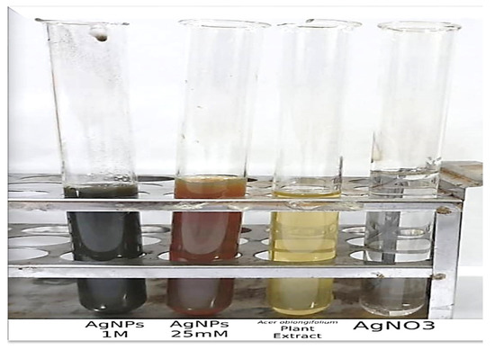
Figure 1.
Synthesis of the silver nanoparticles indicated by color change.
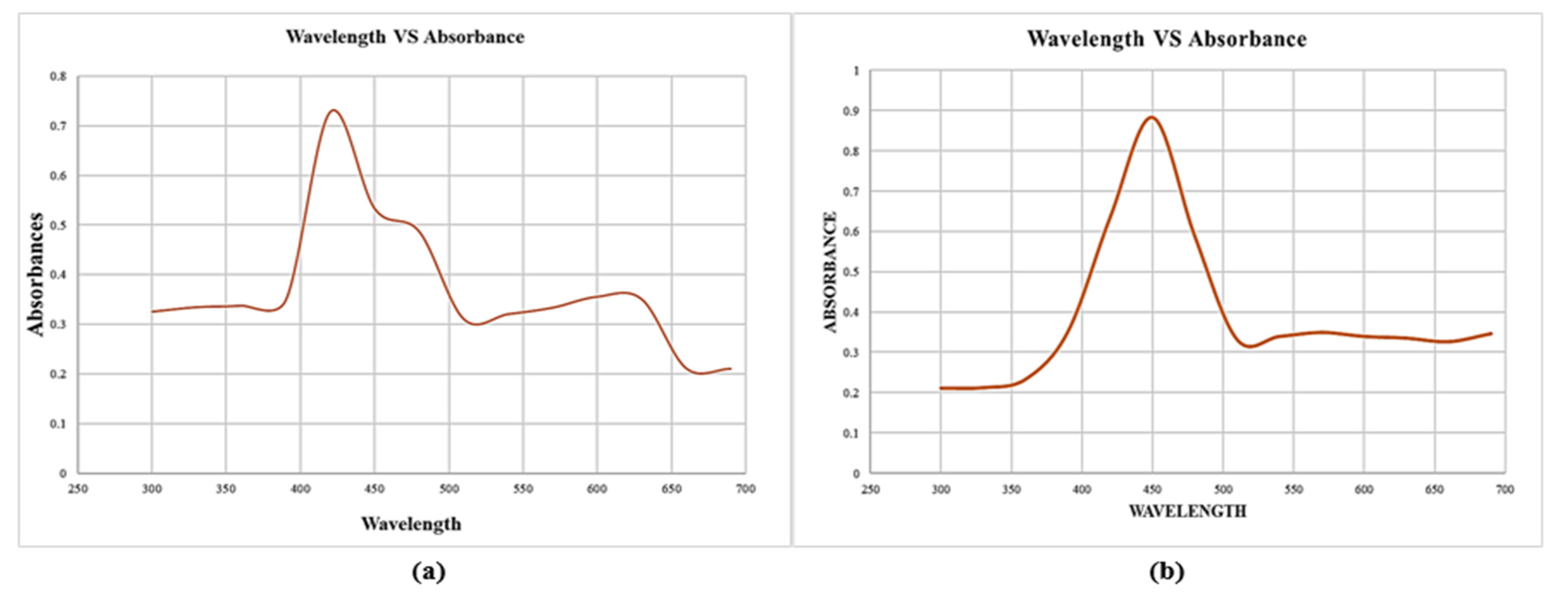
3.2. UV Visible Spectroscopy
The synthesis and stability of reduced AgNPs in colloidal solution were examined using a UV visible spectrophotometer. The maximum absorbance at 450 and 420 nm was observed in the visible UV spectra, as shown in Figure 2, which corresponds to the surface plasmon resonance (SPR) of the AgNPs. Surface Plasmon Resonance (SPR) patterns are commonly used as indicative tools for metal nanoparticle formation, as SPR depends on various parameters, such as size and medium dielectric constant [31]. The Ag+ ions were reduced extracellularly, indicating the formation of silver nanoparticles.
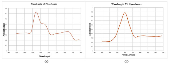
Figure 2.
UV visible spectroscopy for silver nanoparticles synthesized using Acer oblongifolium plant extracts. (a) Synthesized from 25 mM precursor solution of AgNO3, (b) synthesized from 1 M solution of AgNO3.
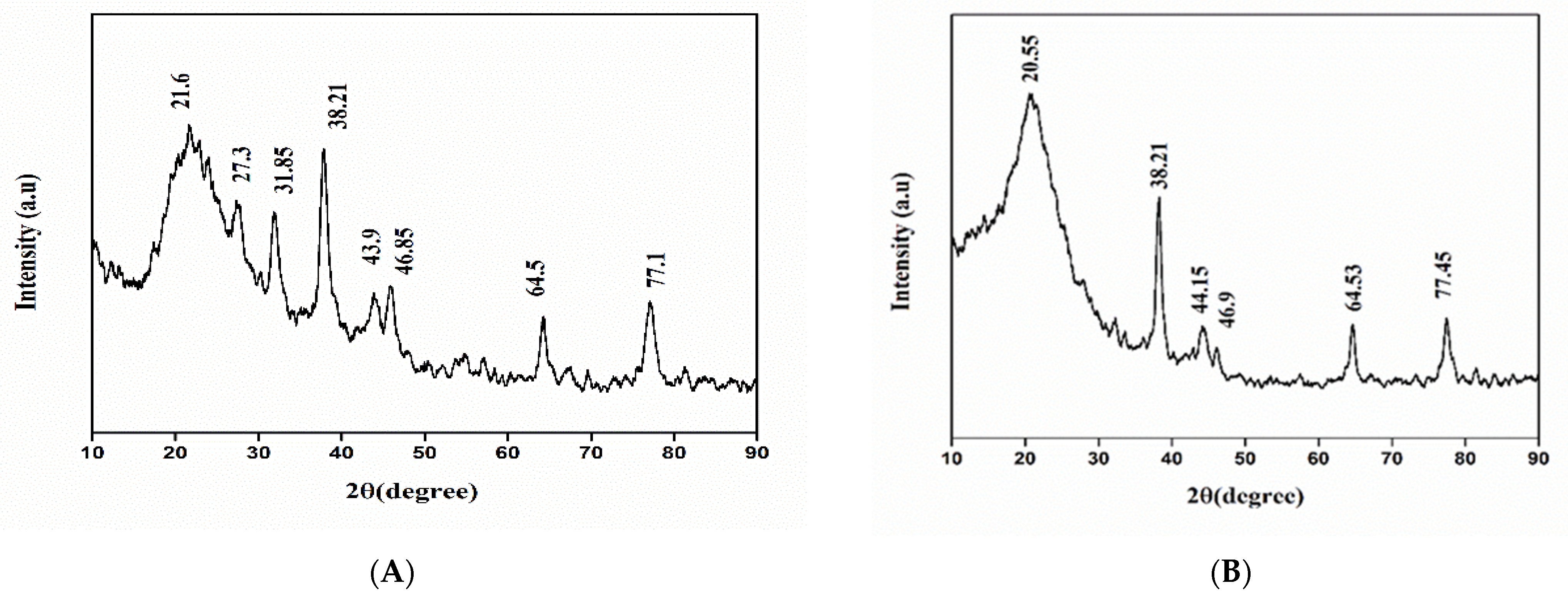
3.3. X-ray Diffraction (XRD) Analysis
Phase identification, purity, and structure of green synthesized AgNPs were determined by X-ray diffraction. Confirmation of crystalline nature and purity of AgNPs were conducted by XRD pattern with strong diffraction peaks analyzed at different 2θ, i.e., 20.55°, 38.21°, 44.15°, 46.9°, 64.53°, and 77.45° for 1M AgNPs, and for 25 mM diffraction peaks were 21.6°, 27.3°, 31.85°, 38.21°, 43.9°, 46.85°, 64.5°, and 77.1°. Diffraction peaks of AgNPs were confirmed at 38.21°, 46.9°, and 64.53° with miller indices of 111, 200, and 220, as shown in Figure 3. The average crystallized sizes of 25 mM and 1 M green synthesized AgNPs were predicted to be 3 nm and 5 nm.
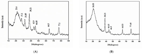
Figure 3.
XRD pattern of silver nanoparticles synthesized using Acer oblongifolium extract. (A) Synthesized from 25 mM solution of AgNO3, (B) synthesized from 1 M solution of AgNO3.
3.4. Fourier Transform Infrared (FTIR) Spectroscopy
The FTIR spectrum (Figure 4) of the reduced AgNO3 solution and capped by secondary metabolites with plant extracts of Acer oblongifolium revealed significant absorption peaks at 2865.16, 2031.92, 1478.96, and 845.35 cm−1. The appearance of the instinct band at 2865.16 cm−1 (C-H) showed alkanes present, the band at 2031.92 cm−1 confirmed the presence of alkynes, and the band at 1478.96 cm−1 revealed the N-C and N=C groups. The C-O group at 845.35 cm−1 depicted the presence of =CH in aromatic compounds in the biosynthesized silver nanoparticles [32], which is in accordance with previous studies [33,34,35].
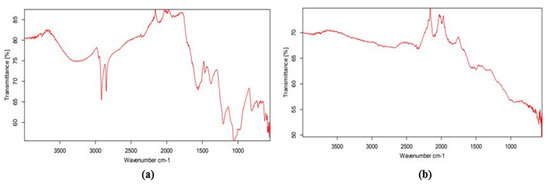
Figure 4.
FTIR spectrum of silver nanoparticles synthesized using the leaf extract of Acer oblongifolium and AgNO3: (a) 25 mM precursor solution of AgNO3, (b) 1 M precursor solution of AgNO3.
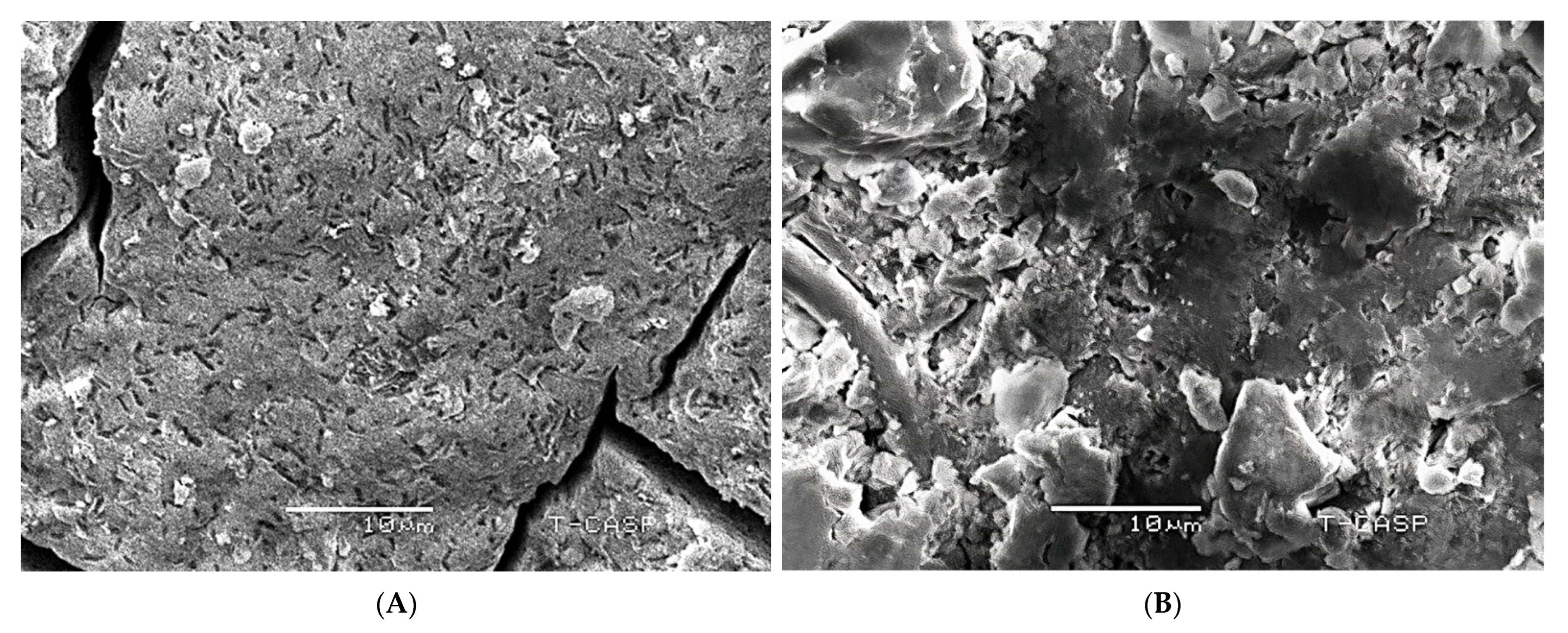
3.5. Scanning Electron Microscope (SEM)
The surface morphology and particle size of the green synthesized AgNPs were determined using a scanning electron microscope (SEM). A typical scanning electron micrograph identifies that AgNPs are rod-like in shape. Moreover, the average crystalline sizes for 25 mM and 1M AgNPs were found to be 5 nm and 8 nm, respectively; this is similar to the size predicted by XRD, as shown in Figure 5.
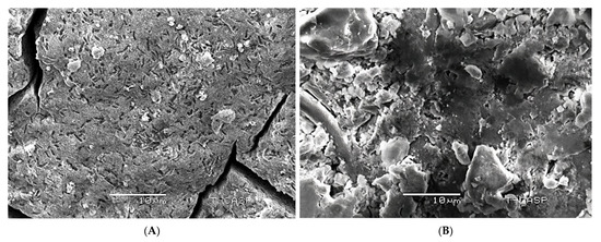
Figure 5.
(A) SEM micrograph of silver nanoparticles synthesized using the 25 mM solution of AgNO3 and leaf extract of Acer oblongifolium; (B) SEM micrograph of silver nanoparticles synthesized using the 1 M solution of AgNO3 and leaf extract of Acer oblongifolium.
3.6. In Vitro Biological Application
3.6.1. Antibacterial Activity
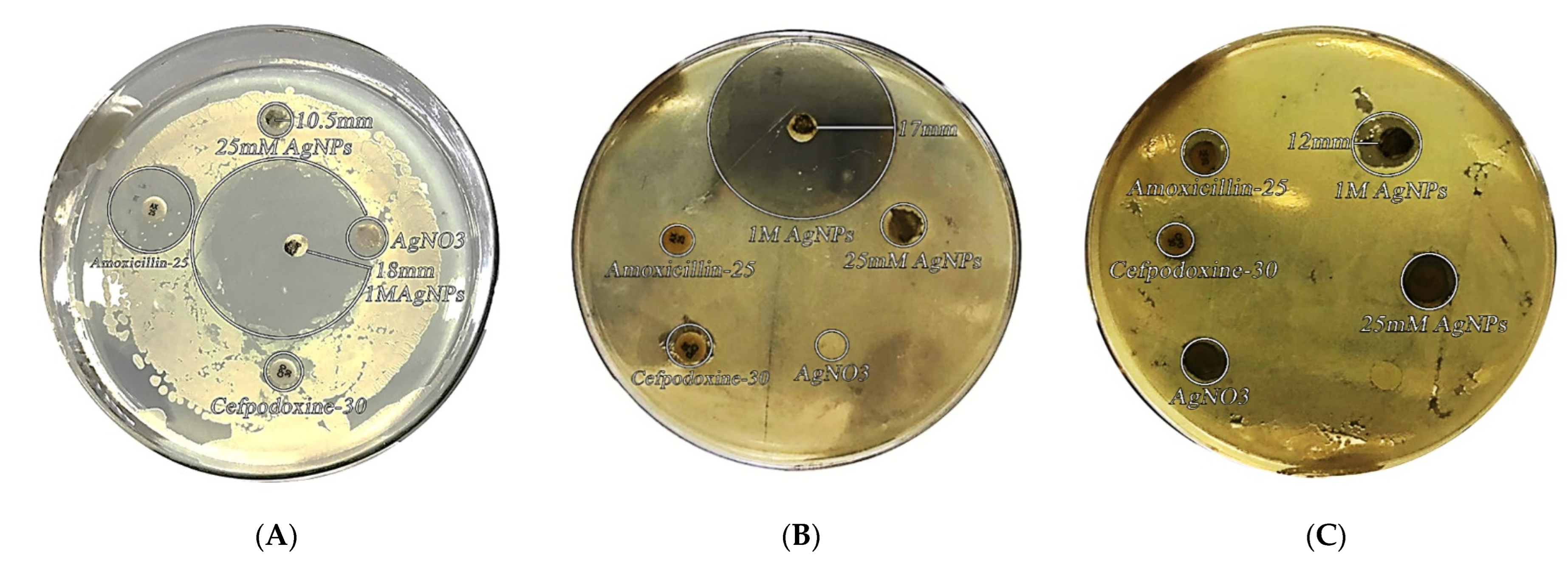
The antimicrobial activity of silver nanoparticles synthesized from Acer oblongifolium plant extract was studied using the disc diffusion method against Bacillus cereus, Bacillus paramycoides, and E. coli. Figure 6 shows the diameter of zones of inhibition around each disc with silver nanoparticle solution. The antibacterial activity of AgNPs synthesized from Acer oblongifolium extracts was shown to be highest against E. coli (18 mm), lower against Bacillus cereus (17 mm), and lowest against Bacillus paramycoides (12 mm).
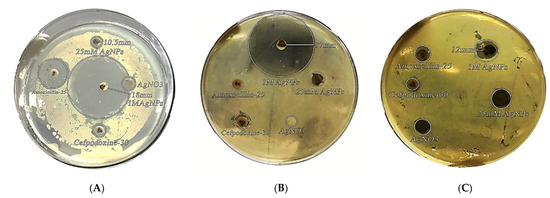
Figure 6.
Bactericidal activity of biosynthesized AgNPs from Acer oblongifolium against (A) E. coli, (B) B. cereus, and (C) B. paramycoides.
3.6.2. Physicochemical Parameters
Density, specific heat, and thermal conductivity were measured using a mathematical equation at various temperature values in the range of 15–45 °C for pure silver nanoparticles (AgNPs) and nanoparticles mixed with water. Furthermore, at the same temperature ranges, the data were taken by mixing the AgNPs with water in different ratios 1:2, 1:4, and 1:6 (in mL). The physicochemical parameters, including density, specific heat, and thermal conductivity data, are given in Table 1, Table 2 and Table 3 and Figure 7a,b.
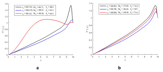
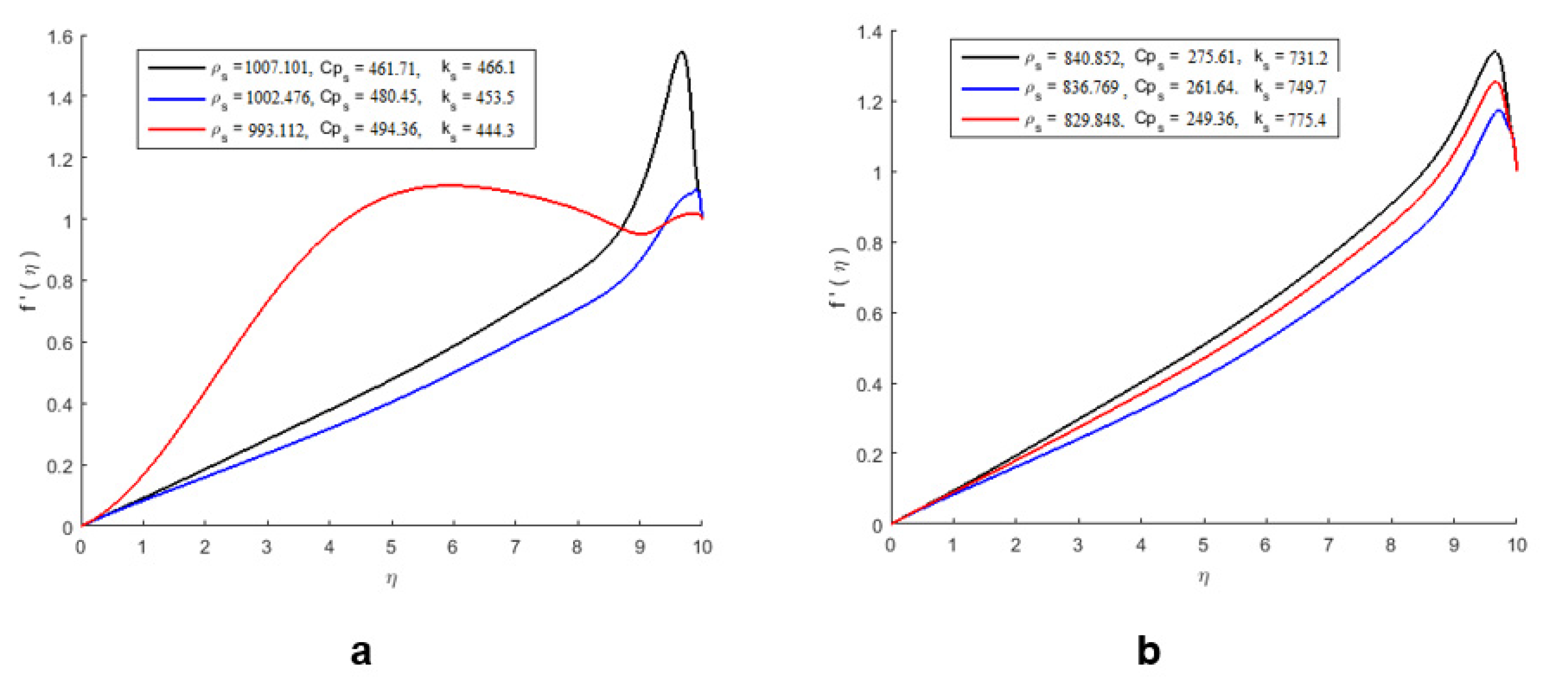
Figure 7.
The effect of pure nanoparticle parameters on the velocity profile of pure nanoparticles and mixed nanoparticles. (a) Velocity profile of pure nanoparticles. (b) Velocity profile of nanoparticles mixed with water.
The effect of pure nanoparticles’ parameters on the velocity profile without mixing with water is shown in Figure 7a, and the effect of nanoparticles on the parameters of the velocity profile after mixing with water is shown in Figure 7b. The deplanement of the volume fraction parameters positively affected the nanofluid velocity. By improving the nanoparticles, the thickness of the boundary layer of the flow reduces, which results in an increase in the velocity of the nanofluid. However, we observed that the behavior of the velocity field is opposite in Figure 7b.
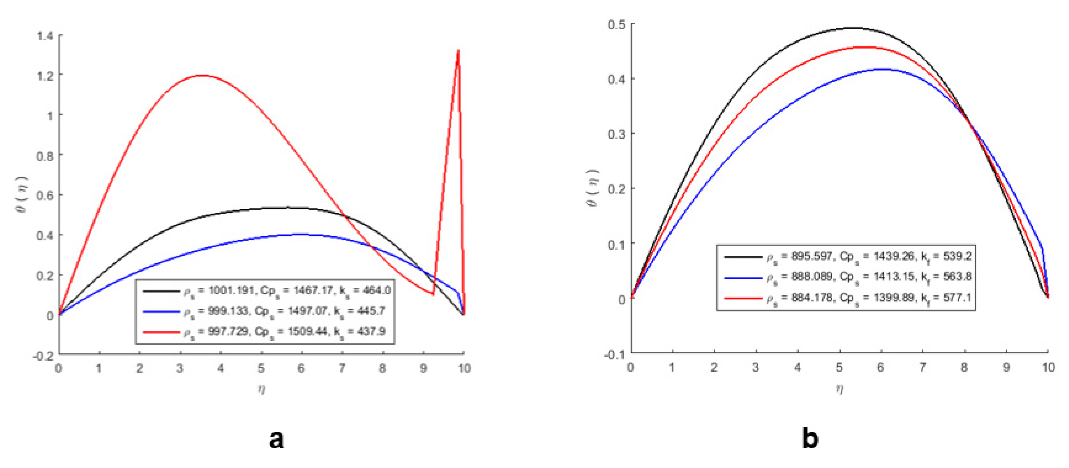
The effect of nanoparticles on the temperature profile is shown in Figure 8a,b, and it can be noticed that the pure nanoparticles show more heat-absorbing capacity, while the nanoparticles mixed with water show less heat-absorbing capacity. As a result, the temperature of the nanofluid increases, which also destroys heat; therefore, the average temperature of the fluid decreases (Figure 8b).
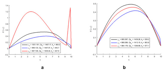
Figure 8.
The effect of nanoparticles on the temperature profile of pure nanoparticles (a) and mixed nanoparticles (b).
The 3D representation of the velocity and temperature of the AgNPs is shown in Figure 9.
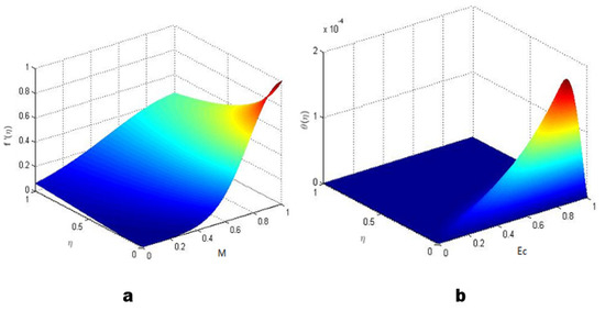
Figure 9.
3D representation of the velocity and temperature of AgNPS: (a) velocity and (b) temperature.
3.6.3. Antiproliferative Activity against MCF-7 and HeLa Cell Lines
IC50 values of AgNPs synthesized from Acer oblongifolium plant extract against MCF-7 and HeLa cell lines. After 48–72 h of incubation, MCF-7 showed high antiproliferative activity in comparison to the HeLa cell line, with values of 9.43 and 6.22 µg/mL, respectively.
4. Discussion
The color transformation to dark brown from the original yellow confirmed the formation of silver NPs from Acer oblongifolium plant extract. Prasad and Elumalai [36] noticed that silver nanoparticles come in a variety of colors, ranging from light yellow to brown. Furthermore, because of surface plasmon excitation fluctuations in silver nanoparticles, Prasad et al. [37] observed that these nanoparticles, in aqueous solutions, gave out a yellowish-brown color. The maximum absorbance peak for silver nanoparticles synthesized from the Acer oblongifolium plant extract was observed at 450 nm using the UV visible spectrum. According to Prasad and Elumalai [36], a 430–450 nm peak of absorption is observed in the silver NPs’ spectra present in the reaction media, whereas Kumar et al. [38] narrowed the gap and reported an absorbance peak of 438 nm. Moreover, unmapped peaks were also determined that indicate the phytochemical’s presence on the surface of AgNPs, which aids in capping. Moreover, the average particle sizes predicted by Image J software for 25 mM and 1 M AgNPs were 5 nm and 8 nm. This was in close proximity to the size obtained from XRD [39]. Plant extracts’ reduction by stabilizing agents plays a role in the reduction of Ag+ ions to Ag nanoparticles, according to FTIR study by Iqbal et al. [40]. Hemlata et al. [22] observed that silver nanoparticles bind to the carboxyl or amino groups of extracted proteins. The amine (–NH), hydroxyl (–OH), and carboxyl (–C=O) groups of leaf extracts are primarily engaged to fabricate silver nanoparticles, according to studies conducted by [41]. Scanning electron microscopy (SEM) was utilized to determine particle size as well as morphology of biosynthesized AgNPs [42]. The SEM micrograph predicted the morphology, i.e., rod-like shape. Relevant morphological studies have also been found in the literature [43].
Acer oblongifolium-derived silver nanoparticles have a potent inhibitory effect on E. coli, Bacillus cereus, and Bacillus paramycoides. Silver nanoparticles synthesized from Acer oblongifolium leaves are very small size, due to which they have unique physical and chemical properties. Nanoparticles have a more germicidal effect than the mass of silver metal because of the reduction of size, increase of the ratio of surface to volume of nanoparticles, and increase of the contact area with microorganisms.
Similarly, Jain et al. [44] published a human pathogen antibacterial assay using silver nanoparticles synthesized from Papaya fruit extract, which was revealed to be highly toxic against bacteria that are resistant to multiple drugs. Kumar [38] also found that AgNPs were moderately toxic to E. coli, Pseudomonas species, including putida, aeruginosa, vulgaris, and B. subtilis. The pathogens’ growth was hindered by the silver nanoparticles of the Acer oblongifolium. We may infer that biosynthesized AgNPs are more toxic to MCF-7 cells than to the HeLa cell line, based on the results of the antiproliferative activity study.
The activation of metallothioneins and the use of Ag+ chelating agents to prevent cytotoxicity support this idea. The potential of the mitochondrial membrane was depolarized by silver nanoparticles. The depolarized mitochondrial membrane potential is a key element in apoptosis signaling pathways. Finally, silver nanoparticles produced early/late apoptosis and necrosis in MCF7 and HeLa cells, with early apoptosis being the primary cause of cell death. The activity of early apoptosis was 19 percent higher than that of late apoptosis and necrosis [45]. Inducing the creation of reactive oxygen species and oxidative stress, which lead to DNA damage and apoptosis, are two possible pathways of toxicity. Toxicity in AgNP-treated cells is primarily induced by the release of Ag+ ions into the cytosol following AgNP uptake via endocytosis and breakdown in an acidic environment. As a result, the oxidative stress, DNA damage, and cell death observed in the presence of AgNPs are mostly attributable to silver ions in the cytosol, impairing natural metabolic and cell cycle mechanisms [46]. For MCF-7 cells, silver nanoparticles synthesized with Artemisia vulgaris leaf extract had an IC50 value close to 60 g/mL [47]. The efficacy of the aqueous leaf extract of Cucumis sativus against MCF-7 cells was also supported by another study [41]. Taraxacum officinale methanolic leaf extract also showed improved activity against MCF-7 cell lines. The current research is the first to compare the cell viability of Acer oblongifolium leaf extract-mediated biosynthesized AgNPs against two cancer cell lines. The Acer oblongifolium AgNPs demonstrated a difference in cytotoxicity toward different cell lines due to the increased cellular absorption and retention of NPs. Because of their small size, NPs can enter cells through endocytosis and are not subjected to efflux by P-glycoprotein [48,49].
5. Conclusions
In the current study, we have revealed the simple use of a natural, low-cost biological reducing agent and Acer oblongifolium leaf extracts through efficient green nanochemistry methodology, avoiding the presence of toxic solvents and waste. Silver nanoparticles made from Acer oblongifolium leaf extract were found to be effective against human infections. The antibacterial activity of the disc diffusion approach is well documented. Because of these applications, this technology is potentially promising for the large-scale production of nanoparticles. Prepared nanoparticles can be employed as antibacterial agents in wound healing, water purification, and medicine.
Author Contributions
Conceptualization, M.N., B.B., T.A., S.Z. and M.A.M.; Original draft, M.N., B.B., A.A.K. and T.A.; Methodology, M.W.A., A.S.D., M.S. and M.A.; Data curation: A.A.A., A.S.A., M.A. and A.A.K.; Writing—review & editing, M.N., B.B., M.W.A. and T.A.; Visualization, M.W.A., M.S., A.A.K. and M.A.; Resources, A.S.D., M.W.A., M.S. and A.A.A.; Project administration, M.N., A.S.D., T.A., S.Z. and M.A.; Funding acquisition, A.A.A., A.S.A. and M.A.; Validation, M.N. and T.A.; Investigation, M.N., B.B., S.Z., T.A., A.A.K. and M.A.M.; Formal analysis, M.W.A., B.B. and M.S.; Supervision, M.N. and T.A. All authors have read and agreed to the published version of the manuscript.
Funding
This research received no external funding.
Institutional Review Board Statement
Not applicable.
Informed Consent Statement
Not applicable.
Data Availability Statement
All major data generated and analyzed in this study are included in this manuscript.
Acknowledgments
Abdulraheem Ali Almalki would like to acknowledge Taif University for supporting this study under Taif University Researchers Supporting Project number (TURSP-2020/296), Taif University, Taif, Saudi Arabia.
Conflicts of Interest
The manuscript was written through contributions of all authors. All authors have given approval to the final version of the manuscript. These authors contributed equally. All authors declare no conflict of interest.
Sample Availability
Samples of the compounds are not available from the authors.
References
- Palei, N.N. Green synthesis of silver nanoparticles using leaf extract of Lantana camara and its antimicrobial activity. Int. J. Green Pharm. 2020, 14, 1–7. [Google Scholar]
- Gogoi, B.; Kumar, R.; Upadhyay, J.; Borah, D. Facile biogenic synthesis of silver nanoparticles (AgNPs) by Citrus grandis (L.) Osbeck fruit extract with excellent antimicrobial potential against plant pathogens. SN Appl. Sci. 2020, 2, 1–7. [Google Scholar] [CrossRef]
- Ajitha, B.; Reddy, Y.A.K.; Reddy, P.S. Green synthesis and characterization of silver nanoparticles using Lantana camara leaf extract. Mater. Sci. Eng. 2015, 49, 373–381. [Google Scholar] [CrossRef] [PubMed]
- Dos Santos, C.A.; Seckler, M.M.; Ingle, A.P.; Gupta, I.; Galdiero, S.; Galdiero, M.; Gade, A.; Rai, M. Silver nanoparticles: Therapeutical uses, toxicity, and safety issues. J. Pharm. Sci. 2014, 103, 1931–1944. [Google Scholar] [CrossRef]
- Jalal, M.; Ansari, M.A.; Alzohairy, M.A.; Ali, S.G.; Khan, H.M.; Almatroudi, A.; Raees, K. Biosynthesis of silver nanoparticles from oropharyngeal Candida glabrata isolates and their antimicrobial activity against clinical strains of bacteria and fungi. Nanomaterials 2018, 8, 586. [Google Scholar] [CrossRef] [Green Version]
- Marinescu, L.; Ficai, D.; Ficai, A.; Oprea, O.; Nicoara, A.I.; Vasile, B.S.; Boanta, L.; Marin, A.; Andronescu, E.; Holban, A.-M. Comparative Antimicrobial Activity of Silver Nanoparticles Obtained by Wet Chemical Reduction and Solvothermal Methods. Int. J. Mol. Sci. 2022, 23, 5982. [Google Scholar] [CrossRef]
- Jain, A.; Malik, A.; Malik, H.K. Mathematical modelling of seed-mediated size-specific growth of spherical silver nanoparticles using Azadirachta indica leaf extract. J. Taibah Univ. Sci. 2020, 14, 873–880. [Google Scholar] [CrossRef]
- Mangindaan, D.; Lin, G.-Y.; Kuo, C.-J.; Chien, H.-W. Biosynthesis of silver nanoparticles as catalyst by spent coffee ground/recycled poly (ethylene terephthalate) composites. Food Bioprod. Process. 2020, 121, 193–201. [Google Scholar] [CrossRef]
- Akinola, P.; Lateef, A.; Asafa, T.; Beukes, L.; Hakeem, A.; Irshad, H. Multifunctional titanium dioxide nanoparticles biofabricated via phytosynthetic route using extracts of Cola nitida: Antimicrobial, dye degradation, antioxidant and anticoagulant activities. Heliyon 2020, 6, e04610. [Google Scholar] [CrossRef]
- Adebayo, E.A.; Ibikunle, J.B.; Oke, A.M.; Lateef, A.; Azeez, M.A.; Oluwatoyin, A.O.; Ayanfe Oluwa, A.V.; Blessing, O.T.; Comfort, O.O.; Adekunle, O.O. Antimicrobial and antioxidant activity of silver, gold and silver-gold alloy nanoparticles phytosynthesized using extract of Opuntia ficus-indica. Rev. Adv. Mater. Sci. 2019, 58, 313–326. [Google Scholar] [CrossRef]
- Padnya, P.; Gorbachuk, V.; Stoikov, I. The Role of Calix[n]arenes and Pillar[n]arenes in the Design of Silver Nanoparticles: Self-Assembly and Application. Int. J. Mol. Sci. 2020, 21, 1425. [Google Scholar] [CrossRef] [PubMed] [Green Version]
- Akkol, E.K.; Göger, F.; Koşar, M.; Başer, K.H.C. Phenolic composition and biological activities of Salvia halophila and Salvia virgata from Turkey. Food Chem. 2008, 108, 942–949. [Google Scholar] [CrossRef]
- van den Berg, A.K.; Perkins, T.D. Contribution of anthocyanins to the antioxidant capacity of juvenile and senescing sugar maple (Acer saccharum) leaves. Funct. Plant Biol. 2007, 34, 714–719. [Google Scholar] [CrossRef]
- An, L.; Wang, J.-W.; Liu, J.-D.; Zhao, Z.-M.; Song, Y.-J. Design, preparation, and characterization of novel calix [4] arene bioactive carrier for antitumor drug delivery. Front. Chem. 2019, 7, 732. [Google Scholar] [CrossRef]
- Ahmed, S.; Saifullah Ahmad, M.; Swami, B.L.; Ikram, S. Green synthesis of silver nanoparticles using Azadirachta indica aqueous leaf extract. J. Radiat. Res. Appl. Sci. 2016, 9, 1–7. [Google Scholar] [CrossRef] [Green Version]
- Escárcega-González, C.E.; Garza-Cervantes, J.A.; Vazquez-Rodríguez, A.; Montelongo-Peralta, L.Z.; Treviño-Gonzalez, M.T.; Castro, E.D.B.; Saucedo-Salazar, E.M.; Morales, R.C.; Soto, D.R.; González, F.T. In vivo antimicrobial activity of silver nanoparticles produced via a green chemistry synthesis using Acacia rigidula as a reducing and capping agent. Int. J. Nanomed. 2018, 13, 2349. [Google Scholar] [CrossRef] [PubMed] [Green Version]
- Anandalakshmi, K.; Venugobal, J.; Ramasamy, V. Characterization of silver nanoparticles by green synthesis method using Pedalium murex leaf extract and their antibacterial activity. Appl. Nanosci. 2016, 6, 399–408. [Google Scholar] [CrossRef] [Green Version]
- Ghiyasiyan-Arani, M.; Salavati-Niasari, M.; Naseh, S. Enhanced photodegradation of dye in wastewater using iron vanadate nanocomposite; ultrasound-assisted preparation and characterization. Ultrason. Sonochem. 2017, 39, 494–503. [Google Scholar] [CrossRef]
- Platania, V.; Kaldeli-Kerou, A.; Karamanidou, T.; Kouki, M.; Tsouknidas, A.; Chatzinikolaidou, M. Antibacterial Effect of Colloidal Suspensions Varying in Silver Nanoparticles and Ions Concentrations. Nanomaterials 2022, 12, 31. [Google Scholar] [CrossRef]
- Khan, M.; Khan, A.U.; Bogdanchikova, N.; Garibo, D. Antibacterial and Antifungal Studies of Biosynthesized Silver Nanoparticles against Plant Parasitic Nematode Meloidogyne incognita, Plant Pathogens Ralstonia solanacearum and Fusarium oxysporum. Molecules 2021, 26, 2462. [Google Scholar]
- Khan, M.S.; Mei, S.; Shabnam Fernandez-Gamiz, U.; Noeiaghdam, S.; Shah, S.A.; Khan, A. Numerical Analysis of Unsteady Hybrid Nanofluid Flow Comprising CNTs-Ferrous oxide/Water with Variable Magnetic Field. Nanomaterials 2022, 12, 180. [Google Scholar] [CrossRef] [PubMed]
- Hemlata, P.R.M.; Singh, A.P.; Tejavath, K.K. Biosynthesis of Silver Nanoparticles Using Cucumis prophetarum Aqueous Leaf Extract and Their Antibacterial and Antiproliferative Activity Against Cancer Cell Lines. ACS Omega 2020, 5, 5520–5528. [Google Scholar] [CrossRef] [PubMed] [Green Version]
- Pathak, M.; Kumar, V.; Pathak, P.; Majee, R.; Ramteke, P.W.; Verma, A. Green Synthesis of Silver Nanoparticles Using Scindapsus officinalis (Gajpipli): In-Vitro Cytotoxic Activity Against HepG-2 & MCF-7 Cancer Cell Lines. Preprints 2019, 2019080118. [Google Scholar]
- Wang, Y.; Chinnathambi, A.; Nasif, O.; Alharbi, S.L. Green synthesis and chemical characterization of a novel anti-human pancreatic cancer supplement by silver nanoparticles containing Zingiber officinale leaf aqueous extract. Arab. J. Chem. 2021, 14, 103081. [Google Scholar] [CrossRef]
- Wypij, M.; Jędrzejewski, T.; Trzcińska-Wencel, J.; Ostrowski, M.; Rai, M.; Golińska, P. Green Synthesized Silver Nanoparticles: Antibacterial and Anticancer Activities, Biocompatibility, and Analyses of Surface-Attached Proteins. Front Microbiol. 2021, 12, 632505. [Google Scholar] [CrossRef]
- Krithiga, N.; Rajalakshmi, A.; Jayachitra, A. Green Synthesis of Silver Nanoparticles Using Leaf Extracts of Clitoria ternatea and Solanum nigrum and Study of Its Antibacterial Effect against Common Nosocomial Pathogens. J. Nanosci. 2015, 8, 928204. [Google Scholar] [CrossRef] [Green Version]
- Pirtarighat, S.; Ghannadnia, M.; Baghshahi, S. Green synthesis of silver nanoparticles using the plant extract of Salvia spinosa grown in vitro and their antibacterial activity assessment. J. Nanostruct. Chem. 2019, 9, 1–9. [Google Scholar] [CrossRef] [Green Version]
- Kivrak, H.; Atbas, D.; Alal, O.; Çögenli, M.S.; Bayrakceken, A.; Mert, S.O.; Sahin, O. A complementary study on novel PdAuCo catalysts: Synthesis, characterization, direct formic acid fuel cell application, and exergy analysis. Int. J. Hydrogen Energy 2018, 43, 21886–21898. [Google Scholar] [CrossRef]
- Bibi, G.; Ullah, N.; Mannan, A.; Mirza, B. Antitumor, cytotoxic and antioxidant potential of Aster thomsonii extracts. Afr. J. Pharm. Pharmacol. 2011, 5, 252–258. [Google Scholar]
- Khan, M.S.; Mei, S.; Fernandez-Gamiz, U.; Noeiaghdam, S.; Khan, A. Numerical Simulation of a Time-Dependent Electroviscous and Hybrid Nanofluid with Darcy-Forchheimer Effect between Squeezing Plates. Nanomaterials 2022, 12, 876. [Google Scholar] [CrossRef]
- Ashour, A.A.; Raafat, D.; El-Gowelli, H.M.; El-Kamel, A.H. Green synthesis of silver nanoparticles using cranberry powder aqueous extract: Characterization and antimicrobial properties. Int. J. Nanomed. 2015, 10, 7207–7221. [Google Scholar]
- Al-Yousef, H.M.; Amina, M.; Alqahtani, A.S.; Alqahtani, M.S.; Malik, A.; Hatshan, M.R.; Siddiqui, M.R.H.; Khan, M.; Shaik, M.R.; Ola, M.S.; et al. Pollen Bee Aqueous Extract-Based Synthesis of Silver Nanoparticles and Evaluation of Their Anti-Cancer and Anti-Bacterial Activities. Processes 2020, 8, 524. [Google Scholar] [CrossRef]
- Vidhu, V.K.; Aromal, S.A.; Philip, D. Green synthesis of silver nanoparticles using Macrotyloma uniflorum. Spectrochim. Acta A Mol. Biomol. Spectrosc. 2011, 83, 392–397. [Google Scholar] [CrossRef] [PubMed]
- Philip, D. Honey mediated green synthesis of silver nanoparticles. Spectrochim. Acta A Mol. Biomol. Spectrosc. 2010, 75, 1078–1081. [Google Scholar] [CrossRef]
- Mude, N.; Ingle, A.; Gade, A.; Ra, M. Synthesis of Silver Nanoparticles Using Callus Extract of Carica papaya—A First Report. J. Plant Biochem. Biotechnol. 2009, 18, 83–86. [Google Scholar] [CrossRef]
- Prasad, T.; Elumalai, E. Biofabrication of Ag nanoparticles using Moringa oleifera leaf extract and their antimicrobial activity. Asian Pac. J. Trop. Biomed. 2011, 1, 439–442. [Google Scholar] [CrossRef] [Green Version]
- Prasad, K.S.; Pathak, D.; Patel, A.; Dalwadi, P.; Prasad, R.; Patel, P.; Selvaraj, K. Biogenic synthesis of silver nanoparticles using Nicotiana tobaccum leaf extract and study of their antibacterial effect. Afr. J. Biotechnol. 2011, 10, 8122–8130. [Google Scholar]
- Kumar, R.; Nirmalya, K.; Pradipta, K. Mohapatra and Suj Chandrashekhar: India’s Global Powerhouses: How They Are Taking on the World; SAGE Publications: Los Angeles, CA, USA, 2009. [Google Scholar]
- Ponnuchamy, K.; Senthamil Selvi, S.; Prabha, L.; Prem Kumar, K.; Ganeshkumar, R.; Govindaraju, M. Synthesis of silver nanoparticles from Sargassum tenerrimum and screening phytochemicals for its antibacterial activity. Nano. Biomed. Eng. 2012, 4, 12–16. [Google Scholar]
- Iqbal, N.; Iqubal, S.S.; Khan, A.A.; Mohammed, T.; Alshabi, A.M.; Aazam, E.S.; Rafiquee, M. Effect of CTABr (surfactant) on the kinetics of formation of silver nanoparticles by Amla extract. J. Mol. Liq. 2021, 329, 115537. [Google Scholar] [CrossRef]
- Tuama, A.A.; Mohammed, A.A. Phytochemical screening and in vitro antibacterial and anticancer activities of the aqueous extract of Cucumis sativus. Saudi. J. Biol. Sci. 2019, 26, 600–604. [Google Scholar] [CrossRef]
- Chinyerenwa, A.C.; Munna, M.K.H.; Rahman, S.; Mia, M.R.; Yousuf, M.; Hasan, J. Ecofriendly Sweet Scented Osmanthus Leaf Extract Mediated Synthesis of Silver Nanoparticles (SNPs). Int. J. Text. Sci. 2018, 7, 35–42. [Google Scholar]
- Iravani, S.; Korbekandi, H.; Mirmohammadi, S.V.; Zolfaghari, B. Synthesis of silver nanoparticles: Chemical, physical and biological methods. Res. Pharm. Sci. 2014, 9, 385. [Google Scholar]
- Jain, J.; Arora, S.; Rajwade, J.M.; Omray, P.; Khandelwal, S.; Paknikar, K.M. Silver nanoparticles in therapeutics: Development of an antimicrobial gel formulation for topical use. Mol. Pharm. 2009, 6, 1388–1401. [Google Scholar] [CrossRef] [PubMed]
- Kap, A.; Kutlu, H.M. Investigation of silver nitrate on cytotoxicity and apoptosis in MCF7 human breast carcinoma cells. Asian Pac. J. Cancer Biol. 2020, 5, 49–56. [Google Scholar]
- Gomes, H.I.O.; Martins, C.S.M.; Prior, J.A.V. Silver Nanoparticles as Carriers of Anticancer Drugs for Efficient Target Treatment of Cancer Cells. Nanomaterials 2021, 11, 964. [Google Scholar] [CrossRef]
- Rasheed, T.; Bilal, M.; Iqbal, H.M.; Li, C. Green biosynthesis of silver nanoparticles using leaves extract of Artemisia vulgaris and their potential biomedical applications. Colloids Surf. B Biointerfaces 2017, 158, 408–415. [Google Scholar] [CrossRef]
- Muhammed, R.A.; Hassawi, D.S.; Ibaheem, N.K. Cytotoxic Activity of Taraxacum officinale Ethanolic Plant Extract against Human Breast Cancer (MCF-7) Cells and Human Hepatic (WRL-68) Cells. Iraqi J. Cancer Med. Genet. (IJCMG) 2018, 11, 16–21. [Google Scholar]
- Sigisted, S.C.; Hooten, C.J.; Callewaert, M.C.; Jenkinsi, A.R.; Romeroi, A.E.; Pullin, M.J.; Korneinko, A.; Lowrey, T.K.; van Salmbrouck, S.; Steelanti, W.F.A. Evaluation of aqueous extracts of Taraxacum officinale on growth and invasion of breast and prostate cancer cells. Int. J. Oncol. 2008, 32, 1085–1090. [Google Scholar] [CrossRef]


Publisher’s Note: MDPI stays neutral with regard to jurisdictional claims in published maps and institutional affiliations. |
© 2022 by the authors. Licensee MDPI, Basel, Switzerland. This article is an open access article distributed under the terms and conditions of the Creative Commons Attribution (CC BY) license (https://creativecommons.org/licenses/by/4.0/).