
Azolo[1,5-a]pyrimidines and Their Condensed Analogs with Anticoagulant Activity

 , ,
, ,  , , ,
, , ,  and
and
Abstract
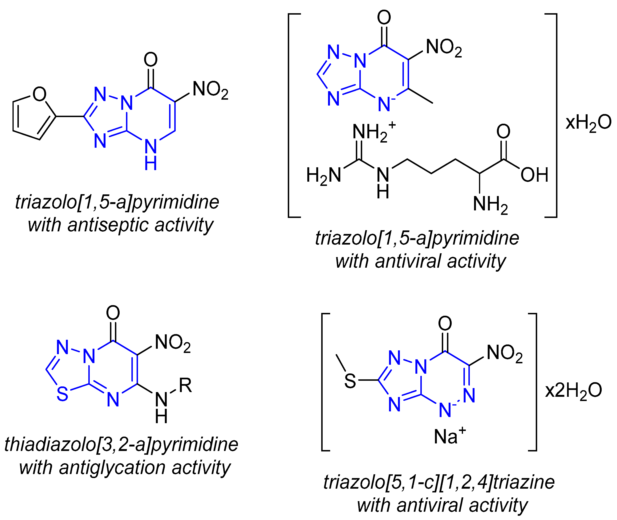
1. Introduction
2. Results
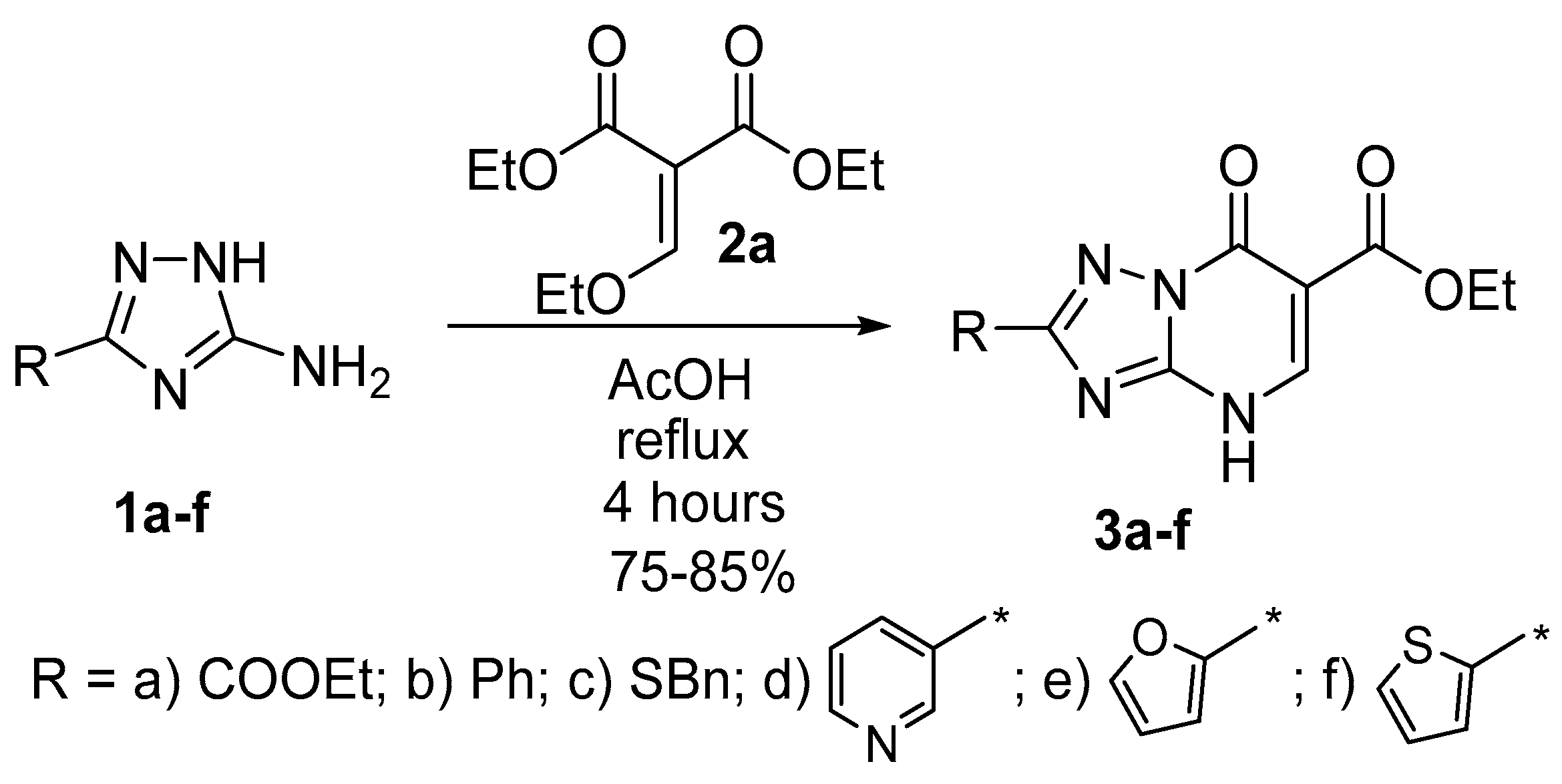
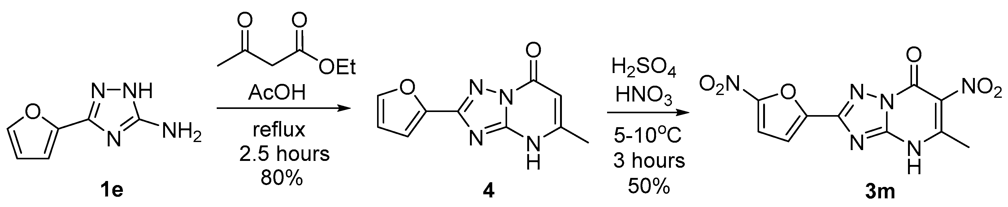
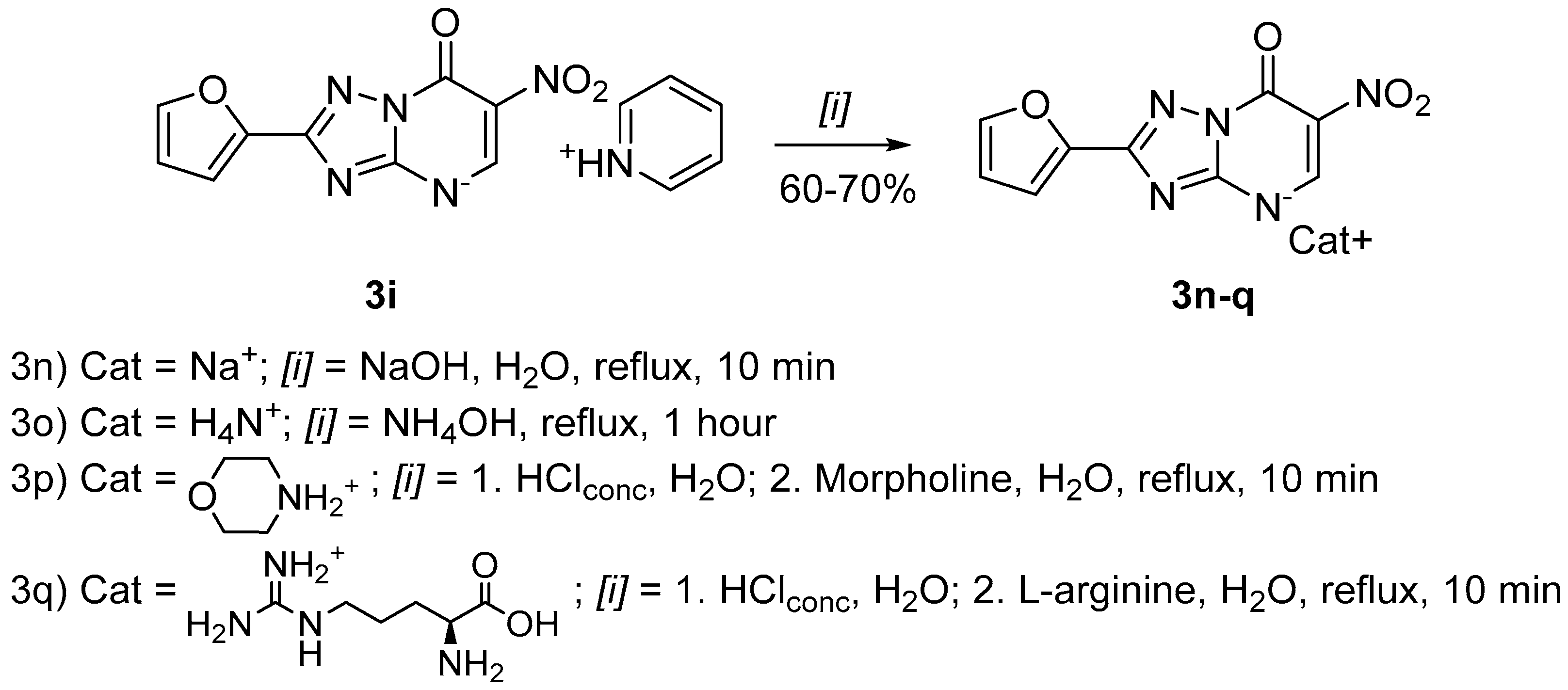
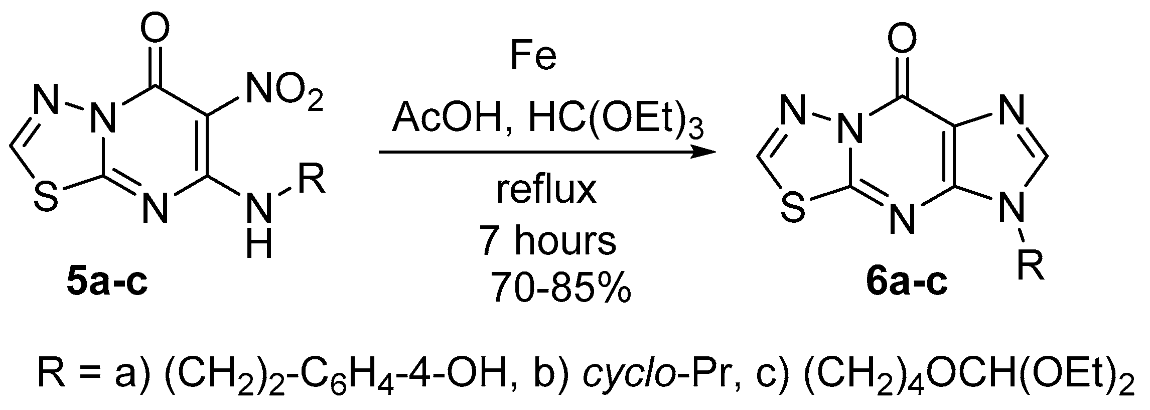
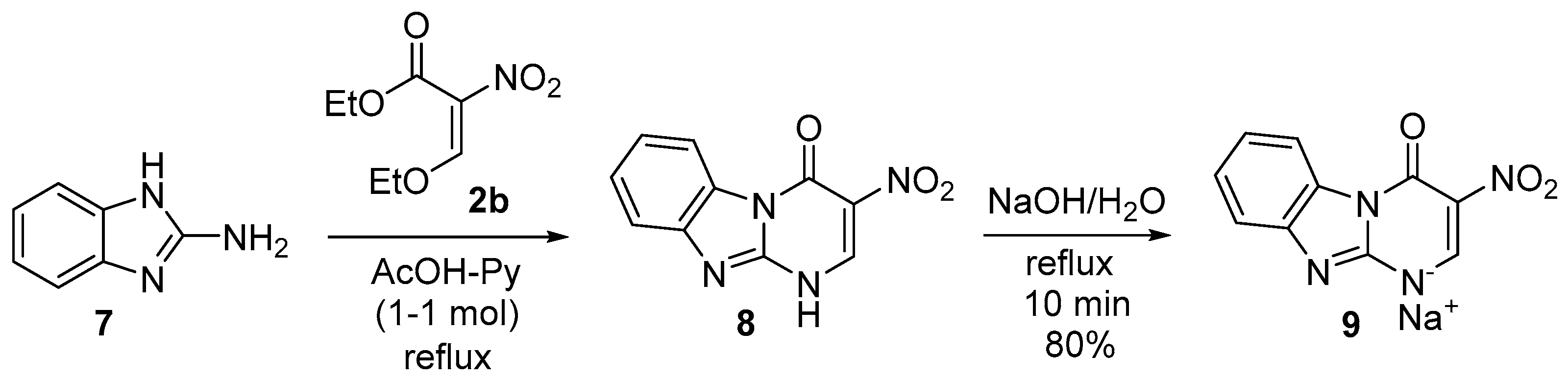
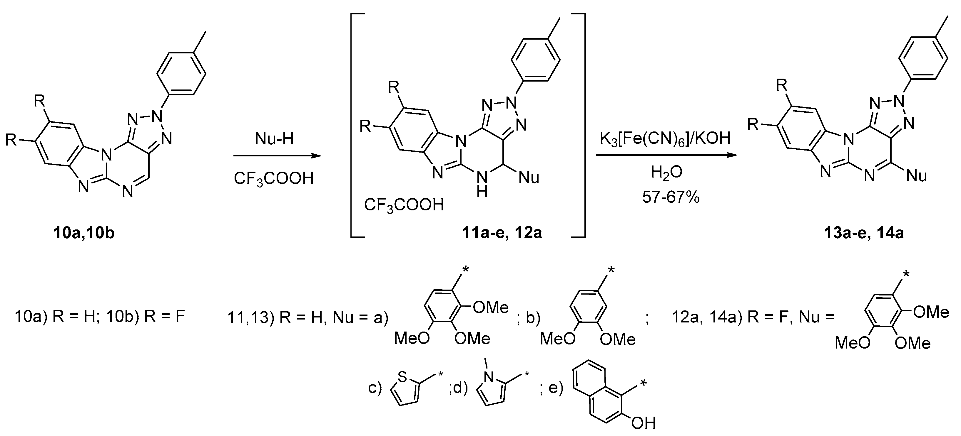
2.1. Chemistry
2.2. Anticoagulant Activity of the Target Compounds In Vitro
2.3. Iterative Neural Network Pharmacophore Analysis
- I.
- Preprocessing of the source data.
- II.
- Iterative neural network modeling.
- In the standard mode of the Statistica program, the initial dataset was divided into training, test, and validation sets in a 70/15/15% ratio. A total of 100 networks were trained with the automatic selection of 25 neural networks with high values of correlation coefficients.
- Out of 25 optimal neural networks one best performing network was picked manually according to the set of three values of the correlation coefficients.
- For the selected best neural network, sensitivity analysis of the input neurons was performed. The dimensionless sensitivity index Sens was calculated, which reflects the relative contribution of each neuron to the formation of the final signal of the output neuron.
- If during sensitivity analysis neurons with Sens < 1.0 were found, they were removed from the initial training sample, and iterative neural network modeling was carried out, starting from step 1 of this scheme.
- Otherwise, the process of iterative training of networks was completed, and for the best neural network, the overall accuracy of the prediction was assessed on the complete data set.
- In the best neural network, the most sensitive input neurons with Sens ≥ 1.1 were identified, which corresponds to the QL descriptors most significantly affecting the level of FIIa-inhibitory activity of the studied compounds.
- III.
- Post-processing of the data obtained.
- Superposition of the significant QL descriptors found in stage II derive the pharmacophore, which provides a high level of FIIa-inhibitory activity of the tested compounds.
- Analysis of entry of the constructed pharmacophore into the structure of the most active compounds was performed.
2.4. Anticoagulant Activity after LPS Treatment
2.5. An Animal Study of Anticoagulant Activity
2.6. Acute Toxicity
2.7. Tail Bleeding Time (TBT)
3. Materials and Methods
3.1. Chemistry
3.2. Biology
3.2.1. Animals
3.2.2. In Vitro Anticoagulant Assay
3.2.3. Anticoagulant Assay in Animals
3.2.4. Acute Toxicity
3.2.5. Tail Bleeding Time (TBT) Model
3.2.6. Statistical Analysis
4. Conclusions
Supplementary Materials
Author Contributions
Funding
Institutional Review Board Statement
Informed Consent Statement
Data Availability Statement
Acknowledgments
Conflicts of Interest
Sample Availability
References
- Mitchell, W.B. Thromboinflammation in COVID-19 Acute Lung Injury. Paediatr. Respir. Rev. 2020, 35, 20–24. [Google Scholar] [CrossRef]
- Llitjos, J.-F.; Leclerc, M.; Chochois, C.; Monsallier, J.-M.; Ramakers, M.; Auvray, M.; Merouani, K. High Incidence of Venous Thromboembolic Events in Anticoagulated Severe COVID-19 Patients. J. Thromb Haemost. 2020, 18, 1743–1746. [Google Scholar] [CrossRef]
- Klok, F.A.; Kruip, M.J.H.A.; van der Meer, N.J.M.; Arbous, M.S.; Gommers, D.; Kant, K.M.; Kaptein, F.H.J.; van Paassen, J.; Stals, M.A.M.; Huisman, M.V.; et al. Confirmation of the High Cumulative Incidence of Thrombotic Complications in Critically Ill ICU Patients with COVID-19: An Updated Analysis. Thromb Res. 2020, 191, 148–150. [Google Scholar] [CrossRef]
- Lodigiani, C.; Iapichino, G.; Carenzo, L.; Cecconi, M.; Ferrazzi, P.; Sebastian, T.; Kucher, N.; Studt, J.-D.; Sacco, C.; Bertuzzi, A.; et al. Venous and Arterial Thromboembolic Complications in COVID-19 Patients Admitted to an Academic Hospital in Milan, Italy. Thromb Res. 2020, 191, 9–14. [Google Scholar] [CrossRef] [PubMed]
- Tang, N.; Bai, H.; Chen, X.; Gong, J.; Li, D.; Sun, Z. Anticoagulant Treatment Is Associated with Decreased Mortality in Severe Coronavirus Disease 2019 Patients with Coagulopathy. J. Thromb Haemost. 2020, 18, 1094–1099. [Google Scholar] [CrossRef]
- Testa, S.; Prandoni, P.; Paoletti, O.; Morandini, R.; Tala, M.; Dellanoce, C.; Giorgi-Pierfranceschi, M.; Betti, M.; Danzi, G.B.; Pan, A.; et al. Direct Oral Anticoagulant Plasma Levels’ Striking Increase in Severe COVID-19 Respiratory Syndrome Patients Treated with Antiviral Agents: The Cremona Experience. J. Thromb Haemost. 2020, 18, 1320–1323. [Google Scholar] [CrossRef] [PubMed]
- Savateev, K.V.; Ulomsky, E.N.; Butorin, I.I.; Charushin, V.N.; Rusinov, V.L.; Chupakhin, O.N. Azoloazines as A2a Receptor Antagonists. Structure–Activity Relationship. Russ. Chem. Rev. 2018, 87, 636–669. [Google Scholar] [CrossRef]
- Vassiliev, P.M.; Spasov, A.A.; Kosolapov, V.A.; Kucheryavenko, A.F.; Gurova, N.A.; Anisimova, V.A. Consensus Drug Design Using IT Microcosm. In Application of Computational Techniques in Pharmacy and Medicine; Gorb, L., Kuz’min, V., Muratov, E., Eds.; Challenges and Advances in Computational Chemistry and Physics; Springer: Dordrecht, The Netherlands, 2014; pp. 369–431. ISBN 978-94-017-9257-8. [Google Scholar]
- Pires, D.E.V.; Blundell, T.L.; Ascher, D.B. pkCSM: Predicting small-molecule pharmacokinetic properties using graph-based signatures. J. Med. Chem 2015, 58, 4066–4072. [Google Scholar] [CrossRef]
- Kůrková, V. Kolmogorov’s Theorem and Multilayer Neural Networks. Neural Net. 1992, 5, 501–506. [Google Scholar] [CrossRef]
- Hilbe, J.M. Statistica 7. Am. Stat. 2007, 61, 91–94. [Google Scholar] [CrossRef]
- Wu, C.; Lu, W.; Zhang, Y.; Zhang, G.; Shi, X.; Hisada, Y.; Grover, S.P.; Zhang, X.; Li, L.; Xiang, B.; et al. Inflammasome Activation Triggers Blood Clotting and Host Death through Pyroptosis. Immunity 2019, 50, 1401–1411.e4. [Google Scholar] [CrossRef] [PubMed]
- Pawlinski, R.; Pedersen, B.; Schabbauer, G.; Tencati, M.; Holscher, T.; Boisvert, W.; Andrade-Gordon, P.; Frank, R.D.; Mackman, N. Role of Tissue Factor and Protease-Activated Receptors in a Mouse Model of Endotoxemia. Blood 2004, 103, 1342–1347. [Google Scholar] [CrossRef] [PubMed]
- Zhang, H.; Zeng, L.; Xie, M.; Liu, J.; Zhou, B.; Wu, R.; Cao, L.; Kroemer, G.; Wang, H.; Billiar, T.R.; et al. TMEM173 Drives Lethal Coagulation in Sepsis. Cell Host Microbe 2020, 27, 556–570.e6. [Google Scholar] [CrossRef] [PubMed]
- Yang, X.; Cheng, X.; Tang, Y.; Qiu, X.; Wang, Y.; Kang, H.; Wu, J.; Wang, Z.; Liu, Y.; Chen, F.; et al. Bacterial Endotoxin Activates the Coagulation Cascade through Gasdermin D-Dependent Phosphatidylserine Exposure. Immunity 2019, 51, 983–996.e6. [Google Scholar] [CrossRef] [PubMed]
- Levi, M.; van der Poll, T. Coagulation and Sepsis. Thromb Res. 2017, 149, 38–44. [Google Scholar] [CrossRef]
- Novinson, T.; Springer, R.H.; O’Brien, D.E.; Scholten, M.B.; Miller, J.P.; Robins, R.K. 2-(Alkylthio)-1,2,4-Triazolo [1,5-a] Pyrimidines as Adenosine Cyclic 3′,5′-Monophosphate Phosphodiesterase Inhibitors with Potential as New Cardiovascular Agents. J. Med. Chem. 1982, 25, 420–426. [Google Scholar] [CrossRef]
- Aghazadeh Tabrizi, M.; Baraldi, P.G.; Ruggiero, E.; Saponaro, G.; Baraldi, S.; Poli, G.; Tuccinardi, T.; Ravani, A.; Vincenzi, F.; Borea, P.A.; et al. Synthesis and Structure Activity Relationship Investigation of Triazolo [1,5-a] Pyrimidines as CB2 Cannabinoid Receptor Inverse Agonists. Eur. J. Med. Chem. 2016, 113, 11–27. [Google Scholar] [CrossRef]
- Fedotov, V.V.; Ulomskiy, E.N.; Gorbunov, E.B.; Eltsov, O.S.; Voinkov, E.K.; Savateev, K.V.; Drokin, R.A.; Kotovskaya, S.K.; Rusinov, V.L. 3-Nitropyrimido [1,2-a] Benzimidazol-4-Ones: Synthesis and Study of Alkylation Reaction. Chem. Heterocycl. Comp. 2017, 53, 582–588. [Google Scholar] [CrossRef]
- Fedotov, V.V.; Ulomsky, E.N.; Belskaya, N.P.; Eltyshev, A.K.; Savateev, K.V.; Voinkov, E.K.; Lyapustin, D.N.; Rusinov, V.L. Benzimidazoazapurines: Design, Synthesis and Photophysical Study. J. Org. Chem. 2021, 86, 8319–8332. [Google Scholar] [CrossRef]
- Du Sert, N.P.; Ahluwalia, A.; Alam, S.; Avey, M.T.; Baker, M.; Browne, W.J.; Clark, A.; Cuthill, I.C.; Dirnagl, U.; Emerson, M.; et al. Reporting Animal Research: Explanation and Elaboration for the ARRIVE Guidelines 2.0. PLoS Biol. 2020, 18, e3000411. [Google Scholar] [CrossRef]
- Spasov, A.A.; Kucheryavenko, A.F.; Gaidukova, K.A.; Kosolapov, V.A.; Zhukovskaya, O.N. Antiplatelet Activity of New Derivatives of Benzimidazole Containing Sterically Hindered Phenolic Group in Their Structure. Res. Results Pharmacol. 2020, 6, 1–9. [Google Scholar] [CrossRef][Green Version]
- Niemi, T.T.; Kuitunen, A.H. Artificial Colloids Impair Haemostasis. An in Vitro Study Using Thromboelastometry Coagulation Analysis. Acta Anaesthesiol. Scand. 2005, 49, 373–378. [Google Scholar] [CrossRef] [PubMed]
- Greene, T.K.; Schiviz, A.; Hoellriegl, W.; Poncz, M.; Muchitsch, E.M. Towards a standardization of the murine tail bleeding model. J. Thromb Haemost. 2010, 12, 2820–2822. [Google Scholar] [CrossRef] [PubMed]


| No. | Compound | Coagulogram Parameter | ||
|---|---|---|---|---|
| APTT, Sec. | TT, Sec. | PT, Sec. | ||
| Control | 47.2 ± 0.3 | 11.7 ± 0.1 | 14.6 ± 0.1 | |
| 1. | Dabigatran etexilate | 79.6 ± 4.6 * | 69.5 ± 4.5 * | 16.7 ± 0.2 |
| 2. | Apixaban | 137.5 ± 2.8 *# | 14.6±0.1 * | 67.8 ± 2.5 *# |
| 3. | 3a | 65.3 ± 3.7 | 108.7 ± 9.9 *# | 13.9 ± 0.4 |
| 4. | 3b | 63.7 ± 4.8 | 37.9 ± 1.6 * | 10.8 ± 0.5 |
| 5. | 3c | 51.8 ± 1.3 | 29.1 ± 3.4 * | 11.0 ± 0.2 |
| 6. | 3d | 58.8 ± 1.9 | 33.7 ± 2.1 * | 11.6 ± 0.2 |
| 7. | 3e | 58.0 ± 1.9 | 30.3 ± 3.5 * | 9.9 ± 0.1 |
| 8. | 3f | 50.8 ± 1.7 | 28.4 ± 1.3 * | 10.1 ± 0.8 |
| 9. | 3g | 63.2 ± 1.8 | 30.1 ± 2.7 * | 10.4 ± 0.4 |
| 10. | 3h | 50.4 ± 3.0 | 29.1 ± 0.6 * | 10.1 ± 0.5 |
| 11. | 3j | 51.8 ± 1.4 | 31.0 ± 4.3 * | 10.4 ± 0.7 |
| 12. | 3k | 50.6 ± 2.1 * | 97.9 ± 19.1 * | 13.9 ± 0.4 |
| 13. | 3l | 61.1 ± 4.1 | 28.5 ± 2.8 * | 10.0 ± 0.6 |
| 14. | 3m | 63.9 ± 0.6 | 134.8 ± 10.4 *# | 14.3 ± 0.6 |
| 15. | 3n | 55.5 ± 0.8 | 146.7 ± 5.2 *# | 13.8 ± 0.1 |
| 16. | 3o | 52.9 ± 1.0 | 31.7 ± 3.9 * | 10.9 ± 0.7 |
| 17. | 3p | 58.0 ± 4.0 | 34.8 ± 3.2 * | 10.4 ± 0.3 |
| 18. | 3q | 59.5 ± 1.9 | 34.8 ± 3.3 * | 10.6 ± 0.2 |
| 19. | 6a | 60.3 ± 2.8 | 30.7 ± 4.9 * | 10.1 ± 0.4 |
| 20. | 6b | 52.4 ± 1.8 | 33.4 ± 5.5 * | 10.5 ± 0.2 |
| 21. | 6c | 64.9 ± 2.7 * | 64.5 ± 9.1 * | 13.9 ± 0.2 |
| 22. | 9 | 63.4 ± 4.0 | 53.2 ± 9.1 * | 11.9 ± 0.6 |
| 23. | 13a | 62.7 ± 8.8 | 37.0 ± 4.0 * | 10.8 ± 0.7 |
| 24. | 13b | 63.7 ± 2.0 | 35.6 ± 1.2 * | 10.8 ± 0.5 |
| 25. | 13c | 53.1 ± 1.6 | 41.6 ± 1.9 * | 10.3 ± 0.4 |
| 26. | 13d | 58.1 ± 1.0 | 40.5 ± 3.6 * | 10.2 ± 0.1 |
| 27. | 13e | 51.8 ± 2.9 | 39.3 ± 1.7 * | 11.5 ± 0.6 |
| 28. | 14a | 64.7 ± 5.8 | 40.0 ± 1.8 * | 11.3 ± 0.9 |
| No. Iteration | Network Architecture | Correlation Coefficient | ||
|---|---|---|---|---|
| Training | Test | Validation | ||
| 1 | MLP126-9-1BFGS65ExpTanh | 0.782 | 0.999 | 0.997 |
| 2 | MLP102-10-1BFGS29ExpIdent | 0.781 | 0.999 | 0.999 |
| 3 | MLP 85-8-1BFGS23ExpTanh | 0.781 | 0.999 | 0.999 |
| 4 | MLP 71-6-1BFGS23ExpTanh | 0.782 | 0.999 | 0.985 |
| 5 | MLP 67-11-1BFGS23LogistTanh | 0.782 | 0.999 | 0.829 |
| 6 | MLP 66-11-1BFGS25TanhIdent | 0.781 | 0.999 | 0.980 |
| 7 | MLP 65-4-1BFGS56ExpTanh | 0.782 | 0.999 | 0.983 |
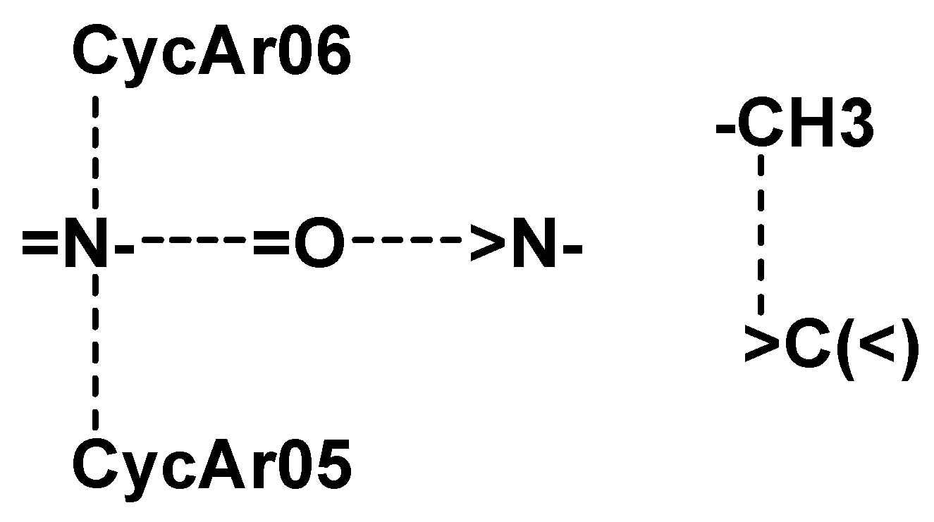
 |  |  |
| 3n | 3a | 3m |
| {-N= … CycAr06}—1 {-N= … CycAr05}—4 {-N< … =O}—1 Total—6 | {-N= … =O}—2 {-N= … CycAr06}—1 {-N= … CycAr05}—2 {-N< … =O}—2 {-CH3 … >C(<)}—2 Total—9 | {-N= … CycAr06}—1 {-N= … CycAr05}—4 {-N< … =O}—1 {CH3 … >C(<)}—1 Total—7 |
 | ||
| Dabigatranetexilate | ||
| {-N= … =O}—2 {-N= … CycAr06}—3 {-N= … CycAr05}—1 {-N< … =O}—3 {-CH3 … >C(<)}—9 Total—18 | ||
| Compound | Parameters of Coagulogram | ||
|---|---|---|---|
| APTT, Sec. | TT, Sec. | PT, Sec. | |
| Control | 47.18 ± 0.34 | 11.65 ± 0.06 | 14.60 ± 0.10 |
| LPS control | 56.28 ± 1.60 * | 13.131 ± 1.63 | 15.10 ± 0.07 |
| Dabigatran etexilate | 145.33 ± 25.17 *# | 125.45 ± 1.54 *# | 16.77 ± 0.20 |
| Apixaban | 156.3 ± 7.5 *# | 15.5 ± 0.1 * | 97.0 ± 5.3 *# |
| 3a | 65.73 ± 2.50 * | 141.62 ± 12.55 *# | 16.38 ± 0.35 * |
| 3k | 51.52 ± 2.26 | 116.9316 ± 16.79 *# | 15.75 ± 0.74 |
| 3m | 62.27 ± 1.07 * | 85.05 ± 4.99 *# | 15.75 ± 0.19 |
| 3n | 61.43 ± 0.49 * | 199.82 ± 4.39 *#$ | 15.20 ± 0.15 |
| Compound | ∆% of Thrombin Time Prolongation Relative to Control | IC50, µM | ||
| 100 µM | 10 µM | 1 µM | ||
| Dabigatran etexilate | 493.6 ± 4.5 * | 302.0 ± 0.3 * | 42.3 ± 0.6 | 24 |
| 3a | 829.1 ± 10.0 *$ | 326.9 ± 1.5 * | 40.6 ± 0.8 | 13 |
| 3n | 1154.1 ± 5.2 *$ | 477.1 ± 3.4 *$ | 48.0 ± 1.3 | 15 |
| Compound | ∆% of thrombin time prolongation relative to control + LPS | IC50, µM | ||
| 100 µM | 10 µM | 1 µM | ||
| Dabigatran etexilate + LPS | 855.4 ± 1.5 # | 292.0 ± 0.6 # | 51.7 ± 0.5 | 11 |
| 3a + LPS | 978.6 ± 12.6 # | 298.3 ± 4.2 # | 66.6 ± 1.2 | 91 |
| 3n + LPS | 1421.8 ± 4.4 #$ | 446.6 ± 6.2 #$ | 70.6 ± 1.0 | 13 |
| Sample | Dose, mg/kg | Time, h | Coagulogram Parameters | ||
|---|---|---|---|---|---|
| APTT, Sec. | TT, Sec. | PT, Sec. | |||
| Control | 38.3 ± 1.7 | 57.7 ± 3.8 | 28.1 ± 1.4 | ||
| Dabigatran etexilate | 12.0 1 | 2 # | 137.53 ± 2.79 * | 637.4 ± 5.1 * | 31.20 ± 1.17 |
| 3a | 5.4 $ | 1 | 32.2 ± 0.8 | 40.9 ± 6.0 | 24.9 ± 0.8 |
| 2 | 37.2 ± 1.2 | 60.7 ± 6.0 | 26.4 ± 0.4 | ||
| 4 | 33.3 ± 0.7 | 64.8 ± 3.4 | 27.7 ± 0.7 | ||
| 3n | 5.8 $ | 1 | 27.9 ± 2.3 | 68.6 ± 3.4 | 29.3 ± 1.3 |
| 2 | 28.5 ± 1.3 | 65.5 ± 6.1 | 28.4 ± 1.6 | ||
| 4 | 27.0 ± 2.7 | 78.5 ± 1.6 * | 31.2 ± 1.2 | ||
| Sample | Dose, mg/kg | Time, h | TEG Parameters | |||
|---|---|---|---|---|---|---|
| R, min | K, min | α–Angle, Deg. | MA, RU | |||
| Control | 6.4 ± 0.9 | 2.5 ± 0.1 | 61.5 ± 2.3 | 65.5 ± 1.2 | ||
| Dabigatran etexilate | 12.0 1 | 2 # | 22.8 ± 3.3 * | 16.8 ± 2.8 * | 14.1 ± 3.1 * | 37.1 ± 7.9 * |
| 3a | 5.4 $ | 1 | 7.3 ± 0.6 | 2.9 ± 0.4 | 55.2 ± 3.5 | 68.2 ± 1.9 |
| 2 | 6.9 ± 0.2 | 3.5 ± 0.3 | 51.7 ± 2.1 | 66.2 ± 0.3 | ||
| 4 | 9.2 ± 1.0 | 2.7 ± 0.5 | 54.6 ± 6.1 | 66.2 ± 0.1 | ||
| 3n | 5.8 $ | 1 | 6.1 ± 1.6 | 2.4 ± 0.8 | 62.8 ± 7.6 | 66.1 ± 5.1 |
| 2 | 5.4 ± 0.4 | 3.2 ± 0.5 | 54.3 ± 2.7 | 63.7 ± 2.5 | ||
| 4 | 7.9 ± 0.5 | 11.6 ± 3.2 * | 37.3 ± 7.8 | 46.9 ± 9.1 | ||
| Sample | Dose, mg/kg | Time, h | Coagulogram Parameters | ||
|---|---|---|---|---|---|
| APTT, Sec. | TT, Sec. | PT, Sec. | |||
| Control | 38.3 ± 1.7 | 57.7 ± 3.8 | 28.1 ± 1.4 | ||
| Dabigatran etexilate | 12.0 1 | 2 # | 140.1 ± 8.1 * | 637.4 ± 5.1 * | 30.5 ± 0.8 |
| 3a | 10.8 | 1 | 35.9 ± 0.3 | 66.5 ± 11.0 | 29.1 ± 1.9 |
| 2 | 35.8 ± 1.5 | 67.2 ± 5.6 | 25.8 ± 0.6 | ||
| 4 | 35.4 ± 1.4 | 57.9 ± 6.4 | 26.2 ± 1.6 | ||
| 3n | 11.6 | 1 | 37.0 ± 0.8 | 194.4 ± 19.8 * | 25.4 ± 1.3 |
| 2 | 32.3 ± 0.2 | 186.9 ± 2.5 | 22.9 ± 4.4 | ||
| 4 | 36.1 ± 1.2 | 198.3 ± 26.7 * | 28.7 ± 1.4 | ||
| 23.2 | 1 | 31.6 ± 1.5 | 655.6 ± 2.1 * | 29.3 ± 0.6 | |
| 2 | 31.7 ± 0.7 | 116.5 ± 13.7 * | 20.9 ± 0.3 | ||
| 4 | 34.4 ± 0.7 | 95.6 ± 25.8 | 21.8 ± 0.9 | ||
| Sample | Dose, mg/kg | Time, h | TEG Parameters | |||
|---|---|---|---|---|---|---|
| R, min | K, min | α–Angle, Deg. | MA, RU | |||
| Control | 6.4 ± 0.9 | 2.5 ± 0.1 | 61.5 ± 2.3 | 65.5 ± 1.2 | ||
| Dabigatran etexilate | 12.0 1 | 2 # | 22.8 ± 3.3 * | 16.8 ± 2.8 * | 14.1 ± 3.1 * | 37.1±7.9 * |
| 3a | 10.8 | 1 | 8.1 ± 0.6 | 2.3 ± 0.1 | 60.3 ± 0.1 | 69.6 ± 2.3 |
| 2 | 5.9 ± 0.4 | 1.8 ± 0.3 | 67.3 ± 0.4 | 69.5 ± 3.3 | ||
| 4 | 5.9 ± 0.6 | 2.3 ± 0.5 | 61.1 ± 3.1 | 73.3 ± 2.3 | ||
| 3n | 11.6 | 1 | 6.2 ± 1.1 | 4.6 ± 1.1 | 41.5 ± 7.6 | 52.8 ± 2.5 |
| 2 | 8.7 ± 0.3 | 6.2 ± 0.4 * | 32.3 ± 1.5 * | 50.3 ± 1.3 | ||
| 4 | 11.9 ± 1.0 * | 8.9 ± 0.9 * | 24.5 ± 1.2 * | 46.7 ± 1.6 * | ||
| 23.2 | 1 | 13.6 ± 1.5 * | 9.2 ± 0.9 * | 29.0 ± 2.8 * | 45.1 ± 3.6 * | |
| 2 | 9.0 ± 0.01 * | 5.0 ± 1.8 | 37.4 ± 6.7 * | 53.5 ± 3.6 | ||
| 4 | 20.1 ± 8.4 | 18.1 ± 7.3 | 14.9 ± 6.4 * | 34.5 ± 2.9 * | ||
| Sample | Dose, mg/kg | TBT, s |
|---|---|---|
| Control | 288.3 ± 29.7 | |
| Dabigatran etexilate | 12.0 | 268.5 ± 17.0 |
| 3n | 23.2 | 313.0 ± 11.4 |
Publisher’s Note: MDPI stays neutral with regard to jurisdictional claims in published maps and institutional affiliations. |
© 2022 by the authors. Licensee MDPI, Basel, Switzerland. This article is an open access article distributed under the terms and conditions of the Creative Commons Attribution (CC BY) license (https://creativecommons.org/licenses/by/4.0/).
Share and Cite
Savateev, K.V.; Fedotov, V.V.; Rusinov, V.L.; Kotovskaya, S.K.; Spasov, A.A.; Kucheryavenko, A.F.; Vasiliev, P.M.; Kosolapov, V.A.; Sirotenko, V.S.; Gaidukova, K.A.; et al. Azolo[1,5-a]pyrimidines and Their Condensed Analogs with Anticoagulant Activity. Molecules 2022, 27, 274. https://doi.org/10.3390/molecules27010274
Savateev KV, Fedotov VV, Rusinov VL, Kotovskaya SK, Spasov AA, Kucheryavenko AF, Vasiliev PM, Kosolapov VA, Sirotenko VS, Gaidukova KA, et al. Azolo[1,5-a]pyrimidines and Their Condensed Analogs with Anticoagulant Activity. Molecules. 2022; 27(1):274. https://doi.org/10.3390/molecules27010274
Chicago/Turabian StyleSavateev, Konstantin V., Victor V. Fedotov, Vladimir L. Rusinov, Svetlana K. Kotovskaya, Alexandr A. Spasov, Aida F. Kucheryavenko, Pavel M. Vasiliev, Vadim A. Kosolapov, Victor S. Sirotenko, Kseniya A. Gaidukova, and et al. 2022. "Azolo[1,5-a]pyrimidines and Their Condensed Analogs with Anticoagulant Activity" Molecules 27, no. 1: 274. https://doi.org/10.3390/molecules27010274
APA StyleSavateev, K. V., Fedotov, V. V., Rusinov, V. L., Kotovskaya, S. K., Spasov, A. A., Kucheryavenko, A. F., Vasiliev, P. M., Kosolapov, V. A., Sirotenko, V. S., Gaidukova, K. A., & Uskov, G. M. (2022). Azolo[1,5-a]pyrimidines and Their Condensed Analogs with Anticoagulant Activity. Molecules, 27(1), 274. https://doi.org/10.3390/molecules27010274