Article

Chemical Composition and Potential Practical Application of 15 Red Algal Species from the White Sea Coast (the Arctic Ocean)

by
Nikolay Yanshin
1,
Aleksandra Kushnareva
2,
Valeriia Lemesheva
1,
Claudia Birkemeyer
3 and
Elena Tarakhovskaya
1,4,*
1
Department of Plant Physiology and Biochemistry, Faculty of Biology, St. Petersburg State University, 199034 St. Petersburg, Russia
2
N. I. Vavilov Research Institute of Plant Industry, 190000 St. Petersburg, Russia
3
Faculty of Chemistry and Mineralogy, University of Leipzig, 04103 Leipzig, Germany
4
Vavilov Institute of General Genetics RAS, St. Petersburg Branch, 199034 St. Petersburg, Russia
*
Author to whom correspondence should be addressed.
Molecules 2021, 26(9), 2489; https://doi.org/10.3390/molecules26092489
Submission received: 28 March 2021 / Revised: 21 April 2021 / Accepted: 21 April 2021 / Published: 24 April 2021

Abstract

:
Though numerous valuable compounds from red algae already experience high demand in medicine, nutrition, and different branches of industry, these organisms are still recognized as an underexploited resource. This study provides a comprehensive characterization of the chemical composition of 15 Arctic red algal species from the perspective of their practical relevance in medicine and the food industry. We show that several virtually unstudied species may be regarded as promising sources of different valuable metabolites and minerals. Thus, several filamentous ceramialean algae (Ceramium virgatum, Polysiphonia stricta, Savoiea arctica) had total protein content of 20–32% of dry weight, which is comparable to or higher than that of already commercially exploited species (Palmaria palmata, Porphyra sp.). Moreover, ceramialean algae contained high amounts of pigments, macronutrients, and ascorbic acid. Euthora cristata (Gigartinales) accumulated free essential amino acids, taurine, pantothenic acid, and floridoside. Thalli of P. palmata and C. virgatum contained the highest amounts of the nonproteinogenic amino acid β-alanine (9.1 and 3.2 μM g−1 DW, respectively). Several red algae tend to accumulate heavy metals; although this may limit their application in the food industry, it makes them promising candidates for phytoremediation or the use as bioindicators.

1. Introduction

Marine red macroalgae are known as sources of numerous valuable natural products that currently have high demand in nutrition, medicine, and different branches of industry. In Asian countries, these algae have long been used as food components in a wide array of applications. As algal food products have proved to be highly effective in diets and healthy nutrition, they are being increasingly utilized all over the world [1,2,3]. A number of substances derived from red algae (e.g., polysaccharides, pigments, lectins, amino acids, phenolic compounds) have antioxidant, antibiotic, and anticancer properties [4,5,6]. Moreover, they are efficient in the prevention and correction of diet-related metabolic disorders, such as obesity and type 2 diabetes [7,8]. Besides nutraceutical applications, red algae can be used for the production of biofuel [9] and natural colorants [10] and for bioremediation [11].
Such multiple uses have drawn the attention of scientists all over the world to biochemical studies of these organisms. The most extensively studied red algal compounds are specific cell wall polysaccharides, i.e., sulfated galactans, agar, and carrageenan. Main sources of agar are the genera Ahnfeltia, Gelidium, and Gracilaria. Carrageenan-producing seaweeds mostly belong to the order Gigartinales (Chondrus, Eucheuma, Furcellaria, Gigartina, Kappaphycus). The content of agar and carrageenan varies from 10 to 60% of dry weight (DW) depending on the species, geographical location, harvesting season, and growth conditions [12]. Both types of galactans are not digested by humans and therefore are used in nutrition as water-soluble dietary fibers and nutraceuticals [1,13].
Red algae have relatively high protein content, up to 30–40% DW, similar to some high-protein vegetables and exceeding the corresponding values for brown and green algae [14,15]. The amino acid composition of red algal protein was thoroughly studied for commercially exploited species, such as Porphyra tenera and Palmaria palmata. It was shown that essential amino acids constitute up to 46% (w/w) of the total amino acid fraction of P. palmata and that at least a few red algae may be valuable sources of lysine, methionine, valine, and leucine [16,17].
Many edible red algae contain relatively high amounts of both macroelements (K, Ca, Mg, P) and trace elements (Mn, Cu, Zn, Mo). Generally, they contain more minerals compared to land plants and animal products and thus may be effective as a low-calorie food supplement for people in regions deficient in essential minerals [18,19].
Currently, the focus of seaweed biochemical studies is shifting more and more to the detailed analysis of low-molecular-weight metabolites, such as free amino acids (both proteinogenic and nonproteinogenic), sugars and polyols, vitamins, and phenolic compounds. Many of these substances feature multiple biological activities and are extensively elaborated as potential components of therapeutic and cosmeceutical products. From this perspective, red algae are sources of a number of specific compounds such as mycosporine-like amino acids, floridoside, and taurine [20,21]. ‘Omic’ techniques have provided powerful new tools for such studies; in particular, GC-MS-based metabolite profiling allows simultaneous analysis of many primary and secondary low-molecular-weight metabolites. Up to now, metabolomics of marine red algae is not among the well-studied fields of plant physiology; likewise, the investigations are typically focused on commercially exploited species (edible seaweeds and agarophytes). Moreover, Asian species have been studied much more intensively than species inhabiting Arctic seas and North Atlantic, and many red algal species growing abundantly on the shores of the White Sea (e.g., Euthora cristata, Cystoclonium purpureum, Ptilota gunneri) are still very poorly investigated. Meanwhile, studies on seaweed chemical composition within different taxa of red algae and different locations show considerable differences in concentrations of compounds of interest [20,22]. Thus, broadening both the list of studied species and the geographical coverage may reveal new candidates for practical use among red seaweeds.
According to literature data, red algae contribute considerably to the White Sea macroalgal community: 41 species of Rhodophyta, 13 species of Chlorophyta, and 33 species of Phaeophyceae have been registered in the phytocoenoses of the red algal belt in the Kandalaksha and Onega bays [23], and the total list of red algae in the White Sea contains 61 species [24]. The dominating representatives of Rhodophyta in the White Sea coastal waters are Palmaria palmata, Odonthalia dentata, Phycodrys rubens, Coccotylus brodiei, C. truncatus, Ahnfeltia plicata, Vertebrata fucoides, Polysiphonia stricta, Lithothamnion glaciale, Ptilota gunneri, P. serrata, etc. [23,24,25,26]. Several of these or systematically close species were reported to have multiple biological activities. Representatives of the order Ceramiales (including Rhodomela confervoides and different species of Ceramium, Polysiphonia, and Odonthalia) synthesize a plethora of halogenated compounds that exhibit antibacterial, antifungal, anti-inflammatory, anti-allergic, and cytotoxic effects [6,27,28,29]. Several Ptilota species were reported to contain different specific lectins [30]. P. palmata is known as a source of antioxidants [5] and kainoids (kainic and domoic acids), which are currently considered promising tools in the study of neurophysiological disorders such as epilepsy and Alzheimer’s and Parkinson’s diseases [1,31].
The objective of this study was to compare the chemical composition of 15 red algal species growing in coastal waters of the White Sea. We analyzed the elemental composition, total carbohydrates and protein, photosynthetic pigments, and several low-molecular-weight metabolites.

2. Results

2.1. General Biochemical Characteristics

The thalli of studied red algae had moisture of 73–89%, except for A. plicata (64%) and C. officinalis (31%) (Table 1). Due to its calcareous thallus, C. officinalis has a relatively high percentage of DW, which should be taken into account during data analysis. Content of total protein varied from 3.4 to 32.3% DW and was especially high in C. virgatum and C. purpureum. Soluble protein fraction contributed to 37.5–74.9% of the total protein. The maximum level of soluble protein (14.3% DW) was found in the thalli of S. arctica (Table 1).
Total carbohydrates contributed to 17.5–56.9% of thallus dry weight, with maximum carbohydrate level found in F. lumbricalis and minimum carbohydrate level found in C. officinalis (Table 1).

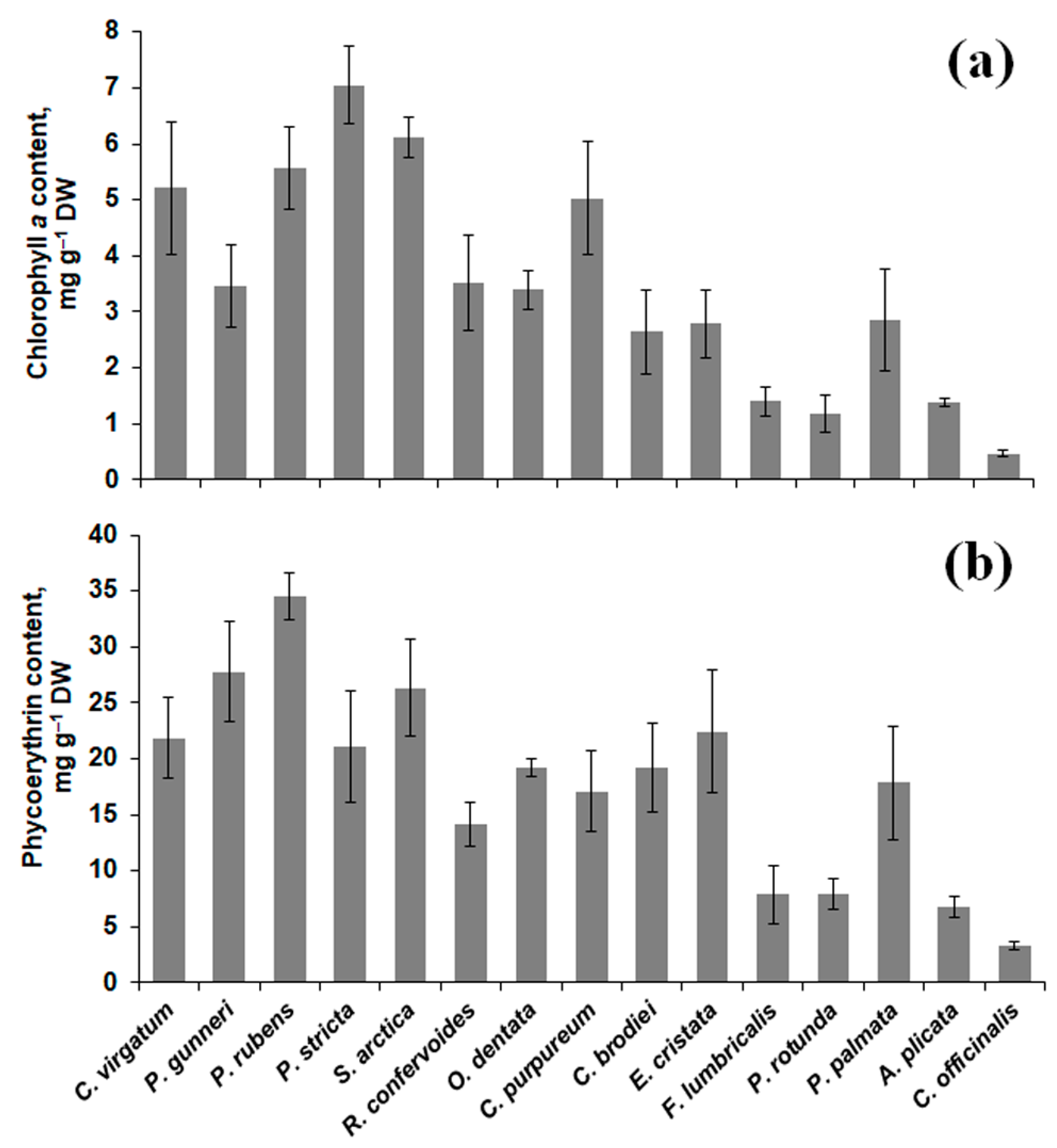
2.2. Pigment Content

The content of major photosynthetic pigments (chlorophyll a and phycoerythrin) in the thalli of different red algal species exhibited considerable variation (Figure 1). Four species (C. officinalis, P. rotunda, A. plicata, and F. lumbricalis) contained relatively low amounts of both pigments (~0.5–1.4 mg g−1 DW for chlorophyll and ~3–8 mg g−1 DW for phycoerythrin). The highest chlorophyll a content (7 mg g−1 DW) was found in P. stricta, and Ph. rubens showed the maximum content of phycoerythrin (34.5 mg g−1 DW) (Figure 1).

2.3. Mineral Composition

Analysis of major element content showed a high correlation (r = 0.93–0.99) among different species of the order Ceramiales; on average, these algae contained relatively high levels of K, Ca, Fe, Mg, and P, as well as a low level of S, compared to the other species (Table 2). The only exception was Ph. rubens, containing lower amounts of Ca and Fe. On the contrary, representatives of the order Gigartinales contained a relatively high level of S (up to 20.36 mg g−1 DW in E. cristata) and lower content of other elements. This group of algae did not show a significant correlation, and the most notable outlier was C. purpureum, containing higher amounts of K and P (close to the corresponding values for Ceramiales). P. palmata contained the maximum level of potassium (~20 mg g−1 DW) but was generally poor in macroelements. Ahnfeltia showed relatively low content of all analyzed elements. Corallina was especially rich in calcium and magnesium deposited in its calcified thalli but contained low amounts of K, P, S, and Fe (Table 2).
Trace metal analysis showed a high correlation among Gigartinales species (r = 0.90–0.99) and relatively high variation in the representatives of Ceramiales (Table 3). Several species accumulated particularly high amounts of specific metals: Mn and Co for O. dentata, Zn and Mo for R. confervoides, Cu for S. arctica, and Ni for C. brodiei. Ahnfeltia and Corallina exhibited the lowest levels of analyzed trace elements. Manganese and cobalt were strongly correlated (r = 0.99) in their distribution in thalli of different red algal species (Table 3).

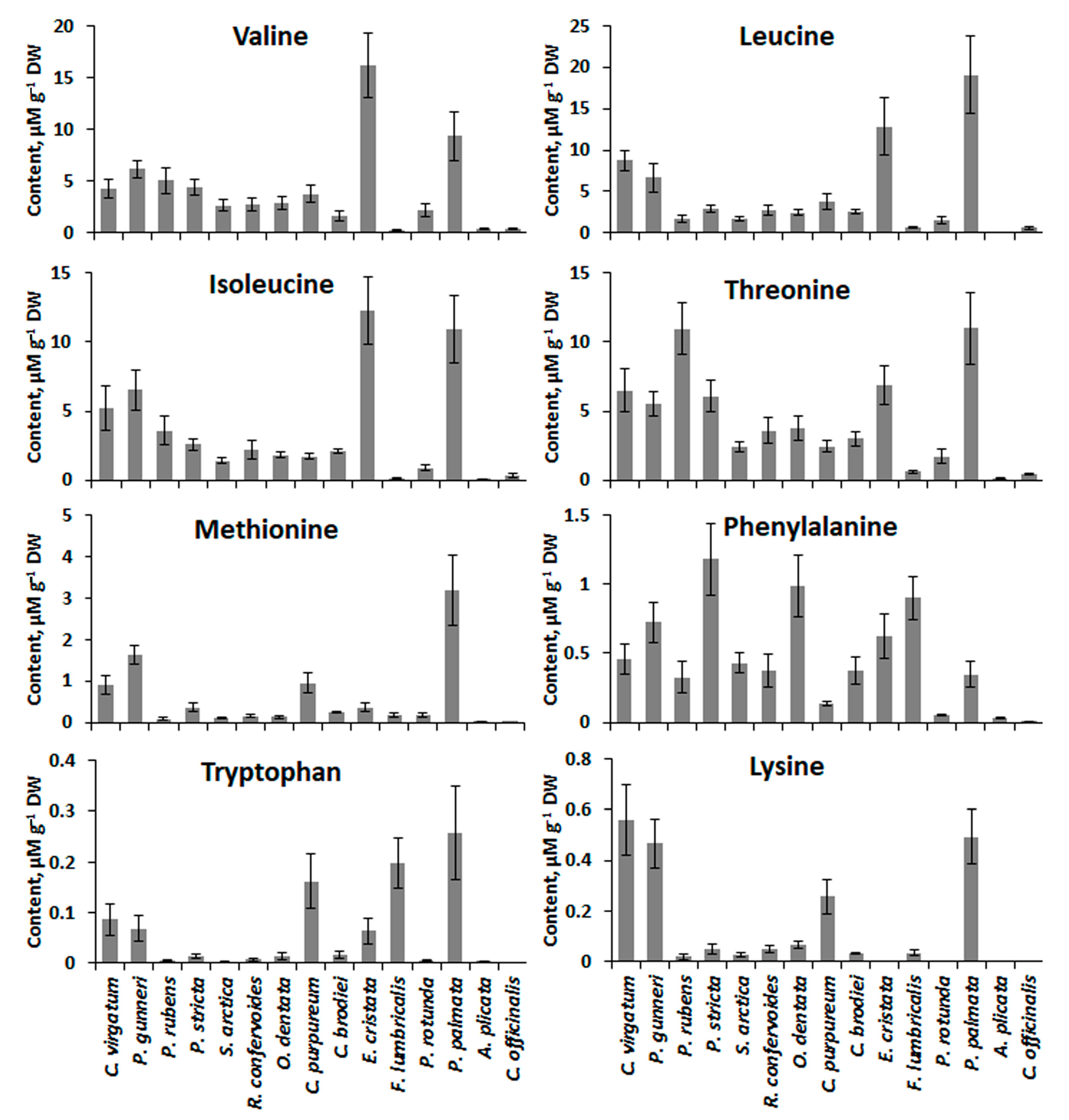
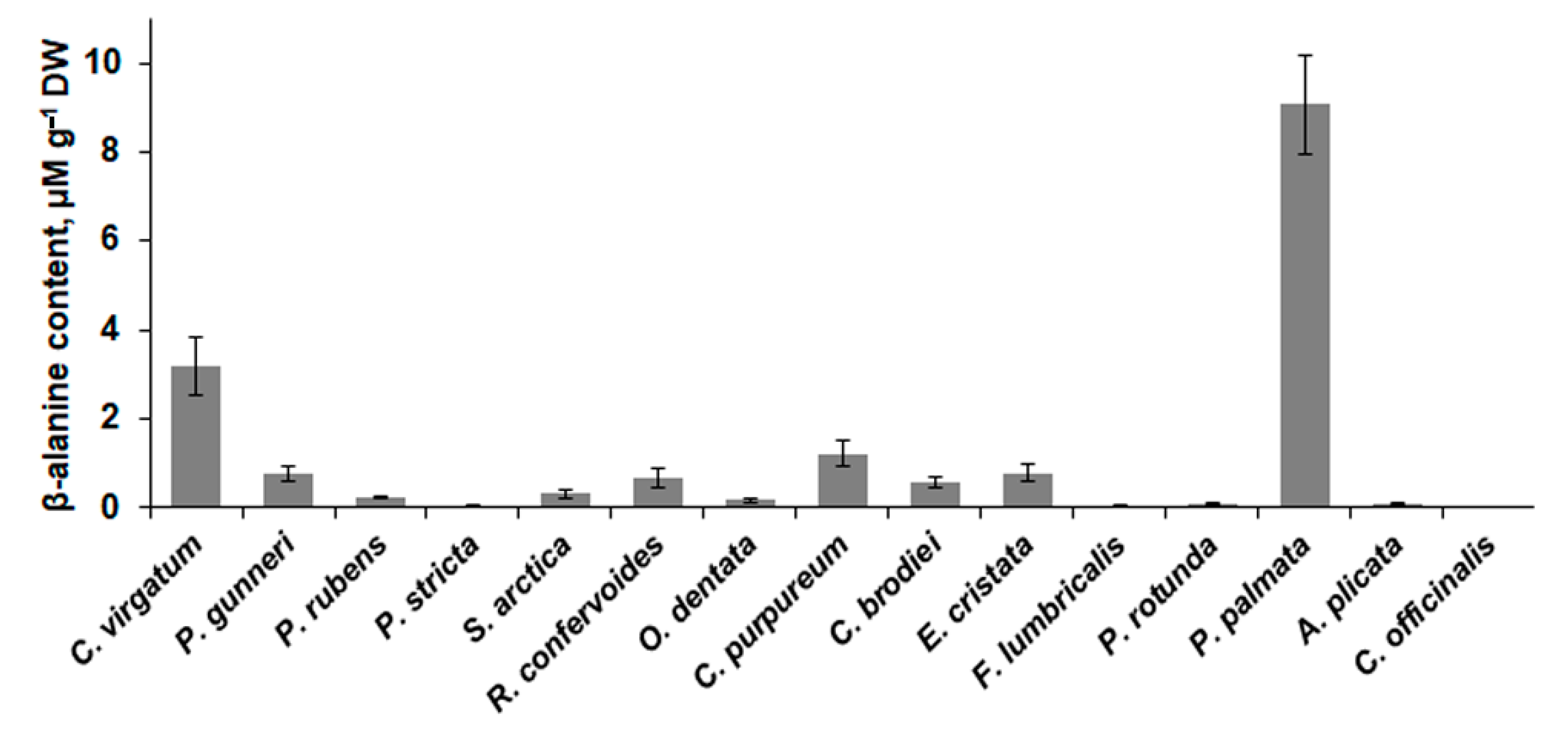
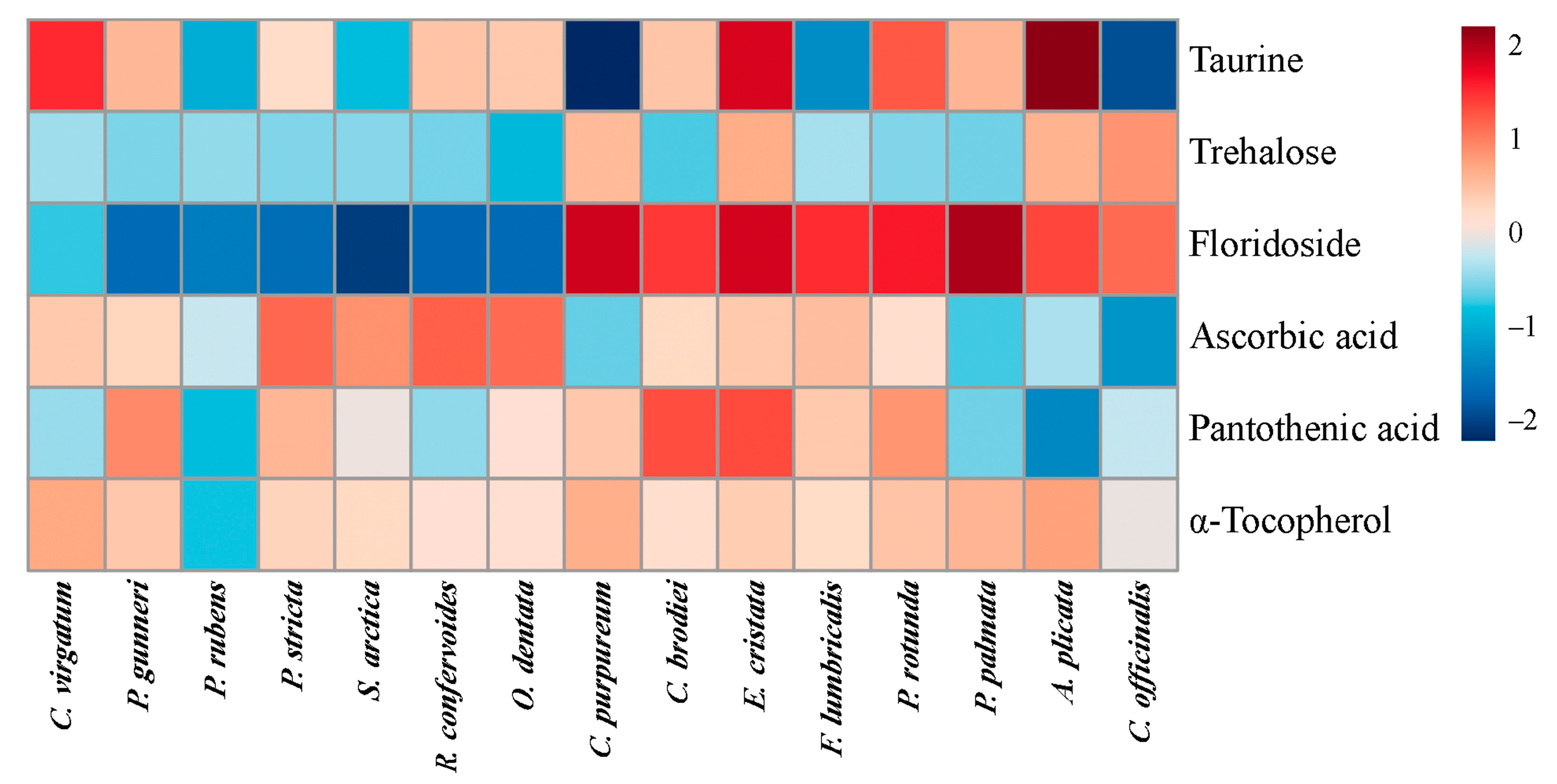
2.4. Low-Molecular-Weight Metabolites

GC-MS analysis allowed detecting eight free essential amino acids in the thalli of the studied red algae, namely valine, leucine, isoleucine, threonine, methionine, phenylalanine, tryptophan, and lysine. The highest amounts of almost all amino acids were found in two species, E. cristata and P. palmata (Figure 2). Moreover, several species showed relatively high content of one particular amino acid: lysine for C. virgatum and P. gunneri, threonine for P. rubens, phenylalanine for P. stricta, and tryptophan for F. lumbricalis and C. purpureum.
Several other valuable metabolites specifically accumulated in thalli of several red algae (Figure 3 and Figure 4). P. palmata and C. virgatum contained the highest amounts of nonproteinogenic amino acid β-alanine (9.1 and 3.2 μM g−1 DW, respectively, Figure 3), and A. plicata, E. cristata, and C. virgatum accumulated taurine (Figure 4). All algae except for representatives of the order Ceramiales contained considerable amounts of specific red algal polyol floridoside. The maximum content of this metabolite was found in P. palmata, C. purpureum, and E. cristata. All studied Ceramiales were also relatively poor in trehalose, the highest level of which was found in C. officinalis.
Three vitamins (ascorbic acid, pantothenic acid, and α-tocopherol) were detected by GC-MS analysis (Figure 4). Ascorbate mostly accumulated in Ceramiales (maximum in R. confervoides), whereas the species containing the highest level of pantothenic acid (C. brodiei and E. cristata) belonged to Gigartinales. Compared to the other vitamins, tocopherol was generally more evenly distributed among the studied species; its maximum content was found in A. plicata, C. virgatum, and C. purpureum (Figure 4).

3. Discussion

Our study provides a comprehensive characterization of the chemical composition of 15 Arctic red algal species from the perspective of their potential practical relevance. We showed that several of the studied species contain different valuable minerals and metabolites.
Currently, seaweeds are regarded as being a cheap alternative source of proteins for human nutrition and animal feeding [22]. Among marine macrophytes, red algae have the highest protein content. According to literature data, different red algae contain from 2.3 (C. officinalis) to 47% DW (Pyropia tenera) of total protein (reviewed in [15,32]). These values are generally in good accordance with our results (Table 1), and for several well-studied algae, our results confirm data previously obtained on the same species (e.g., 18.3–26.5% DW for P. palmata and 2.3–6.9% DW for C. officinalis) [33,34,35,36,37]. Most of the protein-rich species in our study belong to the order Ceramiales (C. virgatum, R. confervoides, S. arctica) (Table 1 and Table 4). As none of the species in this order are extensively exploited, information about the biochemical composition of these algae is limited. However, several analyzed ceramialean algae (Polysiphonia sp., Amansia multifida, Enantiocladia duperreyi) were found to have a total protein content close to our values (19.5–31.3% DW) [35,38].
A general problem in utilizing seaweed protein is its limited bioavailability, especially when algae are consumed as sea vegetables, without previous hydrolysis or preparing protein concentrates. Even cooked, algal thalli are relatively tough and difficult to chew. The digestibility of red algal protein is generally estimated as 50–70% (compared to casein, taken to be 100%). It was suggested that seaweeds contain some compounds interfering with protein digestion and further amino acid absorption. Polysaccharides, phenolic compounds, and membrane-associated glycoproteins (including lectins) were regarded as candidates for this role [39,40]. Polysaccharides are usually present in large quantities in algal cells and are supposed to be the main cause of the limited digestibility of the algal proteins [17,34]. However, our data (Table 1) showed that most of the protein-rich red algae have a relatively low content of total carbohydrates (the only meaningful exception was R. confervoides). A high amount of phenolics is mostly a problem of brown algae utilization, as they may contain up to 25% DW of specific polyphenols (phlorotannins) [41]. Red seaweeds typically contain lesser amounts of phenolic compounds than brown and green algae [39]. At the same time, it was shown that red algae contain more lectins of different types than the other macrophytes [42]. From this perspective, the water-soluble fraction of protein can be of particular interest for the assessment of the potential nutritional value of red algae, as this fraction is more easily available and hardly contains membrane proteins. It was shown that extraction of lectins from algal material is twice less effective without detergent addition [42]. According to our data, the content of soluble protein fraction in red algae varies considerably and does not always show a correlation with the total protein content. For example, P. stricta and P. palmata contained similar total amounts of protein, but content of soluble protein in thalli of P. stricta was twice higher than in the other species (Table 1). A high total protein level in combination with a high proportion of soluble fraction (more than 50% of total protein) was detected in several representatives of the order Ceramiales (P. stricta, S. arctica, R. confervoides). Thus, we may conclude that some ceramialean algae (especially those having filamentous thalli) can be sources of seaweed protein comparable to, or even more efficient than, broadly used P. palmata and different Porphyra species.
Besides bioavailability, protein quality is based also on its amino acid composition. Multiple sources of literature data imply that the protein amino acid score and the essential amino acid index in red algae are higher than those in brown and green macrophytes. In particular, the amino acid profile of P. palmata protein is very close to that of the standard protein ovalbumin [14,15,34]. Much less information can be found about the profiles of free amino acids in the red algal cells. It was shown that different red algae contain detectable amounts of almost all free essential amino acids, except for tryptophan and sometimes methionine [43,44,45]. However, even for extensively studied species such as P. palmata, most papers do not provide quantitative data, reporting just the percentage contribution of different amino acids to the total content of these compounds (e.g., [45,46]). Meanwhile, given the limited digestibility of algal proteins, free amino acids may be regarded as a more easily available source of essential amino acids for human nutrition. In general, the content of essential amino acids in the algae used in our study (Figure 2) is close to the values obtained for the other species, including representatives of Ceramiales, Gigartinales, and Corallinales (e.g., 0.4–7.7 μM g−1 DW for valine and 0.1–3.7 μM g−1 DW for phenylalanine) [43]. Several species contained considerable amounts of free tryptophan, methionine, isoleucine, and lysine (Figure 2), the amino acids that are often limiting in red algal proteins [32]. Analysis of our data shows that the accumulation of amino acids is strongly species-specific. Two species belonging to different orders, P. palmata and E. cristata, showed the highest content of most of the essential amino acids (Figure 2). For P. palmata, we may suppose that its profile of free amino acids partially reflects the specificity of its protein composition, as leucine, valine, and methionine are known to be well represented in the essential amino acid fraction of Palmaria protein [14]. As for E. cristata, to our knowledge, this species was analyzed in detail for the first time.
In the cells of the red algae, a considerable proportion of protein is associated with phycobilins, specific photosynthetic pigments, of which phycoerythrin is the most dominant. Currently, phycoerythrin is widely used in biomedicine, due to its antioxidant, anticarcinogenic, neuroprotective, and anti-inflammatory activities; in biochemical techniques (for producing highly sensitive fluorescence probes); and as a natural protein dye for food, cosmetic, and textile industries [10,47]. In this study, several species belonging to Ceramiales (P. rubens, P. gunneri) showed a phycoerythrin content of about 30 mg g−1 DW (up to 48% of soluble protein or 26% of total protein), which is higher than the values measured in crude or purified extracts of commercially exploited Palmaria species (9–12% of total protein) (Figure 1b; [48,49]). As the main source of phycobilins is still extraction from the natural producers (red algae and cyanobacteria), broadening the list of used organisms is a highly relevant task.
There was no significant correlation between phycoerythrin and total or soluble protein content (r = 0.35–0.4) (Table 1, Figure 1b). Being major light-harvesting pigments in red algal cells, phycobilins are expected to accumulate in the thalli of deep-water species. However, our data imply that a relatively high level of both phycoerythrin and chlorophyll a is rather a species-specific characteristic. For example, P. rubens and O. dentata, two ceramialean species having similar ecology (subtidal seaweeds, frequently growing under the coverage of Saccharina fronds), showed significantly different (p < 0.05) pigment content. At the same time, intertidal P. rotunda and subtidal F. lumbricalis contained practically the same amounts of phycoerythrin and chlorophyll a (Figure 1). Chlorophylls and their derivatives are currently regarded as valuable constituents of healthy nutrition due to their multiple biological activities including cancer prevention and activation of xenobiotic detoxification processes [50]. An additional benefit of chlorophyll consumption is magnesium uptake. Comparison of different plant sources of Mg showed that this element had higher bioavailability as a constituent of chlorophyll (in green vegetables) than in the form of phytin (in cereals) [51]. According to our data, chlorophyll a content in thalli of P. stricta, S. arctica, and P. rubens is comparable to that in green leafy vegetables such as lettuce, spinach, and kale [52,53]. Thus, several ceramialean species can be used as efficient sources of both phycoerythrin and chlorophyll.
Traditionally, seaweeds are valued for their high mineral content, as they contain more essential elements compared to the terrestrial plants [18,19]. The mineral composition of red algae is well documented, and our results are generally in good accordance with literature data [16,33,54,55]. Among the mass of red algal species inhabiting the White Sea coast, the most studied and exploited species is P. palmata. Our study shows that this species was relatively poor in major elements, except for potassium. From the perspective of using algal biomass as a source of diverse macroelements, ceramialean algae might be the most promising, as they were demonstrated to have a well-balanced mineral composition (Table 2).
Assessment of potential practical relevance of any seaweeds should necessarily include a survey of trace metal contents in their thalli. Metals such as copper, manganese, zinc, and cobalt are involved in crucial biochemical processes in the human organism, being components of electron-transport chains, vitamins, and enzymes, and algae may be good sources of these elements. On the other hand, heavy metals are toxic in high concentrations, and seaweeds are known as organisms accumulating them, sometimes up to levels a thousand times higher than those of seawater [56,57]. It was reported that there is significant variation in bioaccumulation capacity of different red algal species, and red algae can accumulate several heavy metals more efficiently than other macrophytes [57,58]. In our study, we revealed four species (O. dentata, R. confervoides, S. arctica, and C. brodiei) containing especially high amounts of several metals (Table 3), with maximum values close to those observed in experiments with artificially contaminated seawater [56]. Though some of these species (R. confervoides and S. arctica) exhibited high protein content, their nutritional value may be limited due to their tendency to accumulate zinc and copper. The high capacity for heavy metal accumulation makes these algae candidates for use as bioindicator species. Currently, red algae are not as extensively exploited for biomonitoring in marine ecosystems as brown seaweeds, though several species have already been reported as reliable bioindicators [59,60].
To provide the most comprehensive characteristic of the studied algae, besides quantitative evaluation of protein, carbohydrate, pigment, and mineral content, we also compared the concentrations of several low-molecular-weight metabolites in the algal cells (Figure 3 and Figure 4). Here we focused on the vitamins and other compounds with confirmed biological activity. According to the literature data, red algae contain relatively high amounts of vitamins: 1–11 mg g−1 DW of ascorbic acid [61,62], 20–80 μg g−1 DW of α-tocopherol [63,64], and 5–150 μg g−1 DW of pantothenic acid [64,65], as well as considerable amounts of thiamine, riboflavin, nicotinic acid, and cobalamin [22,65]. Such levels are comparable with the content of these vitamins in traditionally consumed terrestrial vegetables [22,66]. Our results confirmed a considerable variation of ascorbic and pantothenic acid content between different red algal species. Generally, higher amounts of ascorbate were found in the species of the order Ceramiales, whereas pantothenic acid mostly accumulated in the representatives of Gigartinales (Figure 4). Tocopherol was more evenly distributed among the different algal species. Only one type of tocopherol, namely α-tocopherol, featuring the highest antioxidative activity, was detected in all studied algae, which is in accordance with literature data [63].
Recent studies showed that several specific low-molecular-weight carbohydrates, including floridoside and trehalose, have multiple therapeutic effects. Floridoside (α-D-galactopyranosyl-(1–2)-glycerol) is a unique photosynthetic product of red algae, functioning as a storage compound, osmolyte, and antioxidant [67,68]. In clinical investigations, it was reported that floridoside demonstrated strong immunomodulatory effects, activating a complement cascade [69]. Moreover, this substance possesses anti-inflammatory and antioxidative activities [70,71]. Floridoside content in the red algal cells is taxon-specific, with ceramialean algae containing zero or trace amounts of this metabolite [72]. The highest concentrations of floridoside (up to 1 mM g−1 DW) were reported from P. palmata and several species belonging to the Gigartinales [72,73,74], which corresponds well to our data (Figure 4). Apparently, C. purpureum and E. cristata may be added to the list of floridoside-rich gigartinalean species along with Catenella nipae and Schottera nicaeensis [72,73]. Unlike the situation with floridoside, literature data about the occurrence of another low-molecular-weight carbohydrate, trehalose, in red algae are still fragmentary and sometimes ambiguous. This metabolite was detected in several species of Corallinales (including C. officinalis), Gigartinales (including C. purpureum), Ceramiales, and Batrachospermales [67,72,75]. Recently, it was also found in Porphyra umbilicalis [76]. Our results confirm the presence of trehalose in the cells of C. officinalis and C. purpureum, and we detected similar amounts of this metabolite in A. plicata and E. cristata (Figure 4). To our knowledge, in the latter two species, trehalose was found for the first time. Currently, trehalose is widely used as a stabilizer of foodstuff, vaccines, and cosmetics, and it is becoming more and more popular in medicine, particularly in ophthalmology [77]. Though red algae apparently contain a relatively low concentration of this metabolite [76], its presence in several species should still be considered regarding the development of technologies of multipurpose use of algal biomass.
Red algae contain a variety of nonproteinogenic amino acids and sulfonic acids, and the synthesis and accumulation of these compounds are strictly species-specific [18]. Several algae analyzed in this study exhibited relatively high content of β-alanine and taurine (Figure 3 and Figure 4). Being a common metabolite of many vascular plants, where it contributes to different stress responses [78], β-alanine has been reported from very few of the red algae [43]. Our data show that most of the studied algal species contained 0.05–1.3 μM g−1 DW of this amino acid (Figure 3), which corresponds to the values known from different organs of vascular plants [79]. However, P. palmata contained approximately ten times more β-alanine and thus may be regarded as a valuable natural source of this metabolite. Over the past few decades, β-alanine has become one of the most popular sports nutrition ingredients. Being a metabolic precursor of muscle carnosine, it improves athlete performance during high-intensity exercises in strength and power sports [80]. Moreover, β-alanine is used as an efficient and safe alternative to hormone replacement therapy for menopausal women [81].
Taurine is another valuable metabolite accumulating in thalli of several studied algae (A. plicata, E. cristata, C. virgatum) (Figure 4). According to literature data, red algae contain relatively high concentrations of taurine compared to vascular plants and green and brown algae [82,83]. Regarding the species used in our study, it was shown that P. rubens and S. arctica contained relatively low concentrations of taurine (0.1–0.15 mg g−1 DW), while different Palmaria species contained approximately 10 mg g−1 DW [84,85]. This corresponds to our data, showing the 100-fold difference between taurine contents in P. rubens and P. palmata (Figure 4). Currently, this amino acid draws the attention of pharmacologists due to its multiple beneficial health effects. It was reported that dietary taurine supplementation may prevent cardiovascular diseases, hypertension, and metabolic syndrome [86]. Taurine-rich red algae may be a valuable addition to the list of sea products (shellfish, prawns, etc.) used to obtain functional foods containing naturally occurring taurine.

4. Materials and Methods

4.1. Plant Material Collection

Fifteen species of intertidal and subtidal red algae, representing five orders of the class Florideophyceae (Table 4), were collected in the Keret Archipelago (Kandalaksha Bay, White Sea) in June–August 2017–2020. The list of objects includes a majority of red algae inhabiting the region and having considerable biomass [23,24,25,26]. Thalli were collected from the typical habitats of each species (Table 4). To minimize the possible changes in metabolite content induced by the tidal cycle, all intertidal plants were collected at high tide and directly transported to the laboratory in seawater.

4.2. Fresh Weight and Dry Weight Determination

For the determination of fresh weight (FW), 8–10 individual thalli (or bundles, for filamentous algae) were blotted with filter paper and weighed. For determination of DW, they were weighed again after drying at 60 °C to the constant weight.

4.3. Elemental Analysis

Algal thalli were gently washed with tap water, rinsed with deionized water, and dried for 24 h at 70 °C. One hundred milligrams of dried plant material was ground into fine powder and subjected to a wet-ashing procedure with a mixture of concentrated HNO3 and HClO4 in the ratio 4:1 (v/v). After complete digestion, samples were made up to 25 mL with deionized water. The quantitative analysis of the elemental composition was carried out with inductively coupled plasma atomic emission spectroscopy (ICPE-9000, Shimadzu).

4.4. Carbohydrate Analysis

Samples of dried plant material (5 mg DW) were ground to fine powder and hydrolyzed in sulfuric acid according to [87]. The content of total carbohydrates in the hydrolysates was determined spectrophotometrically using anthrone reagent [88]. Briefly, 3 mL of anthrone reagent was added to 0.3 mL of the sample (diluted as necessary). The mixture was boiled for 7 min and then immediately cooled in the ice bath. The extinction was measured at 620 nm using SPEKOL 1300 spectrophotometer (Analytik Jena AG, Jena, Germany), and the amount of total carbohydrate was calculated based on a calibration curve with glucose as standard.

4.5. Protein Analysis

For extraction of water-soluble proteins, samples of 20 mg FW were homogenized in Tris-HCl buffer (0.02 M, pH 8.3) using a TissueRuptor II homogenizer (QIAGEN, Hilden, Germany). For further extraction of alkali- and detergent-soluble proteins (including membrane proteins), the 3-fold volume of a mixture of 0.5% sodium dodecyl sulfate and 0.5 M NaOH was added to the homogenates, and the samples were boiled for 20 min. Both types of extracts were centrifuged (5000× g, 10 min), and the protein content was measured by Lowry assay with bovine serum albumin as standard [89,90]. Herein, the two protein fractions are referred to as ‘soluble’ and ‘total’ protein.

4.6. Pigment Analysis

For chlorophyll analysis, fragments of algal thalli (10 mg FW) were ground using a mortar and pestle in 100% acetone with small quantities of Na2SO4 and NaHCO3. Several rounds of extraction were completed with additional acetone until the extract was colorless. The concentration of acetone was adjusted to 90% (in distilled water), and the content of chlorophyll a was calculated [91] from data obtained with a SPEKOL 1300 spectrophotometer (Analytik Jena, Jena, Germany).
For determination of phycoerythrin content, fragments of algal thalli (50 mg FW) were homogenized in potassium phosphate buffer (0.1 M, pH 6.8) using a TissueRuptor II homogenizer (QIAGEN, Germany) and left soaking at 4 °C overnight. Then the extracts were centrifuged (5000× g, 10 min), and the content of phycoerythrin was determined spectrophotometrically (SPEKOL 1300, Analytik Jena) [92].

4.7. Metabolite Analysis

Samples (50 mg FW) of plant material were poured with cold methanol (−25 °C), quickly ground in a precooled mortar and left soaking in 1 mL of cold methanol for extraction. Four hundred microliters of methanol extract was transferred to clean 1.5 mL polypropylene Eppendorf tubes (VWR, Dresden, Germany) and vacuum-dried for subsequent analysis.
Metabolite profiling analyses were carried out according to [93]. Briefly, vacuum-dried extracts were incubated by shaking in methoxyamine hydrochloride (Sigma-Aldrich, Taufkirchen, Germany) solution in pyridine and N,O-bis(trimethylsilyl)-trifluoroacetamide (Macherey–Nagel, Düren, Germany). After derivatization, samples were transferred to micro-inserts of glass vials and subjected to GC-MS analysis on an Agilent 6890 gas chromatograph (1 mL/min He flow, splitless mode, injector temperature 250 °C) coupled to an Agilent 5973N quadrupole mass selective detector (Agilent Technologies, Böblingen, Germany) with the standard electron impact ionization. Within each sequence, a mixture of alkanes (C10–C32) in hexane was measured for the calculation of Kovats retention indices (RIs) [94]. A mixture of authentic standards containing 21 amino acids, 20 sugars and polyols, and 19 organic acids was co-spiked to confirm the identity of expected compounds.
Peak deconvolution was accomplished using AMDIS 2.65. The retention indices were automatically calculated using an AMDIS calibration file containing the batch retention times of each alkane. Golm metabolome database (GMD_20100614_VAR5_ALK, 24.09.2010, [95]) and NIST14 (National Institute of Standards and Technology, Gaithersburg, MD, USA) were used for identification of the peaks based on spectra comparison. Quantitation of metabolites was performed by peak integration of the corresponding extracted ion chromatograms (m/z ± 0.5) for representative intense signals at specific retention times using Xcalibur 3.0 software. Where applicable, calibration was performed by the standard addition method using five calibration levels.

4.8. Data Analysis

The measurements were performed with 6 to 12 replicates (from different individuals). Excel 2013 (Microsoft, Redmond, WA, USA) and MetaboAnalyst 4.0 Web application (http://www.metaboanalyst.ca, accessed on 24 November 2020) were used for data processing and normalization procedures, creation of figures, and heatmap construction [96]. The metabolomic data processing included peak area normalization to the median of all areas within a chromatogram, generalized logarithm transformation, and range data scaling (mean-centered and divided by the range of each variable). All values are expressed as means and standard deviations.

5. Conclusions

In this study, we compared the chemical composition of 15 red algal species inhabiting the Russian Arctic. The studied algae showed dramatic differences in concentration of proteins, carbohydrates, minerals, pigments, and several low-molecular-weight metabolites. For several parameters (protein, major elements, floridoside) the values were consistent within the orders of Rhodophyta, whereas the accumulation of free amino acids and vitamins was strongly species-specific. The results allow distinguishing several red algal species not studied in detail earlier. These algae showed similar or higher values of all estimated parameters compared to widely commercially exploited species P. palmata. Among the most promising species are representatives of the order Ceramiales, demonstrating a high content of protein (C. virgatum, P. stricta, R. confervoides, S. arctica), minerals, pigments (P. rubens, P. gunneri), and ascorbic acid (P. stricta, R. confervoides). Gigartinalean alga E. cristata featured a relatively high content of free essential amino acids, taurine, pantothenic acid, and floridoside. Several species showed a tendency to accumulate heavy metals, which makes them candidates for use as bioindicator species.

Author Contributions

Conceptualization, E.T.; data curation, N.Y., C.B., and E.T.; formal analysis, N.Y., C.B., and E.T.; funding acquisition, E.T. and C.B.; investigation, N.Y., A.K., V.L., and E.T.; methodology, C.B. and E.T.; project administration, E.T.; resources, C.B. and E.T.; software, C.B.; supervision, E.T.; validation, C.B. and E.T.; visualization, N.Y. and E.T.; writing—original draft, N.Y., A.K., and E.T.; writing—review and editing, C.B., V.L., and E.T. All authors have read and agreed to the published version of the manuscript.

Funding

This research was funded by the Russian Foundation for Basic Research (project 20-04-00944) and the Universities of Leipzig (CB) and St. Petersburg.

Institutional Review Board Statement

Not applicable.

Informed Consent Statement

Not applicable.

Data Availability Statement

Data associated with the paper are available from the authors upon reasonable request.

Acknowledgments

We thank Marine Biological Station “UNB Belomorskaya” and Chemistry Educational Centre of St. Petersburg State University for providing facilities. Susan Billig (University of Leipzig) helped with the GCMS analyses. We also sincerely thank Natalia Osmolovskaya and Ludmila Kuchaeva for technical assistance.

Conflicts of Interest

The authors declare no conflict of interest. The funders had no role in the design of the study; in the collection, analyses, or interpretation of data; in the writing of the manuscript; or in the decision to publish the results.

References

  1. Holdt, S.L.; Kraan, S. Bioactive compounds in seaweed: Functional food applications and legislation. J. Appl. Phycol. 2011, 23, 543–597. [Google Scholar] [CrossRef]
  2. Tanna, B.; Mishra, A. Metabolites unravel nutraceutical potential of edible seaweeds: An emerging source of functional food. Compr. Rev. Food Sci. Food Saf. 2018, 17, 1613–1624. [Google Scholar] [CrossRef] [PubMed] [Green Version]
  3. Shannon, E.; Abu-Ghannam, N. Seaweeds as nutraceuticals for health and nutrition. Phycologia 2019, 58, 563–577. [Google Scholar] [CrossRef] [Green Version]
  4. Lins, K.O.A.L.; Bezerra, D.P.; Alves, A.P.N.N.; Alencar, N.M.N.; Lima, M.W.; Torres, V.M.; Farias, W.R.L.; Pessoa, C.; de Moraes, M.O.; Costa-Lotufo, L.V. Antitumor properties of a sulfated polysaccharide from the red seaweed Champia feldmannii (Diaz-Pifferer). J. Appl. Toxicol. 2008, 29, 20–26. [Google Scholar] [CrossRef] [PubMed]
  5. Cornish, M.L.; Garbary, D.J. Antioxidants from macroalgae: Potential applications in human health and nutrition. Algae 2010, 25, 155–171. [Google Scholar] [CrossRef]
  6. Kasanah, N.; Hadisaputro, T.; Seto, D.S.; Amelia, W.; Isnansetyo, A. Antibacterial compounds from red seaweeds (Rhodophyta). Indones. J. Chem. 2015, 15, 201–209. [Google Scholar] [CrossRef]
  7. Kim, C.O.; Kim, Y.N.; Lee, D.C. Effects of Gelidium elegans on weight and fat mass reduction and obesity biomarkers in overweight or obese adults: A randomized double-blinded study. Nutrients 2019, 11, 1513. [Google Scholar] [CrossRef] [Green Version]
  8. Sørensen, L.E.; Jeppesen, P.B.; Christiansen, C.B.; Hermansen, K.; Gregersen, S. Nordic seaweed and diabetes prevention: Exploratory studies in KK-Ay mice. Nutrients 2019, 11, 1435. [Google Scholar] [CrossRef] [PubMed] [Green Version]
  9. Kumar, S.; Gupta, R.; Kumar, G.; Sahoo, D.; Kuhad, R.C. Bioethanol production from Gracilaria verrucosa, a red alga, in a biorefinery approach. Biores. Technol. 2013, 135, 150–156. [Google Scholar] [CrossRef] [PubMed]
  10. Azeem, M.; Iqbal, N.; Mir, R.A.; Adeel, S.; Batool, F.; Khan, A.A.; Gul, S. Harnessing natural colorants from algal species for fabric dyeing: A sustainable eco-friendly approach for textile processing. J. Appl. Phycol. 2019, 31, 3941–3948. [Google Scholar] [CrossRef]
  11. Corey, P.; Kim, J.K.; Garbary, D.J.; Prithiviraj, B.; Duston, J. Bioremediation potential of Chondrus crispus (Basin Head) and Palmaria palmata: Effect of temperature and high nitrate on nutrient removal. J. Appl. Phycol. 2012, 24, 441–448. [Google Scholar] [CrossRef]
  12. Titlyanov, E.A.; Titlyanova, T.V.; Belous, O.S. Useful substances of marine red algae (Rhodophyta): Chemical structure and content. Izvestiya TINRO 2011, 165, 305–319. (In Russian) [Google Scholar]
  13. Lahaye, M. Marine algae as sources of fibres: Determination of soluble and insoluble dietary fibre contents in some “Sea vegetables”. J. Sci. Food Agric. 1991, 54, 587–594. [Google Scholar] [CrossRef]
  14. Fleurence, J. Seaweed proteins: Biochemical, nutritional aspects and potential uses. Trends Food Sci. Technol. 1999, 10, 25–28. [Google Scholar] [CrossRef]
  15. Pangestuti, R.; Kim, S.-K. Seaweed proteins, peptides, and amino acids. In Seaweed Sustainability; Tiwari, B.K., Troy, D.J., Eds.; Academic Press: London, UK, 2015; pp. 125–140. [Google Scholar] [CrossRef]
  16. Morgan, K.C.; Wright, J.L.C.; Simpson, F.J. Review of chemical constituents of the red alga Palmaria palmata (Dulse). Econ. Bot. 1980, 34, 27–50. [Google Scholar] [CrossRef]
  17. Fleurence, J.; Morançais, M.; Dumay, J. Seaweed proteins. In Proteins in Food Processing, 7th ed.; Yada, R.Y., Ed.; Woodhead Publishing: Duxford, UK, 2018; pp. 245–262. [Google Scholar] [CrossRef]
  18. Ito, K.; Hori, K. Seaweed: Chemical composition and potential food uses. Food Rev. Int. 1989, 5, 101–144. [Google Scholar] [CrossRef]
  19. Rupérez, P. Mineral content of edible marine seaweeds. Food Chem. 2002, 79, 23–26. [Google Scholar] [CrossRef]
  20. Eggert, A.; Karsten, U. Low molecular weight carbohydrates in red algae—An ecophysiological and biochemical perspective. In Cellular Origins, Life in Extreme Habitats and Astrobiology. Red Algae in the Genomics Age; Seckbach, J., Chapman, D., Weber, A., Eds.; Springer: Berlin, Germany, 2010; pp. 445–456. [Google Scholar] [CrossRef]
  21. Orfanoudaki, M.; Hartmann, A.; Karsten, U.; Ganzera, M. Chemical profiling of mycosporine-like amino acids in twenty-three red algal species. J. Phycol. 2019, 55, 393–403. [Google Scholar] [CrossRef]
  22. Mišurcová, L. Chemical composition of seaweeds. In Handbook of Marine Macroalgae: Biotechnology and Applied Phycology; Kim, S.-K., Ed.; Wiley-Blackwell: Oxford, UK, 2012; pp. 173–192. [Google Scholar] [CrossRef]
  23. Mikhaylova, T.A. Vegetation of the red algal belt of the White Sea (European Arctic, Russia). Nov. Sist. Nizshikh Rastenii 2019, 53, 39–65. [Google Scholar] [CrossRef]
  24. Mikhaylova, T.A. Checklist of Rhodophyta of the White Sea (the Arctic Ocean). Bot. Mar. 2017, 60, 55–65. [Google Scholar] [CrossRef]
  25. Garbary, D.J.; Tarakhovskaya, E.R. Marine macroalgae and associated flowering plants from the Keret Archipelago, White Sea, Russia. Algae 2013, 28, 267–280. [Google Scholar] [CrossRef] [Green Version]
  26. Mikhaylova, T.A.; Aristov, D.A.; Naumov, A.D.; Malavenda, S.S.; Savchenko, O.N.; Bijagov, K.L. Diversity and structure of epibenthic communities of the red algae zone in the White Sea. Polar Biol. 2019, 42, 953–968. [Google Scholar] [CrossRef]
  27. Freile-Pelegrín, Y.; Morales, J.L. Antibacterial activity in marine algae from the coast of Yucatan, Mexico. Bot. Mar. 2004, 47, 140–146. [Google Scholar] [CrossRef]
  28. Jeong Han, E.; Priyan Shanura Fernando, I.; Kim, E.-A.; Kim, J.; Jung, K.; Kim, S.-Y.; Cha, S.-H.; Kim, K.-N.; Heo, S.-J.; Ahn, G. 5-Bromo-3,4-dihydroxybenzaldehyde from Polysiphonia morrowii attenuate IgE/BSA-stimulated mast cell activation and passive cutaneous anaphylaxis in mice. Biochem. Pharmacol. 2020, 114087. [Google Scholar] [CrossRef]
  29. Dong, H.; Dong, S.; Erik Hansen, P.; Stagos, D.; Lin, X.; Liu, M. Progress of bromophenols in marine algae from 2011 to 2020: Structure, bioactivities, and applications. Mar. Drugs 2020, 18, 411. [Google Scholar] [CrossRef]
  30. Sampaio, A.H.; Rogers, D.J.; Barwell, C.J.; Saker-Sampaio, S.; Nascimento, K.S.; Nagano, C.S.; Farias, W.R.L. New affinity procedure for the isolation and further characterization of the blood group B specific lectin from the red marine alga Ptilota plumosa. J. Appl. Phycol. 2002, 14, 489–495. [Google Scholar] [CrossRef]
  31. Smit, A.J. Medicinal and pharmaceutical uses of seaweed natural products: A review. J. Appl. Phycol. 2004, 16, 245–262. [Google Scholar] [CrossRef]
  32. Černá, M. Seaweed proteins and amino acids as nutraceuticals. In Advances in Food and Nutrition Research; Kim, S.-K., Ed.; Academic Press: San Diego, CA, USA, 2011; pp. 297–312. [Google Scholar] [CrossRef]
  33. Macpherson, M.G.; Young, E.G. The chemical composition of marine algae. Can. J. Res. 1949, 27, 73–77. [Google Scholar] [CrossRef]
  34. Galland-Irmouli, A.V.; Fleurence, J.; Lamghari, R.; Luçon, M.; Rouxel, C.; Barbaroux, O.; Bronowicki, J.P.; Villaume, C.; Guéant, J.L. Nutritional value of proteins from edible seaweed Palmaria palmata (Dulse). J. Nutr. Biochem. 1999, 10, 353–359. [Google Scholar] [CrossRef]
  35. Ramos, M.V.; Monteito, A.C.O.; Moreira, R.A.; Carvalho, A. Amino acid composition of some Brazilian seaweed species. J. Food Biochem. 2000, 24, 33–39. [Google Scholar] [CrossRef]
  36. Marsham, S.; Scot, G.W.; Tobin, M.L. Comparison of nutritive chemistry of a range of temperate seaweeds. Food Chem. 2007, 100, 1331–1336. [Google Scholar] [CrossRef]
  37. Mišurcová, L.; Kráčmar, S.; Klejdus, B.; Vacek, J. Nitrogen content, dietary fiber, and digestibility in algal food products. Czech J. Food Sci. 2010, 28, 27–35. [Google Scholar] [CrossRef] [Green Version]
  38. Dere, S.; Dalkiran, N.; Karacaoglu, D.; Yildiz, G.; Dere, E. The determination of total protein, total soluble carbohydrate and pigment contents of some macroalgae collected from Gemlik-Karacaali (Bursa) and Erdek-Ormanli (Balikesir) in the Sea of Marmara. Turk. Oceanol. 2003, 45, 453–471. [Google Scholar]
  39. Wong, K.H.; Cheung, P.C.K. Nutritional evaluation of some subtropical red and green seaweeds. Part II. In vitro protein digestibility and amino acid profiles of protein concentrates. Food Chem. 2001, 72, 11–17. [Google Scholar] [CrossRef]
  40. Vasconcelos, I.M.; Oliveira, J.T.A. Antinutritional properties of plant lectins. Toxicon 2004, 44, 385–403. [Google Scholar] [CrossRef] [PubMed]
  41. Birkemeyer, C.; Lemesheva, V.; Billig, S.; Tarakhovskaya, E. Composition of intracellular and cell wall-bound phlorotannin fractions in fucoid algae indicates specific functions of these metabolites dependent on the chemical structure. Metabolites 2020, 10, 369. [Google Scholar] [CrossRef] [PubMed]
  42. Rogers, D.J.; Hori, K. Marine algal lectins: New developments. Hydrobiologia 1993, 260, 589–593. [Google Scholar] [CrossRef]
  43. Impellizzeri, G.; Mangiafico, S.; Oriente, G.; Piattelli, M.; Sciuto, S. Amino acids and low-molecular-weight carbohydrates of some marine red algae. Phytochemistry 1975, 14, 1549–1557. [Google Scholar] [CrossRef]
  44. Nylund, G.M.; Weinberger, F.; Rempt, M.; Pohnert, G. Metabolomic assessment of induced and activated chemical defense in the invasive red alga Gracilaria vermiculophylla. PLoS ONE 2011, 6, e29359. [Google Scholar] [CrossRef] [PubMed]
  45. Mouritsen, O.G.; Dawczynski, C.; Duelund, L.; Jahreis, G.; Vetter, W.; Schröder, M. On the human consumption of the red seaweed dulse (Palmaria palmata (L.) Weber a Mohr). J. Appl. Phycol. 2013, 25, 1777–1791. [Google Scholar] [CrossRef]
  46. Mai, K.; Mercer, J.P.; Donlon, J. Comparative studies on the nutrition of two species of abalone, Haliotis tuberculata L. and Haliotis discus Hannai Ino. II. Amino acid composition of abalone and six species of macroalgae with an assessment of their nutritional value. Aquaculture 1994, 128, 115–130. [Google Scholar] [CrossRef]
  47. Sonani, R.R.; Rastogi, R.P.; Patel, R.; Madamwar, D. Recent advances in production, purification and applications of phycobiliproteins. World J. Biol. Chem. 2016, 7, 100–109. [Google Scholar] [CrossRef]
  48. Galland-Irmouli, A.V.; Pons, L.; Luçon, M.; Villaume, C.; Mrabet, N.T.; Guéant, J.L.; Fleurence, J. One-step purification of R-phycoerythrin from the red macroalga Palmaria palmata using preparative polyacrylamide gel electrophoresis. J. Chromatogr. B Biomed. Sci. Appl. 2000, 739, 117–123. [Google Scholar] [CrossRef]
  49. Martínez, B.; Rico, J.M. Seasonal variation of P content and major N pools in Palmaria palmata (Rhodophyta). J. Phycol. 2002, 38, 1082–1089. [Google Scholar] [CrossRef] [Green Version]
  50. Ferruzzi, M.G.; Blakeslee, J. Digestion, absorption, and cancer preventative activity of dietary chlorophyll derivatives. Nutr. Res. 2007, 27, 1–12. [Google Scholar] [CrossRef]
  51. Fairweather-Tait, S.; Hurrell, R.F. Bioavailability of minerals and trace elements. Nutr. Res. Rev. 1996, 9, 295–324. [Google Scholar] [CrossRef] [Green Version]
  52. Khachik, F.; Beecher, G.R.; Whittaker, N.F. Separation, identification, and quantification of the major carotenoid and chlorophyll constituents in extracts of several green vegetables by liquid chromatography. J. Agric. Food Chem. 1986, 34, 603–616. [Google Scholar] [CrossRef]
  53. Perucka, I.; Olszówka, K.; Chilczuk, B. Changes in the chlorophyll content in stored lettuce Lactuca sativa L. after pre-harvest foliar application of CaCl2. Acta Agrobot. 2013, 66, 137–142. [Google Scholar] [CrossRef] [Green Version]
  54. Kaliaperumal, N.; Chennubhotla, V.; Kalimuthu, S.; Ramalingam, J.; Selvaraj, M.; Najmuddin, M. Chemical composition of seaweeds. Cent. Mar. Fish. Res. Inst. CMFRI Bull. 1987, 41, 31–99. [Google Scholar]
  55. Frikha, F.; Kammoun, M.; Hammami, N.; Mchirgui, R.A.; Belbahri, L.; Gargouri, Y.; Miled, N.; Ben-Rebah, F. Chemical composition and some biological activities of marine algae collected in Tunisia. Cienc. Mar. 2011, 37, 113–124. [Google Scholar] [CrossRef] [Green Version]
  56. Burdin, K.S.; Bird, K.T. Heavy metal accumulation by carrageenan and agar producing algae. Bot. Mar. 1994, 37, 467–470. [Google Scholar] [CrossRef]
  57. Ali, A.Y.A.; Idris, A.M.; Eltayeb, M.A.H.; El-Zahhar, A.A.; Ashraf, I.M. Bioaccumulation and health risk assessment of toxic metals in red algae in Sudanese Red Sea coast. Toxin Rev. 2019. [Google Scholar] [CrossRef]
  58. Strezov, A.; Nonova, T. Influence of macroalgal diversity on accumulation of radionuclides and heavy metals in Bulgarian Black Sea ecosystems. J. Environ. Radioact. 2009, 100, 144–150. [Google Scholar] [CrossRef] [PubMed]
  59. Boubonari, T.; Malea, P.; Koyro, H.W.; Kevrekidis, T. The red macroalga Gracilaria bursa-pastoris as a bioindicator of metals (Fe, Zn, Cu, Pb, Cd) in oligohaline coastal environments. Fresenius Environ. Bull. 2008, 17, 2207–2216. [Google Scholar]
  60. Ryan, S.; McLoughlin, P.; O’Donovan, O.A. comprehensive study of metal distribution in three main classes of seaweed. Environ. Pollut. 2012, 167, 171–177. [Google Scholar] [CrossRef] [PubMed]
  61. Munda, I.M. Preliminary information on the ascorbic acid content in some Adriatic seaweeds. Hydrobiologia 1987, 151–152, 477–481. [Google Scholar] [CrossRef]
  62. Tanna, B.; Brahmbhatt, H.R.; Mishra, A. Phenolic, flavonoid, and amino acid compositions reveal that selected tropical seaweeds have the potential to be functional food ingredients. J. Food Process. Preserv. 2019, 43, e14266. [Google Scholar] [CrossRef]
  63. Jensen, A. Tocopherol content of seaweed and seaweed meal: 1.-Analytical methods and distribution of tocopherols in benthic algae. J. Sci. Food Agric. 1969, 20, 449–453. [Google Scholar] [CrossRef]
  64. Vinothkumar, R.; Murugesan, S.; Sivamurugan, V. Vitamin content of marine red alga Champia parvula. Asian J. Pharm. Clin. Res. 2019, 12, 207–209. [Google Scholar] [CrossRef]
  65. Teeri, A.E.; Bieber, R.E. B-Complex vitamins in certain brown and red algae. Science 1958, 127, 1500. [Google Scholar] [CrossRef]
  66. James, D.P. Nicotinic acid, pantothenic acid and biotin in fruits, vegetables and nuts. Br. J. Nutr. 1952, 6, 341–356. [Google Scholar] [CrossRef] [Green Version]
  67. Craigie, J.S. Storage products. In Algal Physiology and Biochemistry; Stewart, W.D.P., Ed.; Blackwell Press: Oxford, UK, 1974; pp. 206–235. [Google Scholar]
  68. Reed, R.H. Osmoacclimation in Bangia atropurpurea (Rhodophyta, Bangiales): The osmotic role of floridoside. Br. Phycol. J. 1985, 20, 211–218. [Google Scholar] [CrossRef]
  69. Courtois, A.; Simon-Colin, C.; Boisset, C.; Berthou, C.; Deslandes, E.; Guézennec, J.; Bordron, A. Floridoside extracted from the red alga Mastocarpus stellatus is a potent activator of the classical complement pathway. Mar. Drugs 2008, 6, 407–417. [Google Scholar] [CrossRef] [PubMed]
  70. Kim, M.J.; Li, Y.X.; Dewapriya, P.; Ryu, B.; Kim, S.K. Floridoside suppresses proinflammatory responses by blocking MAPK signaling in activated microglia. BMB Rep. 2013, 46, 398–403. [Google Scholar] [CrossRef] [PubMed]
  71. Kang, H.K.; Seo, C.H.; Park, Y. The effects of marine carbohydrates and glycosylated compounds on human health. Int. J. Mol. Sci. 2015, 16, 6018–6056. [Google Scholar] [CrossRef] [Green Version]
  72. Karsten, U.; Görs, S.; Eggert, A.; West, J.A. Trehalose, digeneaside and floridoside in the Florideophyceae (Rhodophyta)—A re-evaluation of its chemotaxonomic value. Phycologia 2007, 46, 143–150. [Google Scholar] [CrossRef]
  73. Kirst, G.O. Low MW carbohydrates and ions in Rhodophyceae: Quantitative measurement of floridoside and digeneaside. Phytochemistry 1980, 19, 1107–1110. [Google Scholar] [CrossRef]
  74. Rødde, R.S.H.; Vårum, K.M.; Larsen, B.A.; Myklestad, S.M. Seasonal and geographical variation in the chemical composition of the red alga Palmaria palmata (L.) Kuntze. Bot. Mar. 2004, 47, 125–133. [Google Scholar] [CrossRef]
  75. Elbein, A.D. The metabolism of α,α-trehalose. Adv. Carbohydr. Chem. Biochem. 1974, 30, 227–256. [Google Scholar] [CrossRef]
  76. Cao, Y.; Ashline, D.J.; Ficko-Blean, E.; Klein, A.S. Trehalose and (iso)floridoside production under desiccation stress in red alga Porphyra umbilicalis and the genes involved in their synthesis. J. Phycol. 2020, 56, 1468–1480. [Google Scholar] [CrossRef]
  77. Feofilova, E.P.; Usov, A.I.; Mysyakina, I.S.; Kochkina, G.A. Trehalose: Chemical structure, biological functions, and practical application. Microbiology 2014, 83, 184–194. [Google Scholar] [CrossRef]
  78. Parthasarathy, A.; Savka, M.A.; Hudson, A.O. The synthesis and role of β-alanine in plants. Front. Plant Sci. 2019, 10, 921. [Google Scholar] [CrossRef] [PubMed] [Green Version]
  79. Duhazé, C.; Gagneul, D.; Leport, L.; Larher, F.R.; Bouchereau, A. Uracil as one of the multiple sources of β-alanine in Limonium latifolium, a halotolerant β-alanine betaine accumulating Plumbaginaceae. Plant Physiol. Biochem. 2003, 41, 993–998. [Google Scholar] [CrossRef]
  80. Trexler, E.T.; Smith-Ryan, A.E.; Stout, J.R.; Hoffman, J.R.; Wilborn, C.D.; Sale, C.; Kreider, R.B.; Jäger, R.; Earnest, C.P.; Bannock, L.; et al. International society of sports nutrition position stand: Beta-alanine. J. Int. Soc. Sports Nutr. 2015, 12, 30. [Google Scholar] [CrossRef] [PubMed] [Green Version]
  81. Andreeva, E.; Tkeshelashvili, B. Women dealing with hot flushes: The role of β-alanine. Eur. Rev. Med. Pharmacol. Sci. 2020, 24, 5148–5154. [Google Scholar] [CrossRef]
  82. Kawasaki, A.; Ono, A.; Mizuta, S.; Kamiya, M.; Takenaga, T.; Murakami, S. The taurine content of Japanese seaweed. In Taurine 10. Advances in Experimental Medicine and Biology; Lee, D.H., Schaffer, S.W., Park, E., Kim, H.W., Eds.; Springer: Dordrecht, The Netherlands, 2017; pp. 1105–1113. [Google Scholar] [CrossRef]
  83. Mehdinia, A.; Rostami, S.; Dadkhah, S.; Fumani, N.S. Simultaneous screening of homotaurine and taurine in marine macro-algae using liquid chromatography–fluorescence detection. J. Iran. Chem. Soc. 2017, 14, 2135–2142. [Google Scholar] [CrossRef]
  84. Kadnikova, I.A.; Selivanova, O.N.; Shcherbakova, N.S. Chemical composition of the Palmariales algae (Rhodophyta) from the coast of Kamchatka. Izv. TINRO 2012, 169, 246–254. (In Russian) [Google Scholar]
  85. Klindukh, M.P.; Ryzhik, I.V.; Dobuchina, E.O. Composition and content of free amino acids of sublittoral red algae from Murmansk coast of the Barents Sea. Tr. Kolskogo Nauchnogo Cent. RAN 2020, 5, 92–101. (In Russian) [Google Scholar]
  86. Yamori, Y.; Taguchi, T.; Hamada, A.; Kunimasa, K.; Mori, H.; Mori, M. Taurine in health and diseases: Consistent evidence from experimental and epidemiological studies. J. Biomed. Sci. 2010, 17, 1–14. [Google Scholar] [CrossRef] [Green Version]
  87. Huijgen, W.J.J.; Cobussen-Pool, E.M.; van Egmond, B.F.; van Hal, J.W. Determination of carbohydrate composition of macroalgae. In Protocols for Macroalgae Research; Charrier, B., Wichard, T., Reddy, C.R.K., Eds.; CRC Press: Boca Raton, FL, USA, 2018; pp. 199–210. [Google Scholar] [CrossRef] [Green Version]
  88. Hewitt, B.R. Spectrophotometric determination of total carbohydrate. Nature 1958, 182, 246–247. [Google Scholar] [CrossRef]
  89. Lowry, D.H.; Rosebrough, N.J.; Farr, A.; Randall, R.J. Protein measurement with the Folin-phenol reagent. J. Biol. Chem. 1951, 193, 265–275. [Google Scholar] [CrossRef]
  90. Lees, M.B.; Paxman, S. Modification of the Lowry procedure for the analysis of proteolipid protein. Anal. Biochem. 1972, 47, 184–192. [Google Scholar] [CrossRef]
  91. Ritchie, R.J. Consistent sets of spectrophotometric chlorophyll equations for acetone, methanol and ethanol solvents. Photosynth. Res. 2006, 89, 27–41. [Google Scholar] [CrossRef] [PubMed]
  92. Lüder, U.H.; Wiencke, C.; Knoetzel, J. Acclimation of photosynthesis and pigments during and after six months of darkness in Palmaria decipiens (Rhodophyta): A study to simulate Antarctic winter sea ice cover. J. Phycol. 2002, 38, 904–913. [Google Scholar] [CrossRef]
  93. Hutschenreuther, A.; Kiontke, A.; Birkenmeier, G.; Birkemeyer, C. Comparison of extraction conditions and normalization approaches for cellular metabolomics of adherent growing cells with GC-MS. Anal. Meth. 2012, 4, 1953–1963. [Google Scholar] [CrossRef]
  94. Kovats, V.E. Characterization of organic compounds by gas chromatography. Part 1. Retention indices of aliphatic halides, alcohols, aldehydes and ketones. Helv. Chim. Acta 1958, 41, 1915–1932. [Google Scholar] [CrossRef]
  95. Kopka, J.; Schauer, N.; Krueger, S.; Birkemeyer, C.; Usadel, B.; Bergmüller, E.; Dörmann, P.; Weckwerth, W.; Gibon, Y.; Stitt, M.; et al. GMD@ CSB. DB: The Golm metabolome database. Bioinformatics 2005, 21, 1635–1638. [Google Scholar] [CrossRef] [Green Version]
  96. Chong, J.; Soufan, O.; Li, C.; Caraus, I.; Li, S.; Bourque, G.; Wishart, D.S.; Xia, J. MetaboAnalyst 4.0: Towards more transparent and integrative metabolomics analysis. Nucleic Acids Res. 2018, 46, W486–W494. [Google Scholar] [CrossRef] [Green Version]
Figure 1. Chlorophyll a (a) and phycoerythrin (b) content in 15 red algal species (means ± SD).
Molecules 26 02489 g001
Figure 2. Essential amino acid contents in 15 red algal species (means ± SD).
Molecules 26 02489 g002
Figure 3. Content of β-alanine in 15 red algal species (means ± SD).
Molecules 26 02489 g003
Figure 4. Relative content of several biologically active metabolites in 15 red algal species. Mean values of 5–6 samples are presented on a log10 scale.
Molecules 26 02489 g004
Table 1. Moisture, protein, and carbohydrate content in 15 red algal species (means ± SD).
SpeciesMoisture, %Protein, % DWTotal Carbohydrates, % DW
Soluble ProteinTotal Protein
Ceramium virgatum86.77 ± 3.137.79 ± 1.9032.25 ± 2.1832.33 ± 4.31
Ptilota gunneri82.90 ± 3.056.69 ± 0.368.92 ± 0.5540.26 ± 4.06
Phycodrys rubens76.71 ± 2.667.16 ± 0.7213.21 ± 1.3240.83 ± 3.00
Polysiphonia stricta86.38 ± 4.3211.63 ± 1.9218.58 ± 4.1829.38 ± 2.25
Savoiea arctica81.93 ± 2.1614.28 ± 2.4524.95 ± 4.7132.50 ± 8.19
Rhodomela confervoides80.87 ± 2.9812.15 ± 0.7824.26 ± 1.2644.38 ± 3.18
Odonthalia dentata79.64 ± 2.406.43 ± 0.4414.78 ± 2.6438.09 ± 4.78
Cystoclonium purpureum89.42 ± 3.7111.88 ± 1.2427.10 ± 2.3627.24 ± 7.36
Coccotylus brodiei72.53 ± 3.186.96 ± 0.7415.87 ± 1.3833.27 ± 3.59
Euthora cristata84.24 ± 2.566.52 ± 0.6213.06 ± 2.4741.59 ± 4.50
Furcellaria lumbricalis77.20 ± 2.0210.45 ± 1.6615.98 ± 1.4456.92 ± 4.49
Polyides rotunda73.26 ± 4.013.46 ± 0.549.23 ± 1.5333.02 ± 0.95
Palmaria palmata88.21 ± 2.655.60 ± 0.5719.94 ± 3.5824.06 ± 4.17
Ahnfeltia plicata64.03 ± 2.013.02 ± 0.395.57 ± 1.1441.27 ± 3.63
Corallina officinalis31.28 ± 1.901.39 ± 0.233.37 ± 0.4617.46 ± 3.76
Table 2. Major elements in 15 red algal species (means ± SD).
SpeciesMineral Content, mg g−1 DW
KCaMgPSFe
Ceramium virgatum10.18 ± 0.155.78 ± 0.836.47 ± 0.442.52 ± 0.016.99 ± 0.090.32 ± 0.02
Ptilota gunneri8.72 ± 0.216.63 ± 0.633.34 ± 0.282.42 ± 0.035.08 ± 0.250.43 ± 0.03
Phycodrys rubens1.42 ± 0.266.66 ± 0.928.55 ± 0.632.68 ± 0.0811.92 ± 0.750.25 ± 0.03
Polysiphonia stricta10.33 ± 0.354.49 ± 0.623.50 ± 0.081.22 ± 0.0310.00 ± 0.320.72 ± 0.16
Savoiea arctica8.56 ± 0.244.19 ± 0.602.57 ± 0.101.12 ± 0.068.93 ± 0.671.84 ± 0.59
Rhodomela confervoides14.32 ± 0.045.44 ± 0.055.03 ± 0.022.45 ± 0.0112.81 ± 0.030.53 ± 0.11
Odonthalia dentata10.70 ± 0.154.07 ± 0.332.86 ± 0.131.25 ± 0.039.25 ± 0.391.34 ± 0.07
Cystoclonium purpureum14.99 ± 0.083.95 ± 0.204.61 ± 0.052.28 ± 0.086.96 ± 0.050.43 ± 0.06
Coccotylus brodiei7.05 ± 0.354.50 ± 0.163.13 ± 0.121.38 ± 0.0415.36 ± 0.890.39 ± 0.06
Euthora cristata1.85 ± 0.226.73 ± 0.554.83 ± 0.302.37 ± 0.0120.36 ± 0.260.39 ± 0.09
Furcellaria lumbricalis6.09 ± 0.122.18 ± 0.105.55 ± 0.121.29 ± 0.0213.44 ± 0.280.43 ± 0.02
Polyides rotunda4.86 ± 0.243.69 ± 0.163.15 ± 0.100.99 ± 0.018.61 ± 0.190.29 ± 0.04
Palmaria palmata19.99 ± 2.001.61 ± 0.130.92 ± 0.112.12 ± 0.051.73 ± 0.010.72 ± 0.08
Ahnfeltia plicata3.47 ± 0.356.54 ± 0.402.05 ± 0.091.41 ± 0.024.37 ± 0.180.26 ± 0.03
Corallina officinalis1.57 ± 0.21309.7 ± 9.714.7 ± 0.710.56 ± 0.024.03 ± 0.210.28 ± 0.09
Table 3. Trace elements in 15 red algal species (means ± SD).
SpeciesMineral Content, μg g−1 DW
MnCuZnMoCoNi
Ceramium virgatum83.64 ± 5.166.79 ± 1.6359.15 ± 5.521.41 ± 0.08<0.13.71 ± 0.25
Ptilota gunneri210.4 ± 3.669.64 ± 0.9475.66 ± 5.510.53 ± 0.071.09 ± 0.3919.99 ± 4.02
Phycodrys rubens43.10 ± 16.6319.76 ± 2.31100.3 ± 6.091.44 ± 0.23<0.19.36 ± 0.28
Polysiphonia stricta279.1 ± 12.1626.97 ± 2.6486.33 ± 1.671.84 ± 0.101.35 ± 0.186.35 ± 0.39
Savoiea arctica63.62 ± 8.8142.54 ± 3.02235.8 ± 8.381.23 ± 0.260.19 ± 0.0310.57 ± 0.82
Rhodomela confervoides4.72 ± 0.5015.40 ± 0.73286.7 ± 0.724.33 ± 0.190.08 ± 0.017.25 ± 0.81
Odonthalia dentata1751.2 ± 56.230.30 ± 0.6564.10 ± 2.960.70 ± 0.1014.05 ± 0.3014.82 ± 0.17
Cystoclonium purpureum778.1 ± 31.287.39 ± 0.39154.8 ± 8.672.00 ± 0.396.11 ± 0.329.78 ± 0.70
Coccotylus brodiei597.0 ± 53.066.70 ± 0.0867.85 ± 4.970.76 ± 0.033.69 ± 0.2045.32 ± 3.62
Euthora cristata132.9 ± 16.1814.62 ± 0.2675.70 ± 2.502.16 ± 0.550.76 ± 0.0111.59 ± 0.75
Furcellaria lumbricalis321.4 ± 14.357.14 ± 0.2434.93 ± 2.170.52 ± 0.021.25 ± 0.165.14 ± 0.47
Polyides rotunda157.2 ± 12.314.40 ± 0.8834.80 ± 1.640.37 ± 0.031.02 ± 0.1111.16 ± 0.45
Palmaria palmata81.64 ± 10.1911.06 ± 0.4698.56 ± 9.970.52 ± 0.020.43 ± 0.0211.17 ± 0.36
Ahnfeltia plicata56.72 ± 3.274.63 ± 0.4034.78 ± 1.290.56 ± 0.070.34 ± 0.015.79 ± 0.40
Corallina officinalis171.1 ± 5.873.60 ± 0.2131.05 ± 3.710.06 ± 0.010.30 ± 0.043.68 ± 0.18
Table 4. Red algal species used as objects in this study and their typical habitat on shores of the Kandalaksha Bay, White Sea.
OrderSpeciesTypical Habitat
CeramialesCeramium virgatumLow intertidal–subtidal: exposed rocky shores
Ptilota gunneriSubtidal: rocks, Saccharina holdfasts
Phycodrys rubensSubtidal: rocks, Saccharina stipes and holdfasts
Polysiphonia strictaSubtidal: rocks, Saccharina stipes and holdfasts
Savoiea arcticaSubtidal: fucoid beds, Saccharina holdfasts
Rhodomela confervoidesLow intertidal–subtidal: rock pools, fucoid beds, Saccharina holdfasts
Odonthalia dentataSubtidal: rocks, Saccharina stipes and holdfasts
GigartinalesCystoclonium purpureumSubtidal: rocky shores, frequently growing on Ahnfeltia thalli
Coccotylus brodieiSubtidal: rocks, Saccharina stipes and holdfasts
Euthora cristataSubtidal: rocks, Saccharina stipes and holdfasts
Furcellaria lumbricalisSubtidal: rocks, Saccharina holdfasts
Polyides rotundaLow intertidal–subtidal: rock pools, sandy and rocky bottom
PalmarialesPalmaria palmataMid-intertidal–subtidal: rock pools, Saccharina stipes
AhnfeltialesAhnfeltia plicataSubtidal: rocky shores
CorallinalesCorallina officinalisSubtidal: rocks, fucoid beds, Saccharina stipes and holdfasts
Publisher’s Note: MDPI stays neutral with regard to jurisdictional claims in published maps and institutional affiliations.

