Article

Metabolome-Based Discrimination Analysis of Five Lilium Bulbs Associated with Differences in Secondary Metabolites

1
Beijing Radiation Center, Beijing 100875, China
2
Key Lab of Beam Technology and Material Modification of Ministry of Education, College of Nuclear Science and Technology, Beijing Normal University, Beijing 100875, China
*
Author to whom correspondence should be addressed.
Molecules 2021, 26(5), 1340; https://doi.org/10.3390/molecules26051340
Submission received: 15 January 2021 / Revised: 23 February 2021 / Accepted: 24 February 2021 / Published: 2 March 2021

Abstract

:
The bulbs of several Lilium species are considered to be both functional foods and traditional medicine in northern and eastern Asia. Considering the limited information regarding the specific bioactive compounds contributing to the functional properties of these bulbs, we compared the secondary metabolites of ten Lilium bulb samples belonging to five different species, using an ultrahigh-performance liquid chromatography-electrospray ionization-tandem mass spectrometry (UPLC-ESI-MS/MS)-based secondary metabolomics approach. In total, 245 secondary metabolites were detected; further, more metabolites were detected from purple Lilium bulbs (217 compounds) than from white bulbs (123–171 compounds). Similar metabolite profiles were detected in samples within the same species irrespective of where they were collected. By combining herbal analysis and screening differential metabolites, steroid saponins were considered the key bioactive compounds in medicinal lilies. Of the 14 saponins detected, none were accumulated in the bulbs of L. davidii var. willmottiae, also called sweet lily. The purple bulbs of L. regale accumulated more secondary metabolites, and, notably, more phenolic acid compounds and flavonoids. Overall, this study elucidates the differential metabolites in lily bulbs with varying functions and colors and provides a reference for further research on functional foods and the medicinal efficacy of Lilium species.

1. Introduction

The genus Lilium comprises more than 100 species worldwide, and China is the main distribution center of Lilium, with 55 species [1,2]. The lily bulb, without its enveloping protective sheath, is composed of firm, fleshy scales that provide nourishment for the developing plant. The scales are modified leaves that are thickened and shortened to act as a store of food reserves [3]. The carbohydrates stored by lily plants include starch, sugar, and so on. The starch content accounts for 11–22% of the bulb’s fresh weight (Table 1), and intrinsic free sugar content, mainly including sucrose, glucose, and fructose, accounts for 2–14% of the fresh weight [4].
In addition to providing nutrients to the plant itself, the lily bulb has accumulated a variety of secondary metabolites serving as defenses for plants, including major groups such as phenolics, terpenoids, and nitrogen-containing compounds [5]. The ancient Chinese people discovered the medicinal properties of lily bulbs and recorded them in ancient medical books. The earliest known record of lily bulb efficacy is found in the book Shen Nong Ben Cao Jing, which was compiled before the second century AD [6]. Nowadays, the dry scales of three Lilium bulbs, including L. pumilum (Shan Dan, SD), L. lancifolium (Juan Dan, JD), and L. brownii var. viridulum (Bai He, and Longya Baihe for trade name, LY), have been officially listed in the Chinese Pharmacopeia (2020 edition) and are considered medicative for bronchitis, pneumonia, and cough [7]. Many natural products have been separated and identified from lily bulbs, including phenylpropenoid glycerides [8,9], phenolic compounds [10], and steroidal saponins [11], which exhibit excellent antioxidant, antibacterial, and anti-inflammatory effects [12]. However, the specific bioactive compounds responsible for the efficacy of lily bulbs remain unclear.
In addition to its medicinal uses, the lily bulb is also considered a good root vegetable that provides carbohydrates, proteins, and a low fat content [13]. Lily bulbs can be used for daily consumption and are thus known as medicinal and edible (M&E) plants. According to the traditional theories of Chinese medicine, eating lily bulbs is beneficial as they include nourishing yin, moisten the lungs, and clear heartburn [9]. Aside from the three abovementioned Lilium species, the bulbs of L. davidii var. willmottiae (Lanzhou Baihe, LZ) are also popular products in Chinese vegetable markets, and are called ”sweet lily bulbs” [14]. In contrast, JD is called a “bitter lily bulb.” Their different tastes may result from the differential accumulation of secondary metabolites because many plant-based phenols and flavonoids are bitter or astringent [15]. Some bitter substances have been isolated from other Lilium bulbs, mainly phenylpropanoid glycosides and derivatives, including regaloside A and B, and some ferulic acid esters of sucrose [16,17,18,19]. Whether there are other substances that contribute to the bitterness of lily is unclear.
These four lily bulbs that are commonly used as edible and as medicine are mostly light-colored. In nature, Lilium bulbs exist in a wide range of color traits from white to yellow and deep purple. When exposed to light, many light-colored lily bulbs take on a rosy-purple coloration [20]. Even closely related Lilium species may have different bulb colors. For example, LY and L. regale (Minjiang Baihe, MJ), both of which possess trumpet white flowers, have mature bulbs that are white and purple, respectively. Nowadays, it is generally believed that dark-colored vegetables contain more bioactive phytochemicals, such as flavonoids and anthocyanins. Consequently, pigmented root vegetables such as sweet potatoes have attracted increased attention from scientists and consumers [21]. However, the purple MJ bulbs are not considered edible because of their highly bitter taste [13]. Overall, purple lilies are speculated to accumulate more anthocyanins. However, whether they contain other differential metabolites remains unclear.
Therefore, the objectives of this study were (1) to screen out the key active metabolites in medicinal lily bulbs, (2) to discuss the reasons for the difference in taste between sweet and bitter lily bulbs, and (3) to explore the differential secondary metabolites between deep purple lily bulbs and white lily bulbs. This study provides a comprehensive understanding of the bioactive natural compounds in lily bulbs, and it is beneficial to the further development and utilization of secondary metabolites in Lilium species.

2. Results and Discussion

2.1. Metabolite Profiling Analysis of Five Lilium Bulbs

In total, 245 metabolites (87 flavonoids, 59 phenolic acids, 14 steroids, 10 alkaloids, 6 coumarins, 11 lignans, 8 terpenoids, and 50 other compounds) were identified or annotated in all ten lily samples. Most of the metabolites were detected in the purple bulbs of MJ (217 compounds), whereas eight lily samples belonging to three M&E Lilium species accumulated fewer secondary metabolites (139–171 compounds). The number of secondary metabolites in the edible LZ samples was the least (123 compounds) (Figure 1a). Compared with the three cultivated JD samples (JD-LS, JD-MSH, and JD-YX), more secondary metabolites were accumulated in samples of JD-AB (Figure 1a), which grew at high altitudes (about 2000 m) in the wild environment. Ninety-nine common metabolites were detected in all lily bulbs, and the correlation coefficients of white lily bulbs were high (Pearson’s r > 0.7) (Figure S1, Supplementary Materials), possibly because light-colored lily samples had fewer metabolites, whereas purple MJ bulbs accumulated more specific metabolites (Figure 1b).
Both hierarchical cluster analysis (HCA) and principal component analysis (PCA) were performed, and the edible lilies with white bulbs were separated from MJ with purple bulbs. The relative contents of many metabolites were higher in MJ samples (Figure 1c,d). The results showed that samples within the same species clustered together except for JD-AB (Figure 1c). However, the results of PCA plots indicated that except for a sample of LY bulbs which was closer to the SD group, other samples were distributed together by species (Figure 1d). This suggested that samples within the same species demonstrated similar metabolite profiles regardless of where they were planted. Overall, each Lilium species was found to possess its own unique and distinguishable metabolite profile.

2.2. Differential Metabolite Analysis in Medical Lily Bulbs

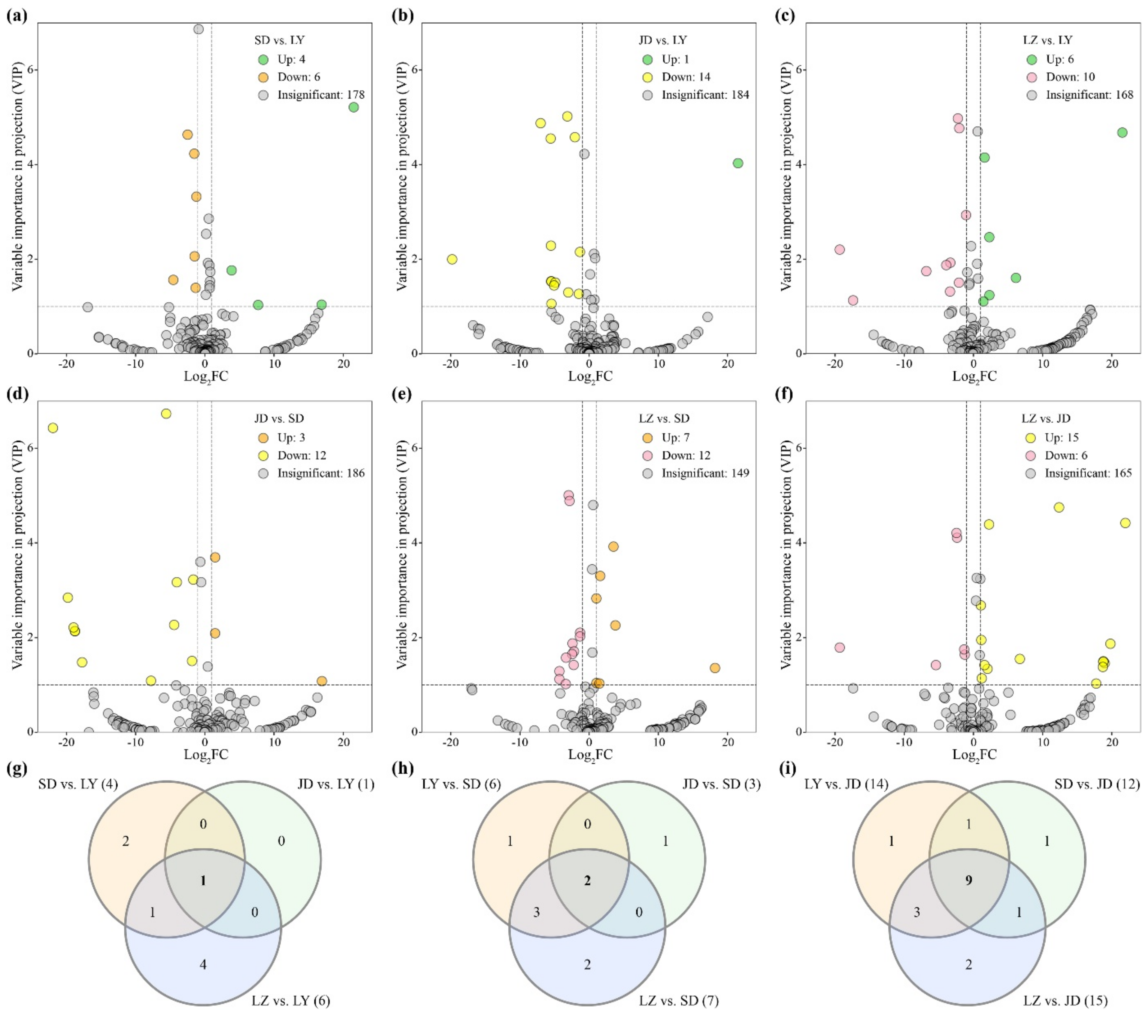
By comparing the relative metabolite contents between four edible Lilium species, the upregulated (log2FC (fold change) ≥1, VIP ≥ 1) and downregulated (log2FC ≤ −1, VIP ≥ 1) metabolites were screened (Figure 2a–f). Based on the speculation that higher bioactive ingredients were accumulated in medicinal lilies, the commonly upregulated metabolites in each medicinal Lilium species were chosen and analyzed (Figure 2g–i).

2.2.1. Differential Metabolites in LY Bulbs

Although the bulbs of three Lilium species were considered medicative, recent systematic studies on their origin and quality based on textual research and investigation indicated that Lilium plants with white trumpet flowers and white bulbs are an authentic medicinal material [6,22,23]. Therefore, the bulbs of L. brownii and L. brownii var. viridulum, which were the most common and widespread species at that time, were considered to be of good quality in the ancient herbal literature (Figure S2).
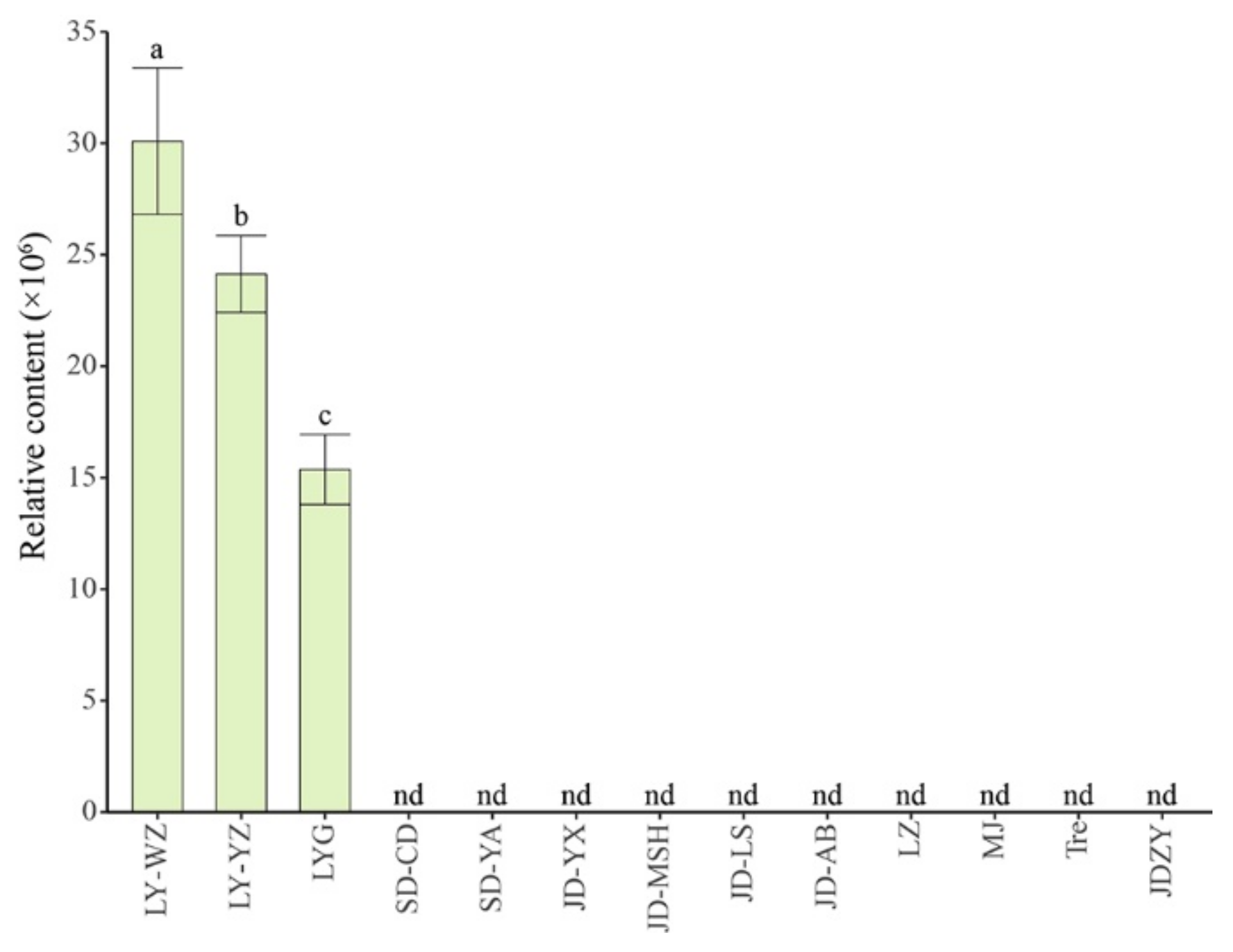
There were four upregulated metabolites between SD and LY (Figure 2a), only one upregulated metabolite between JD and LY (Figure 2b), and six upregulated metabolites between LZ and LY (Figure 2c). In total, there was only one commonly differential metabolite in LY (Figure 2g), which was not detected in other lily samples; this was a steroidal saponin with the molecular formula C45H74O18 (MW 902.43), which was temporarily identified as 26-O-glucopyranosyl-furost-5-3,26-diol 3-O-[rhamnopyranosyl-(1→2)]-glucopyranoside [11]. Combined with the relative amounts of this compound in three other lily samples in our pre-experiment, including the dry scales of LY, the bulbils of JD-AB, and the bulbs of Lilium ‘Tresor’, we found that this steroidal saponin was exclusively detected in LY scales regardless of whether they were fresh or dried (Figure 3). As steroidal saponins have multiple spatial configurations and isomers in L. brownii [24,25], it is difficult to determine their specific structure based only on the MS spectrum (Figure S3). Because some saponins were not detected [26], we speculated that steroidal saponins may be the key bioactive metabolites in medicinal lily bulbs.

2.2.2. Differential Metabolites in SD Bulbs

Although SD is also used as a medicinal plant, its efficacy differs from that of LY. The traditional pharmacopoeia records indicate that SD can be used for treating various skin conditions including bruises, swelling, and wounds. There were two commonly upregulated differential metabolites among the comparison groups LY vs. SD, JD vs. SD, and LZ vs. SD (Figure 2a,d,e,h); 1,2-O-diferuloylglycerol and 1,3-O-diferuloyl-glycerol were both detected in all lily samples in this study. However, after one-way analysis of variance (ANOVA) with other samples, their content difference between SD and some JD samples was not significant (Figure S4). These two ferulic acid derivatives with antioxidant and ultraviolet light-absorbing properties accumulated slightly higher only in the SD samples [8,9,27]; thus, further studies are needed to determine whether they are the key active components in SD.

2.2.3. Differential Metabolites in JD Bulbs

Similarly, there were nine commonly upregulated differential metabolites found in the JD samples (Figure 2b,d,f,i). After excluding compounds that were not significantly different from other lily samples, eight metabolites were screened out, including one flavonoid, three phenolic compounds, three steroids, and one coumarin (Figure S5). The easily identified JD has orange flowers with black spots and bulbils, and the Ben Cao Tu Jing (Illustrated Pharmacopoeia), which dates from about 1080, clearly mentions that “it is not suitable for medicinal use” [6]. Subsequent pharmacy books have also recorded that JD does not have the same medicinal effects as LY. However, in Korea, the bulbs of JD are used to treat bronchitis, pneumonia, and so on [28]. JD bulbs are also reported to possess potent antioxidant and anticancer characteristics [8,29]. Other medicinal effects of JD remain to be studied.

2.3. Differential Metabolites Analysis Sweet and Bitter Lily Bulbs

The taste description of LY and SD bulbs is controversial; therefore, we chose JD and LZ samples to represent bitter and sweet lily bulbs, respectively. Notably, no steroidal saponins were detected in LZ samples in this study (Figure 1a). After screening the differential metabolites (Figure 2f) and excluding the metabolites with insignificant differences between LZ and JD, there were 12 upregulated differential metabolites including 5 phenolic acids, 4 steroids, 1 flavonoid, 1 coumarin, and 1 other compound, and 6 downregulated differential metabolites, including 3 phenolic acids, 1 flavonoid, and 2 other compounds (Figure S6).
Although many phenolic compounds are bitter substances, both JD and LZ accumulated differential phenolic acids, and it was difficult to distinguish their contribution to the bitter taste of lily bulbs. Moreover, some saponins also have a bitter taste [30]. A previous study found that the taste of quinoa varieties (Chenopodium quinoa), bitter or sweet, was related to their saponin contents [31], and that sweeteners such as sugars were commonly used agents to reduce perceived bitterness [32]. Our previous research found that the intrinsic free sugar content in LZ bulbs was higher (11.2–11.7%) than that in JD bulbs (5.1–7.6%) [4]. Therefore, we speculated that the bitter taste of JD bulbs might relate to their low content of free sugars and high accumulation of saponins.

2.4. Differential Metabolite Analysis between Purple and White Lily Bulbs

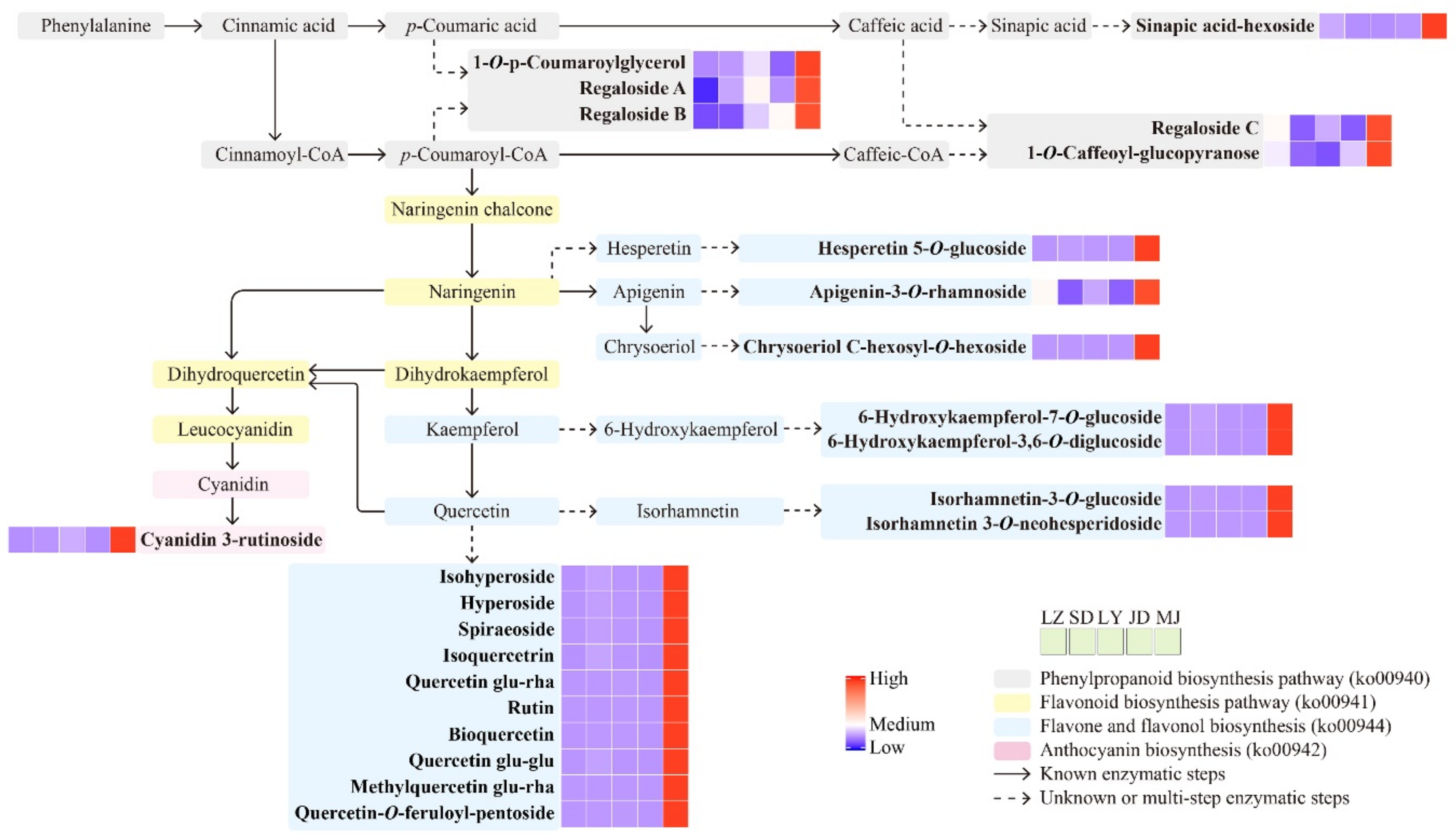
In addition to more metabolites being detected in the purple bulbs of MJ, 32 metabolites were exclusively detected in these bulbs (Figure 2b). Combining the relative contents and orthogonal partial least square discriminant analysis (OPLS-DA) results with four edible lily samples (log2FC ≥ 1, VIP ≥ 1), we found 38 upregulated metabolites between LY and MJ (Figure 4a), 36 upregulated metabolites between SD and MJ (Figure 4b), 32 upregulated metabolites between JD and MJ (Figure 4c), and 33 upregulated metabolites between LZ and MJ (Figure 4d). In total, there were 30 commonly upregulated differential metabolites between purple MJ bulbs and edible white bulbs (Figure 4e, Table S1), including 18 flavonoids, 6 phenolic acids, 1 coumarin, 1 lignan, 1 steroid, and 3 other compounds (Figure 4f). These upregulated differential metabolites in MJ bulbs were mapped to the KEGG database to analyze their biochemical synthesis pathways. The metabolic pathways found to be differentially altered between the purple and white lily bulbs mainly included phenylpropanoid biosynthesis (ko00940), flavonoid biosynthesis (ko00941), flavone and flavonol biosynthesis (ko00944), and anthocyanin biosynthesis (ko00942) (Figure 5).
Five of the six phenolic acid compounds were phenolic glycerides and their derivatives. The synthesis pathway of these phenolic glycerides (glycosides) is still unclear. However, it is speculated that they might be catalyzed by glycerol O-hydroxycinnamoyltransferase or glycerol transferase and then by glycosyltransferase, of which the former can employ p-coumaroyl-CoA or caffeoyl-CoA as an acyl donor and use glycerol as an acceptor [33].
Among these differential flavonoid metabolites, the relative contents of cyanidin 3-rutinoside, the main anthocyanin compound, were higher in the MJ samples as expected (Figure 5), which is also a major component of red and pink lily flowers [34]. In addition, 14 of 18 differential flavonoids are flavonol glycosides, including 2 kaempferol derivates and 12 quercetin derivates. This is consistent with previous research showing that the quercetin and rutin contents in MJ bulbs were significantly higher than those in SD, JD, and LZ bulbs [10]. Although quercetin and its derivatives are pharmacologically active and have various biological activities, their bitter flavors, which contribute to the bitter taste of MJ bulbs, make them unpopular [35]. Although studies are underway to obtain functional foods with more biological activities and improved taste, introducing these bioactive substances from MJ bulbs into edible lilies is a topic worthy of further in-depth study.

3. Materials and Methods

3.1. Plant Material

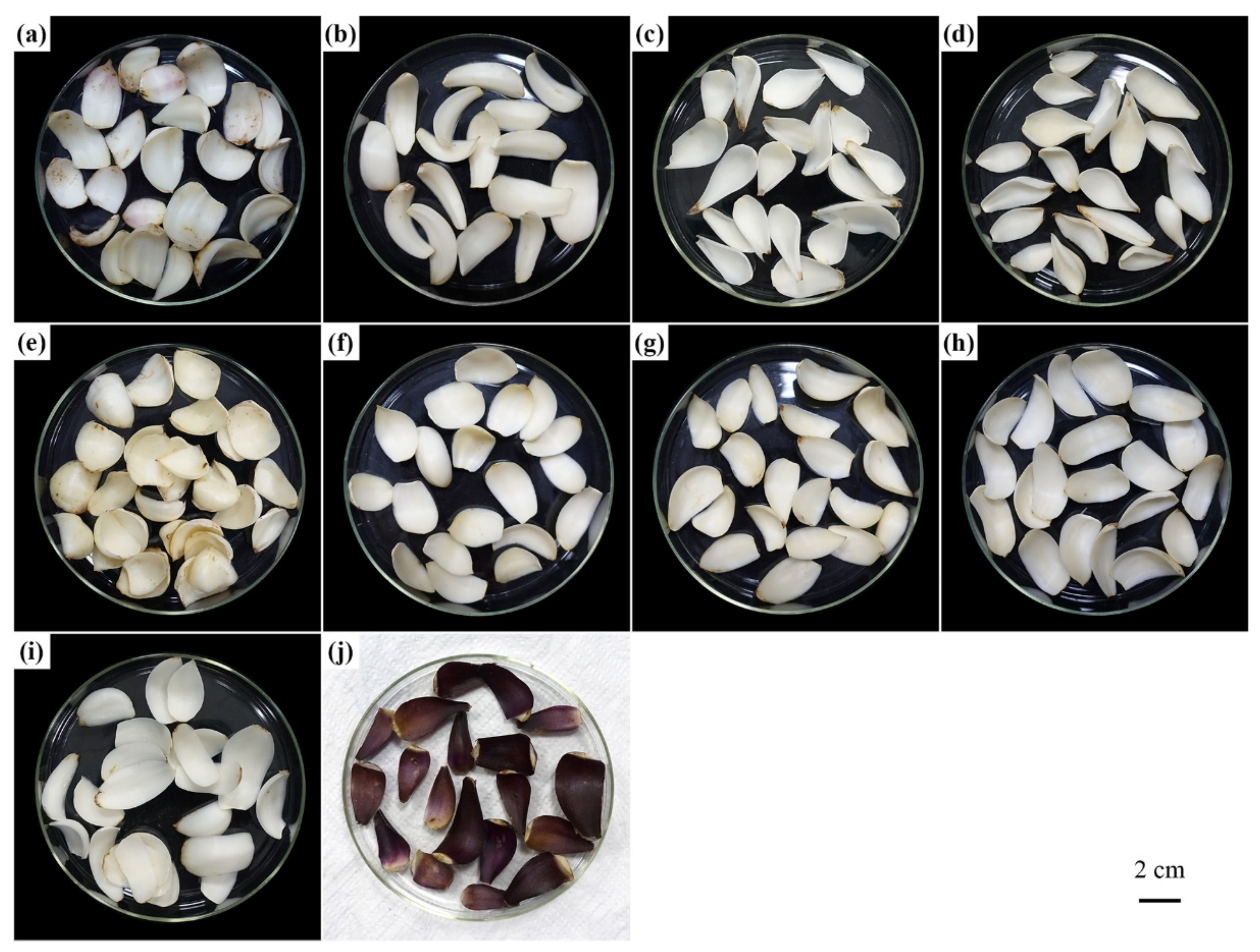
Light-colored bulbs of four edible Lilium species were collected, mainly from their natural habitats. Bulbs of L. regale, which were propagated from seeds, collected from Sichuan Province and planted in the Lily Germplasm Resource Preservation Centre (116°43′ N, 40°16′ E) for three to four years, were also used. Samples were collected from September to October 2019, and their information is listed in Table 1, Figure 6, and Figure S2.
Fresh bulbs of Lilium species were first stored at −4°C for one week and then washed and dried at room temperature for 15 min. Three bulbs were used for each sample. The scales were cut into 1 × 1 cm2 pieces and frozen in liquid nitrogen. After freeze-drying (SCIENTZ-50ND, SCIENTZ), the samples were ground into a powder using a mixer mill (MM 400, Retsch) with zirconia beads for 1.5 min at 30 Hz, and then stored at −80°C until extraction.

3.2. Reagents

Methanol and acetonitrile (HPLC grade) were purchased from Merck (Darmstadt, Germany). Acetic acid was obtained from Sigma (St. Louis, MO, USA). MilliQ water (Millipore, Bradford, PA, USA) was used in all experiments.

3.3. Sample Extraction

A fine powder sample of a dried lily bulb (100 mg) was extracted overnight at 4 ℃ using 1.0 mL of 70% aqueous methanol. The homogenate was centrifuged at 10,000× g for 10 min, and the extracts were cleaned up by solid-phase extraction (SPE, CNWBOND Carbon-GCB cartridge, 250 mg, 3 mL; ANPEL, Shanghai, China) and were filtrated (SCAA-104, 0.22 μm pore size; ANPEL, Shanghai, China) before LC–MS analysis.

3.4. HPLC–MS/MS Analysis

Samples were injected into an LC–ESI–MS/MS system (HPLC, Shim-pack UFLC SHIMADZU CBM30A system; MS, Applied Biosystems 4500 Q TRAP). The analytical conditions were as follows, HPLC: column, Waters ACQUITY UPLC HSS T3 C18 column (1.8 µm, 2.1 mm × 100 mm); solvent system, ultrapure water (Solvent A, 0.04% acetic acid): acetonitrile (Solvent B, 0.04% acetic acid); gradient program, 100/0, 5/11.0, 5/12.0, 95/12.1, 95/15.0 (min/% A, v/v), flow rate, 0.40 mL/min; temperature, 40 °C; injection volume: 5 μL. The effluent was alternatively connected to an ESI–triple quadrupole–linear ion trap (Q TRAP)–MS system. The ESI–Q TRAP–MS/MS analytical conditions used were as reported by Chu et al. [36].

3.5. Metabolite Annotation and Quantification

Metabolite identification was based on the primary and secondary spectrometry data, annotated and compared against public databases, including MassBank, KNApSAcK, HMDB, METLIN, and the MWDB Database (MetWare Biological Science and Technology Co. Ltd., Wuhan, China), following the standard metabolic operating procedures. The phenolic compounds were divided into different subgroups based on their chemical structures, such as phenolic acids, flavonoids, coumarins, and lignans, as reported by Gan et al. [37].
The metabolites were quantified via the multiple reaction monitoring mode (MRM) using triple quadrupole mass spectrometry as reported by Wang et al. [38]. After obtaining metabolite data from different samples, the peak area of the mass spectra of all metabolites was integrated, and the mass spectra of the same metabolites in different samples were integrated and corrected.

3.6. Screening of Differential Metabolites

Hierarchical cluster analysis (HCA), principal component analysis (PCA), and correlation analysis were performed using the ComplexHeatmap, factoextra, and corrplot packages in RStudio, respectively [39]. For HCA, after normalizing the signal intensities of each metabolite (unit variance scaling), the results of the samples and metabolites were presented as heatmaps with dendrograms. After data were subjected to Pareto scaling by SIMCA software (V14.1, MKS Data Analytics Solutions, Sweden), orthogonal partial least square discriminant analysis (OPLS-DA) was performed, and the validity of the OPLS-DA model was evaluated [40]. The variable importance in projection (VIP) of metabolites was calculated for the subsequent screening of differential metabolites. To compare the differential metabolite variation, Venn diagrams were plotted using the InteractiVenn tool [41]. Significant differences (p < 0.05) were determined by one-way analysis of variance (ANOVA) using SPSS 23 (SPSS Inc., Chicago, IL, USA). Differential metabolites were mapped to the Kyoto Encyclopaedia of Genes and Genomes (KEGG) database (www.kegg.jp/kegg/pathway.html, accessed on 15 January 2021) for metabolic network analysis.

4. Conclusions

The bulbs of several Lilium species are esteemed as culinary delicacies in northern and eastern Asia and are also considered to have medicinal properties. In this study, we analyzed the secondary metabolites of 10 lily bulb samples from different habitats, belonging to five species. In total, 245 metabolites (including 87 flavonoids, 59 phenolic acids, 14 steroids, 10 alkaloids, 6 coumarins, 11 lignans, 8 terpenoids, and 50 other compounds) were identified. Combined literature analysis and metabolomic analysis suggested that L. brownii var. viridulum was considered the authentic lily for traditional medical use and that steroid saponins may be the key active metabolites in medicinal lily bulbs. The bitter taste of L. lancifolium might be related to its low content of free sugars and high accumulation of saponins. The purple bulbs of L. regale showed a differential accumulation of 30 metabolites, including 18 flavonoids and 6 phenolic acids. Overall, this study elucidates the differences in secondary metabolites among different Lilium bulbs, provides a reference for the utilization of and research on lilies as plant medicine and functional food, and may guide the development and genetic enhancement of functional lily bulb varieties.

Supplementary Materials

The following are available online. Figure S1: Correlation analysis on the metabolite profiles of ten lily samples, Figure S2: The distribution area and collection place of ten lily samples belonging to five different species, Figure S3: The MS/MS spectrum of the [M-H]+ ion of 26-O-glucopyranosyl-furost-5-3,26-diol 3-O-[rhamnopyranosyl-(1→2)]-glucopyranoside, Figure S4: The relative contents of upregulated metabolites in SD samples compared with other edible lily samples, Figure S5: The relative contents of upregulated metabolites in JD samples compared with other edible lily samples, Figure S6. The relative contents of differential metabolites in JD and LZ samples. Table S1: A list of upregulated metabolites identified between the purple and white lily bulbs.

Author Contributions

Y.K.: conceptualization, methodology, data curation, writing—review and editing, funding acquisition. H.W.: investigation, writing—review and editing. L.L.: investigation, resources. X.D.: resources, writing—review and editing. J.B.: conceptualization, supervision, project administration, funding acquisition. All authors have read and agreed to the published version of the manuscript.

Funding

This project was supported by the National Natural Science Foundation of China (No. 32002082), the Beijing Municipal Finance Bureau (No. PXM2019_178217_000001), and the Innovation and Development Program of Beijing Academy of Science and Technology (PY2020JK42).

Data Availability Statement

Data presented are available in the manuscript and Supplementary Materials.

Conflicts of Interest

The authors declare no conflict of interest.

Sample Availability

Samples of the compounds are not available from the authors.

References

  1. Liang, S.; Minoru, N.T. Lilium. In Flora of China. Volume 24: Flagellariaceae through Marantaceae; Wu, Z., Raven, P.H., Hong, D., Eds.; Scidence Press & Missouri Botanical Garden Press: Beijing, China; St. Louis, MO, USA, 2000; pp. 135–149. [Google Scholar]
  2. Kong, Y.; Bai, J.; Lang, L.; Bao, F.; Dou, X.; Wang, H.; Shang, H. Floral scents produced by Lilium and Cardiocrinum species native to China. Biochem. Syst. Ecol. 2017, 70, 222–229. [Google Scholar] [CrossRef]
  3. McRae, E.A. Lilies: A Guide for Growers and Collectors; Timber Press: Portland, OR, USA, 1998; p. 19. [Google Scholar]
  4. Kong, Y.; Wang, H.; Lang, L.; Dou, X.; Bai, J. Analysis on the contents of 13 intrinsic free sugars in different lily samples. Food Sci. in press (In Chinese).
  5. Erb, M.; Kliebenstein, D.J. Plant secondary metabolites as defenses, regulators, and primary metabolites: The blurred functional trichotomy. Plant Physiol. 2020, 184, 39–52. [Google Scholar] [CrossRef] [PubMed]
  6. Haw, S.G. The history of lilies in China. In The Lilies of China; Haw, S.G., Ed.; Timber Press: London, UK, 1986; pp. 43–56. [Google Scholar]
  7. Chinese Pharmacopoeia Commission. Pharmacopoeia of The Peoples Republic Of China (Part I), 2020th ed.; China Pharmaceutical Science and Technology Press: Beijing, China, 2020; p. 137. [Google Scholar]
  8. Luo, J.; Li, L.; Kong, L. Preparative separation of phenylpropenoid glycerides from the bulbs of Lilium lancifolium by high-speed counter-current chromatography and evaluation of their antioxidant activities. Food Chem. 2012, 131, 1056–1062. [Google Scholar] [CrossRef]
  9. Ma, T.; Wang, Z.; Zhang, Y.M.; Luo, J.-G.; Kong, L.-Y. Bioassay-guided isolation of anti-inflammatory components from the bulbs of Lilium brownii var. viridulum and identifying the underlying mechanism through acting on the NF-κB/MAPKs pathway. Molecules 2017, 22, 506. [Google Scholar] [CrossRef] [Green Version]
  10. Jin, L.; Zhang, Y.; Yan, L.; Guo, Y.; Niu, L. Phenolic compounds and antioxidant activity of bulb extracts of six Lilium species native to China. Molecules 2012, 17, 9361–9378. [Google Scholar] [CrossRef]
  11. Zhu, M.; Luo, J.; Lv, H.; Kong, L. Determination of anti-hyperglycaemic activity in steroidal glycoside rich fraction of lily bulbs and characterization of the chemical profiles by LC-Q-TOF-MS/MS. J. Funct. Foods 2014, 6, 585–597. [Google Scholar] [CrossRef]
  12. Xu, Z.; Wang, H.; Wang, B.; Fu, L.; Yuan, M.; Liu, J.; Zhou, L.; Ding, C. Characterization and antioxidant activities of polysaccharides from the leaves of Lilium lancifolium Thunb. Int. J. Biol. Macromol. 2016, 92, 148–155. [Google Scholar] [CrossRef] [PubMed]
  13. Hu, Y.; Du, Y.; Zhang, M.; Zhang, X.; Ren, J. Characters and comprehensive evaluation of nutrients and active components of 12 Lilium species. Nat. Prod. Res. Dev. 2019, 31, 292–298. (In Chinese) [Google Scholar] [CrossRef]
  14. Wang, F.; Wang, W.; Niu, X.; Huang, Y.; Zhang, J. Isolation and structural characterization of a second polysaccharide from bulbs of Lanzhou lily. Appl. Biochem. Biotech. 2018, 186, 535–546. [Google Scholar] [CrossRef]
  15. Drewnowski, A.; Gomez-Carneros, C. Bitter taste, phytonutrients, and the consumer: A review. Am. J. Clin. Nutr. 2000, 72, 1424–1435. [Google Scholar] [CrossRef] [PubMed]
  16. Shimomura, H.; Sashida, Y.; Mimaki, Y. Bitter phenylpropanoid glycosides from Lilium speciosum var. rubrum. Phytochemistry 1986, 25, 2897–2899. [Google Scholar] [CrossRef]
  17. Shoyama, Y.; Hatano, K.; Nishioka, I.; Yamagishi, T. Phenolic glycosides from Lilium longiflorum. Phytochemistry 1987, 26, 2965–2968. [Google Scholar] [CrossRef]
  18. Shimomura, H.; Sashida, Y.; Mimaki, Y. Phenolic glycerides from Lilium auratum. Phytochemistry 1987, 26, 844–845. [Google Scholar] [CrossRef]
  19. Shimomura, H.; Sashida, Y.; Mimaki, Y.; Iida, N. Regaloside A and B, acylated glycerol glucosides from Lilium regale. Phytochemistry 1988, 27, 451–454. [Google Scholar] [CrossRef]
  20. Fox, D. Growing Lilies; Croom Helm Ltd.: Wolfeboro, NH, USA, 1985; p. 10. [Google Scholar]
  21. Liu, Y.; Lin-Wang, K.; Deng, C.; Warran, B.; Wang, L.; Yu, B.; Yang, H.; Wang, J.; Espley, R.V.; Zhang, J.; et al. Comparative transcriptome analysis of white and purple potato to identify genes involved in anthocyanin biosynthesis. PLoS ONE 2015, 10, e0129148. [Google Scholar] [CrossRef] [PubMed]
  22. Wang, C.; Shu, S.; Yin, F.; Zhao, J.; Zhang, Z.; Liu, X.; Zhan, Z. Textual research on origin and genuine of Lilii Bulbus. China J. Chin. Meter. Med. 2018, 43, 1732–1736. (In Chinese) [Google Scholar] [CrossRef]
  23. Zhang, W.; Wang, J.; Zhang, Z.; Peng, H.; Zhan, Z.; Yang, H. Herbal textual research on triditional Chinese medicine “Baihe” (Lilii Bulbus). China J. Chin. Mater. Med. 2019, 44, 5007–5011. (In Chinese) [Google Scholar] [CrossRef]
  24. Hong, X.-X.; Luo, J.-G.; Guo, C.; Kong, L.-Y. New steroidal saponins from the bulbs of Lilium brownii var. viridulum. Carbohydr. Res. 2012, 361, 19–26. [Google Scholar] [CrossRef] [PubMed]
  25. Hou, X.; Chen, F. Studies on chemical constituents of Lilium brownii. Acta Pharm. Sin. 1998, 33, 923–926. (In Chinese) [Google Scholar]
  26. Munafo, J.P.; Gianfagna, T.J., Jr. Chemistry and biological activity of steroidal glycosides from the Lilium genus. Nat. Prod. Rep. 2015, 32, 454–477. [Google Scholar] [CrossRef] [PubMed]
  27. Kharrat, N.; Aissa, I.; Dgachi, Y.; Aloui, F.; Chabchoub, F.; Bouaziz, M.; Gargouri, Y. Enzymatic synthesis of 1,3-dihydroxyphenylacetoyl-sn-glycerol: Optimization by response surface methodology and evaluation of its antioxidant and antibacterial activities. Bioorg. Chem. 2017, 75, 347–356. [Google Scholar] [CrossRef]
  28. Lim, T.K. Lilium lancifolium. In Edible Medicinal and Non Medicinal Plants (Volume 8, Flowers); Lim, T.K., Ed.; Springer: Dordrecht, The Netherlands, 2014; pp. 215–220. [Google Scholar] [CrossRef]
  29. Zhang, Y.-Y.; Luo, L.-M.; Wang, Y.-X.; Zhu, N.; Zhao, T.-J.; Qin, L. Total saponins from Lilium lancifolium: A promising alternative to inhibit the growth of gastric carcinoma cells. J. Cancer 2020, 11, 4261–4273. [Google Scholar] [CrossRef]
  30. Heng, L.; Vincken, J.-P.; van Koningsveld, G.; Legger, A.; Gruppen, H.; Van Boekel, T.; Roozen, J.; Voragen, F. Bitterness of saponins and their content in dry peas. J. Sci. Food Agric. 2006, 86, 225–1231. [Google Scholar] [CrossRef]
  31. Fiallos-Jurado, J.; Pollier, J.; Moses, T.; Arendt, P.; Barriga-Medina, N.; Morillo, E.; Arahana, V.; Torres, M.D.L.; Goossens, A.; Leon-Reyes, A. Saponin determination, expression analysis and functional characterization of saponin biosynthetic genes in Chenopodium quinoa leaves. Plant Sci. 2016, 250, 188–197. [Google Scholar] [CrossRef]
  32. Mennella, J.A.; Spector, A.C.; Reed, D.R.; Coldwell, S.E. The bad taste of medicines: Overview of basic research on bitter taste. Clin. Ther. 2013, 35, 1225–1246. [Google Scholar] [CrossRef] [PubMed] [Green Version]
  33. Kim, I.A.; Kim, B.-G.; Kim, M.; Ahn, J.-H. Characterization of hydroxycinnamoyltransferase from rice and its application for biological synthesis of hydroxycinnamoyl glycerols. Phytochemistry 2012, 76, 25–31. [Google Scholar] [CrossRef]
  34. Yamagishi, M.; Yoshida, Y.; Nakayama, M. The transcription factor LhMYB12 determines anthocyanin pigmentation in the tepals of Asiatic hybrid lilies (Lilium spp.) and regulates pigment quantity. Mol. Breed. 2012, 30, 913–925. [Google Scholar] [CrossRef]
  35. Khor, C.M.; Ng, W.K.; Kanaujia, P.; Chan, K.P.; Dong, Y. Hot-melt extrusion microencapsulation of quercetin for taste-masking. J. Microencapsul. 2017, 34, 29–37. [Google Scholar] [CrossRef]
  36. Chu, C.; Du, Y.; Yu, X.; Shi, J.; Yuan, X.; Liu, X.; Liu, Y.; Zhang, H.; Zhang, Z.; Yan, N. Dynamics of antioxidant activities, metabolites, phenolic acids, flavonoids, and phenolic biosynthetic genes in germinating Chinese wild rice (Zizania latifolia). Food Chem. 2020, 318, 126483. [Google Scholar] [CrossRef] [PubMed]
  37. Gan, R.-Y.; Chan, C.-L.; Yang, Q.-Q.; Li, H.-B.; Zhang, D.; Ge, Y.-Y.; Gunaratne, A.; Ge, J.; Corke, H. Bioactive compounds and beneficial functions of sprouted grains. In Sprouted Grains; Feng, H., Nemzer, B., DeVries, J.W., Eds.; AACC International Press: Duxford, UK, 2019; pp. 191–246. [Google Scholar] [CrossRef]
  38. Wang, D.; Zhang, L.; Huang, X.; Wang, X.; Yang, R.; Mao, J.; Wang, X.; Wang, X.; Zhang, Q.; Li, P. Identification of nutritional components in black sesame determined by widely targeted metabolomics and traditional Chinese medicines. Molecules 2018, 23, 1180. [Google Scholar] [CrossRef] [PubMed] [Green Version]
  39. Gu, Z.; Eils, R.; Schlesner, M. Complex heatmaps reveal patterns and correlations in multidimensional genomic data. Bioinformatics 2016, 32, 2847–2849. [Google Scholar] [CrossRef] [PubMed] [Green Version]
  40. Yan, N.; Du, Y.; Liu, X.; Chu, M.; Shi, J.; Zhang, H.; Liu, Y.; Zhang, Z. A comparative UHPLC-QqQ-MS-based metabolomics approach for evaluating Chinese and North American wild rice. Food Chem. 2019, 275, 618–627. [Google Scholar] [CrossRef] [PubMed]
  41. Heberle, H.; Meirelles, G.V.; da Silva, F.R.; Telles, G.P.; Minghim, R. InteractiVenn: A web-based tool for the analysis of sets through Venn diagrams. BMC Bioinform. 2015, 16, 169. [Google Scholar] [CrossRef]
Figure 1. Secondary metabolite profiles of ten lily samples belonging to five different species. (a) The number of secondary metabolites detected in ten lily samples; (b) Venn diagrams for displaying the number of commonly and specific metabolites among five Lilium species; (c) Hierarchical cluster analysis and heatmap of metabolite profiles of ten lily samples. Clustering distance method ”Pearson”, hierarchical clustering method “complete”; (d) Principal component analysis and scores plot for ten lily samples.
Molecules 26 01340 g001
Figure 2. Differential metabolites in four edible Lilium bulbs. Upregulated metabolites are indicated in green (L. brownii var. viridulum, LY), orange (L. pumilum, SD), yellow (L. lancifolium, JD), and pink (L. davidii var. willmottiae, LZ). (a) Volcano plot of the differential metabolites between SD and LY. (b) Volcano plot of the differential metabolites between JD and LY. (c) Volcano plot of the differential metabolites between LZ and LY. (d) Volcano plot of the differential metabolites between JD and SD. (e) Volcano plot of the differential metabolites between LZ and SD. (f) Volcano plot of the differential metabolites between LZ and JD. (g) Venn plot indicating the number (in bold) of commonly upregulated metabolites among the comparison groups of SD vs. LY, JD vs. LY, and LZ vs. LY. (h) Venn plot indicating the number (in bold) of commonly upregulated metabolites among the comparison groups of LY vs. SD, JD vs. SD, and LZ vs. SD. (i) Venn plot indicating the number (in bold) of commonly upregulated metabolites among the comparison groups of SD vs. LY, JD vs. LY, and LZ vs. LY.
Molecules 26 01340 g002
Figure 3. The relative contents of a steroidal saponin that was temporarily identified as 26-O-glucopyranosyl-furost-5-3,26-diol 3-O-[rhamnopyranosyl-(1→2)]-glucopyranoside in different lily samples. Data are presented as the mean ± standard error (SE, n = 3). Different lowercase letters indicate statistically significant differences (p < 0.05). LYG: dry scales of LY bulbs, Tre: bulbs of Lilium “Tresor”, JDZY: bulbils of L. lancifolium. nd: not detected.
Molecules 26 01340 g003
Figure 4. Differential metabolites between purple L. regale (MJ) bulbs and edible white bulbs. Upregulated metabolites are indicated in blue (MJ). (a) Volcano plot of the differential metabolites between L. brownii var. viridulum (LY) and MJ. (b) Volcano plot of the differential metabolites between L. pumilum (SD) and MJ. (c) Volcano plot of the differential metabolites between L. lancifolium (JD) and MJ. (d) Volcano plot of the differential metabolites between L. davidii var. willmottiae (LZ) and MJ. (e) Venn plot indicating the number (in bold) of commonly upregulated metabolites in MJ samples. (f) The number of differential metabolites in different compound groups.
Molecules 26 01340 g004
Figure 5. Upregulated differential phenolic acids and flavonoids (in bold) between purple L. regale (MJ) bulbs and edible white bulbs were mapped to the metabolic pathway networks. LZ: L. davidii var. willmottiae, SD: L. pumilum, LY: L. brownii var. viridulum, JD: L. lancifolium.
Molecules 26 01340 g005
Figure 6. Photos of ten Lilium bulb samples belonging to five different species. (a) LY-WZ; (b) LY-YZ; (c) SD-CD; (d) SD-YA; (e) JD-AB; (f) JD-LS; (g) JD-MSH; (h) JD-YX; (i) LZ; (j) MJ.
Molecules 26 01340 g006
Table 1. Information of ten Lilium samples.
NameApplication aSection bBulb ColorProvinceLocationCodeCircumference (cm) cStarch Content (%)
L. brownii var. viridulumM&ELWhiteJiangxiWanzaiLY-WZ19.80 ± 1.1722.80
HunanYongzhouLY-YZ23.13 ± 0.38/
L. pumilumM&ESWhiteHebeiChengdeSD-CD7.27 ± 0.1314.55
ShaanxiYan’anSD-YA6.73 ± 0.3312.06
L. lancifoliumM&ESWhiteSichuanA’baJD-AB13.80 ± 0.3119.16
HunanLongshanJD-LS25.97 ± 0.15 d15.82
AnhuiManshuiheJD-MSH22.50 ± 0.26 d11.86
JiangsuYixingJD-YX27.03 ± 0.69 d17.99
L. davidii var. willmottiaeESWhiteGansuLanzhouLZ15.97 ± 0.2611.64
L. regale/LPurpleBeijingHuairouMJ14.40 ± 0.8713.33
a M, medicinal; E, edible. b L, Section Leucolirion; S, Section Sinomartagon. c Values present mean ± standard error (SE) of triplicate tests. d The bulbs of cultivated L. lancifolium are connate with 4–5 smaller bulbs.
Publisher’s Note: MDPI stays neutral with regard to jurisdictional claims in published maps and institutional affiliations.