Article

Chemophenetic Significance of Anomalocalyx uleanus Metabolites Are Revealed by Dereplication Using Molecular Networking Tools

by
José Assis Gomes de Brito
1,2,
Luciano da Silva Pinto
3,
Cintia Folly Chaves
1,
Antônio Jorge Ribeiro da Silva
1,
Maria Fátima das Graças Fernandes da Silva
3 and
Fernando Cotinguiba
1,*
1
Instituto de Pesquisas de Produtos Naturais “Walter Mors”, Centro de Ciencas da Saude, Universidade Federal do Rio de Janeiro (UFRJ), Avenida Carlos Chagas Filho, 373, Bloco H, Cidade Universitaria, CEP 21941-902 Rio de Janeiro, Brazil
2
Instituto Federal de Rondônia, Campus de Ji-Parana, Rua Rio Amazonas, 151, Jardim dos Migrantes, CEP 78960-000 Ji-Paraná-RO, Brazil
3
Departamento de Quimica, Universidade Federal de São Carlos, Rodovia Washington Luís km 235, CEP 13565-905 São Carlos-SP, Brazil
*
Author to whom correspondence should be addressed.
Molecules 2021, 26(4), 925; https://doi.org/10.3390/molecules26040925
Submission received: 14 January 2021 / Revised: 28 January 2021 / Accepted: 4 February 2021 / Published: 9 February 2021
(This article belongs to the Special Issue LC-MS Approach in Specialized Natural Products Research)

Abstract

:
Anomalocalyx uleanus (Pax & K. Hoffm.) Ducke (Euphorbiaceae) is a singular species in the genus and is restricted and exclusive to the Brazilian Amazon. A phytochemical study of A. uleanus leaves was performed, yielding the isolation of five major compounds: catechin/epicatechin, afzelin, quercetin 3-O-α-L-rhamnopyranoside, and astilbin. The phytochemical compositions of the methanolic extracts of leaves, roots, bark, and stem bark were determined using a dereplication approach. Forty-six compounds were annotated from the liquid chromatography-mass spectrometry (LC-MS/MS) data, while four lipids were identified using gas chromatography-mass spectrometry (GC-MS). In total, fifty compounds were detected, and they belonged to the primary metabolism and several classes of natural products such as flavonoids, flavonoids O-glycosides, flavonoids C-glycosides, biflavonoids, procyanidin, triterpene, triterpenes esterified with phenylpropanoids, phenylpropanoid derivatives, flavonolignans, coumarins, quinic acid derivatives, and benzoic acid derivatives. This is the first report on the phytochemical data of the genus Anomalocalyx, and the results of this study will contribute to the chemosystematic knowledge of the Euphorbiaceae family and justify the need for investigation of the pharmacological potential of the species A. uleanus.

1. Introduction

The Euphorbiaceae family comprises 219 genera and 6300 species, which are distributed in tropical and subtropical regions, and many of them are endemic to Brazil [1]. This plant family has a very diverse secondary metabolism, and their metabolites are characterized by their medicinal properties [2,3]. The species of the Euphorbiaceae family have suffered throughout history, with several reorganizations within the subfamilies, genera, and even segregations of other families. Chemosystematic and phylogenetic studies have contributed immensely to the understanding of the classification of this family [4,5].
Recently, the term chemophenetics has been used to describe a variety of secondary metabolites specialized in a given taxon and are important for the description of classified organisms with the help of modern molecular methods. Chemophenetics do not aim to elucidate phylogenetic relationships but to describe the matrix of natural products and use them for the phenetic characterization of clades [6]. Several studies using chemophenetic and chemotaxonomic approaches have been described in the literature, including several genera of the Euphorbiaceae such as Senefelderopsis [7], Euphorbia [8,9,10], Mallotus [5], Chrozophora [11], Croton [12], Alchornea [13], Sapium [14], and Sebastiania [15], among other examples.
Anomalocalyx uleanus (Pax & K. Hoffm.) Ducke is a unique species in the genus Anomalocalyx, and it is popularly known as “arataciú-preto”. It is a tree that can reach 40 m, has no latex, and is restricted and exclusive to the Brazilian Amazon. This species is found mainly in flooded areas and has never been studied chemically [3]. Metabolite profiling using high-resolution mass spectrometry (HRMS) grants access to large volumes of high-quality spectral data from a minimal amount of samples, and appropriate data analysis workflows allow the efficient mining of such data [16]. An approach to dereplicating the extracts of leaves, roots, bark, and stem bark of A. uleanus was adopted in order to discover as much as possible the classes of natural products present in that species using tools such as MZmine 2, MS-DIAL, MSFINDER, and GNPS. The aim of this work was to describe, as extensively as possible, the array of natural products present in A. uleanus and to establish chemophenetic correlations with other species in Euphorbiaceae [3,17].

2. Results and Discussion

2.1. Isolation of Majoritary Compound from A. uleanus Leaves

A phytochemical study of A. uleanus leaves was performed using traditional approaches for extract preparation; liquid-liquid partition, thin layer chromatography, and high-performance liquid chromatography were performed. The ethyl acetate partition was used for separation of the major compounds using high-performance liquid chromatography (HPLC), and five compounds were isolated—namely, catechin (5a), epicatechin (5b), afzelin (12), quercetin 3-O-α-L-rhamnopyranoside (15), and astilbin (16). The purified compound samples were analyzed using 1H-NMR and MS, and the experimental data were compared with data from the literature to confirm their structures. These five compounds were classified as level L1 identification, because their structures were characterized using NMR spectroscopy (see Table 1) [18,19].

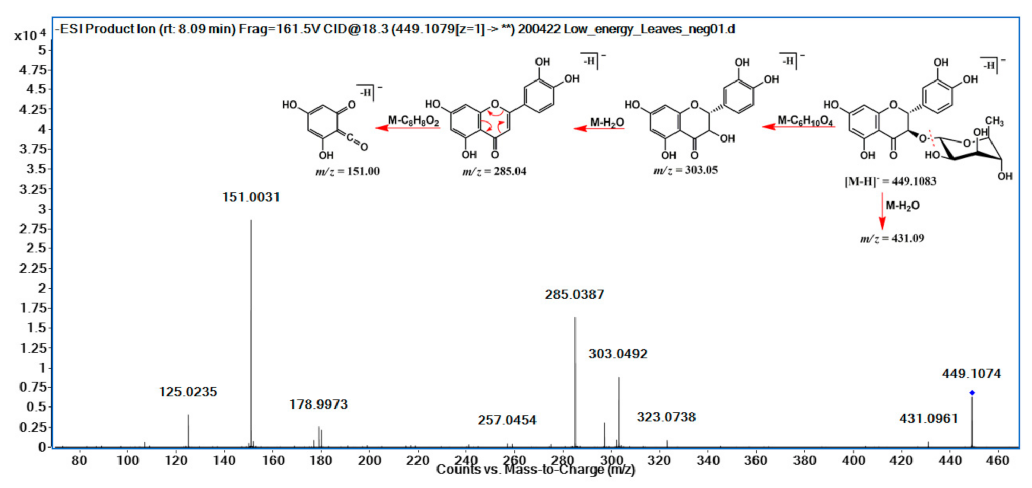
2.2. Dereplicated Compounds from A. uleanus of Methanolic Roots, Bark, Stem Bark, and Leaf Extracts of A. uleanus and the Fragmentation Proposed to the Main Compounds

The methanolic extracts of roots, bark, stem bark, and leaves from A. uleanus were analyzed using Ultra-High Performance Liquid Chromatography with Quadrupole Time-of-Flight Mass Spectrometry (UPLC-QTOF-MS/MS) to obtain MS2 spectra data of compounds. Thus, MS2 data were converted using MSConverter and processed using MassHunter and MS-DIAL software to submit them to the GNPS and METLIN libraries to create Feature-Based Molecular Networking (FBMN) (Figure 1), which was visualized using Cytoscape v.3.8.0. The MN annotation was verified using a mass accuracy error below 5 ppm, and the fragmentation mechanism was proposed to the main compound.
A purified astilbin sample (16), after being structurally characterized by 1D and 2D NMR spectroscopy data, was used as a reference in the dereplication of other similar metabolites in the cluster network (Figure 2). Initially, the fragmentation mechanism for astilbin was proposed, and then, the values of the fragment ions were compared with the data of the other nodes of the same cluster. Derivatives of flavonols have fragmentation characteristics, such as loss of the sugar molecule, elimination of water, and a Retro-Diels-Alder reaction. It was noticed that the nodes in this cluster were all purple in color (referring to the leaves), showing that all substances were main present in the plant organs (Figure 3).
Flavonolignans was another class of natural products detected in the extracts of A. uleanus. The most evident was cinchonain Ib (33), which is one of the bioactive compounds found in catuaba (Trichilia catigua) [20]. A comparison was made between the extracts from A. uleanus and T. catigua, and both showed exactly the same MS signals from the base peak chromatogram (BPC) (Figure 4), and the molecular formula was calculated under a mass error below 3 ppm to give C24H20O9, [M − H] = 451.1030. Thus, the MS2 spectra data comparison showed equal signals from the fragment mechanism (Figure 5). The fragmentation mechanism proposed is shown below with the first loss of the catechol unit (C6H6O2), then to the dehydration reaction (M-H2O) to give m/z = 323.06, and, lastly, to the Retro-Diels–Alder reaction (RDA) at the C ring to give fragment ions at m/z = 189.02. In addition, two catechol loss sequentially to give fragment ions at m/z = 231.03, then carbon monoxide loss to give 203.03 or an RDA fragmentation reaction at the C ring to give m/z = 189.02, and, lastly, carbon monoxide loss to give m/z = 162.02 (Figure 6).
The comparison of the base peak chromatogram (BPC) from the extract and stem bark of A. uleanus (A) and T. catigua (B) showed retention times that support the presence of the same substance as observed in the fragmentation profile for the same ion in their respective MS2 spectra.
The fragmentation profile observed for cinchonain Ib (Figure 6) and the fragmentation proposal supported the annotation of the other substances, as shown in Figure 7. The monitoring of the fragment ions m/z 341.07, 217.01, and 189.02 were key points to confirm the similar fragmentation profile information in the nodes of the molecular network presented in Figure 7, and the direct differences between each node helped in the proposal of the other structures.
In addition to flavonoid aglycones, O-glycosylated flavonoids, C-glycosylated flavonoids, and flavonolignans, other classes of natural products such as biflavonoids, procyanidin, triterpene, triterpenes esterified with phenylpropanoids, phenylpropanoid derivatives, coumarins, quinic acid derivatives, and benzoic acid derivatives were also identified.
Gas chromatography-mass spectrometry (GC-MS) was also used to generate data on the chemical composition of A. uleanus. The hexane partition of the leaf extract was fractionated with silica gel (adsorption chromatography), and the 9D subfraction was derivatized for analysis using GC-MS. The GC-MS data were compared with the NIST library, making it possible to identify four fatty acid esters: palmitic acid (methyl ester) (47), linoleic acid (methyl ester) (48), stearic acid (methyl ester) (49), and oleic acid (methyl ester) (50).
Then, forty-six compounds were determined using LC-MS/MS data, while four lipid derivatives were determined using GC-MS, totaling fifty compounds identified from extracts of different parts of A. uleanus. These compounds were classified with the following levels of identification: level 1 (L1): structure confirmed by the reference standard or elucidated using NMR spectroscopy, level 2a (L2a): probable structure using library spectrum match, and level 3 (L3): tentative candidates based on MS and MS2 experimental data [18,19] (Figure 8).

2.3. Chemophenetic Significance

The species of the Euphorbiaceae family have high chemical complexity and accumulate a series of metabolites from different classes of natural products.
Flavonoids and O-glycosylated flavonoids are compounds present in several genera of Euphorbiaceae, such as Alchornea [21], Chrozophora [11], Cnidoscolus [22], Croton [23], Euphorbia [24], Macaranga [25], Phyllanthus [26], Jatropha [27], Ricinus [28], Pedilanthus [29], and others. Several flavonoids, such as apigenin (1), naringenin (2), kaempferol (3), catechin/epicatechin (5), and quercitrin (15), identified in A. uleanus are present in most of the genera mentioned above. However, flavonoids eriodictyol (4) and astilbin (16) have more restricted distributions within the Euphorbiaceae family. Eriodictyol (4) is a flavonoid widely distributed in the plant kingdom; however, in the Euphorbiaceae family, it was identified only in three species of the genus Phyllanthus: P. emblica [30], P. niruri [31], and P. amarus [32]. Phyllanthus is placed in the subfamily Phyllanthoideae and has been classified in the tribe Phyllantheae, which was divided into six subtribes and 18 genera [33]. A prenylated derivative of eriodictyol has also been identified in one species of the genus Macaranga: M. triloba [34]. Astilbin (16) is present in considerable amounts in the leaves of A. uleanus. Despite having a relatively common structure, it is the second time that this glycosylated flavonoid has been identified in the Euphorbiaceae family. Until that time, this substance has only been identified in species of the genus Mallotus: M. apelta and M. metcalfianu [35].
Coincidentally, Mallotus, Macaranga, and Phyllanthus genera share chemical similarities. Macaranga and Mallotus are closely related, are large paleo(sub)tropical genera, both share very similar ecological strategies, and have similar geographical distributions and a recent common ancestry [36]. Mallotus and Phyllanthus genera have been extensively used in folk medicine in India, China, Vietnam, and other countries for thousands of years for the treatment of a broad spectrum of diseases, such as chronic hepatitis, enteritis, urinary bladder, intestinal infections, and kidney disease. Thiangthum et al., using tools such as HPLC and multivariate analysis, analyzed the similarities and differences in the compositions of compounds having antioxidant properties from 36 samples from six species of Mallotus and Phyllanthus. The results were used to compare and differentiate species from both genera [37].
One C-glycosylated flavonoid, isovitexin (20), was also detected in the extracts of A. uleanus. This class of compound is found in some genera of Euphorbiaceae, such as Aleurites [38], Croton [39], Jatropha [40], and Phyllanthus [41]. One biflavonoid was also annotated as 3′′′-O-methylfukugetin (21). Biflavonoids also occur in the Euphorbiaceae family, in the genera Euphorbia [42] and Senefelderopsis [43].
Procyanidins (22) are dimers and higher polymers formed from catechin and epicatechin molecules that occur in several plant families, including Euphorbiaceae. This class of compounds can be found in several species of the genera, such as Croton [2], Antidesma, and Phyllanthus [44].
Flavonolignans are a very rare class of natural products in Euphorbiaceae and have been identified just once in a species. Rivière et al. identified a mixture of two pairs of new diastereoisomeric flavonolignans (±)-hydnocarpin-7-O-(4″-O-(E)-coumaroyl)-β-glucopyranoside)/(±)-hydnocarpin-D-7-O-(4″-O-(E)-coumaroyl)-β-glucopyranoside) in a ratio of 2:1 in Mallotus metcalfianus [45]. The identification of this class of natural products reiterates the need for chemotaxonomy, since they are the only two records indicating the presence of flavonolignans in species of the Euphorbiaceae family.
Despite the presence of flavonolignans in Euphorbiaceae, this is the first report of cinchonain Ib (33) in the family. This bioactive compound is found in the bark of Trichilia catigua (Meliaceae). An infusion of the bark, which is called catuaba, is used in traditional Brazilian medicine as an aphrodisiac and central nervous system stimulant [20]. The families Euphorbiaceae and Meliaceae belong to the Rosids group; Meliaceae is included in the order Sapindales, while Euphorbiaceae is included in the order Malpighiales [46]. In addition to Meliaceae, cinchonain Ib (33) has already been identified in several families, such as Elaeagnaceae [47], Hypericaceae [48], Lauraceae [49], Rhizophoraceae [50], Rosaceae [51], Rubiaceae [52], Smilacaceae [53], and Theaceae [54].
Chlorogenic acid (25) and its derivatives are present in Euphorbiaceae and could be isolated from some species of Euphorbiaceae, such as Jatropha aethiopica [55], Euphorbia peplus [56], Euphorbia hirta, Phyllanthus emblica, Ricinus communis [57], Sapium insigne [58], and Croton antisyphiliticus [39]. Coumarins are relatively common in many Euphorbiaceae genera, such as Pedilanthus [59], Cnidoscolus [22], Macaranga [60], Mallotus [61], Phyllanthus [62], Jatropha [63], Euphorbia [64], and others. Four coumarins were detected in A. uleanus: aesculin (28), phyllocoumarin (29), fraxin (30), and fraxidin (31). The compounds 40, 41, and 42 were annotated as triterpenes esterified with phenylpropanoids. Triterpenes esterified with phenylpropanoids can be found in several species of the following genera: Cnidoscolus [65], Drypetes (Putranjivaceae family) [66], Glochidion (Phyllanthaceae family) [67], and Jatropha [68].

3. Materials and Methods

3.1. Plant Material

On 4 May 2017, samples of the A. uleanus species was collected at the Jarú Biological Reserve (REBIO-Jarú) in Ji-Paraná (RO), Brazil. The samples were left in a bleach solution (5%) for 5 min and then washed with running water. Fresh leaves (1029 g), bark (242 g), stem bark (147 g), and roots (123 g) were separated and dried in a circulating air oven for 15 days at 37 °C. Following this, all the materials were crushed separately in a mill. The collected plants were previously identified by a team of botanists from the Jarú Reserve. An exsiccate (number RB01331744) of this species was deposited in the herbarium of the Rio de Janeiro Botanical Garden (JBRJ), and its identification was confirmed again by Prof. Cássia Mônica Sakuragui (Institute of Biology/UFRJ/Rio de Janeiro, Brazil).

3.2. Preparation of Crude Extracts and Fractionation

Crude extracts were prepared using methanol (HPLC grade) by maceration for three times during three days. The extracts were concentrated using a reduced pressure in a rotary evaporator R-100 (Büchi, Flawil, Switzerland). The extracts from the bark (BMe), stem bark (TBMe), roots (RMe), and leaves (LMe) were subject to chromatographic separations via solid-phase extraction (SPE) using C-18 as the stationary phase and acetonitrile and ultrapure water as the mobile phases. To isolate the compounds, the crude leaves extract was resuspended in a mixture of water/methanol (1:1), followed by extraction with hexane and ethyl acetate. The compounds in the ethyl acetate fraction were separated by HPLC (see Section 3.3, Purification of compounds). For the study on dereplication, see Section 3.5 (UPLC-MS/MS analysis for molecular networking).

3.3. Purification of Compounds

The ethyl acetate fraction (204 mg) was dissolved in 1.5 mL of acetonitrile/ultrapure water solution (20:80), acidified with 1% formic acid, and subsequently, injected for semipreparative HPLC. HPLC analysis was performed on a Dionex UltiMate 3000 system (Thermo Scientific, Waltham, MA, USA). A C18 Inertsil ODS-4 column (5 µm × 6.0 × 250 mm2) was used. Ultrapure water (with 1% formic acid) and methanol (with 1% formic acid) were used as the mobile phase. Samples were separated by gradient elution under the following conditions: t = 0 min, 10% B; t = 40 min, 33% B; t = 41 min, 100% B; t = 56 min, 100% B; and t = 57 min, 10% B. The flow rate was 2.8 mL/min, and the wavelength (λ) was 280 nm. Samples corresponding to five peaks were collected and subjected to 1H-NMR analysis.

3.4. Structure Elucidation by 1H-NMR Spectroscopy

1H-NMR spectra were obtained using the 400- and 500-MHz VNMR-500 (Varian, Palo Alto, CA, USA) NMR spectrometer. The spectra were calibrated using tetramethylsilane (TMS). MestReNova software (version 6.0.2) was used to process the spectra. Coupling constants and chemical shifts were expressed in Hz and parts per million (ppm), respectively.

3.4.1. Catechin (5a)

Brownish yellow amorphous powder (1.4 mg). QTOF-MS/MS spectrum (negative ionization mode) displayed a molecular ion peak at m/z 289.0720 [M−H] (calculated for C15H14O6, 290.0790). 1H-NMR (500 MHz, CD3OD): δ 6.84 (d, J = 1.8 Hz, H-2′), δ 6.76 (d; J = 8.1 Hz, H-5′), δ 6.72 (dd; J = 8.1 and 1.8 Hz, H-6′), δ 5.85 (d; J = 2.2 Hz, H-6), 5.92 (d, J = 2.2 Hz, H-8), δ 4.56 (d; J = 7.7 Hz, 1H, H-2), δ 3.97 (dt, J = 7.7 and 5.5 Hz, H-3), δ 2.84 (dd, J = 16.0 and 5.4 Hz, H-4b), δ 2.50 (dd, J = 16.0 and 8.4 Hz, H-4a). These data are consistent with the catechin structure reported in the literature [69].

3.4.2. Epicatechin (5b)

Brownish yellow amorphous powder (1.1 mg). QTOF-MS/MS spectrum (negative ionization mode) displayed a precursor ion peak at m/z 289.0720 [M − H] (calculated for C15H14O6, 290.0790). 1H-NMR (500 MHz, CD3OD): δ 5.94 (d, J = 2.0, Hz, H-6) and 5.91 (d, J = 2.0 Hz, H-8), δ 6.97 (d, J = 1.3 Hz, H-2′), δ 6.76 (d; J = 8.1 Hz, H-5′), δ 6.80 (dd; J = 8.1 and 1.3 Hz, H-6′), δ 4.82 (br s, 1H, H-2), δ 4.18 (br s, H-3), δ 2.86 (dd, J = 16.9 and 4.6 Hz, H-4a), 2.73 (dd, J = 16.9 and 2.7 Hz, H-4b). These data are consistent with the epicatechin structure reported in the literature [70].

3.4.3. Afzelin (12)

Yellow powder (2.8 mg). QTOF-MS/MS spectrum (negative ionization mode) displayed a precursor ion peak at m/z 431.0977 [M − H] (calculated for C21H20O10, 432.1056). 1H-NMR (400 MHz, CD3OD): δ 7.77 (d, J = 8.7 Hz, H-2′ and H-6′), δ 6.38 (d, J = 1.9 Hz, H-6), δ 6.21 (d, J = 1.9 Hz, H-8), δ 6.94 (d, J = 8.7 Hz, H-3′ and H-5′), δ 5.38 (d, J = 1.5 Hz, H-1″), δ 4.22 (m, H-5”), δ 3.71 (m, H-3″), δ 3.48 (m, H-2″), δ 3.33 (m, H-4″), δ 0.92 (d; J = 5.6 Hz, H-6″). These data are consistent with the afzelin structure reported in the literature [71].

3.4.4. Quercetin (quercetin 3-O-α-L-rhamnopyranoside) (15)

Yellow powder (1.1 mg). QTOF-MS/MS spectrum (negative ionization mode) displayed a precursor ion peak at m/z 447.0926 [M − H] (calculated for C21H20O11, 448.10056). 1H-NMR (400 MHz, CD3OD): δ 7.34 (d, J = 1.9 Hz, H-2′), δ 7.31 (dd, J = 8.2 and 1.9 Hz, H-6′), δ 6.91 (d, J = 8.2, H-5′), 6.37 (d, J = 1.5 Hz, H-8), δ 6.20 (d, J = 1.5 Hz, H-6), δ 5.35 (d, J = 1.5 Hz, H-1″), δ 4.22 (m, H-2″), δ 3.42 (m, H-5″), δ 3.75 (dd, J = 9.3 and 3.2, H-3″), 3.33 (br s, H-4″), δ 0.94 (d, J = 6.1, H-6″). These data are consistent with the quercetin structure reported in the literature [72].

3.4.5. Astilbin (16)

Yellowish-green amorphous powder (7.4 mg). QTOF-MS/MS spectrum (negative ionization mode) displayed a molecular ion peak at m/z 449.1083 [M − H] (calculated for C21H22O11, 450.1162). 1H-NMR (400 MHz, CD3OD): δ 6.96 (d, J = 1.7 Hz, H-2′), δ 6.82 (m, H-5′and H-6′), δ 5.92 (d, J = 2.0 Hz, H-8), δ 5.90 (d, J = 2.0 Hz, H-6), δ 5.07 (d, J = 10.7, H-2), δ 4.58 (d, J = 10.7, H-3), δ 4.25 (m, H- 2″), δ 4.04 (d, J = 1.6 Hz, H-1″), δ 3.66 (dd, J = 9.5 and 3.3, H-3″), 3.54 (dd, J = 3.3 and 1.7, H-4″), δ 3.33 (br s, H-5″), δ 1.19 (d, J = 6.2, H-6″). These data are consistent with the astilbin structure reported in the literature [73].

3.5. UPLC-MS/MS Analysis for Molecular Networking

MS1 and MS2 analyses were performed on an Agilent 6545 Q-TOF mass spectrometer (Agilent Technologies Inc., Santa Clara, CA, USA) equipped with an electrospray ionization (ESI) source. MassHunter® workstation software (version B.08.00) was used for data acquisition and processing. The samples were analyzed in triplicate of authentic biological replicates. The chromatographic separation was carried out on an Agilent Zorbax SB-C18 column (3.0 × 50 mm) and water–acetonitrile with 0.1% formic acid (LC-MS grade). The mobile phase flowed at a rate of 0.3 mL/min after injecting 5 µL of the analytical solutions. The mass data were acquired with a positive (+) and negative (−) ion ESI source in the TOF-MS mode for the molecular ions and in the auto-MS/MS mode for the fragment ions using collision energy (Table S2, Supplementary Materials). The operating source parameters for the TOF-MS mode were as follows: capillary voltage, 2.400 KV; skimmer voltage, 65 V; fragmentor voltage, 110 V; nebulizer gas pressure, 28 psi; dry gas flow, 10 L/min; gas temperature, 300 °C; sheath gas flow, 10 L/min; sheath gas temperature, 350 °C, acquisition rate, 3 spectrum per second; and a resolution of 32,000. The mode of acquisition of auto-MS or target MS/MS and the acquisition of second-order spectra followed pre-established processes of collision energies in the form of mini-ramps of small intervals of m/z—100−300 Da, for example (Table 1). The acquired data were processed by the MassHunter® molecule features extraction software to find the compounds, and the ionized molecules ([M + H]+ and [M − H]) obtained in the TOF-MS mode were identified.

3.6. Molecular Networking Full Imaging

The molecular networking visualization (MN) was constructed using each MS2 spectrum as a node, and the connection between nodes were made by the mass difference from the precursor ion (edges); then, each individual molecular networking has a similar fragmentation profile with a minimum fragment-ion characteristic. Further, Figure 1 suggests different colors of the crude extracts of the roots (blue), bark (green), stem bark (red), leaves (purple), and blank (black). The annotation of the secondary metabolites was performed using the attempt to rationalize the fragment ions by means of fragmentation proposals applying concepts of organic chemistry, with an emphasis on acid/base mechanisms together with several databases for secondary metabolites (FooDB, PlantCyc, ChEBI, LipidMAPS, DrugBank, KNApSAcK, NANPDB, PubChem, UNPD, and METLIN). For this purpose, deconvolution of the MS2 spectra and peak alignment was performed using the MS-Dial software (http://prime.psc.riken.jp/compms/msdial/main.html). Then, the processed, deconvolved, and aligned MS2 spectra were extracted and submitted for processing in the MS-FINDER software in order to achieve a greater number of databases aimed at the annotation of the secondary metabolites Aiming to process at MS-DIAL, MS1 at ±0.02 Da and MS2 at ±0.06 Da mass tolerance, minimum peak height of 1 × 10⁴, MS/MS amplitude abundance cut-off at 30, retention time tolerance of ±0.5 min, and MS1 tolerance of ±0.02 Da were used to align the chromatogram. Thus, the processed data were exported to the .mgf format postprocessing and subjected to a feature-based molecular networking (FBMN) analysis at the global natural product social molecular networking (GNPS) platform (https://gnps.ucsd.edu/). The confidence annotated metabolites are presented in this site: https://gnps.ucsd.edu/ProteoSAFe/status.jsp?task=deca49b0f49d4af087be4e852e9a4b91). The MN was processed using Cytoscape version 3.8.0 software (https://cytoscape.org/) to construct the molecular network. Parent ions for all the extracts were different. Besides, 5 fragment ions were used to construct the molecular network, and 4 fragment ions were used for library annotation.

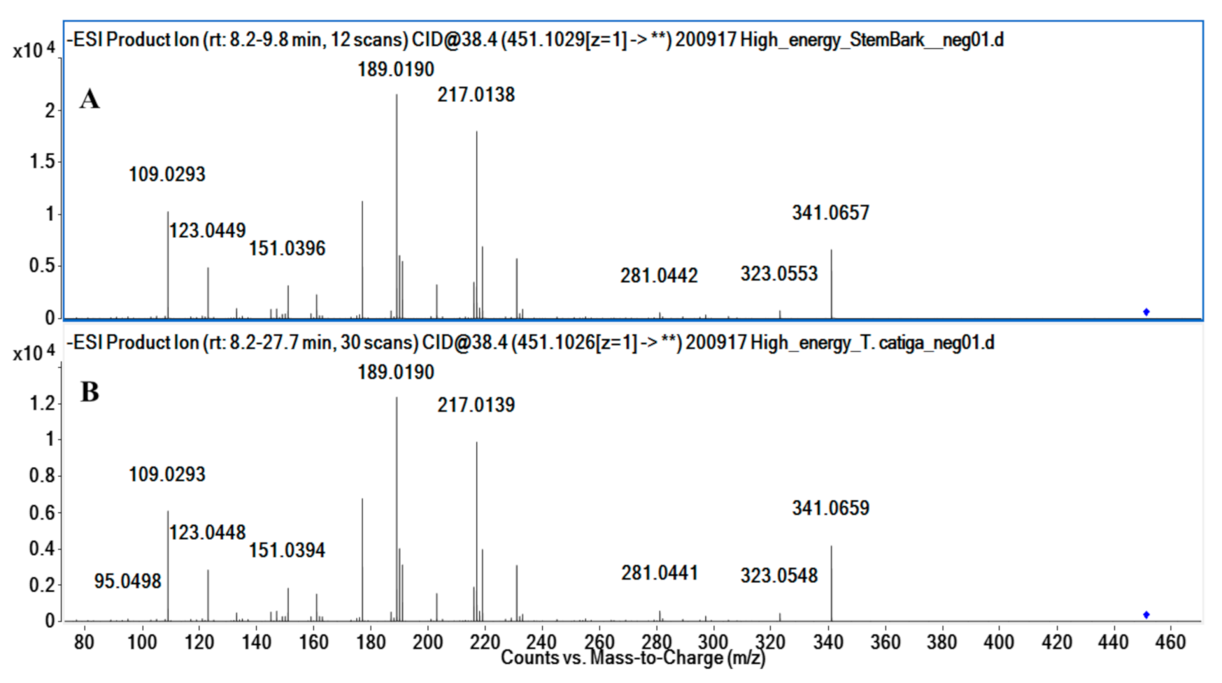
3.7. Comparison between A. uleanus Extracts and Cinchonain Ib Pattern from Trichilia catigua (catuaba)

A commercial sample of catuaba tea (Trichilia catigua, brand “Chá do Brasil”®) was used to prepare the extracts following the protocol described by Beltrame et al. (2006) [20]. A. uleanus extract and catuaba standard extract containing cinchonain Ib were subjected to the same chromatographic conditions, ionization energy, and collision energy. The chromatographic separation was performed on a reverse column using the Agilent 1200 system. The column temperature and injection volume were 33 °C and 3 µL, respectively. The proportion of solvent B (acetonitrile) was linearly varied as follows: 0–4 min, 5–15%; 4–15 min, 15–60%; 15–24 min, 60–100%; 24–27 min, 100% (isocratic elution); 27–27.50 min, 100–5%; and 27.50–30 min, 5% (isocratic elution). The ESI source conditions were supported by the mass ranges 200–1500 Da to MS1 and 70–1500 Da to MS2 data. The other parameters were as follows: capillary voltage, 2.5 kV; nozzle voltage, 0 (zero); fragmentor voltage, 125 V; skimmer voltage, 65 V; octupole RF peak, 750 V; gas temperature, 300 °C; gas flow, 12 L/min; nebulizer pressure, 35 psi; sheath gas temp, 350 °C; and sheath gas flow, 10 L/min. MS2 spectra acquisition was performed under the following collision energies: 100–300 Da (30–35 eV), 300–500 Da (35–40 eV), 500–700 Da (40–45 eV), 70–1000 Da (45–50 eV), and 100–1500 Da (50–60 eV).

3.8. GC-MS Analysis

Sample 9D (58 mg), obtained from the hexane fraction of A. uleanus leaves, was dissolved in 4 mL of 0.5-N NaOH solution in methanol and subjected to methylation according to the procedure described by Ichihara and Fukubayashi (2010). This derivatized form of sample 9D was analyzed on a GC-MS chromatograph (Shimadzu, GCMS-QP2010 model, Kyoto, Japan), using a DB-5 MS column (30 m × 0.25 mm × 0.25 µm). The chromatography was conducted in the split injection mode, with a column oven temperature of 100 °C and injection temperature of 225 °C. Column flow rate was 1 mL/min, equilibration time was 3.0 min, and helium was used as the carrier gas. The results were compared with the NIST library database, and it was possible to identify four esters (4750) [74].

4. Conclusions

The chemical composition of A. uleanus was investigated using both phytochemical and dereplication approaches. Five major compounds were isolated from the leaf extracts and identified using NMR and MS: catechin/epicatechin (5a/5b), afzelin (12), quercetin 3-O-α-L-rhamnopyranoside (15), and astilbin (16). All the compounds were used as reference standards (level 1 of confirmation, L1) and were important in confirming the accuracy of the MS/MS data in the dereplication study.
UPLC-Q-TOF-MS/MS and GC-MS analyses provided abundant information for the genus Anomalocalyx. It was the first time that the chemistry of the genus was described in the literature. Dereplication was the main investigative process adopted to avoid the re-isolation of the compounds already described, revealing the metabolic pathways present in A. uleanus. The use of the molecular networking approach is extremely important and powerful as a strategy for analyzing clusters of the main compounds. It was possible to establish a structural relationship between these known substances and other structures present in the cluster by analyzing the fragmentation pattern. Forty-six compounds were annotated from the LC-MS/MS data and four from GC-MS, totaling fifty compounds. These compounds belong to the primary metabolism and several classes of natural products, such as flavonoids, flavonoids O-glycosides, flavonoids C-glycosides, biflavonoids, procyanidin, triterpene, triterpenes esterified with phenylpropanoids, phenylpropanoid derivatives, flavonolignans, coumarins, quinic acid derivatives, and benzoic acid derivatives.
One of the compounds annotated in the dereplication study was flavonolignan cinchonain Ib (33). This compound was obtained from Trichilia catigua bark extract, and it was used as a standard to confirm its presence and derivatives in A. uleanus barks by comparing MS spectral data. This is the first time that cinchonain Ib (33) and derivatives were detected in a species of the Euphorbiaceae family.
All the information obtained about the metabolism of the genus Anomalocalyx will contribute greatly to the chemosystemic and chemophenetic knowledge of the genus and suggest the need for its pharmacological potential, which has never been investigated, to be investigated.

Supplementary Materials

The following are available online: Scheme S1.-Molecular networking fractions on negative polarity, Table S2. Collision energy, Fragmentation mechanism propose to dereplicated compounds, Mass spectrometry data (MS1 and MS2)-Figures S1–S100.

Author Contributions

Conceptualization, F.C. and J.A.G.d.B.; methodology, F.C., J.A.G.d.B., M.F.d.G.F.d.S., A.J.R.d.S. and L.d.S.P.; formal analysis, F.C., J.A.G.d.B., C.F.C., A.J.R.d.S. and L.d.S.P.; investigation, F.C., J.A.G.d.B., C.F.C., A.J.R.d.S. and L.d.S.P.; resources, F.C., M.F.d.G.F.d.S. and A.J.R.d.S.; data curation, F.C., J.A.G.d.B. and L.d.S.P.; validation, J.A.G.d.B., L.d.S.P., A.J.R.d.S. and F.C.; writing—original draft preparation, F.C., J.A.G.d.B., A.J.R.d.S., M.F.d.G.F.d.S. and L.d.S.P.; writing—review and editing, F.C., J.A.G.d.B., A.J.R.d.S., M.F.d.G.F.d.S. and L.d.S.P.; supervision, M.F.d.G.F.d.S., A.J.R.d.S. and F.C.; project administration, F.C.; and funding acquisition, M.F.d.G.F.d.S., A.J.R.d.S. and F.C. All authors have read and agreed to the published version of the manuscript.

Funding

This research was supported by FAPERJ, CAPES, and FAPESP. J.A.G.B. thanks CAPES (DINTER-IFRO-UFRJ Project, support number 1887/2017) for the PhD scholarship. L.S.P. thanks FAPESP (15/26320-7) for the PhD scholarship. C.F.C. thanks FAPERJ for the undergraduate scholarship (E-26/202.206/2017).

Institutional Review Board Statement

Not applicable.

Informed Consent Statement

Not applicable.

Data Availability Statement

The MS/MS data presented in this study are available in the Supporting Information.

Acknowledgments

The authors are grateful to Professor Cássia Mônica Sakuragui (Institute of Biology/UFRJ) for identifying the botanical material and Jarú Biological Reserve (Ji-Paraná, Rondônia, Brazil) for providing the facilities to collect the plant material. The authors thank SisGen (http://sisgen.gov.br) for the authorization to access the samples from Brazilian Genetic Heritage (AE0D819).

Conflicts of Interest

The authors declare no conflict of interest.

References

  1. Wurdack, K.J.; Davis, C.C. Malpighiales phylogenetics: Gaining ground on one of the most recalcitrant clades in the angiosperm tree of life. Am. J. Bot. 2009, 96, 1551–1570. [Google Scholar] [CrossRef] [PubMed]
  2. Salatino, A.; Salatino, M.L.F.; Negri, G. Traditional uses, chemistry and pharmacology of Croton species (Euphorbiaceae). J. Braz. Chem. Soc. 2007, 18, 11–33. [Google Scholar] [CrossRef]
  3. Secco, R.S. Flora da Reserva Ducke, Amazonas, Brasil. Rodriguésia 2005, 56, 143–168. [Google Scholar] [CrossRef] [Green Version]
  4. Tokuoka, T. Molecular phylogenetic analysis of Euphorbiaceae sensu stricto based on plastid and nuclear DNA sequences and ovule and seed character evolution. J. Plant Res. 2007, 120, 511–522. [Google Scholar] [CrossRef]
  5. Rivière, C.; Van Nguyen, T.H.; Nam, N.H.; Dejaegher, B.; Tistaert, C.; Kiem, P.V.; Heyden, Y.v.; Minh, C.V.; Quetin-Leclercq, J. N-methyl-5-carboxamide-2-pyridone from Mallotus barbatus: A chemosystematic marker of the Euphorbiaceae genus Mallotus. Biochem. Syst. Ecol. 2012, 44, 212–215. [Google Scholar] [CrossRef]
  6. Zidorn, C. Plant chemophenetics—A new term for plant chemosystematics/plant chemotaxonomy in the macro-molecular era. Phytochemistry 2019, 163, 147–148. [Google Scholar] [CrossRef]
  7. Suárez, A.I. Chemical constituents from Senefelderopsis croizatii Steyerm. Biochem. Syst. Ecol. 2020, 90, 104016. [Google Scholar] [CrossRef]
  8. Noori, M.; Chehreghani, A.; Kaveh, M. Flavonoids of 17 species ofEuphorbia(Euphorbiaceae) in Iran. Toxicol. Environ. Chem. 2009, 91, 631–641. [Google Scholar] [CrossRef]
  9. Elshamy, A.I.; Mohamed, T.A.; Al-Rowaily, S.L.; Abd-Elgawad, A.M.; Abd-Elgawad, A.M.; Shahat, A.A.; Hegazy, M.E.F. Euphosantianane E–G: Three New Premyrsinane Type Diterpenoids from Euphorbia sanctae-catharinae with Contribution to Chemotaxonomy. Molecules 2019, 24, 2412. [Google Scholar] [CrossRef] [Green Version]
  10. Lan, Y.-H.; Yen, C.-H.; Leu, Y.-L. Chemical constituents from the aerial parts of Euphorbia formosana Hayata and their chemotaxonomic significance. Biochem. Syst. Ecol. 2020, 88, 103967. [Google Scholar] [CrossRef]
  11. Marzouk, M.M.; Hussein, S.R.; Kassem, M.E.; Kawashty, S.A.; El Negoumy, S.I. Phytochemical constituents and chemosystematic significance of Chrozophora tinctoria (L.) Raf. Nat. Prod. Res. 2016, 30, 1537–1541. [Google Scholar] [CrossRef]
  12. Rossi, D.; Guerrini, A.; Maietti, S.; Bruni, R.; Paganetto, G.; Poli, F.; Scalvenzi, L.; Radice, M.; Saro, K.; Sacchetti, G. Chemical fingerprinting and bioactivity of Amazonian Ecuador Croton lechleri Müll. Arg. (Euphorbiaceae) stem bark essential oil: A new functional food ingredient? Food Chem. 2011, 126, 837–848. [Google Scholar] [CrossRef]
  13. Cui, G.; Tan, R. Lignans and tannins from Alchornea davidii (Euphorbiaceae) and their chemotaxonomic significance. Biochem. Syst. Ecol. 2004, 32, 99–102. [Google Scholar] [CrossRef]
  14. Al Muqarrabun, L.R.; Ahmat, N.; Aris, S.R.S.; Norizan, N.; Shamsulrijal, N.; Yusof, F.Z.; Suratman, M.N.; Yusof, M.I.M.; Salim, F. A new triterpenoid from Sapium baccatum (Euphorbiaceae). Nat. Prod. Res. 2014, 28, 1003–1009. [Google Scholar] [CrossRef] [PubMed]
  15. Machado, D.N.M.; Palmeira, S.F., Jr.; Conserva, L.M.; Lemos, R.P.L. Quinoline alkaloids from Sebastiania corniculata (Euphorbiaceae). Biochem. Syst. Ecol. 2005, 33, 555–558. [Google Scholar] [CrossRef]
  16. Rutz, A.; Dounoue-Kubo, M.; Ollivier, S.; Bisson, J.; Bagheri, M.; Saesong, T.; Ebrahimi, S.N.; Ingkaninan, K.; Wolfender, J.-L.; Allard, P.-M. Taxonomically Informed Scoring Enhances Confidence in Natural Products Annotation. Front. Plant Sci. 2019, 10, 1329. [Google Scholar] [CrossRef] [PubMed] [Green Version]
  17. Secco, R.D.S.; Cordeiro, I.; Senna-Vale, L.; Sales, M.F.; Lima, L.R.; Medeiros, D.; Haiad, B.S.; Oliveira, A.S.; Caruzo, M.B.R.; Carneiro-Torres, D.; et al. An overview of recent taxonomic studies on Euphorbiaceae s.l. in Brazil. Rodriguésia 2012, 63, 227–242. [Google Scholar] [CrossRef] [Green Version]
  18. Rivera-Mondragón, A.; Tuenter, E.; Ortiz, O.; Sakavitsi, M.E.; Nikou, T.; Halabalaki, M.; Caballero-George, C.; Apers, S.; Pieters, L.; Foubert, K. UPLC-MS/MS-based molecular networking and NMR structural determination for the untargeted phytochemical characterization of the fruit of Crescentia cujete (Bignoniaceae). Phytochemistry 2020, 177, 112438. [Google Scholar] [CrossRef]
  19. Schymanski, E.L.; Jeon, J.; Gulde, R.; Fenner, K.; Ruff, M.; Singer, H.P.; Hollender, J. Identifying Small Molecules via High Resolution Mass Spectrometry: Communicating Confidence. Environ. Sci. Technol. 2014, 48, 2097–2098. [Google Scholar] [CrossRef]
  20. Beltrame, F.L.; Filho, E.R.; Barros, F.A.P.; Cortez, D.A.; Cass, Q.B. A validated higher-performance liquid chromatography method for quantification of cinchonain Ib in bark and phytopharmaceuticals of Trichilia catigua used as Catuaba. J. Chromatogr. A 2006, 1119, 257–263. [Google Scholar] [CrossRef]
  21. Okoye, F.; Osadebe, P. A new anti-inflammatory flavonol glycoside from Alchornea floribunda leaves. Nat. Prod. Res. 2010, 24, 266–273. [Google Scholar] [CrossRef]
  22. Yuan, W.; Li, S.; Ownby, S.; Zhang, Z.; Wang, P.; Zhang, W.; Beasley, R. Flavonoids, Coumarins and Triterpenes from the Aerial Parts of Cnidoscolus texanus. Planta Med. 2007, 73, 1304–1308. [Google Scholar] [CrossRef] [PubMed] [Green Version]
  23. Zou, G.-A.; Su, Z.-H.; Zhang, H.-W.; Wang, Y.; Yang, J.-S.; Zou, Z.-M. Flavonoids from the Stems of Croton caudatus Geisel. var. tomentosus Hook. Molecules 2010, 15, 1097–1102. [Google Scholar] [CrossRef] [Green Version]
  24. Rao, C.V.; Gupta, S.S.; Azmi, L.; Mohapatra, P.K. Flavonoids from whole plant of Euphorbia hirta and their evaluation against experimentally induced gastroesophageal reflux disease in rats. Pharmacogn. Mag. 2017, 13, S127–S134. [Google Scholar] [CrossRef]
  25. Yang, D.-S.; Li, Z.-L.; Peng, W.-B.; Yang, Y.-P.; Wang, X.; Liu, K.-C.; Li, X.-L.; Xiao, W.-L. Three new prenylated flavonoids from Macaranga denticulata and their anticancer effects. Fitoterapia 2015, 103, 165–170. [Google Scholar] [CrossRef]
  26. Navarro-Hoyos, M.; Moreira, I.; Arnaez, E.; Quesada, S.; Azofeifa, G.; Vargas, F.; Alvarado, D.; Chen, P. Flavonoids and Ellagitannins Characterization, Antioxidant and Cytotoxic Activities of Phyllanthus acuminatus Vahl. Plants 2017, 6, 62. [Google Scholar] [CrossRef] [PubMed] [Green Version]
  27. Félix-Silva, J.; Gomes, J.A.; Fernandes, J.M.; Moura, A.K.; De Menezes, Y.A.S.; Dos Santos, E.C.G.; Tambourgi, D.V.; Da Silva-Júnior, A.A.; Zucolotto, S.M.; Fernandes-Pedrosa, M.D.F. Comparison of two Jatropha species (Euphorbiaceae) used popularly to treat snakebites in Northeastern Brazil: Chemical profile, inhibitory activity against Bothrops erythromelas venom and antibacterial activity. J. Ethnopharmacol. 2018, 213, 12–20. [Google Scholar] [CrossRef]
  28. Upasani, S.M.; Kotkar, H.M.; Mendki, P.S.; Maheshwari, V. Partial characterization and insecticidal properties ofRicinus communis L foliage flavonoids. Pest Manag. Sci. 2003, 59, 1349–1354. [Google Scholar] [CrossRef] [PubMed]
  29. Abreu, P.J.M.; Matthew, S.; González, T.; Vaníčková, L.; Costa, D.; Gomes, A.; Segundo, M.A.; Fernandes, E. Isolation and identification of antioxidants from Pedilanthus tithymaloides. J. Nat. Med. 2007, 62, 67–70. [Google Scholar] [CrossRef] [PubMed]
  30. Sharma, S.; Kapoor, A.; Aslam, M. Phytochemical constituents from flowering twigs of Emblica officinalis Gaertn. Oriental J. Chem. 2007, 23, 695–700. [Google Scholar]
  31. Masrur, H.; Ulfa, A.; Ardiansyah, R. Pharmacopore modeling and molecular docking studies on Phyllanthus niruri as a target for diabetes mellitus. Aust. J. Basic Appl. Sci. 2015, 9, 389–395. [Google Scholar]
  32. Kumar, S.; Chandra, P.; Bajpai, V.; Singh, A.; Srivastava, M.; Mishra, D.; Kumar, B. Rapid qualitative and quantitative analysis of bioactive compounds from Phyllanthus amarus using LC/MS/MS techniques. Ind. Crop. Prod. 2015, 69, 143–152. [Google Scholar] [CrossRef]
  33. Kathriarachchi, H.-S.; Samuel, R.; Hoffmann, P.; Mlinarec, J.; Wurdack, K.J.; Ralimanana, H.; Stuessy, T.F.; Chase, M.W. Phylogenetics of tribe Phyllantheae (Phyllanthaceae; Euphorbiaceae sensu lato) based on nrITS and plastid matK DNA sequence data. Am. J. Bot. 2006, 93, 637–655. [Google Scholar] [CrossRef] [PubMed]
  34. Zakaria, I.; Ahmat, N.; Jaafar, F.M.; Widyawaruyanti, A. Flavonoids with antiplasmodial and cytotoxic activities of Macaranga triloba. Fitoterapia 2012, 83, 968–972. [Google Scholar] [CrossRef]
  35. Rivière, C.; Hong, V.N.T.; Hong, Q.T.; Chataigné, G.; Hoai, N.N.; Dejaegher, B.; Tistaert, C.; Kim, T.N.T.; Heyden, Y.V.; Van, M.C.; et al. Mallotus species from Vietnamese mountainous areas: Phytochemistry and pharmacological activities. Phytochem. Rev. 2009, 9, 217–253. [Google Scholar] [CrossRef]
  36. Van Welzen, P.C.; Strijk, J.S.; Cittert, J.H.A.V.K.-V.; Nucete, M.; Merckx, V.S.F.T. Dated Phylogenies of the Sister Genera Macaranga and Mallotus (Euphorbiaceae): Congruence in Historical Biogeographic Patterns? PLoS ONE 2014, 9, e85713. [Google Scholar] [CrossRef]
  37. Thiangthum, S.; Dejaegher, B.; Goodarzi, M.; Tistaert, C.; Gordien, A.; Hoai, N.N.; Van, M.C.; Quetin-Leclercq, J.; Suntornsuk, L.; Heyden, Y.V. Potentially antioxidant compounds indicated from Mallotus and Phyllanthus species fingerprints. J. Chromatogr. B 2012, 910, 114–121. [Google Scholar] [CrossRef] [PubMed]
  38. Meyre-Silva, C.; Yunes, R.; Santos, A.; Magro, J.; Monache, F.D.; Odone-Filho, V. Isolation of aC-Glycoside Flavonoid with Antinociceptive Action fromAleurites moluccanaLeaves. Planta Med. 1999, 65, 293–294. [Google Scholar] [CrossRef]
  39. Dos Reis, G.O.; Vicente, G.; De Carvalho, F.K.; Heller, M.; Micke, G.A.; Pizzolatti, M.G.; Fröde, T.S. Croton antisyphiliticus Mart. attenuates the inflammatory response to carrageenan-induced pleurisy in mice. Inflammopharmacology 2013, 22, 115–126. [Google Scholar] [CrossRef]
  40. Pilon, A.C.; Carneiro, R.L.; Neto, F.C.; Bolzani, V.S.; Castro-Gamboa, I. Interval Multivariate Curve Resolution in the Dereplication of HPLC-DAD Data from Jatropha gossypifolia. Phytochem. Anal. 2013, 24, 401–406. [Google Scholar] [CrossRef]
  41. Francioso, A.; Franke, K.; Villani, C.; Mosca, L.; D’Erme, M.; Frischbutter, S.; Brandt, W.; Sanchez-Lamar, A.; Wessjohann, L.A.A. Insights into the Phytochemistry of the Cuban Endemic Medicinal Plant Phyllanthus orbicularis: Fideloside, a Novel Bioactive 8-C-glycosyl 2,3-Dihydroflavonol. Molecules 2019, 24, 2855. [Google Scholar] [CrossRef] [Green Version]
  42. Yang, A.M.; Yu, H.T.; Liu, J.L.; Shi, X.L.; Men, Y.; Wu, R.; Guo, W.J. Biflavonoids from Euphorbia altotibetica. Chem. Nat. Compd. 2015, 51, 1162–1163. [Google Scholar] [CrossRef]
  43. Canelón, D.J.; Compagnone, R.S.; Castillo, A.; Suarez, A.I. Chemical constituents from Senefelderopsis chiribiquetensis. Biochem. Syst. Ecol. 2005, 33, 1303–1306. [Google Scholar] [CrossRef]
  44. Álvarez, Á.L.; Del Barrio, G.; Kouri, V.; Martínez, P.A.; Suarez, B.; Parra, F. In vitro anti-herpetic activity of an aqueous extract from the plant Phyllanthus orbicularis. Phytomedicine 2009, 16, 960–966. [Google Scholar] [CrossRef] [Green Version]
  45. Rivière, C.; Hong, V.N.T.; Pieters, L.; Dejaegher, B.; Heyden, Y.V.; Van, M.C.; Quetin-Leclercq, J. Polyphenols isolated from antiradical extracts of Mallotus metcalfianus. Phytochemistry 2009, 70, 86–94. [Google Scholar] [CrossRef] [PubMed]
  46. Simpson, M.G. Diversity and Classification of Flowering Plants: Eudicots. In Plant Systematics; Elsevier BV: Amsterdam, The Netherlands, 2010; pp. 275–448. [Google Scholar]
  47. Ma, Q.-G.; Wei, R.-R.; Shang, D.-L.; Sang, Z.-P.; Dong, J.-H. Structurally Diverse Flavonolignans with Immunosuppressive and Neuroprotective Activities from the Fruits of Hippophae rhamnoides L. J. Agric. Food Chem. 2020, 68, 6564–6575. [Google Scholar] [CrossRef] [PubMed]
  48. Yan, X.-T.; An, Z.; Huangfu, Y.; Zhang, Y.-T.; Li, C.-H.; Chen, X.; Liu, P.-L.; Gao, J.-M. Polycyclic polyprenylated acylphloroglucinol and phenolic metabolites from the aerial parts of Hypericum elatoides and their neuroprotective and anti-neuroinflammatory activities. Phytochemistry 2019, 159, 65–74. [Google Scholar] [CrossRef]
  49. Tang, W.; Lu, W.; Cao, X.; Zhang, Y.; Zhang, H.; Lv, X.; Li, J. Two New Dihydrostilbenoid Glycosides Isolated from the Leaves of Litsea coreana and their Anti-inflammatory Activity. Nat. Prod. Commun. 2013, 8, 479–480. [Google Scholar] [CrossRef] [PubMed] [Green Version]
  50. Andrade-Cetto, A.; Escandón-Rivera, S.M.; Torres-Valle, G.M.; Quijano, L. Phytochemical composition and chronic hypoglycemic effect of Rhizophora mangle cortex on STZ-NA-induced diabetic rats. Rev. Bras. Farm. 2017, 27, 744–750. [Google Scholar] [CrossRef]
  51. Bijttebier, S.; Van Der Auwera, A.; Voorspoels, S.; Noten, B.; Hermans, N.; Pieters, L.; Apers, S. A First Step in the Quest for the Active Constituents in Filipendula ulmaria (Meadowsweet): Comprehensive Phytochemical Identification by Liquid Chromatography Coupled to Quadrupole-Orbitrap Mass Spectrometry. Planta Medica 2016, 82, 559–572. [Google Scholar] [CrossRef] [Green Version]
  52. Wang, Y.; Fang, Y.-D.; Su, J.-L.; Li, R.-S.; Wang, F.; Wang, K. Chemical constituents of Uncaria rhynchophylloides How and their chemotaxonomic significance. Biochem. Syst. Ecol. 2020, 91, 104051. [Google Scholar] [CrossRef]
  53. He, L.; Zhang, Z.; Liu, Y.; Chen, D.; Yuan, M.; Dong, G.; Luo, P.; Yan, Z. Rapid discrimination of raw and sulfur-fumigated Smilax glabra based on chemical profiles by UHPLC-QTOF-MS/MS coupled with multivariate statistical analysis. Food Res. Int. 2018, 108, 226–236. [Google Scholar] [CrossRef]
  54. Zhou, Z.-H.; Zhang, Y.-J.; Xu, A.M.; Yang, C.-R. Puerins A and B, Two New 8-C Substituted Flavan-3-ols from Pu-er Tea. J. Agric. Food Chem. 2005, 53, 8614–8617. [Google Scholar] [CrossRef]
  55. Gamiotea-Turro, D.; Camaforte, N.A.; Valerino-Diaz, A.B.; Nuñez, Y.O.; Rinaldo, D.; Dokkedal, A.L.; Bosqueiro, J.R.; Dos Santos, L.C. Qualitative and Quantitative Analysis of Ethanolic Extract and Phenolic Fraction of Jatropha aethiopica (Euphorbiaceae) Leaves and Their Hypoglycemic Potential. J. Agric. Food Chem. 2018, 66, 1419–1427. [Google Scholar] [CrossRef]
  56. Frezza, C.; Venditti, A.; Sciubba, F.; Tomai, P.; Antonetti, M.; Franceschin, M.; Di Cocco, M.E.; Gentili, A.; Delfini, M.; Serafini, M.; et al. Phytochemical profile of Euphorbia peplus L. collected in Central Italy and NMR semi-quantitative analysis of the diterpenoid fraction. J. Pharm. Biomed. Anal. 2018, 160, 152–159. [Google Scholar] [CrossRef] [PubMed]
  57. Chaowuttikul, C.; Palanuvej, C.; Ruangrungsi, N. Quantification of chlorogenic acid, rosmarinic acid, and caffeic acid contents in selected Thai medicinal plants using RP-HPLC-DAD. Braz. J. Pharm. Sci. 2020, 56, 56. [Google Scholar] [CrossRef] [Green Version]
  58. Devkota, H.P.; Basnet, P.; Yahara, S. Diterpene esters and phenolic compounds from Sapium insigne (ROYLE) BENTH. ex HOOK. fil. Chem. Pharm. Bull. 2009, 57, 1289–1291. [Google Scholar] [CrossRef] [PubMed] [Green Version]
  59. Sandjo, L.P.; Foster, A.J.; Rheinheimer, J.; Anke, H.; Opatz, T.; Thines, E. Coumarin derivatives from Pedilanthus tithymaloides as inhibitors of conidial germination in Magnaporthe oryzae. Tetrahedron Lett. 2012, 53, 2153–2156. [Google Scholar] [CrossRef]
  60. Darmawan, A.; Kosela, S.; Kardono, L.B.S.; Syah, Y.M. Scopoletin, a coumarin derivative compound isolated from Macaranga gigantifolia Merr. J. Appl. Pharm. Sci. 2012, 2, 175–177. [Google Scholar]
  61. Ma, J.; Jones, S.H.; Hecht, S.M. A coumarin from Mallotus resinosus that mediates DNA cleavage. J. Nat. Prod. 2004, 67, 1614–1616. [Google Scholar] [CrossRef] [PubMed]
  62. Kaur, N.; Kaur, B.; Sirhindi, G. Phytochemistry and Pharmacology ofPhyllanthus niruriL.: A Review. Phytother. Res. 2017, 31, 980–1004. [Google Scholar] [CrossRef]
  63. Cavalcante, N.B.; Santos, A.D.D.C.; Almeida, J.R.G.D.S. The genus Jatropha (Euphorbiaceae): A review on secondary chemical metabolites and biological aspects. Chem. Interact. 2020, 318, 108976. [Google Scholar] [CrossRef] [PubMed]
  64. Masamoto, Y.; Ando, H.; Murata, Y.; Shimoishi, Y.; Tada, M.; Takahata, K. Mushroom Tyrosinase Inhibitory Activity of Esculetin Isolated from Seeds ofEuphorbia lathyrisL. Biosci. Biotechnol. Biochem. 2003, 67, 631–634. [Google Scholar] [CrossRef] [PubMed]
  65. De Lemos, T.L.; Silveira, E.R.; Oliveira, M.F.; Braz-Filho, R.; Hufford, C.D. Terpenoids from Cnidoscolus phyllacanthus Pax et Hoff. J. Braz. Chem. Soc. 1991, 2, 105–110. [Google Scholar] [CrossRef]
  66. Awanchiri, S.S.; Trinh-Van-Dufat, H.; Shirri, J.C.; Dongfack, M.D.J.; Nguenang, G.M.; Boutefnouchet, S.; Fomum, Z.T.; Seguin, E.; Vérité, P.; Tillequin, F.; et al. Triterpenoids with antimicrobial activity from Drypetes inaequalis. Phytochemistry 2009, 70, 419–423. [Google Scholar] [CrossRef] [PubMed]
  67. Liu, X.-Q.; Huang, H.-L.; Yao, M.-J.; Han, G.-T.; Liu, N.; Yuan, J.-C.; Yuan, C.-S. Oleanane-Type Triterpenoids from Glochidion assamicum. Helvetica Chim. Acta 2011, 94, 2264–2271. [Google Scholar] [CrossRef]
  68. García, A.; Delgado, G.; García-Zepeda, E.A. Uncommon Sesquiterpenoids and New Triterpenoids fromJatropha neopauciflora (Euphorbiaceae). Helvetica Chim. Acta 2006, 89, 16–29. [Google Scholar] [CrossRef]
  69. Ayres, M.C.C.; Chaves, M.H.; Rinaldo, D.; Vilegas, W.; Júnior, G.M.V. Constituintes químicos e atividade antioxidante de extratos das folhas de Terminalia fagifolia Mart. et Zucc. Química Nova 2009, 32, 1509–1512. [Google Scholar] [CrossRef]
  70. Shahat, A.A. Procyanidins from Adansonia digitata. Pharm. Biol. 2006, 44, 445–450. [Google Scholar] [CrossRef]
  71. Santana, J.S.; Sartorelli, P.; Lago, J.H.G.; Matsuo, A.L. Isolamento e avaliação do potencial citotóxico de derivados fenólicos de Schinus terebinthifolius Raddi (Anacardiaceae). Química Nova 2012, 35, 2245–2248. [Google Scholar] [CrossRef] [Green Version]
  72. Isidoro, M.M.; das Graças Fernandes da Silva, M.F.; Fernandes, J.B.; Vieira, P.C.; Arruda, A.C.; da Cruz Silva, S. Fitoquímica e quimiossistemática de Euxylophora paraensis (Rutaceae). Química Nova 2012, 35, 2119–2124. [Google Scholar] [CrossRef] [Green Version]
  73. Huang, H.; Cheng, Z.; Shi, H.; Xin, W.; Wang, T.T.Y.; Yu, L. (Lucy) Isolation and Characterization of Two Flavonoids, Engeletin and Astilbin, from the Leaves of Engelhardia roxburghiana and Their Potential Anti-inflammatory Properties. J. Agric. Food Chem. 2011, 59, 4562–4569. [Google Scholar] [CrossRef] [PubMed]
  74. Ichihara, K.; Fukubayashi, Y. Preparation of fatty acid methyl esters for gas-liquid chromatography. J. Lipid Res. 2010, 51, 635–640. [Google Scholar] [CrossRef] [PubMed] [Green Version]
Figure 1. Molecular networking of different methanolic extracts from Anomalocalyx uleanus with MS2 data obtained in the negative mode and clustered by the Cytoscape 3.80 software and emphasizing the main compound annotations obtained from the GNPS and METLIN libraries. Color nodes: roots (blue), bark (green), stem bark (red), leaves (purple), and blank (black).
Molecules 26 00925 g001
Figure 2. Astilbin (16) molecular network: molecular family of flavonol glycosides and phenolic compounds from a methanolic extract of A. uleanus. Color nodes: roots (blue), bark (green), stem bark (red), leaves (purple), and blank (black).
Molecules 26 00925 g002
Figure 3. Proposed fragmentation mechanism of flavonoid O-glycoside astilbin (16).
Molecules 26 00925 g003
Figure 4. Extracted ion chromatogram (EIC) of [M − H] = 451.1029 for cinchonain Ib (33) from stem bark (A) and the crude extract of Trichilia catigua (B).
Molecules 26 00925 g004
Figure 5. MS2 spectra of [M − H] = 451.1029 for cinchonain Ib (33) from the stem bark of A. uleanus (A) and crude extract of T. catigua (B) at the same collision energy.
Molecules 26 00925 g005
Figure 6. Proposed fragmentation mechanism of cinchonain Ib (33) from the A. uleanus and T. catigua crude extracts.
Molecules 26 00925 g006
Figure 7. Molecular network of the molecular family of cinchonain Ib (33) and derivatives from the methanolic extract of A. uleanus. Color nodes: roots (blue), bark (green), stem bark (red), leaves (purple), and blank (black).
Molecules 26 00925 g007
Figure 8. Dereplicated compounds from A. uleanus leaves, roots, bark, and stem bark extracts.
Molecules 26 00925 g008
Table 1. Compounds detected in the Anomalocalyx uleanus methanolic extracts by UPLC-Q-TOF-MS/MS (compounds 146) and gas chromatography-mass spectrometry (GC-MS) (4750). Level of confirmation: Level 1 (L1): structure confirmed by reference standard or structure elucidation by NMR spectroscopy, level 2a (L2a): probable structure by library spectrum match, and level 3 (L3): tentative candidates based on MS and MS2 experimental data [18,19].
No.AnnotationRTFormulaIdentification ConfidenceMSError (ppm)MS/MS
Flavonoids
1apigenin11.67C15H10O5L2a269.0451 [M − H]0.4225.05; 201.06; 181.07; 151.00; 117.04
2naringenin11.63C15H12O5L2a271.0610 [M − H]1.5185.06; 151.00; 119.05; 107.01
3kaempferol10.53C15H10O6L2a285.0399 [M − H]0.0223.04; 183.04; 151.00; 133.03; 107.02
4eriodictyol8.04C15H12O6L2a287.0551 [M − H]−1.7177.02; 133.03; 109.03
5a/5bcatechin/epicatechin6.74C15H14O6L1289.0720 [M − H]-2.4173.06; 151.04; 137.02; 123.05; 109.03
6taxifolin8.35C15H12O7L2a303.0506 [M − H]0.3285.04; 217.05; 181.01; 177.02; 137.02; 125.02
7apometzgerin8.66C17H14O7L2a331.0808 [M + H]+−3.0316.06; 301.03; 288.06; 273.04; 245.04; 167.03; 153.02
8chrysoeriol8.66C16H12O6L2a301.0704 [M + H]+−2.7286.05; 269.04; 285.05; 153.02
9luteolin10.55C15H10O6L2a285.0397 [M − H]−0.7175.04; 151.00; 133.03
10aromadendrin8.01C15H12O6L2a287.0553 [M − H]−1.0259.06; 125.02
Flavonoids O-glycosides
113-(arabinofuranosyloxy)-2,3-dihydro-5,7-dihydroxy-2-(4-hydroxyphenyl)-4H-1-benzopyran-4-one8.14C20H20O10L2a419.0980 [M − H]0.5287.06; 269.05; 259.06; 180.00; 152.01; 151.00; 125.02; 107.01
12afzelin9.86C21H20O10L1431.0977 [M − H]−0.2285.04; 151.00
13naringenin 3-O-glucoside8.92C21H22O10L2a433.1136 [M − H]0.2287.06; 269.05; 259.06; 180.01; 152.01; 151.00; 125.02; 107.01
14taxifolin 3-xyloside7.47C20H20O10L2a435.0930 [M − H]0.7417.08; 309.06; 303.05; 285.04; 259.06; 151.00; 125.02; 107.01
15quercitrin8.47C21H20O11L1447.0926 [M − H]−0.2301.04; 300.03; 255.03; 151.00
16astilbin8.25C21H22O11L1449.1083 [M − H]−0.2431.10; 303.05; 297.10; 285.04; 151.00; 125.02; 107.02
17quercetin 3-galactoside
(isoquercetin)
7.99C21H20O12L2a463.0873 [M − H]−0.9343.05; 323.08; 301.04; 300.03; 271.02; 161.02; 151.00; 125.02; 107.01
183″,6″-di-O-p-coumaroyltrifolin7.78C39H32O15L2a739.1657 [M − H]−0.8587.11; 569.11; 459.07; 133.09; 417.06; 339.04; 289.07; 245.08; 177.02; 161.02; 137.02; 125.02
19kaempferide 3-rhamnoside8.40C22H23O10L2a447.1282 [M + H]+−2.0301.07; 286.05
Flavonoids C-glycosides
20isovitexin7.88C21H20O10L2a431.0980 [M − H]0.5341.07; 323.06; 311.06; 283.06
Biflavonoid
213‴-O-methylfukugetin9.08C31H22O11L2a571.1230 [M − H]1.8553.26; 529.11; 377.06; 283.02; 123.04
Procyanidin
22procyanidin6.43C30H26O12L2a577.1333 [M − H]−2.3451.10; 407.08; 339.08; 289.07; 161.02; 137.02; 125.02;
Quinic acid derivatives
233-O-caffeoylshikimic acid7.52C16H16O8L2a335.0764 [M − H]−0.9179.04; 173.05; 161.02; 135.04
243-O-p-coumaroylquinic acid6.85C16H18O8L2a337.0923 [M − H]0.0191.06; 176.05; 163.04; 137.02; 119.05
25chlorogenic acid5.91C16H18O9L2a353.0871 [M − H]−0.6191.06; 173.05; 161.03; 135.04; 109.03
263-O-caffeoyl-4-O-methylquinic acid7.62C17H20O9L2a367.1027 [M − H]−0.5191.05; 173.05; 161.02; 135.05
273,5-dicaffeoylquinic acid8.97C25H24O12L2a515.1183 [M − H]−1.4353.09; 335.07; 191.06; 179.03; 173.05; 161.02; 135.04
Coumarins
28aesculin5.44C15H16O9L2a339.0721 [M − H]1.5177.02; 149;02; 133.03; 105.04
29phyllocoumarin8.14C18H14O7L2a341.0661 [M − H]0.0323.06; 231.03; 203.03; 189.02; 189.02; 187.04; 151.04; 123.04; 109.03
30fraxin6.38C16H18O10L2a393.0794 [M + Na]+−1.0231.03
31fraxidin8.38C11H10O5L2a223.0599 [M + H]+−3.1208.04
32naringenin-(3→8)-5,7-dihydroxychromone10.12C24H16O9L2a447.0717 [M − H]0.2323.02; 295.03; 267.03; 151.04; 123.05
Flavonolignans
33cinchonain Ib8.77C24H20O9L1451.1030 [M − H]0.2341.07; 323.06; 297.08; 289.07; 231.03; 217.01; 189.02; 177.02; 151.04
34apocynin (A, B or C)7.21C24H20O10L2a467.0980 [M − H]0.4357.06; 327.05; 305.07; 299.06; 231.03; 217.01; 189.02; 177.02; 139.04
35cinchonain Ib derivative I7.99C29H28O13L3583.1444 [M − H]−1.4451.10; 431.06; 341.07; 329.06; 299.06; 289.07; 161.03
36cinchonain Ib derivative II9.91C33H26O12L3613.1349 [M − H]0.5503.10; 461.09; 451.10; 393.06; 379.05; 351.05; 341.07; 323.06; 161.02
37cinchonain Ib derivative III9.39C48H38O18L3901.1961 [M − H]−2.1451.10; 417.06; 353.06; 341.07; 299.05; 287.06; 177.02; 161.02
38cinchonain II7.78C39H32O15L3739.1650 [M − H]−1.8569.11; 459.07; 417.06; 339.05; 289.07; 177.02; 161.02
Triterpenes
3911-oxooleanolic acid20.71C30H46O4L2a471.3459 [M + H]3.2453.34; 425.34; 407.33; 341.24; 219.17; 159.12; 95.09
40esterified triterpene with ferulic acid21.76C40H56O7L3647.3941 [M − H]−1.1573.36; 465.26; 153.33; 193.05; 175.04; 149.06
41esterified triterpene with p-coumaric acid23.72C39H54O6L3617.3826 [M − H]2.6463.28; 161.02; 134.04
42esterified triterpene with caffeic acid21.19C39H54O7L3633.3788 [M − H]0.5589.39; 497.32; 479.28; 179.04; 161.02; 135.05
Phenylpropanoid derivative
43syringin6.01C17H24O9L2a395.1313 [M + Na]+−1.3232.07; 185.04
Benzoic acid derivative
44gaultherin7.08C19H26O12L2a469.1314 [M + Na]+−1.7317.08
Primary metabolism
45sucrose0.77C12H22O11L2a365.1055 [M + Na]+−1.4203.05; 185.04
46pheophorbide A24.08C35H36N4O5L2a593.275 [M + H]+−2.4533.25
GC-MS
Fat acids
47palmitic acid (methyl ester)33.57C17H34O2L2a270 [M+ •]--
48linoleic acid (methyl ester)38.77C19H34O2L2a294 [M+ •]--
49stearic acid (methyl ester)39.86C19H38O2L2a298 [M+ •]--
50oleic acid (methyl ester)39.00C19H36O2L2a296 [M+ •]--
Publisher’s Note: MDPI stays neutral with regard to jurisdictional claims in published maps and institutional affiliations.

