
Ellagitannins and Oligomeric Proanthocyanidins of Three Polygonaceous Plants

 , and
, and 
Abstract
:1. Introduction
2. Results and Discussion
2.1. Polyphenols of Persicaria capitata
2.2. Polyphenols from Persicaria Chinensis
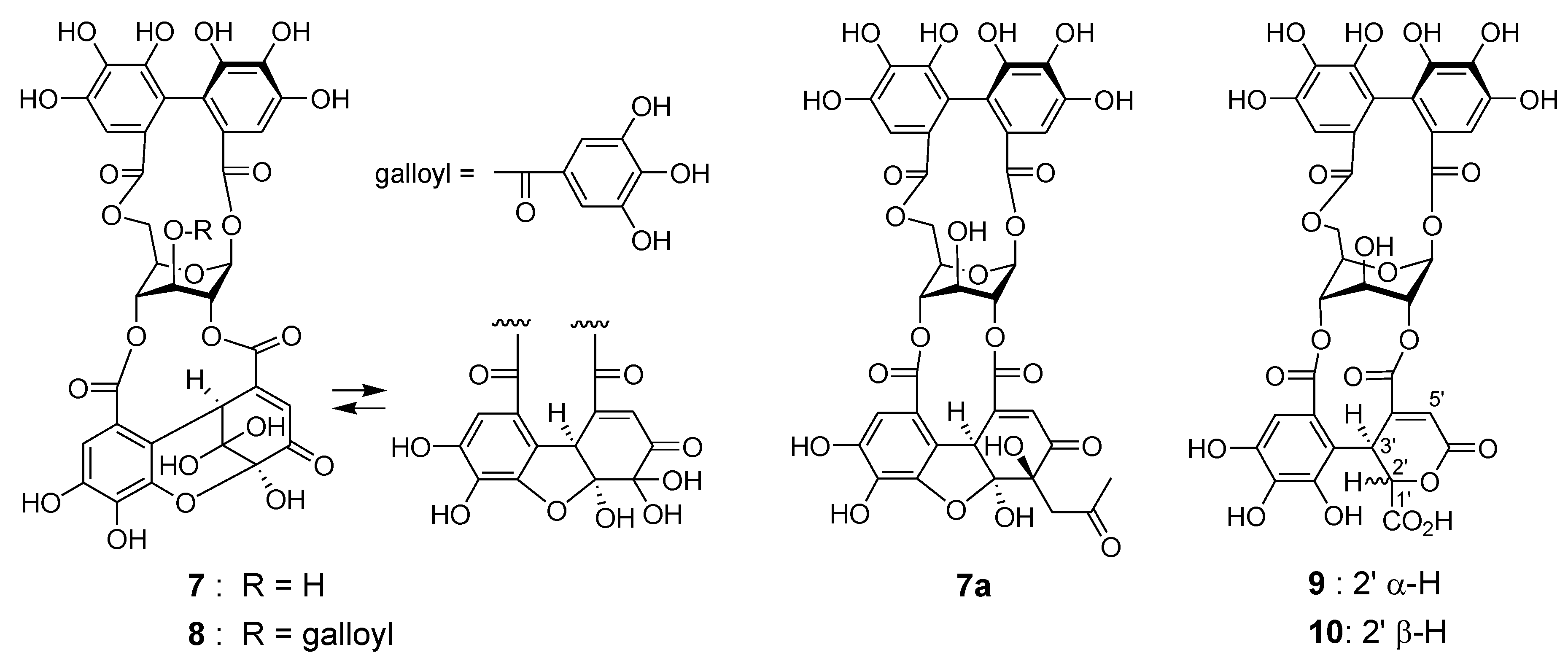
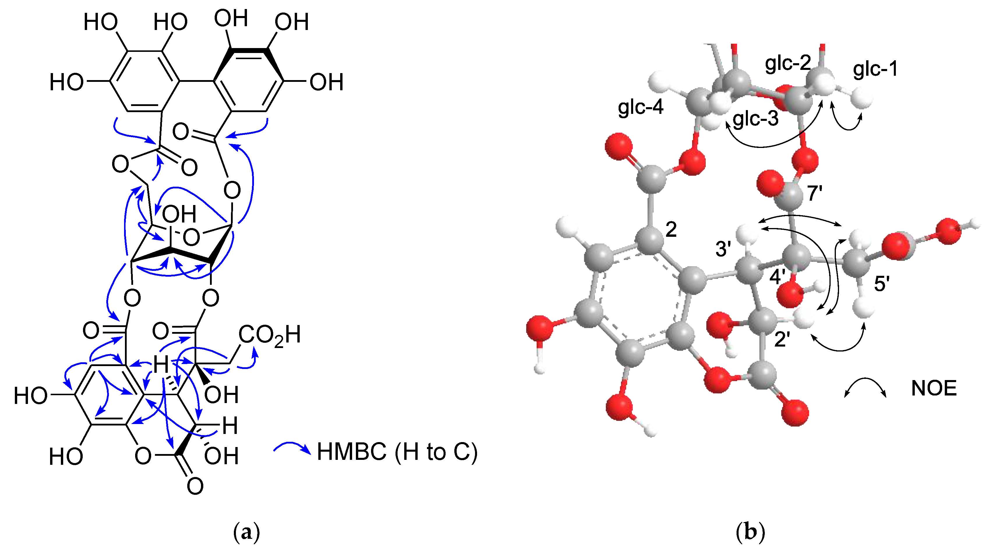
2.3. Hydrolyzable Tannin from Polygonum Runcinatum var. Sinense
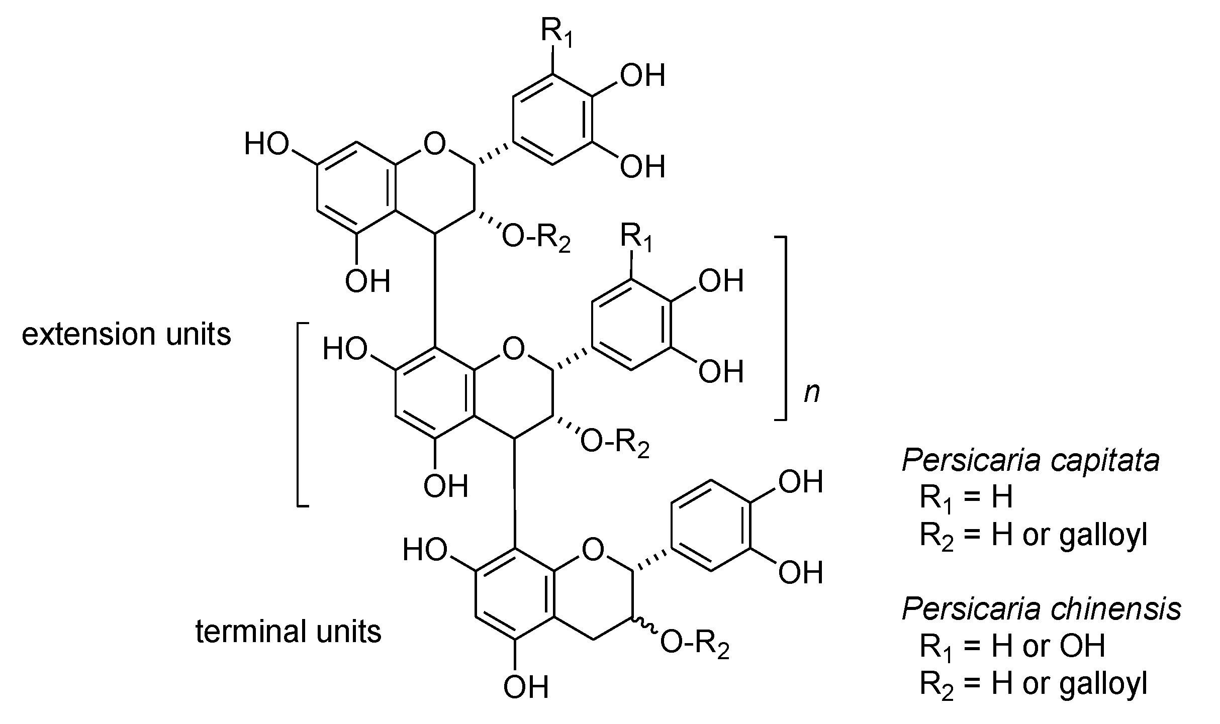
2.4. Proanthocyanidins of Persicaria Capitata and Persicaria Chinensis
3. Materials and Methods
3.1. General Information
3.2. Plant Material
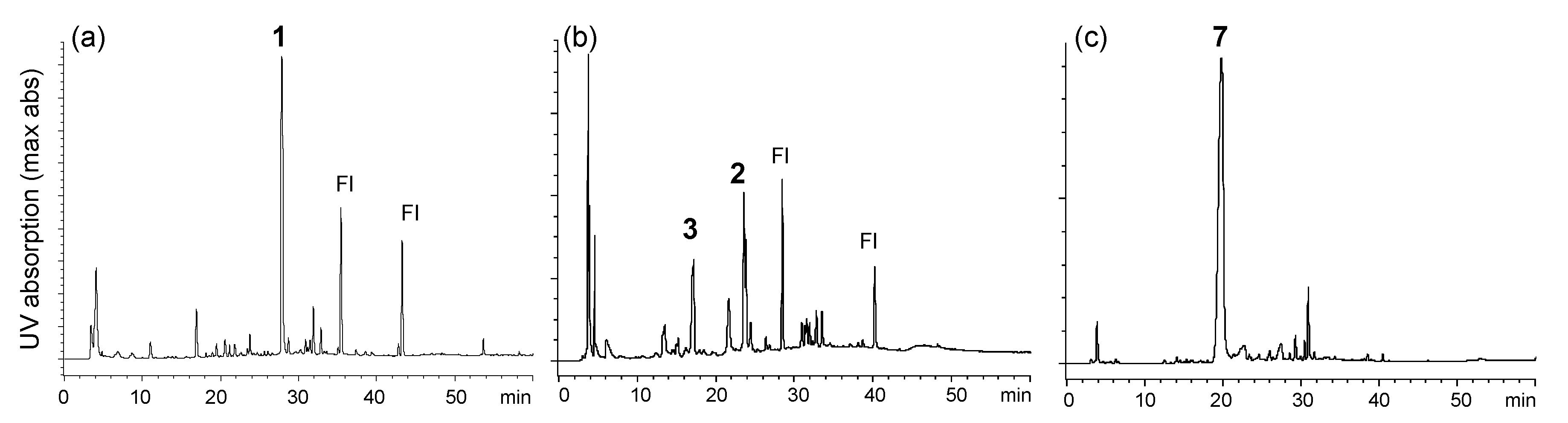
3.3. HPLC Analysis
3.4. Extraction and Separation
3.4.1. Persicaria Capitata
3.4.2. Persicaria Chinensis
3.4.3. Polygonum Runcinatum var. Sinense
3.5. Spectroscopic Data
3.5.1. Persicarianin (3)
3.5.2. Polygonanin A (11)
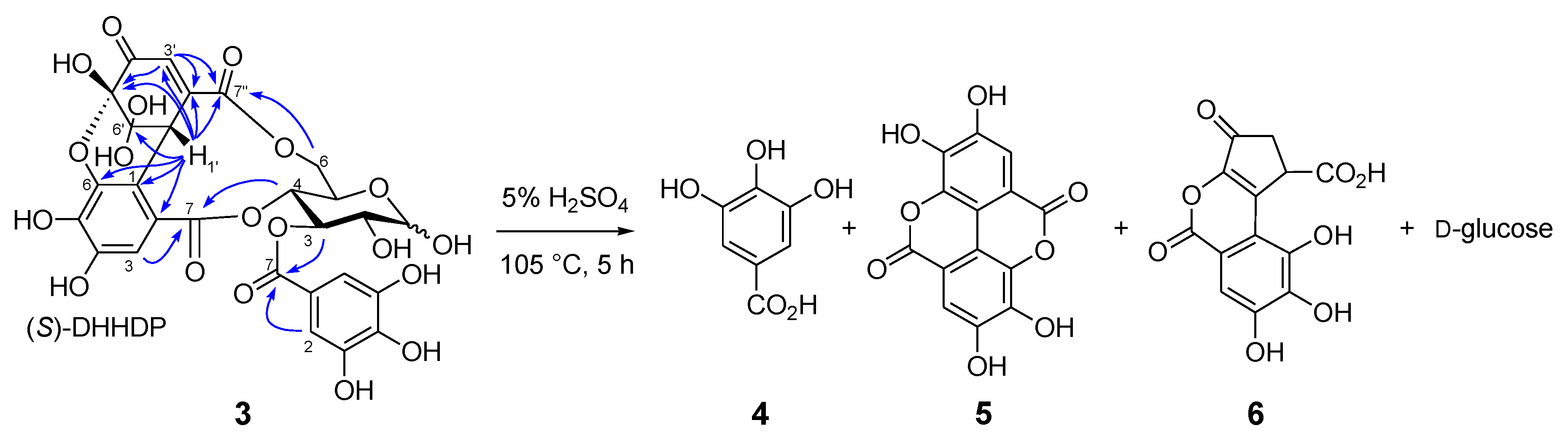
3.6. Acid Hydrolysis of 3
3.7. Computational Calculation of 8
3.8. Thiol Degradation
4. Conclusions
Supplementary Materials
Author Contributions
Funding
Acknowledgments
Conflicts of Interest
Sample Availability
References
- Okuda, T.; Yoshida, T.; Hatano, T. Hydrolyzable tannins and related polyphenols. In Progress in the chemistry of organic natural products; Herz, W., Kirby, G.W., Moore, R.E., Steglich, W., Tamm, C., Eds.; Springer: New York, NY, USA, 1995; Volume 66, pp. 1–117. [Google Scholar]
- Haslam, E.; Cai, Y. Plant polyphenols (vegetable tannins): Gallic acid metabolism. Nat. Prod. Rep. 1994, 11, 41–66. [Google Scholar] [CrossRef] [PubMed]
- Yoshida, T.; Hatano, T.; Ito, H.; Okuda, T. Structural diversity and antimicrobial activities of ellagitannins. In Chemistry and Biology of Ellagitannins, An Underestimated Class of Bioactive Plant Polyphenols; Quideau, S., Ed.; World Scientific Publishing: Singapore, 2009; pp. 55–93. [Google Scholar]
- Quideau, S.; Feldman, K.S. Ellagitannin chemistry. Chem. Rev. 1996, 96, 475–504. [Google Scholar] [CrossRef] [PubMed]
- Quideau, S.; Deffieux, D.; Douat-Casassus, C.; Pouységu, L. Plant Polyphenols: Chemical Properties, Biological Activities, and Synthesis. Angew. Chem. Int. Ed. 2011, 50, 586–621. [Google Scholar] [CrossRef] [PubMed]
- Fu, J.; Ma, J.-Y.; Zhang, X.-F.; Wang, Y.; Feng, R.; Chen, Y.-C.; Tan, X.-S.; Zhang, Y.Y.; Sun, Y.-P.; Zhou, Y.; et al. Identification of metabolites of FR429, a potential antitumor ellagitannin, transformed by rat intestinal bacteria in vitro, based on liquid chromatography-ion trap-time of flight mass spectrometry analysis. J. Pharm. Biomed. Anal. 2012, 71, 162–167. [Google Scholar] [CrossRef]
- Xie, L.-M.; Yau, L.-F.; Jiang, Z.-H.; Zhang, L.-Y.; Xia, Y.; Wang, J.-R. Sphingolipidomic study of davidiin-treated HepG2 human hepatocellular carcinoma cells using UHPLC-MS. RSC Advances 2017, 7, 55249–55256. [Google Scholar] [CrossRef] [Green Version]
- Ma, J.-Y.; Zhou, X.; Fu, J.; He, C.-Y.; Feng, R.; Huang, M.; Shou, J.-W.; Zhao, Z.-X.; Li, X.-Y.; Zhang, L.; et al. In vivo metabolite profiling of a purified ellagitannin isolated from Polygonum capitatum in rats. Molecules 2016, 21, 1110. [Google Scholar] [CrossRef] [Green Version]
- Wang, Y.; Ma, J.; Chow, S.-C.; Li, C.-H.; Xiao, Z.; Feng, R.; Fu, J.; Chen, Y. A potential antitumor ellagitannin, davidiin, inhibited hepatocellular tumor growth by targeting EZH2. Tumor Biology 2014, 35, 205–212. [Google Scholar] [CrossRef]
- Zhang, H.; Yu, M.; Jia, H.; Zhang, T.; Shang, H.; Zhang, M.; Zhu, Z.; Zou, Z. Comprehensive identification of potential antioxidant components in the aerial parts of Polygonum chinense L. var. hispidum using ultra high performance liquid chromatography coupled with quadrupole time-of-flight mass spectrometry. J. Sep. Sci. 2020, 43, 2380–2392. [Google Scholar]
- Hatano, T.; Ogawa, N.; Kira, R.; Yasuhara, T.; Okuda, T. Tannins of Cornaceous plants. I. Cornusiins A, B and C, dimeric monomeric and trimeric hydrolyzable tannins from Cornus officinalis, and orientation of valoneoyl group in related tannins. Chem. Pharm. Bull. 1989, 37, 2083–2090. [Google Scholar] [CrossRef] [Green Version]
- Zhu, M.; Phillipson, J.D.; Greengrass, P.M.; Bowery, N.E.; Cai, Y. Plant polyphenols: Biologically active compounds or non-selective binders to protein? Phytochemistry 1997, 44, 441–447. [Google Scholar] [CrossRef]
- Yoshida, T.; Nakazawa, T.; Hatano, T.; Yang, R.C.; Yang, L.L.; Yen, K.Y.; Okuda, T. A dimeric hydrolysable tannin from Camellia oleifera. Phytochemistry 1994, 37, 241–244. [Google Scholar] [CrossRef]
- Fortes, G.A.C.; da Silva, A.J.R.; Ferri, P.H.; Santos, S.C. Phenolic compounds from the leaves of Eucalyptus microcorys F. Muell. Rec. Nat. Prod 2015, 9, 292–296. [Google Scholar]
- Chen, X.; Yoshida, T.; Hatano, T.; Fukushima, M.; Okuda, T. Tannins and related polyphenols of the Saxifragaceae. Part, I. Galloylarbutin and other polyphenols from Bergenia purpurascens. Phytochemistry 1987, 26, 515–517. [Google Scholar]
- Saijo, R.; Nonaka, G.; Nishioka, I. Tannins and related compounds. Part 82. Phenol glucoside gallates from Mallotus japonicus. Phytochemistry 1989, 28, 2443–2446. [Google Scholar] [CrossRef]
- Markham, K.R.; Ternai, B.; Stanley, R.; Geiger, H.; Mabry, T.J. Carbon-13 NMR studies of flavonoids. III. Naturally occurring flavonoid glycosides and their acylated derivatives. Tetrahedron 1978, 34, 1389–1397. [Google Scholar] [CrossRef]
- Moharram, F.A.; Marzouk, M.S.A.; Ibrahim, M.T.; Mabry, T.J. Antioxidant galloylated flavonol glycosides from Calliandra haematocephala. Nat. Prod. Res. 2006, 20, 927–934. [Google Scholar] [CrossRef]
- Tanaka, T.; Nonaka, G.; Nishioka, I.; Miyahara, K.; Kawasaki, T. Tannins and related compounds. part 37. Isolation and structure elucidation of elaeocarpusin, a novel ellagitannin from Elaeocarpus sylvestris var. ellipticus. J. Chem. Soc. Perkin Trans. 1 1986, 369–376. [Google Scholar] [CrossRef]
- Tanaka, T.; Nonaka, G.; Nishioka, I. Tannins and related compounds. Part 14. 7-O-Galloyl-(+)-catechin and 3-O-galloylprocyanidin B-3 from Sanguisorba officinalis. Phytochemistry 1983, 22, 2575–2578. [Google Scholar] [CrossRef]
- Okuda, T.; Yoshida, T.; Hatano, T. Constituents of Geranium thunbergii Sieb. et Zucc. Part 12. Hydrated stereostructure and the equilibration of geraniin. J. Chem. Soc. Perkin Trans. 1 1982, 9–14. [Google Scholar] [CrossRef]
- Furusawa, M.; Tanaka, T.; Ito, T.; Nakaya, K.; Iliya, I.; Ohyama, M.; Iinuma, M.; Murata, H.; Inatomi, Y.; Inada, A.; et al. Flavonol glycosides in leaves of two Diospyros Species. Chem. Pharm. Bull. 2005, 53, 591–593. [Google Scholar] [CrossRef] [Green Version]
- Nonaka, G.; Matsumoto, Y.; Nishioka, I. Trapain, a new hydrolyzable tannin from Trapa japonica Flerov. Chem. Pharm. Bull. 1981, 29, 1184–1187. [Google Scholar] [CrossRef] [Green Version]
- Okuda, T.; Yoshida, T.; Hatano, T.; Koga, T.; Tho, N.; Kuriyama, K. Circular dichroism of hydrolysable tannins-II dehydroellagitannins. Tetrahedron Lett. 1982, 23, 3941–3944. [Google Scholar] [CrossRef]
- Tanaka, T.; Nakashima, T.; Ueda, T.; Tomii, K.; Kouno, I. Facile discrimination of aldose enantiomers by reversed-phase HPLC. Chem. Pharm. Bull. 2007, 55, 899–901. [Google Scholar] [CrossRef] [Green Version]
- Era, M.; Matsuo, Y.; Shii, T.; Saito, Y.; Tanaka, T.; Jiang, Z.H. Diastereomeric Ellagitannin Isomers from Penthorum chinense. J. Nat. Prod. 2015, 78, 2104–2109. [Google Scholar] [CrossRef] [PubMed]
- Era, M.; Matsuo, Y.; Saito, Y.; Tanaka, T. Production of ellagitannin hexahydroxydiphenoyl ester by spontaneous reduction of dehydrohexahydroxydiphenoyl ester. Molecules 2020, 25, 1051. [Google Scholar] [CrossRef] [PubMed] [Green Version]
- Kojima, D.; Shimizu, K.; Aritake, K.; Era, M.; Matsuo, Y.; Saito, Y.; Tanaka, T.; Nonaka, G. Highly oxidized ellagitannins of Carpinus japonica and their oxidation-reduction disproportionation. J. Nat. Prod. 2020, 83, 3424–3434. [Google Scholar] [CrossRef]
- Tanaka, T.; Nonaka, G.; Nishioka, I. Tannins and related compounds. Part 30. Punicafolin, an ellagitannin from the leaves of Punica granatum. Phytochemistry 1985, 24, 2075–2078. [Google Scholar] [CrossRef]
- Tanaka, T.; Fujisaki, H.; Nonaka, G.; Nishioka, I. Tannins and related compounds. CXVIII. Structures, preparation, high-performance liquid chromatography and some reactions of dehydroellagitannin-acetone condensates. Chem. Pharm. Bull. 1992, 40, 2937–2944. [Google Scholar] [CrossRef] [Green Version]
- Lee, S.H.; Tanaka, T.; Nonaka, G.; Nishioka, I. Tannins and related compounds. XCV. Isolation and characterization of helioscopinins and helioscopins, four new hydrolyzable tannins from Euphorbia helioscopia L. (1). Chem. Pharm. Bull. 1990, 38, 1518–1523. [Google Scholar] [CrossRef] [Green Version]
- Esumi, A.; Aoyama, H.; Shimozu, Y.; Taniguchi, S.; Hatano, T. Modified dehydroellagitannins from Davidia involucrata leaves. Heterocycles 2019, 98, 895–903. [Google Scholar]
- Lin, J.-H. Studies on tannins from the bark of Macaranga sinensis (Baill.) Muell.-Arg. J. Food Drug Anal. 1994, 2, 201–209. [Google Scholar] [CrossRef]
- Tanaka, T.; Takahashi, R.; Kouno, I.; Nonaka, G. Chemical evidence for the de-astringency (insolubilization of tannins) of persimmon fruit. J. Chem. Soc. Perkin Trans. 1 1994, 3013–3022. [Google Scholar] [CrossRef]
- Yanagida, A.; Shoji, T.; Shibusawa, Y. Separation of proanthocyanidins by degree of polymerization by means of size-exclusion chromatography and related techniques. J. Biochem. Biophy. Meth. 2003, 56, 311–322. [Google Scholar] [CrossRef]
- Lodewyk, M.W.; Siebert, M.R.; Tantillo, D.J. Computational prediction of 1H and 13C chemical shifts: A useful tool for natural product, mechanistic, and synthetic organic chemistry. Chem. Rev. 2012, 112, 1839–1862. [Google Scholar] [CrossRef] [PubMed]
- Grimblat, N.; Sarotti, A.M. Computational chemistry to the rescue: Modern toolboxes for the assignment of complex molecules by GIAO NMR calculations. Chem. Eur. J. 2016, 22, 12246–12261. [Google Scholar] [CrossRef]
- Zanardi, M.M.; Marcarino, M.O.; Sarotti, A.M. Redefining the impact of boltzmann analysis in the stereochemical assignment of polar and flexible molecules by NMR calculations. Org. Lett. 2020, 22, 52–56. [Google Scholar] [CrossRef]

Publisher’s Note: MDPI stays neutral with regard to jurisdictional claims in published maps and institutional affiliations. |
© 2021 by the authors. Licensee MDPI, Basel, Switzerland. This article is an open access article distributed under the terms and conditions of the Creative Commons Attribution (CC BY) license (http://creativecommons.org/licenses/by/4.0/).
Share and Cite
Li, Y.-Q.; Kitaoka, M.; Takayoshi, J.; Wang, Y.-F.; Matsuo, Y.; Saito, Y.; Huang, Y.-L.; Li, D.-P.; Nonaka, G.-i.; Jiang, Z.-H.; et al. Ellagitannins and Oligomeric Proanthocyanidins of Three Polygonaceous Plants. Molecules 2021, 26, 337. https://doi.org/10.3390/molecules26020337
Li Y-Q, Kitaoka M, Takayoshi J, Wang Y-F, Matsuo Y, Saito Y, Huang Y-L, Li D-P, Nonaka G-i, Jiang Z-H, et al. Ellagitannins and Oligomeric Proanthocyanidins of Three Polygonaceous Plants. Molecules. 2021; 26(2):337. https://doi.org/10.3390/molecules26020337
Chicago/Turabian StyleLi, Yun-Qiu, Masako Kitaoka, Juri Takayoshi, Ya-Feng Wang, Yosuke Matsuo, Yoshinori Saito, Yong-Lin Huang, Dian-Peng Li, Gen-ichiro Nonaka, Zhi-Hong Jiang, and et al. 2021. "Ellagitannins and Oligomeric Proanthocyanidins of Three Polygonaceous Plants" Molecules 26, no. 2: 337. https://doi.org/10.3390/molecules26020337