Review

Xanthone Glucosides: Isolation, Bioactivity and Synthesis

1
School of Pharmacy, Shanghai University of Traditional Chinese Medicine, Shanghai 201203, China
2
Engineering Research Center of Shanghai Colleges for TCM New Drug Discovery, Shanghai 201203, China
*
Authors to whom correspondence should be addressed.
These authors contributed equally to this work.
Molecules 2021, 26(18), 5575; https://doi.org/10.3390/molecules26185575
Submission received: 17 August 2021 / Revised: 6 September 2021 / Accepted: 9 September 2021 / Published: 14 September 2021
(This article belongs to the Special Issue A Themed Issue in Memory of Academician Xing Qiyi (1911–2002))

Abstract

:
Xanthones are secondary metabolites found in plants, fungi, lichens, and bacteria from a variety of families and genera, with the majority found in the Gentianaceae, Polygalaceae, and Clusiaceae. They have a diverse range of bioactivities, including anti-oxidant, anti-bacterial, anti-malarial, anti-tuberculosis, and cytotoxic properties. Xanthone glucosides are a significant branch of xanthones. After glycosylation, xanthones may have improved characteristics (such as solubility and pharmacological activity). Currently, no critical review of xanthone glucosides has been published. A literature survey including reports of naturally occurring xanthone glucosides is included in this review. The isolation, structure, bioactivity, and synthesis of these compounds were all explored in depth.

Graphical Abstract

1. Introduction

In natural product chemistry, xanthones are one of the most abundant types of chemicals. They are secondary metabolites found in higher plant families, fungi, lichen, and bacteria, and are primarily found in Gentianaceae, Polygalaceae, Clusiaceae, and others [1,2,3]. They have a variety of health-promoting properties, including anti-bacterial, anti-carcinogenic, anti-oxidant, and anti-diabetic properties [4,5,6,7,8].
The structure of xanthone determines its bioactivity, and different substitutions might result in a variable bioactivity [9,10,11]. The chemical formula of xanthone is C13H8O2. Its main structure is 9H-xanthen-9-one with a dibenzo-γ-pirone scaffold. Research on xanthones has received much attention in recent years [12,13,14,15]. In general, xanthones are categorized into six classes based on substitutions on the basic structure of xanthones: simple xanthones, xanthone glucosides (or glycosylated xanthones), prenylated xanthones, xanthonolignoids, bis-xanthones, and miscellaneous xanthones [16,17]. The main distribution of these xanthones varies, as prenylated xanthones are widely distributed in the Clusiaceae and most compounds of simple xanthones and xanthone glucosides are from the Gentianaceae. These primary groupings are further subdivided into non, mono-, di-, tri-, tetra-, penta-, hexa-, and hepta-oxygenated xanthones based on the degree of oxygenation [18,19,20].
Molecules 26 05575 i001
More recently, xanthone glucosides have been explored, and the mutation of these glycosyl groups can change the biological activity of xanthone, which has a wide range of clinical applications [21,22]. However, xanthones usually have poor solubility; herein, many studies are being devoted to the synthesis of glycosylated xanthones to improve their solubility and activity and minimize their toxicity [23,24]. Xanthone glucosides are an important class of xanthones that are extensively dispersed in the plant families Gentianaceae and Polygalaceae. For natural xanthone glucosides, each xanthone site can be connected to a sugar group, which can be either monosaccharide or disaccharide. Recent research has revealed that xanthone glucosides have anti-oxidant [25], anti-inflammatory [26], anti-cancer [21,27], and other pharmacological properties. We separated xanthone glucosides into xanthone C-glucoside and xanthone O-glucoside and classified the substances accordingly. CC bonds connect the sugar moiety to the xanthone nucleus in C-glucosides, which are usually resistant to acidic and enzymatic hydrolysis, whereas O-glucosides have normal glycosidic linkages. In glucosides whose glycosyl group is disaccharide, the second sugar residue is often glucose, xylose, or rhamnose and is usually associated with C-6 of the first glucose unit. However, when the second residue is rhamnose, it is linked to the C-2 of the first residue. The structures and connection site of sugars to the xanthone core that may be used in their full names are shown below.
Molecules 26 05575 i002
In general, xanthone glucosides have received much interest due to their unique structures and significant bioactivities. As a result, we examined the separation, bioactivity, and synthesis of naturally occurring xanthone glucosides, with the goal of providing a reference for future relevant studies.

2. Structure, Isolation and Bioactivity of Xanthone Glucoside

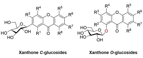
2.1. Xanthone C-Glucoside

This class of xanthone glucosides is composed of xanthone and sugar groups that are linked together by carbon atoms in the structure. D-glucose is a sugar group that is commonly found in these compounds. The majority of the sugar binding sites are located at position 2, and glycosylation can often boost the activity to a certain amount [28]. All of the xanthones have hydroxyl substitutions on their skeletons, and some of them have methoxy groups. The scavenging of free radicals and the anti-oxidant activity of these compounds are their most notable impacts. We will classify these compounds by distinct genera in the order in which they were discovered, followed by a description of their biological activity.

2.1.1. Xanthone C-Glucoside from Liliaceae

Mangiferin (1) is the most widely studied xanthone C-glucoside for pharmaceutical purposes [29,30], and it may be obtained from a variety of plants, including Anemarrhena asphodeloides Bge (Liliaceae) [31], A. senkakuinsulare (Aristolochiaceae) [32], Mahkota dewa (Phaleria macrocarpa (Scheff.) Boerl) [33], Coffea pseudozanguebariae (Rubiaceae) [34], and Lomatogonium carinthiacum (Gentianaceae) [35]. There is a glucose substitution at position 2 of the xanthone skeleton in mangiferin, as well as hydroxyl substitutions at positions 1, 3, 6, and 7. Mangiferin’s C-glycosidic bond, which mimics the nucleophilic substitution of phloroglucinol, improves bio-availability and is responsible for its anti-oxidant properties [36]. Mangiferin has been shown to have anti-inflammatory activity [37,38,39], anti-oxidant activity [40,41,42], anti-diabetic activity [43,44,45], cardio-protective effects [46,47,48], and anti-cancer activity [49,50,51]. The anti-inflammatory and anti-oxidant activities were due to the free radical scavenging capacity of mangiferin [41,52,53]. Mangiferin is a potent inhibitor of the NF-Kappa B signaling pathway [54], and the anti-oxidant activity of mangiferin is also related to its iron-chelating properties [55].
Molecules 26 05575 i003
In 1970, Aritomi and Kawasaki isolated homomangiferin (2) and isomangiferin (3) from Anemarrhena asphodeloides Bunge [56]. These two compounds were similar to mangiferin in structure. Homomangiferin has a methoxy group at position 3 compared to mangiferin, while the sugar group of isomangiferin is attached at position 4. Isomangiferin can also be isolated from Cyclopia genistoides (L.) Vent. (honeybush) and has a strong effect in the treatment of rheumatoid arthritis [57].
In 1997, Guo’s team isolated neomangiferin (4) from Anemarrhena asphodeloides Bge. The structure of the compound was 7-O-β-d-glucopyranosyl-mangiferin [31], which thus far is the only xanthone glucoside that contains both C-glucoside and O-glucoside. Neomangiferin was found to modulate the Th17/Treg balance and ameliorate colitis in mice [58].
Molecules 26 05575 i004

2.1.2. Xanthone C-Glucoside from Iridaceae

In 1973, Takemoto’s team isolated irisxanthone (5) from Iris florentina L. Compared to mangiferin, irisxanthone has a methoxy group at the 5-position and no hydroxyl group at the 7-position [59]. Irisxanthone can also be isolated from the leaves of I. albicans Lange [60], Iris adriatica [61], and Iris germanica [62]. In 1995, Alkhalil’s team isolated 2-β-d-glucopyranosyl-l,3,5,8-tetrahydroxyxanthone (nigricanside) (6) from the rhizomes of Iris nigricans, and the structure of the compound was 2-β-d-glucopyranosyl-l,3,5,8-tetrahydroxy-9H-xanthene-9-one [63].
Molecules 26 05575 i005

2.1.3. Xanthone C-Glucoside from Arrabidaea

In 2003, Bolzani’s team isolated 2-(2′-O-trans-caffeoyl)-C-β-d-glucopyranosyl-1,3,6,7-tetrahydroxyxanthone (7), 2-(2′-O-trans-cinnamoyl)-C-β-d-glucopyranosyl-1,3,6,7-tetrahydroxyxanthone (8), and 2-(2′-O-trans-coumaroyl)-C-β-d-glucopyranosyl-1,3,6,7-tetrahydroxyxanthone (9) from the stems of Arrabidaea samydoides. These compounds showed moderate free radical scavenging activity against 1,1-diphenyl-2-picrylhydrazyl (DPPH) [64]. In 2008, Hostettmann’s team isolated 3′-O-p-hydroxybenzoylmangiferin (10), 3′-O-trans-coumaroylmangiferin (11), 6′-O-trans-coumaroylmangiferin (12), 3′-O-trans-cinnamoylmangiferin (13), 3′-O-trans-caffeoylmangiferin (14), and 3′-O-benzoylmangiferin (15) from the leaves of Arrabidaea patellifera. Compounds 1012 have demonstrated anti-plasmodial activity (IC50: 26.5 μM, 18.1 μM and 38.2 μM, respectively). In addition, compounds 1015 (shown in Table 1) have shown radical-scavenging and anti-oxidant activities [65].
Molecules 26 05575 i006

2.1.4. Xanthone C-Glucoside from Polygalaceae

In 1999, sibiricaxanthones A (16) and sibiricaxanthones B (17) were isolated from the roots of Polygala sibirica by Miyase’s group. The structure of 16 was 2-C-[β-d-apiofuranosyl-(1→6)-β-d-glucopyranosyl]-1,3,7-trihydroxyxanthone, and 17 was shown to be 2-C-[β-d-apiofuranosyl-(1→2)-β-d-glucopyranosyl]-1,3,7-trihydroxyxanthone [66]. In 2005, Tu’s group isolated polygalaxanthones VIII (18) and polygalaxanthones XI (19) from the cortexes of Polygala tenuifolia. The structure of 18 was defined as 2-C-[β-d-arabinopyranosyl-(1→6)-β-d-glucopyranosyl]-1,3,7-trihydroxy-6-methoxyxanthone and 19 was 2-C-[β-d-apiofuranosyl-(1→2)-β-d-glucopyranosyl]-1,3,7-trihydroxy-6-methoxyxanthone [67].
Telephioxanthones A (20) and Telephioxanthones B (21) are two xanthone C-glucosides isolated from Polygala telephioides by Tu’s group in 2007. Compound 20 was shown to be 6’-O-[(E)-cinnamoyl]mangiferin), and compound 21 was 4’-O-[(E)-cinnamoyl]mangiferin [68]. Polygalaxanthone III (22), is a xanthone glucoside isolated from polygala root [69] that showed a potential scavenging effect on DPPH and hydroxy radicals and reductive activity to Fe3+ with IC50 values of 76.1, 83.5, and 54.9 mM [70].
In 2013, a new xanthone C-glucoside, tenuiside A (23), along with three known xanthone C-glucosides, lancerin (24) [71], neolancerin (25) [72], and 7-O-methylmangiferin (26) [73] were isolated from Polygala tenuifolia by Jiang’s group. Compounds 2325 have NO inhibitory activity and low cytotoxicity. Compound 24 showed stronger activity than compound 25, indicating that glycosidation at C-4 is superior to glycosidation at C-2 in terms of inhibition of NO [74].
Molecules 26 05575 i007
Molecules 26 05575 i008

2.1.5. Xanthone C-Glucoside from Gentianaceae

In 1991, Cordell’s team isolated swertipunicosid (27) from Swertia punicea Hemal., which was the first bisxanthone C-glucoside. The structure was 1,5,8-trihydroxy-3-methoxy-7-(1′,3′,6′,7′-tetrahydroxy-9′-oxo-4′-xanthyl) xanthone 2′-C-β-d-glucopyranoside [75]. In 1992, the same team isolated 3-O-demethylswertipunicoside (28) from Swertia punicea, the structure of which was 1,3,5,8-tetrahydroxy-7-(1′,3′,6′,7’-tetrahydroxy-9′-oxo-4′-xanthyl) xanthone 2′-C-β-d-glucopyranoside [76]. According to later research, the compound 28 showed potent neuro-protective activity against H2O2-induced PC12 cell damage [77].
In 2010, Guo’s team isolated two new xanthone C-glucosides, puniceaside D (29) and puniceaside E (30), from Swertia punicea. Puniceasides D and E are two unique trimeric xanthone C-glucosides [77]. In 2013, 3,5,6,8-tetrahydroxyxanthone-1-C-β-d-glucoside (31), which has excellent anti-oxidant activity, was isolated from Swertia mussotii. According to the research, glycosylated xanthones are more active than those that are not [78]. In 2016, Zhang’s team isolated apigenin-7-O-gluco (1″→3‴) glucoside (32) from Gentianella turkestanorum, the structure of which is similar to mangiferin, with a hydroxyl group missing at the 7-position [79].
Molecules 26 05575 i009
Molecules 26 05575 i010

2.1.6. Xanthone C-Glucoside from Bombacaceae

Shamimoside (33) was isolated from the leaves of Bombax ceiba L. by Versiani’s team. The structure of the compound was 4-C-β-d-glucopyranosyl-1,3,6,8-tetrahydroxy-7-O-(p-hydroxybenzoyl)-9H-xanthen-9-one and it is the first naturally occurring xanthone containing a benzoate moiety directly attached to an aromatic ring. The DPPH anti-oxidant assay shows that the compound has moderate anti-oxidant activity (IC50 = 150 μg/mL) [29].
Molecules 26 05575 i011

2.1.7. Others

Mangiferoxanthone A (34) is a xanthone dimer isolated from M. indica by bioassay in 2014, and is a symmetric homodimer of mangiferin. The compound showed moderate influenza neuraminidase inhibition activity. According to the research, dimerization increased the activity of the compound compared with mangiferin [80].
Molecules 26 05575 i012

2.2. Xanthone O-Glucoside

In contrast to xanthone C-glucosides, xanthone O-glucosides are glucosides that are linked to the tricyclic body of xanthones by an oxygen atom. Xanthone glucoside is generally found at the C-1 position of the xanthone nucleus. Glucosides are typically monosaccharides or disaccharides that contain glucose, xylose, rhamnose, and other glycosyl groups. At present, most xanthone O-glucosides isolated from natural resources contain hydroxyl, and methoxy groups, and a few have methyl groups, aliphatic side chains, or aromatic rings. Xanthone O-glucosides, in general, are a well-studied class of compounds. The glycosylation of xanthones improves not only their physical properties (such as solubility) but also their biological activity.

2.2.1. Xanthone O-Glucoside from Gentianaceae

In 1969, Stout and Balkenhol identified a xanthone O-glucoside whose structure is 1-(β-d-glucosyloxy)-8-hydroxy-3,5-dimethoxyxanthone (35) from the root of Frasera carolinicnsis Walt [81].
Molecules 26 05575 i013
In the same year, Tomimori and Komatsu obtained norswertianolin (36) from Swertia macrosperma for the first time [82]. Four years later, Tomimori’s team discovered a new xanthone O-glucoside in Swertia spp., named norswertianin-1-glucoside (37) [83].
Molecules 26 05575 i014
Four compounds (3841) were isolated from Gentiana bavarica L. by Hostettmann’s group in 1974: gentiabavaroside (38), gentiabavarutinoside (39), isogentiakochianoside (40), and norswertiaprimevdroside (41). Structurally, they all contain disaccharide substituents, with the other substituents being hydroxyl or methoxyl groups, respectively [84].
Molecules 26 05575 i015
In 1977, a new xanthone diglucoside (42) was isolated from the aerial parts of Swertia perennis L. (Gentianaceae) by means of column chromatography on polyamide, followed by preparative TLC. Its structure has been established as 1,3-di-β-d-glucopyranosyl-7,8-dihydroxyxanthone or norswertianine-1,3-diglucoside [85].
Molecules 26 05575 i016
In 1978, Ghosal extracted and isolated five compounds (4347) that had not been reported before from Swertia angustifoh Buch.-Ham. Their study showed that xanthone O-glucosides in the plant could be identified after the onset of maturity (i.e., 4- to 6-week-old plants) and were not present at the beginning of growth [86].
Molecules 26 05575 i017
Dhasmana and Garg isolated 2,3,7-trimethoxyxanthone-1-O-glucoside (48) and 2,3,5-trimethoxyxanthone-1-O-glucoside (49) from Halenia elliptrca D. Don. in 1989, and parts from an alcoholic plant extract containing these two compounds showed anti-amoebic activity. The structural difference between these two compounds is that the methoxy groups are at sites 2, 3, and 7 in 48 and 2,3, and 5 in 49 [87].
Molecules 26 05575 i018
After extraction and analysis, Sun’s group obtained three compounds from Swertia mussotii Franch. in 1991, namely 7-O-β-d-xylopyranosyl-1,8-dihydroxy-3-methoxyxanthone (50), 7-O-[α-ʟ-rhanopyranosyl-(1–2)-β-d-xylopyranosyl]-1,8-dihydroxy-3-methoxyxanthone (51), and 3-O-β-d-glucopyranosy-1,8-dihydroxy-5-methoxyxanthone (52). They all have hydroxyl groups at positions 1 and 8. These three compounds were isolated from water-soluble components, demonstrating that the glycosyl group in the structure was the main factor influencing their solubility [88].
Molecules 26 05575 i019
Hosteyitman and coworkers discovered and identified three compounds in 1992: 2,3,5-trimethoxy-1-O-gentiobiosyloxyxanthone (53), 2,3,5-trimethoxy-l-O-primeverosyloxyxanthone (54), and 2,3,4,5-tetramethoxy-1-O-primeverosyloxyxanthone (55). The other substituents of these three compounds, such as those of compounds 48 and 49, are all methoxy groups, and their glycosidic bond is at the 1-position. These three compounds, however, are disaccharide substituted, in contrast to the former [89].
Molecules 26 05575 i020
In 1995, Hostettmann’s group isolated and identified eight xanthone O-glucosides (5663, shown in Table 2) from Halenia corniculata. These eight compounds share the following characteristics: (1) they all have three or four methoxy groups, and (2) they are disaccharides with gentiobiose or primeverose at the C-1 position. Their structures are similar to those discovered by Hosteyitman (5355) [90].
Molecules 26 05575 i021
From the aerial parts of Tripterospermum japonicum, five new xanthone glucosides, named triptexanthosides A-E (6468), were isolated along with a known xanthone C-glucoside, mangiferin, by Hideaki Ostuka in 1999. Their structures were elucidated as 1,2,6,8-tetrahydroxyxanthone 1-O-β-d-glucopyranoside (64), 1-O-β-gentiobioside (65), 1,2,8-trihydroxy-5,6-dimethoxyxanthone 2-O-β-d-glucopyranoside (66), 2-O-β-primeveroside (67), and 1-O-gentiobioside (68) [91].
Molecules 26 05575 i022
Milosavljević isolated two compounds, 1-O-primeverosyl-3,8-dihydroxy-5-methoxyxanthone (69) and 1-O-gentiobiosyl-3,7-dimethoxy-8-hydroxyxanthone (70), from another plant of the Gentianaceae (Swertia punctate) in 2002. They have disaccharide substituents such as gentiobiosyl and primeverosyl, which are the same as the substituents of the compound discovered in 5663 [92].
Molecules 26 05575 i023
Tan and colleagues isolated a xanthone O-glucoside, 5-O-β-d-glucopyranosyl-1,3,8-trihydroxyxanthone (71), from Swertia davidii Franch in 2004 [93]. Meanwhile, 8-O-β-d-glucopyranosyl-1,3,5-trihydroxyxanthone (norswertianolin, 72, isomers of 71 and 64), discovered with 1,5-dihydroxy-3-methoxyxanthone-8-O-β-d-glucopyranoside (73) from Gentiana campestris by Kaldas and co-workers in 1974 [94], was isolated for the first time from Swertia davidii Franch. by the same group [93]. Molecules 26 05575 i024
In 2005, Rana’s team isolated 6-hydroxy-3,5-dimethoxy-1-[(6-O-β-d-xylopyranosyl-β-d-glucopyranosyl)oxy]-9H-xanthen-9-one (74) from the rhizomes of Swertia speciosa, and the compound had moderate 2,2-di(4-tert-octylphenyl)-1-picryl-hydrazyl (DPPH) radical scavenging activity [95].
Molecules 26 05575 i025
One year later, from the aerial parts of Swertia longifolia Boiss., which grows in northern of Iran, two diglycosidic xanthones were isolated. The structures were confirmed by means of their spectral data as 1,5-dihydroxy-3-methoxy-6-O-primeverosyl xanthone (75) and 8-hydroxy-3,5-dimethoxy-1-O-primeverosyl xanthone (76), which are new derivatives in the plant kingdom [96].
Molecules 26 05575 i026
In 2008, corymbiferin 3-O-β-d-glucopyranoside (77) and swertiabisxanthone-I 8′-O-β-d-glucopyranoside (78) were isolated and identified from Gentianella amarella ssp. acuta (Michx.) J.M.Gillett by Hostettmann, in which 78 was a dimer of xanthones. Moreover, these two compounds also showed a weak inhibitory effect on acetylcholinesterase (AChE) and monoamine oxidases (MAO) A and B, which are associated with Alzheimer’s disease [97,98,99]. The inhibitory rates of 77 on MAO A and B were 40.2 ± 2.6% and 47.8 ± 2.2%, respectively. The inhibitory rates of 78 to MAO A and B were 21.4 ± 4.9% and 39.1 ± 1.2%, respectively [100].
Molecules 26 05575 i027
In 2010, two new dimeric xanthone O-glucosides, puniceasides A (79) and B (80), a new trimeric O-glucoside, puniceaside C (81), and a known xanthone O-glucoside swertiabisxanthone-I 8′-O-β-d-glucopyranoside (82) were isolated from the entire plant of Swertia punicea. The compounds were evaluated for their potential neuroprotective activities against H2O2-induced PC12 cell damage using an MTT assay. Compounds 80 and 82 displayed potent neuroprotective activity [77].
Molecules 26 05575 i028
Feng and co-workers found comastomaside A (83) in Comastoma pedunlulatum (Rogle eX D. Dou) Holub in 2011, which is a traditional Tibetan medicine named Zangyinchen. Structurally, 83 is different from other compounds in that it has an aryl side chain on its glucoside chain [101].
Molecules 26 05575 i029
In 2011, Ding’s group conducted several activity tests on 1-O-β-d-glucoside-7-hydroxyl-3,8-dimethoxyxanthone (84) from Gentianopsis paludosa Ma. The compound exhibited significant cytotoxicity, with IC50 values of 18.00 ± 0.84 (μg/mL) in HepG2 cells and 24.80 ± 1.79 (μg/mL) in HL-60 cells. At the same time, the compound can inhibit cell proliferation while inducing apoptosis in both types of cells [102].
Molecules 26 05575 i030
Li’s group found that 1-O-β-d-glucopyranosyl-3,5,6-trimethoxy-xanthone (85) from Swertia mussotii Franch. had a weak inhibitory effect on α-glucosidase, with an inhibition rate of 5.4% when the concentration was 40 μM. The other compound 1-O-[β-d-xylopyranosyl-(1→6)-β-d-glucopyranosyl]-3,5,6-trimethoxy-xanthone (86) from Swertia mussotii Franch had a weak inhibitory effect on dipeptidyl peptidase IV (DPP-IV) with 2.1% inhibition at 10−5 M [103].
Molecules 26 05575 i031
In 2013, Luo’s group isolated 7-hydroxy-3,4,8-trimethoxyxanthone-1-O-(β-d-glucoside) (87), 6-hydroxy-3,5-dimethoxyxanthone-1-O-(β-d-glucoside) (88), and 3,4,7,8-tetramethoxyxanthone-1-O-(β-d-glucoside) (89) from Swertia mussotii. These three compounds were found to have moderate anti-oxidant activity. Their oxygen radical absorbance capacity (ORAC) values at a concentration of 3.1 μM were 30.2 ± 0.2, 33.1 ± 0.2 and 33.2 ± 0.7, respectively. The experiment in this study also showed that the bio-activity of glycosylated xanthones was higher than that of xanthones without glycosylation [78].
Molecules 26 05575 i032
From an n-butanol fraction of Swertia kouitchensis, ten new xanthone glucosides, kouitchensides A–J (9099, shown in Table 3), were isolated. The structures of these glucosides were determined by interpreting extensive spectroscopic data. In an in vitro test, compounds 91, 93, 94, and 95 (IC50 values ranging from 126 to 451 μM) inhibited α-glucosidase activity more effectively than acarbose, the positive control (IC50 value of 627 μM) [104].
Molecules 26 05575 i033
In 2014, seven new xanthone glucosides (100106, shown in Table 4) were isolated from the n-butanol extract of Swertia bimaculate. Compounds 102, 103, and 106 were found to have significant α-glucosidase inhibitory activities in vitro (IC50 values of 142 µM, 136 µM, and 258 µM, respectively), and the assay showed that glucoside units at C-1 exhibited more potent inhibitory activity than the units located at C-8 [105].
Molecules 26 05575 i034
Hu’s group found a disaccharide substituted xanthone O-glucoside from the whole plant of Lomatogonium carinthiacum (Wulfen) Rchb. and identified its structure as 1,4,8-trimethoxyxanthone-6-O-β-d-glucoronyl-(1→6)-O-β-d-glucoside in 2014 (107) [106].
Molecules 26 05575 i035
1,2-Dihydroxy-6-methoxyxanthone-8-O-β-d-xylopyranosyl (108) is one of the main constituents of petroleum ether and ethyl acetate extracts from Swertia corymbosa (Gentinaceae), a medicinal plant used in traditional Indian systems to treat diabetes. In STZ-induced diabetic rats, the compound aids in the management of diabetes and the prevention of its vascular complications and may be useful in the treatment of anti-hyperglycemia and anti-hyperlipidemia in diabetic patients [107].
Molecules 26 05575 i036
Six new tetrahydroxanthone glucosides (amarellins A–F (109114)) were isolated from the aerial parts of the Mongolian medicinal plant Gentianella amarelle ssp. acuta (Gentianaceae) by Yoshiki Kashiwada and colleagues in 2016. Amarellins A–C (109111) were assigned as 8-O-β-d-glucoside, 8-O-β-d-xyloside, and 1-O-β-d-glucoside of the trans-tetrahydroxanthone, respectively, while amarellins D–F (112114) were elucidated to be 8-O-β-d-xyloside, 1-O-β-d-glucoside, and 3-O-β-d-glucoside of the cis-tetrahydroxanthone, respectively [108].
Molecules 26 05575 i037
Kuang’s laboratory isolated a new compound, 5-hydroxy-3,4,6-trimethoxyxanthone-1-O-β-d-glucopyranoside (115), and two known compounds, norswertianolin (72) and swertianolin (116) [109,110,111,112] from Gentianella acuta (Michx.) Hulten in 2018 [46]. A mixture of extracts that included 72 and 116 was verified to provide protection against myocardial I/R injury through their anti-oxidative and anti-apoptotic effects [113].
Molecules 26 05575 i038

2.2.2. Xanthone O-Glucoside from Clusiaceae

From the stem of Poeciloneuron pauciflorum, a new xanthone, 1,6-dihydroxy-7-methoxyxanthone 6-O-β-d-glucoside (117) was isolated by the Riswan group in 1997. The glucose moiety was located at C-6 of the xanthone [114].
Molecules 26 05575 i039

2.2.3. Xanthone O-Glucoside from Hypericaceae

From the aerial part of Hypericum japonicum, one new xanthone glucoside, 1,5-dihydroxyxanthone-6-O-β-d-glucoside (118), was isolated from Hypericum japonicum in 1998 by Wu. Compound 118 was found to exert interesting coagulant activity in an in vitro test, showing prothrombin coagulation activity [18]. Zhang also isolated and identified 1,6-dihydroxyisojacereubin-5-O-β-d-glucoside (119), which is a tetracyclic compound containing a 2H-pyran ring, from the same plant in 2006 [115].
Molecules 26 05575 i040
Ishiguro and colleagues isolated two new compounds, patuloside A (120) and patuloside B (121) in 1999 from cell suspension cultures of Hypericum patulum [116]. This is the first report on the isolation of 1,3,5,6-tetrahydroxyxanthone glucosides from cell suspension cultures of H. patulum.
Molecules 26 05575 i041
In 2000, Kitanov and Nedialkov extracted and identified an innovative compound from Hypericum annulatum and named it xanthohypericoside (122) [117].
Molecules 26 05575 i042
A phytochemical study on the aerial parts of Hypericum elatoides led to the isolation of five previously undescribed phenolic metabolites, hyperelatones E–H (123126), along with tenuiside A (127) in 2019 by Gao’s group. Compound 123 has a hydroxyethyl group at the C-1 position and 126 is a compound with only a glucoside side chain. It was experimentally verified that 125, 126 and 127 had neuroprotective activity and could improve the survival rate of PC-12 cells in a dose-dependent manner, among which 126 and 127 had the strongest activity. Compounds 125, 126 and 127 also inhibited neuroinflammation induced by lipopolysaccharide (LPS) in BV-2 microglial cells without cytotoxicity to cells with IC50 values of 3.84 ± 0.15, 0.75 ± 0.02, and 1.39 ± 0.03 μM, respectively. In addition, 125, 126, and 127 showed stronger activity than 123 and 124 [118].
Molecules 26 05575 i043

2.2.4. Xanthone O-Glucoside from Iridaceae

An and coworkers separated 1-hydroxy-3,5-dimethoxy-xanthone-6-O-β-d-glucoside (128) from Iris minutiaurea Makino in 2016. To assess the anti-inflammatory activity of this compound, they measured its inhibitory rate of it on nitric oxide (NO) production, and tumor necrosis factor-α (TNF-α), interleukin-1β (IL-1β), and IL-6 release by LPS-induced RAW 264.7 macrophage cells. The results showed that the compound could exert an anti-inflammatory effect by inhibiting the production of the pro-inflammatory cytokine NO [119].
Molecules 26 05575 i044

2.2.5. Xanthone O-Glucoside from Polygalaceae

Li’s group isolated polycaudoside A (129) from the roots of Polygala caudata Reld et Wils in 1999. As seen from the structure, the glucoside side chains of 129 and 121 are the same, but the difference is that 121 has two more hydroxyl groups than 129 [120].
Molecules 26 05575 i045

2.2.6. Xanthone O-Glucoside from Polygonaceae

In 2013, Nafady’s group isolated a new xanthone O-glucoside (130) from the methanol extract of the aerial part of the plant Polygonum bellardii growing in Egypt. The structure of the compound was 1,8-dihydroxy-3,6-dimethoxy-xanthone-5-O-[α-ʟ-rhamnopyranosyl-(1″→2′)]-β-d-glucopyranoside. The DPPH scavenging test of all obtained compounds found that 130 had certain anti-oxidant potential. The scavenging rates of 130 were 18.2 ± 1.56%, 28.4 ± 1.93%, 41.1 ± 0.99%, 51.0 ± 0.98%, and 66.1 ± 0.87% at concentrations of 10, 25, 50, 100, and 200 µg/mL, respectively, and the IC50 value was 79.3 ± 2.65 μM [121].
Molecules 26 05575 i046
In 2005, Tu’ group isolated tricornosides B–F (131135) from the roots of Polygala tricornis. With the exception of 134, all of the compounds were diglucosides, and all of the remaining four compounds, with the exception of 131, contained hydroxyl groups at the C-1 position [122].
Molecules 26 05575 i047
Polygalaxanthones IX (136) and X (137) were isolated from the cortexes of Polygala tenuifolia by Jiang’s group. Compound 136 was identified as 3-O-[α-ʟ-rhamnopyranosyl-(1→2)-β-d-glucopyranosyl]-1,7-dihydroxyxanthone and 137 was identified as 6-O-[α-ʟ-rhamnopyranosyl-(1→2)-β-d-glucopyranosyl]-1,2,3,7-tetramethoxyxanthone [67].
Molecules 26 05575 i048
In 2008, two xanthone glucosides, polyhongkongenosides A (138) and B (139) and a known compound called polygalaxanthone V (140) [123], were isolated from Polygala hongkongensis [70].
Molecules 26 05575 i049

2.2.7. Xanthone O-Glucoside from Polypodiaceae

3,5,7,8-Tetramethoxyxanthone-1-O-β-d-glucopyranoside (141) was isolated and identified from Pyrrosia sheareri (Bak.) Ching by Du and coworkers in 2019. Since Pyrrosia mainly contains xanthone C-glucoside, this compound can be used as a characteristic component of Pyrrosia sheareri to assist in the identification of Pyrrosia sheareri [124].
Molecules 26 05575 i050

2.2.8. Others

Xanthone glucosides have frequently been described in higher plants, but only a few reports that describe the presence of glucosides from lichens have been published. Rezanka and Dembitsky extracted and identified 16 compounds (142157) from Umbilicaria proboscidea in 2003. As shown below, umbilicaxanthosides A (142) and B (150) are mono- and di-prenyl xanthones, and other compounds are their 6-O-acylated derivatives (142149, 150157) [125,126].
Molecules 26 05575 i051
Microluside A (158) is a unique O-glycosylated disubstituted xanthone isolated from the broth culture of Micrococcus sp. EG45 cultivated from the Red Sea sponge Spheciospongia vagabunda. Anti-microbial activity evaluations showed that 158 exhibited anti-bacterial potential against Enterococcus faecalis JH212 and Staphylococcus aureus NCTC 8325 with MIC values of 10 and 13 μM, respectively [127].
Molecules 26 05575 i052
Recently, Xiong’s group isolated sporormielloside (159) from an EtOAc extract of Sporormiella irregularis in 2016. The presence of a methyl group in the structure of compound 159 is unusual [128].
Molecules 26 05575 i053
Recently, Yoneyama’s team isolated and identified a new compound (160) (whose structure is 3-O-(4-O-methyl-β-d-glucopyranosyl) xanthone) from the culture of Conoideocrella luteorostrata NBRC106950 in 2021 [129]. To date, only two xanthone glucosides with methyl substituents have been isolated, including 159.
Molecules 26 05575 i054

3. NMR Difference of Xanthone Glucosides

After investigation on the NMR data of xanthone C-glucosides and xanthone O-glucosides reported in the literature, it was discovered that there was no significant difference in the chemical shift of protons in 1H NMR spectrum. However, the 13C NMR data showed regular difference in the chemical shifts of C-1 of sugars which connected to the xanthone structures.
Generally, the chemical shifts of the sugar group appear among the range of δ 60–110 (13C NMR). It was found that the chemical shift of C-1 on the sugar group in xanthone C-glucosides is obviously smaller than that of xanthone O-glucosides. The chemical shift value of the former is basically distributed around δ 74, while that of the latter is mainly distributed between δ 100–110. Conversely, for the chemical shifts of C-3 and C-5 of sugar group, xanthone C-glucosides is slightly greater than xanthone O-glucosides. For example, neomangiferin is a compound bearing both C- and O-glycosides. The chemical shifts of C-1, C-3, and C-5 of the sugar group via O-linker are 103.1, 76.5, and 77.2, respectively, while the chemical shifts of C-1, C-3, and C-5 via C-linker are 73.2, 79.1, and 81.4, respectively [31]. For more examples, please see the chemical shifts listed in the Table 5 below.

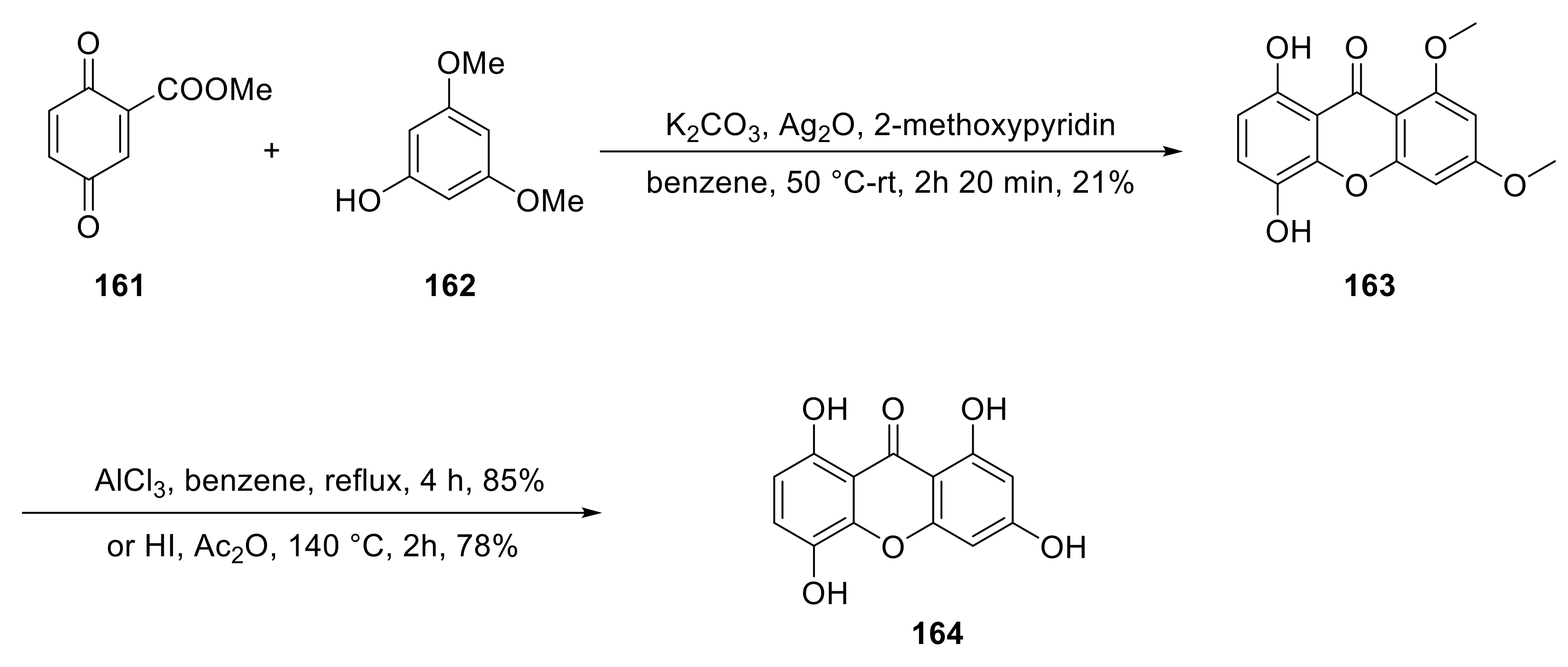
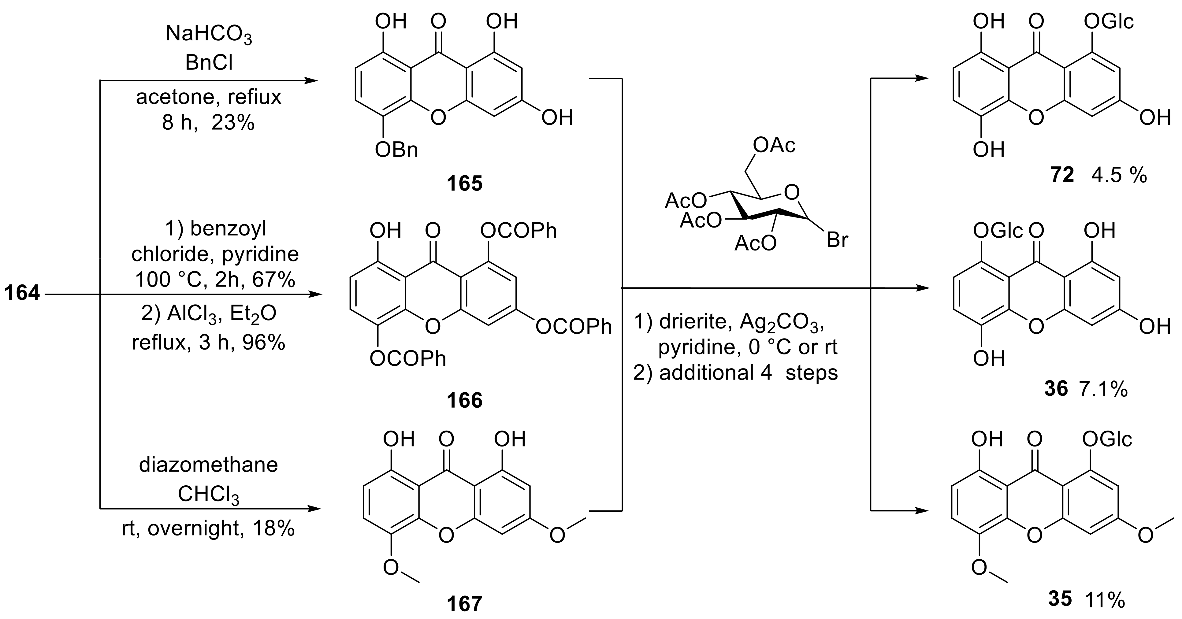
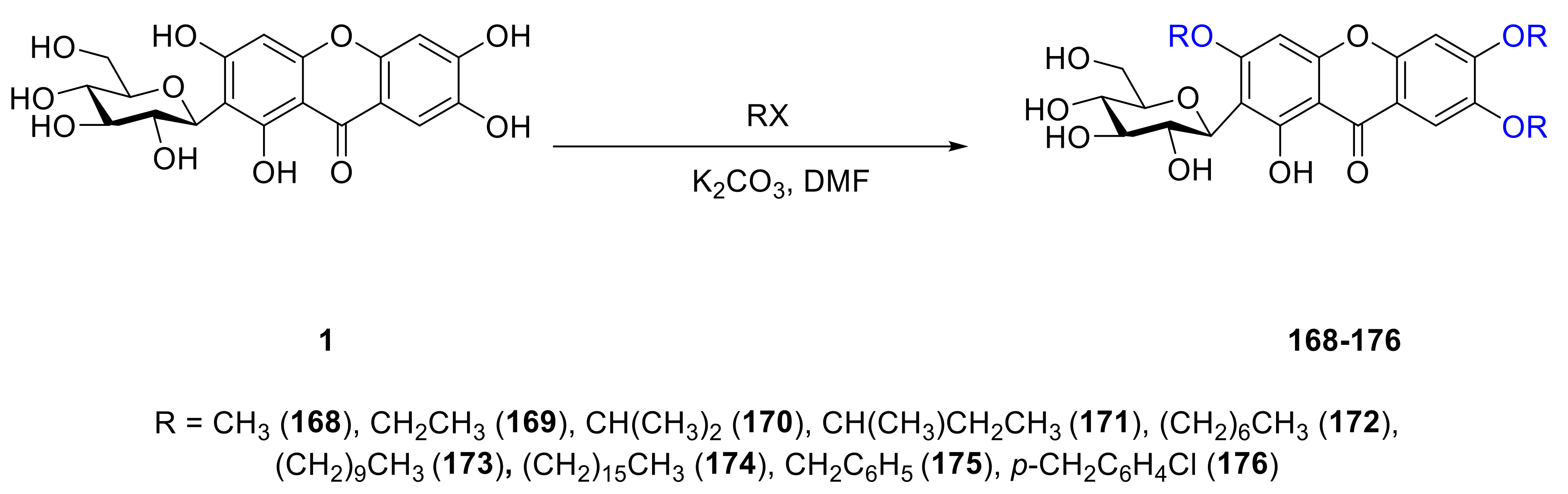
4. Synthesis of Xanthone Glucosides or Derivatives

The first synthesis of xanthone glucosides was accomplished by Wagner in 1985 (Scheme 1) [130]. Three xanthones, 1-(β-d-glucosyloxy)-8-hydroxy-3,5-dimethoxyxanthone (35) [80], 8-(β-d-glucosyloxy)-1,3,5-trihydroxyxanthone (36) [81] and norswertianolin (72) [92], were synthesized in one sequence. Starting with the nucleophilic addition of 3,5-dimethoxyphenol (162) to 2-methoxycarbonyl-1,4-benzoquinone (161), 5,8-dihydroxy-1,3-dimethoxyxanthone (163) was obtained at 21% yield. The compound 1,3,5,8-tetrahydroxyxanthone (164) was completed by refluxing 163 with AlCl3 in benzene for 4 h or heating 163 with HI and Ac2O at 140 °C Compound 164 then went through different steps to form three compounds: 5-benzyloxy-l,3,8-trihydroxyxanthone (165), 1,3,5-tribenzoyloxy-8-hydroxyxanthone (166), and 1,8-dihydroxy-3,5-dimethoxy (167). Then, taking 165167 as aglycons, coupling with α-acetobromoglucose successfully afforded the corresponding products 72, 36, and 35.
The structure of mangiferin (1) is 2-(β-d-glucosyloxy)-1,3,6,7-tetrahydroxyxanthone, which is distributed in a variety of plants and has demonstrated many biological activities. To improve the solubility of 1, several mangiferin derivatives were synthesized by Wu and coworkers (Scheme 2). They used nucleophilic substitution to add alkyl or benzyl groups to the skeleton of mangiferin and nine derivatives 168176 were obtained [131].
Neomangiferin (4) is a derivative of 1. Li and coworkers solved the problem of hydroxyl selectivity and realized the semi-synthesis of 4 from 1 in 2014 (Scheme 3). First, compound 177 was synthesized by acylation in high yield, which is a suitable intermediate for selective benzylation at the 1-, 3- and 6-positions. After de-acylation, only the remaining 7-OH can be coupled with α-d-glucopyranosyl bromide under optimized conditions to give the corresponding product 4 after the removal of all the protective groups [132].
As a continuing work, the Li group completed the total synthesis of three xanthone glucosides including 1, homomangiferin (2) and 4 using an alternative method in 2016. They chose tetrabenzylglucose (181), phloroglucinol derivatives (182183) and bromobenzene derivatives (184) as the starting materials. Compounds 1 and 2 were synthesized by a series of steps, including glycosylation, Vilsmeier formylation, de-protection, selective reprotection, and ring formation reactions. Then, according to the research in 2014, the construction of 4 was completed (Scheme 4) [133].
In addition to chemical methods, enzyme catalysis can also be used to synthesize xanthone glucosides. For example, Zarena et al. used enzyme catalysis to achieve glycosylation of α−mangostin (193) in a supercritical carbon dioxide system [134], and Sohng completed the diversified glycosylation of 193 by a one-pot enzymatic catalysis [135]. In addition, Kim and coworkers modified 1 with glucansucrase to obtain the disglycation product mangiferin-(1→6)-α-d-glucopyranoside (194), thus improving the activity and solubility of mangiferin [24].
Molecules 26 05575 i055

5. Conclusions and Outlook

In this review, we summarized 160 xanthone glucosides, of which xanthone O-glucoside was the most abundant (136 included). These compounds are derived from a variety of sources, with mangiferin being the most widely distributed and having the most investigated pharmacological activities. There was no significant difference in bioactivity between glucosylxanones and xanthones, but glycosylation can usually improve bioactivity.
We reviewed 93 monosaccharide xanthone glucosides and 66 disaccharide xanthone glucosides. Disaccharide xanthone glucosides are composed primarily of two glucose or glucose and xylose sugars, with a small amount of glucose combined with rhamnose, apiose, or arabinose. In terms of sugar binding sites, xanthone C-glucosides have glucosyl groups primarily at C-2, whereas xanthone O-glucosides have glucosyl groups primarily at C-1. Hydroxyl and methoxy groups are the most common substituents on the xanthone skeleton. Only two compounds out of 160 contain a methyl group (159 and 160). Prenylated xanthone glucosides are also extremely rare and have only been discovered in lichens (142157). With the exception of a few examples containing tetrahydroxanthones, xanthone glucosides all have a xanthone skeleton (8081, 109114).
Despite the fact that a number of xanthone glucosides have been discovered, the medicinal study and health benefits of this type of compound have largely been limited to mangiferin. Synthesis and structural modification based on xanthones and glucosyl groups are also underdeveloped. Future research could concentrate on the synthesis of xanthone glucoside derivatives and the investigation of their pharmacological activities.

Funding

We are grateful to the National Natural Science Foundation of China (81973438), the NSFC-Joint Foundation of Yunnan Province (U1902213), Fok Ying-Tong Education Foundation (161039), the Guangdong Province Key Area R&D Program of China (2020B1111110003), The Three-year development plan project for Traditional Chinese Medicine (ZY(2018-2020)-CCCX-2001-02) for financial support.

Institutional Review Board Statement

Not applicable.

Informed Consent Statement

Not applicable.

Conflicts of Interest

The authors declare no conflict of interest.

References

  1. Fiesel, T.; Gaid, M.; Muller, A.; Bartels, J.; El-Awaad, I.; Beuerle, T.; Ernst, L.; Behrends, S.; Beerhues, L. Molecular Cloning and Characterization of a Xanthone Prenyltransferase from Hypericum calycinum Cell Cultures. Molecules 2015, 20, 15616–15630. [Google Scholar] [CrossRef] [PubMed]
  2. Mandal, S.; Das, P.C.; Joshi, P.C. Naturally-occurring xanthones from terrestrial flora. J. Indian Chem. Soc. 1992, 69, 611–636. [Google Scholar]
  3. Peres, V.; Nagem, T.J. Naturally occurring, pentaoxygenated, hexaoxygenated and dimeric xanthones: A literature survey. Quim. Nova 1997, 20, 388–397. [Google Scholar] [CrossRef] [Green Version]
  4. Nhan, N.T.; Nguyen, P.H.; Tran, M.H.; Nguyen, P.D.; Tran, D.T.; To, D.C. Anti-inflammatory xanthone derivatives from Garcinia delpyana. J. Asian Nat. Prod. Res. 2021, 23, 414–422. [Google Scholar] [CrossRef] [PubMed]
  5. Banik, K.; Harsha, C.; Bordoloi, D.; Lalduhsaki Sailo, B.; Sethi, G.; Leong, H.C.; Arfuso, F.; Mishra, S.; Wang, L.; Kumar, A.P.; et al. Therapeutic potential of gambogic acid, a caged xanthone, to target cancer. Cancer Lett. 2018, 416, 75–86. [Google Scholar] [CrossRef] [PubMed]
  6. Moon, K.M.; Kim, C.Y.; Ma, J.Y.; Lee, B. Xanthone-related compounds as an anti-browning and antioxidant food additive. Food Chem. 2019, 274, 345–350. [Google Scholar] [CrossRef] [PubMed]
  7. Akao, Y.; Nakagawa, Y.; Iinuma, M.; Nozawa, Y. Anti-Cancer Effects of Xanthones from Pericarps of Mangosteen. Int. J. Mol. Sci. 2008, 9, 355–370. [Google Scholar] [CrossRef]
  8. Rukachaisirikul, V.; Kamkaew, M.; Sukavisit, D.; Phongpaichit, S.; Sawangchote, P.; Taylor, W.C. Antibacterial xanthones from the leaves of Garcinia nigrolineata. J. Nat. Prod. 2003, 66, 1531–1535. [Google Scholar] [CrossRef]
  9. He, L.; Zhu, C.F.; Yuan, Y.; Xu, Z.F.; Qiu, S.X. Specific glycosylated metabolites of α-mangostin by Cunninghamella blakesleana. Phytochem. Lett. 2014, 9, 175–178. [Google Scholar] [CrossRef]
  10. Gales, L.; Damas, A.M. Xanthones–A Structural Perspective. Curr. Med. Chem. 2005, 12, 2499–2515. [Google Scholar] [CrossRef]
  11. Klein, L.C.; Campos, A.; Niero, R.; Correa, R.; Vander Heyden, Y.; Cechinel, V. Xanthones and Cancer: From Natural Sources to Mechanisms of Action. Chem. Biodivers. 2020, 17, 30. [Google Scholar]
  12. Shan, T.; Ma, Q.; Guo, K.; Liu, J.; Li, W.; Wang, F.; Wu, E. Xanthones from Mangosteen Extracts as Natural Chemopreventive Agents: Potential Anticancer Drugs. Curr. Mol. Med. 2011, 11, 666–677. [Google Scholar] [CrossRef] [PubMed]
  13. Han, Q.B.; Xu, H.X. Caged Garcinia Xanthones: Development since 1937. Curr. Med. Chem. 2009, 16, 3775–3796. [Google Scholar] [CrossRef] [PubMed]
  14. El-Seedi, H.R.; El-Ghorab, D.M.H.; El-Barbary, M.A.; Zayed, M.F.; Goransson, U.; Larsson, S.; Verpoorte, R. Naturally Occurring Xanthones; Latest Investigations: Isolation, Structure Elucidation and Chemosystematic Significance. Curr. Med. Chem. 2009, 16, 2581–2626. [Google Scholar] [CrossRef] [PubMed]
  15. Na, Y. Recent cancer drug development with xanthone structures. J. Pharm. Pharmacol. 2009, 61, 707–712. [Google Scholar] [CrossRef] [PubMed]
  16. Araujo, J.; Fernandes, C.; Pinto, M.; Tiritan, M.E. Chiral Derivatives of Xanthones with Antimicrobial Activity. Molecules 2019, 24, 314. [Google Scholar] [CrossRef] [Green Version]
  17. Vieira, L.M.M.; Kijjoa, A. Naturally-Occurring Xanthones: Recent Developments. Curr. Med. Chem. 2005, 12, 2413–2446. [Google Scholar] [CrossRef]
  18. Wu, Q.L.; Wang, S.P.; Du, L.J.; Yang, J.S.; Xiao, P.G. Xanthones from Hypericum japonicum and H-Henryi. Phytochemistry 1998, 49, 1395–1402. [Google Scholar] [CrossRef]
  19. Perest, V.; Nagem, T.J. Trioxygenated naturally occurring xanthones. Phywchemistry 1997, 44, 191–214. [Google Scholar] [CrossRef]
  20. Valdir, P.; Nagem, T.J.; de Oliveira, F.F. Tetraoxygenated naturally occurring xanthones. Phytochemistry 2000, 55, 683–710. [Google Scholar]
  21. Mangangcha, I.R.; Singh, R.K.B.; Lebeche, D.; Ali, S. Xanthone glucoside 2-beta-d-glucopyranosyl-1,3,6,7-tetrahydroxy-9H-xanthen-9-one binds to the ATP-binding pocket of glycogen synthase kinase 3 beta and inhibits its activity: Implications in prostate cancer and associated cardiovascular disease risk. J. Biomol. Struct. Dyn. 2021. [Google Scholar] [CrossRef] [PubMed]
  22. Ghosal, S.; Sharma, P.V.; Chaudhuri, R.K. Chemical constituents of gentianaceae. X. Xanthone-O-glucosides of Swertia purpurascens Wall. J. Pharm. Sci. 1974, 63, 1286–1290. [Google Scholar] [CrossRef] [PubMed]
  23. Kren, V.; Martinkova, L. Glycosides in medicine: “The role of glycosidic residue in biological activity”. Curr. Med. Chem. 2001, 8, 1303–1328. [Google Scholar] [CrossRef]
  24. Septiana, I.; Nguyen, T.T.H.; Lim, S.; Lee, S.; Park, B.; Kwak, S.; Park, S.; Kim, S.B.; Kim, D. Enzymatic synthesis and biological characterization of a novel mangiferin glucoside. Enzym. Microb. Technol. 2020, 134, 109479. [Google Scholar] [CrossRef]
  25. Feng, S.T.; Wang, Z.Z.; Yuan, Y.H.; Sun, H.M.; Chen, N.H.; Zhang, Y. Mangiferin: A multipotent natural product preventing neurodegeneration in Alzheimer’s and Parkinson’s disease models. Pharmacol. Res. 2019, 146, 12. [Google Scholar] [CrossRef]
  26. Jo, C.; Yoon, K.Y.; Jang, E.J.; Kim, T.H. Degradation products of mangiferin by gamma irradiation with inhibitory effects on NO production. Biosci. Biotechnol. Biochem. 2016, 80, 2022–2024. [Google Scholar] [CrossRef] [Green Version]
  27. Yoshimi, N.; Matsunaga, K.; Katayama, M.; Yamada, Y.; Kuno, T.; Qiao, Z.; Hara, A.; Yamahara, J.; Mori, H. The inhibitory effects of mangiferin, a naturally occurring glucosylxanthone, in bowel carcinogenesis of male F344 rats. Cancer Lett. 2001, 163, 163–170. [Google Scholar] [CrossRef]
  28. Kim, G.E.; Kang, H.K.; Seo, E.S.; Jung, S.H.; Park, J.S.; Kim, D.H.; Kim, D.W.; Ahn, S.A.; Sunwoo, C.; Kim, D. Glucosylation of the flavonoid, astragalin by Leuconostoc mesenteroides B-512FMCM dextransucrase acceptor reactions and characterization of the products. Enzym. Microb. Technol. 2012, 50, 50–56. [Google Scholar] [CrossRef]
  29. Faizi, S.; Zikr-ur-Rehman, S.; Naz, A.; Versiani, M.A.; Dar, A.; Naqvi, S. Bioassay-guided studies on Bombax ceiba leaf extract: Isolation of shamimoside, a new antioxidant xanthone C-glucoside. Chem. Nat. Compd. 2012, 48, 774–779. [Google Scholar] [CrossRef]
  30. Finnegan, R.A.; Stephani, R.A.; Ganguli, G.; Ganguly, S.N.; Bhattacharya, A.K. Occurrence of mangiferin in Hiptage madablota Geartn. J. Pharm. Sci. 1968, 57, 1039–1040. [Google Scholar] [CrossRef] [PubMed]
  31. Hong, Y.F.; Han, G.Y.; Guo, X.M. Isolation and structure determination of xanthone glycosides of Anemarrhena asphodeloides. Acta Pharm. Sin. 1997, 32, 473–475. [Google Scholar]
  32. Iwashina, T.; Kitajima, J.; Shiuchi, T.; Itou, Y. Chalcones and other flavonoids from Asarum sensu lato (Aristolochiaceae). Biochem. Syst. Ecol. 2005, 33, 571–584. [Google Scholar] [CrossRef]
  33. Zhang, Y.B.; Xu, X.J.; Liu, H.M. Chemical constituents from Mahkota dewa. J. Asian Nat. Prod. Res. 2006, 8, 119–123. [Google Scholar] [CrossRef]
  34. Talamond, P.; Mondolot, L.; Gargadennec, A.; de Kochko, A.; Hamon, S.; Fruchier, A.; Campa, C. First report on mangiferin (C-glucosyl-xanthone) isolated from leaves of a wild coffee plant, Coffea pseudozanguebariae (Rubiaceae). Acta Bot. Gallica 2008, 155, 513–519. [Google Scholar] [CrossRef] [Green Version]
  35. Li, L.; Li, M.H.; Zhang, N.; Huang, L.Q. Chemical constituents from Lomatogonium carinthiacum (Gentianaceae). Biochem. Syst. Ecol. 2011, 39, 766–768. [Google Scholar] [CrossRef]
  36. Khare, P.; Shanker, K. Mangiferin: A review of sources and interventions for biological activities. Biofactors 2016, 42, 504–514. [Google Scholar]
  37. Wei, Z.; Yan, L.; Chen, Y.; Bao, C.; Deng, J.; Deng, J. Mangiferin inhibits macrophage classical activation via downregulating interferon regulatory factor 5 expression. Mol. Med. Rep. 2016, 14, 1091–1098. [Google Scholar] [CrossRef] [PubMed] [Green Version]
  38. Feng, Z.L.; Lu, X.Q.; Gan, L.S.; Zhang, Q.W.; Lin, L.G. Xanthones, A Promising Anti-Inflammatory Scaffold: Structure, Activity, and Drug Likeness Analysis. Molecules 2020, 25, 598. [Google Scholar] [CrossRef] [Green Version]
  39. Sha, H.; Zeng, H.R.; Zhao, J.; Jin, H.Y. Mangiferin ameliorates gestational diabetes mellitus-induced placental oxidative stress, inflammation and endoplasmic reticulum stress and improves fetal outcomes in mice. Eur. J. Pharmacol. 2019, 859, 7. [Google Scholar] [CrossRef]
  40. Dar, A.; Faizi, S.; Naqvi, S.; Roome, T.; Zikr-ur-Rehman, S.; Ali, M.; Firdous, S.; Moin, S.T. Analgesic and antioxidant activity of mangiferin and its derivatives: The structure activity relationship. Biol. Pharm. Bull. 2005, 28, 596–600. [Google Scholar] [CrossRef] [Green Version]
  41. He, L.Y.; Peng, X.F.; Zhu, J.F.; Chen, X.; Liu, H.; Tang, C.Y.; Dong, Z.; Liu, F.Y.; Peng, Y.M. Mangiferin Attenuate Sepsis-Induced Acute Kidney Injury via Antioxidant and Anti-Inflammatory Effects. Am. J. Nephrol. 2014, 40, 441–450. [Google Scholar] [CrossRef]
  42. PardO-Andreu, G.L.; Barrios, M.F.; Curti, C.; Hernandez, I.; Merino, N.; Lemus, Y.; Martinez, L.; Riano, A.; Delgado, R. Protective effects of Mangifera indica L. extract (Vimang), and its major component mangiferin, on iron-induced oxidative damage to rat serum and liver. Pharmacol. Res. 2008, 57, 79–86. [Google Scholar] [CrossRef] [PubMed]
  43. Zheng, H.H.; Luo, C.T.; Chen, H.R.; Lin, J.N.; Ye, C.L.; Mao, S.S.; Li, Y.L. Xanthones from Swertia mussotii as Multitarget-Directed Antidiabetic Agents. Chemmedchem 2014, 9, 1374–1377. [Google Scholar] [CrossRef] [PubMed]
  44. Muruganandan, S.; Srinivasan, K.; Gupta, S.; Gupta, P.K.; Lal, J. Effect of mangiferin on hyperglycemia and atherogenicity in streptozotocin diabetic rats. J. Ethnopharmacol. 2005, 97, 497–501. [Google Scholar] [CrossRef]
  45. Miura, T.; Ichiki, H.; Hashimoto, I.; Iwamoto, N.; Kato, M.; Kubo, M.; Ishihara, E.; Komatsu, Y.; Okada, M.; Ishida, T.; et al. Antidiabetic activity of a xanthone compound, mangiferin. Phytomedicine 2001, 8, 85–87. [Google Scholar] [CrossRef] [PubMed]
  46. Wang, Z.B.; Wu, G.S.; Yu, Y.; Liu, H.; Yang, B.Y.; Kuang, H.X.; Wang, Q.H. Xanthones isolated from Gentianella acuta and their protective effects against H2O2-induced myocardial cell injury. Nat. Prod. Res. 2018, 32, 2171–2177. [Google Scholar] [CrossRef]
  47. Muruganandan, S.; Gupta, S.; Kataria, M.; Lal, J.; Gupta, P.K. Mangiferin protects the streptozotocin-induced oxidative damage to cardiac and renal tissues in rats. Toxicology 2002, 176, 165–173. [Google Scholar] [CrossRef]
  48. Prabhu, S.; Jainu, M.; Sabitha, K.E.; Devi, C.S.S. Effect of mangiferin on mitochondrial energy production in experimentally induced myocardial infarcted rats. Vasc. Pharmacol. 2006, 44, 519–525. [Google Scholar] [CrossRef]
  49. Zeng, Z.; Lin, C.J.; Wang, S.W.; Wang, P.F.; Xu, W.W.; Ma, W.Q.; Wang, J.L.; Xiang, Q.; Liu, Y.T.; Yang, J.M.; et al. Suppressive activities of mangiferin on human epithelial ovarian cancer. Phytomedicine 2020, 76, 9. [Google Scholar] [CrossRef]
  50. Deng, Q.; Tian, Y.X.; Liang, J.J. Mangiferin inhibits cell migration and invasion through Rac1/WAVE2 signalling in breast cancer. Cytotechnology 2018, 70, 593–601. [Google Scholar] [CrossRef]
  51. Lv, J.Z.; Wang, Z.J.; Zhang, L.; Wang, H.L.; Liu, Y.D.; Li, C.Y.; Deng, J.G.; Yi, W.; Bao, J.K. Mangiferin Induces Apoptosis and Cell Cycle Arrest in MCF-7 Cells Both in vitro and in vivo. J. Anim. Vet. Adv. 2013, 12, 352–359. [Google Scholar]
  52. Pinto, M.M.M.; Sousa, M.E.; Nascimento, M.S.J. Xanthone derivatives: New insights in biological activities. Curr. Med. Chem. 2005, 12, 2517–2538. [Google Scholar] [CrossRef]
  53. Sato, T.; Kawamoto, A.; Tamura, A.; Tatsumi, Y.; Fujii, T. Mechanism of antioxidant action of pueraria glycoside (PG)-1 (an isoflavonoid) and mangiferin (a xanthonoid). Chem. Pharm. Bull. 1992, 40, 721–724. [Google Scholar] [CrossRef] [PubMed] [Green Version]
  54. Vyas, A.; Syeda, K.; Ahmad, A.; Padhye, S.; Sarkar, F.H. Perspectives on Medicinal Properties of Mangiferin. Mini-Rev. Med. Chem. 2012, 12, 412–425. [Google Scholar] [CrossRef] [PubMed]
  55. Andreu, G.P.; Delgado, R.; Velho, J.A.; Curti, C.; Vercesi, A.E. Iron complexing activity of mangiferin, a naturally occurring glucosylxanthone, inhibits mitochondrial lipid peroxidation induced by Fe2+-citrate. Eur. J. Pharmacol. 2005, 513, 47–55. [Google Scholar] [CrossRef] [PubMed]
  56. Aritomi, M.; Kawasaki, T. A new xanthone C-glucoside, position isomer of mangiferin, from Anemarrhena asphodeloides Bunge. Chem. Pharm. Bull. 1970, 18, 2327–2333. [Google Scholar] [CrossRef] [Green Version]
  57. Kokotkiewicz, A.; Luczkiewicz, M.; Pawlowska, J.; Luczkiewicz, P.; Sowinski, P.; Witkowski, J.; Bryl, E.; Bucinski, A. Isolation of xanthone and benzophenone derivatives from Cyclopia genistoides (L.) Vent. (honeybush) and their pro-apoptotic activity on synoviocytes from patients with rheumatoid arthritis. Fitoterapia 2013, 90, 199–208. [Google Scholar] [CrossRef] [PubMed]
  58. Lim, S.M.; Kang, G.D.; Jeong, J.J.; Choi, H.S.; Kim, D.H. Neomangiferin modulates the Th17/Treg balance and ameliorates colitis in mice. Phytomedicine 2016, 23, 131–140. [Google Scholar] [CrossRef]
  59. Arisawa, M.; Morita, N.; Kondo, Y.; Takemoto, T. The constiuents of Iris florentina L. (3). structure of irisxanthone, a know C-glucosylxanthone. Chem. Pharm. Bull. 1973, 21, 2562–2565. [Google Scholar] [CrossRef] [Green Version]
  60. Abdel-Mageed, W.M.; Al-Wahaibi, L.H.; Al-Saleem, M.S.M.; Gouda, Y.G.; Abdel-Kader, M.S.; Ibraheim, Z.Z. Phytochemical and chemotaxonomic study on Iris albicans Lange leaves. Biochem. Syst. Ecol. 2018, 76, 32–34. [Google Scholar] [CrossRef]
  61. Bukvicki, D.; Novakovic, M.; Ab Ghani, N.; Marin, P.D.; Asakawa, Y. Secondary metabolites from endemic species Iris adriatica Trinajstic ex Mitic (Iridaceae). Nat. Prod. Res. 2018, 32, 1849–1852. [Google Scholar] [CrossRef]
  62. Xie, G.Y.; Chen, Y.J.; Wen, R.; Xu, J.Y.; Wu, S.S.; Qin, M.J. Chemical constituents from rhizomes of Iris germanica. China J. Chin. Mater. Med. 2014, 39, 846–850. [Google Scholar]
  63. Alkhalil, S.; Tosa, H.; Iinuma, M. A xanthone C-glycoside from Iris Nigricans. Phytochemistry 1995, 38, 729–731. [Google Scholar] [CrossRef]
  64. Pauletti, P.M.; Castro-Gamboa, I.; Silva, D.H.S.; Young, M.C.M.; Tomazela, D.M.; Eberlin, M.N.; Bolzani, V.D. New antioxidant C-glucosylxanthones from the stems of Arrabidaea samydoides. J. Nat. Prod. 2003, 66, 1384–1387. [Google Scholar] [CrossRef]
  65. Martin, F.; Hay, A.E.; Cressend, D.; Reist, M.; Vivas, L.; Gupta, M.P.; Carrupt, P.A.; Hostettmann, K. Antioxidant C-Glucosylxanthones from the Leaves of Arrabidaea patellifera. J. Nat. Prod. 2008, 71, 1887–1890. [Google Scholar] [CrossRef] [PubMed]
  66. Miyase, T.; Noguchi, H.; Chen, X.M. Sucrose Esters and Xanthone C-Glycosides from the Roots of Polygala sibirica. J. Nat. Prod. 1999, 62, 993–996. [Google Scholar] [CrossRef]
  67. Jiang, Y.; Zhang, W.; Tu, P.F.; Xu, X.J. Xanthone Glycosides from Polygala tenuifolia and Their Conformational Analyses. J. Nat. Prod. 2005, 68, 875–879. [Google Scholar] [CrossRef]
  68. Chang, H.T.; Tu, P.F. New oligosaccharide esters and xanthone C-glucosides from Polygala telephioides. Helv. Chim. Acta 2007, 90, 944–950. [Google Scholar] [CrossRef]
  69. Tsujimoto, T.; Nishihara, M.; Osumi, Y.; Hakamatsuka, T.; Goda, Y.; Uchiyama, N.; Ozeki, Y. Structural Analysis of Polygalaxanthones, C-Glucosyl Xanthones of Polygala tenuifolia Roots. Chem. Pharm. Bull. 2019, 67, 1242–1247. [Google Scholar] [CrossRef] [PubMed] [Green Version]
  70. Wu, J.F.; Chen, S.B.; Gao, J.C.; Song, H.L.; Wu, L.J.; Chen, S.L.; Tu, P.F. Xanthone glycosides from herbs of Polygala hongkongensis Hemsl and their antioxidant activities. J. Asian Nat. Prod. Res. 2008, 10, 673–678. [Google Scholar] [CrossRef] [PubMed]
  71. Jankovic, T.; KrstiC-Milosevic, D.; Aljancic, I.; Savikin, K.; Menkovic, N.; Radanovic, D.; Milosavljevic, S. Phytochemical re-investigation of Gentiana utriculosa. Nat. Prod. Res. 2009, 23, 466–469. [Google Scholar] [CrossRef]
  72. Schaufelberger, D.; Hostettmann, K. Chemistry and pharmacology of Gentiana lactea. Planta Med. 1988, 54, 219–221. [Google Scholar] [CrossRef]
  73. Fujita, M.; Inoue, T. Studies on the Constituents of Iris florentina L. II. C-Glucosides of Xanthones and Flavones from the Leaves. Chem. Pharm. Bull. 1982, 30, 2342–2348. [Google Scholar] [CrossRef] [Green Version]
  74. Shi, T.X.; Wang, S.; Zeng, K.W.; Tu, P.F.; Jiang, Y. Inhibitory constituents from the aerial parts of Polygala tenuifolia on LPS-induced NO production in BV2 microglia cells. Bioorg. Med. Chem. Lett. 2013, 23, 5904–5908. [Google Scholar] [CrossRef]
  75. Tan, P.; Hou, C.Y.; Liu, Y.L.; Lin, L.J.; Cordell, G.A. Swertipunicoside. The First Bisxanthone C-Glycoside. J. Org. Chem. 1991, 56, 7130–7133. [Google Scholar] [CrossRef]
  76. Tan, P.; Hou, C.Y.; Liu, Y.L.; Lin, L.J.; Cordell, G.A. 3-O-demethylswertipunicoside from Swertia punicea. Phytochemistry 1992, 31, 4313–4315. [Google Scholar] [CrossRef]
  77. Du, X.G.; Wang, W.; Zhang, S.P.; Pu, X.P.; Zhang, Q.Y.; Ye, M.; Zhao, Y.Y.; Wang, B.R.; Khan, I.A.; Guo, D.A. Neuroprotective Xanthone Glycosides from Swertia punicea. J. Nat. Prod. 2010, 73, 1422–1426. [Google Scholar] [CrossRef]
  78. Luo, C.T.; Mao, S.S.; Liu, F.L.; Yang, M.X.; Chen, H.; Kurihara, H.; Li, Y. Antioxidant xanthones from Swertia mussotii, a high altitude plant. Fitoterapia 2013, 91, 140–147. [Google Scholar] [CrossRef] [PubMed]
  79. Zhang, L.; Zou, D.Z.; Bai, S.; Li, Z.H.; Zhang, C.H.; Li, M.H. Chemical constituents from Gentianella turkestanorum (Gentianaceae). Biochem. Syst. Ecol. 2016, 65, 89–92. [Google Scholar] [CrossRef]
  80. Abdel-Mageed, W.M.; Bayoumi, S.A.H.; Chen, C.; Vavricka, C.J.; Li, L.; Malik, A.; Dai, H.; Song, F.; Wang, L.; Zhang, J.; et al. Benzophenone C-glucosides and gallotannins from mango tree stem bark with broad-spectrum anti-viral activity. Bioorg. Med. Chem. 2014, 22, 2236–2243. [Google Scholar] [CrossRef] [PubMed]
  81. Stout, G.H.; Balkenhol, W.J. Xanthones of the Gentianaceae-I Frasera caroliniensis Walt. Tetrahedron 1969, 25, 1947–1960. [Google Scholar] [CrossRef]
  82. Tomimori, T.; Komatsu, M. Studies on the Constituents of Swertia japonica. VI. on the Flavonoid and Xanthone Constituents of Swertia randaiensis. HAYATA amd S. swertopsis MAKINO. Yakugaku Zasshi 1969, 89, 1276–1282. [Google Scholar] [CrossRef] [Green Version]
  83. Tomimori, T.; Yoshizaki, M.; Nanba, T. Studies on the Nepalese Crude Drugs.I. On the Flavonoid and Xanthone Constituents of the Plants of Swertia spp. Yakugaku Zasshi 1973, 93, 442–447. [Google Scholar] [CrossRef] [PubMed] [Green Version]
  84. Hostettmann, K.; Tabacchi, R.; Jacot-Guillarmod, A. Contribution à la phytochimie du genre Gentiana, VI. Etude des xanthones dans les feuilles de Gentiana bavarica L. Helv. Chim. Acta 1974, 57, 294–301. [Google Scholar] [CrossRef]
  85. Hostettmann, K.; Miura, I. A New Xanthone Diglucoside from Swertia perennis L. Helv. Chim. Acta 1977, 60, 262–264. [Google Scholar] [CrossRef]
  86. Ghosal, S.; Sharma, P.V.; Jaiswal, D.K. Chemical constituents of Gentianaceae XXIII: Tetraoxygenated and pentaoxygenated xanthones and xanthone O-glucosides of Swertia angustifolia Buch.-Ham. J. Pharm. Sci. 1978, 67, 55–60. [Google Scholar] [CrossRef]
  87. Dhasmana, H.; Garg, H.S. Two xanthone glucosides from Halenia elliptica. Phytochemrstry 1989, 28, 2819–2821. [Google Scholar] [CrossRef]
  88. Sun, H.F.; Hu, B.L.; Ding, J.Y.; Fan, S.F. The glucosides from Swertia mussotii Franch. Acta Bot. Sin. 1991, 33, 31–37. [Google Scholar]
  89. Recio-Iglesias, M.C.; Marston, A.; Hosteyitman, K. Xanthones and secoiridoid glucosides of Halenia campanulata. Phytochemistry 1992, 31, 1387–1389. [Google Scholar] [CrossRef]
  90. Rodriguez, S.; Wolfender, J.L.; Odontuya, G.; Purev, O.; Hostettmann, K. Xanthones, secoiridoids and flavonoids from Halenia corniculata. Phytochemistry 1995, 40, 1265–1272. [Google Scholar] [CrossRef]
  91. Otsuka, H.; Triptexanthosides, A.-E. Xanthone Glycosides from Aerial Parts of Tripterospermum japonicum. Chem. Pharm. Bull. 1999, 47, 962–965. [Google Scholar] [CrossRef] [Green Version]
  92. Menkovic, N.; Savikin-Fodulovic, K.; Bulatovic, V.; Aljancic, I.; Juranic, N.; Macura, S.; Vajs, V.; Milosavljevic, S. Xanthones from Swertia punctata. Phytochemistry 2002, 61, 415–420. [Google Scholar] [CrossRef]
  93. Zeng, G.Y.; Tan, G.S.; Xu, K.P.; Xu, X.P.; Li, F.S.; Tan, J.B.; Hu, G.Y. Water-soluble chemical constituents of Swertia davidi Franch. Acta Pharm. Sin. 2004, 39, 351–353. [Google Scholar]
  94. Kaldas, M.; Hostettmann, K.; Jacot-Guillarmod, A. Contribution à la phytochimie du genre Gentiana IX. Etude de composés flavoniques et xanthoniques dans les feuilles de gentiana CampestrisL. 1ère communication. Helv. Chim. Acta 1974, 57, 2557–2561. [Google Scholar] [CrossRef]
  95. Rana, V.S.; Rawat, M.S.M. A New Xanthone Glycoside and Antioxidant Constituents from the Rhizomes of Swertia speciosa. Chem. Biodivers. 2005, 2, 1310–1315. [Google Scholar] [CrossRef]
  96. Hajimehdipoor, H.; Dijoux-Franca, M.G.; Mariotte, A.M.; Amanzadeh, Y.; Sadat-Ebrahimi, S.E.; Ghazi-Khansari, M. Two new xanthone diglycosides from Swertia longifolia Boiss. Nat. Prod. Res. 2006, 20, 1251–1257. [Google Scholar] [CrossRef]
  97. Murray, A.P.; Faraoni, M.B.; Castro, M.J.; Alza, N.P.; Cavallaro, V. Natural AChE Inhibitors from Plants and their Contribution to Alzheimer’s Disease Therapy. Curr. Neuropharmacol. 2013, 11, 388–413. [Google Scholar] [CrossRef] [Green Version]
  98. Nalivaeva, N.N.; Turner, A.J. AChE and the amyloid precursor protein (APP)—Cross-talk in Alzheimer’s disease. Chem.-Biol. Interact. 2016, 259, 301–306. [Google Scholar] [CrossRef] [PubMed]
  99. Sherif, F.; Gottfries, C.G.; Alafuzoff, I.; Oreland, L. Brain gamma-aminobutyrate aminotransferase (GABA-T) and monoamine-oxidase (MAO) in patients with alzheimers-disease. J. Neural Transm.-Park. Dis. Dement. Sect. 1992, 4, 227–240. [Google Scholar] [CrossRef] [PubMed]
  100. Urbain, A.; Marston, A.; Grilo, L.S.; Bravo, J.; Purev, O.; Purevsuren, B.; Batsuren, D.; Reist, M.; Carrupt, P.A.; Hostettmann, K. Xanthones from Gentianella amarella ssp acuta with acetylcholinesterase and monoamine oxidase inhibitory activities. J. Nat. Prod. 2008, 71, 895–897. [Google Scholar] [CrossRef] [PubMed]
  101. Tang, L.; Xu, X.M.; Rinderspacher, K.A.; Cai, C.Q.; Ma, Y.; Long, C.L.; Feng, J.C. Two new compounds from Comastoma pedunlulatum. J. Asian Nat. Prod. Res. 2011, 13, 895–900. [Google Scholar] [CrossRef] [PubMed]
  102. Ding, L.; Liu, B.; Zhang, S.D.; Hou, Q.; Qi, L.L.; Zhou, Q.Y. Cytotoxicity, apoptosis-inducing effects and structure-activity relationships of four natural xanthones from Gentianopsis paludosa Ma in HepG2 and HL-60 cells. Nat. Prod. Res. 2011, 25, 669–683. [Google Scholar] [CrossRef]
  103. Gao, L.; Zhou, Y.; Yan, H.; Huang, F.; Wen, R.; Li, G. Two new xanthone glucosides from Swertia mussotii Franch. Heterocycles 2011, 83, 1897–1902. [Google Scholar]
  104. Wan, L.S.; Min, Q.X.; Wang, Y.L.; Yue, Y.D.; Chen, J.C. Xanthone glycoside constituents of Swertia kouitchensis with α-glucosidase inhibitory activity. J. Nat. Prod. 2013, 76, 1248–1253. [Google Scholar] [CrossRef]
  105. Yue, Y.D.; Zhang, Y.T.; Liu, Z.X.; Min, Q.X.; Wan, L.S.; Wang, Y.L.; Xiao, Z.Q.; Chen, J.C. Xanthone Glycosides from Swertia bimaculata with α-Glucosidase Inhibitory Activity. Planta Med. 2014, 80, 502–508. [Google Scholar] [CrossRef] [PubMed]
  106. Chen, Y.L.; Tong, Y.F.; Wang, Q.H. Two new xanthones from Lomatogonium carinthiacum. Chin. J. Nat. Med. 2014, 12, 693–696. [Google Scholar]
  107. Mahendran, G.; Manoj, M.; Murugesh, E.; Sathish Kumar, R.; Shanmughavel, P.; Rajendra Prasad, K.J.; Narmatha Bai, V. In vivo anti-diabetic, antioxidant and molecular docking studies of 1,2,8-trihydroxy-6-methoxy xanthone and 1,2-dihydroxy-6-methoxyxanthone-8-O-β-d-xylopyranosyl isolated from Swertia corymbosa. Phytomedicine 2014, 21, 1237–1248. [Google Scholar] [CrossRef]
  108. Lu, S.; Tanaka, N.; Kawazoe, K.; Murakami, K.; Damdinjav, D.; Dorjbal, E.; Kashiwada, Y. Tetrahydroxanthones from Mongolian medicinal plant Gentianella amarella ssp. acuta. J. Nat. Med. 2016, 70, 780–788. [Google Scholar] [CrossRef]
  109. Shi, K.L.; Wang, Y.Q.; Jiang, Q.; Liao, Z.X. Chemical Constituents of Gentianella azurea. Chin. J. Nat. Med. 2010, 8, 425–428. [Google Scholar]
  110. Xu, K.P.; Li, F.S.; Liu, J.F.; Tan, J.B.; Zhang, L.H.; Zeng, G.Y.; Tan, G.S. Studies on Chemical Constituents of Swertia nervosa (G.Don) Wall. Chin. Pharm. J. 2008, 43, 1612–1615. [Google Scholar]
  111. Tan, G.S.; Xu, P.S.; Tian, H.Y.; Xu, K.P.; Dai, Z.Y. Studies on the chemical constituents of Swertia davidi. Chin. Pharm. J. 2000, 35, 441–443. [Google Scholar]
  112. Tian, C.W.; Zhang, T.J.; Wang, L.L.; Shan, Q.; Jiang, L.H. The hepatoprotective effect and chemical constituents of total iridoids and xanthones extracted from Swertia mussotii Franch. J. Ethnopharmacol. 2014, 154, 259–266. [Google Scholar] [CrossRef] [PubMed]
  113. Wang, Z.; Wu, G.; Liu, H.; Xing, N.; Sun, Y.; Zhai, Y.; Yang, B.; Kong, A.T.; Kuang, H.; Wang, Q. Cardioprotective effect of the xanthones from Gentianella acuta against myocardial ischemia/reperfusion injury in isolated rat heart. Biomed. Pharmacother. 2017, 93, 626–635. [Google Scholar] [CrossRef] [PubMed]
  114. Tosa, H.; Iinuma, M.; Murakami, K.; Ito, T.; Tanaka, T.; Chelladurai, V.; Riswan, S. Three xanthones from Poeciloneuron pauciflorum and Mammea acuminata. Phytochemistry 1997, 45, 133–136. [Google Scholar] [CrossRef]
  115. Fu, P.; Zhang, W.D.; Liu, R.H.; Li, T.Z.; Shen, Y.H.; Li, H.L.; Zhang, W.; Chen, H.S. Two new xanthones from Hypericum japonicum. Nat. Prod. Res. 2006, 20, 1237–1240. [Google Scholar] [CrossRef] [PubMed]
  116. Ishiguro, K.; Yamamota, R.; Oku, H. Patulosides A and B, novel xanthone glycosides from cell suspension cultures of Hypericum patulum. J. Nat. Prod. 1999, 62, 906–908. [Google Scholar] [CrossRef]
  117. Kitanov, G.M.; Nedialkov, P.T. Xanthohypericoside, a new xanthone-O-glucoside from Hypericum annulatum. Pharmazie 2000, 55, 397–398. [Google Scholar]
  118. Yan, X.T.; An, Z.; Huangfu, Y.; Zhang, Y.T.; Li, C.H.; Chen, X.; Liu, P.L.; Gao, J.M. Polycyclic polyprenylated acylphloroglucinol and phenolic metabolites from the aerial parts of Hypericum elatoides and their neuroprotective and anti-neuroinflammatory activities. Phytochemistry 2019, 159, 65–74. [Google Scholar] [CrossRef]
  119. Woo, K.W.; Lee, K.H.; Jang, J.H.; Kim, M.S.; Cho, H.W.; Cho, J.H.; An, B. Anti-inflammatory Constituents from the Aerial Parts of Iris minutiaurea. Nat. Prod. Commun. 2016, 11, 817–819. [Google Scholar] [CrossRef] [PubMed] [Green Version]
  120. Li, W.K.; Chan, C.L.; Leung, H.W.; Yeung, H.W.; Xiao, P.G. Xanthones from Polygala caudata. Phytochemistry 1999, 51, 953–958. [Google Scholar] [CrossRef]
  121. Abd El-Kader, A.M.; Ahmed, A.S.; Nafady, A.M.; Ibraheim, Z.Z. Xanthone and lignan glycosides from the aerial parts of Polygonum bellardii all growing in Egypt. Pharmacogn. Mag. 2013, 9, 135–143. [Google Scholar] [PubMed]
  122. Li, J.; Jiang, Y.; Tu, P.F. Xanthone O-Glycosides and Benzophenone O-Glycosides from the Roots of Polygala tricornis. J. Nat. Prod. 2005, 68, 1802–1804. [Google Scholar] [CrossRef] [PubMed]
  123. Jiang, Y.; Tu, P.F. Xanthone O-glycosides from Polygala tenuifolia. Phytochemistry 2002, 60, 813–816. [Google Scholar] [CrossRef]
  124. He, K.; Fan, L.L.; Wu, T.T.; Du, J. A new xanthone glycoside from Pyrrosia sheareri. Nat. Prod. Res. 2019, 33, 2982–2987. [Google Scholar] [CrossRef]
  125. Rezanka, T.; Dembitsky, V.M. Identification of acylated xanthone glycosides by liquid chromatography-atmospheric pressure chemical ionization mass spectrometry in positive and negative modes from the lichen Umbilicaria proboscidea. J. Chromatogr. A 2003, 995, 109–118. [Google Scholar] [CrossRef]
  126. Rezanka, T.; Jachymova, J.; Dembitsky, V.M. Prenylated xanthone glucosides from Ural’s lichen Umbilicaria proboscidea. Phytochemistry 2003, 62, 607–612. [Google Scholar] [CrossRef]
  127. Eltamany, E.E.; Abdelmohsen, U.R.; Ibrahim, A.K.; Hassanean, H.A.; Hentschel, U.; Ahmed, S.A. New antibacterial xanthone from the marine sponge-derived Micrococcus sp. EG45. Bioorg. Med. Chem. Lett. 2014, 24, 4939–4942. [Google Scholar] [CrossRef]
  128. Yang, B.J.; Chen, G.D.; Li, Y.J.; Hu, D.; Guo, L.D.; Xiong, P.; Gao, H. A New Xanthone Glycoside from the Endolichenic Fungus Sporormiella irregularis. Molecules 2016, 21, 764. [Google Scholar] [CrossRef]
  129. Yoneyama, T.; Iguchi, M.; Yoshii, K.; Elshamy, A.I.; Ban, S.; Noji, M.; Umeyama, A. Xanthone glucoside from an insect pathogenic fungus Conoideocrella luteorostrata NBRC106950. Nat. Prod. Res. 2021, 1–4. [Google Scholar] [CrossRef] [PubMed]
  130. Vermes, B.; Seligmann, O.; Wagner, H. Synthesis of xanthone O-glycosides. III. synthesis of 1-O and 8-O-β-d-glycosides of 5-O-methylbellidifolin and de-O-methylbellidifolin. Helv. Chim. Acta 1985, 68, 2359–2366. [Google Scholar] [CrossRef]
  131. Hu, H.G.; Wang, M.J.; Zhao, Q.J.; Liao, H.L.; Cai, L.Z.; Song, Y.; Zhang, J.; Yu, S.C.; Chen, W.S.; Liu, C.M.; et al. Synthesis of mangiferin derivatives as protein tyrosine phosphatase 1B inhibitors. Chem. Nat. Compd. 2007, 43, 663–666. [Google Scholar] [CrossRef]
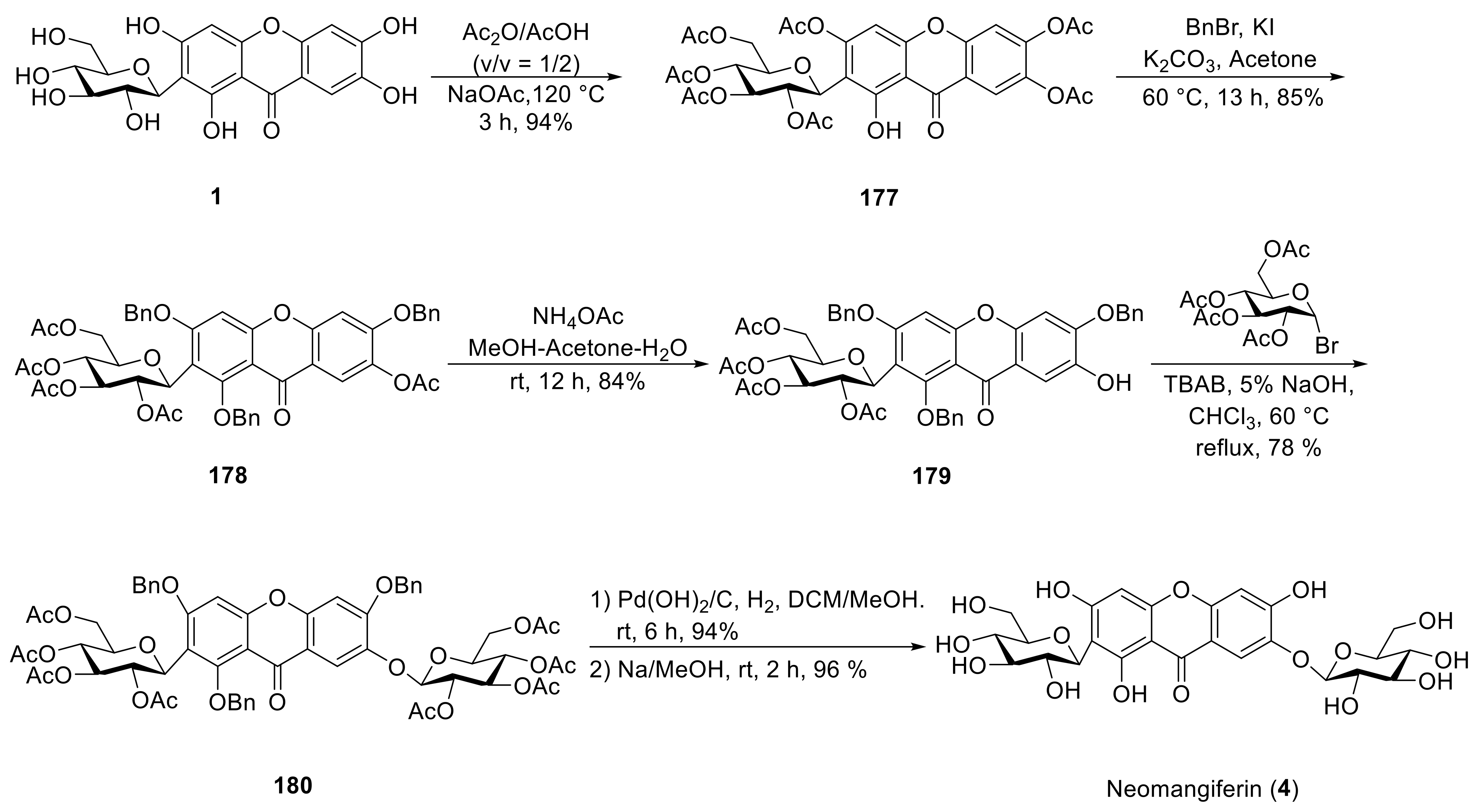
  132. Wei, X.; Liang, D.; Ning, M.; Wang, Q.; Meng, X.; Li, Z. Semi-synthesis of neomangiferin from mangiferin. Tetrahedron Lett. 2014, 55, 3083–3086. [Google Scholar] [CrossRef]
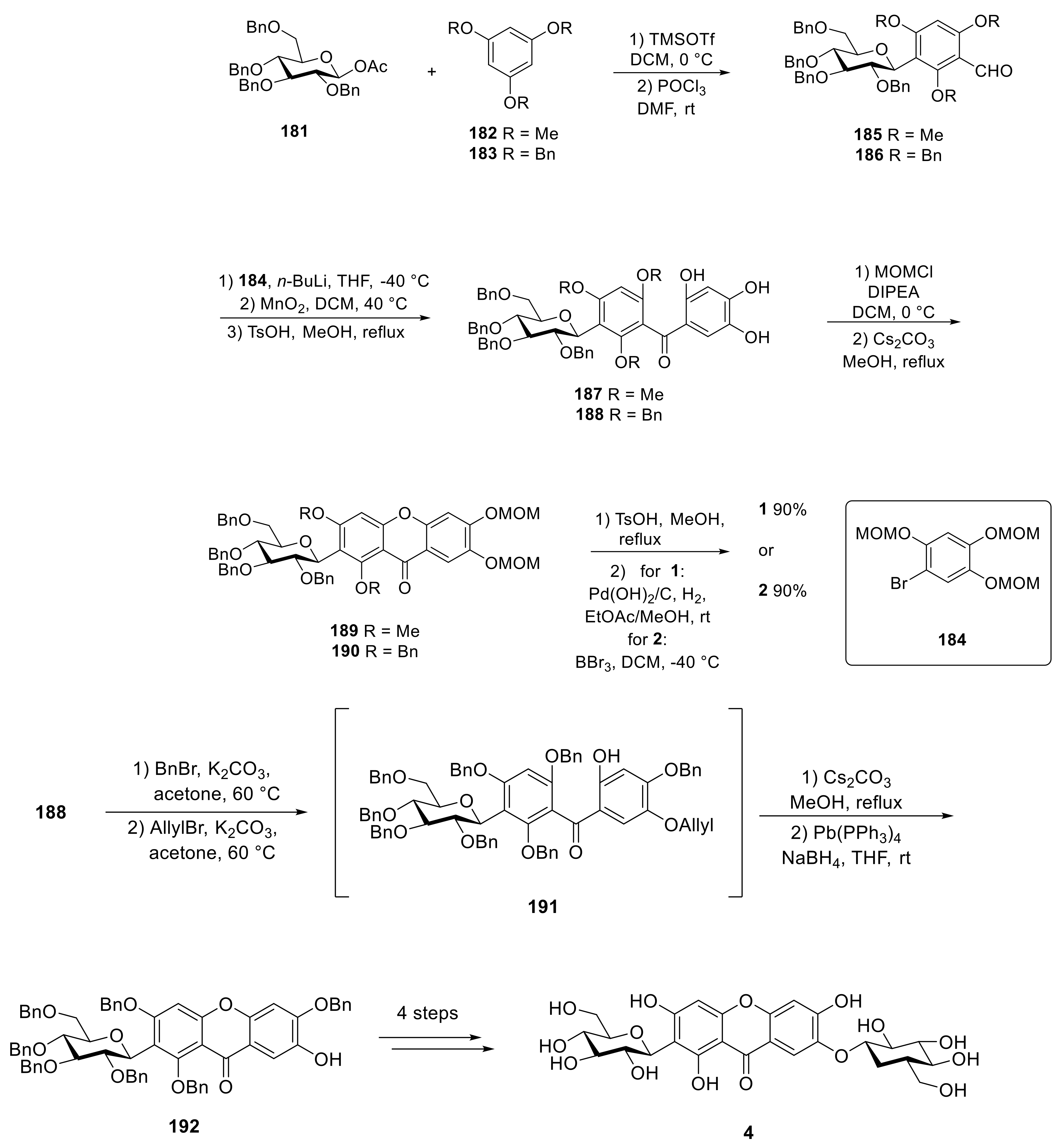
  133. Wei, X.; Liang, D.; Wang, Q.; Meng, X.; Li, Z. Total synthesis of mangiferin, homomangiferin, and neomangiferin. Org. Biomol. Chem. 2016, 14, 8821–8831. [Google Scholar] [CrossRef]
  134. Zarena, A.S.; Sankar, K.U. Synthesis of α-mangostin-d-glucoside in supercritical carbon dioxide media. J. Food Sci. Technol. 2015, 52, 6547–6555. [Google Scholar] [CrossRef] [PubMed] [Green Version]
  135. Tuoi, T.L.; Pandey, R.P.; Gurung, R.B.; Dhakal, D.; Sohng, J.K. Efficient enzymatic systems for synthesis of novel α-mangostin glycosides exhibiting antibacterial activity against Gram-positive bacteria. Appl. Microbiol. Biotechnol. 2014, 98, 8527–8538. [Google Scholar]
Scheme 1. The synthesis of 35, 36 and 72.
Molecules 26 05575 sch001aMolecules 26 05575 sch001b
Scheme 2. The synthesis of mangiferin derivatives.
Molecules 26 05575 sch002
Scheme 3. The synthesis of neomangiferin.
Molecules 26 05575 sch003
Scheme 4. The synthesis of mangiferin, homomangiferin, and neomangiferin.
Molecules 26 05575 sch004
Table 1. The structure of compounds 1015.
R1R2
10 Molecules 26 05575 i056H
11 Molecules 26 05575 i057H
12H Molecules 26 05575 i058
13 Molecules 26 05575 i059H
14 Molecules 26 05575 i060H
15 Molecules 26 05575 i061H
Table 2. The structure of compounds 5663.
R1R2R3R4R5R6
56HOMeGentiobiosylOMeOMeH
57HOMePrimeverosylOMeOMeH
58OMeHGentiobiosylOMeOMeOMe
59HOMePrimeverosylOMeOMeOMe
60OMeOHGentiobiosylOMeOMeOMe
61OMeOHPrimeverosylOMeOMeOMe
62OMeOMeGentiobiosylOMeOMeOMe
63OMeOMePrimeverosylOMeOMeOMe
Table 3. The structure of compounds 9099.
R1R2R3R4R5R6
90O-glc(6–1)-xylOMeOMeHOHOMe
91O-glc(6–1)-xylOMeOMeHOMeOMe
92O-glcOMeOHHOHOMe
93O-glc(6–1)-xylOMeHOMeHOMe
94O-glc(6–1)-xylOMeHOMeOMeOMe
95OHO-glc(6–1)-xylOMeOMeHOH
96O-glc(6–1)-glcOMeHOMeHH
97O-glc(6–1)-glcOMeHOMeHOH
98OHOMeHHO-glc(2–1)-rhaOH
99OHOMeHHO-rhaO-glc
Table 4. The structure of compounds 100106.
R1R2R3R4R5R6R7
100O-glc(6–1)-xylOMeOMeOMeOMeHOH
101OHOMeOMeOMeOMeHO-glc(6–1)-xyl
102O-glc(6–1)-xylHOMeOMeOMeHOH
103OHHOMeOMeOMeHO-glc(6–1)-xyl
104O-glc(6–1)-xylHOHOMeOMeHOH
105O-glc(6–1)-glcHOHOMeOMeHOH
106O-glc(6–1)-glcHOHOMeOMeHOH
Table 5. 13C NMR data of typical compounds.
CategoryCompound [Ref.]C-1′C-3′C-5′
Xanthone C-glucoside1 [34]73.679.582.1
10 [62]75.481.682.7
22 [69]74.0479.6080.55
29 [77]74.177.981.3
31 [78]74.480.382.9
Xanthone O-glucoside69 [92]10276.476.3
79 [77]103.676.077.5
87 [78]103.776.277.8
119 [115]105.477.276.0
123 [118]100.476.477.2
Publisher’s Note: MDPI stays neutral with regard to jurisdictional claims in published maps and institutional affiliations.

