Article

Delta Sleep-Inducing Peptide Recovers Motor Function in SD Rats after Focal Stroke

by
Elena A. Tukhovskaya
1,*,
Alina M. Ismailova
1,
Elvira R. Shaykhutdinova
1,
Gulsara A. Slashcheva
1,
Igor A. Prudchenko
2,
Inessa I. Mikhaleva
2,
Oksana N. Khokhlova
1,
Arkady N. Murashev
1 and
Vadim T. Ivanov
2
1
Biological Testing Laboratory, Branch of Shemyakin and Ovchinnikov, Institute of Bioorganic Chemistry, Russian Academy of Sciences, Pushchino, Prospekt Nauki, 6, 142290 Moscow, Russia
2
Laboratory of Peptide Chemistry, Shemyakin and Ovchinnikov Institute of Bioorganic Chemistry, Russian Academy of Sciences, Miklukho-Maklaya Street, 16/10, 117997 Moscow, Russia
*
Author to whom correspondence should be addressed.
Molecules 2021, 26(17), 5173; https://doi.org/10.3390/molecules26175173
Submission received: 14 July 2021 / Revised: 12 August 2021 / Accepted: 16 August 2021 / Published: 26 August 2021
(This article belongs to the Special Issue Bioactive Peptides and Proteins)

Abstract

:
Background and Objectives: Mutual effect of the preliminary and therapeutic intranasal treatment of SD rats with DSIP (8 days) on the outcome of focal stroke, induced with intraluminal middle cerebral occlusion (MCAO), was investigated. Materials and Methods: The groups were the following: MCAO + vehicle, MCAO + DSIP, and SHAM-operated. DSIP or vehicle was applied nasally 60 (±15) minutes prior to the occlusion and for 7 days after reperfusion at dose 120 µg/kg. The battery of behavioral tests was performed on 1, 3, 7, 14, and 21 days after MCAO. Motor coordination and balance and bilateral asymmetry were tested. At the end of the study, animals were euthanized, and their brains were perfused, serial cryoslices were made, and infarction volume in them was calculated. Results: Although brain infarction in DSIP-treated animals was smaller than in vehicle-treated animals, the difference was not significant. However, motor performance in the rotarod test significantly recovered in DSIP-treated animals. Conclusions: Intranasal administration of DSIP in the course of 8 days leads to accelerated recovery of motor functions.

1. Introduction

Delta sleep-inducing peptide (DSIP) is a multifunctional regulatory peptide. It was first isolated from cerebral venous blood of rabbits after low-frequency (‘hypnogenic’) electrical stimulation of the intralaminar thalamic nuclei by the Schoenenberger–Monnier group from Basel in 1977 [1]. Subsequently, the functions of the DSIP were actively investigated. Consequently, in the work of Iyer et al. (1988), the effect of DSIP on the induction of slow-wave sleep and sleep-related growth hormone release was shown [2]. In a later study, the absence of the effect of DSIP in the formation of sleep was shown when injected into the nucleus raphe dorsalis of rats [3]. A wide range of studies conducted to identify the biological activities of DSIP showed that endogenous DSIP or DSIP-like peptides have regulatory activity and play an important role in endocrine regulation. Thus, it has been shown that DSIP reduces the basal level of corticotropin [4,5,6,7,8,9,10,11,12,13,14] and stimulates the secretion of luteinizing hormone and the release of somatoliberin and somatotropin [8]. DSIP has a wide range of physiological activities, some of which are not related to each other [15]. It has stress-protective and adaptive activity [16,17,18,19,20]. DSIP acts as an adaptogen at amphetamine-induced stereotypy, which is a model of schizophrenia-like condition in humans. It normalizes brain metabolism injured with long-term amphetamine treatment. It is also shown that DSIP normalizes MAO activities through serotonin adrenergic systems [21]. Konorova et al. elucidated the ability of DSIP (as the medical product deltaran) to reduce the lethality of stress in low-resistant rats [22]. DSIP also has neuroprotective properties. Accordingly, the treatment of Wistar rats with DSIP several hours prior to global brain ischemia improved locomotor functions and reduced lethality [23]. DSIP reduced neuronal activity and improved blood supply of the brain of stressed animals subjected to brain ischemia [24]. There are several reports suggesting that DSIP reduces stress-induced overproduction of free radicals in CNS and prevents neuronal death [16,25]. Investigation of DSIP at a model of carotid arteries occlusion in different age groups of male rats revealed DSIP’s ability to correct the impaired balance of brain neuromediators hypoxia [25]. DSIP has also been shown to potentiate the action of the anticonvulsant drug Valproate in metaphit-provoked generalized epilepsy induced by audiogenic stimulation in adult rats, indicating the potential for using DSIP in combination with antiepileptic drugs for the treatment of seizures [26].
The aim of the present study was to investigate the effect of mutual preventive and therapeutic intranasal treatment of SD rats with DSIP on the consequences of focal stroke.

2. Materials and Methods

2.1. Animals

Adult male Sprague-Dawley (SD) rats of 320–380 g were used. All animals were SPF and were obtained from the Pushchino Nursery for Laboratory Animals (Pushchino, Russia). All procedures and manipulations with animals were approved by the Committee for Control over Care and Use of Laboratory Animals of BIBCh RAS and were carried out in accordance with the EU Directive 2010/63/EU for animal experiments and Animal Control and Use Committee (IACUC) protocol number 683/19. The Biological Testing Laboratory, Branch of Shemyakin, and the Ovchinnikov Institute of Bioorganic Chemistry, Pushchino, Moscow Region, Russia, where the experiments were followed, has AAALAC accreditation (can be found as Russian Academy of Sciences Biological Testing Laboratory-Branch of Shemyakin & Ovchinnikov Institute of Bioorganic Chemistry Pushchino, Moscow Region, Russia by clicking on the link https://www.aaalac.org/accreditation-program/directory/directory-of-accredited-organizations-search-result/#R, (accessed on 24 August 2021). The rats were maintained on 12 h light/dark cycle with unlimited access to food and water.

2.2. Drugs

DSIP (H-Trp-Ala-Gly-Gly-Asp-Ala-SerGly-Glu-OH) was synthesized in the Laboratory of Peptide Chemistry, Shemyakin, and the Ovchinnikov Institute of Bioorganic Chemistry, Russian Academy of Sciences, Moscow, Russia, as described elsewhere [26].

2.3. Middle Cerebral Artery Occlusion Procedure (MCAO)

A total of 26 mature male SD rats were used. Animals were anesthetized intramuscularly with a ketamine–xylazine mixture (30 mg/kg ketamine and 12 mg/kg xylazine). The total injection volume was 1.2 mL/kg. The right temporal muscle was dissected, temporal bone was dried with cotton balls, and a laser Doppler flow (LDF) probe (PeriFlux 5001, PR407-1 Small Straight Probe) was attached to the temporal bone to measure regional cerebrovascular blood flow (rCBF). A midline cervical incision was made to expose the right common carotid artery (CCA). Loose ligatures were placed on CCA and internal carotid artery (ICA), followed by two loose ligatures on the external carotid artery (ECA) at a point 2–3 mm distal from the bifurcation of CCA. The occipital artery was ligated permanently and the pterygopalatine artery (PPA) was ligated for the MCAO procedure duration. Occluding sutures were prepared using a silicon–Teflon monofilament of 0.13 mm diameter (STROFT GTM, Germany) with a heat blunted end (0.3 mm diameter) coated with 0.5% (w/v) poly-l-lysine solution and dried under a heating lamp for 1 h [27]. The intraluminal suture was inserted through a small puncture opening in ECA and advanced into the ICA to approximately 18 mm from the bifurcation (depending on a rat weight) until a slight resistance occurred. When decreased blood flow was observed, the occlusion suture was left in place for 90 min. Ischemia was confirmed by a more than 70% rCBF decrease compared with the baseline value. After 90 min of occlusion, the suture was withdrawn. Blood flow restitution to 90–120% of baseline in 10 min was recognized as a successful reperfusion. The ligatures were removed from the CCA, ICA, and PPA, while the ECA was permanently ligated above and below the opening. The LDF probe was detached, and the wounds were closed with a twisted suture. Sham-operated rats were subjected to the same surgical procedure except for the MCA occlusion. The rectal temperature was maintained at 37 °C using a heating pad until the animals recovered from anesthesia. After surgery, the rats were returned to their cages and allowed free access to food and water.

2.4. Groups and Dosing Regimen

The animals were divided into three groups: animals subjected to MCAO and treated with vehicle—MCAO + vehicle (saline) (n = 9); animals subjected to MCAO and treated with DSIP—MCAO + DSIP (n = 10); sham-operated animals—SHAM (n = 7). The animals received DSIP nasally 60 (±15) minutes prior to the occlusion and for 7 days after reperfusion. The DSIP dosage was 120 µg/kg (volume of treatment was 100 µL/kg).

2.5. Behavioral Testing

Each rat was subjected to a series of behavioral tests on days 1, 3, 5, 7, 14, and 21 after a stroke. Motor deficits of balance and coordination were assessed by rotarod testing. Drug-induced rotations asymmetry was tested. All behavioral testing was performed during the animals’ light cycle.

2.5.1. Rotarod Test

Quantitative assessment of motor coordination and balance was performed using an automated rotarod (Dual Species Economex Rota-Rod, Columbus Instruments). All animals were trained on the rotarod before the MCAO procedure for 3 consecutive days. This training consisted of three trials per day on each of these 3 days (nine trials in total). Each trial consisted of placing the animal onto the rotarod, which was revolving at a constant speed (5 rpm/min), and then the rotarod was switched to acceleration mode (0.6 rpm/min). Fall latency was defined as the time during which the animal balanced on the rotating rod from the moment the acceleration was switched on until the moment it fell off the rod. The countdown started from point 0 when acceleration was turned on and continued until the animal fell or was held on a rotating rod (squeezing its paws) for two full revolutions (720°). According to the results of training and preliminary testing, the animals were selected into groups so that in all groups there were animals with homogeneous results. The purpose of this pre-testing was to identify groups of animals with similar performance in the rotarod test in order to more accurately determine how MCAO and MCAO + DSIP treatments might affect this performance. Starting from the first day after MCAO, consecutive sessions of rotarod testing were started. In each daily session, testing was repeated three times with 5 min intervals between each trial. Animals unable to grasp the rotating rod were given a latency value of 0 s. Latency times were measured as described above.

2.5.2. Drug-Induced Rotation Asymmetry

Drug-induced rotation asymmetry was recorded manually for 5 min following ketamine injection (10 mg/kg intramuscular) [28,29] in a plastic cylinder with a smooth round bottom (30 cm bottom diameter and 40 cm walls height). Intramuscular injection of ketamine in subanesthetic dose to the rats with unilateral occlusion of the middle cerebral artery causes an increase in ipsilateral (toward the injured hemisphere) rotation. A full 360° arc of turning in the same direction scored as one rotation. The percentage of rotations to the right (ipsilateral to the injured hemisphere) was assessed relative to the total number of rotations in both directions.

2.6. Brain Infarct Analysis

After 21 days of testing, the animals were sacrificed by CO2 inhalation, and their brains were perfusion-fixed. The heart was exposed by a median sternotomy, and the ascending aorta was catheterized via the left ventricle. The blood was removed from animals with 150 mL saline solution containing heparin (100 Units/L) perfusion at a constant perfusion rate of 30 mL/min. Then the perfusion medium was switched to a mixture of 3% (w/v) formaldehyde buffered solution (pH 7.4) and 1% (v/v) glutaraldehyde solution (150 mL). Following brain perfusion-fixation, the animals were decapitated, and the brain was removed from the skull. The brain was cryoprotected in sucrose buffered solutions (pH 7.4) of graded concentrations (10%, 20%, 30%) and then immediately frozen in the cryostat (Microm HM 525, Carl Zeiss) at −65°C. From each brain, coronal cryosections of 50 μm thickness at 1 mm intervals were obtained and stained with cresyl violet. The stained sections were scanned and processed with the 3D-reconstruction program ‘Reconstruct’ https://synapseweb.clm.utexas.edu/software-0 (accessed on 24 August 2021). Volumes of both hemispheres were calculated using the Cavalieri method [30]. The indirect lesion area was calculated as the intact area of the ipsilateral hemisphere subtracted from the area of the contralateral (uninjured) hemisphere. Lesion volume was calculated as a percentage in which the lesion volume was compared with the contralateral hemisphere volume [31].

2.7. Statistical Analysis

The behavioral data and infarct size were analyzed using Statistica for Windows 7.0. Test for the normality was performed for all data. All the values are presented as mean ± standard deviation. The significance of the differences in the rotarod test and ketamine-induced rotation test was evaluated by repeated measures ANOVA with post hoc Duncan analysis. The significance of the differences when comparing the volumes of cerebral infarction was assessed using one-way ANOVA.

3. Results

3.1. Behavioral Testing

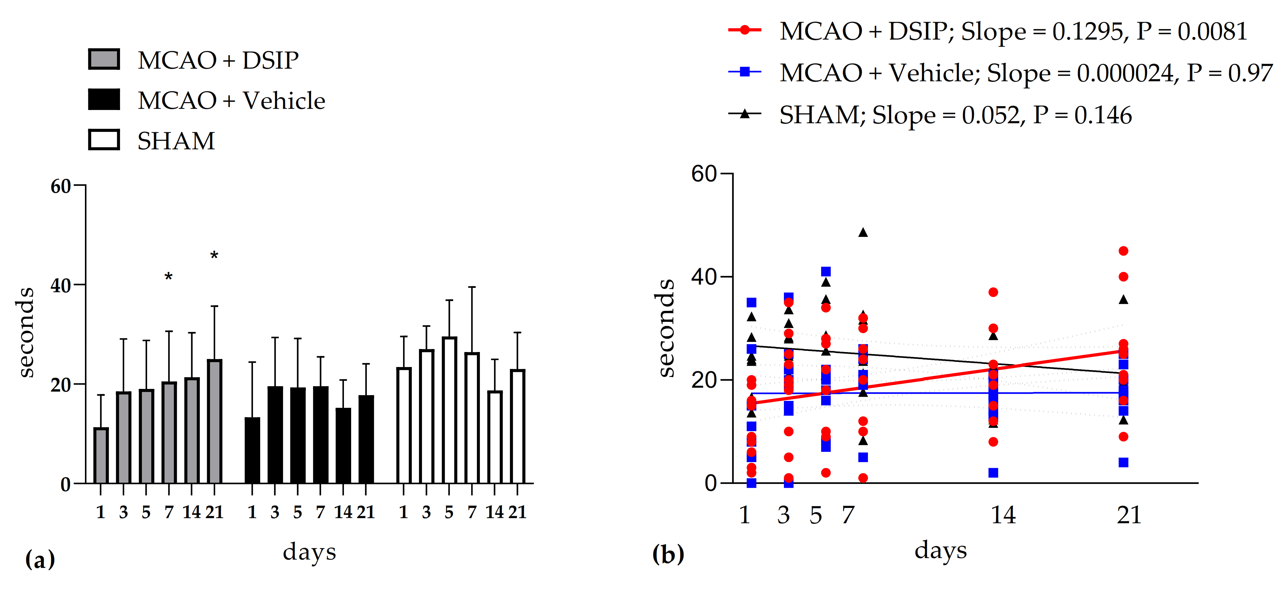
3.1.1. Motor Performance after MCAO in Rotarod Test

Animals treated with DSIP showed a significantly improved performance in a rotarod test (Figure 1). Motor coordination recovered on the 7-th day of testing, which was confirmed by the repeated measures ANOVA analysis. Linear regression analysis was applied to assess the dynamics of the fall latency. Only in animals receiving DSIP were there positive dynamics of this parameter, which was confirmed by statistical assessment of the slope of the linear regression line (Figure 1). These findings elucidated a positive DSIP effect on the recovery of motor performance after stroke. Individual data for all animals are given in the Appendix A.

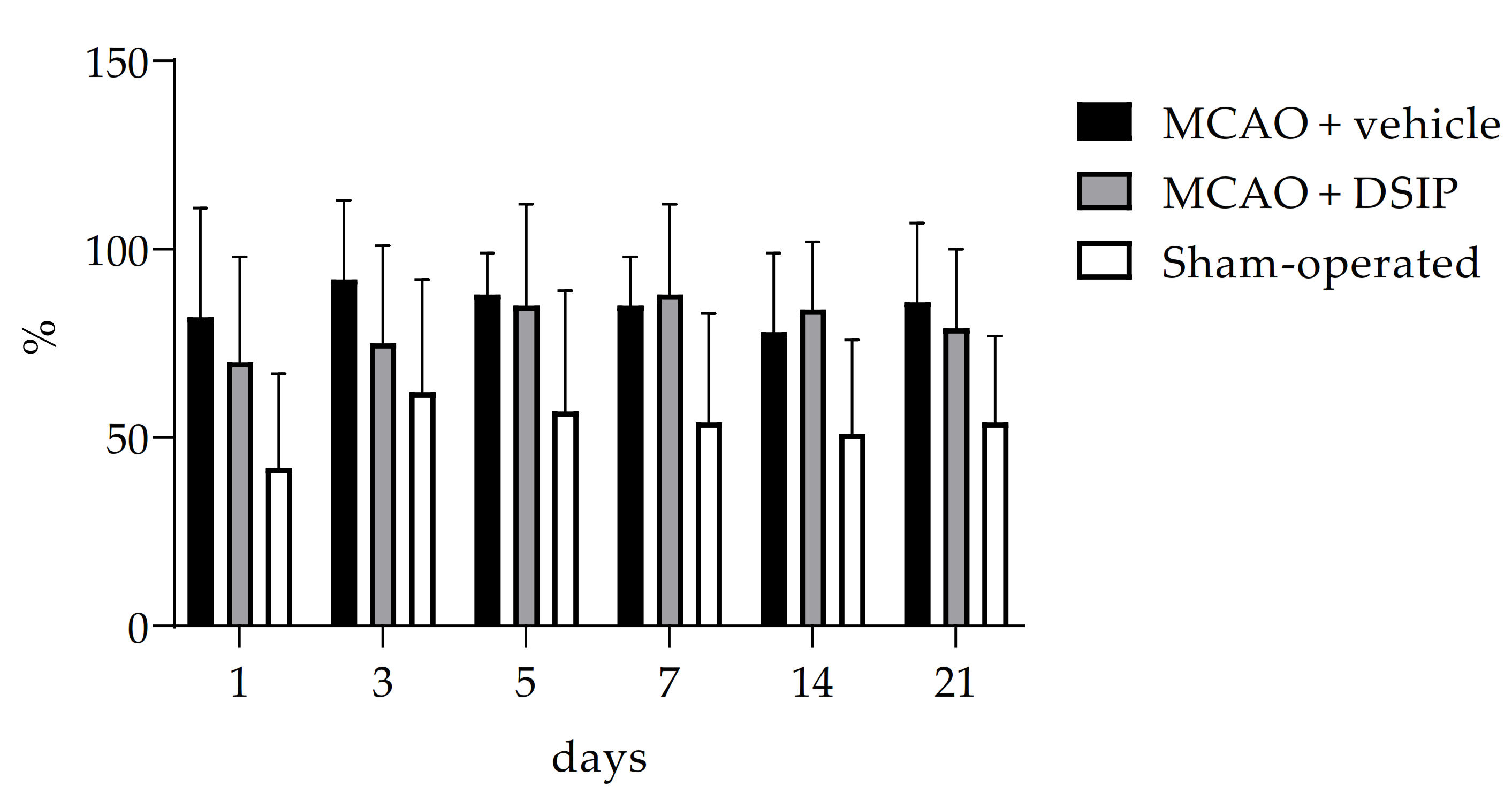
3.1.2. Ketamine-Induced Rotation

As can be seen from Figure 2, there was no significant influence of DSIP on rotation asymmetry tested by the ketamine-induced rotation test. Individual data for all animals are given in the Appendix A.

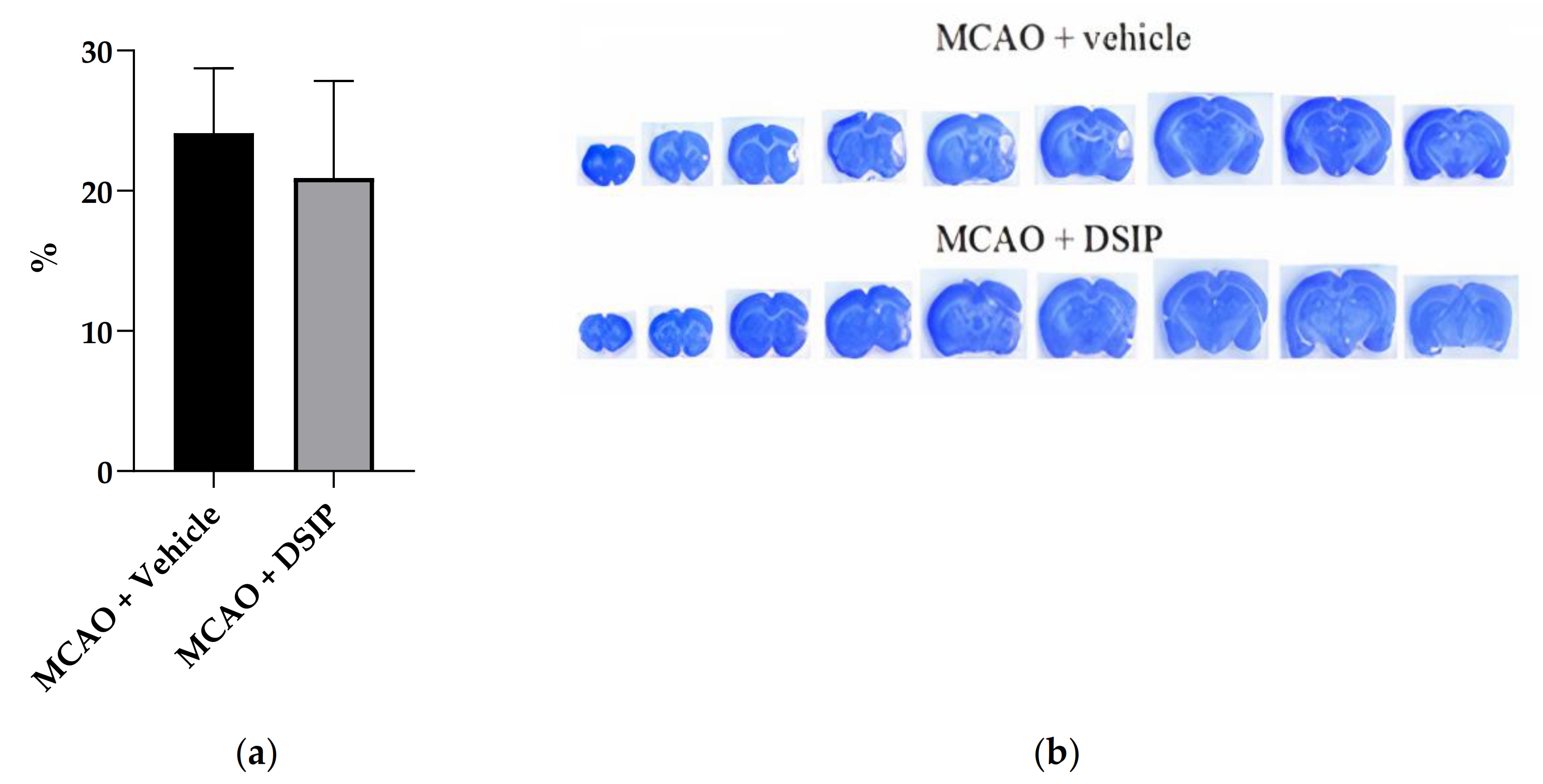
3.1.3. Brain Infarction Volume

The brain infarction volume in the group MCAO+DSIP was 20.9 ± 6.9% versus 24.1 ± 4.6% in the group MCAO + vehicle, which had no statistical significance (Figure 3). Individual data for all animals are given in the Appendix A.

4. Discussion and Conclusions

Although the infarct volume of the affected hemisphere in DSIP-treated animals was less than in vehicle-treated animals, this difference was not significant. In addition, there was not much difference in the ketamine-induced rotation between DSIP-treated animals and vehicle-treated animals. However, in the test of motor performance, the latent time of falling from the rotarod significantly increased only in DSIP-treated animals. This increase was confirmed by both linear regression analysis and repeated measures ANOVA. Motor dysfunction is one of the most serious complications of stroke [32]. Loss of motor ability is thought to be caused by abnormalities in the neural network that controls movement [33,34,35,36,37]. Motor recovery is crucial for regaining independence [38]. Rehabilitation of stroke survivors includes maximal recovery of motor functions and coordination [39,40,41]. Using different biomarkers to determine the motor functions of patients is helpful in the prediction of motor recovery [42,43,44,45,46,47]. We assessed motor function dynamics for stroke-subjected animals. The obtained results allow us to reveal a rather significant positive DSIP effect on the recovery of motor coordination, which is an important parameter in stroke-subjected animals. This DSIP effect could be associated with the rescuing of neurons of the motor cortex and subcortical structures associated with motor control. Neurons rescuing mechanisms may include DSIP effects on biosynthetic processes in the brain, as has been previously reported [48]. It is suggested that the protective effects of peptide could be based on a decline in the synthesis rate of stress protein factors [49]. The motor function recovery of animals treated with DSIP may also be associated with its effect on glutamate receptors and GABA receptors of neurons of the cortex, thalamus, hypothalamus, hippocampus, and cerebellum. Thus, Sudakov and colleagues showed that DSIP-caused activation of neurons at the sensorimotor cortex, dorsal hippocampus, ventral thalamic nuclei, and lateral hypothalamus could be explained by its action on NMDA receptors of these structures [50]. Grigor’ev et al. showed that DSIP potentiated GABA-activated currents in hippocampal and cerebellar neurons and blocked NMDA-activated potentiation in cortical and hippocampal neurons [51]. There are available data that DSIP improves mitochondrial respiratory activity. Oxygen utilization by mitochondria is carried out through the respiratory chain—a system of specific peptides involved in electron transport and ATP synthesis. The combination of these processes ensures the normal functioning of the enzymatic energy system. Khvatova et al. showed that pretreatment of rats with DSIP prior to their subjection to hypoxia completely inhibited the hypoxia-induced reduction of mitochondrial respiratory activity [52].
It is known that in the process of ischemia, a cascade of metabolic reactions is activated, some of which are involved in the production of reactive oxygen species (ROS) and reactive nitrogen species (RNS), such as superoxide, hydrogen peroxide [H2O2], hydroxyl radical, nitric oxide [NO], and peroxynitrite, which cause most of the damage observed in ischemia-reperfusion [53,54,55,56]. The antioxidant mechanism of DSIP is an increase in the activity of superoxide dismutase (SOD) and glutamate peroxidase (GPO) [56], mediated by an increase in the expression of genes encoding these enzymes, as shown in aging animals [57,58].
In our other study [59], it was shown that the administration of DSIP and its analog KND-peptide during occlusion led to the death of animals, and when administered during the reperfusion, a decrease in the size of the brain and heart infarction was observed. Presumably, mortality could be a consequence of the inactivation of the genes for the early response of c-Fos upon administration of DSIP during occlusion, whose activity is associated with the activation of NMDA receptors [60,61]. In the present study, a combined administration of DSIP was used preventively (before the onset of occlusion) and then within 7 days after reperfusion. Thus, we managed to avoid the negative consequences of the introduction of DSIP during the period of occlusion and, on the contrary, to demonstrate a positive effect on the restoration of motor functions. In our study, we demonstrated that DSIP might have clinical benefits for stroke therapy in terms of accelerating the recovery of motor functions after a stroke. Research into the DSIP effect on stroke should be focused on in future studies of optimal dosage regimens to provide more effective and fast recovery from stroke.

Author Contributions

Conceptualization, I.A.P. and V.T.I.; formal analysis, E.R.S.; investigation, E.A.T., E.R.S., A.M.I., G.A.S., I.I.M. and O.N.K.; methodology, E.A.T., E.R.S. and I.A.P.; project administration, V.T.I.; resources, A.N.M.; software, V.T.I.; supervision, I.A.P., I.I.M. and V.T.I.; A.N.M.; writing—original draft, E.A.T.; writing—review and editing, V.T.I. All authors have read and agreed to the published version of the manuscript.

Funding

This research received no external funding.

Institutional Review Board Statement

All procedures and manipulations with animals were approved by the Committee for Control over Care and Use of Laboratory Animals of BIBCh RAS and were carried out in accordance with the EU Directive 2010/63/EU for animal experiments and Animal Control and Use Committee (IACUC), protocol number 683/19, 27 February 2019.

Informed Consent Statement

Not applicable.

Data Availability Statement

All raw data are presented in Appendix A.

Conflicts of Interest

There is no conflict of interest.

Appendix A

Appendix A.1. Raw Data on Rotarod

Table A1. Latent time of falling from rotarod for SHAM-operated animals, sec.
Animal
Number
1234567Mean ± SD
Day 11425321724282423 ± 6
Day 32821312522283427 ± 5
Day 52126393621352930 ± 7
Day 7218491832332426 ± 13
Day 141212292216172319 ± 6
Day 212325121920263623 ± 7
Table A2. Latent time of falling from rotarod for vehicle-treated animals, sec.
Animal
Number
8910111213141516Mean ± SD
Day 15111526515320813 ± 11
Day 32015193614252502220 ± 10
Day 5168212220214171819 ± 10
Day 72019192621242152120 ± 6
Day 141617142021181621315 ± 5
Day 212314171823162542018 ± 6
Table A3. Latent time of falling from rotarod for DSIP-treated animals, sec.
Animal
Number
17181920212223242526Mean ± SD
Day 115892166319201511 ± 6
Day 318251011953520232919 ± 11
Day 518271021892228223419 ± 10
Day 7243010120122030263220 ± 10
Day 14303712823192119153021 ± 9
Day 21452116925202127264025 ± 11

Appendix A.2. Raw Data on Ketamine-Induced Rotation

Table A4. The percentage of the animal’s rotation in the direction of damage relative to rotations in both directions for SHAM-operated animals, %.
Animal
Number
1234567Mean ± SD
Day 1582683252338342 ± 25
Day 3726418255510010062 ± 30
Day 5925903067599357 ± 32
Day 72261169252657154 ± 29
Day 14495245662488551 ± 25
Day 216441195837649254 ± 23
Table A5. The percentage of the animal’s rotation in the direction of damage relative to rotations in both directions for vehicle-treated animals, %.
Animal
Number
8910111213141516Mean ± SD
Day 1943982100251001001009682 ± 31
Day 390939290751001001008592 ± 24
Day 5767610010010088936110088 ± 13
Day 796968593777365899585 ± 13
Day 1470100938590959916778 ± 22
Day 21728583768386100989286 ± 21
Table A6. The percentage of the animal’s rotation in the direction of damage relative to rotations in both directions for DSIP-treated animals, %.
Animal
Number
17181920212223242526Mean ± SD
Day 171590696771100751008770 ± 32
Day 37390641004167100331008085 ± 29
Day 510010081100010010079959185 ± 29
Day 783100811001001007750929588 ± 24
Day 14831003899919510067649884 ± 18
Day 21841005310092799540559479 ± 22

Appendix A.3. Raw Data on Brain Damage

Table A7. The percentage of damage to the right hemisphere for vehicle-operated animals, %.
Animal
Number
8910111213141516Mean ± SD
21.025.926.219.126.823.522.022.618.924.1 ± 4.6
Table A8. The percentage of damage to the right hemisphere for DSIP-treated animals, %.
Animal
Number
17181920212223242526Mean ± SD
18.020.418.926.215.524.231.06.820.627.820.9 ± 6.9

References

  1. Schoenenberger, G.A.; Maier, P.F.; Tobler, H.J.; Monnier, M. A Naturally Occurring Delta-EEG Enhancing Nonapeptide in Rabbits X. Final Isolation, Characterization and Activity Test. Pflfigers Arch. 1977, 369, 99–109. [Google Scholar] [CrossRef] [PubMed]
  2. Iyer, K.S.; Marks, G.A.; Kastin, A.J.; Mccann, S.M. Evidence for a role of delta sleep-inducing peptide in slow-wave sleep and sleep-related growth hormone release in the rat. Proc. NatI. Acad. Sci. USA 1988, 85, 3653–3656. [Google Scholar] [CrossRef] [PubMed] [Green Version]
  3. Karl, B.E.; Cespuglio, R.; Leger, L.; Marinesco, S.; Jouvet, M. Is the nucleus raphe dorsalis a target for the peptides possessing hypnogenic properties? Brain Res. 1994, 637, 211–221. [Google Scholar]
  4. Graf, M.V.; Kastin, A.J. Delta Sleep-Inducing Peptide (DSIP): A review. Neurosci. Biobehav. Rev. 1984, 8, 83–93. [Google Scholar] [CrossRef]
  5. Graf, M.V.; Kastin, A.J. Delta Sleep-Inducing Peptide (DSIP): Update. Peptides 1986, 7, 1165–1187. [Google Scholar] [CrossRef]
  6. Graf, M.V.; Kastin, A.J.; Coy, D.H.; Zadina, J.E. DSIP reduces amphetamine-induced hyperthermia in mice. Physiol. Behav. 1984, 33, 291–295. [Google Scholar] [CrossRef]
  7. Schoenenberger, G.A. Characterization, properties and multivariate functions of Delta-Sleep-Inducing Peptide (DSIP). Eur. Neurol. 1984, 23, 321–345. [Google Scholar] [CrossRef]
  8. Kovalzon, V.M. DSIP: A sleep peptide or unknown hypothalamic hormone? J. Evol. Biochem. Physiol. 1994, 30, 195–199. [Google Scholar]
  9. Yehuda, S.; Carasso, R. DSIP–a tool for investigating the sleep onset mechanism: A review. Int. J. Neurosci. 1988, 38, 345–353. [Google Scholar] [CrossRef]
  10. Chiodera, A.P.; Volpi, R.; Carpetti, L.; Giacalone, G.; Caffarri, G.; Davoli, C.; Nigro, E.; Coiro, V. Different effects of delta-sleep-inducing peptide on arginine-vasopressin and ACTH secretion in normal men. Horm. Res. 1994, 42, 267–272. [Google Scholar] [CrossRef]
  11. Prudchenko, I.A.; Mikhaleva, I.I. A problem of endogenity of Delta Sleep-Induced Peptide. Uspechi Sovremennoy Biol. 1994, 114, 727–740. [Google Scholar]
  12. Lysenko, A.V.; Mendzheritsky, A.M. Characteristics and mechanisms of realizing biological effects of the peptide inducing delta-sleep. Uspekhi Sovremennoy Biol. 1995, 115, 729–739. [Google Scholar]
  13. Strekalova, T.V. DSIP: Problems of its endogenous origin and biological activity. Neurokhimia 1998, 15, 227–239. [Google Scholar]
  14. Pollard, B.J.; Pomfrett, C.J.D. Delta sleep-inducing peptide. Eur. J. Anaesthesiol. 2001, 18, 419–422. [Google Scholar] [CrossRef]
  15. Kovalzon, V.M.; Strekalova, T.V. Delta sleep-inducing peptide (DSIP): A still unresolved riddle. J. Neurochem. 2006, 97, 303–309. [Google Scholar] [CrossRef] [Green Version]
  16. Mikhaleva, I.I.; Voitenkov, B.O. The delta-sleep inducing peptide and Deltaran: A way from biochemical studies to medicine. Novye Lekarstvennye Preparaty 2007, 3, 6–20. [Google Scholar]
  17. Shustanova, T.A.; Bondarenko, T.I.; Milyutina, N.P.; Mikhaleva, I.I. Regulation of freeradical processes in rat tissues by the delta-sleep inducing peptide during cold stress. Biokhimiya 2001, 66, 632–639. [Google Scholar] [CrossRef]
  18. Rikhireva, G.T.; Sokolova, I.S.; Rylova, A.V.; Kopylovskiĭ, S.A.; Mikhaleva, I.I.; Prudchenko, I.A. Changes in the rate of protein biosynthesis in the organs of mice under the action of the delta sleep-inducing peptide and psychoemotional stress. Izv. Akad. Nauk. Ser. Biol. 1995, 2, 142–148. [Google Scholar]
  19. Shmarin, I.P. Signaling molecules and social behavior. Neurochemistry 2001, 18, 243–250. [Google Scholar]
  20. Sudakov, K.V.; Ivanov, V.T.; Kolpik, E.V. Delta-sleep Indusing Peptide (DSIP) as factor facilitation animals resistance to acute emotional stress. Pavlov Biol. Sci. 1983, 18, 1–5. [Google Scholar] [CrossRef]
  21. Gershteĭn, L.M.; Dovedova, E.L.; Khrustalev, D.A. Morphochemical characteristic of rat brain structures after exposure to delta-sleep inducing peptide following long-term amphetamine administration. Morfologiia 2004, 125, 74–77. [Google Scholar]
  22. Konorova, I.L.; Gannushkina, I.V.; Koplik, E.V.; Antelava, A.L. Deltaran prevents an adverse effect of emotional stress on the course of cerebral ischemia in stress-low-resistant animals. Bull. Exp. Biol. Med. 2006, 141, 564–566. [Google Scholar] [CrossRef]
  23. Shandra, A.A.; Godlevskii, L.S.; Brusentsov, A.I.; Vast’yanov, R.S.; Karlyuga, V.A.; Dzygal, A.F.; Nikel, B. Effects of delta-sleep-inducing peptide in cerebral ischemia in rats. Neurosci. Behav. Physiol. 1998, 28, 443–446. [Google Scholar] [CrossRef]
  24. Kolpik, E.V. Delta-sleep indusing peptide and drug deltaran: Possible approaches to antistress protection. Zh. Nevrol. Psikhiatr. Im. SS Korsakova 2007, 107, 50–54. [Google Scholar]
  25. Kim, T.K.; Karantysh, G.V.; Mendzheritskiĭ, A.M.; Ryzhak, G.A. Effect of deltaran on the mediatory balance in the brain of young and old rats with left-side laterality profile in case of carotid arteries occlusion. Adv. Gerontol. 2007, 20, 138–142. [Google Scholar] [PubMed]
  26. Hrnčić, D.; Stanojlović, O.; Živanović, D.; Šušić, V. Delta-Sleep-Inducing Peptide Potentiates Anticonvulsive Activity of Valproate against Metaphit-Provoked Audiogenic Seizure in Rats. Pharmacology 2006, 77, 78–84. [Google Scholar] [CrossRef] [PubMed]
  27. Belayev, L.; Alonso, O.F.; Busto, R.; Zhao, W.; Ginsberg, M.D. Middle cerebral artery occlusion in the rat by intraluminal suture. Neurological and pathological evaluation of an improved model. Stroke 1996, 27, 1616–1622. [Google Scholar] [CrossRef]
  28. Lebedev, S.V.; Petrov, S.V.; Blinov, D.V.; Lazarenko, I.P.; Chekhonin, V.P. Ketamine-induced rotational asymmetry in evaluation of motor disturbances in rats with middle cerebral artery occlusion. Bull. Exp. Biol. Med. 2003, 135, 424–427. [Google Scholar] [CrossRef]
  29. Tukhovskaya, E.A.; Yukin, A.Y.; Khokhlova, O.N.; Murashev, A.N. COG1410, a Novel Apolipoprotein-E Mimetic, Improves Functional and Morphological Recovery in a Rat Model of Focal Brain Ischemia. J. Neurosci. Res. 2009, 87, 677–682. [Google Scholar] [CrossRef] [Green Version]
  30. Gundersen, H.J.; Jensen, E.B. The efficiency of systematic sampling in stereology and its prediction. J. Microsc. 1987, 147, 229–263. [Google Scholar] [CrossRef]
  31. Swanson, R.A.; Morton, M.T.; Tsao-Wu, G.; Savalos, R.A.; Davidson, C.; Sharp, F.R. A semiautomated method for measuring brain infarct volume. J. Cereb. Blood Flow Metab. 1990, 10, 290–293. [Google Scholar] [CrossRef]
  32. Rathore, S.S.; Hinn, A.R.; Cooper, L.S.; Tyroler, H.A.; Rosamond, W.D. Characterization of incident stroke signs and symptoms: Findings from the atherosclerosis risk in communities study. Stroke 2002, 33, 2718–2721. [Google Scholar] [CrossRef] [Green Version]
  33. Chen, X.; Xie, P.; Zhang, Y.; Chen, Y.; Cheng, S.; Zhang, L. Abnormal functional corticomuscular coupling after stroke. NeuroImage Clin. 2018, 19, 147–159. [Google Scholar] [CrossRef]
  34. Chen, X.L.; Xie, P.; Zhang, Y.Y.; Liu, L.X.; Du, Y.H.; Chen, X.L.; Xie, P.; Zhang, Y.Y.; Liu, L.X.; Du, Y.H. Distinction of functional corticomuscular coupling in synkinetic and separate movement following stroke. Brain Stimul. 2017, 10, 435–436. [Google Scholar] [CrossRef]
  35. Fang, Y.; Daly, J.J.; Sun, J.; Hvorat, K.; Fredrickson, E.; Pundik, S.; Sahgal, V.; Guang, H.Y. Functional Corticomuscular Connection During Reaching Is Weakened Following Stroke. Clin. Neurophysiol. 2009, 120, 994–1002. [Google Scholar] [CrossRef] [Green Version]
  36. Gardiner, R. The pathophysiological and clinical implications of neuro-muscular changes following cerebrovascular accident. Aust. J. Physiother. 1996, 42, 139–146. [Google Scholar] [CrossRef] [Green Version]
  37. Mima, T.; Hallett, M. Corticomuscular coherence: A review. Clin. Neurophysiol. 1999, 16, 501–511. [Google Scholar] [CrossRef]
  38. Langhorne, P.; Coupar, F.; Pollock, A. Motor recovery after stroke: A systematic review. Lancet Neurol. 2009, 8, 741–754. [Google Scholar] [CrossRef]
  39. Stinear, C.M. Prediction of motor recovery after stroke: Advances in biomarkers. Lancet Neurol. 2017, 16, 826–836. [Google Scholar] [CrossRef]
  40. Schwamm, L.H.; Pancioli, A.; Acker, J.E., 3rd; Goldstein, L.B.; Zorowitz, R.D.; Shephard, T.J.; Moyer, P.; Gorman, M.; Johnston, S.C.; Duncan, P.W.; et al. Recommendations for the Establishment of Stroke Systems of Care Recommendations From the American Stroke Association’s Task Force on the Development of Stroke Systems. Circulation 2005, 111, 1078–1091. [Google Scholar] [CrossRef]
  41. Desrosiers, J.; Rochette, A.; Corriveau, H. Validation of a New Lower-Extremity Motor Coordination Test. Arch. Phys. Med. Rehabil. 2005, 86, 993–998. [Google Scholar] [CrossRef] [PubMed]
  42. Winters, C.; van Wegen, E.E.; Daffertshofer, A.; Kwakkel, G. Generalizability of the proportional recovery model for the upper extremity after an ischemic stroke. Neurorehabil. Neural Repair. 2015, 29, 614–622. [Google Scholar] [CrossRef] [PubMed]
  43. Buch, E.R.; Rizk, S.; Nicolo, P.; Cohen, L.G.; Schnider, A.; Guggisberg, A.G. Predicting motor improvement after stroke with clinical assessment and diffusion tensor imaging. Neurology 2016, 86, 1924–1925. [Google Scholar] [CrossRef] [PubMed] [Green Version]
  44. Byblow, W.D.; Stinear, C.M.; Barber, P.A.; Petoe, M.A.; Ackerley, S.J. Proportional recovery after stroke depends on corticomotor integrity. Ann. Neurol. 2015, 78, 848–859. [Google Scholar] [CrossRef]
  45. Feng, W.; Wang, J.; Chhatbar, P.Y.; Doughty, C.; Landsittel, D.; Lioutas, V.A.; Kautz, S.A.; Schlaug, G. Corticospinal tract lesion load: An imaging biomarker for stroke motor outcomes. Ann. Neurol. 2015, 78, 860–870. [Google Scholar] [CrossRef]
  46. Stinear, C.M.; Byblow, W.D.; Ackerley, S.J.; Smith, M.C.; Borges, V.M.; Barber, P.A. Proportional motor recovery after stroke: Implications for trial design. Stroke 2017, 48, 795–798. [Google Scholar] [CrossRef]
  47. Yeh, S.C.; Stewart, J.; McLaughlin, M.; Parsons, T.; Winstein, C.J.; Rizzo, A. Evaluation. Approach for Post-stroke Rehabilitation Via Virtual Reality Aided Motor Training. In International Conference on Ergonomics and Health Aspects of Work with Computers; Springer: Berlin/Heidelberg, Germany, 2007; LNCS 4566; pp. 378–387. [Google Scholar]
  48. Monnier, M.; Dudler, L.; Gächter, R.; Schoenenberger, G.A. Delta sleep-inducing peptide (DSIP): EEG and motor activity in rabbits following intravenous administration. Neurosci. Lett. 1977, 6, 9–13. [Google Scholar] [CrossRef]
  49. Makletsova, M.G.; Mikhaleva, I.I.; Prudchenko, I.A.; Rikhireva, G.T. Effect of delta sleep-inducing peptide on macromolecule biosynthesis in brain tissue of stressed rodents. Bull. Exp. Biol. Med. 2006, 141, 416–419. [Google Scholar] [CrossRef]
  50. Sudakov, K.V.; Umriukhin, P.E.; Rayevsky, K.S. Delta-sleep inducing peptide and neuronal activity after glutamate microiontophoresis: The role of NMDA-receptors. Pathophysiology 2004, 11, 81–86. [Google Scholar] [CrossRef]
  51. Grigor’ev, V.V.; Ivanova, T.A.; Kustova, E.A.; Petrova, L.N.; Serkova, T.P.; Bachurin, S.O. Effects of delta sleep-inducing peptide on pre- and postsynaptic glutamate and postsynaptic GABA receptors in neurons of the cortex, hippocampus, and cerebellum in rats. Bull. Exp. Biol. Med. 2006, 142, 186–188. [Google Scholar] [CrossRef]
  52. Khvatova, E.M.; Samartzev, V.N.; Zagoskin, P.P.; Prudchenko, I.A.; Mikhaleva, I.I. Delta sleep inducing peptide (DSIP): Effect on respiration activity in rat brain mitochondria and stress protective potency under experimental hypoxia. Peptides 2003, 24, 307–311. [Google Scholar] [CrossRef]
  53. Li, F.; Silva, M.D.; Liu, K.F.; Helmer, K.G.; Omae, T.; Fenstermacher, J.D.; Sotak, C.H.; Fisher, M. Secondary decline in apparent diffusion coefficient and neurological outcome after a short period of focal brain ischemia in rats. Ann. Neurol. 2000, 48, 236–244. [Google Scholar] [CrossRef]
  54. Kidwell, C.S.; Saver, J.L.; Mattiello, J.; Starkman, S.; Vinuela, F.; Duckwiler, G.; Gobin, Y.P.; Jahan, R.; Vespa, P.; Kalafut, M.; et al. Thrombolytic reversal of acute human cerebral ischemic injury shown by diffusion/perfusion magnetic resonance imaging. Ann. Neurol. 2000, 47, 462–469. [Google Scholar] [CrossRef]
  55. Lo, E.H.; Dalkara, T.; Moskowitz, M.A. Mechanisms, challenges and opportunities in stroke. Nat. Rev. Neurosci. 2003, 4, 399–415. [Google Scholar] [CrossRef]
  56. Warner, D.S.; Sheng, H.; Batini-Haberle, I. Oxidants, antioxidants and the ischemic brain. J. Exp. Biol. 2004, 207, 3221–3231. [Google Scholar] [CrossRef] [Green Version]
  57. Bondarenko, T.I.; Maiboroda, E.A.; Mikhaleva, I.I.; Prudchenko, I.A. Mechanism of geroprotective action of delta-sleep inducing peptide. Adv. Gerontol. 2011, 1, 328–339. [Google Scholar] [CrossRef]
  58. Kutilin, D.S.; Bondarenko, T.I.; Kornienko, I.V.; Mikhaleva, I.I. Effect of Delta Sleep-Inducing Peptide on the Expression of Antioxidant Enzyme Genes in the Brain and Blood of Rats during Physiological Aging. Bull. Exp. Biol. Med. 2014, 157, 616–619. [Google Scholar] [CrossRef]
  59. Tukhovskaya, E.A.; Shaykhutdinova, E.R.; Ismailova, A.M.; Slashcheva, G.A.; Prudchenko, I.A.; Mikhaleva, I.I.; Khokhlova, O.N.; Murashev, A.N.; Ivanov, V.T. DSIP-Like KND Peptide Reduces Brain Infarction in C57Bl/6 and Reduces Myocardial Infarction in SD Rats When Administered during Reperfusion. Biomedicines 2021, 9, 407. [Google Scholar] [CrossRef]
  60. Sudakov, K.V.; Umryukhin, P.E.; Koplik, E.V.; Anokhin, K.V. Expression of the c-fos gene during emotional stress in rats: The clocking effect of delta sleep-inducing peptide. Neurosci. Behav. Physiol. 2001, 31, 635–640. [Google Scholar] [CrossRef]
  61. Umriukhin, P.; Koplik, E.; Sudakov, K. Dizocilpine and cycloheximide prevent inhibition of c-Fos gene expression by delta sleep-inducing peptide in the paraventricular nucleus of the hypothalamus in rats with different resistance to emotional stress. Neurosci. Lett. 2012, 506, 184–187. [Google Scholar] [CrossRef]
Figure 1. Latent time of falling from a rotating rod: (a) Results are presented as group mean ± standard deviation. * p < 0.01 relative to the first day of testing according to repeated measures ANOVA, Duncan’s test. (b) Linear regression was used to assess the dynamic changes in the latency time of falling. The individual values are given for all groups for each day of testing. The slopes of the linear regression lines for each group are shown. The slope of the regression line for the DSIP-treated group was significantly different from 0, indicating that the time of fall is dependent on the day of testing. Thus, there was a dynamic improvement in the performance of the rotarod test over 21 days of testing in DSIP-treated animals.
Molecules 26 05173 g001
Figure 2. The percentage of the animal’s rotation in the direction of damage relative to rotations in both directions. Results are presented as group mean + standard deviation.
Molecules 26 05173 g002
Figure 3. Brain damage: (a): The percentage of damage to the right hemisphere. Results are presented as group mean ± standard deviation. (b) Representative slices of MCAO + vehicle animal and MCAO + DSIP animal. Cryoslices stained with cresil violet, 50 µm thick.
Molecules 26 05173 g003
Publisher’s Note: MDPI stays neutral with regard to jurisdictional claims in published maps and institutional affiliations.

