Article

Fluorescent Azasteroids through Ultrasound Assisted Cycloaddition Reactions

by
Costel Moldoveanu
1,*,
Ionel Mangalagiu
1,2 and
Gheorghita Zbancioc
1,*
1
Chemistry Department, Alexandru Ioan Cuza University of Iasi, 11 Carol 1st Bvd, 700506 Iasi, Romania
2
Institute of Interdisciplinary Research-CERNESIM Centre, Alexandru Ioan Cuza University of Iasi, 11 Carol I, 700506 Iasi, Romania
*
Authors to whom correspondence should be addressed.
Molecules 2021, 26(16), 5098; https://doi.org/10.3390/molecules26165098
Submission received: 26 July 2021 / Revised: 10 August 2021 / Accepted: 21 August 2021 / Published: 23 August 2021
(This article belongs to the Special Issue Advances in Ultrasound Chemistry)

Abstract

:
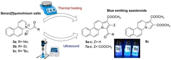
We report here the synthesis and optical spectral properties of several new azasteroid derivatives. The formation of these compounds was explained based on the most probable mechanism. The luminescent heterocycles were synthesized by 1,3-dipolar cycloaddition reactions between benzo[f]quinoline and methylpropiolate or dimethyl acetylenedicarboxylate (DMAD). A selective and efficient way for [3+2]-dipolar cycloaddition of benzo[f]quinolinium ylides under ultrasound (US) irradiation (20 kHz processing frequency) is presented. We report substantially higher yields under US irradiation, whereas the solvent amounts required are at least three-fold less compared to classical heating. The azasteroid derivatives are blue emitters with λmax of fluorescence around 430–450 nm. A certain influence of the azasteroid substituents concerning absorption and fluorescent properties was observed. Compounds anchored with a bulky pivaloyl group or without a C=O carbonyl group have shown increased fluorescence intensity.

Graphical Abstract

1. Introduction

Azasteroids are an important class of heterocycles that has received increasing interest in recent years [1], due to a wide range of potential applications for medicinal chemistry and optoelectronics. As materials with potential applications in medicinal chemistry, azasteroid derivatives were investigated as potential antimicrobial [2], antifungal [3], anticancer [4,5], and antituberculosis [6] agents. The optoelectronic properties of the azasteroids were investigated regarding their fluorescence emission [7,8,9]. The fluorescence of the azasteroid derivatives makes them very attractive materials in optoelectronics, whereas a combined use of the two distinct properties (biological and optical) has suggested interesting applications as fluorescent biomarkers [10,11,12,13].
Azasteroid derivatives are in fact benzo-azaindolizines, a class with an 18 π-electron N-fused heterocycle, containing a bridgehead nitrogen atom shared by an electron-excessive pyrrole and an azine electron-deficient six-membered ring. This structural arrangement being a ‘pure’ blue-emitting moiety. [13,14,15,16,17]. This uneven π-electron distribution between the two fused rings is an important feature that leads to electron delocalization. The electron delocalization within the entire heterocycle skeleton can be possible without a planar geometry of the indolizine. The planarity of azasteroid derivatives is provided by the sp2 hybridization of all the atoms in the fused ring and is preserved upon substitution with different groups.
In recent years, ultrasound-assisted reactions have proved to be a new versatile tool in synthetic organic chemistry [18,19], offering a facile and usefully alternative in a large variety of syntheses [20,21,22,23,24,25,26,27]. In comparison with conventional thermal heating (TH), ultrasound irradiation has several important advantages in terms of higher yields, increased purity of the compounds and selectivity, shorter reaction times, lower costs, and simplicity in handling and processing. All these advantages make US-assisted reactions environmentally friendly [22,23,24,28].
Our group has presented in previous work several studies on the synthesis of azine derivatives and their biological activities [29,30,31,32,33,34]. A special focusing was paid to the dipolar cycloaddition reactions of azinium ylides with various dipolarophiles in order to obtain new compounds with blue-fluorescent properties. Herein, we add further contributions in the field of using US irradiation in organic chemistry [20,21,22,23,24] and propose a facile, efficient, and environmentally friendly method for the synthesis of blue-emitting derivatives using US technologies.

2. Results and Discussion

Scheme 1 presents the general strategy adopted for the synthesis of the new fluorescent azasteroids. As can be observed, the synthesis of all pyrrolobenzo[f]quinoline derivatives, 5ac and 7ac, involves two steps. In the first step, an N-alkylation of the benzo[f]quinoline with bromoketones 3ac take place and benzo[f]quinolinium bromides are obtained as previously reported [34]. The following step is a 3+2 dipolar cycloaddition of benzo[f]quinolinium ylides 4ac (generated in situ from the corresponding salts 3ac, in the presence of 1,2-butylene oxide as a catalyst) to the alkyne dipolarophiles (dimethyl acetylenedicarboxylate–DMAD or methyl propiolate), which are leading to final products, the cycloadducts 5ac, 7ac, 8c.
The cycloaddition reactions were completed after 48 h of reflux. In the case of reactions of all salts with methyl propiolate, and in the case of the reactions of 3a,b salts with DMAD only the fully aromatized compounds 5ac and 7ab, respectively, were obtained. In the case of the reaction of 3c salt with DMAD, a mixture of fully aromatized compound 7c and decarbonylated derivative 8c was obtained.
The most probable reaction mechanism which explains the formation of these adducts is depicted in Scheme 2.
This mechanism involves, in the first step, the nucleophilic attack of the bromide ion on the 1,2-butylene oxide, the oxirane ring is opened, the alkoxide ion extracts a proton from the methylene group and leads to the ylide in 1,2-dipole form 4ac. The 1,2-dipole form adopts the 1,3-dipole 4ac form, which gives a Huisgen 3+2 cycloaddition with dipolarophiles via a concerted mechanism. The obtained reaction products 6ac are unstable (and non-isolable) because are non-aromatized, suffering a rearrangement to the more thermodynamic stable adducts 6ac (since the double bond in the pyrrole ring is in a conjugated system). The intermediate 6ac can be aromatized either by an oxidative dehydrogenation to a fully aromatized cycloadduct 7ac, or by a pivalaldehyde elimination in the case of the intermediate 6c to a fully aromatized and decarbonylated derivative 8c. The formation of the compound 8c could be explained by the steric hindrance of the tert-butyl group from the 6c intermediates. The partially aromatized derivatives 6ac are unstable when isolated, but were observed in the NMR spectra of the reaction mixture of all three benzo[f]quinolinium ylides 4ac. In the case of the benzo[f]quinolinium ylide 4a, we have studied the time conversion of the 6a intermediate (isolated after 24 h of reflux) to the final cycloadduct 7a (see Figure S15 from supporting information). As can be observed from this study, the conversion of the intermediate 6a to final cycloadduct 7a takes several days in the absence of the 1,2-butylene oxide catalyst. If the reaction reflux occurs in 48 h, the amount of the intermediate 6ac is negligible. In addition, this study is solid prove for the proposed mechanism.
The final azasteroids were obtained in moderate to good yield (40 to 69% see Table 1). The long reaction time (48 h) and the high energy consumption are major disadvantages of the synthesis carried out under conventional conditions.
As a more energy-efficient alternative, we have performed the synthesis of the azasteroid derivatives under US irradiation, using a Bandelin reactor (Sonopuls GM 3200, Berlin, Germany), with a nominal power of 200 W, and an operating frequency of 20 kHz. The used reactor allowed us to control the pulse sequence, as well as the amplitude (mean percent of the nominal power) and the irradiation time. All these parameters are expected to influence the reaction. The cycloaddition reactions were performed using 80% of the instrument nominal power and were completed after 2 h of irradiation.
The data from Table 1 shows that under US irradiation the reaction times decrease substantially from 2 days to 2 h. At the same time, the solvent amounts used in the former were at least three times lower than the corresponding quantities used under conventional conditions (see Experimental). This qualifies the former reactions as environmentally friendly. We may also notice that under US irradiation the yields were slightly higher (by 12 to 27%). In the case of the reaction of bromide 3c with DMAD, under US irradiation, we isolated less decarbonylated cycloadduct 8c which makes this reaction more selective to the fully aromatized cycloadduct 7c.
Optical properties of the synthesized azasteroids were investigated on diluted solutions (less than 10−5 mol/L) prepared in cyclohexane and trichloromethane, respectively. The dilution of each solution was adjusted, thus the absorbances maxima were measured in the 340–440 nm range, and reported on a 10 mm cuvette, to fit in a 0.5–0.9 unit range.
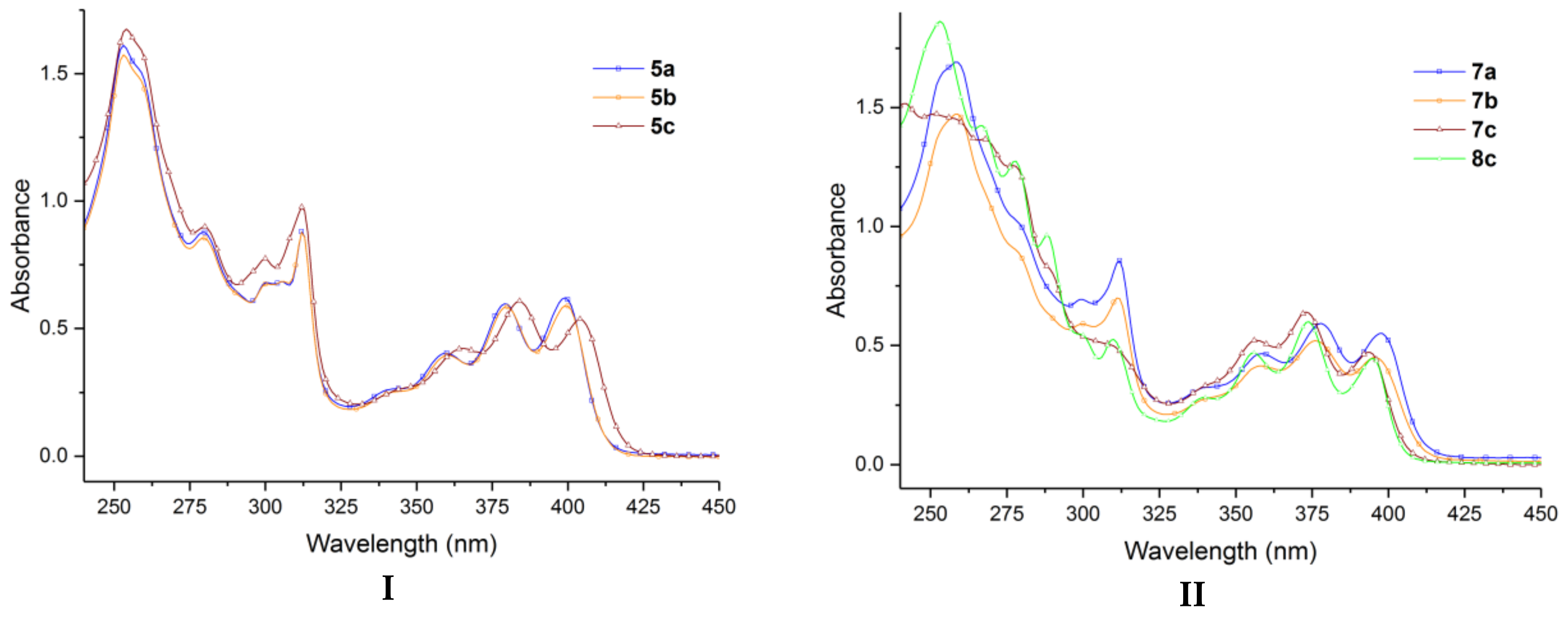
Since the compounds 5ac, 7ac, and 8c have a relatively similar structure, they exhibit small differences in their experimental UV-Vis absorption spectra, as can be seen from Figure 1 and Figure 2.
The absorption maxima of the seven azasteroid derivatives in cyclohexane and in trichloromethane are summarized in Table 2.
As we can observe in Table 2 and Figure 1 and Figure 2, the position of the absorption bands shows the influence of the substituents on the pyrrole ring.
In the case of all compounds (Figure 1 and Figure 2), in both solvents, three relatively well-separated absorption regions are observed (the first between 240–280 nm, the second between 280–325 nm, and the third between 325–425 nm, respectively). The absorption bands from the first region have a higher (double or more) intensity than the absorption bands from the second region, and the latter ones have a higher (less than double) intensity than the absorption bands from the third region. The absorption bands responsible for the blue emission of the azasteroids are situated in the third region (325–425 nm). In this region, four more or less separated absorption bands are visible. In cyclohexane, the four absorption bands are better separated than in trichloromethane.
In the case of the samples 5ac, the profile of the third absorption region and the absorption maxima are similar for the sample 5a and 5b (402 nm for 5a and 403 nm for 5b in trichloromethane, and 399 nm for both 5a and 5b in cyclohexane), while for the sample 5c the maximum of the absorption is situated on 386 nm in trichloromethane and 384 nm in cyclohexane. Sample 5c presents the same absorption bands as 5a and 5b with a small bathochromic shift.
In the case of the samples 7ac and 8c, the absorption bands have similar intensities and profiles. In the case of the samples 7a and 7b, the absorption bands are not so well separated in trichloromethane but well separated in cyclohexane, while for the samples 7c and 8c these bands are separated both in trichloromethane and in cyclohexane. The maximum of the absorption of the sample 7a presents a small bathochromic shift in comparison with the sample 7bc and 8c. The maxima of the absorption are 381 nm for 7a, 375 nm for 7b, 370 nm for 7c, and 369 nm for 8c in trichloromethane, and 378 nm for 7a, 376 nm for 7b, 373 nm for 7c, and 374 nm for 8c in cyclohexane.
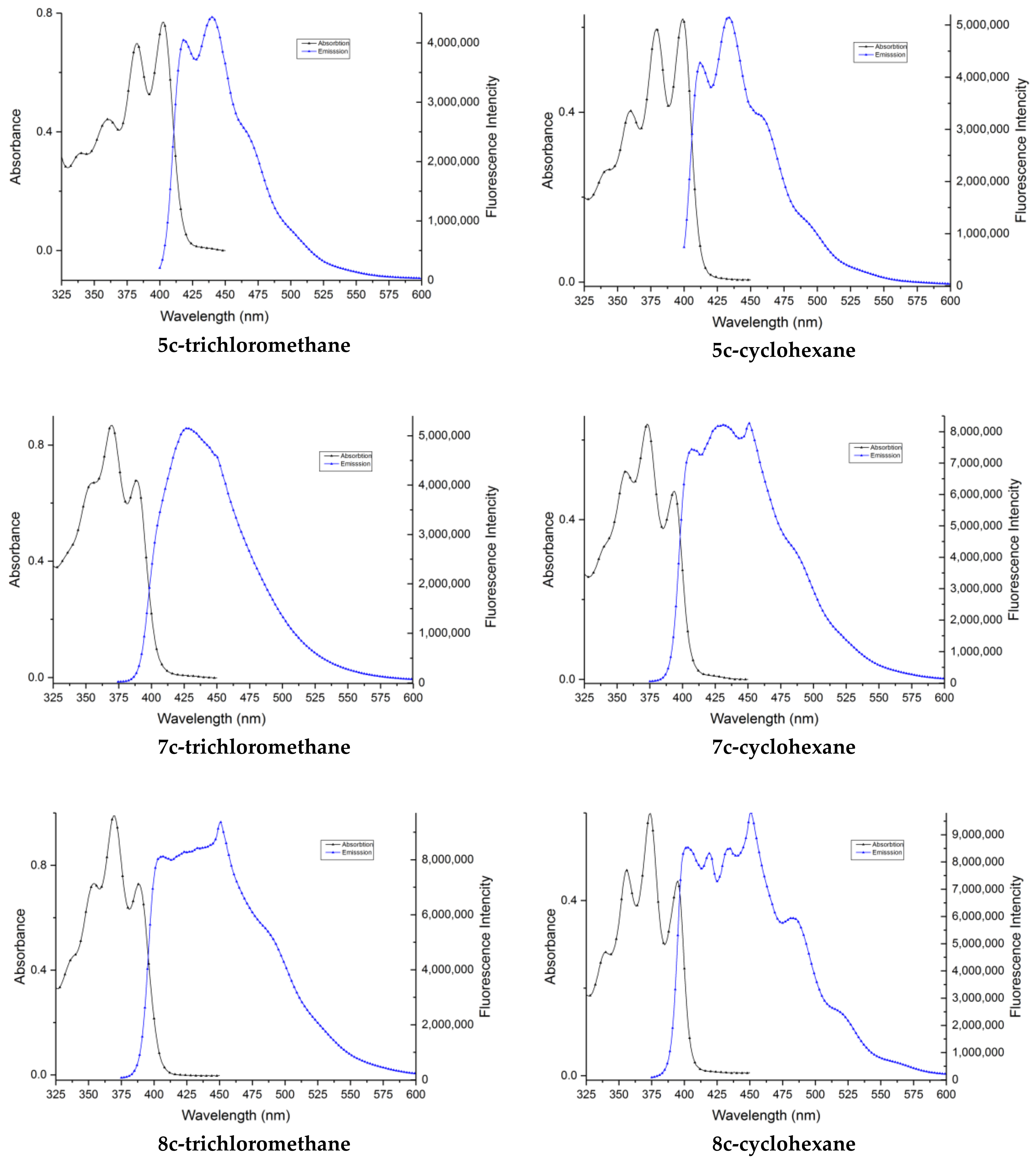
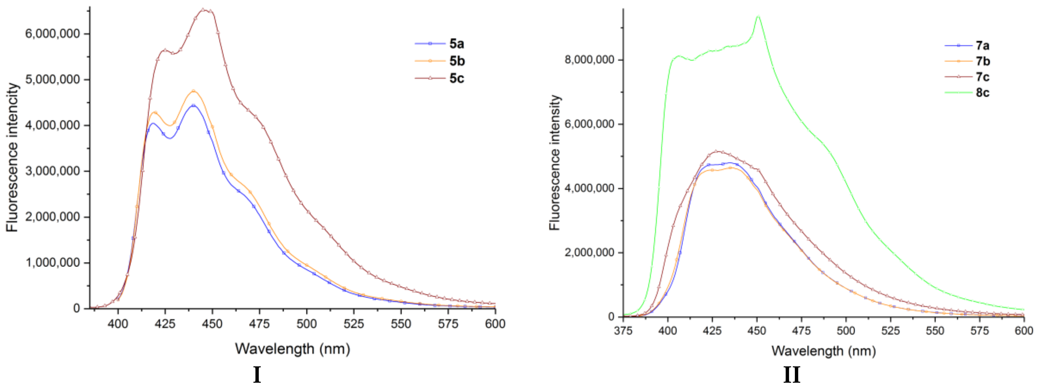
All the studied azasteroid derivatives have emission spectra consisting of one structured band in the 400–500 nm region (Figure 3, Figure 4, and Figure 5), indicating a planar structure of the molecules. The position of the band is significantly influenced by the presence of a carbomethoxy group at the 2nd position of the azasteroid skeleton. A hypsochromic shift of Δmax = 7 nm (5a compared with 7a), of Δmax = 7 nm (5b compared with 7b) and of Δmax = 15 nm (5c compared with 7c), could be observed in trichloromethane (Table 2). The same hypsochromic shift of Δmax = 2 nm (5a compared with 7a), of Δmax = 2 nm (5b compared with 7b), and of Δmax = 3 nm (5c compared with 7c), could be observed in cyclohexane (Table 2 and Figure 3).
When the substituent from the 3rd position of the azasteroid skeleton is the bulky pivaloyl group, an increase in the fluorescence intensity can be observed. A possible explanation can be the fact that the bulky group determines the deviation of the ketone group out of the molecule’s plane which breaks the conjugation between C=O carbonyl and the rest of the molecule, thus, the quenching of the fluorescence by this carbonyl group is reduced. When this carbonyl group from the 3rd position is missing (sample 8c) the fluorescence intensity is maximum.
Figure 3 illustrate the absorption and emission spectra of compounds 5c, 7c, and 8c in trichloromethane (left column) and in cyclohexane (right column), the absorption and emission spectra of the other compounds (5a,b and 7a,b) are presented in supporting info (Figures S37 and S38).
The optical absorption and emission maxima of the azasteroid derivatives in cyclohexane and trichloromethane are summarized in Table 2.
Table 2, Figure 4 and Figure 5 show that all the compounds are blue emitters (λmax of fluorescence around 430–450 nm). Compounds 7c (in cyclohexane) and 8c (both in trichloromethane and cyclohexane) have higher intensity in the emission spectra. Different behavior (in terms of the maximum of the emission) of the samples 5a,b comparing with 5c, and of the samples 7a,b comparing with 7c shown in Table 2 should relate to the difference in the electronic structures of 5a,b and 7a,b related to 5c and 7c, respectively. In the case of the compounds 5a,b and 7a,b, the substituent from the C=O keto group is methyl and ethyl, respectively, the difference between them is negligible, while in the case of the compounds 5c and 7c the substituent from the C=O keto group is tert-butyl, a bulkier one.

3. Experimental Section

3.1. General Procedure

All the reagents and solvents were purchased from commercial sources and used without further purification except bromoacetone which was synthesized by the reaction of acetone with bromine in acetic acid as the catalyst. Melting points were recorded on an Electrothermal MEL-TEMP (Barnstead International, Dubuque, IA, USA) apparatus in open capillary tubes and are uncorrected. Analytical thin-layer chromatography was performed with commercial silica gel plates 60 F254 (Merck KGaA, Darmstadt, Germany) and visualized with UV light. The NMR spectra were recorded on a (Bruker Vienna, Wien Austria) Avance III 500 MHz spectrometer operating at 500 MHz for 1H and 125 MHz for 13C. The following abbreviations were used to designate chemical shift multiplicities: s = singlet, d = doublet, t = triplet, m = multiplet. Chemical shifts were reported in delta (δ) units, part per million (ppm), and coupling constants (J) in Hz. Infrared (IR) data were recorded in powder on the diamond crystal ATR mode (Pike Technologies, Fitchburg, MA, USA) on an FT-IR Vertex-70 (Bruker Optik, Leipzig, Germany) spectrophotometer. Ultrasound-assisted reactions were carried out using a Bandelin Ultrasound reactor (Sonopuls GM 3200, Berlin, Germany), with a nominal power of 200 W and a frequency of 20 kHz. The booster horn SH 213 G was fixed tightly to the ultrasonic converter. The titanium flat probe tip TT13 (diameter: 12.7 mm; length: 7 mm) was fixed tightly to the booster horn. The titanium probe tip was immersed in the used solvent. UV-Vis spectra were recorded on an (Shimadzu, Kyoto, Japan) 1800 PC spectrophotometer in cyclohexane and trichloromethane (spectroscopic grade) solution. The fluorescence measurements were made using an (Edinburgh Instruments, Livingstone, UK) F900 photoluminescence spectrometer, in the same solvents as for the UV-Vis spectra, with the excitation wavelength set to the absorption band maximum. For all spectral determinations, the solutions were kept in 10 mm path length quartz cells. The fluorescence quantum yield was determined at room temperature with an (Edinburgh Instruments, Livingstone, UK).

3.1.1. General Procedure for Synthesis of Azasteroids Derivatives, 5ab, 7ab and 8c under Conventional TH Conditions

A mixture of benzo[f]quinolinium salt 3ac (0.791 g, 2.5 mmol of 3a or 0,826 g, 2.5 mmol of 3b or 0.896 g, 2.5 mmol of 3c) and methyl propiolate (0.31 mL, 3.5 mmol) or dimethyl acetylenedicarboxylate (0.43 mL, 3.5 mmol) was suspended in 30 mL 1,2-butylene oxide. The stirring and refluxing were continued for 48 h. After the reaction was finished (TLC), the obtained solution was cooled down at room temperature and evaporated under reduced pressure to give the crude product. The purification of the crude product was done by column chromatography on silica gel (eluted with 99.5/0.5 CH2Cl2/CH3OH).

3.1.2. General Procedure for Synthesis of Azasteroids Derivatives, 5ab, 7ab and 8c under US Irradiation

Under US irradiation, the mixture of reagents benzo[f]quinolinium salt 3ac (0.791 g, 2.5 mmol of 3a or 0,826 g, 2.5 mmol of 3b or 0.896 g, 2.5 mmol of 3c) and methyl propiolate (0.31 mL, 3.5 mmol) or dimethyl acetylenedicarboxylate (0.43 mL, 3.5 mmol) in 10 mL 1,2-butylene oxide was placed into the reaction vessel and exposed to irradiation (from 2 h; see Table 1). Once the irradiation cycle was completed, the reaction tube was removed from the reactor, and processed as indicated above for TH condition.
Methyl 3-acetylbenzo[f]pyrrolo[1,2-a]quinoline-1-carboxylate (5a). (0.516 g, 65% (under classical heating) and 0.635 g, 80% (under ultrasounds)) as a straw-yellow crystals, m.p. 215–216 °C); Rf (99.5/0.5 CH2Cl2/CH3OH) 0.44; IR (cm−1): 3034, 3013 (C-H arom.), 2963, 2927 (C-H aliph.), 1704 (C=O, ester), 1639 (C=O, keto), 1610, 1547, 1497, 1442, 1415, 1406, 1353 (aromatic and heteroaromatic ring), 1243, 1178, 1157, 1098, 1064 (C–O–C, ester); 1H NMR (500 MHz, CDCl3): δ 8.56 (2H, m, overlapped peaks, H-10, H-11), 8.47 (1H, d, J = 9.5 Hz, H-12), 8.10 (2H, m, overlapped peaks, H-2, H-5), 7.96 (2H, m, overlapped peaks, H-6, H-7), 7.71 (1H, t, J = 7.5 Hz, H-9), 7.63 (1H, t, J = 7.5 Hz, H-8), 3.97 (3H, s, CH3 of methoxycarbonyl group from 1st position), 2.75 (3H, s, CH3 of acetyl group from 3rd position); 13C NMR (125 MHz, CDCl3): δ 186.8 (CO keto group from 3rd position), 164.6 (CO keto ester from 1st position), 140.5 (C-12a), 132.5 (C-4a), 131.0 (C-6a), 129.7 (C-10a), 129.1 (C-6), 128.9 (C-7), 128.6 (C-5), 128.6 (C-3), 127.7 (C-9), 126.8 (C-8), 124.2 (C-11), 123.0 (C-10), 121.2 (C-10b), 120.4 (C-2), 117.5 (C-12), 106.7 (C-1), 51.6 (CH3 of methoxycarbonyl group from 1st position), 28.4 (CH3 of acetyl group from 3rd position). Anal. calc. for C20H15NO3 (317.34): C 75.70, H 4.76, N 4.41; found: C 75.65, H 4.70, N 4.35.
Methyl 3-propionylbenzo[f]pyrrolo[1,2-a]quinoline-1-carboxylate (5b). (0.572 g, 69% (under classical heating) and 0.671 g, 81% (under ultrasounds)) as a straw-yellow crystals, m.p. 186–187 °C); Rf (99.5/0.5 CH2Cl2/CH3OH) 0.58; IR (cm−1): 3034, 3001 (C-H arom.), 2978, 2940 (C-H aliph.), 1705 (C=O, ester), 1645 (C=O, keto), 1610, 1547, 1496, 1442, 1414, 1359 (aromatic and heteroaromatic ring), 1238, 1180, 1156, 1128, 1088, 1073 (C–O–C, ester); 1H NMR (500 MHz, CDCl3): δ 8.56 (1H, d, J = 8.5 Hz, H-10), 8.54 (1H, d, J = 9.5 Hz, H-11), 8.46 (1H, d, J = 9.5 Hz, H-12), 8.08 (1H, s, H-2), 8.01 (1H, d, J = 9.0 Hz, H-5), 7.96 (2H, m, overlapped peaks, H-6, H-7), 7.70 (1H, t, J = 7.0, 8.5 Hz, H-9), 7.62 (1H, t, J = 7.0 Hz, H-8), 3.97 (3H, s, CH3 of methoxycarbonyl group from 1st position), 3.10 (2H, q, J = 7.5 Hz, CH2 of propionyl group from 3rd position), 1.38 (3H, t, J = 7.5 Hz, CH3 of propionyl group from 3rd position); 13C NMR (125 MHz, CDCl3): δ 190.9 (CO keto group from 3rd position), 164.6 (CO keto ester from 1st position), 140.3 (C-12a), 132.5 (C-4a), 131.0 (C-6a), 129.7 (C-10a), 129.1 (C-6), 128.8 (C-7), 128.6 (C-3), 127.7 (C-8), 127.6 (C-2), 126.8 (C-9), 123.9 (C-11), 123.0 (C-10), 121.2 (C-10b), 120.3 (C-5), 117.6 (C-12), 106.7 (C-1), 51.5 (CH3 of methoxycarbonyl group from 1st position), 33.7 (CH2 of propionyl group from 3rd position), 9.9 (CH3 of propionyl group from 3rd position). Anal. calc. for C21H17NO3 (331.37): C 76.12, H 5.17, N 4.23; found: C 76.07, H 5.13, N 4.17.
Methyl 3-pivaloylbenzo[f]pyrrolo[1,2-a]quinoline-1-carboxylate (5c). (0.575 g, 64% (under classical heating) and 0.701 g, 78% (under ultrasounds)) as a straw-yellow crystals, m.p. 192–193 °C); Rf (99.5/0.5 CH2Cl2/CH3OH) 0.67; IR (cm−1): 3057 (C-H arom.), 2982, 2950, 2930 (C-H aliph.), 1700 (C=O, ester), 1648 (C=O, keto), 1611, 1545, 1507, 1493, 1444, 1415, 1367 (aromatic and heteroaromatic ring), 1235, 1152, 1138, 1119, 1084 (C–O–C, ester); 1H NMR (500 MHz, CDCl3): δ 8.50 (1H, d, J = 8.0 Hz, H-10), 8.40 (2H, m, overlapped peaks, H-11, H-12), 7.92 (1H, s, H-2), 7.90 (2H, m, overlapped peaks, H-5, H-7), 7.65 (1H, t, J = 7.5, 8.0 Hz, H-9), 7.57 (1H, t, J = 7.5 Hz, H-8), 7.54 (1H, d, J = 9.0 Hz, H-6), 3.96 (3H, s, CH3 of methoxycarbonyl group from 1st position), 1.58 (9H, s, 3xCH3 of tert-butyl group from 3rd position); 13C NMR (125 MHz, CDCl3): δ 199.7 (CO keto group from 3rd position), 164.8 (CO keto ester from 1st position), 138.9 (C-12a), 131.9 (C-4a), 130.6 (C-6a), 129.9 (C-10a), 129.3 (C-5), 128.8 (C-7), 127.7 (C-9), 126.9 (C-3), 126.6 (C-8), 124.1 (C-2), 122.9 (C-10), 122.8 (C-11), 120.8 (C-10b), 119.2 (C-6), 117.6 (C-12), 105.9 (C-1), 51.4 (CH3 of methoxycarbonyl group from 1st position), 44.4 (C of tert-butyl group from 3rd position) 29.1 (3xCH3 of tert-butyl group from 3rd position). Anal. calc. for C23H21NO3 (359.43): C 76.86, H 5.89, N 3.90; found: C 76.80, H 5.86, N 3.85.
Dimethyl 3-acetylbenzo[f]pyrrolo[1,2-a]quinoline-1,2-dicarboxylate (7a). (0.507 g, 54% (under classical heating) and 0.666 g, 71% (under ultrasounds)) as a straw-yellow crystals, m.p. 197–198 °C); Rf (99.5/0.5 CH2Cl2/CH3OH) 0.26; IR (cm−1): 3067, 3003 (C-H arom.), 2952 (C-H aliph.), 1739, 1695 (C=O, ester), 1662 (C=O, keto), 1613, 1547, 1491, 1468, 1444, 1405 (aromatic and heteroaromatic ring), 1247, 1219, 1197, 1166, 1154, 1096 (C–O–C, ester); 1H NMR (500 MHz, CDCl3): δ 8.55 (1H, d, J = 8.5 Hz, H-10), 8.51 (1H, d, J = 9.5 Hz, H-11), 8.40 (1H, d, J = 9.5 Hz, H-12), 7.95 (2H, d, overlapped peaks, H-5, H-7), 7.73 (1H, t, J = 7.5 Hz, H-9), 7.64 (2H, m, overlapped peaks, H-6, H-8), 4.05 (3H, s, CH3 of methoxycarbonyl group from 1st position), 3.95 (3H, s, CH3 of methoxycarbonyl group from 2nd position), 2.60 (3H, s, CH3 of acetyl group from 3rd position); 13C NMR (125 MHz, CDCl3): δ 189.0 (CO keto group from 3rd position), 167.0 (CO keto ester from 2nd position), 163.6 (CO keto ester from 1st position), 137.8 (C-12a), 131.7 (C-4a), 130.9 (C-6a), 130.5 (C-10a), 129.7 (C-5), 129.7 (C-2), 128.9 (C-7), 128.1 (C-9), 127.1 (C-8), 126.9 (C-3), 123.9 (C-11), 123.0 (C-10), 121.5 (C-10b), 119.3 (C-6), 117.9 (C-12), 104.9 (C-1), 53.4 (CH3 of methoxycarbonyl group from 1st position), 52.0 (CH3 of methoxycarbonyl group from 2nd position), 29.5 (CH3 of acetyl group from 3rd position). Anal. calc. for C22H17NO5 (375.38): C 70.39, H 4.56, N 3.73; found: C 70.33, H 4.52, N 3.69.
Dimethyl 3-propionylbenzo[f]pyrrolo[1,2-a]quinoline-1,2-dicarboxylate (7b). (0.526 g, 54% (under classical heating) and 0.672 g, 69% (under ultrasounds)) as a straw-yellow crystals, m.p. 206–207 °C); Rf (99.5/0.5 CH2Cl2/CH3OH) 0.30; IR (cm−1): 3032, 3000 (C-H arom.), 2981, 2951 (C-H aliph.), 1731, 1697 (C=O, ester), 1668 (C=O, keto), 1612, 1546, 1494, 1467, 1444, 1406, 1364 (aromatic and heteroaromatic ring), 1266, 1223, 1182, 1163, 1145, 1095, 1089 (C–O–C, ester); 1H NMR (500 MHz, CDCl3): δ 8.54 (1H, d, J = 8.5 Hz, H-10), 8.46 (1H, d, J = 9.5 Hz, H-11), 8.38 (1H, d, J = 9.5 Hz, H-12), 7.95 (2H, d, overlapped peaks, H-5, H-7), 7.72 (1H, t, J = 7.5 Hz, H-9), 7.63 (1H, t, J = 7.5 Hz, H-8), 7.56 (1H, d, J = 9.5 Hz, H-6), 4.03 (3H, s, CH3 of methoxycarbonyl group from 1st position), 3.94 (3H, s, CH3 of methoxycarbonyl group from 2nd position), 2.85 (2H, q, J = 7.5 Hz, CH2 of propionyl group from 3rd position), 1.31 (3H, t, J = 7.5 Hz, CH3 of propionyl group from 3rd position); 13C NMR (125 MHz, CDCl3): δ 194.3 (CO keto group from 3rd position), 166.7 (CO keto ester from 2nd position), 163.7 (CO keto ester from 1st position), 137.2 (C-12a), 131.5 (C-4a), 130.8 (C-6a), 129.8 (C-5), 129.7 (C-10a), 128.9 (C-7), 128.8 (C-2), 128.1 (C-9), 127.1 (C-8), 127.0 (C-3), 123.2 (C-11), 123.0 (C-10), 121.4 (C-10b), 118.7 (C-6), 118.1 (C-12), 104.7 (C-1), 53.3 (CH3 of methoxycarbonyl group from 1st position), 51.9 (CH3 of methoxycarbonyl group from 2nd position), 35.6 (CH2 of propionyl group from 3rd position), 9.1 (CH3 of propionyl group from 3rd position). Anal. calc. for C23H19NO5 (389.41): C 70.94, H 4.92, N 3.60; found: C 70.87, H 4.88, N 3.55.
Dimethyl 3-pivaloylbenzo[f]pyrrolo[1,2-a]quinoline-1,2-dicarboxylate (7c). (0.397 g, 38% (under classical heating) and 0.678 g, 65% (under ultrasounds)) as a straw-yellow crystals, m.p. 184–185 °C); Rf (99.5/0.5 CH2Cl2/CH3OH) 0.35; IR (cm−1): 3067, 3023 (C-H arom.), 2981, 2971, 2953 (C-H aliph.), 1729, 1694 (C=O, ester), 1682 (C=O, keto), 1612, 1559, 1546, 1505, 1464, 1418 (aromatic and heteroaromatic ring), 1258, 1208, 1177, 1161, 1119, 1091, 1059 (C–O–C, ester); 1H NMR (500 MHz, CDCl3): δ 8.56 (1H, d, J = 8.0 Hz, H-10), 8.35 (2H, m, overlapped peaks, H-11, H-12), 7.95 (2H, m, overlapped peaks, H-5, H-7), 7.73 (2H, m, overlapped peaks, H-6, H-9), 7.64 (1H, t, J = 7.0 Hz, H-8), 3.95 (3H, s, CH3 of methoxycarbonyl group from 1st position), 3.93 (3H, s, CH3 of methoxycarbonyl group from 2nd position), 1.15 (9H, s, 3xCH3 of tert-butyl group from 3rd position); 13C NMR (125 MHz, CDCl3): δ 207.9 (CO keto group from 3rd position), 165.6 (CO keto ester from 2nd position), 164.0 (CO keto ester from 1st position), 134.4 (C-12a), 131.8 (C-4a), 130.8 (C-6a), 130.1 (C-5), 129.8 (C-10a), 128.9 (C-7), 128.3 (C-9), 127.0 (C-8), 126.4 (C-2), 123.0 (C-10), 122.9 (C-3), 120.9 (C-11), 120.7 (C-10b), 118.7 (C-12), 116.9 (C-6), 104.6 (C-1), 52.8 (CH3 of methoxycarbonyl group from 1st position), 51.9 (CH3 of methoxycarbonyl group from 2nd position), 47.2 (C of tert-butyl group from 3rd position), 27.4 (3xCH3 of tert-butyl group from 3rd position). Anal. calc. for C25H23NO5 (417.16): C 71.93, H 5.55, N 3.36; found: C 71.88, H 5.50, N 3.32.
Dimethyl benzo[f]pyrrolo[1,2-a]quinoline-1,2-dicarboxylate (8c). (0.108 g, 13% (under classical heating) and 0.042 g, 5% (under ultrasounds)) as a straw-yellow crystals, m.p. 198–199 °C); Rf (99.5/0.5 CH2Cl2/CH3OH) 0.28; IR (cm−1): 3145, 3091, 3066 (C-H arom.), 2982, 2963, 2942 (C-H aliph.), 1710, 1675 (C=O, ester), 1610, 1563, 1545, 1507, 1463, 1441, 1406 1369 (aromatic and heteroaromatic ring), 1270, 1255, 1231, 1217, 1197, 1164 1095, 1060 (C–O–C, ester); 1H NMR (500 MHz, CDCl3): δ 8.41 (1H, d, J = 8.5 Hz, H-10), 8.21 (1H, s, H-3), 8.11 (1H, d, J = 9.5 Hz, H-11), 8.06 (1H, d, J = 9.5 Hz, H-12), 7.88 (3H, m, overlapped peaks, H-5, H-6, H-7), 7.66 (1H, t, J = 8.0 Hz, H-9), 7.56 (1H, t, J = 8.0 Hz, H-8), 3.97 (3H, s, CH3 of methoxycarbonyl group from 1st position), 3.96 (3H, s, CH3 of methoxycarbonyl group from 2nd position); 13C NMR (125 MHz, CDCl3): δ 165.0 (CO keto ester from 2nd position), 164.5 (CO keto ester from 1st position), 134.9 (C-12a), 130.8 (C-6a), 130.5 (C-6), 130.1 (C-4a), 129.8 (C-10a), 129.0 (C-7), 128.0 (C-9), 126.5 (C-8), 122.7 (C-10), 120.7 (C-2), 119.5 (C-11), 119.3 (C-10b), 118.6 (C-12), 116.1 (C-3), 113.9 (C-5), 105.4 (C-1), 52.3 (CH3 of methoxycarbonyl group from 1st position), 51.6 (CH3 of methoxycarbonyl group from 2nd position). Anal. calc. for C20H15NO4 (333.34): C 72.06, H 4.54, N 4.20; found: C 72.00, H 4.49, N 4.16.

4. Conclusions

In conclusion, we report herein an efficient and straightforward pathway for obtaining a new class of blue fluorescent azasteroid derivatives, under conventional (thermal) heating as well as under unconventional (ultrasound) irradiation. Blue fluorescent azasteroid derivatives have been obtained using a cycloaddition reaction of benzo[f]quinolinium ylides with symmetrically and unsymmetrically activated alkynes. Under conventional heating, in the case of reactions with methyl propiolate only the fully aromatized compounds 5ac were obtained, while in the case of reactions with DMAD fully aromatized compounds 7ab and a mixture of fully aromatized compound 7c and a fully aromatized and decarbonylated derivative 8c were obtained. A feasible reaction mechanism for the azasteroid derivatives formation is described in this study. Under ultrasound irradiation, we isolated fully aromatized cycloadducts with increased yield, and in the case of the reaction of bromide 3c with DMAD, we isolated less decarbonylated cycloadduct 8c which make this reaction more selective to the fully aromatized cycloadduct 7c.
The absorption and emission maxima of the obtained azasteroid derivatives were studied, some of these compounds being blue emitters. The absorption and emission spectra are dependent on their structure, compounds with a similar structure having spectra with small differences, whereas the compounds with a bulky pivaloyl group or without a C=O keto group in the 3rd position present more intense fluorescence emission.
We also note that under ultrasound irradiation, the reactions occur with increased selectivity regarding the decarbonylated compound and offers several advantages in terms of yield, easier workup of the reaction, a substantial decrease in consumed solvents, a substantial reduction in reaction time (from days to hours), thus, consequent diminution in energy consumption. Taking into consideration these advantages, the proposed method should be considered environmentally friendly.

Supplementary Materials

The following are available online. Details of the IR, UV-Vis, Emission and NMR spectra (1H NMR and 13C NMR) of the synthesized compounds can be found in the Supporting Information.

Author Contributions

Design, synthesis, and writing, I.M., G.Z. and C.M.; evaluation of fluorescent properties, G.Z. and C.M.; experimental work and structure elucidation, G.Z. and C.M.; revision of the first draft of the paper, I.M., G.Z. and C.M. All authors have read and agreed to the final version of the manuscript.

Funding

This work was supported by a grant of the Romanian Ministry of Education and Research, CNCS-UEFISCDI, project number PN-III-P4-ID-PCE-2020-0371, within PNCDI III.

Institutional Review Board Statement

Not available.

Informed Consent Statement

Not available.

Data Availability Statement

Not available.

Acknowledgments

Acknowledgment to the infrastructure support from Operational Program Competitiveness 2014–2020, Axis 1, under POC/448/1/1 Research infrastructure projects for public R&D institutions/Sections F 2018, through the Research Center with Integrated Techniques for Atmospheric Aerosol Investigation in Romania (RECENT AIR) project, under grant agreement MySMIS no. 127324. Authors are thankful to UEFISCDI Bucharest, Romania, project PN-III-P1-1.1-TE-2016-1205 and PN-III-P4-ID-PCE-2020-0371, for financial support and the POSCCE-O 2.2.1, SMIS-CSNR 13984-901, No. 257/28.09.2010 Project, CERNESIM, for NMR experiments.

Conflicts of Interest

The authors declare no conflict of interest.

Sample Availability

Samples of the all compounds are available from the authors.

References

  1. Singh, R.; Panda, G. An overview of synthetic approaches for heterocyclic steroids. Tetrahedron 2013, 69, 2853–2884. [Google Scholar] [CrossRef]
  2. Karnik, A.V.; Malviya, N.J.; Kulkarni, A.M.; Jadhav, B.L. Synthesis and in vitro antibacterial activity of novel heterocyclic derivatives of 18-nor-equilenin. Eur. J. Med. Chem. 2006, 41, 891–895. [Google Scholar] [CrossRef]
  3. Burbiel, J.; Bracher, F. Azasteroids as antifungals. Steroids 2003, 68, 587–594. [Google Scholar] [CrossRef]
  4. Robinson, A.J.; DeLucca, I.; Drummond, S.; Boswell, G.A. Steroidal nitrone inhibitors of 5α-reductase. Tetrahedron Lett. 2003, 44, 4801–4804. [Google Scholar] [CrossRef]
  5. Singh, J.; Singh, R.; Gupta, P.; Rai, S.; Ganesher, A.; Badrinarayan, P.; Sastry, G.N.; Konwar, R.; Panda, G. Targeting progesterone metabolism in breast cancer with L-proline derived new 14-azasteroids. Bioorg. Med. Chem. 2017, 25, 4452–4463. [Google Scholar] [CrossRef]
  6. Thomas, S.T.; Yang, X.; Sampson, N.S. Inhibition of the M. tuberculosis 3β-hydroxysteroid dehydrogenase by azasteroids. Bioorganic Med. Chem. Lett. 2011, 21, 2216–2219. [Google Scholar] [CrossRef] [Green Version]
  7. Bagnich, S.A.; Gulyakevich, O.V.; Mikhalchuk, A.L. Spectral-Luminescent properties of 12-oximino derivatives of 8-AZA-D-homogona-12,17a-diones and their concentration dependence. J. Fluoresc. 2008, 18, 277–283. [Google Scholar] [CrossRef]
  8. Borisevich, N.A.; Raichyonok, T.F.; Sukhodola, A.A.; Tolstorozhev, G.B. Delayed fluorescence and phosphorescence of 8-aza-d-homogonane in the gas and condensed phases. J. Fluoresc. 2006, 16, 649–653. [Google Scholar] [CrossRef]
  9. Akhrem, A.A.; Borisevich, N.A.; Gulyakevich, O.V.; Mikhalchuk, A.L.; Raichyonok, T.F.; Tikhomirov, S.A.; Tolstorozhev, G.B. Specific fluorescence properties and picosecond transient absorption of 8-azasteroids. J. Fluoresc. 1999, 9, 357–361. [Google Scholar]
  10. Li, Z.; Askim, J.R.; Suslick, K.S. The optoelectronic nose: Colorimetric and fluorometric sensor arrays. Chem. Rev. 2019, 119, 231–292. [Google Scholar] [CrossRef]
  11. Wu, L.; Qu, X. Cancer biomarker detection: Recent achievements and challenges. Chem. Soc. Rev. 2015, 44, 2963–2997. [Google Scholar] [CrossRef]
  12. Queralto, N.; Berliner, A.N.; Goldsmith, B.; Martino, R.; Rhodes, P.; Lim, S.H. Detecting cancer by breath volatile organic compound analysis: A review of array-based sensors. J. Breath Res. 2014, 8, 027112. [Google Scholar] [CrossRef]
  13. Moldoveanu, C.; Amariucai-Mantu, D.; Mangalagiu, V.; Antoci, V.; Maftei, D.; Mangalagiu, I.I.; Zbancioc, G. Microwave assisted reactions of fluorescent pyrrolodiazine building blocks. Molecules 2019, 24, 3760. [Google Scholar] [CrossRef] [Green Version]
  14. Tisler, M. Structure and reactivity correlation of bicyclic 10-π electron systems with bridgehead nitrogen. Pure Appl. Chem. 1980, 52, 1611–1621. [Google Scholar] [CrossRef] [Green Version]
  15. Maftei, D.; Zbancioc, G.; Humelnicu, I.; Mangalagiu, I. Conformational effects on the lowest excited states of benzoyl-pyrrolopyridazine: Insights from PCM time-dependent DFT. J. Phys. Chem. A 2013, 117, 3165–3175. [Google Scholar] [CrossRef]
  16. Zbancioc, G.; Mangalagiu, I.I. Pyrrolopyridazine derivatives as blue organic luminophores: Synthesis and properties. Part 2. Tetrahedron 2010, 66, 278–282. [Google Scholar] [CrossRef]
  17. Zbancioc, G.; Huhn, T.; Groth, U.; Deleanu, C.; Mangalagiu, I.I. Pyrrolodiazine derivatives as blue organic luminophores: Synthesis and properties. Part 3. Tetrahedron 2010, 66, 4298–4306. [Google Scholar] [CrossRef] [Green Version]
  18. Shingare, M.S.; Shingate, B.B. Ultrasound in synthetic applications and organic chemistry. In Handbook on Applications of Ultrasound: Sonochemistry for Sustainability, 1st ed.; Chen, D., Sharma, S.K., Mudhoo, A., Eds.; CRC Press: Boca Raton, FL, USA, 2012; ISBN 9781439842065. [Google Scholar]
  19. Mason, T.J.; Peters, D. Practical Sonochemistry. Power Ultrasound Uses and Applications, 2nd ed.; Ellis Horwood: Chichester, UK, 2002; ISBN 9781898563839. [Google Scholar]
  20. Bejan, V.; Moldoveanu, C.; Mangalagiu, I.I. Ultrasounds assisted reactions of steroid analogous of anticipated biological activities. Ultrason. Sonochem. 2009, 16, 312–315. [Google Scholar] [CrossRef]
  21. Zbancioc, G.; Zbancioc, A.M.; Mangalagiu, I.I. Ultrasound and microwave assisted synthesis of dihydroxyacetophenone derivatives with or without 1,2-diazine skeleton. Ultrason. Sonochem. 2014, 21, 802–811. [Google Scholar] [CrossRef]
  22. Zbancioc, G.; Moldoveanu, C.; Mangalagiu, I.I. Ultrasound assisted synthesis of imidazolium salts: An efficient way to ionic liquids. Ultrason. Sonochem. 2015, 23, 376–384. [Google Scholar] [CrossRef]
  23. Antoci, V.; Moldoveanu, C.; Danac, R.; Mangalagiu, V.; Zbancioc, G. Huisgen 3+2 dipolar cycloadditions of phthalazinium ylides to activated symmetric and non-symmetric alkynes. Molecules 2020, 25, 4416. [Google Scholar] [CrossRef]
  24. Mantu, D.; Moldoveanu, C.; Nicolescu, A.; Deleanu, C.; Mangalagiu, I.I. A facile synthesis of pyridazinone derivatives under ultrasonic irradiation. Ultrason. Sonochem. 2009, 16, 312–315. [Google Scholar] [CrossRef]
  25. Borsoi, A.F.; Paz, J.D.; Pissinate, K.; Rambo, R.S.; Pestana, V.Z.; Bizarro, C.V.; Basso, L.A.; Machado, P. Ultrasound-Assisted synthesis of 4-alkoxy-2-methylquinolines: An efficient method toward antitubercular drug candidates. Molecules 2021, 26, 1215. [Google Scholar] [CrossRef]
  26. Banik, B.K.; Sahoo, B.M.; Kumar, B.V.V.R.; Panda, K.C.; Jena, J.; Mahapatra, M.K.; Borah, P. Green synthetic approach: An efficient eco-friendly tool for synthesis of biologically active oxadiazole derivatives. Molecules 2021, 26, 1163. [Google Scholar] [CrossRef]
  27. Rodríguez-Villar, K.; Yépez-Mulia, L.; Cortés-Gines, M.; Aguilera-Perdomo, J.D.; Quintana-Salazar, E.A.; Olascoaga Del Angel, K.S.; Cortés-Benítez, F.; Palacios-Espinosa, J.F.; Soria-Arteche, O.; Pérez-Villanueva, J. Synthesis, antiprotozoal activity and cheminformatic analysis of 2-phenyl-2H-indazole derivatives. Molecules 2021, 26, 2145. [Google Scholar] [CrossRef]
  28. Anastas, P.T.; Warner, J.C. Green Chemistry: Theory and Practice; Oxford University Press: Oxford, UK; New York, NY, USA, 1988. [Google Scholar]
  29. Moldoveanu, C.; Zbancioc, G.; Mantu, D.; Maftei, D.; Mangalagiu, I.I. The cycloaddition of the benzimidazolium ylides with alkynes: New mechanistic insights. PLoS ONE 2016, 11, e0156129. [Google Scholar] [CrossRef] [Green Version]
  30. Zbancioc, A.M.; Miron, A.; Tuchilus, C.; Rotinberg, P.; Mihai, C.T.; Mangalagiu, I.I.; Zbancioc, G. Synthesis and in vitro analysis of novel dihydroxyacetophenone derivatives with antimicrobial and antitumor activities. Med. Chem. 2014, 10, 476–483. [Google Scholar] [CrossRef]
  31. Antoci, V.; Mantu, D.; Cozma, D.G.; Usru, C.; Mangalagiu, I.I. Hybrid anticancer 1,2-diazine derivatives with multiple mechanism of action. Part 3. Med. Hypotheses 2014, 82, 11–15. [Google Scholar] [CrossRef]
  32. Lungu, C.N.; Bratanovici, B.I.; Grigore, M.M.; Antoci, V.; Mangalagiu, I.I. Hybrid Imidazole-Pyridne Derivatives: An Approach to Nevel Anticancer DNA Intercalators. Curr. Med. Chem. 2020, 27, 154–169. [Google Scholar] [CrossRef]
  33. Antoci, V.; Cucu, D.; Zbancioc, G.; Moldoveanu, C.; Mangalagiu, V.; Amariucai-Mantu, D.; Aricu, A.; Mangalagiu, I.I. Bis-(imidazole/benzimidazole)-pyridine derivatives: Synthesis, structure and antimycobacterial activity. Future Med. Chem. 2020, 12, 207–222. [Google Scholar] [CrossRef] [Green Version]
  34. Antoci, V.; Oniciuc, L.; Amariucai-Mantu, D.; Moldoveanu, C.; Mangalagiu, V.; Amarandei, A.M.; Lungu, C.N.; Dunca, S.; Mangalagiu, I.I.; Zbancioc, G. Benzoquinoline derivatives: A straightforward and efficient route to antibacterial and antifungal agents. Pharmaceuticals 2021, 14, 335. [Google Scholar] [CrossRef]
Scheme 1. Reaction pathway to generate azasteroid derivatives.
Molecules 26 05098 sch001
Scheme 2. The most probable mechanism of the cycloaddition reaction.
Molecules 26 05098 sch002
Figure 1. The absorption spectra recorded in trichloromethane solution of the azasteroid derivatives: (I)-5ac; (II)-7ac, and 8c.
Molecules 26 05098 g001
Figure 2. The absorption spectra recorded in cyclohexane solution of the azasteroid derivatives: (I)-5ac; (II)-7ac and 8c.
Molecules 26 05098 g002
Figure 3. The absorption (black line) and emission (blue line) spectra of azasteroid derivatives 5c, 7c, and 8c in trichloromethane (left column) and cyclohexane (right column). The excitation wavelength was: 380 nm for sample 5c and 365 nm for samples 7c and 8c.
Molecules 26 05098 g003
Figure 4. The emission spectra recorded in trichloromethane solution of the azasteroid derivatives: (I)-5ac; (II)-7ac and 8c.
Molecules 26 05098 g004
Figure 5. The emission spectra recorded in cyclohexane solution of the azasteroid derivatives: (I)-5ac; (II)-7ac and 8c.
Molecules 26 05098 g005
Table 1. The synthesis of azasteroid derivatives under US irradiation and conventional TH.
Compound
Data Availability Statement
UltrasoundsConventional TH
Reaction Time, (h)Yield, %Reaction Time, (h)Yield, %
5a2804865
5b8169
5c7864
7a7154
7b6954
7c6538
8c513
Table 2. λmax (nm) of absorption spectra and λmax (nm) of emission spectra of compounds 5ac, 7ac, and 8c.
CompoundFluorescence (λmax, nm)Absorption (λmax, nm)
CyclohexaneTrichloromethaneCyclohexaneTrichloromethane
5a434439399403
5b434440399402
5c440445384386
7a432432378381
7b432433376375
7c451430373370
8c450449374369
λexc = 396 nm for samples 5a and 5b; λexc = 380 nm for sample 5c; λexc = 370 nm for samples 7a and 7b; λexc = 365 nm for samples 7c and 8c.
Publisher’s Note: MDPI stays neutral with regard to jurisdictional claims in published maps and institutional affiliations.