
Antifungal Effect of Chitosan/Nano-TiO2 Composite Coatings against Colletotrichum gloeosporioides, Cladosporium oxysporum and Penicillium steckii

 , and
, and
Abstract
:1. Introduction
2. Materials and Methods
2.1. Materials
2.2. Coating Preparation
2.3. SEM Analysis
2.4. Determination of Mold Mortality
2.5. Inhibition Rate and Dry Weight of Mycelium
2.6. Determination of Conductivity, Protein Dissolution and Nucleic Acids Exosmosis
2.7. Statistical Analysis
3. Results
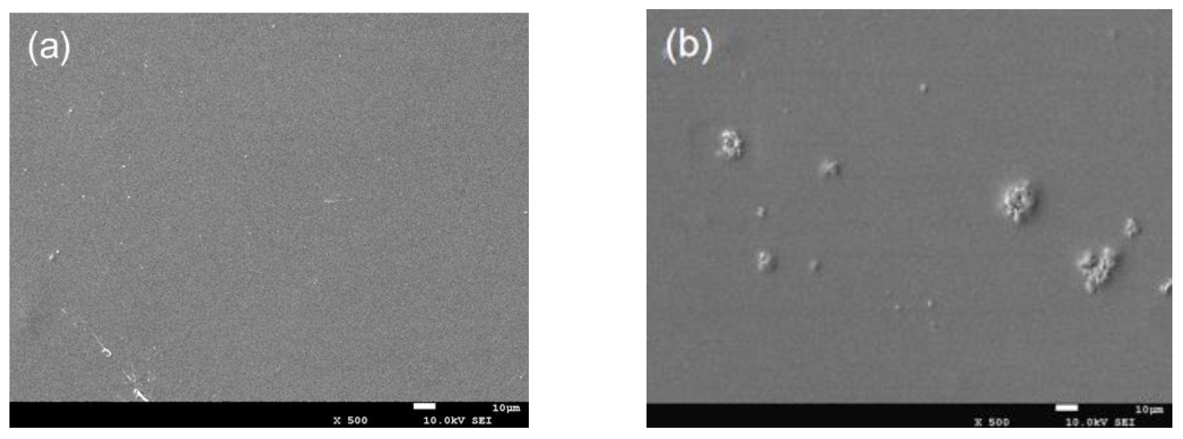
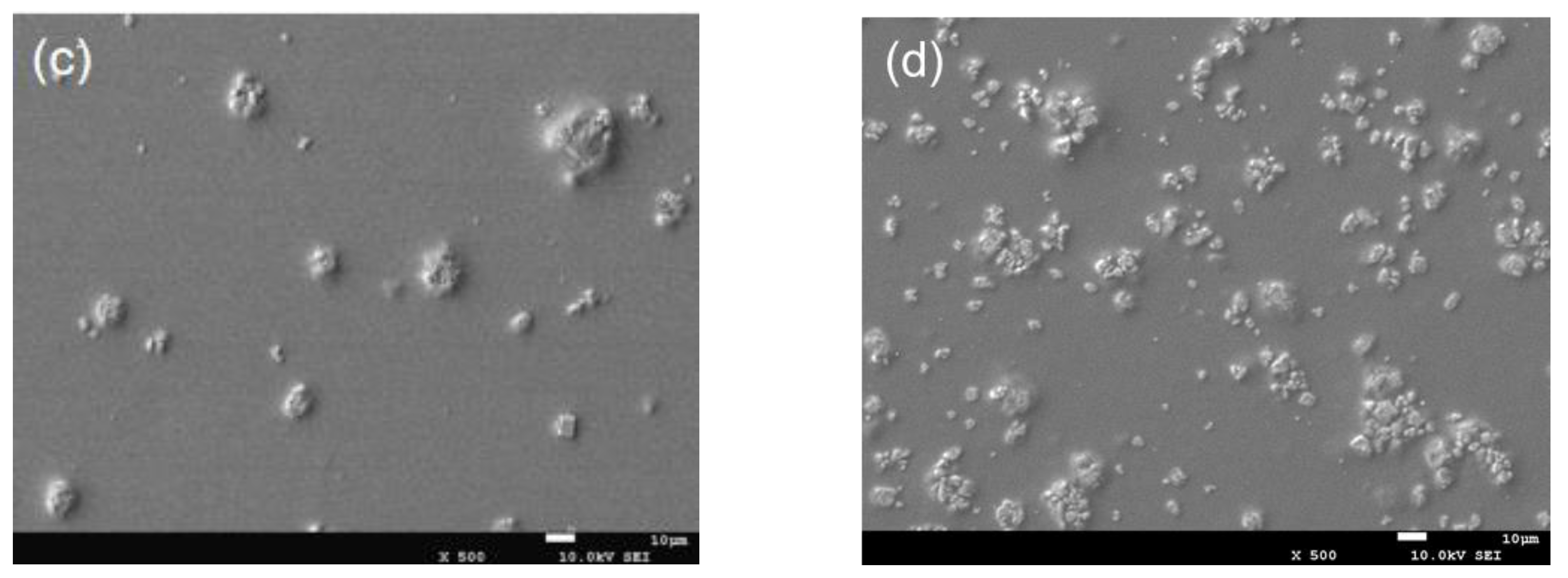
3.1. SEM Analysis
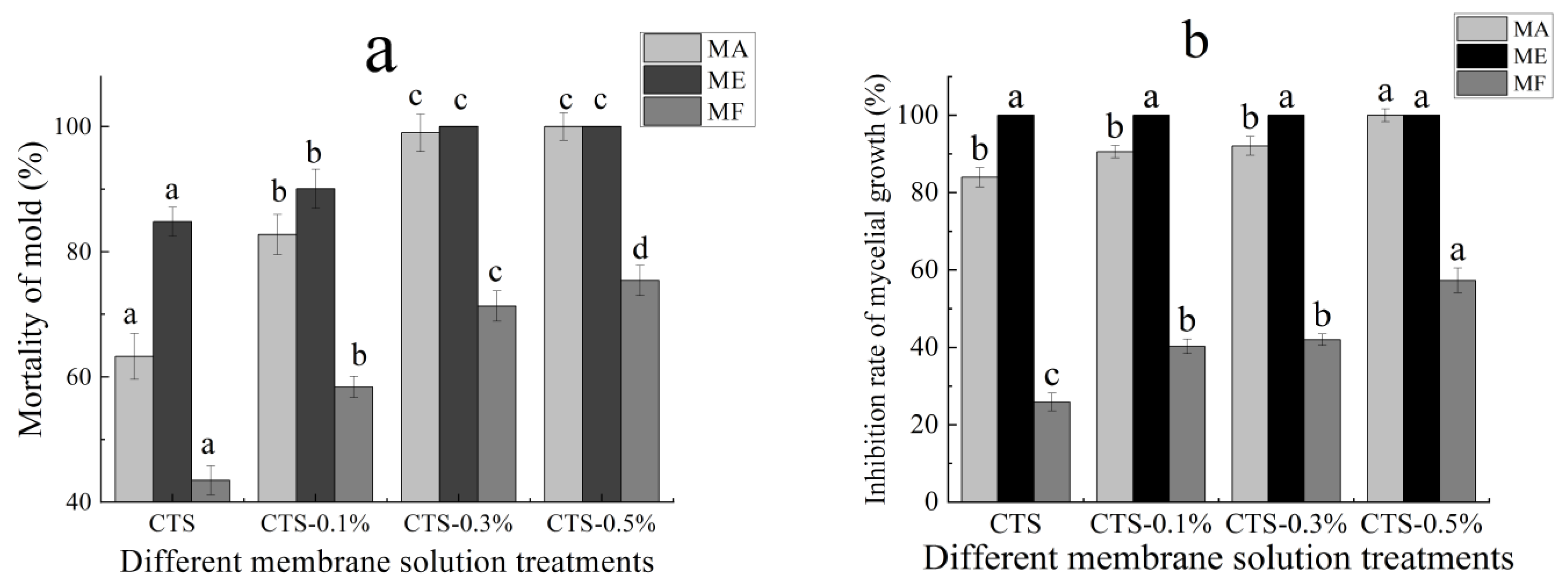
3.2. Effect on the Mortality of Mold and Inhibition Rate of Mycelial Growth
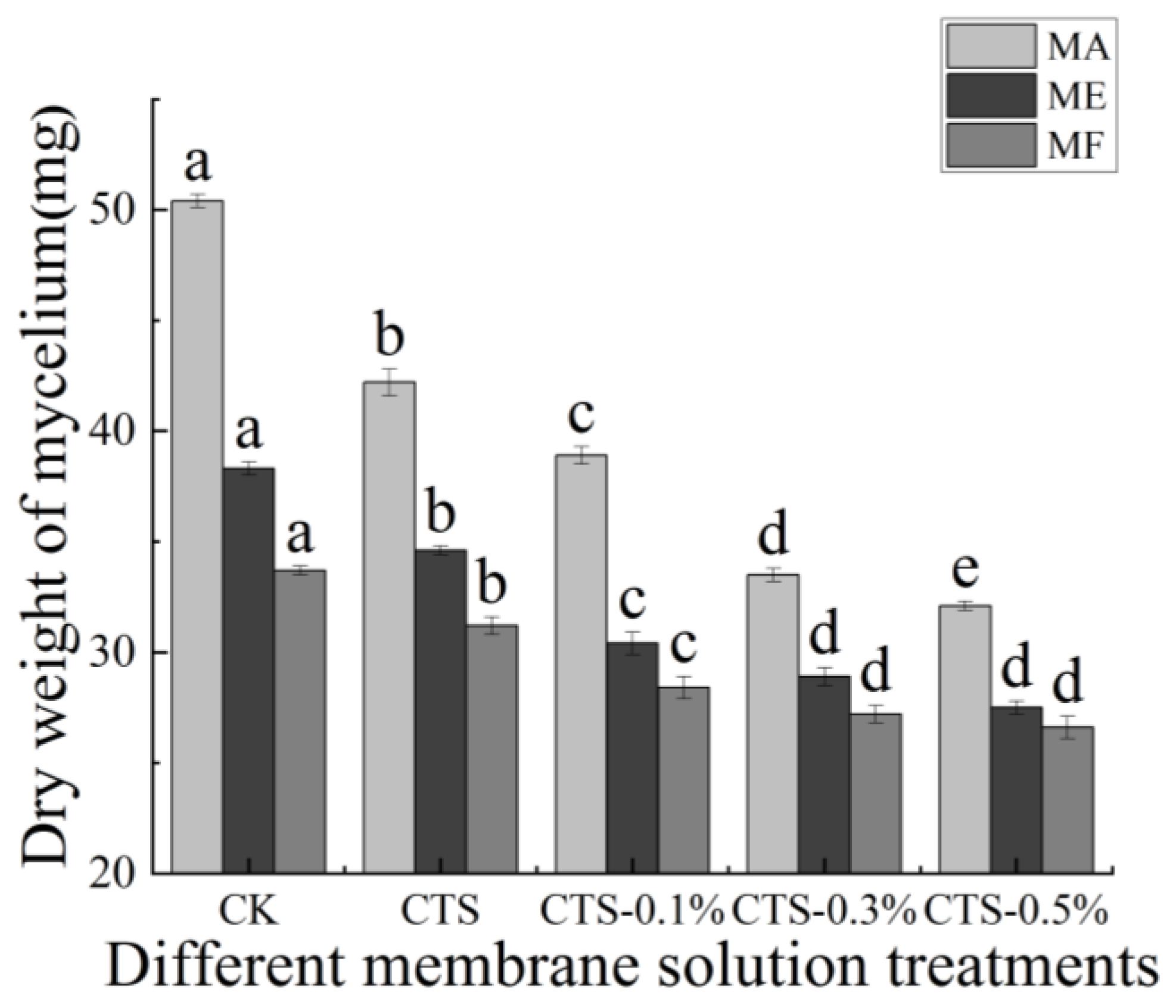
3.3. Effects of CTS/Nano-TiO2 Composite on Dry Weight of Mycelium
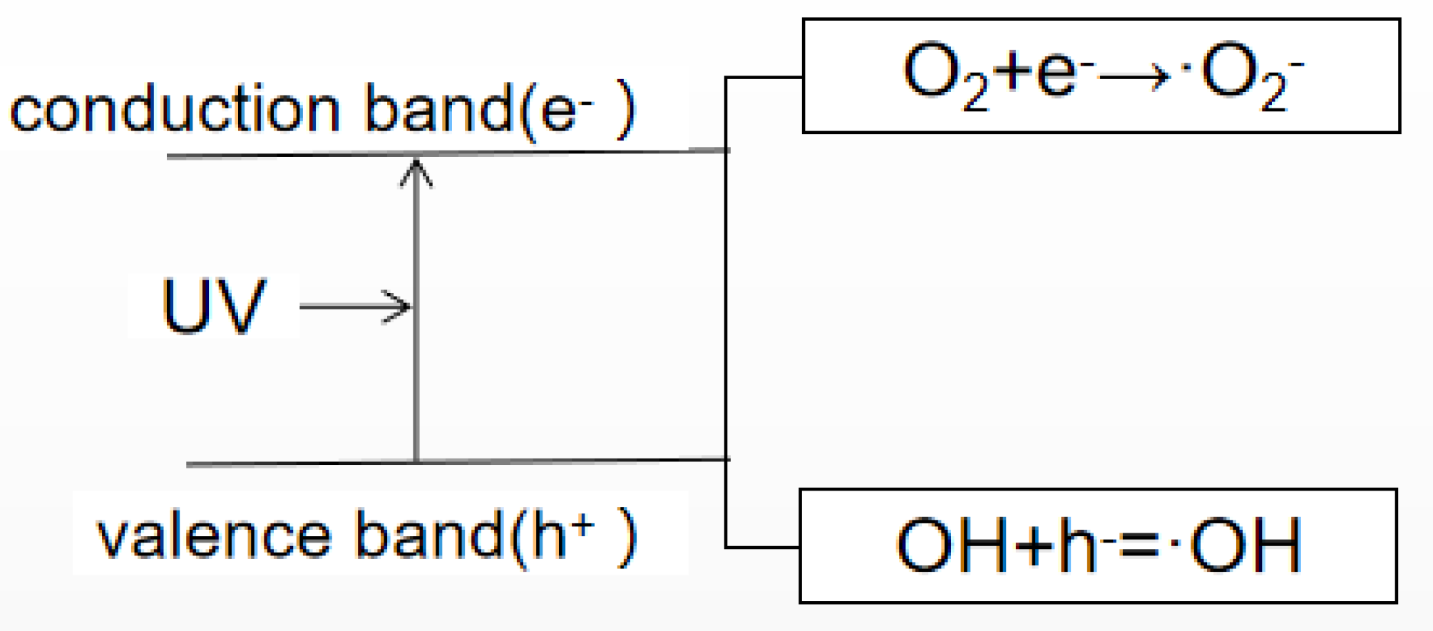
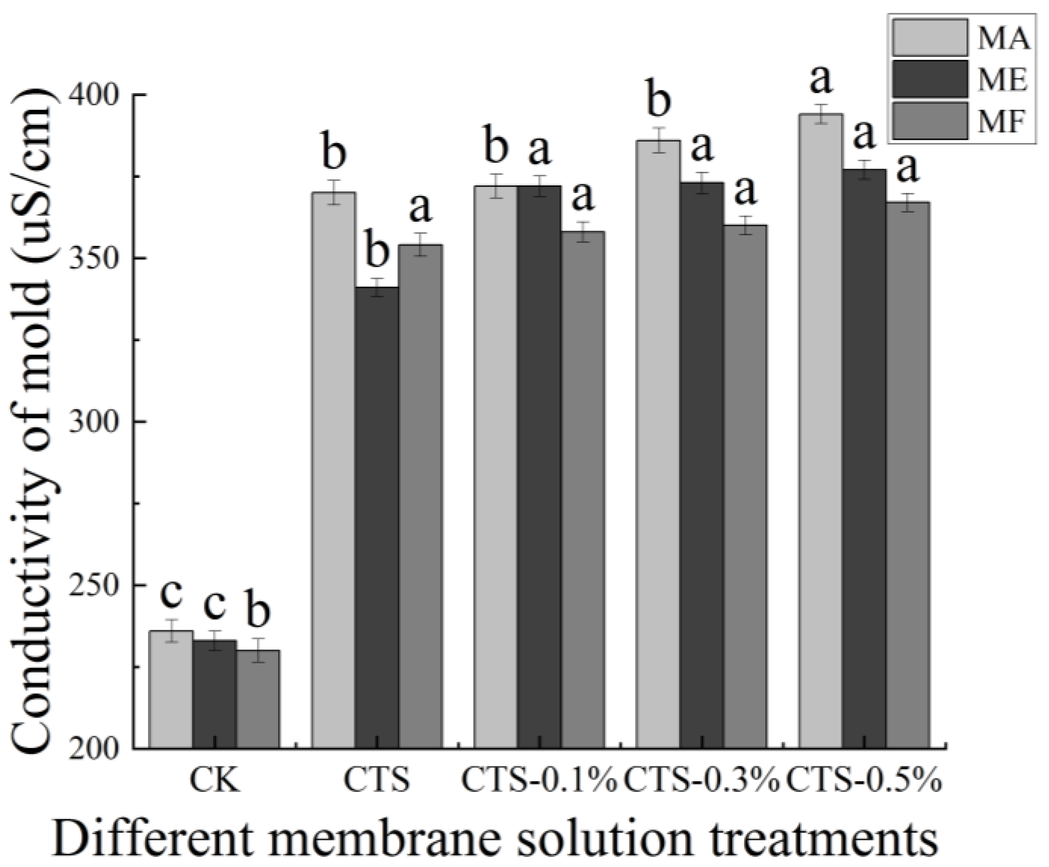
3.4. Effect on the Conductivity of Mold
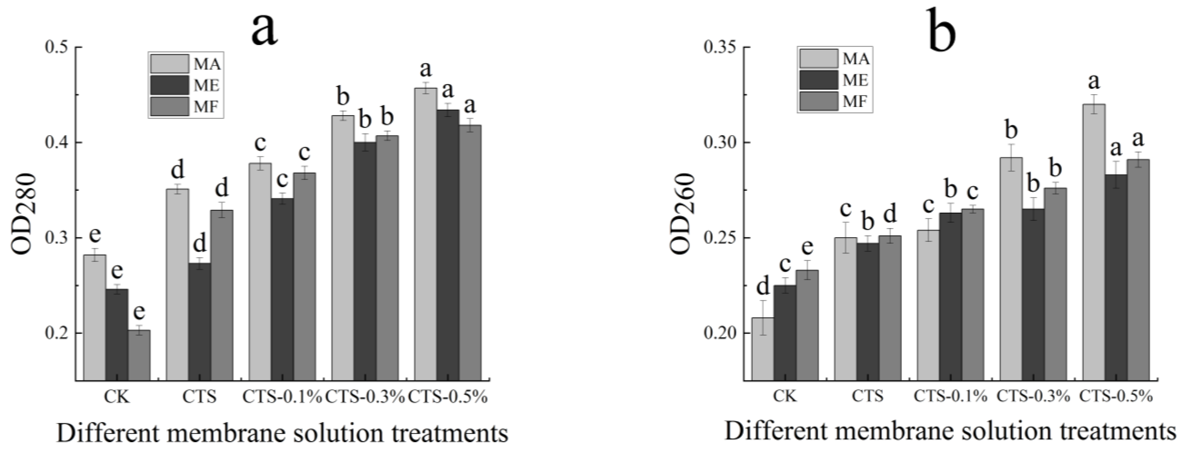
3.5. Effect on Protein Dissolution and Determination of Nucleic Acids Exosmosis
4. Conclusions
Author Contributions
Funding
Institutional Review Board Statement
Informed Consent Statement
Data Availability Statement
Conflicts of Interest
Sample Availability
References
- Stefania, Q.; Loredana, V. Antimicrobial food packaging in meat industry. Meat Sci. 2002, 62, 373–380. [Google Scholar]
- Wanchaitanawong, P.; Chaungwanit, P.; Poovarodom, N.; Nitisin-prasert, S. In vitro antifungal activity of Thai herb and spice extracts against food spoilage fungi. Kasetsart J. 2005, 39, 400–405. [Google Scholar]
- Li, X.; Xing, Y.; Jiang, Y.; Ding, Y.; Li, W. Antimicrobial activities of ZnO powder-coated PVC film to inactivate food pathogens. Int. J. Food Sci. Technol. 2009, 44, 2161–2168. [Google Scholar] [CrossRef]
- Zhang, Z.; Yang, Z.; Yang, B.; Gao, Z.; Li, M.; Jiang, Y.; Hu, M. β-Aminobutyricacid induces resistance of mango fruit to postharvest anthracnose caused by Colletorichum gloeosporiodes and enhances activity of fruit defense mechanisms. Sci. Hortic. 2013, 160, 78–84. [Google Scholar] [CrossRef]
- Li, X. Preparation of Lemongrass/Beeswax Microspheres by Electrospraying and Their Applications in Mango Preservation. Master’s Thesis, Zhejiang Gongshang University, Zhejiang, China, 2019. [Google Scholar]
- You, J.; Guo, H.; Zeng, S.; Liu, Y.; Wang, M. Effect of processing methods on preservatives pesticide residues in dried mango. Chin. J. Trop. Crop. 2016, 37, 2420–2426. [Google Scholar]
- Wang, M.; Chen, X.; Yang, Y.; Xu, R.; Yang, N.; Zhou, H. Resistance of mango stem-end rot to eight fungicides in Hainan. Agrochemicals 2015, 54, 384–386. [Google Scholar]
- Zhou, M.; Ye, Z. Resistance of plant pathogens to benzimidazole and related fungicides. Plant Prot. 1987, 2, 31–33. [Google Scholar]
- Yang, H. Study on the Inhibition Effect of CTS/Nano-TiO2 Composite Coating on Spoilage Molds of Mango. Master’s Thesis, Xihua University, Sichuan, China, 2020. [Google Scholar]
- Xu, J.; Zhao, X.; Han, X.; Du, Y. Antifungal activity of oligochitosan against Phytophthora capsici and other plant pathogenic fungi in vitro. Pestic. Biochem. Physiol. 2006, 87, 220–228. [Google Scholar] [CrossRef]
- Hamidreza, B.; Ronak, B.; Jamal, H.S.; Mojgan, B.; Hadi, S.; Laila, H. Antibacterial/Antifungal activity of extracted chitosan from american cockroach (Dictyoptera: Blattidae) and german cockroach (Blattodea: Blattellidae). J. Med. Entomol. 2021, 56, 1208–1214. [Google Scholar]
- Huang, X.; You, Z.; Luo, Y.; Yang, C.; Ren, J.; Liu, Y.; Wei, G.; Dong, P.; Ren, M. Antifungal activity of chitosan against Phytophthora infestans, the pathogen of potato late blight. Int. J. Biol. Macromol. 2021, 166, 1365–1376. [Google Scholar] [CrossRef]
- Yan, H.; Liu, J.; Meng, X. Antibacterial mechanism of oligochitosan on E. coli. Acad. Period. Farm Prod. Process. 2014, 7, 1–4. [Google Scholar]
- Yadav, S.; Jaiswar, G. Review on undoped/doped TiO2 nanomaterial; synthesis and photocatalytic and antimicrobial activity. J. Chin. Chem. Soc. 2017, 64, 103–116. [Google Scholar] [CrossRef]
- Pan, X.; Du, Y.; Chen, W.; Ji, M.; Wang, L.; Zhou, G.; Lei, C. Applied properties of Ag/TiO2 in antibacterial plastics. Eng. Plast. Appl. 2012, 40, 101–105. [Google Scholar]
- Zhao, F.; Zhou, X.; Hong, Z.; Zhou, S.; Fan, X. Research progress of TiO2-based antibacterial material. Mater. Rep. 2005, 11, 35–38. [Google Scholar]
- Gasparro, F.P.; Mitchnick, M.; Nash, J.F. Areviewof sun-screen safety and efficacy. Photochem. Photobiol. 1998, 68, 243–256. [Google Scholar] [CrossRef]
- Tao, X.; Wang, M.; Yuan, Z.; Wang, L. Study on scorage Jinqiu Pear with chitosan/nano-TiO2. Food Ferment. Ind. 2009, 35, 210–213. [Google Scholar]
- Peri, K.; Li, L.; Liu, Z.; Aleksandr, M.; Wang, T. Antibacterial mechanisms of a novel type picosecond laser-generated silver-titanium nanoparticles and their toxicity to human cells. Int. J. Nanomed. 2018, 13, 89–101. [Google Scholar]
- Jiang, L. Preparation of Nano-Ag/TiO2 Polyamide Antibacterial Fiber. Master’s Thesis, Central South University, Hunan, China, 2012. [Google Scholar]
- Valan, A.A.; Dhinesh, K.D.; Khan, A.I. Experimental investigation of thermal conductivity and stability of TiO2-Ag/water nanocompositefluid with SDBS and SDS surfactants. Thermochim. Acta 2019, 678, 178308. [Google Scholar]
- Wu, Y.; Li, S.; Song, J.; Jiang, B.; Chen, S.; Sun, H.; Li, X. Acetylated distarch phosphate/chitosan films reinforced with sodium Laurate-Modified nano-TiO2: Effects of sodium laurate concentration. J. Food Sci. 2018, 83, 2819–2826. [Google Scholar] [CrossRef] [PubMed]
- Stephen, R.; Lorenzo, N.; Michael, N. Modification of TiO2 with metal chalcogenide nanoclusters for hydrogen evolution. J. Phys. Energy 2021, 3, 025001. [Google Scholar]
- Maneerat, C.; Hayata, Y. Antifungal activity of TiO2 photocatalysis against Penicillium expansum in vitro and in fruit tests. Int. J. Food Microbiol. 2006, 107, 99–103. [Google Scholar] [CrossRef]
- Chawengkijwanich, C.; Hayata, Y. Development of TiO2 powder-coated food packaging film and its ability to inactivate Escherichia coli in vitro and in actual tests. Int. J. Food Microbiol. 2008, 123, 288–292. [Google Scholar] [CrossRef]
- Yetria, R.; Dita, D.; Syukri, S.; Anthoni, A.; Hilfi, P. Enhancement of antifungal capability of cotton textiles coated with TiO;–SiO;/chitosan using citric acid and sodium hypophosphite catalyst. J. Dispers. Sci. Technol. 2021, 42, 784–790. [Google Scholar]
- Yan, J.; Sun, Y.; Shi, L.; Chen, H. Chitosan/nano TiO2 composite materials and its application. J. Cap. Norm. Univ. 2011, 32, 40–46. [Google Scholar]
- Vahhab, S.; Ali, M. A green, and eco-friendly bionanocomposite film (poly(vinyl alcohol)/TiO2/chitosan/chlorophyll) by photocatalytic ability, and antibacterial activity under visible-light irradiation. J. Photochem. Photobiol. A Chem. 2021, 404, 112906. [Google Scholar]
- Han, J.; Su, H.; Tan, T. Study on sterilizing action on ecoli of nano-TiO2-chitosan multiplex film. New Chem. Mater. 2006, 7, 65–68. [Google Scholar]
- Arain, R.A.; Khatri, Z.; Memon, M.H. Antibacterial property and characterization of cotton fabric treated with chitosan/AgCl-TiO2 colloid. Carbohydr. Polym. 2013, 96, 326–331. [Google Scholar] [CrossRef] [PubMed]
- Rhim, J.W.; Weller, C.L.; Ham, K.S. Characteristics of chitosan films as affected by the type of solvent acid. Food Sci. Biotechnol. 1998, 7, 35–40. [Google Scholar]
- Yuan, Z.; Wang, M.; Li, X. Effects of chitosan/TiO2 composite coating on keeping-fresh of stauntonvine. Adv. Mater. Res. 2012, 530, 68–73. [Google Scholar] [CrossRef]
- Wang, H. Effects of Chitosan on Postharvest grey mildew and penicillium in Cherry Tomato and Its Molecular Mechanism. Master’s Thesis, Hefei University of Technology, Zhejiang, China, 2017. [Google Scholar]
- Cao, L. Study on the Inhibitory Mechanism of Ozone on Spoilage Molds of Fresh-Peeled Garlic. Master’s Thesis, Xihua University, Sichuan, China, 2019. [Google Scholar]
- Wang, G.; Peng, X.; Wu, Z.; Li, W.; Zhang, Z.; Wu, B. Inhibitory effect of extracts on several postharvest fungal diseases of walnut green husk. Sci. Technol. Food Ind. 2014, 35, 142–145. [Google Scholar]
- Zeng, R. Study on Antimicrobial Active Ingredients, Antimicrobial Mechanism and Antiseptic Effect on Citrus. Master’s Thesis, Nanchang University, Jiangxi, China, 2012. [Google Scholar]
- Xu, J. Preliminary Study on Antifungal Activity and Mode of Action of Oligochitosan against Phytopathogen In Vitro. Ph.D. Thesis, Graduate University of Chinese Academy of Sciences, Dalian, China, 2007. [Google Scholar]
- Dong, M.; Su, W.; Li, C.; Jing, X.; Dai, H.; Bian, H. Preparation and characterization of ligcellulosic nanofibril/nano titanium dioxide composite film. J. Cell. Sci. Technol. 2021, 29, 22–28. [Google Scholar]
- Hu, J.; Liu, S.; Wu, D.; Zhou, T.; Feng, Q. Preperation and properties of PVA/PA6/TiO2 composite nanofibers. J. Funct. Mater. 2020, 51, 7148–7154. [Google Scholar]
- Skjak-Braek, G.; Grasdalen, H.; Smidsrod, O. Inhomogeneous polysaccharide ionic gels. Carbohydr. Polym. 1989, 10, 31–54. [Google Scholar] [CrossRef]
- Jackon, S.l.; Heath, I.B. Roles of calcium ions in hyphal tip growth. Microbiol. Mol. Biol. Rev. 1993, 57, 367–382. [Google Scholar]
- Leuba, J.L.; Stossel, P. Chitosan and other polyamines: Antifungal activity and interaction with biological membrace. In Chitin in Nature and Technology; Springer: Boston, MA, USA, 1986; pp. 279–286. [Google Scholar]
- Muzzarelli, R.A.A. Chitosan-based dietary foods. Carbohydr. Polym. 1996, 29, 309–316. [Google Scholar]
- Jia, L.; Wang, X.; Tao, W.; Zhang, H.; Qing, X. Preparation and antibacterial property of polyacrylonitrile antibacterial composite nanofiber membranes. J. Text. Res. 2020, 41, 14–20. [Google Scholar]
- Liu, N.; Chang, Y.; Feng, Y.; Cheng, Y.; Sun, X.; Jian, H.; Feng, Y.; Li, X.; Zhang, H. {101}–{001} Surface Heterojunction-Enhanced Antibacterial Activity of Titanium Dioxide Nanocrystals Under Sunlight Irradiation. ACS Appl. Mater. Interfaces 2017, 9, 5907–5915. [Google Scholar] [CrossRef]
- Arjunan, N.; Linda, J.H.; Karuppannan, R.; Kandasamy, R.; Kulandaivel, J.; Kandasamy, J. A versatile effect of chitosan-silver nanocomposite for surface plasmonic photocatalytic and antibacterial activity. J. Photochem. Photobiol. B Biol. 2015, 153, 412–422. [Google Scholar]
- Ashkarran, A.A.; Hamidinezhad, H.; Haddadi, H.; Mahmoudi, M. Double-doped TiO2 nanoparticles as an efficient visible-light-active photocatalyst and antibacterial agent under solar simulated light. Appl. Surf. Sci. 2014, 301, 338–345. [Google Scholar] [CrossRef]
- Pham, T.D.; Lee, B.K. Cu doped TiO2/GF for photocatalytic disinfection of Escherichia coli in bioaerosols under visible light irradiation: Application and mechanism. Appl. Surf. Sci. 2014, 296, 15–23. [Google Scholar] [CrossRef]
- Gao, F. Preparation, Characterization and Antifungal Properties of Sulfonated and Oxidized Chitosan with Different Molecular Weights. Master’s Thesis, Hebei Normal University of Science & Technology, Hebei, China, 2021. [Google Scholar]
- Guo, X. Preparation and Properties of Chitosan Nano-TiO2 Composite Coating Film. Master’s Thesis, Xihua Univercity, Sichuan, China, 2019. [Google Scholar]
- Li, J.; Wu, Z.; Su, M.; He, S.; Chen, Y.; Qin, D. Antifungal properties of mesoporous TiO2 films modified with [M(NH3)4]2+ (M=Cu, Zn) against Trichoderma viride and Penicillium citrinum. Vacuum 2020, 176, 109346. [Google Scholar] [CrossRef]
- Karthikeyan, K.T.; Nithya, A.; Jothivenkatachalam, K. Photocatalytic and antimicrobial activities of chitosan-TiO2 nanocomposite. Int. J. Biol. Macromol. 2017, 104, 1762–1773. [Google Scholar] [CrossRef] [PubMed]
- Huang, Q.; Jiao, Z.; Li, M.; Qiu, D.; Liu, K.; Shi, H. Preparation, Characterization, Antifungal Activity, and Mechanism of Chitosan/TiO2 Hybrid Film against Bipolaris maydis. J. Appl. Polym. Sci. 2013, 128, 2623–2629. [Google Scholar] [CrossRef]
- Cheng, K.; Mao, W.; Xu, B.; Liu, Y.; Guo, Y.; Chen, J.; Zhang, L.; Zhang, S. Effect of different microwave and ultraviolet mutagenesis methods on the growth of TrichodermaT-YS. Grassl. Turf. 2019, 39, 79–83. [Google Scholar]
- Gao, Y.; Wang, W.; Su, F.; Min, F.; Liu, Z.; Wei, Q.; Wan, S. Antifungal activity of dryocrassine on potato dry rot. Chin. Potato J. 2020, 34, 238–244. [Google Scholar]
- Kang, J.M.; Jin, W.Y.; Wang, J.F. Antibacterial and anti-biofilm activities of peppermint essential oil against Staphylococcus aureus. LWT Food Sci. Technol. 2019, 101, 639–645. [Google Scholar] [CrossRef]
- Li, X. Study on Antibacterial Activity and Mechanism of Chitosan. Master’s Thesis, Lanzhou University, Gansu, China, 2009. [Google Scholar]
- Yu, X. Study on the Antibacterial Mechanism of Chitosan. Master’s Thesis, Wuhan Polytechnic University, Hubei, China, 2011. [Google Scholar]
- Kong, M.; Chen, X.G.; Xing, K.; Park, H.J. Antimicrobial properties of chitosan and mode of action: A state of the art review. Int. J. Food Microbiol. 2010, 144, 51–63. [Google Scholar] [CrossRef] [PubMed]
- Ye, X.; Li, X.; Yuan, L.; He, H. Effect of the surface activity on the antibacterial activity of octadecanoyl acetal sodium sulfite series. Colloids Surf. A Physicochem. Eng. Asp. 2005, 268, 85–89. [Google Scholar] [CrossRef]
- Fujishima, A.; Zhang, X.; Tryk, D.A. TiO2 photocatalysis and related surface phenomena. Surf. Sci. Rep. 2008, 63, 515–582. [Google Scholar] [CrossRef]
- Wei, X.; Yang, Z.; Tay, S.L.; Gao, W. Photocatalytic TiO2 nanoparticles enhanced polymer antimicrobial coating. Appl. Surf. Sci. 2014, 290, 274–279. [Google Scholar] [CrossRef]
- Hao, Y. Antifungal Activity and Mechanism of Endophytic Fungus SYS-5-2 from Walnut. Master’s Thesis, Northwest A&F University, Shanxi, China, 2020. [Google Scholar]
- Zhang, Y.; Liu, X.; Jiang, P.; Li, W.; Wang, Y. Mechanism and Antibacterial Activity of Cinnamaldehyde against Escherichia coli and Staphylococcus aureus. Mod. Food Sci. Technol. 2015, 31, 31–35. [Google Scholar]


| Fungi | Treatment | Antimicrobial Effect |
|---|---|---|
| Fusarium graminearum | 100 μg/mL chitosan | Inhibition rate, 97.39% [49] |
| P. Steckii, A. oryzae | Red and blue light combined with CTS/TiO2 treatment for 120 h | Zone of inhibition, 4.7 mm, 54.8 mm [50] |
| Phytophthora infestans | 0.05 g/L chitosan for 12 h | Spore germination rate, 1.29% [12] |
| T. Viride, P. citrinum | ZnTB and CuTB ((Cu, Zn)/TiO2 film-coated) for 14 and 28 days | Inhibition efficiency, 100% [51] |
| C. albicans | Fungi were treated with CTS/TiO2 at 37 °C for 24 h | Zone of inhibition, 14.66667 ± 0.5773 mm [52] |
| B. maydis | CTS/TiO2 treatment for 1 h | Inhibition rate, 100% [53] |
Publisher’s Note: MDPI stays neutral with regard to jurisdictional claims in published maps and institutional affiliations. |
© 2021 by the authors. Licensee MDPI, Basel, Switzerland. This article is an open access article distributed under the terms and conditions of the Creative Commons Attribution (CC BY) license (https://creativecommons.org/licenses/by/4.0/).
Share and Cite
Xing, Y.; Yi, R.; Yang, H.; Xu, Q.; Huang, R.; Tang, J.; Li, X.; Liu, X.; Wu, L.; Liao, X.; et al. Antifungal Effect of Chitosan/Nano-TiO2 Composite Coatings against Colletotrichum gloeosporioides, Cladosporium oxysporum and Penicillium steckii. Molecules 2021, 26, 4401. https://doi.org/10.3390/molecules26154401
Xing Y, Yi R, Yang H, Xu Q, Huang R, Tang J, Li X, Liu X, Wu L, Liao X, et al. Antifungal Effect of Chitosan/Nano-TiO2 Composite Coatings against Colletotrichum gloeosporioides, Cladosporium oxysporum and Penicillium steckii. Molecules. 2021; 26(15):4401. https://doi.org/10.3390/molecules26154401
Chicago/Turabian StyleXing, Yage, Rumeng Yi, Hua Yang, Qinglian Xu, Ruihan Huang, Jing Tang, Xuanlin Li, Xiaocui Liu, Lin Wu, Xingmei Liao, and et al. 2021. "Antifungal Effect of Chitosan/Nano-TiO2 Composite Coatings against Colletotrichum gloeosporioides, Cladosporium oxysporum and Penicillium steckii" Molecules 26, no. 15: 4401. https://doi.org/10.3390/molecules26154401
APA StyleXing, Y., Yi, R., Yang, H., Xu, Q., Huang, R., Tang, J., Li, X., Liu, X., Wu, L., Liao, X., Bi, X., & Yu, J. (2021). Antifungal Effect of Chitosan/Nano-TiO2 Composite Coatings against Colletotrichum gloeosporioides, Cladosporium oxysporum and Penicillium steckii. Molecules, 26(15), 4401. https://doi.org/10.3390/molecules26154401