Article

Prenylated Flavonoid Glycosides with PCSK9 mRNA Expression Inhibitory Activity from the Aerial Parts of Epimedium koreanum

College of Pharmacy and Research Institute of Pharmaceutical Sciences, Seoul National University, Seoul 08826, Korea
*
Author to whom correspondence should be addressed.
Molecules 2021, 26(12), 3590; https://doi.org/10.3390/molecules26123590
Submission received: 1 May 2021 / Revised: 2 June 2021 / Accepted: 8 June 2021 / Published: 11 June 2021
(This article belongs to the Special Issue Isolation and Structure Determination of Bioactive Natural Products)

Abstract

:
Phytochemical investigation on the n-BuOH-soluble fraction of the aerial parts of Epimedium koreanum using the PCSK9 mRNA monitoring assay led to the identification of four previously undescribed acylated flavonoid glycosides and 18 known compounds. The structures of new compounds were elucidated by NMR, MS, and other chemical methods. All isolated compounds were tested for their inhibitory activity against PCSK9 mRNA expression in HepG2 cells. Of the isolates, compounds 6, 7, 10, 15, and 1722 were found to significantly inhibit PCSK9 mRNA expression. In particular, compound 7 was shown to increase LDLR mRNA expression. Thus, compound 7 may potentially increase LDL uptake and lower cholesterol levels in the blood.

1. Introduction

The dried aerial parts of Epimedium koreanum Nakai (Berberidaceae), Herba Epimedii, have been used as a tonic or for the treatment of dementia, hypertension, impotence, rheumatic, and paralytic diseases [1,2]. Previous phytochemical studies reported that lignans, phenol glycosides, and prenylated flavonoids are present as chemical constituents of this plant [3,4,5,6]. Individual constituents, including icariin and extracts of E. koreanum, demonstrated a variety of biological activities such as anti-hepatotoxic, anti-inflammatory, anti-osteoporosis, anti-tumor, and immunoadjuvant activities, as well as the improvement of sexual function [7,8,9,10,11,12,13].
Proprotein convertase subtilisin/kexin type 9 (PCSK9) is involved in degrading LDLR via clathrin-dependent endocytosis and preventing LDLR recycling, resultantly decreasing the capacity of LDL uptake into cells [14]. Thus, high expression of PCSK9 is often associated with the incidence of hypercholesterolemia, and inhibition of PCSK9 expression or activity has been suggested as a tool to treat patients with familial hypercholesterolemia [15]. Currently, two antibody drugs are prescribed clinically since 2015 [16].
As part of our ongoing project to discover PCSK9 expression inhibitory compounds from medicinal plants [17,18,19,20], the n-BuOH-soluble fraction of the aerial parts of E. koreanum was selected for further investigation due to its initial PCSK9 mRNA expression inhibitory activity (Supplementary Material Figure S1-1). However, there are no reports regarding PCSK9 inhibitory substances from this plant. Thus, herein, we describe the isolation and identification of four new acylated flavonoid glycosides and 18 known compounds, and their effects on PCSK9 and LDLR mRNA expression in the HepG2 cells.

2. Results

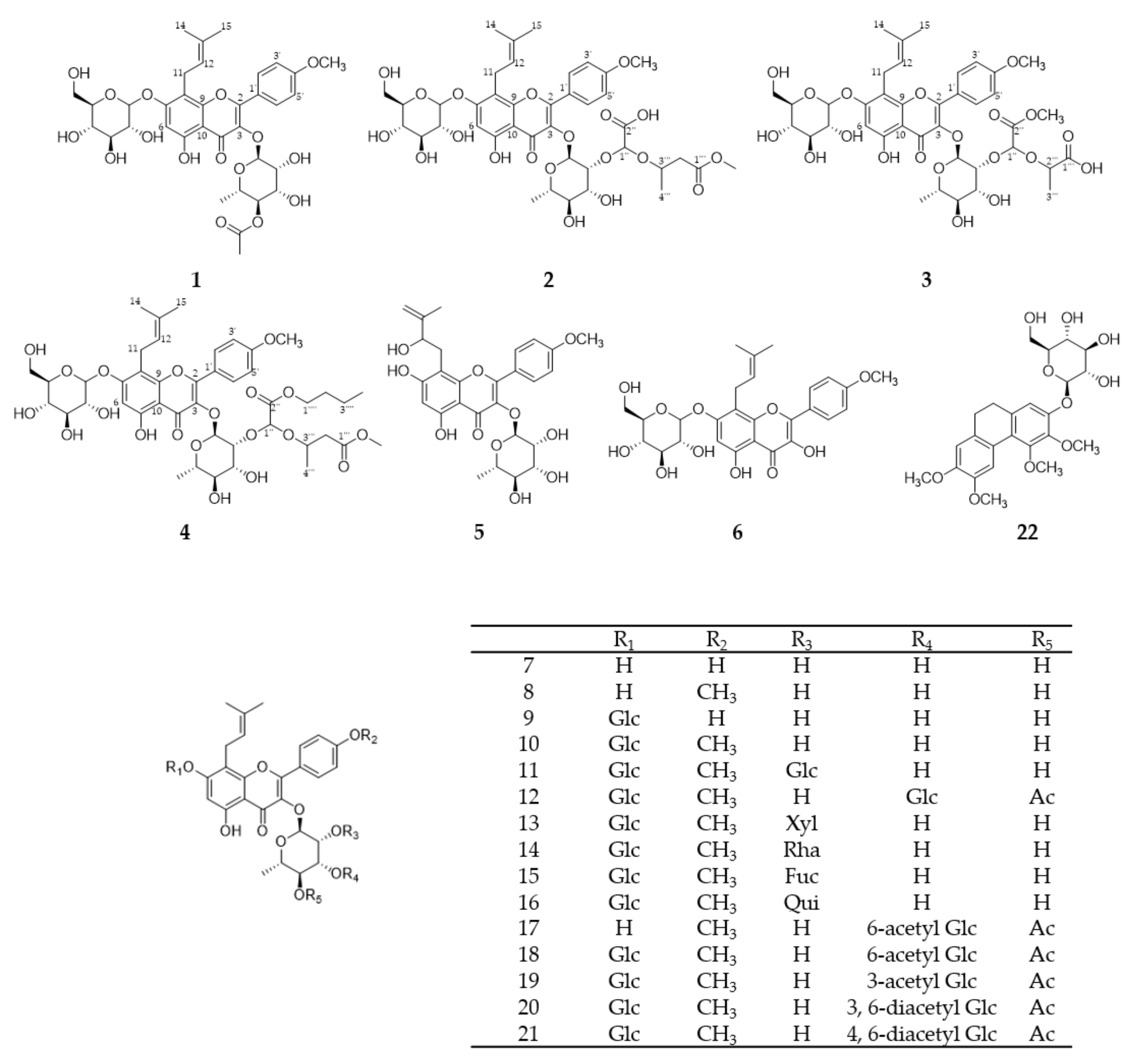
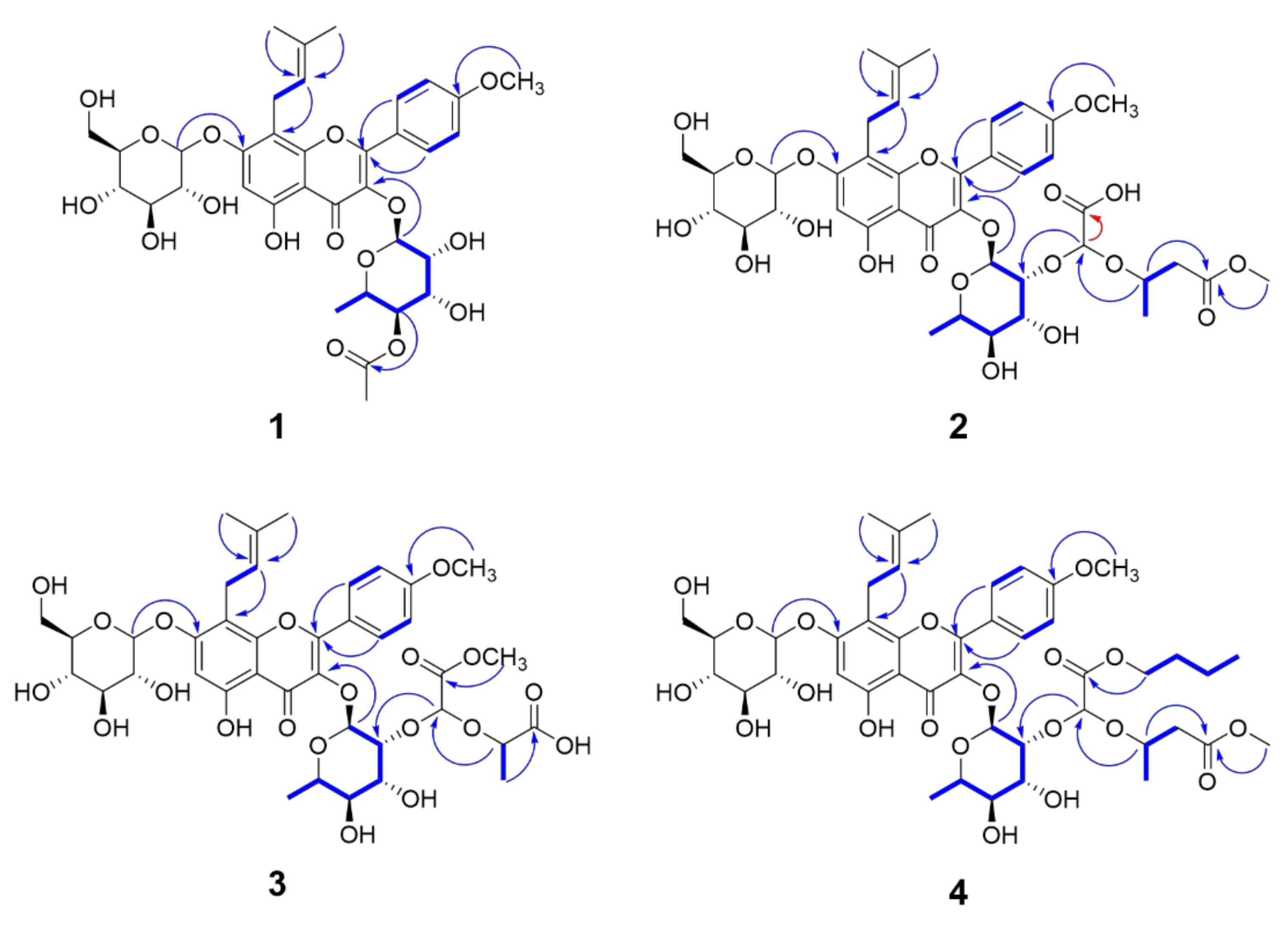
2.1. Isolation of Compounds from E. koreanum

The known compounds 5-22 (Figure 1) were confirmed by NMR and MS as koreanoside E (5) [21], icariside I (6) [22], ikarisoside A (7) [23], icariside II (8) [24], epimedoside A (9) [6,25], icariin (10) [26], epimedin A (11) [3], korepimedoside C (12) [27], epimedin B (13) [3], epimedin C (14) [3], anhydroicaritin 3-O-β-d-fucopyranosyl(1→2)-α-l-rhamnopyranoside-7-O-β-d-glucopyranoside (15) [28], icarisid I (16) [29], korepimedoside A (17) [30], epimedokoreanoside I (18) [6], korepimeoside C (19) [31], epimedin L (20) [5], caohuoside B (21) [32], and epimedoicarisoside A (22) [33].
Compound 1 was obtained as a yellow amorphous powder and its molecular formula was determined to be C35H42O16 by the pseudomolecular ion peak [M + H]+ at m/z 719.2529 (calcd. for C35H43O16, 719.2551) in the positive mode ESI-QTOF-HRMS. In the 1H-NMR spectrum of 1 (Table 1), a singlet proton signal at δH 6.65 (1H, s, H-6), two doublet signals at δH 7.85 (2H, d, J = 8.8 Hz, H-2′ and H-6′) and 7.10 (2H, d, J = 8.8 Hz, H- 3′ and H-5′) corresponding to a flavonol skeleton, and the signals responsible for an isoprenyl unit at δH 3.52 (1H, m, H-11a), 3.57 (1H, m, H-11b), 5.19 (1H, t, J = 6.8 Hz, H-12), 1.64 (3H, s, H-14), and 1.73 (3H, s, H-15) were observed. In addition, one methoxy signal at δH 3.89 (3H, s, 4′-OMe) and the signals for glucose (Glc) with β-conformer (δH 5.07 (1H, d, J = 7.2 Hz, Glc H-1)) and rhamnose (Rha) with α-conformer (δH 5.50 (1H, brs, Rha H-1)) were detected. All these data indicate that this compound was similar to icariin (10), one of the known main constituents in this plant [26]. However, in the 1H-NMR and 13C-NMR spectroscopic data of 1, there were additional singlet proton signals at δH 2.01 (3H, s) and an ester carbon signal at δC 172.3 belonging to an acetyl group. The position of this acetyl group was assigned to C-4 of rhamnose by the HMBC correlations of δH 4.82 (Rha H-4) to δC 172.3, and δH 2.01 to δC 172.3 (Figure 2). Further HMBC correlations of δH 5.07 (Glc H-1) to δC 161.0 (C-7) and δH 5.50 (Rha H-1) to δC 135.9 (C-3) confirmed the locations of glucose and rhamnose at C-7 and C-3, respectively. The absolute configuration of these sugars was determined as d-glucose and l-rhamnose using HPLC analysis of the acid hydrolysate [34]. Thus, the structure of 1 turned out to be icaritin 3-O-[4-O-acetyl-α-l-rhamnopyranoside]-7-O-β-d-glucopyranoside.
Compound 2, a yellow amorphous powder, had the molecular formula C40H50O20, supported by the pseudomolecular ion peak [M–H] at m/z 849.2815 (calcd. for C40H49O20, 849.2817) in the negative mode ESI-QTOF-HRMS. The 1H- and 13C-NMR spectroscopic data of 2 were similar to those of icariin (10), except for the additional signals derived from the presence of a 2-hydroxy-2-((4-methoxy-4-oxobutan-2-yl)oxy)acetic acid unit, a 2,2-dihyroxyacetic acid moiety, and a methyl 3-hydroxybutanoate moiety. The 1H NMR signal at δH 5.21 (1H, s, H-1″) and 13C NMR signals at δC 101.1 (C-1″) and 170.0 (C-2″) were assignable to the 2,2-dihyroxyacetic acid moiety, which was supported by the HMBC correlation (optimized at long range J = 2.0 Hz) between H-1″ (δH 5.21, s) and C-2″ (δC 170.0). The remaining signals for a methyl 3-hydroxybutanoate moiety were assigned by the sequential correlations of H-2‴ (δH 2.60 and 2.46)/H-3‴(δH 4.18, sextet, J = 6.0 Hz)/H-4‴ (δH 1.21, d, J = 6.4 Hz) in the 1H-1H COSY spectrum, and the HMBC correlations of both H-3‴ and a methoxy signal at δH 3.65 to C-1‴ (δC 173.1). The connectivity between the 2,2-dihyroxyacetic acid moiety and the methyl 3-hydroxybutanoate moiety was confirmed by the HMBC correlation of H-3‴ (δH 4.18) to C-1″ (δC 101.1), constructing 2-hydroxy-2-((4-methoxy-4-oxobutan-2-yl)oxy)acetic acid unit. This 2-hydroxy-2-((4-methoxy-4-oxobutan-2-yl)oxy)acetic acid unit was linked to Rha C-2 via an ether linkage by observing HMBC correlation of H-1″ to δC 80.0 (Rha C-2). However, the absolute configurations of C-1″ and C-3‴ were not resolved in this study. Therefore, the structure of compound 2 was determined to be icaritin 3-O-[2-O-2-((4-methoxy-4-oxobutan-2-yl)oxy)acetic acid-α-L-rhamnopyranoside]-7-O-β-d-glucopyranoside.
The molecular formula of compound 3 was assigned to be C39H48O20 by pseudomolecular ion peak [M–H] at m/z 835.2672 (calcd. for C39H47O20, 835.2661) in the negative mode ESI-QTOF-HRMS. The 1H and 13C-NMR spectra of 3 were similar to those of 2 except for the presence of a 2-hydroxypropanoic acid moiety instead of the methyl 3-hydroxybutanoate moiety in 2. The signals responsible for the 2-hydroxypropanoic acid moiety were observed at δH 4.53 (1H, quintet, J = 6.8 Hz H-2‴), and 1.39 (3H, d, J = 6.8 Hz H-3‴); δC 174.4 (C-1‴), 73.2 (C-2‴), and 18.8 (C-3‴). The connectivity of 3 was further confirmed by the HMBC correlations of the methoxy signal at δH 3.78 to C-2″, H-2‴ to C-1″(δC 100.4), H-1″(δH 5.25) to Rha C-2 (δC 80.0), and a long-range COSY correlation of H-1″ to the methoxy signal. The absolute configuration of sugars was determined as D-glucose and L-rhamnose using HPLC analysis of the acid hydrolysate, while the absolute configurations of C-1″ and C-2‴ were not resolved in this study. Accordingly, compound 3 was characterized as icaritin 3-O-[2-O-2-((4-oxopropan-2-yl)oxy)acetic acid methyl ester-α-l-rhamnopyranoside]-7-O-β-d-glucopyranoside.
Compound 4 was isolated as a yellow amorphous powder. Its molecular formula was determined to be C44H58O20 by the pseudomolecular ion peak [M + HCOO] at m/z 951.3529 (calcd. for C45H59O22, 951.3498) in the negative mode ESI-QTOF-HRMS. The 1H- and 13C-NMR spectra of 4 resembled those of 2 except for the presence of an additional butyl group. The additional butyl group appeared at δH 4.16 (2H, m, H-1⁗), 1.66 (2H, m, H-2⁗), 1.42 (2H, m, H-3⁗), and 0.95 (3H, t, J = 7.2 Hz, H-4⁗). In addition, the sequential correlations from H-1⁗ to H-4⁗ were observed in the 1H-1H COSY spectrum. The HMBC correlation between H-1⁗ (δH 4.16, m) and C-2″ (δC 169.7) suggested the position of a butyl group to be at C-2″ via an ester linkage. Therefore, The structure of 4 was determined to be icaritin 3-O-[2-O-2-((4-methoxy-4-oxobutan-2-yl)oxy)acetic acid butyl ester-α-l-rhamnopyranoside]-7-O-β-d-glucopyranoside.

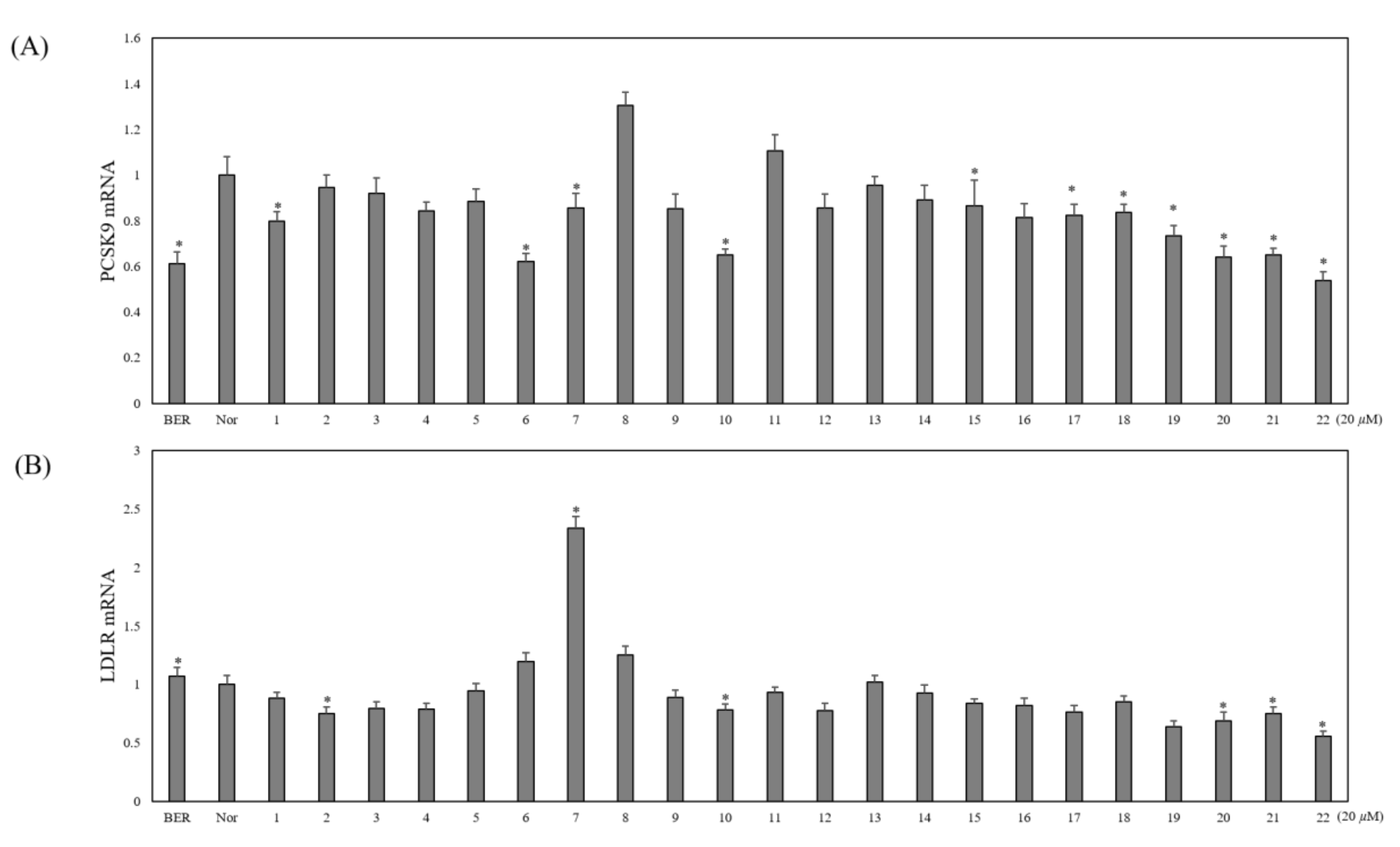
2.2. Bioactivity Evaluation

All isolates (122) were tested for their PCSK9 and LDLR mRNA expression in the HepG2 cells. As shown in the Figure 3, compounds 6, 7, 10, 15, and 1722 were found to inhibit PCSK9 mRNA expression significantly while other flavonoid glycosides seemed to be inactive. Of the active compounds, compound 7 (ikarisoside A) also significantly increased LDLR mRNA expression. Thus, it seems that compound 7 may have potential to increase LDL uptake and lower cholesterol levels in the blood.

3. Discussion and Conclusions

The uptake of LDL-cholesterol into the hepatocytes may control cholesterol levels in the blood; this LDL-cholesterol uptake is mediated by LDLR. Hence, adequate LDLR expression in the cells may clear cholesterol in the blood. PCSK9 facilitates the degradation of LDLR after endocytosis of the PCSK-LDLR complex. Upon endocytosis of LDLR-LDL in the absence of PCSK9, LDLR usually dissociates with LDL in the endosomes and then moves back to the cell surface; meanwhile, in the presence of PCSK9, LDLR is degraded in the lysosomes and, resultantly, less LDLR in the cell surface appear, leading to a decrease in the uptake of LDL into cells [35]. Recently, two antibody drugs which interfere the binding of PCSK9 and LDLR were approved for cholesterol-lowering drugs. However, due to some adverse effects of these antibody drugs, small molecules from synthetic molecules or natural molecules were pursued as PCSK9 inhibitory substances [36]. In particular, small molecules from natural sources were found to participate in inhibiting PCSK9 transcriptional or translational expression, PCSK9 secretion, and interaction of PCSK9 and LDLR [37,38]. In this study, PCSK9 transcriptional expressions by the compounds 6, 7, 10, 15, and 1722 isolated from E. koreanum were significantly downregulated. Concomitantly, LDLR transcriptional expression was upregulated by ikarisoside A (7). Previously, prenylated flavonoids [20] were able to downregulate PCSK9 expression, but their upregulation of LDLR expression was not documented. As natural compounds with downregulation of PCSK9 expression and upregulation of LDLR expression, α-mangostin [37] and sauchinone [38] were reported and demonstrated an increase in LDL uptake, implying the potential in lowering blood cholesterol. Likewise, ikarisoside A (7) may have the positive potential for a cholesterol-lowering effect. Thus, ikarisoside A (7) may have strong merits for further investigation in vitro and in vivo.

4. Materials and Methods

4.1. General Experimental Procedures

Optical rotations were measured using a Jasco P-2000 digital polarimeter (Jasco, Tokyo, Japan). UV spectra were recorded on a UV-VIS spectrometer lamda 25 (Perkin Elmer, Waltham, MA, USA). IR spectra were recorded using Jasco FT/IR-4200 spectrophotometer. Waters Xevo G2 Q-TOF, (Waters, Milford, MA, USA) spectra were measured on a Q-TOF mass spectrometer.
One-dimensional (1H and 13C) and two-dimensional (1H-1H COSY, HSQC, HMBC, NOESY) NMR spectra were obtained with a Jeol 400, 600 (JEOL, Tokyo, Japan)—400, 600MHz and Bruker 500 (Bruker, AVANCE 500, Billerica, MA, USA)—500MHz. Column chromatography was performed on silica gel (60-200μm, Zeochem, Switzerland) and diaion HP20 (Mitsubishi chemical, Tokyo, Japan). TLC analysis was run on silica gel 60 F254 plates (Merck, Darmstadt, Germany) and visualization of the TLC plates was performed under UV radiation and spraying with 10% aqueous H2SO4. High-performance liquid chromatography (HPLC) was performed on a Gilson 305/306 pump, equipped with a Gilson UV/VIS 151 detector. Luna 5μ C18 column 250 × 21.20 mm (Phenomenex) and Synergi 4μ hydro-RP column 250 × 21.20 mm (Phenomenex) as HPLC columns were used. Medium-pressure liquid chromatography (MPLC) was run on Isolera One (Biotage, Cardiff, UK). LC grade acetonitrile (MeCN) were purchased from SK Chemicals (Seoul, Korea). Water was purified using a Milli-Q system (Millipore, Bedford, MA, USA). l-cysteine methyl ester hydrochloride and O-tolylisothiocyanate were purchased from Tokyo Chemical Industry (Tokyo, Japan).

4.2. Plant Material

The aerial parts of E. koreanum were purchased from Daerim Pharmaceutical Wholesale Company (Cheongju, Korea) and identified by one of the authors (J. Kim). A voucher specimen (CYWSNUKP-00019) was deposited at the medicinal plant garden in the College of Pharmacy, Seoul National University.

4.3. Extraction and Isolation

The air-dried aerial parts of E. koreanum (2.0 kg) were extracted with MeOH at room temperature, giving the crude extract (238 g). The crude extract was suspended in water and partitioned successively with n-hexane, CHCl3, and n-BuOH. The n-BuOH fraction (94.2 g) was subjected to Diaion HP-20 column chromatography eluted with 20, 40, 60, 80, 100% MeOH to give five fractions (Bu20- Bu100). Bu80 (21.1 g) was subjected to silica gel column chromatography eluted with gradient mixtures of CH2Cl2/MeOH/H2O (50:5:1–7:5:1) and gave 10 subfractions (Bu80.1– Bu80.10). A solid precipitate was separated from Bu80.9 and recrystallized from MeOH to give compound 10 (2.5 g).
Bu80.3 (275 mg) was subjected to reversed-phase (RP) medium pressure liquid chromatography (25 g) and eluted with MeOH/H2O (4:6–10:0, step-gradient system, 20 mL/min) to give five fractions (Bu80.3.1- Bu80.3.5). Bu80.3.3 (89.9 mg) was purified using a HPLC column (Luna 5μ C18, 250 × 21.20 mm) and isocratic elution with 41% aqueous MeCN (4 mL/min) to afford compounds 5 (12 mg), 2 (18.7 mg) and subfraction Bu80.3.3.2 (15.7 mg). HPLC purification (Luna 5μ C18, 250 × 21.20 mm, 37% aqueous MeCN, 4 mL/min) of Bu80.3.3.2 furnished compound 3 (3.4 mg).
Bu80.6 (342.1 mg) was subjected to RP-MPLC column chromatography (25 g) using gradient mixtures of MeOH/H2O (3:7–10:0, 20 mL/min) to give three fractions (Bu80.6.1- Bu80.6.3). Bu80.6.2 (148.3 mg) was separated by HPLC (Luna 5μ C18, 250 × 21.20 mm) and eluted with 41% aqueous MeCN (4 mL/min), furnishing compounds 21 (5.5 mg), 7 (8.4 mg) and subfraction Bu80.6.2.2 (49.3 mg). From Bu80.6.2.2, compound 20 (29.7 mg) was purified by HPLC (Luna 5μ C18, 250 × 21.20 m, 4 mL/min m) using isocratic elution of 37% aqueous MeCN.
Bu80.8 (475.1 mg) was fractionated into four subfractions (Bu80.8.1- Bu80.8.4) by RP-MPLC (25 g), and eluted with gradient mixtures of MeOH/H2O (3:7–10:0, 20 mL/min). Bu80.8.2 (322.8 mg) was subjected to HPLC separation (Luna 5μ C18, 250 × 21.20 mm) and eluted with 39% aqueous MeCN (4 mL/min) to give subfractions Bu80.8.2.2 (33.2 mg) and Bu80.8.2.3 (27.5 mg). Compound 18 (28.3 mg) was isolated from Bu80.8.2.2 by HPLC separation (Synergi 4μ hydro-RP, 250 × 21.20 mm, 35% aqueous MeCN, 4 mL/min). From Bu80.8.2.3, compound 19 (17.4 mg) was purified by HPLC (Synergi 4μ hydro-RP, 250 × 21.20 mm, 4 mL/min) and isocratically eluted with 35% aqueous MeCN.
Bu80.10 (8.5 g) was subjected to RP-MPLC (50 g) using gradient mixtures of MeOH/H2O (3:7–10:0, 40 mL/min) to give 8 fractions (Bu80.10.1- Bu80.10.8). Bu80.10.3 (405.5 mg) was purified using HPLC (Synergi 4μ hydro-RP, 250 × 21.20 mm, 25% aqueous MeCN, 8 mL/min) to give compound 9 (14.7 mg). Bu80.10.4 (5.8 g) was separated by HPLC (Synergi 4μ hydro-RP, 250 × 21.20 mm) and eluted with 29% aqueous MeCN (8 mL/min) to obtain compounds 11 (19.4 mg), 13 (32.8 mg), 14 (29.5 mg), 15 (7.1 mg), 16 (3.6 mg) and 12 (7.6 mg).
Bu100 (16.9 g) was subjected to silica gel column chromatography and eluted with gradient mixtures of CH2Cl2/MeOH/H2O (50:5:1–7:5:1) to give 10 fractions (Bu100.1–Bu100.10). Bu100.4 (1.1 g) was separated into four fractions (Bu100.4.1- Bu100.4.4) by RP-MPLC and eluted with gradient mixtures of MeOH/H2O (4:6-10:0, 40 mL/min). From Bu100.4.1 (46.3 mg), compound 22 (4.4 mg) was isolated by HPLC separation (Synergi 4μ hydro-RP, 250 × 21.20 mm, 28% aqueous MeCN, 8 mL/min). HPLC purification (Synergi 4μ hydro-RP, 250 × 21.20 mm, 42% aqueous MeCN, 8 mL/min) of Bu100.4.3 (402.3 mg) furnished compounds 6 (16.5 mg), 8 (66.7 mg), 4 (8.2 mg) and 17 (21.3 mg).
Bu100.7 (1.4 g) was subjected to RP-MPLC (50 g) using gradient mixtures of MeOH/H2O(4:6–10:0, 40 mL/min) to give four fractions (Bu100.7.1–Bu100.7.4). From Bu100.7.2 (552.9 mg), compound 1 (12.5 mg) was purified using HPLC (Synergi 4μ hydro-RP, 250 × 21.20 mm) using isocratic elution of 22% aqueous MeCN (8 mL/min).

4.4. Characterization

(1): Yellow amorphous powder; α D 25 -137.2 (c 0.1, MeOH); UV (MeOH) λmax nm (log ε) 228 (3.28), 264 (3.23), 314 (2.79), 353 (2.40); IR (KBr) νmax 3409, 2923, 1647, 1597, 1261 cm−1; 1H-NMR (400 MHz) and 13C-NMR (100 MHz) data, see Table 1; HRMS (ESI-TOF) m/z 719.2529 (calcd. for C35H43O16, 719.2551).
(2): Yellow amorphous powder; α D 20 -79.3 (c 0.1, MeOH); UV (MeOH) λmax nm (log ε) 267 (1.25), 313 (0.70), 344 (0.56); IR (KBr) νmax 3383, 2931, 1738, 1654, 1596, 1511, 1437, 1376, 1342, 1303, 1259, 1220, 1181, 1143 cm−1; 1H-NMR (400 MHz) and 13C-NMR (100 MHz) data, see Table 1; HRMS (ESI-TOF) m/z 849.2815 (calcd. for C40H49O20, 849.2817).
(3): Yellow amorphous powder; α D 20 -56.1 (c 0.1, MeOH); UV (MeOH) λmax nm (log ε) 267 (1.01), 313 (0.57), 342 (0.47); IR (KBr) νmax 2924, 1748, 1595, 1508, 1489, 1339, 1259, 1181 cm−1; 1H-NMR (400 MHz) and 13C-NMR (100 MHz) data, see Table 1; HRMS (ESI-TOF) m/z 835.2672 (calcd. for C39H47O20, 835.2661).
(4): Yellow amorphous powder; α D 20 -65.2 (c 0.1, MeOH UV (MeOH) λmax nm (log ε) 267 (1.15), 313 (0.64), 345 (0.53); IR (KBr) νmax 3414, 2932, 1739, 1653, 1597, 1511, 1438, 1375, 1304, 1259, 1219, 1181 cm−1; 1H-NMR (400 MHz) and 13C-NMR (100 MHz) data, see Table 1; HRMS (ESI-TOF) m/z 951.3529 (calcd. for C45H59O22, 951.3498).

4.5. Acid Hydrolysis

Compounds were hydrolyzed using 1 N H2SO4 (200 µL) and heated with a water bath at 90 °C for 2 h, then neutralized with saturated aqueous Na2CO3 solution. After the solutions were dried under a stream of N2, the products and standard sugars (d-Glc, l-Rha) were dissolved in pyridine (200 µL) containing D-cysteine methyl ester hydrochloride (1 mg). After that, they were heated at 60 °C for 1 h. The solutions were treated with 2 µL (1.11 mg) of O-tolylisothiocyanate and then heated again at 60 °C for 1 h. Each final mixture was directly analyzed by analytical RP-HPLC (Hypersil™ BDS C18 column, 150 × 4.60 mm, 25% aqueous. MeCN, 0.8 mL/min). The peaks at 19.20 and 31.92 min of the derivatives of d-glucose and l-rhamonse, respectively, were coincided with the peaks of the derivatives of d-glucose and l-rhamnose in compounds 14.

4.6. Cell Culture, Drugs and Chemicals

HepG2 (human hepatocellular liver cell line) was obtained from the Korea Research Institute of Bioscience and Biotechnology (Daejeon, Korea) and grown in Eagle’s minimum essential medium (EMEM), supplemented with 10% fetal bovine serum and 100 U/mL penicillin/streptomycin sulfate. Cells were incubated in a humidified incubator at 37 °C in a 5% CO2 atmosphere. EMEM, penicillin, and streptomycin were purchased from HyClone Laboratories (Logan, UT, USA). Oligonucleotide primers for LDLR, PCSK9, and GAPDH were purchased from Bioneer Corp. (Daejeon, Korea). Berberine·HCl was purchased from Chengdu Biopurify Phytochemicals Ltd. (Sichuan, China).

4.7. Quantitative Real-Time RT-PCR

Total cellular RNA was isolated using a Trizol RNA extraction kit (Thermo Fisher Scientific, Waltham, MA, USA) according to the manufacturer’s instructions. Total RNA (1 μg) was then converted to cDNA using 200 units of iScript cDNA Synthesis Kit (Bio-Rad, Hercules, CA, USA) at 25 °C for 5 min and at 46 °C for 20 min. The reaction was stopped by incubating the solution at 95 °C for 1 min, after which 1 μL of cDNA mixture was used for enzymatic amplification. PCR reactions were performed using 4 μL of the cDNA and 6 μL master mix containing iQ SYBR Green Supermix (Bio-Rad), 5 pmol of forward primer, and 5 pmol of reverse primer, in a CFX96 real-time PCR detection system (Bio-Rad). Reaction conditions were 3 min at 95 °C, followed by 40 cycles of 10 s at 95 °C and 30 s at 55 °C. The plate was read subsequently. The fluorescence signal generated with SYBR Green I DNA dye was measured during the annealing step. The specificity of the amplification was confirmed using a melting curve analysis. Data were collected and recorded with CFX Manager Software (Bio-Rad) and expressed as a function of the threshold cycle (CT). The relative quantity of the gene of interest was then normalized to the relative quantity of GAPDH (ΔΔCT). The mRNA abundance in the sample was calculated using the 2−(ΔΔCT) method. The following specific primer sets were used (5′ to 3′): human GAPDH: GAAGGTGAAGGTCGGAGTCA (forward), AATGAAGGGGTCATTGATGG (reverse); human LDLR: GTGCTCCTCGTCTTCCTTTG (forward), TAGCTGTAGCCGTCCTGGTT (reverse); human PCSK9: GGTACTGACCCCCAACCTG (forward), CCGAGTGTGCTGACCATACA (reverse). Gene-specific primers were custom-synthesized by Bioneer (Daejeon, Korea).

4.8. Statistical Analysis

For multiple comparisons, one-way analysis of variance (ANOVA) was performed followed by Dunnett’s t test. Data from experiments are presented as means ± standard error of the mean. The number of independent experiments analyzed is given in the figure captions. P-values of less than 0.05 were regarded as statistically significant.

Supplementary Materials

The following are available online, Figures S1-1–S5-3: NMR, MS, and sugar analysis for new compounds (14) and NMR and MS data for compound 7, Table S5-1: NMR chemical shifts of compound 7.

Author Contributions

Conceptualization, J.K. and Y.-W.C.; experimental, E.K., J.A., H.-S.C., and Y.-M.K.; writing, E.K., Y.-W.C., and J.K.; review, Y.-W.C. and J.K. All authors have read and agreed to the published version of the manuscript.

Funding

This research was funded by the National Research Foundation of Korea (NRF) in the Korean government (MSIT) [grant number NRF-2019R1A2C2009053].

Institutional Review Board Statement

Not applicable.

Informed Consent Statement

Not applicable.

Data Availability Statement

Not applicable.

Conflicts of Interest

The authors declare no conflict of interest.

Sample Availability

Samples of the compounds are available from the authors.

References

  1. Perry, L.M. Medicinal Plants of East and Southeast Asia: Attributed Properties and Uses; The MIT Press: Cambridge, MA, USA, 1980; p. 55. ISBN 0262160765. [Google Scholar]
  2. Tang, W.; Eisenbrand, G. Chinese Drugs of Plant. Origin: Chemistry, Pharmacology, and Use in Traditional and Modern Medicine; Springer: Berlin/Heidelberg, German, 1992; ISBN 3-642-73739-0. [Google Scholar]
  3. Ito, Y.; Hirayama, F.; Suto, K.; Sagara, K.; Toshida, T. Three flavonol glycosides from Epimedium koreanum. Phytochemistry 1988, 27, 911–913. [Google Scholar] [CrossRef]
  4. Kang, S.S.; Kang, Y.J.; Lee, M.W. Flavonoids from Epimedium Koreanum. J. Nat. Prod. 1991, 54, 542–546. [Google Scholar] [CrossRef]
  5. Li, W.; Xiao, P.; Tu, G.; Ma, L.; Zhang, R. Flavonol glycosides from Epimedium Koreanum. Phytochemistry 1995, 38, 263–265. [Google Scholar] [CrossRef]
  6. Su, X.; Li, W.; Ma, J.; Kim, Y. Chemical constituents from Epimedium Korenum Nakai and their chemotaxonomic significance. Nat. Prod. Res. 2018, 32, 2347–2351. [Google Scholar] [CrossRef]
  7. Lee, M.K.; Choi, Y.J.; Sung, S.H.; Shin, D.I.; Kim, J.W.; Kim, Y.C. Antihepatotoxic activity of icariin, a major constituent of Epimedium Koreanum. Planta Med. 1995, 61, 523–526. [Google Scholar] [CrossRef] [PubMed]
  8. Makarova, M.N.; Pozharitskyay, O.N.; Shikov, A.N.; Tesakova, S.V.; Makarov, V.G.; Tikhonov, V.P. Effects of lipid-based suspenstion of Epimedium Koreanum Nakai extract on sexual behavior in rats. J. Ethnopharmacol. 2007, 114, 412–416. [Google Scholar] [CrossRef] [PubMed]
  9. Cho, N.J.; Sung, S.H.; Lee, H.S.; Jeon, M.H.; Kim, Y.C. Anti-hepatotoxic activity of icariside II, a constituent of Epimedium koreanum. Arch. Pharm. Res. 1995, 18, 289–292. [Google Scholar] [CrossRef]
  10. Meng, F.; Xiong, Z.; Jiang, Z.; Li, F. Osteoblastic proliferation stimulating activity of Epimedium koreanum Nakai extracts and its flavonol glycosides. Pharm. Biol. 2005, 43, 92–95. [Google Scholar] [CrossRef]
  11. Rhew, K.Y.; Han, Y. Immunoadjuvant activity of icarrin that induces Th1-type antibody in mice. Arch. Pharm. Res. 2012, 26, 796–806. [Google Scholar]
  12. Lin, X.; Li, W.; Xiao, P. Effects of icariside II from Epimedium koreanum on tumor cell lines in-vitro. J. Pharm. Pharmacol. 1999, 5, 701–703. [Google Scholar]
  13. Choi, H.J.; Eun, J.S.; Park, Y.R.; Kim, D.K.; Li, R.; Moon, W.S.; Park, J.M.; Kim, H.S.; Cho, N.P.; Cho, S.D.; et al. Ikarisoside A inhibits inducible nitric oxide synthase in lipopolysaccharide-stimulated RAW 264.7 cells via p38 kinase and nuclear factor-κB signaling pathways. Eur. J. Pharmacol. 2008, 601, 171–178. [Google Scholar] [CrossRef]
  14. Sabatine, M.S. PCSK9 inhibitors: Clinical evidence and implementation. Nat. Rev. Cardiol. 2019, 16, 155–165. [Google Scholar] [CrossRef]
  15. Dadu, R.T.; Ballantyne, C.M. Lipid lowering with PCSK9 inhibitors. Nat. Rev. Cardiol. 2014, 11, 563–575. [Google Scholar] [CrossRef] [PubMed]
  16. Stoekenbroek, R.M.; Lambert, G.; Cariou, B.; Hovingh, G.K. Inhibiting PCSK9—biology beyond LDL control. Nat. Rev. Endocrinol. 2018, 15, 52–62. [Google Scholar] [CrossRef]
  17. Ahn, J.; Chae, H.S.; Pel, P.; Kim, Y.M.; Choi, Y.H.; Kim, J.; Chin, Y.W. Dilignans with a chromanol motif discovered by molecular networking from the stem barks of Magnolia obovata and their proprotein convertase subtilisin/kexin type 9 expression inhibitory activity. Biomolecules 2021, 11, 463. [Google Scholar] [CrossRef]
  18. Nhoek, P.; Chae, H.S.; Kim, Y.M.; Pel, P.; Huh, J.; Kim, H.W.; Choi, Y.H.; Lee, K.; Chin, Y.W. Sesquiterpenoids from the aerial parts of Salvia plebeia with inhibitory activities on proprotein convertase subtilisin/kexin type 9 expression. J. Nat. Prod. 2021, 84, 220–229. [Google Scholar] [CrossRef]
  19. Pel, P.; Chae, H.S.; Nhoek, P.; Kim, Y.M.; Khiev, P.; Kim, G.J.; Nam, J.W.; Choi, H.; Choi, Y.H.; Chin, Y.W. A stilbene dimer and flavonoids from the aerial parts of Chromolaena odorata with proprotein convertase subtilisin/kexin type 9 expression inhibitory activity. Bioorg. Chem. 2020, 99, 103869. [Google Scholar] [CrossRef] [PubMed]
  20. Ahn, J.; Kim, Y.M.; Chae, H.S.; Choi, Y.H.; Ahn, H.C.; Yoo, H.; Kang, M.; Kim, J.; Chin, Y.W. Prenylated flavonoids from the roots and rhizomes of Sophora tonkinensis and their effects on the expression of inflammatory mediators and proprotein convertase subtilisin/kexin type 9. J. Nat. Prod. 2019, 82, 309–317. [Google Scholar] [CrossRef]
  21. Li, H.; Zhou, C.; Chen, C.; Li, R.; Lee, K. Flavonoids isolated from heat-processed Epimedium koreanum and their anti-HIV-1 activities. Helv. Chim. Acta 2015, 98, 1177–1187. [Google Scholar] [CrossRef]
  22. Mei, Q.; Wang, C.; Zhao, Z.; Yuan, W.; Zhang, G. Synthesis of icariin from kaempferol through regioselective methylation and para-claisen-cope rearrangement. Beilstein J. Org. Chem. 2015, 11, 1220–1225. [Google Scholar] [CrossRef] [Green Version]
  23. Fukai, T.; Nomura, T. Seven prenylated flavonol glycosides from two Epimedium species. Phytochemistry 1998, 27, 259–266. [Google Scholar] [CrossRef]
  24. Liu, R.; Li, A.; Sun, A.; Cui, J.; Kong, L. Preparative isolation and purification of three flavonoids from the Chinese medicinal plant Epimedium koreanum Nakai by high-speed counter-current chromatography. J. Chromator. A 2005, 1064, 53–57. [Google Scholar] [CrossRef]
  25. Mizuno, M.; Kanie, Y.; Iinuma, M.; Tanaka, T.; Lang, F.A. Two flavonol glycosides, hexandrasides C and D, from the underground parts of Vancouveria hexandra. Phytochemistry 1991, 30, 2765–2768. [Google Scholar] [CrossRef]
  26. Kim, E.S.; Kim, M.K.; Kang, H.K.; Park, Y.I.; Dong, M.S.; Kim, D.H.; Chung, H.S. Flavonol glycosides with antioxidant activity from the aerial parts of Epimedium koreanum Nakai. Nat. Prod. Sci. 2008, 14, 233–238. [Google Scholar]
  27. Sun, P.; Chem, Y.; Shimizu, N.; Takeda, T. Studies on the constituents of Epimedium koreanum. III. Chem. Pharm. Bull. 1998, 46, 355–358. [Google Scholar] [CrossRef] [Green Version]
  28. Ueda, T.; Nakajima, K.; Chem, M.; Mihashi, H. Flavonoid glucoside and 5-lipoxygenase inhibitor containing the flavonoid as active ingredient. JP Patent: JP04159295A 2 June 1992. [Google Scholar]
  29. Ye, Z.; Wu, J.; Li, J.; Chang, X.; Tan, J.; Lv, W.; Zhu, H.; Sun, H.; Wnag, W.; Chem, Z.; et al. New prenylflavonol glycosides with xanthine oxidase inhibitory activity from the leaves of Cyclocarya paliurus. Bioorg. Chem. 2020, 101, 104018. [Google Scholar] [CrossRef]
  30. Sun, P.; Ye, W.; Zhao, J.; Pei, Y.; Wang, Z.; Chen, Y.; Ogihara, Y.; Takeda, T. Studies on the constituents of Epimedium Koreanum 1995, 43, 703–704. Chem. Pharm. Bull. 1995, 43, 703–704. [Google Scholar] [CrossRef] [Green Version]
  31. Li, J.; LI, H.; Liu, D.; Chen, X.; Chen, C.; Li, R. Three new acylated prenylflavonol glycosides from Epimedium Koreanum. Phytochem. Lett. 2016, 17, 206–212. [Google Scholar] [CrossRef]
  32. Li, W.; Pan, J.; Lü, M.; Zhang, R.; Xiao, P. A 9,10-dihydrophenanthrene derivate from Epimedium koreanum. Phytochemistry 1995, 39, 231–233. [Google Scholar] [CrossRef]
  33. Tanaka, T.; Nakashima, T.; Ueda, T.; Tomii, K.; Kouno, I. Facile discrimination of aldose enantiomers by reversed-phase HPLC. Chem. Pharm. Bull. 2007, 55, 899. [Google Scholar] [CrossRef] [Green Version]
  34. Taechalertpaisarn, J.; Zhao, B.; Liang, X.; Burgess, K. Small molecule inhibitors of the PCSK9·LDLR interaction. J. Am. Chem. Soc. 2018, 140, 3242–3249. [Google Scholar] [CrossRef] [PubMed]
  35. Xu, S.; Luo, S.; Zhu, Z.; Xu, J. Small molecules as inhibitors of PCSK9: Current status and future challenges. Eur. J. Med. Chem. 2019, 162, 212–233. [Google Scholar] [CrossRef] [PubMed]
  36. Adorni, M.P.; Zimetti, F.; Lupo, M.G.; Ruscica, M.; Ferri, N. Naturally occurring PCSK9 inhibitors. Nutrients 2020, 12, 1440. [Google Scholar] [CrossRef]
  37. Chae, H.S.; Kim, H.J.; Ko, H.J.; Lee, C.H.; Choi, Y.H.; Chin, Y.W. Transcriptome analysis illuminates a hub role of SREBP2 in cholesterol metabolism by α-mangostin. ACS Omega 2020, 5, 31126–31136. [Google Scholar] [CrossRef] [PubMed]
  38. Chae, H.S.; You, B.H.; Kim, D.Y.; Lee, H.; Ko, H.W.; Ko, H.J.; Choi, Y.H.; Choi, S.S.; Chin, Y.W. Sauchinone controls hepatic cholesterol homeostasis by the negative regulation of PCSK9 transcriptional network. Sci. Rep. 2018, 8, 6737. [Google Scholar] [CrossRef]
Figure 1. Structures of compounds 1–22.
Molecules 26 03590 g001
Figure 2. Key 1H-1H COSY (bold line), HMBC (long range J = 8 Hz blue arrow) and HMBC (long range J = 2 Hz red arrow) correlations of new compounds.
Molecules 26 03590 g002
Figure 3. Effects of compounds 122 and berberine·HCl (BER) on PCSK9 and LDLR regulation in the HepG2 human hepatocellular liver carcinoma cell line. The mRNA expressions of PCSK9 (A) and LDLR (B) were assayed by qRT-PCR in cells treated with compounds 122 for 24 h. * p < 0.05.
Molecules 26 03590 g003
Table 1. 1H and 13C-NMR Data of compounds 1-4 (CD3OD).
Position1234
δH (J in Hz)δCδH (J in Hz)δCδH (J in Hz)δCδH (J in Hz)δC
2 159.4 159.2 159.4 159.3
3 135.9 136.8 136.6 136.9
4 180.0 180.0 180.0 180.1
5 161.0 161.0 160.9 161.1
66.65, s99.46.65, s99.46.67, s99.96.67, s99.4
7 162.1 162.1 162.1 162.2
8 110.5 110.6 110.7 110.6
9 155.0 155.0 155.0 155.1
10 107.5 107.5 107.5 107.5
113.52, m, 3.57, m22.73.51, m, 3.57, m22.73.52, m, 3.58, m22.73.53, m, 3.57, m22.8
125.19, t (6.8)123.55.18, m123.65.20, m123.45.19, m123.6
13 132.6 132.7 132.8 132.7
141.64, s25.91.64, s25.91.64, s25.91.64, s25.9
151.73, s18.31.72, s18.31.75, s18.31.72, s18.3
1′ 123.9 123.8 123.7 123.9
2′, 6′7.85, d (8.8)131.97.86, d (8.8)131.97.90, d (8.4)131.97.89, d (8.8)132.0
3′, 5′7.10, d (8.8)115.27.08, d (8.8)115.27.10, d (8.4)115.27.10, d (8.8)115.3
4′ 163.6 163.5 163.6 163.6
4’-OMe3.89, s56.13.89, s56.13.90, s56.13.89, s56.1
Glucose
15.07, d (7.2)101.95.07, d (6.8)101.95.07, d (7.2)101.85.07, d (7.2)101.9
23.53, m74.93.53, m74.93.54, m74.93.53, m74.9
33.51, m78.23.51, m78.33.52, m78.33.51, m78.4
43.43, m71.13.43, m71.13.43, m71.13.42, m71.2
53.48, m78.33.48, m78.23.48, m78.23.49, m78.3
63.92, m
3.74, m
62.43.92, m
3.74, m
62.43.92, m
3.74, m
62.33.92, m
3.74, m
63.4
Rhamnose
15.50, brs102.75.45, d (1.6)102.25.45, d (2.0)102.05.45, brs102.2
24.21, brs71.74.33, brs80.04.36, brs80.04.34, brs79.9
33.85, dd (9.8, 3.0)70.03.80, dd (9.2, 3.2)71.93.92, m72.23.80, dd (9.6, 3.2)71.9
44.82, t (10.0)74.93.38, m73.33.41, m71.73.39, m73.4
53.23, m69.63.33, m72.23.34, m72.93.29, m72.2
60.77, d (6.0)17.50.95, d (6.0)17.70.94, d (6.4)17.60.95, d (5.2)17.7
4-O-Ac 172.3
2.01, s20.9
Terminal
1″ 5.21, s101.15.25, s100.45.21, s101.3
2″ 170.0 169.4 169.7
2″-OMe 3.78, s52.9
1‴ 173.1 174.4 173.1
2‴ 2.46, m
2.60, m
42.54.53, q (6.8)73.22.46, m
2.62, m
42.6
3‴ 4.18, sextet (6.0)73.31.39, d (6.8)18.84.18, m73.4
4‴ 1.21, d (6.4)21.6 1.21, d (6.0)21.7
1‴-OMe 3.65, s52.2 3.66, s52.2
1⁗ 4.16, m66.4
2⁗ 1.66, m31.7
3⁗ 1.42, m20.2
4⁗ 0.95, t (7.2)14.1
1H and 13C-NMR spectra were obtained from 400 and 100 MHz, respectively.
Publisher’s Note: MDPI stays neutral with regard to jurisdictional claims in published maps and institutional affiliations.

