
Fluorescent Probes for Live Cell Thiol Detection

Abstract
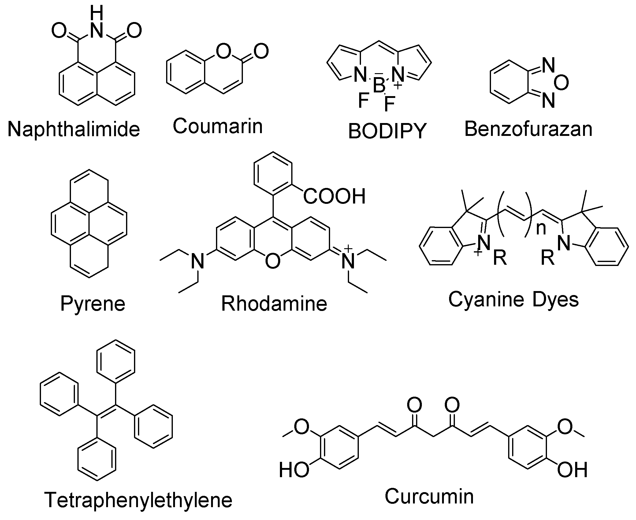
1. Introduction
2. Detection of Thiols in Cells
2.1. NPSH, GSH, Cys, and Hcy
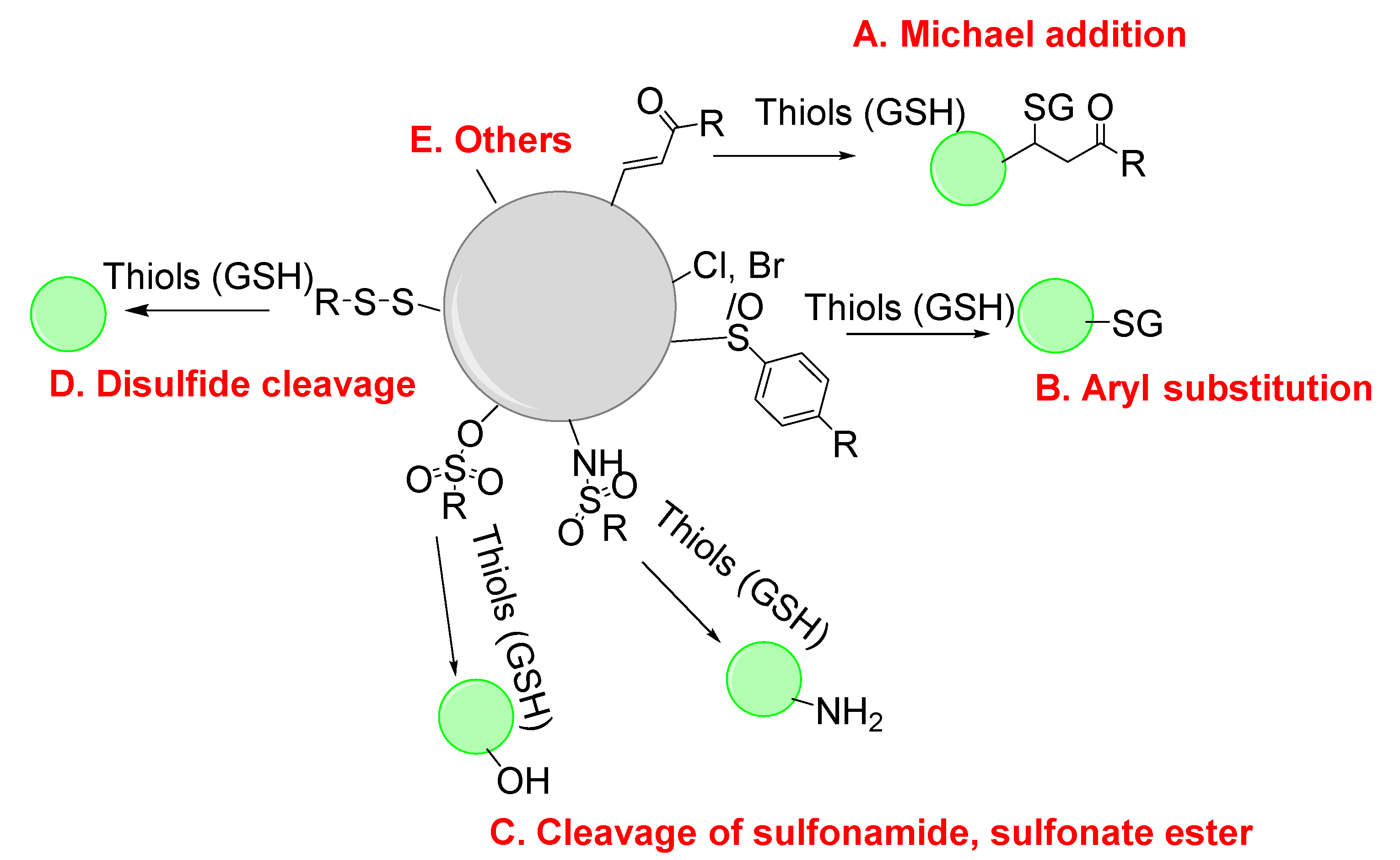
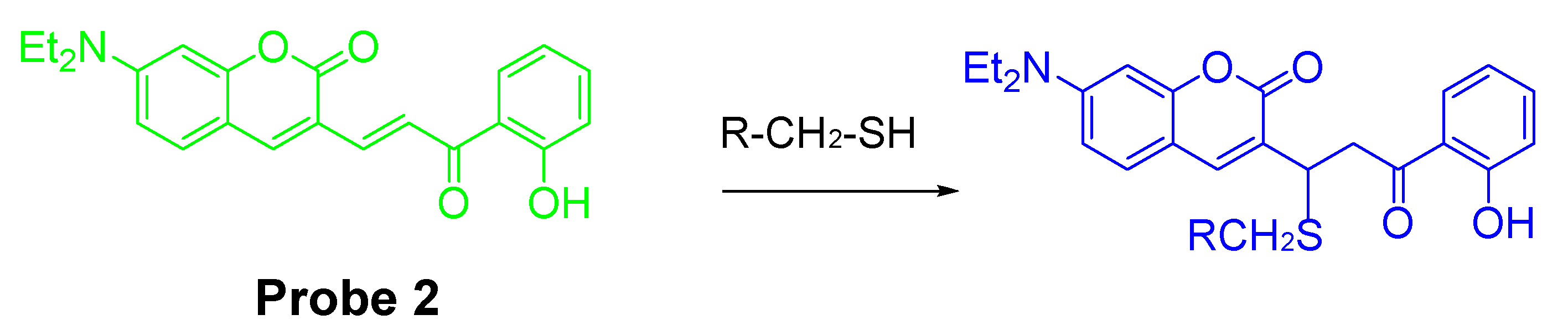
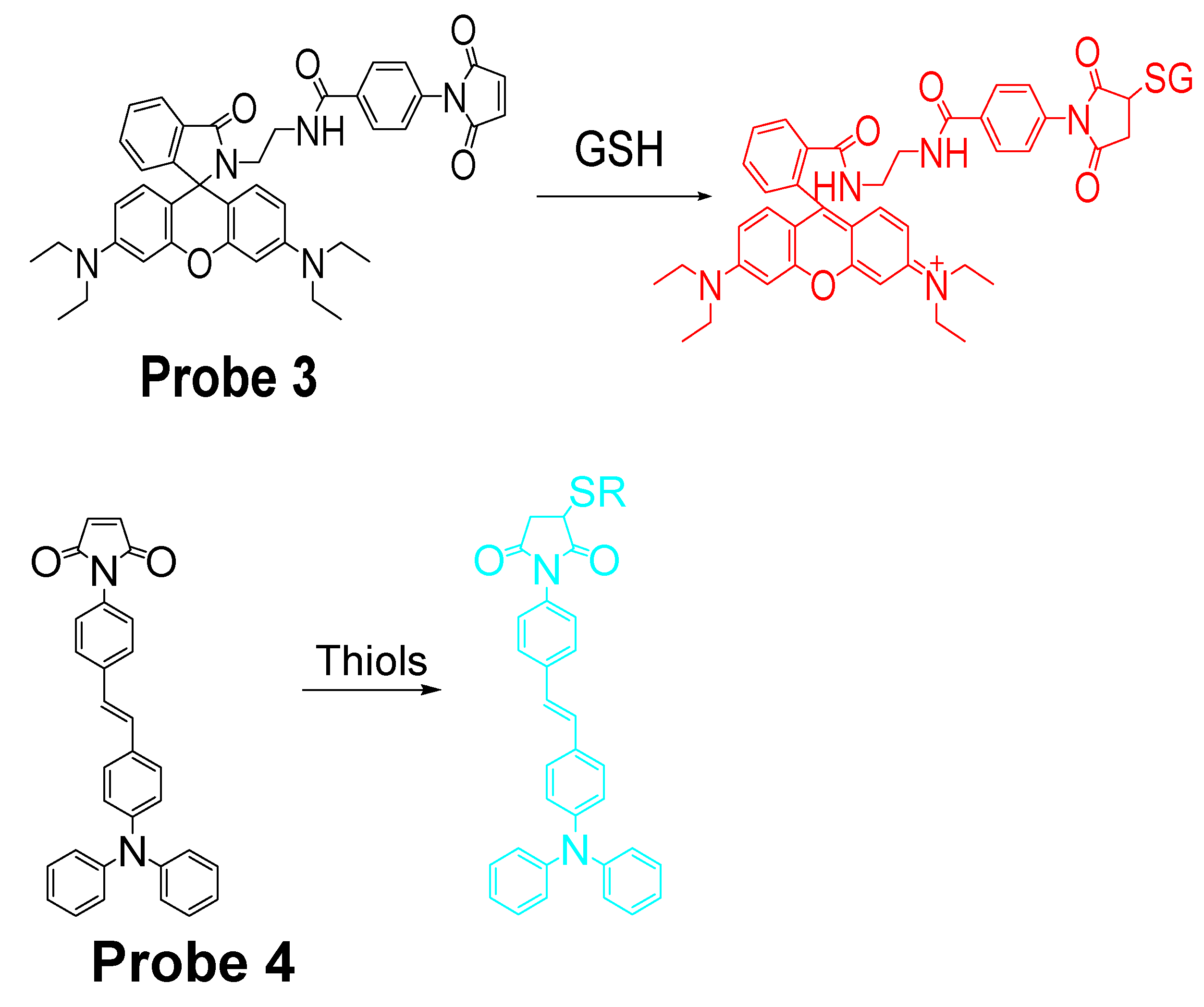
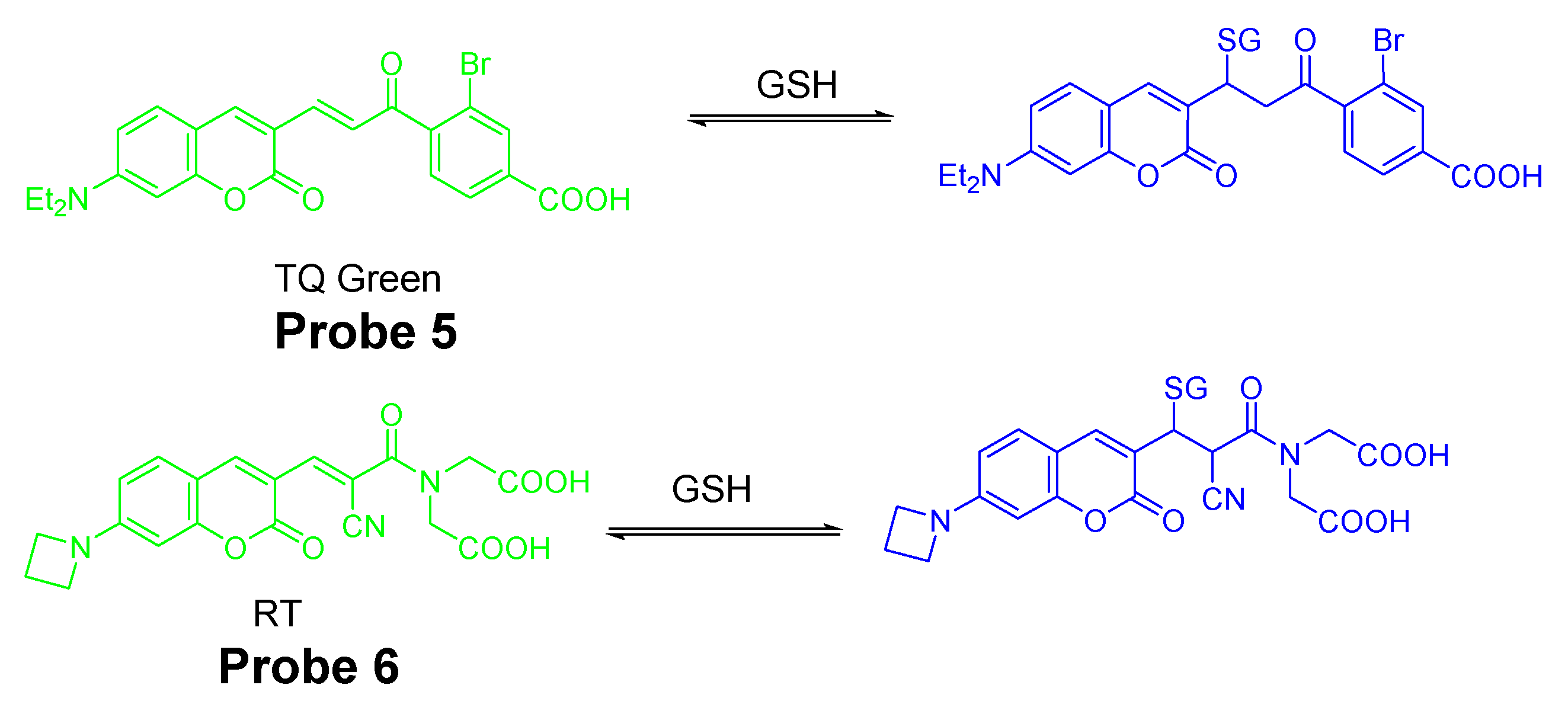
2.1.1. Detection of NPSH via a Michael Addition Reaction
2.1.2. Detection of NPSH via a Nucleophilic Aromatic Substitution (SNAr) Reaction Using Halogen, Ether, Thioether as a Leaving Group
Detection of NPSH via a SNAr Reaction Using Halogen as a Leaving Group
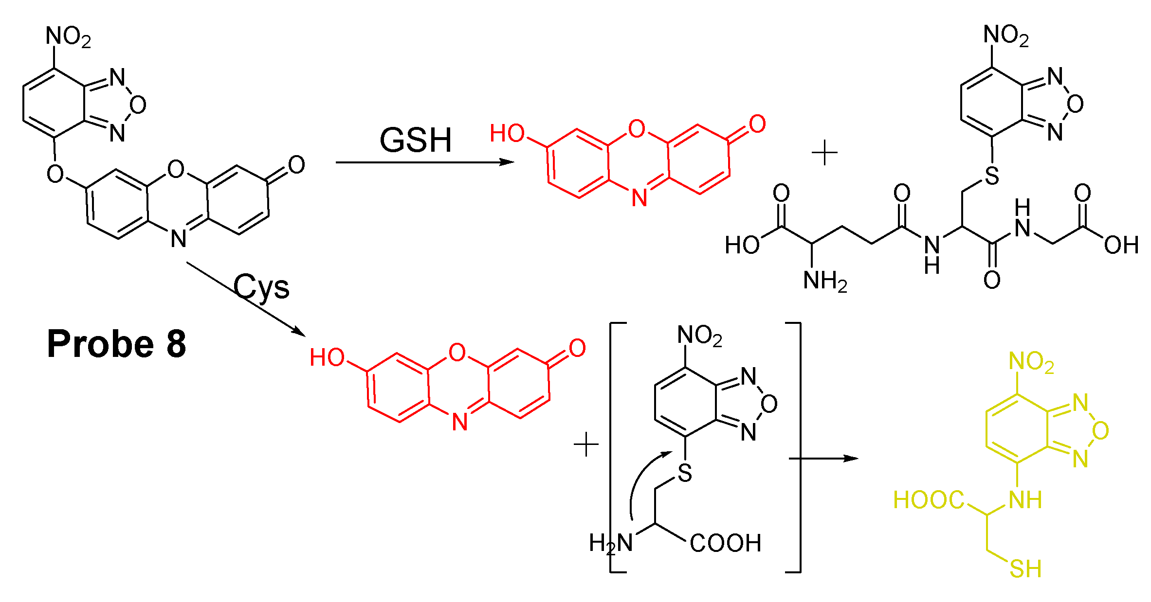
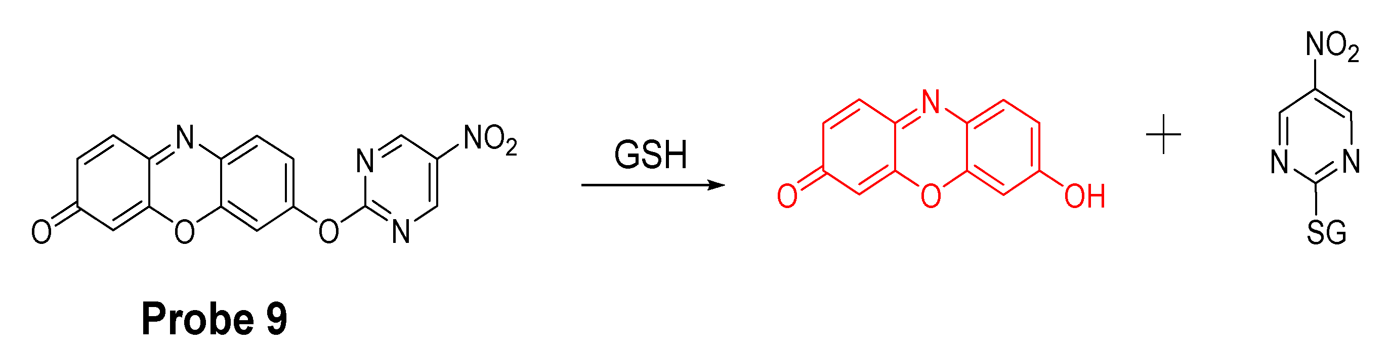
Detection of NPSH via a SNAr Reaction Using ether as a Leaving Group
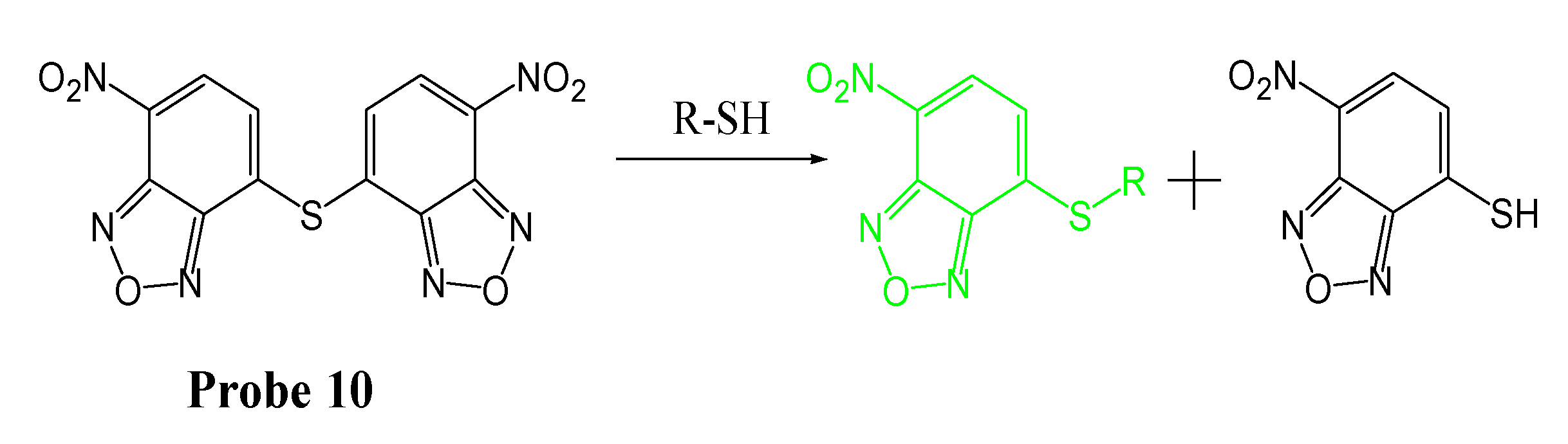
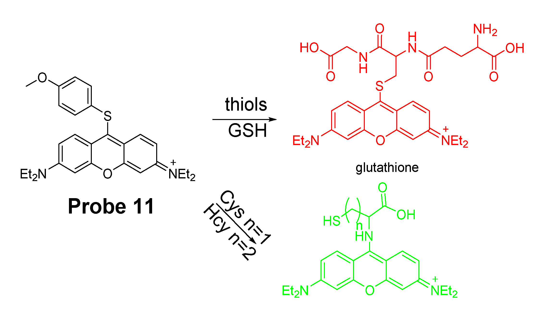
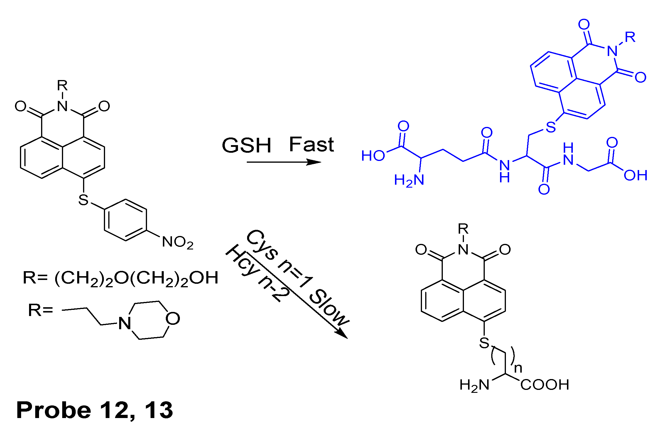
Detection of NPSH via a SNAr Reaction Using Thioether as a Leaving Group
2.1.3. Detection of NPSH via Cleavage of Sulfonamide or Sulfonate Ester
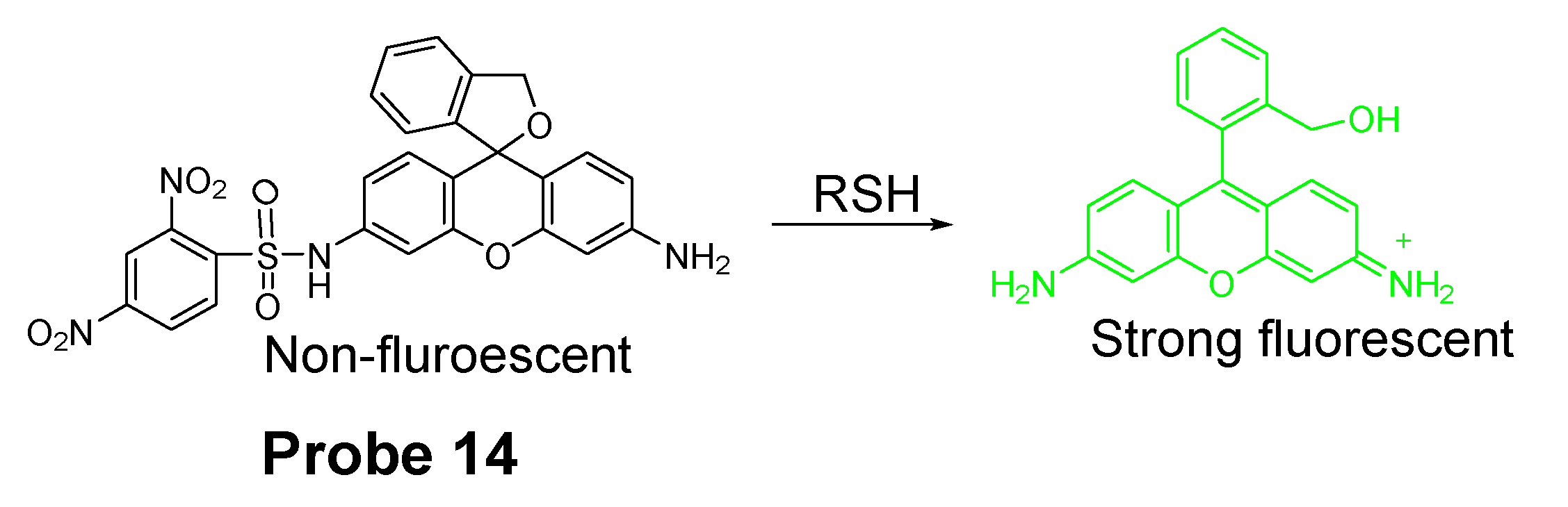
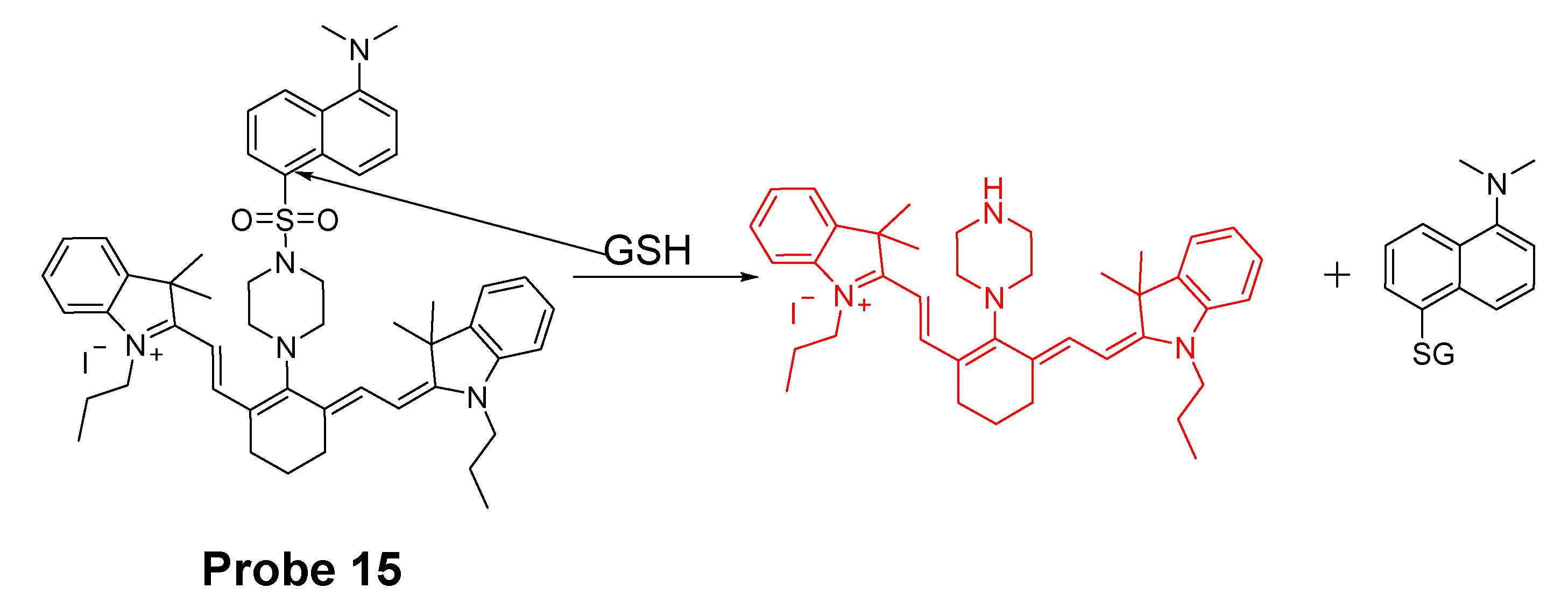
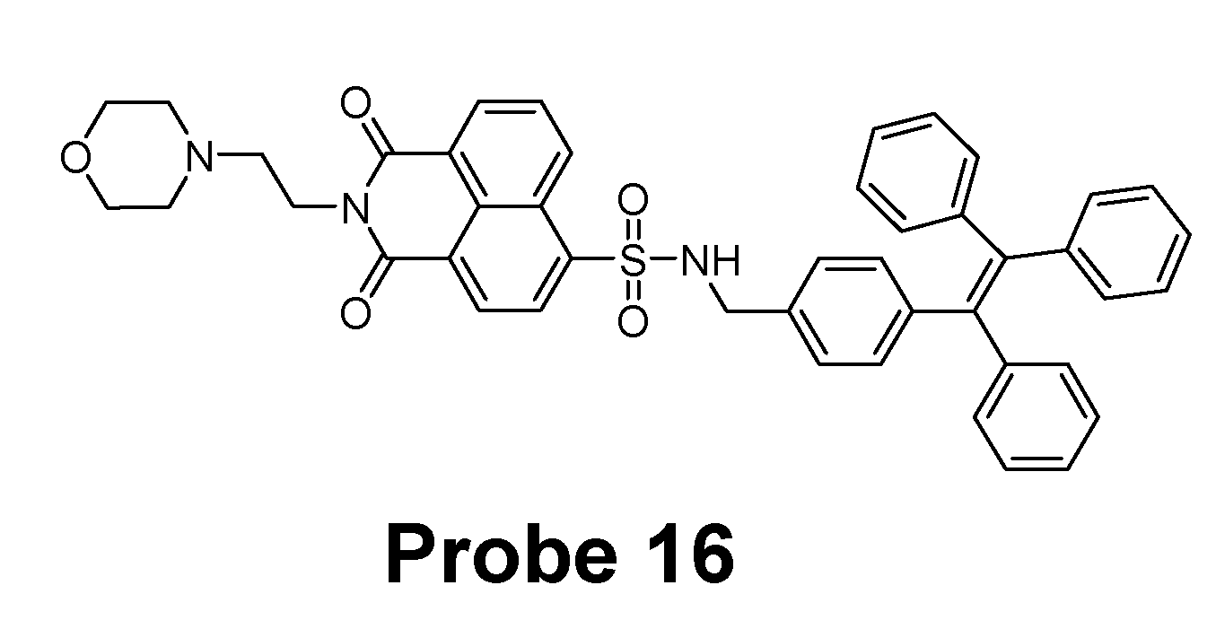
Detection of NPSH via Cleavage of Sulfonamide
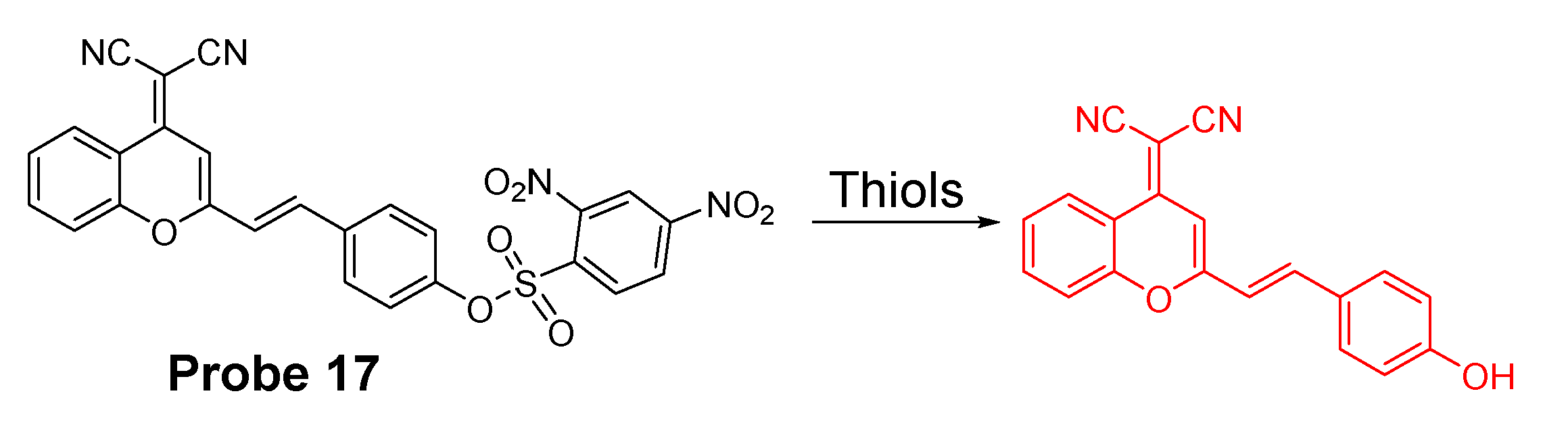
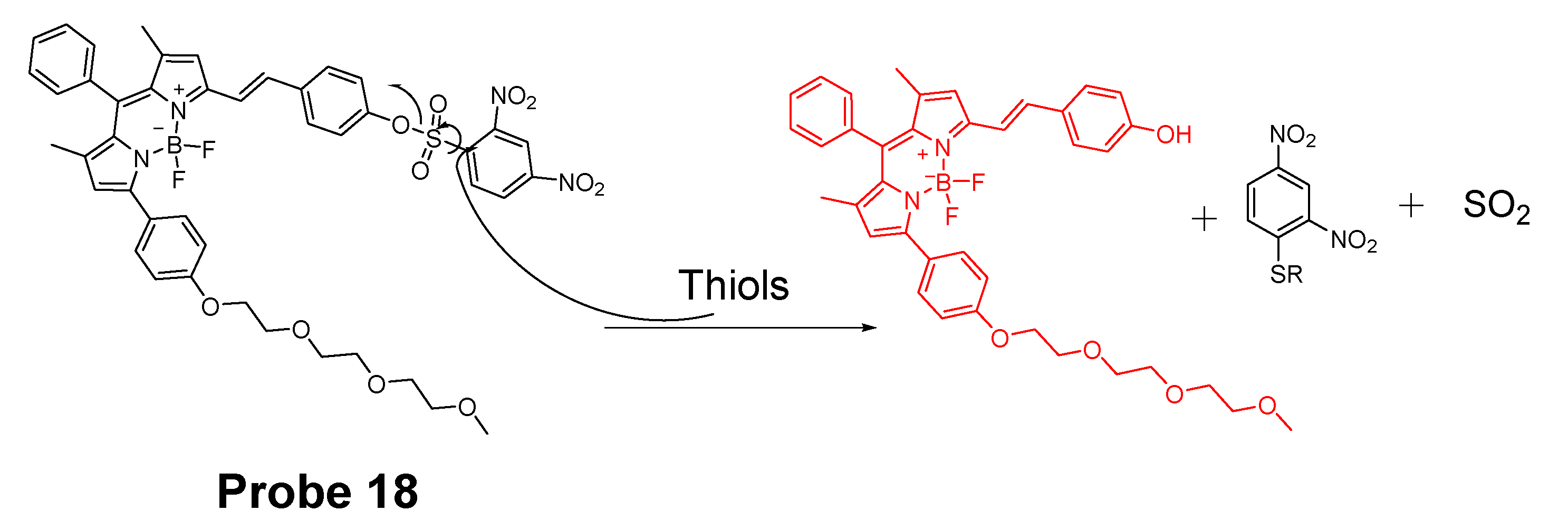
Detection of NPSH via Cleavage of Sulfonate Ester
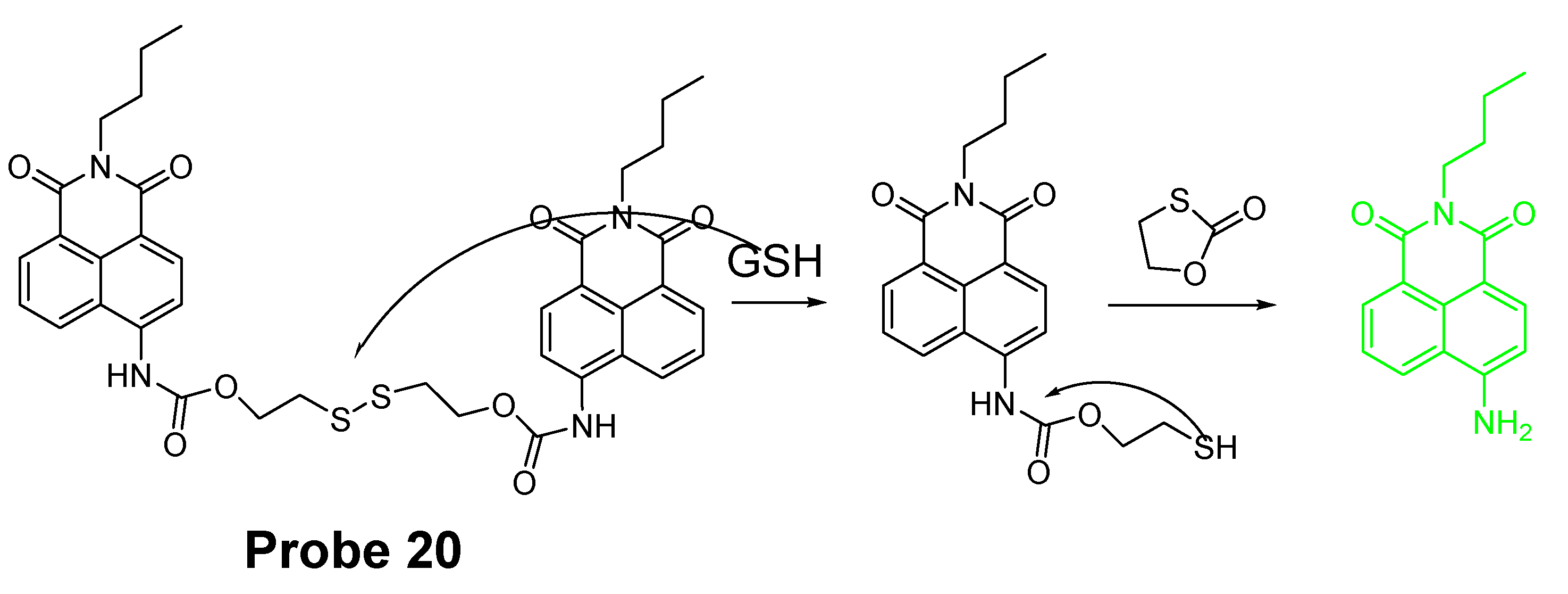
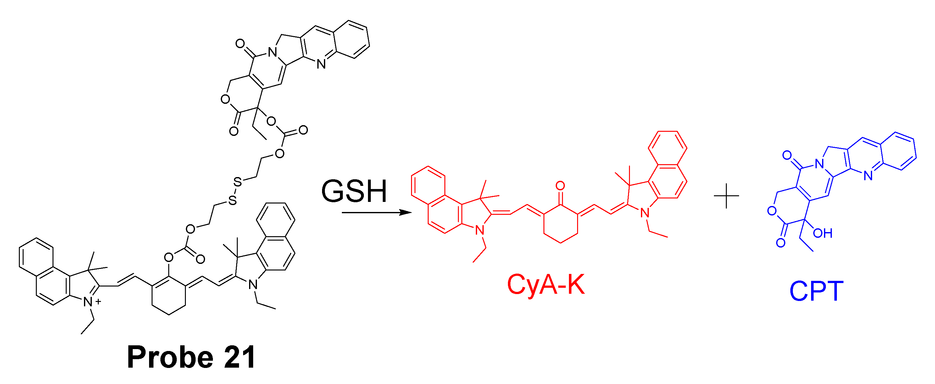
2.1.4. Detection of NPSH via Cleavage of a Disulfide Bond
2.1.5. Detection of NPSH via Other Strategies
2.2. Selective Detection of Cys and Hcy
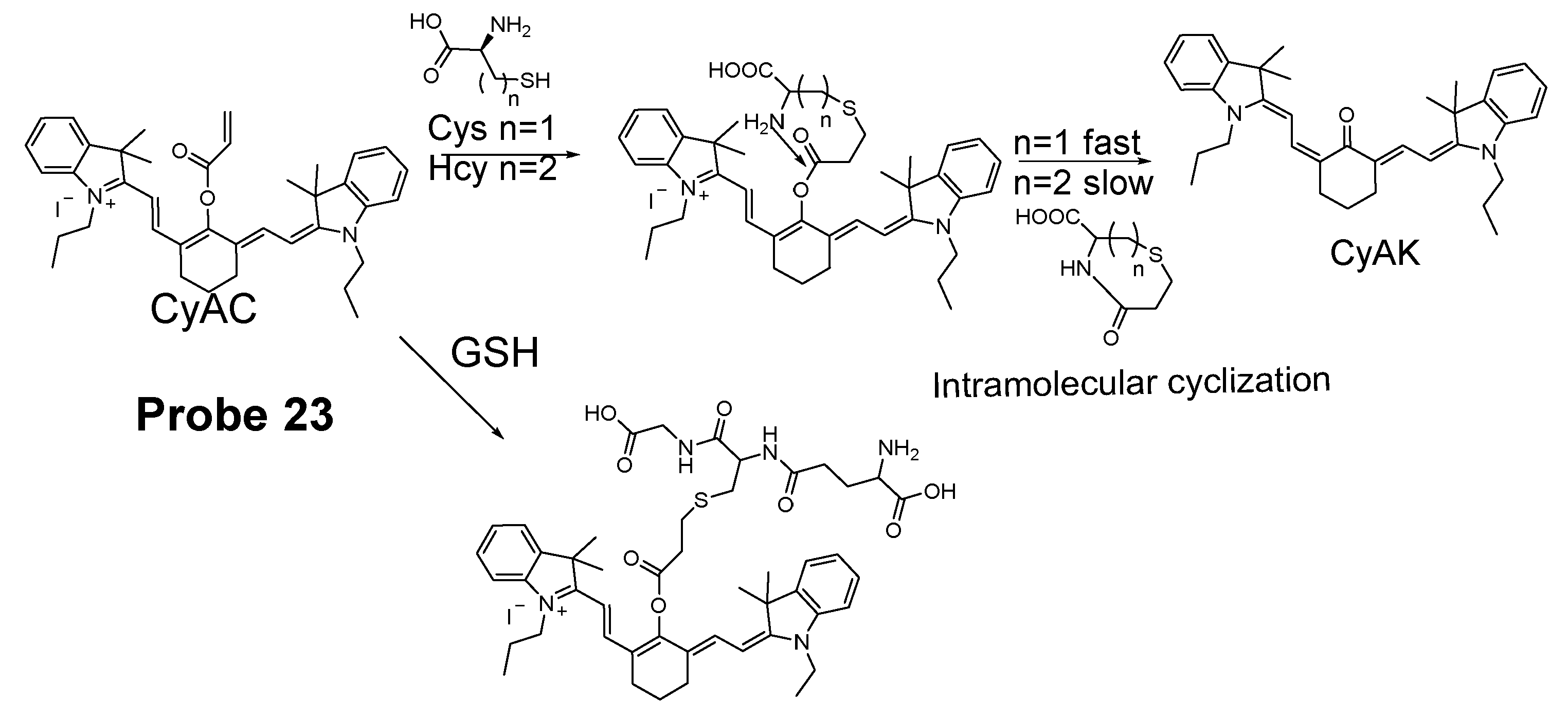
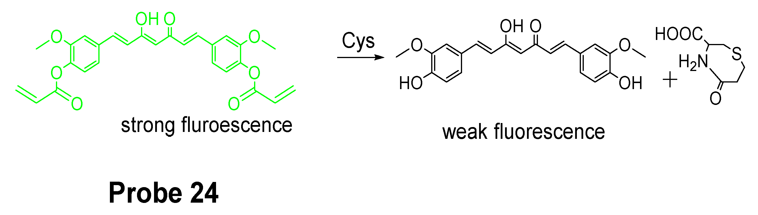
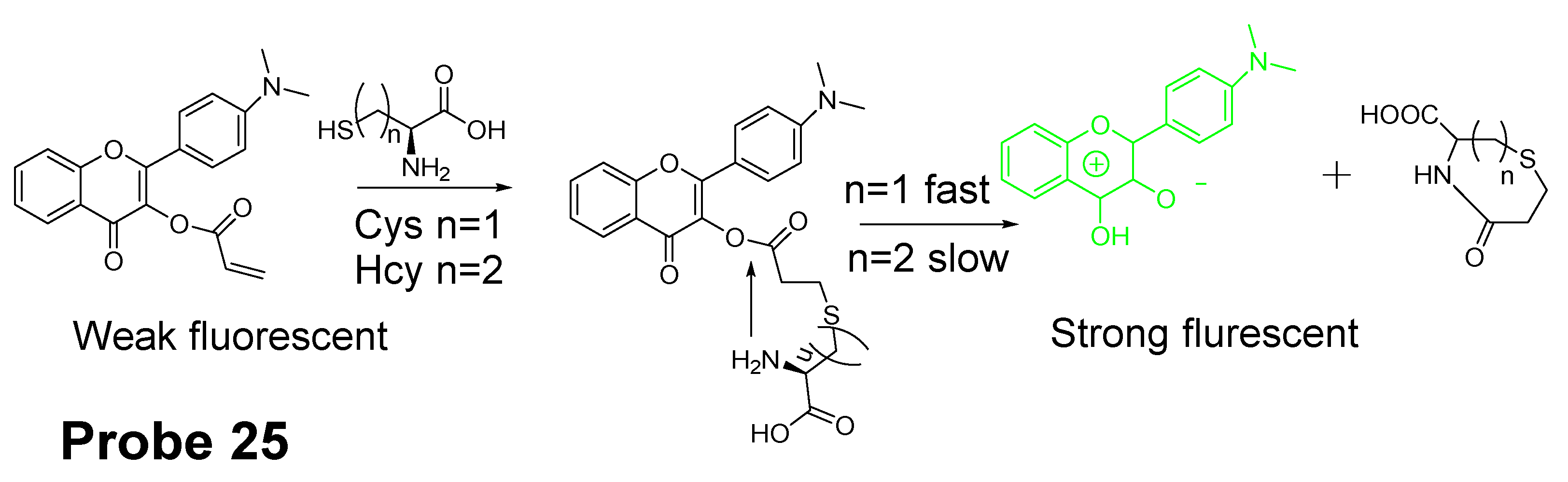
2.2.1. Selective Detection of Cys and Hcy through Cyclization of Cys/Hcy with Acrylates or Aldehydes
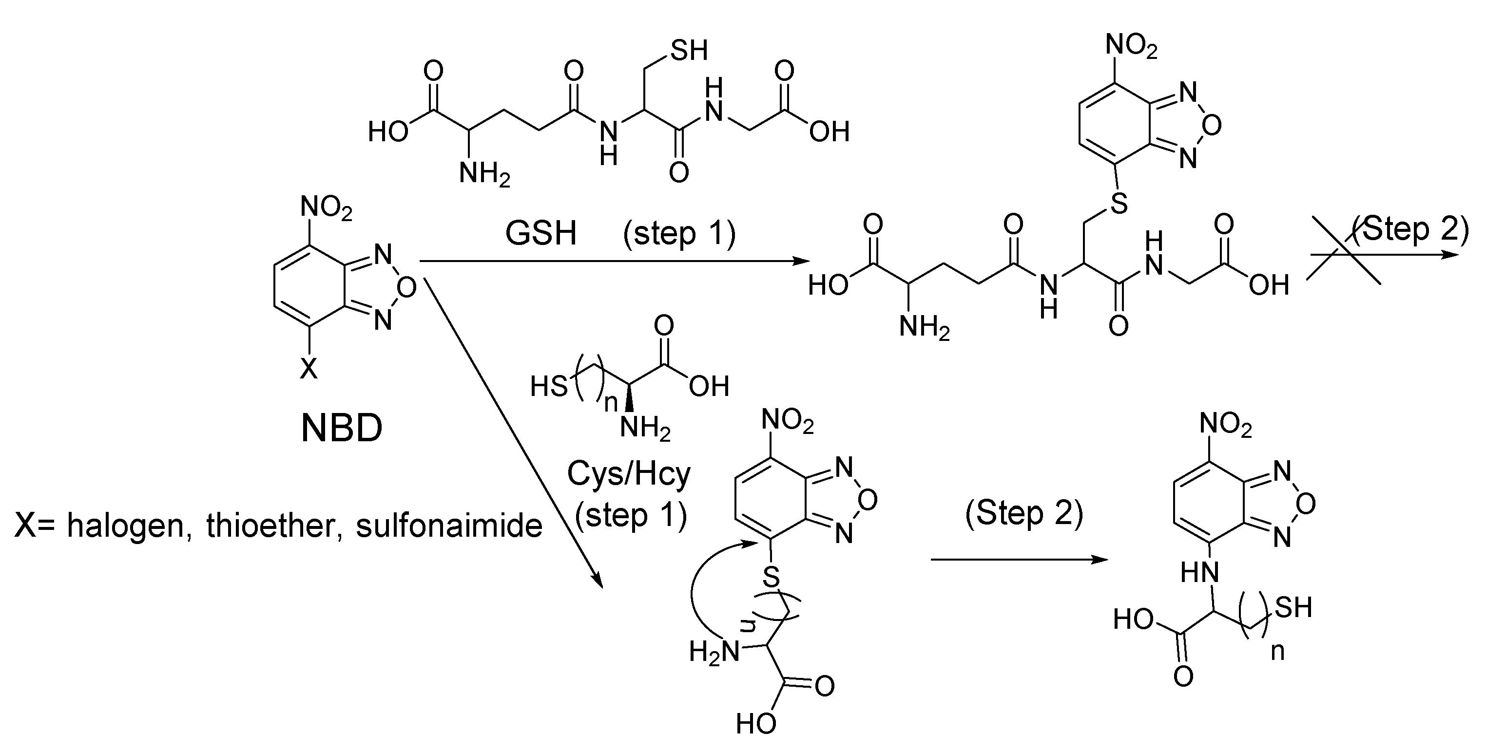
2.2.2. Selective Detection of Cys and Hcy through Cys-Induced SNAr Substitution−Rearrangement Reaction
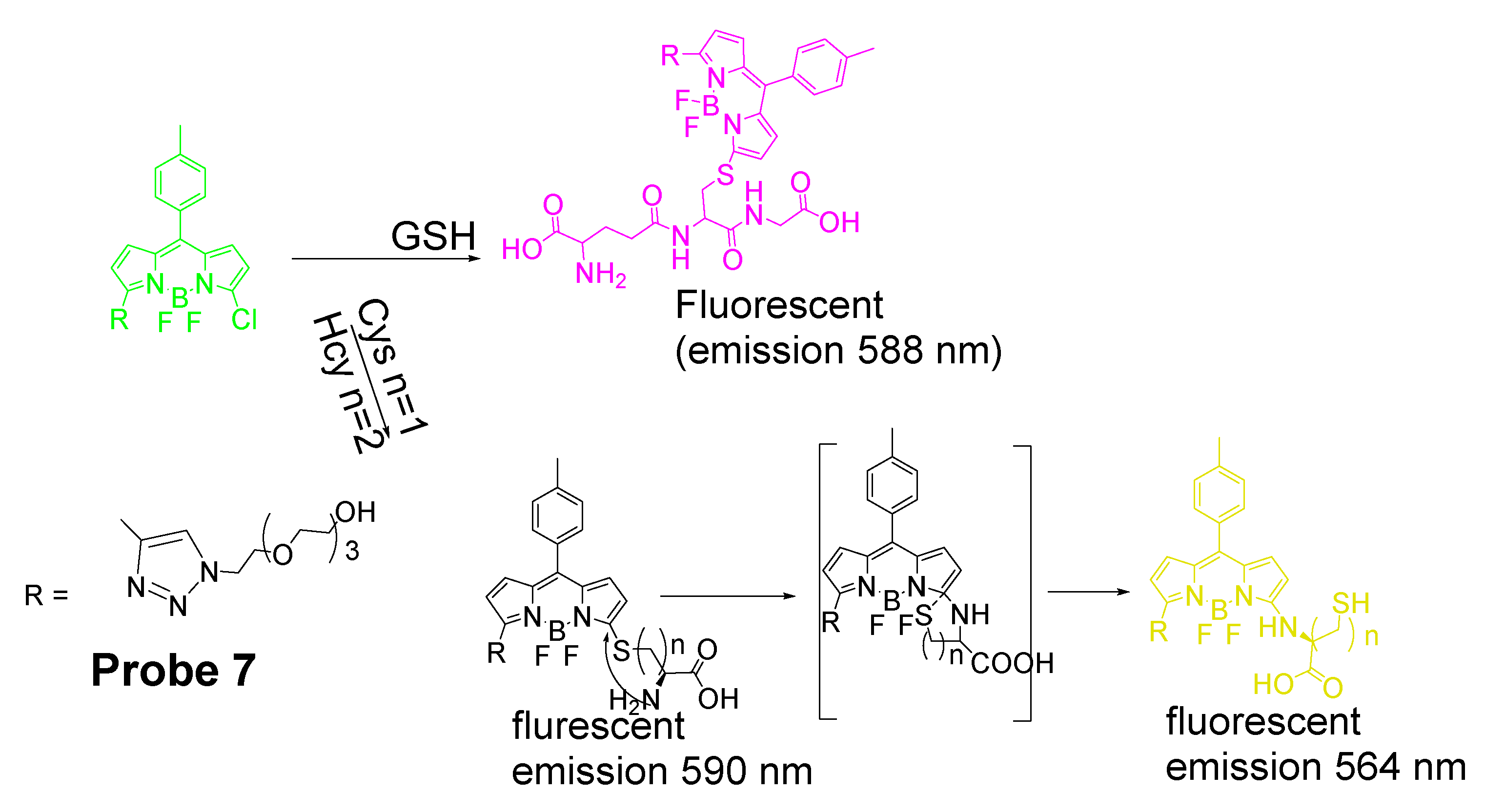
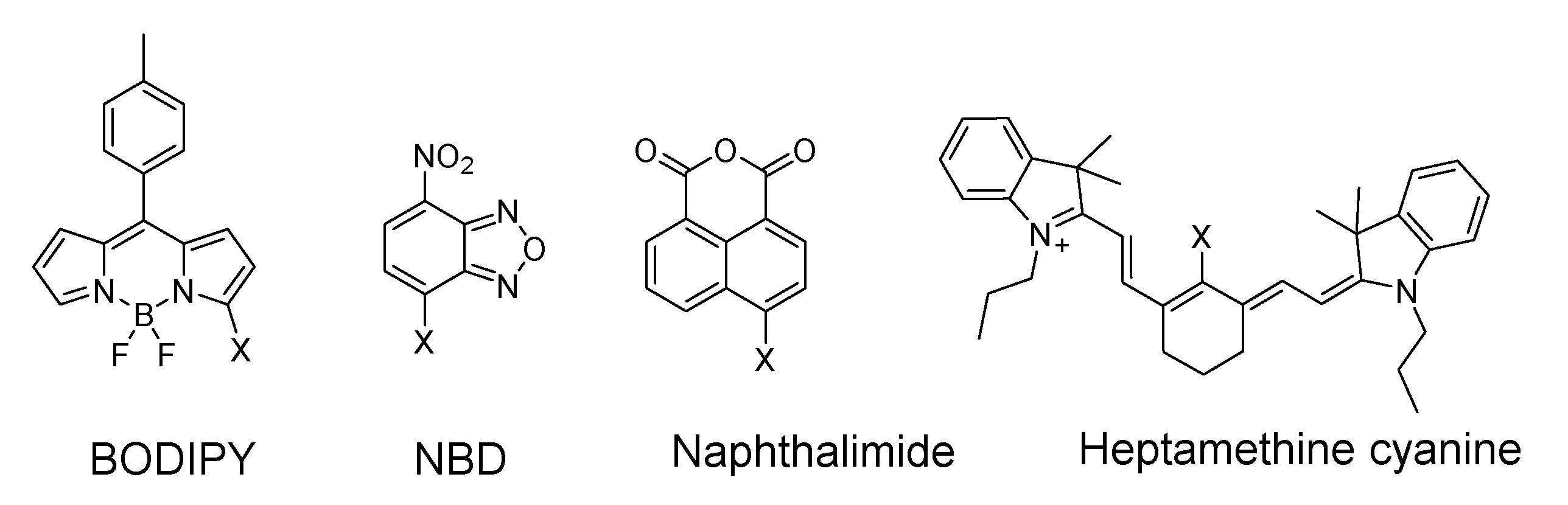
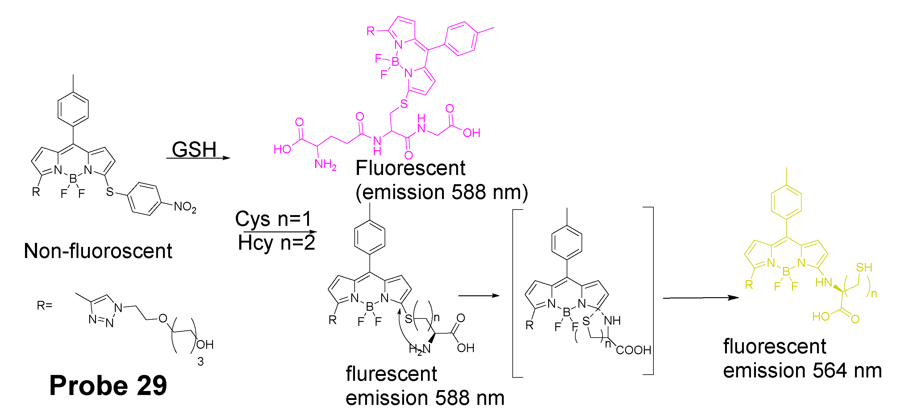
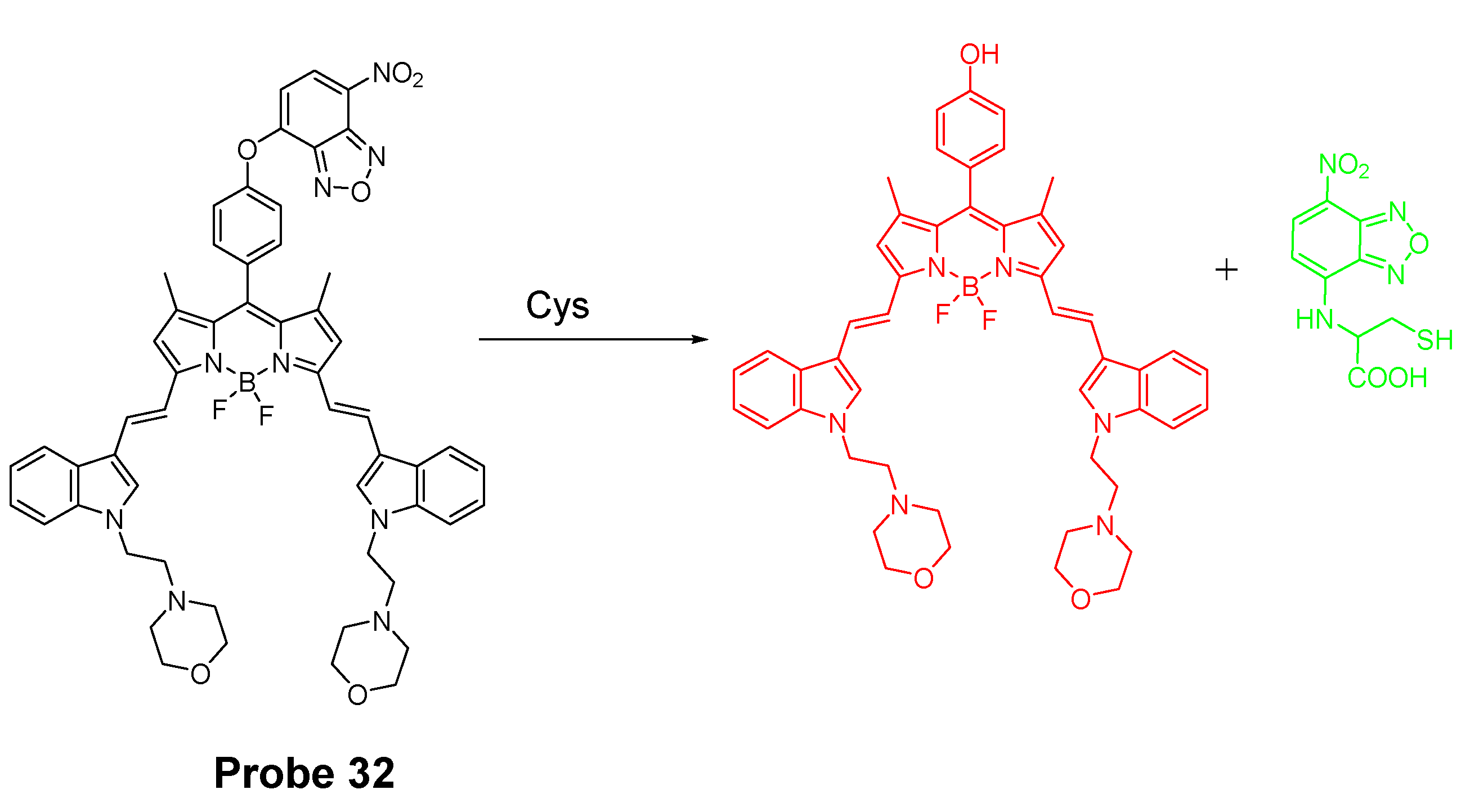
BODIPY
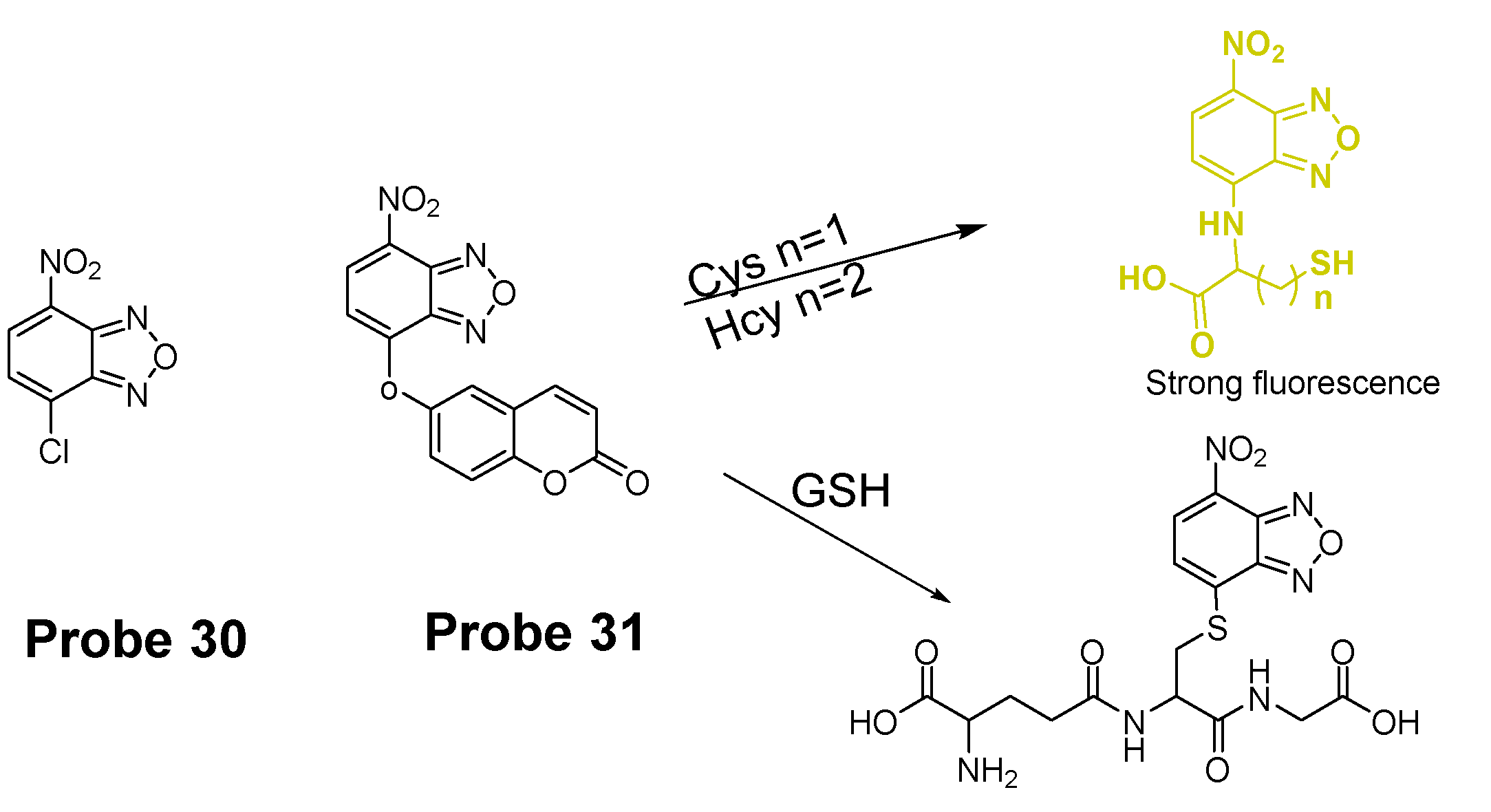
7-Nitrobenzofurazan (NBD)
Other Fluorophores
2.2.3. Selective Detection of Cys and Hcy via Other Mechanisms
3. Detection of Thiols in Subcellular Organelles
3.1. Mitochondrial Total Thiols and GSH
3.1.1. Detection of Mitochondrial Thiols via a Michael Addition Reaction
3.1.2. Detection of Mitochondrial NPSH via a SNAr Reaction Using Halogen, Ether, or Thioether as a Leaving Group
Detection of Mitochondrial NPSH via a SNAr Reaction Using Halogen as a Leaving Group
Detection of Mitochondrial NPSH via a SNAr Reaction Using Thioether as a Leaving Group
Detection of Mitochondrial NPSH via a SNAr Reaction Using Ether as a Leaving Group
3.1.3. Detection of Mitochondrial NPSH via Cleavage of Sulfonamide or Sulfonate Ester
Detection of Mitochondrial NPSH via Cleavage of Sulfonamide
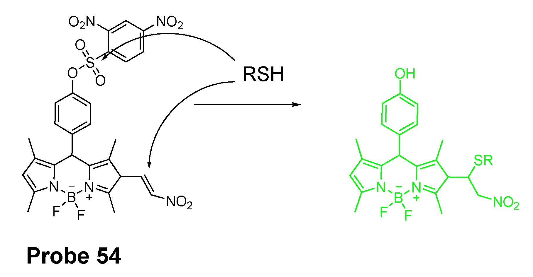
Detection of Mitochondrial NPSH via Cleavage of Sulfonate Ester
3.1.4. Detection of Mitochondria NPSH via Cleavage of Disulfide Bond
3.1.5. Detection of Mitochondrial NPSH through Other Methods
3.2. Selective Detection of Mitochondrial Cys/Hcy
3.2.1. Selective Detection of Mitochondrial Cys and Hcy through Cyclization of Cys/Hcy with Acrylates or Aldehydes
3.2.2. Selective Detection of Mitochondrial Cys/Hcy through Cys-induced SNAr Substitution−Rearrangement Reaction
3.2.3. Selective Detection of Mitochondrial Cys/Hcy through Other Mechanisms
3.3. Lysosomal Total Thiols and GSH
3.3.1. Detection of Lysosomal NPSH via a Michael Addition Reaction
3.3.2. Detection of Lysosomal NPSH via SNAr Reactions Using Ether, Thioether as a Leaving Group
Detection of Lysosomal NPSH via SNAr Reactions Using Ether as a Leaving Group
Detection of Lysosomal NPSH via SNAr Reactions Using Thioether as a Leaving Group
3.3.3. Detection of Lysosome NPSH via Cleavage of Sulfonamide and Sulfonate
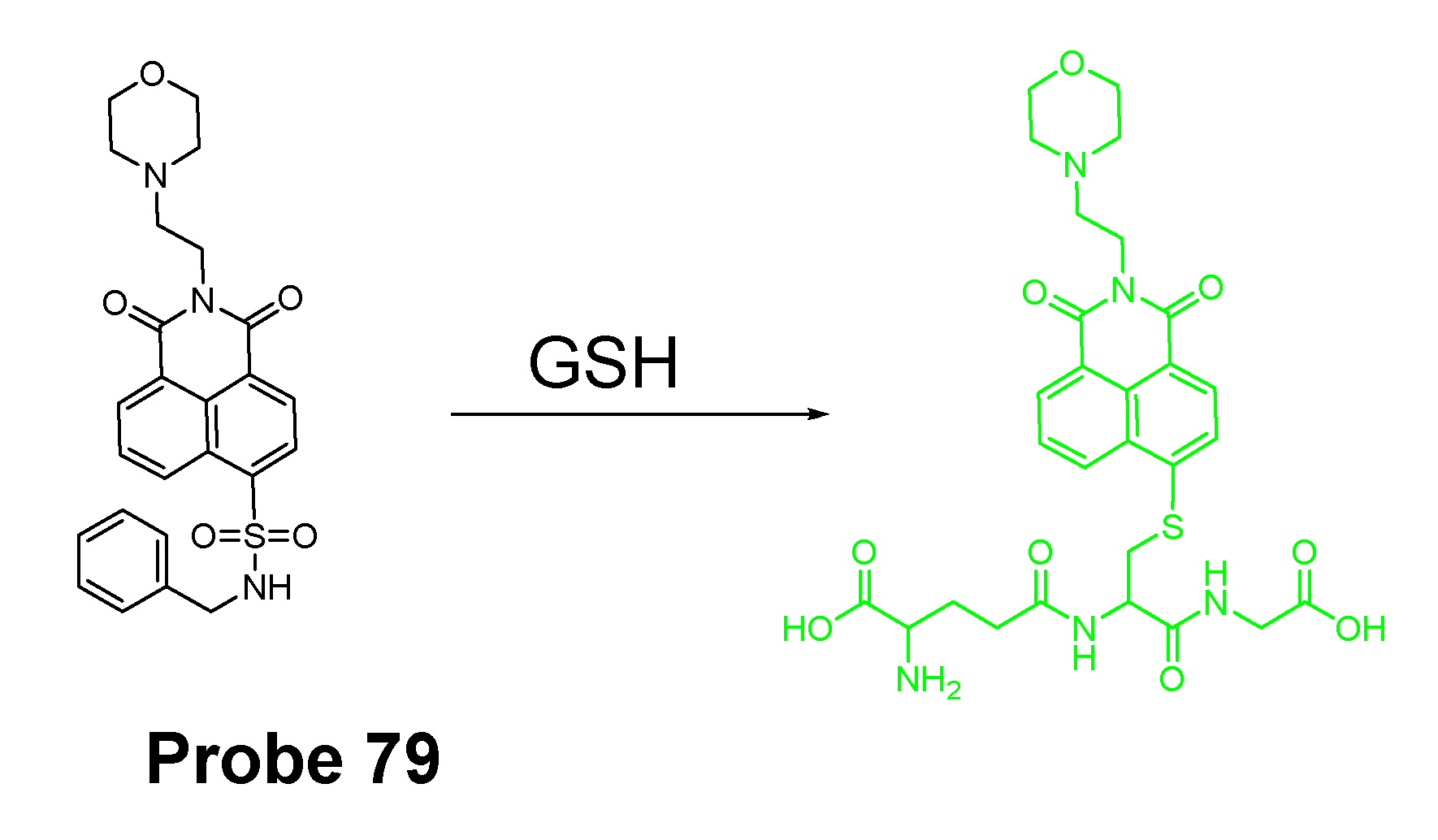
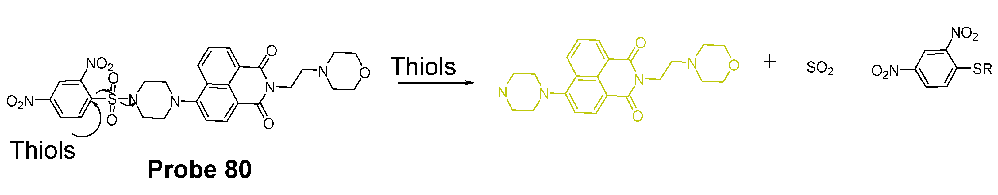
Detection of Lysosomal NPSH via Cleavage of Sulfonamide
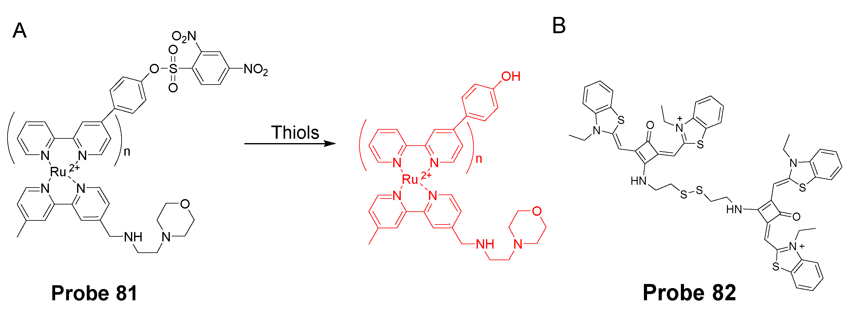
Detection of Lysosomal NPSH via Cleavage of Sulfonate
3.3.4. Detection of Lysosomal NPSH via Cleavage of Disulfide
3.3.5. Detection of Lysosome NPSH via Other Methods
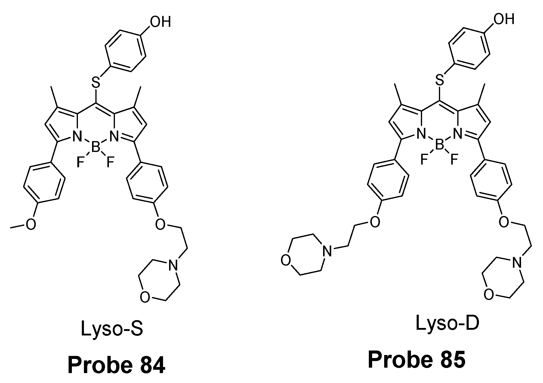
3.4. Selective Detection of Lysosomal Cys and Hcy
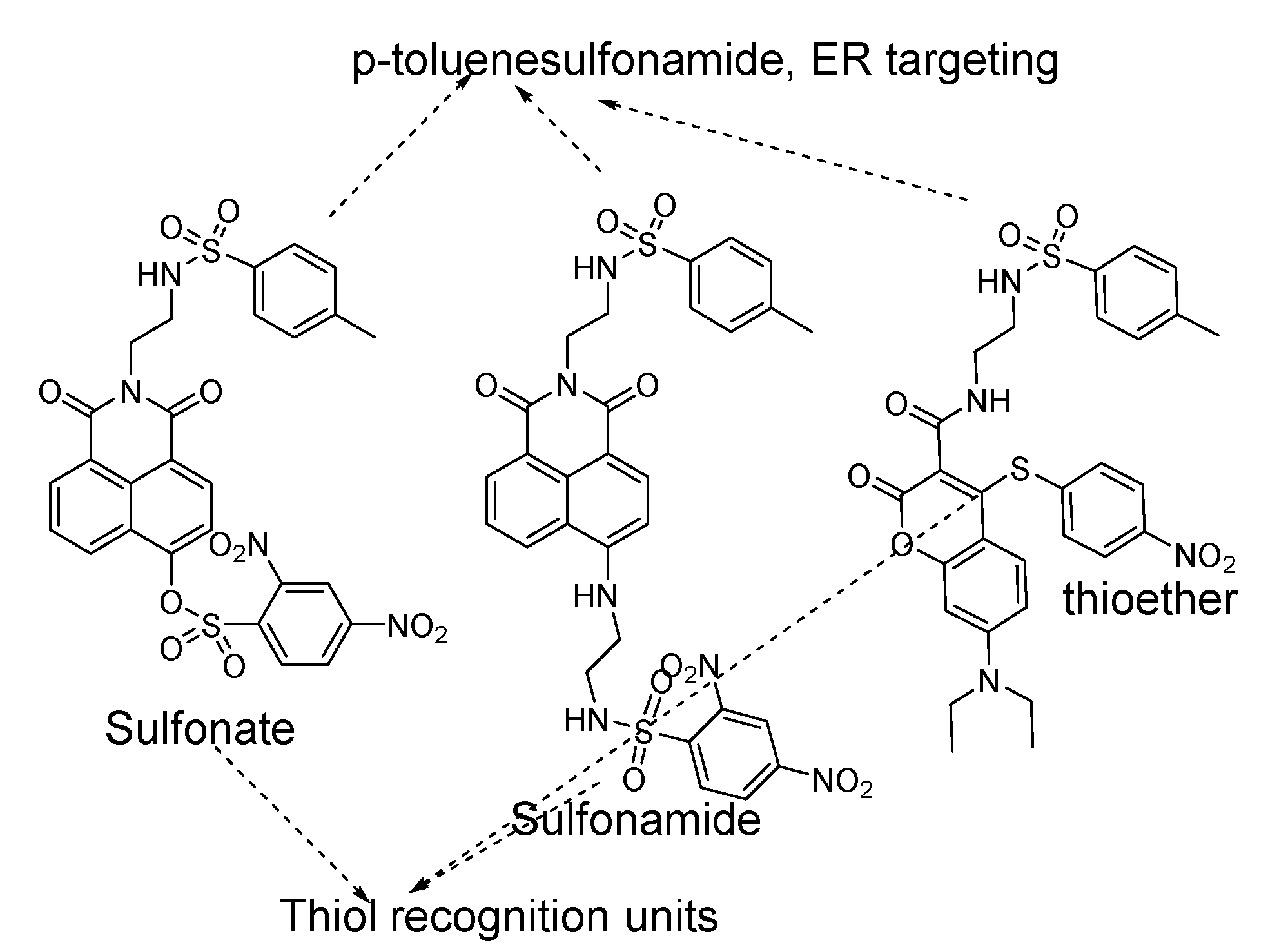
3.5. Detection of NPSH in Endoplasmic Reticulum
4. Conclusions
Author Contributions
Funding
Institutional Review Board Statement
Informed Consent Statement
Data Availability Statement
Conflicts of Interest
References
- Xu, Y.; Li, R.; Zhou, X.; Li, W.; Ernest, U.; Wan, H.; Li, L.; Chen, H.; Yuan, Z. A visible and near-infrared, dual emission fluorescent probe based on thiol reactivity for selectively tracking mitochondrial glutathione in vitro. Talanta 2019, 205, 120–125. [Google Scholar] [CrossRef] [PubMed]
- Dai, J.; Ma, C.; Zhang, P.; Fu, Y.; Shen, B. Recent progress in the development of fluorescent probes for detection of biothiols. Dye. Pigment. 2020, 177, 108321. [Google Scholar] [CrossRef]
- Hansen, R.E.; Roth, D.; Winther, J.R. Quantifying the global cellular thiol–disulfide status. Proc. Natl. Acad. Sci. USA 2009, 106, 422–427. [Google Scholar] [CrossRef] [PubMed]
- Chen, H.; Tang, Y.; Lin, W. Recent progress in the fluorescent probes for the specific imaging of small molecular weight thiols in living cells. Trac Trends Anal. Chem. 2016, 76, 166–181. [Google Scholar] [CrossRef]
- Liu, Y.; Teng, L.; Chen, L.; Ma, H.; Liu, H.-W.; Zhang, X.-B. Engineering of a near-infrared fluorescent probe for real-time simultaneous visualization of intracellular hypoxia and induced mitophagy. Chem. Sci. 2018, 9, 5347–5353. [Google Scholar] [CrossRef]
- Kumar, N.; Bhalla, V.; Kumar, M. Development and sensing applications of fluorescent motifs within the mitochondrial environment. Chem. Commun. 2015, 51, 15614–15628. [Google Scholar]
- Xu, W.; Zeng, Z.; Jiang, J.H.; Chang, Y.T.; Yuan, L. Discerning the chemistry in individual organelles with small-molecule fluorescent probes. Angew. Chem. Int. Ed. 2016, 55, 13658–13699. [Google Scholar] [CrossRef] [PubMed]
- Hansen, J.M.; Go, Y.-M.; Jones, D.P. Nuclear and mitochondrial compartmentation of oxidative stress and redox signaling. Annu. Rev. Pharmacol. Toxicol. 2006, 46, 215–234. [Google Scholar] [CrossRef]
- Moriarty-Craige, S.E.; Jones, D.P. Extracellular thiols and thiol/disulfide redox in metabolism. Annu. Rev. Nutr. 2004, 24, 481–509. [Google Scholar] [CrossRef]
- Reed, D.; Babson, J.; Beatty, P.; Brodie, A.; Ellis, W.; Potter, D. HPLC analysis of nanogram levels of glutathione, glutathione disulphide and related thiols and disulphides. Anal Biochem. 1980, 106, 55–62. [Google Scholar] [CrossRef]
- Newton, G.L.; Dorian, R.; Fahey, R.C. Analysis of biological thiols: Derivatization with monobromobimane and separation by reverse-phase high-performance liquid chromatography. Anal. Biochem. 1981, 114, 383–387. [Google Scholar] [CrossRef]
- Chen, W.; Zhao, Y.; Seefeldt, T.; Guan, X. Determination of thiols and disulfides via HPLC quantification of 5-thio-2-nitrobenzoic acid. J. Pharm. Biomed. Anal. 2008, 48, 1375–1380. [Google Scholar] [CrossRef]
- Guan, X.; Hoffman, B.; Dwivedi, C.; Matthees, D.P. A simultaneous liquid chromatography/mass spectrometric assay of glutathione, cysteine, homocysteine and their disulfides in biological samples. J. Pharm. Biomed. Anal. 2003, 31, 251–261. [Google Scholar] [CrossRef]
- Wei, W.; Liang, X.; Hu, G.; Guo, Y.; Shao, S. A highly selective colorimetric probe based on 2,2′,2″-trisindolylmethene for cysteine/homocysteine. Tetrahedron Lett. 2011, 52, 1422–1425. [Google Scholar] [CrossRef]
- Ren, M.; Zhou, K.; He, L.; Lin, W. Mitochondria and lysosome-targetable fluorescent probes for HOCl: Recent advances and perspectives. J. Mater. Chem. B 2018, 6, 1716–1733. [Google Scholar] [CrossRef]
- Ren, T.-B.; Zhang, Q.-L.; Su, D.; Zhang, X.-X.; Yuan, L.; Zhang, X.-B. Detection of analytes in mitochondria without interference from other sites based on an innovative ratiometric fluorophore. Chem. Sci. 2018, 9, 5461–5466. [Google Scholar] [CrossRef] [PubMed]
- Maeda, H.; Matsuno, H.; Ushida, M.; Katayama, K.; Saeki, K.; Itoh, N. 2, 4-Dinitrobenzenesulfonyl fluoresceins as fluorescent alternatives to Ellman’s reagent in thiol-quantification enzyme assays. Angew. Chem. Int. Ed. 2005, 44, 2922–2925. [Google Scholar] [CrossRef] [PubMed]
- Hurd, T.R.; Prime, T.A.; Harbour, M.E.; Lilley, K.S.; Murphy, M.P. Detection of reactive oxygen species-sensitive thiol proteins by redox difference gel electrophoresis implications for mitochondrial redox signaling. J. Biol. Chem. 2007, 282, 22040–22051. [Google Scholar] [CrossRef]
- Mao, Z.; Jiang, H.; Li, Z.; Zhong, C.; Zhang, W.; Liu, Z. An N-nitrosation reactivity-based two-photon fluorescent probe for the specific in situ detection of nitric oxide. Chem. Sci. 2017, 8, 4533–4538. [Google Scholar] [CrossRef] [PubMed]
- Rao, J.; Dragulescu-Andrasi, A.; Yao, H. Fluorescence imaging in vivo: Recent advances. Curr. Opin. Biotechnol. 2007, 18, 17–25. [Google Scholar] [CrossRef]
- Giustarini, D.; Colombo, G.; Garavaglia, M.L.; Astori, E.; Portinaro, N.M.; Reggiani, F.; Badalamenti, S.; Aloisi, A.M.; Santucci, A.; Rossi, R. Assessment of glutathione/glutathione disulphide ratio and S-glutathionylated proteins in human blood, solid tissues, and cultured cells. Free Radic. Biol. Med. 2017, 112, 360–375. [Google Scholar] [CrossRef]
- Wang, S.; Yin, H.; Huang, Y.; Guan, X. Thiol specific and mitochondria selective fluorogenic benzofurazan sulfide for live cell nonprotein thiol imaging and quantification in mitochondria. Anal. Chem. 2018, 90, 8170–8177. [Google Scholar] [CrossRef]
- Ding, D.; Li, K.; Liu, B.; Tang, B.Z. Bioprobes based on AIE fluorogens. Acc. Chem. Res. 2013, 46, 2441–2453. [Google Scholar] [CrossRef]
- Song, Z.; Mao, D.; Sung, S.H.; Kwok, R.T.; Lam, J.W.; Kong, D.; Ding, D.; Tang, B.Z. Activatable fluorescent nanoprobe with aggregation-induced emission characteristics for selective in vivo imaging of elevated peroxynitrite generation. Adv. Mater. 2016, 28, 7249–7256. [Google Scholar] [CrossRef]
- Shi, L.; Li, K.; Li, L.-L.; Chen, S.-Y.; Li, M.-Y.; Zhou, Q.; Wang, N.; Yu, X.-Q. Novel easily available purine-based AIEgens with colour tunability and applications in lipid droplet imaging. Chem. Sci. 2018, 9, 8969–8974. [Google Scholar] [CrossRef]
- Niu, G.; Zhang, P.; Liu, W.; Wang, M.; Zhang, H.; Wu, J.; Zhang, L.; Wang, P. Near-infrared probe based on rhodamine derivative for highly sensitive and selective lysosomal pH tracking. Anal. Chem. 2017, 89, 1922–1929. [Google Scholar] [CrossRef] [PubMed]
- Caskey, C.T. Disease diagnosis by recombinant DNA methods. Science 1987, 236, 1223–1229. [Google Scholar] [CrossRef] [PubMed]
- Li, X.; Qian, S.; He, Q.; Yang, B.; Li, J.; Hu, Y. Design and synthesis of a highly selective fluorescent turn-on probe for thiol bioimaging in living cells. Org. Biomol. Chem. 2010, 8, 3627–3630. [Google Scholar] [CrossRef] [PubMed]
- Niu, L.-Y.; Guan, Y.-S.; Chen, Y.-Z.; Wu, L.-Z.; Tung, C.-H.; Yang, Q.-Z. BODIPY-based ratiometric fluorescent sensor for highly selective detection of glutathione over cysteine and homocysteine. J. Am. Chem. Soc. 2012, 134, 18928–18931. [Google Scholar] [CrossRef]
- Tang, B.; Xing, Y.; Li, P.; Zhang, N.; Yu, F.; Yang, G. A rhodamine-based fluorescent probe containing a Se-N bond for detecting thiols and its application in living cells. J. Am. Chem. Soc. 2007, 129, 11666–11667. [Google Scholar] [CrossRef]
- Shibata, A.; Furukawa, K.; Abe, H.; Tsuneda, S.; Ito, Y. Rhodamine-based fluorogenic probe for imaging biological thiol. Bioorganic Med. Chem. Lett. 2008, 18, 2246–2249. [Google Scholar] [CrossRef]
- Hedley DW, C.S. Evaluation of methods for measuring cellular glutathione content using flow cytometry. Cytometry 1994, 15, 9. [Google Scholar] [CrossRef] [PubMed]
- Shiu, H.Y.; Chong, H.C.; Leung, Y.C.; Wong, M.K.; Che, C.M. A Highly Selective FRET-Based Fluorescent Probe for Detection of Cysteine and Homocysteine. Chem. A Eur. J. 2010, 16, 3308–3313. [Google Scholar] [CrossRef] [PubMed]
- JungáKim, M. A thiol-specific fluorescent probe and its application for bioimaging. Chem. Commun. 2010, 46, 2751–2753. [Google Scholar]
- Gu, Y.; Zhao, Z.; Niu, G.; Zhang, R.; Zhang, H.; Shan, G.-G.; Feng, H.-T.; Kwok, R.T.; Lam, J.W.; Yu, X. Ratiometric detection of mitochondrial thiol with a two-photon active AIEgen. ACS Appl. Bio Mater. 2019, 2, 3120–3127. [Google Scholar] [CrossRef]
- Kim, G.-J.; Lee, K.; Kwon, H.; Kim, H.-J. Ratiometric fluorescence imaging of cellular glutathione. Org. Lett. 2011, 13, 2799–2801. [Google Scholar] [CrossRef] [PubMed]
- Lin, W.; Yuan, L.; Cao, Z.; Feng, Y.; Long, L. A sensitive and selective fluorescent thiol probe in water based on the conjugate 1, 4-addition of thiols to α, β-unsaturated ketones. Chem. A Eur. J. 2009, 15, 5096–5103. [Google Scholar] [CrossRef]
- Pang, L.; Zhou, Y.; Gao, W.; Zhang, J.; Song, H.; Wang, X.; Wang, Y.; Peng, X. Curcumin-based fluorescent and colorimetric probe for detecting cysteine in living cells and zebrafish. Ind. Eng. Chem. Res. 2017, 56, 7650–7655. [Google Scholar] [CrossRef]
- Ahn, Y.H.; Lee, J.S.; Chang, Y.T. Combinatorial rosamine library and application to in vivo glutathione probe. J. Am. Chem. Soc. 2007, 129, 4510–4511. [Google Scholar] [CrossRef]
- Lee, M.H.; Han, J.H.; Kwon, P.S.; Bhuniya, S.; Kim, J.Y.; Sessler, J.L.; Kang, C.; Kim, J.S. Hepatocyte-targeting single galactose-appended naphthalimide: A tool for intracellular thiol imaging in vivo. J. Am. Chem. Soc. 2012, 134, 1316–1322. [Google Scholar] [CrossRef]
- Tang, L.; Yu, F.; Tang, B.; Yang, Z.; Fan, W.; Zhang, M.; Wang, Z.; Jacobson, O.; Zhou, Z.; Li, L. Tumor Microenvironment-Activated Ultrasensitive Nanoprobes for Specific Detection of Intratumoral Glutathione by Ratiometric Photoacoustic Imaging. ACS Appl. Mater. Interfaces 2019, 11, 27558–27567. [Google Scholar] [CrossRef]
- Li, Y.; Yang, Y.; Guan, X. Benzofurazan sulfides for thiol imaging and quantification in live cells through fluorescence microscopy. Anal. Chem. 2012, 84, 6877–6883. [Google Scholar] [CrossRef]
- Lee, H.Y.; Choi, Y.P.; Kim, S.; Yoon, T.; Guo, Z.; Lee, S.; Swamy, K.M.K.; Kim, G.; Lee, J.Y.; Shin, I.; et al. Selective homocysteine turn-on fluorescent probes and their bioimaging applications. Chem. Commun. 2014, 50, 6967–6969. [Google Scholar] [CrossRef]
- Liu, Y.; Yu, D.; Ding, S.; Xiao, Q.; Guo, J.; Feng, G. Rapid and ratiometric fluorescent detection of cysteine with high selectivity and sensitivity by a simple and readily available probe. ACS Appl. Mater. Interfaces 2014, 6, 17543–17550. [Google Scholar] [CrossRef] [PubMed]
- Yue, Y.; Huo, F.; Li, X.; Wen, Y.; Yi, T.; Salamanca, J.; Escobedo, J.O.; Strongin, R.M.; Yin, C. pH-Dependent Fluorescent Probe That Can Be Tuned for Cysteine or Homocysteine. Org. Lett. 2016, 19, 82–85. [Google Scholar] [CrossRef] [PubMed]
- Lee, S.; Li, J.; Zhou, X.; Yin, J.; Yoon, J. Recent progress on the development of glutathione (GSH) selective fluorescent and colorimetric probes. Coord. Chem. Rev. 2018, 366, 29–68. [Google Scholar] [CrossRef]
- Chen, X.; Zhou, Y.; Peng, X.; Yoon, J. Fluorescent and colorimetric probes for detection of thiols. Chem. Soc. Rev. 2010, 39, 2120–2135. [Google Scholar] [CrossRef]
- Yan, F.; Sun, X.; Zu, F.; Bai, Z.; Jiang, Y.; Fan, K.; Wang, J. Fluorescent probes for detecting cysteine. Methods Appl. Fluoresc. 2018, 6, 042001. [Google Scholar] [CrossRef] [PubMed]
- Wang, S.; Shen, S.; Zhang, Y.; Dai, X.; Zhao, B. Recent Progress in Fluorescent Probes for the Detection of Biothiols. Chin. J. Org. Chem. 2014, 34, 1717–1729. [Google Scholar] [CrossRef]
- Liu, L.; Li, T.; Ruan, Z.; Yuan, P.; Yan, L. Reduction-sensitive polypeptide nanogel conjugated BODIPY-Br for NIR imaging-guided chem/photodynamic therapy at low light and drug dose. Mater. Sci. Eng. C 2018, 92, 745–756. [Google Scholar] [CrossRef]
- Liu, Y.; Niu, L.Y.; Liu, X.L.; Chen, P.Z.; Yao, Y.S.; Chen, Y.Z.; Yang, Q.Z. Synthesis of N, O, B-Chelated Dipyrromethenes through an Unexpected Intramolecular Cyclisation: Enhanced Near-Infrared Emission in the Aggregate/Solid State. Chem. A Eur. J. 2018, 24, 13549–13555. [Google Scholar] [CrossRef] [PubMed]
- Sadhu, S.S.; Wang, S.; Dachineni, R.; Averineni, R.K.; Seefeldt, T.; Xie, J.; Tummala, H.; Bhat, G.J.; Guan, X. In vitro and in vivo antimetastatic effect of glutathione disulfide liposomes. Cancer Growth Metastasis 2017, 10, 11790644–17695255. [Google Scholar] [CrossRef] [PubMed]
- Meister, A.; Anderson, M.E. Glutathione. Annu. Rev. Biochem. 1983, 52, 711–760. [Google Scholar] [CrossRef]
- Ghezzi, P. Protein glutathionylation in health and disease. Biochim. Et Biophys. Acta (Bba) Gen. Subj. 2013, 1830, 3165–3172. [Google Scholar] [CrossRef]
- Balendiran, G.K.; Dabur, R.; Fraser, D. The role of glutathione in cancer. Cell Biochem. Funct. Cell. Biochem. Its Modul. By Act. Agents Or Dis. 2004, 22, 343–352. [Google Scholar] [CrossRef]
- Wu, G.; Fang, Y.-Z.; Yang, S.; Lupton, J.R.; Turner, N.D. Glutathione metabolism and its implications for health. J. Nutr. 2004, 134, 489–492. [Google Scholar] [CrossRef] [PubMed]
- Townsend, D.M.; Tew, K.D.; Tapiero, H. The importance of glutathione in human disease. Biomed. Pharmacother. 2003, 57, 145–155. [Google Scholar] [CrossRef]
- Wang, K.; Peng, H.; Wang, B. Recent advances in thiol and sulfide reactive probes. J. Cell. Biochem. 2014, 115, 1007–1022. [Google Scholar] [CrossRef]
- Yu, J.-G.; Zhao, X.-H.; Yu, L.-Y.; Yang, H.; Chen, X.-Q.; Jiang, J.-H. Fluorescent probes for selective probing thiol-containing amino acids. Curr. Org. Synth. 2014, 11, 377–402. [Google Scholar] [CrossRef]
- Bao, B.; Liu, M.; Liu, Y.; Zhang, X.; Zang, Y.; Li, J.; Lu, W. NIR absorbing DICPO derivatives applied to wide range of pH and detection of glutathione in tumor. Tetrahedron 2015, 71, 7865–7868. [Google Scholar] [CrossRef]
- Li, M.; Wu, X.; Wang, Y.; Li, Y.; Zhu, W.; James, T.D. A near-infrared colorimetric fluorescent chemodosimeter for the detection of glutathione in living cells. Chem. Commun. 2014, 50, 1751–1753. [Google Scholar] [CrossRef] [PubMed]
- Xu, K.; Qiang, M.; Gao, W.; Su, R.; Li, N.; Gao, Y.; Xie, Y.; Kong, F.; Tang, B. A near-infrared reversible fluorescent probe for real-time imaging of redox status changes in vivo. Chem. Sci. 2013, 4, 1079–1086. [Google Scholar] [CrossRef]
- Xu, G.; Tang, Y.; Lin, W. Development of a two-photon ratiometric fluorescent probe for Glutathione and its applications in living cells. Chem. Res. Chin. Univ. 2018, 34, 523–527. [Google Scholar] [CrossRef]
- Xiong, K.; Huo, F.; Chao, J.; Zhang, Y.; Yin, C. Colorimetric and NIR fluorescence probe with multiple binding sites for distinguishing detection of Cys/Hcy and GSH in vivo. Anal. Chem. 2018, 91, 1472–1478. [Google Scholar] [CrossRef]
- Dai, X.; Wang, Z.-Y.; Du, Z.-F.; Cui, J.; Miao, J.-Y.; Zhao, B.-X. A colorimetric, ratiometric and water-soluble fluorescent probe for simultaneously sensing glutathione and cysteine/homocysteine. Anal. Chim. Acta 2015, 900, 103–110. [Google Scholar] [CrossRef] [PubMed]
- Li, X.; Huo, F.; Yue, Y.; Zhang, Y.; Yin, C. A coumarin-based “off-on” sensor for fluorescence selectivily discriminating GSH from Cys/Hcy and its bioimaging in living cells. Sens. Actuators B: Chem. 2017, 253, 42–49. [Google Scholar] [CrossRef]
- Xia, X.; Qian, Y.; Shen, B. Synthesis of a BODIPY disulfonate near-infrared fluorescence-enhanced probe with high selectivity to endogenous glutathione and two-photon fluorescent turn-on through thiol-induced SN Ar substitution. J. Mater. Chem. B 2018, 6, 3023–3029. [Google Scholar] [CrossRef]
- Hou, X.; Li, Z.; Li, B.; Liu, C.; Xu, Z. An “off-on” fluorescein-based colormetric and fluorescent probe for the detection of glutathione and cysteine over homocysteine and its application for cell imaging. Sens. Actuators B: Chem. 2018, 260, 295–302. [Google Scholar] [CrossRef]
- Gilli, P.; Bertolasi, V.; Ferretti, V.; Gilli, G. Evidence for intramolecular N−H···O resonance-assisted hydrogen bonding in β-enaminones and related heterodienes. A combined crystal-structural, IR and NMR spectroscopic, and quantum-mechanical investigation. J. Am. Chem. Soc. 2000, 122, 10405–10417. [Google Scholar] [CrossRef]
- Lee, K.-S.; Kim, H.-J.; Kim, G.-H.; Shin, I.; Hong, J.-I. Fluorescent chemodosimeter for selective detection of cyanide in water. Org. Lett. 2008, 10, 49–51. [Google Scholar] [CrossRef]
- Shu, H.; Wu, X.; Zhou, B.; Han, Y.; Jin, M.; Zhu, J.; Bao, X. Synthesis and evaluation of a novel fluorescent chemosensor for glutathione based on a rhodamine B and N-[4-(carbonyl) phenyl] maleimide conjugate and its application in living cell imaging. Dye. Pigment. 2017, 136, 535–542. [Google Scholar] [CrossRef]
- Liu, T.; Huo, F.; Yin, C.; Li, J.; Chao, J.; Zhang, Y. A triphenylamine as a fluorophore and maleimide as a bonding group selective turn-on fluorescent imaging probe for thiols. Dye. Pigment. 2016, 128, 209–214. [Google Scholar] [CrossRef]
- Jiang, X.; Yu, Y.; Chen, J.; Zhao, M.; Chen, H.; Song, X.; Matzuk, A.J.; Carroll, S.L.; Tan, X.; Sizovs, A. Quantitative imaging of glutathione in live cells using a reversible reaction-based ratiometric fluorescent probe. ACS Chem. Biol. 2015, 10, 864–874. [Google Scholar] [CrossRef]
- Jiang, X.; Chen, J.; Bajić, A.; Zhang, C.; Song, X.; Carroll, S.L.; Cai, Z.-L.; Tang, M.; Xue, M.; Cheng, N. Quantitative real-time imaging of glutathione. Nat. Commun. 2017, 8, 1–13. [Google Scholar] [CrossRef] [PubMed]
- Liu, Z.; Zhou, X.; Miao, Y.; Hu, Y.; Kwon, N.; Wu, X.; Yoon, J. A reversible fluorescent probe for real-time quantitative monitoring of cellular glutathione. Angew. Chem. Int. Ed. 2017, 56, 5812–5816. [Google Scholar] [CrossRef] [PubMed]
- Tian, M.; Yang, M.; Liu, Y.; Jiang, F.-L. Rapid and reversible reaction-based ratiometric fluorescent probe for imaging of different glutathione levels in living cells. ACS Appl. Bio Mater. 2019, 2, 4503–4514. [Google Scholar] [CrossRef]
- Tian, M.; Liu, X.-Y.; He, H.; Ma, X.-Z.; Liang, C.; Liu, Y.; Jiang, F.-L. Real-time imaging of intracellular glutathione levels based on a ratiometric fluorescent probe with extremely fast response. Anal. Chem. 2020, 92, 10068–10075. [Google Scholar] [CrossRef]
- Morozumi, A.; Kamiya, M.; Uno, S.-N.; Umezawa, K.; Kojima, R.; Yoshihara, T.; Tobita, S.; Urano, Y. Spontaneously blinking fluorophores based on nucleophilic addition/dissociation of intracellular glutathione for live-cell super-resolution imaging. J. Am. Chem. Soc. 2020, 142, 9625–9633. [Google Scholar] [CrossRef]
- Becker, P.S.; Cohen, C.; Lux, S.E. The effect of mild diamide oxidation on the structure and function of human erythrocyte spectrin. J. Biol. Chem. 1986, 261, 4620–4628. [Google Scholar] [CrossRef]
- Gao, X.; Li, X.; Li, L.; Zhou, J.; Ma, H. A simple fluorescent off–on probe for the discrimination of cysteine from glutathione. Chem. Commun. 2015, 51, 9388–9390. [Google Scholar] [CrossRef]
- Xie, X.; Li, M.; Tang, F.; Li, Y.; Zhang, L.; Jiao, X.; Wang, X.; Tang, B. Combinatorial strategy to identify fluorescent probes for biothiol and thiophenol based on diversified pyrimidine moieties and their biological applications. Anal. Chem. 2017, 89, 3015–3020. [Google Scholar] [CrossRef] [PubMed]
- Yang, Y.; Guan, X. Rapid and Thiol-Specific High-Throughput Assay for Simultaneous Relative Quantification of Total Thiols, Protein Thiols, and Nonprotein Thiols in Cells. Anal. Chem. 2014, 87, 649–655. [Google Scholar] [CrossRef]
- Liu, J.; Sun, Y.-Q.; Zhang, H.; Huo, Y.; Shi, Y.; Guo, W. Simultaneous fluorescent imaging of Cys/Hcy and GSH from different emission channels. Chem. Sci. 2014, 5, 3183–3188. [Google Scholar] [CrossRef]
- Song, L.; Tian, H.; Pei, X.; Zhang, Z.; Zhang, W.; Qian, J. Colorimetric and fluorescent detection of GSH with the assistance of CTAB micelles. RSC Adv. 2015, 5, 59056–59061. [Google Scholar] [CrossRef]
- Yoshida, M.; Kamiya, M.; Yamasoba, T.; Urano, Y. A highly sensitive, cell-membrane-permeable fluorescent probe for glutathione. Bioorganic Med. Chem. Lett. 2014, 24, 4363–4366. [Google Scholar] [CrossRef]
- Zhu, W.; Huang, X.; Guo, Z.; Wu, X.; Yu, H.; Tian, H. A novel NIR fluorescent turn-on sensor for the detection of pyrophosphate anion in complete water system. Chem. Commun. 2012, 48, 1784–1786. [Google Scholar] [CrossRef]
- Guo, Z.; Zhu, W.; Tian, H. Dicyanomethylene-4H-pyran chromophores for OLED emitters, logic gates and optical chemosensors. Chem. Commun. 2012, 48, 6073–6084. [Google Scholar] [CrossRef] [PubMed]
- Yin, J.; Kwon, Y.; Kim, D.; Lee, D.; Kim, G.; Hu, Y.; Ryu, J.-H.; Yoon, J. Cyanine-based fluorescent probe for highly selective detection of glutathione in cell cultures and live mouse tissues. J. Am. Chem. Soc. 2014, 136, 5351–5358. [Google Scholar] [CrossRef] [PubMed]
- Han, X.; Liu, Y.; Liu, G.; Luo, J.; Liu, S.H.; Zhao, W.; Yin, J. A Versatile Naphthalimide–Sulfonamide-Coated Tetraphenylethene: Aggregation-Induced Emission Behavior, Mechanochromism, and Tracking Glutathione in Living Cells. Chem. Asian J. 2019, 14, 890–895. [Google Scholar] [CrossRef]
- Huang, X.; Guo, Z.; Zhu, W.; Xie, Y.; Tian, H. A colorimetric and fluorescent turn-on sensor for pyrophosphate anion based on a dicyanomethylene-4 H-chromene framework. Chem. Commun. 2008, 41, 5143–5145. [Google Scholar] [CrossRef]
- Huang, L.; Duan, R.; Li, Z.; Zhang, Y.; Zhao, J.; Han, G. BODIPY-Based Nanomicelles as Near-Infrared Fluorescent “Turn-On” Sensors for Biogenic Thiols. ChemNanoMat 2016, 2, 396–399. [Google Scholar] [CrossRef]
- Zhu, B.; Zhang, X.; Li, Y.; Wang, P.; Zhang, H.; Zhuang, X. A colorimetric and ratiometric fluorescent probe for thiols and its bioimaging applications. Chem. Commun. 2010, 46, 5710–5712. [Google Scholar] [CrossRef]
- Ye, M.; Wang, X.; Tang, J.; Guo, Z.; Shen, Y.; Tian, H.; Zhu, W.-H. Dual-channel NIR activatable theranostic prodrug for in vivo spatiotemporal tracking thiol-triggered chemotherapy. Chem. Sci. 2016, 7, 4958–4965. [Google Scholar] [CrossRef] [PubMed]
- Yin, C.; Tang, Y.; Li, X.; Yang, Z.; Li, J.; Li, X.; Huang, W.; Fan, Q. A Single Composition Architecture-Based Nanoprobe for Ratiometric Photoacoustic Imaging of Glutathione (GSH) in Living Mice. Small 2018, 14, 1703400. [Google Scholar] [CrossRef] [PubMed]
- Zhu, B.; Zhang, X.; Jia, H.; Li, Y.; Chen, S.; Zhang, S. The determination of thiols based using a probe that utilizes both an absorption red-shift and fluorescence enhancement. Dye. Pigment. 2010, 86, 87–92. [Google Scholar] [CrossRef]
- Yuan, L.; Lin, W.; Xie, Y.; Zhu, S.; Zhao, S. A Native-Chemical-Ligation-Mechanism-Based Ratiometric Fluorescent Probe for Aminothiols. Chem. A Eur. J. 2012, 18, 14520–14526. [Google Scholar] [CrossRef] [PubMed]
- Yang, X.; Huang, Q.; Zhong, Y.; Li, Z.; Li, H.; Lowry, M.; Escobedo, J.; Strongin, R. A dual emission fluorescent probe enables simultaneous detection of glutathione and cysteine/homocysteine. Chem. Sci. 2014, 5, 2177–2183. [Google Scholar] [CrossRef]
- Zhang, Y.; Shao, X.; Wang, Y.; Pan, F.; Kang, R.; Peng, F.; Huang, Z.; Zhang, W.; Zhao, W. Dual emission channels for sensitive discrimination of Cys/Hcy and GSH in plasma and cells. Chem. Commun. 2015, 51, 4245–4248. [Google Scholar] [CrossRef]
- Yang, X.; Guo, Y.; Strongin, R.M. Conjugate addition/cyclization sequence enables selective and simultaneous fluorescence detection of cysteine and homocysteine. Angew. Chem. 2011, 123, 10878–10881. [Google Scholar] [CrossRef]
- Dai, X.; Wu, Q.-H.; Wang, P.-C.; Tian, J.; Xu, Y.; Wang, S.-Q.; Miao, J.-Y.; Zhao, B.-X. A simple and effective coumarin-based fluorescent probe for cysteine. Biosens. Bioelectron. 2014, 59, 35–39. [Google Scholar] [CrossRef]
- Guo, Z.; Nam, S.; Park, S.; Yoon, J. A highly selective ratiometric near-infrared fluorescent cyanine sensor for cysteine with remarkable shift and its application in bioimaging. Chem. Sci. 2012, 3, 2760–2765. [Google Scholar] [CrossRef]
- Liu, J.; Sun, Y.-Q.; Huo, Y.; Zhang, H.; Wang, L.; Zhang, P.; Song, D.; Shi, Y.; Guo, W. Simultaneous fluorescence sensing of Cys and GSH from different emission channels. J. Am. Chem. Soc. 2014, 136, 574–577. [Google Scholar] [CrossRef] [PubMed]
- Liu, S.-R.; Chang, C.-Y.; Wu, S.-P. A fluorescence turn-on probe for cysteine and homocysteine based on thiol-triggered benzothiazolidine ring formation. Anal. Chim. Acta 2014, 849, 64–69. [Google Scholar] [CrossRef] [PubMed]
- Wang, F.; Zhou, L.; Zhao, C.; Wang, R.; Fei, Q.; Luo, S.; Guo, Z.; Tian, H.; Zhu, W.-H. A dual-response BODIPY-based fluorescent probe for the discrimination of glutathione from cystein and homocystein. Chem. Sci. 2015, 6, 2584–2589. [Google Scholar] [CrossRef] [PubMed]
- Niu, L.-Y.; Yang, Q.-Q.; Zheng, H.-R.; Chen, Y.-Z.; Wu, L.-Z.; Tung, C.-H.; Yang, Q.-Z. BODIPY-based fluorescent probe for the simultaneous detection of glutathione and cysteine/homocysteine at different excitation wavelengths. RSC Adv. 2015, 5, 3959–3964. [Google Scholar] [CrossRef]
- Wang, P.; Liu, J.; Lv, X.; Liu, Y.; Zhao, Y.; Guo, W. A naphthalimide-based glyoxal hydrazone for selective fluorescence turn-on sensing of Cys and Hcy. Org. Lett. 2012, 14, 520–523. [Google Scholar] [CrossRef]
- Sun, Y.-Q.; Liu, J.; Zhang, H.; Huo, Y.; Lv, X.; Shi, Y.; Guo, W. A mitochondria-targetable fluorescent probe for dual-channel NO imaging assisted by intracellular cysteine and glutathione. J. Am. Chem. Soc. 2014, 136, 12520–12523. [Google Scholar] [CrossRef]
- Lim, S.; Escobedo, J.O.; Lowry, M.; Xu, X.; Strongin, R. Selective fluorescence detection of cysteine and N-terminal cysteine peptide residues. Chem. Commun. 2010, 46, 5707–5709. [Google Scholar] [CrossRef] [PubMed][Green Version]
- Shu, W.; Wang, Y.; Wu, L.; Wang, Z.; Duan, Q.; Gao, Y.; Liu, C.; Zhu, B.; Yan, L. Novel carbonothioate-based colorimetric and fluorescent probe for selective detection of mercury ions. Ind. Eng. Chem. Res. 2016, 55, 8713–8718. [Google Scholar] [CrossRef]
- Yue, Y.; Yin, C.; Huo, F.; Chao, J.; Zhang, Y. The application of natural drug-curcumin in the detection hypochlorous acid of real sample and its bioimaging. Sens. Actuators B: Chem. 2014, 202, 551–556. [Google Scholar] [CrossRef]
- Rohanizadeh, R.; Deng, Y.; Verron, E. Therapeutic actions of curcumin in bone disorders. Bonekey Rep. 2016, 5, 793. [Google Scholar] [CrossRef]
- Niu, W.; Guo, L.; Li, Y.; Shuang, S.; Dong, C.; Wong, M.S. Highly selective two-photon fluorescent probe for ratiometric sensing and imaging cysteine in mitochondria. Anal. Chem. 2016, 88, 1908–1914. [Google Scholar] [CrossRef] [PubMed]
- Ali, F.; Anila, H.; Taye, N.; Gonnade, R.G.; Chattopadhyay, S.; Das, A. A fluorescent probe for specific detection of cysteine in the lipid dense region of cells. Chem. Commun. 2015, 51, 16932–16935. [Google Scholar] [CrossRef] [PubMed]
- Lee, H.; Kim, D.I.; Kwon, H.; Kim, H.-J. Bromoacetylfluorescein monoaldehyde as a fluorescence turn-on probe for cysteine over homocysteine and glutathione. Sens. Actuators B Chem. 2015, 209, 652–657. [Google Scholar] [CrossRef]
- Xiao, H.; Li, P.; Hu, X.; Shi, X.; Zhang, W.; Tang, B. Simultaneous fluorescence imaging of hydrogen peroxide in mitochondria and endoplasmic reticulum during apoptosis. Chem. Sci. 2016, 7, 6153–6159. [Google Scholar] [CrossRef] [PubMed]
- Loudet, A.; Burgess, K. BODIPY dyes and their derivatives: Syntheses and spectroscopic properties. Chem. Rev. 2007, 107, 4891–4932. [Google Scholar] [CrossRef]
- Niu, L.-Y.; Guan, Y.-S.; Chen, Y.-Z.; Wu, L.-Z.; Tung, C.-H.; Yang, Q.-Z. A turn-on fluorescent sensor for the discrimination of cystein from homocystein and glutathione. Chem. Commun. 2013, 49, 1294–1296. [Google Scholar] [CrossRef]
- Niu, L.-Y.; Zheng, H.-R.; Chen, Y.-Z.; Wu, L.-Z.; Tung, C.-H.; Yang, Q.-Z. Fluorescent sensors for selective detection of thiols: Expanding the intramolecular displacement based mechanism to new chromophores. Analyst 2014, 139, 1389–1395. [Google Scholar] [CrossRef]
- Kand, D.; Saha, T.; Talukdar, P. Off-on type fluorescent NBD-probe for selective sensing of cysteine and homocysteine over glutathione. Sens. Actuators B Chem. 2014, 196, 440–449. [Google Scholar] [CrossRef]
- Zhai, L.; Shi, Z.; Tu, Y.; Pu, S. A dual emission fluorescent probe enables simultaneous detection and discrimination of Cys/Hcy and GSH and its application in cell imaging. Dye. Pigment. 2019, 165, 164–171. [Google Scholar] [CrossRef]
- Ye, Z.; Duan, C.; Hu, Q.; Zhang, Y.; Qin, C.; Zeng, L. A dual-channel responsive near-infrared fluorescent probe for multicolour imaging of cysteine in living cells. J. Mater. Chem. B 2017, 5, 3600–3606. [Google Scholar] [CrossRef]
- Das, P.; Mandal, A.K.; Chandar, N.B.; Baidya, M.; Bhatt, H.B.; Ganguly, B.; Ghosh, S.K.; Das, A. New chemodosimetric reagents as ratiometric probes for cysteine and homocysteine and possible detection in living cells and in blood plasma. Chem. A Eur. J. 2012, 18, 15382. [Google Scholar] [CrossRef] [PubMed]
- Wang, H.; Zhou, G.; Gai, H.; Chen, X. A fluorescein-based probe with high selectivity to cysteine over homocysteine and glutathione. Chem. Commun. 2012, 48, 8341–8343. [Google Scholar] [CrossRef]
- Ding, S.; Feng, G. Smart probe for rapid and simultaneous detection and discrimination of hydrogen sulfide, cysteine/homocysteine, and glutathione. Sens. Actuators B Chem. 2016, 235, 691–697. [Google Scholar] [CrossRef]
- He, L.; Yang, X.; Xu, K.; Kong, X.; Lin, W. A multi-signal fluorescent probe for simultaneously distinguishing and sequentially sensing cysteine/homocysteine, glutathione, and hydrogen sulfide in living cells. Chem. Sci. 2017, 8, 6257–6265. [Google Scholar] [CrossRef]
- Wang, P.; Wang, Y.; Li, N.; Huang, J.; Wang, Q.; Gu, Y. A novel DCM-NBD conjugate fluorescent probe for discrimination of Cys/Hcy from GSH and its bioimaging applications in living cells and animals. Sens. Actuators B Chem. 2017, 245, 297–304. [Google Scholar] [CrossRef]
- Lee, D.; Kim, G.; Yin, J.; Yoon, J. An aryl-thioether substituted nitrobenzothiadiazole probe for the selective detection of cysteine and homocysteine. Chem. Commun. 2015, 51, 6518–6520. [Google Scholar] [CrossRef] [PubMed]
- Ma, L.; Qian, J.; Tian, H.; Lan, M.; Zhang, W. A colorimetric and fluorescent dual probe for specific detection of cysteine based on intramolecular nucleophilic aromatic substitution. Analyst 2012, 137, 5046–5050. [Google Scholar] [CrossRef] [PubMed]
- Jung, H.S.; Han, J.H.; Pradhan, T.; Kim, S.; Lee, S.W.; Sessler, J.L.; Kim, T.W.; Kang, C.; Kim, J.S. A cysteine-selective fluorescent probe for the cellular detection of cysteine. Biomaterials 2012, 33, 945–953. [Google Scholar] [CrossRef]
- Zhou, X.; Jin, X.; Sun, G.; Li, D.; Wu, X. A cysteine probe with high selectivity and sensitivity promoted by response-assisted electrostatic attraction. Chem. Commun. 2012, 48, 8793–8795. [Google Scholar] [CrossRef] [PubMed]
- Baba, S.P.; Bhatnagar, A. Role of thiols in oxidative stress. Curr. Opin. Toxicol. 2018, 7, 133–139. [Google Scholar] [CrossRef]
- Ow, Y.-L.P.; Green, D.R.; Hao, Z.; Mak, T.W. Cytochrome c: Functions beyond respiration. Nat. Rev. Mol. Cell Biol. 2008, 9, 532–542. [Google Scholar] [CrossRef] [PubMed]
- Hoye, A.T.; Davoren, J.E.; Wipf, P.; Fink, M.P.; Kagan, V.E. Targeting mitochondria. Acc. Chem. Res. 2008, 41, 87–97. [Google Scholar] [CrossRef]
- Dickinson, B.C.; Srikun, D.; Chang, C.J. Mitochondrial-targeted fluorescent probes for reactive oxygen species. Curr. Opin. Chem. Biol. 2010, 14, 50–56. [Google Scholar] [CrossRef] [PubMed]
- Su, P.; Zhu, Z.; Tian, Y.; Liang, L.; Wu, W.; Cao, J.; Cheng, B.; Liu, W.; Tang, Y. A TAT peptide-based ratiometric two-photon fluorescent probe for detecting biothiols and sequentially distinguishing GSH in mitochondria. Talanta 2020, 218, 121–127. [Google Scholar] [CrossRef]
- Gao, P.; Pan, W.; Li, N.; Tang, B. Fluorescent probes for organelle-targeted bioactive species imaging. Chem. Sci. 2019, 10, 6035–6071. [Google Scholar] [CrossRef]
- Yang, Y.; Zhou, T.; Jin, M.; Zhou, K.; Liu, D.; Li, X.; Huo, F.; Li, W.; Yin, C. Thiol–Chromene “Click” reaction triggered self-immolative for NIR visualization of thiol flux in physiology and pathology of living cells and mice. J. Am. Chem. Soc. 2019, 142, 1614–1620. [Google Scholar] [CrossRef]
- Chen, J.; Jiang, X.; Zhang, C.; MacKenzie, K.R.; Stossi, F.; Palzkill, T.; Wang, M.C.; Wang, J. Reversible reaction-based fluorescent probe for real-time imaging of glutathione dynamics in mitochondria. ACS Sens. 2017, 2, 1257–1261. [Google Scholar] [CrossRef] [PubMed]
- Umezawa, K.; Yoshida, M.; Kamiya, M.; Yamasoba, T.; Urano, Y. Rational design of reversible fluorescent probes for live-cell imaging and quantification of fast glutathione dynamics. Nat. Chem. 2017, 9, 279. [Google Scholar] [CrossRef]
- Liu, X.-L.; Niu, L.-Y.; Chen, Y.-Z.; Zheng, M.-L.; Yang, Y.; Yang, Q.-Z. A mitochondria-targeting fluorescent probe for the selective detection of glutathione in living cells. Org. Biomol. Chem. 2017, 15, 1072–1075. [Google Scholar] [CrossRef]
- Qi, S.; Liu, W.; Zhang, P.; Wu, J.; Zhang, H.; Ren, H.; Ge, J.; Wang, P. A colorimetric and ratiometric fluorescent probe for highly selective detection of glutathione in the mitochondria of living cells. Sens. Actuators B Chem. 2018, 270, 459–465. [Google Scholar] [CrossRef]
- Xu, C.; Li, H.; Yin, B. A colorimetric and ratiometric fluorescent probe for selective detection and cellular imaging of glutathione. Biosens. Bioelectron. 2015, 72, 275–281. [Google Scholar] [CrossRef] [PubMed]
- Xu, Z.; Zhang, M.-X.; Xu, Y.; Liu, S.H.; Zeng, L.; Chen, H.; Yin, J. The visualization of lysosomal and mitochondrial glutathione via near-infrared fluorophore and in vivo imaging application. Sens. Actuators B: Chem. 2019, 290, 676–683. [Google Scholar] [CrossRef]
- Pawlicki, M.; Collins, H.A.; Denning, R.G.; Anderson, H.L. Two-photon absorption and the design of two-photon dyes. Angew. Chem. Int. Ed. 2009, 48, 3244–3266. [Google Scholar] [CrossRef]
- Ding, D.; Goh, C.C.; Feng, G.; Zhao, Z.; Liu, J.; Liu, R.; Tomczak, N.; Geng, J.; Tang, B.Z.; Ng, L.G. Ultrabright organic dots with aggregation-induced emission characteristics for real-time two-photon intravital vasculature imaging. Adv. Mater. 2013, 25, 6083–6088. [Google Scholar] [CrossRef]
- Dong, Y.; Lam, J.W.; Qin, A.; Sun, J.; Liu, J.; Li, Z.; Sun, J.; Sung, H.H.; Williams, I.D.; Kwok, H.S. Aggregation-induced and crystallization-enhanced emissions of 1, 2-diphenyl-3, 4-bis (diphenylmethylene)-1-cyclobutene. Chem. Commun. 2007, 31, 3255–3257. [Google Scholar] [CrossRef] [PubMed]
- Zheng, X.; Peng, Q.; Zhu, L.; Xie, Y.; Huang, X.; Shuai, Z. Unraveling the aggregation effect on amorphous phase AIE luminogens: A computational study. Nanoscale 2016, 8, 15173–15180. [Google Scholar] [CrossRef]
- Liang, J.; Chen, Z.; Yin, J.; Yu, G.-A.; Liu, S.H. Aggregation-induced emission (AIE) behavior and thermochromic luminescence properties of a new gold (I) complex. Chem. Commun. 2013, 49, 3567–3569. [Google Scholar] [CrossRef]
- Kwok, R.T.; Leung, C.W.; Lam, J.W.; Tang, B.Z. Biosensing by luminogens with aggregation-induced emission characteristics. Chem. Soc. Rev. 2015, 44, 4228–4238. [Google Scholar] [CrossRef]
- Yuan, W.Z.; Lu, P.; Chen, S.; Lam, J.W.; Wang, Z.; Liu, Y.; Kwok, H.S.; Ma, Y.; Tang, B.Z. Changing the behavior of chromophores from aggregation-caused quenching to aggregation-induced emission: Development of highly efficient light emitters in the solid state. Adv. Mater. 2010, 22, 2159–2163. [Google Scholar] [CrossRef]
- Luo, J.; Xie, Z.; Lam, J.W.; Cheng, L.; Chen, H.; Qiu, C.; Kwok, H.S.; Zhan, X.; Liu, Y.; Zhu, D. Aggregation-induced emission of 1-methyl-1,2,3,4,5-pentaphenylsilole. Chem. Commun. 2001, 18, 1740–1741. [Google Scholar] [CrossRef]
- Pan, L.; Cai, Y.; Wu, H.; Zhou, F.; Qin, A.; Wang, Z.; Tang, B.Z. Tetraphenylpyrazine-based luminogens with full-colour emission. Mater. Chem. Front. 2018, 2, 1310–1316. [Google Scholar] [CrossRef]
- Yu, T.; Ou, D.; Yang, Z.; Huang, Q.; Mao, Z.; Chen, J.; Zhang, Y.; Liu, S.; Xu, J.; Bryce, M.R. The HOF structures of nitrotetraphenylethene derivatives provide new insights into the nature of AIE and a way to design mechanoluminescent materials. Chem. Sci. 2017, 8, 1163–1168. [Google Scholar] [CrossRef] [PubMed]
- Yamaguchi, M.; Ito, S.; Hirose, A.; Tanaka, K.; Chujo, Y. Control of aggregation-induced emission versus fluorescence aggregation-caused quenching by bond existence at a single site in boron pyridinoiminate complexes. Mater. Chem. Front. 2017, 1, 1573–1579. [Google Scholar] [CrossRef]
- Mei, J.; Leung, N.L.; Kwok, R.T.; Lam, J.W.; Tang, B.Z. Aggregation-induced emission: Together we shine, united we soar! Chem. Rev. 2015, 115, 11718–11940. [Google Scholar] [CrossRef] [PubMed]
- Wang, Y.; Chen, M.; Alifu, N.; Li, S.; Qin, W.; Qin, A.; Tang, B.Z.; Qian, J. Aggregation-induced emission luminogen with deep-red emission for through-skull three-photon fluorescence imaging of mouse. ACS Nano 2017, 11, 10452–10461. [Google Scholar] [CrossRef]
- Leung, N.L.; Xie, N.; Yuan, W.; Liu, Y.; Wu, Q.; Peng, Q.; Miao, Q.; Lam, J.W.; Tang, B.Z. Restriction of intramolecular motions: The general mechanism behind aggregation-induced emission. Chem. A Eur. J. 2014, 20, 15349–15353. [Google Scholar] [CrossRef] [PubMed]
- Zhao, Z.; Gao, S.; Zheng, X.; Zhang, P.; Wu, W.; Kwok, R.T.; Xiong, Y.; Leung, N.L.; Chen, Y.; Gao, X. Rational Design of Perylenediimide-Substituted Triphenylethylene to Electron Transporting Aggregation-Induced Emission Luminogens (AIEgens) with High Mobility and Near-Infrared Emission. Adv. Funct. Mater. 2018, 28, 1705609. [Google Scholar] [CrossRef]
- Ni, J.-S.; Liu, H.; Liu, J.; Jiang, M.; Zhao, Z.; Chen, Y.; Kwok, R.T.; Lam, J.W.; Peng, Q.; Tang, B.Z. The unusual aggregation-induced emission of coplanar organoboron isomers and their lipid droplet-specific applications. Mater. Chem. Front. 2018, 2, 1498–1507. [Google Scholar] [CrossRef]
- Zhang, J.; Bao, X.; Zhou, J.; Peng, F.; Ren, H.; Dong, X.; Zhao, W. A mitochondria-targeted turn-on fluorescent probe for the detection of glutathione in living cells. Biosens. Bioelectron. 2016, 85, 164–170. [Google Scholar] [CrossRef] [PubMed]
- Cui, M.; Li, W.; Wang, L.; Gong, L.; Tang, H.; Cao, D. Twisted intramolecular charge transfer and aggregation-enhanced emission characteristics based quinoxaline luminogen: Photophysical properties and a turn-on fluorescent probe for glutathione. J. Mater. Chem. C 2019, 7, 3779–3786. [Google Scholar] [CrossRef]
- Xu, Z.; Huang, X.; Han, X.; Wu, D.; Zhang, B.; Tan, Y.; Cao, M.; Liu, S.H.; Yin, J.; Yoon, J. A visible and near-infrared, dual-channel fluorescence-on probe for selectively tracking mitochondrial glutathione. Chem 2018, 4, 1609–1628. [Google Scholar] [CrossRef]
- Xu, Z.; Huang, X.; Zhang, M.-X.; Chen, W.; Liu, S.H.; Tan, Y.; Yin, J. Tissue imaging of glutathione-specific naphthalimide–cyanine dye with two-photon and near-infrared manners. Anal. Chem. 2019, 91, 11343–11348. [Google Scholar] [CrossRef] [PubMed]
- Liu, Z.; Wang, Q.; Wang, H.; Su, W.; Dong, S. A FRET based two-photon fluorescent probe for visualizing mitochondrial thiols of living cells and tissues. Sensors 2020, 20, 1746. [Google Scholar] [CrossRef]
- Fan, J.; Han, Z.; Kang, Y.; Peng, X. A Two-Photon Fluorescent Probe for Lysosomal Thiols in Live Cells and Tissues. Sci. Rep. 2016, 6. [Google Scholar] [CrossRef]
- Zhang, W.; Huo, F.; Liu, T.; Wen, Y.; Yin, C. A rapid and highly sensitive fluorescent imaging materials for thiophenols. Dye. Pigment. 2016, 133, 248–254. [Google Scholar] [CrossRef]
- Wang, F.-F.; Liu, Y.-J.; Wang, B.-B.; Gao, L.-X.; Jiang, F.-L.; Liu, Y. A BODIPY-based mitochondria-targeted turn-on fluorescent probe with dual response units for the rapid detection of intracellular biothiols. Dye. Pigment. 2018, 152, 29–35. [Google Scholar] [CrossRef]
- Tekdaş, D.A.; Viswanathan, G.; Topal, S.Z.; Looi, C.Y.; Wong, W.F.; Tan, G.M.Y.; Zorlu, Y.; Gürek, A.G.; Lee, H.B.; Dumoulin, F. Antimicrobial activity of a quaternized BODIPY against Staphylococcus strains. Org. Biomol. Chem. 2016, 14, 2665–2670. [Google Scholar] [CrossRef]
- Jiang, N.; Fan, J.; Liu, T.; Cao, J.; Qiao, B.; Wang, J.; Gao, P.; Peng, X. A near-infrared dye based on BODIPY for tracking morphology changes in mitochondria. Chem. Commun. 2013, 49, 10620–10622. [Google Scholar] [CrossRef]
- Gao, T.; He, H.; Huang, R.; Zheng, M.; Wang, F.-F.; Hu, Y.-J.; Jiang, F.-L.; Liu, Y. BODIPY-based fluorescent probes for mitochondria-targeted cell imaging with superior brightness, low cytotoxicity and high photostability. Dye. Pigment. 2017, 141, 530–535. [Google Scholar] [CrossRef]
- Li, Y.; Wang, K.-N.; Liu, B.; Lu, X.-R.; Li, M.-F.; Ji, L.-N.; Mao, Z.-W. Mitochondria-targeted two-photon fluorescent probe for the detection of biothiols in living cells. Sens. Actuators B Chem. 2018, 255, 193–202. [Google Scholar] [CrossRef]
- Lin, X.; Hu, Y.; Yang, D.; Chen, B. Cyanine-coumarin composite NIR dye based instantaneous-response probe for biothiols detection and oxidative stress assessment of mitochondria. Dye. Pigment. 2020, 174, 107956. [Google Scholar] [CrossRef]
- Lim, C.S.; Masanta, G.; Kim, H.J.; Han, J.H.; Kim, H.M.; Cho, B.R. Ratiometric Detection of Mitochondrial Thiols with a Two-Photon Fluorescent Probe. J. Am. Chem. Soc. 2011, 133, 11132–11135. [Google Scholar] [CrossRef] [PubMed]
- Wang, L.; Wang, J.; Xia, S.; Wang, X.; Yu, Y.; Zhou, H.; Liu, H. A FRET-based near-infrared ratiometric fluorescent probe for detection of mitochondria biothiol. Talanta 2020, 219, 121296. [Google Scholar] [CrossRef]
- Long, L.; Lin, W.; Chen, B.; Gao, W.; Yuan, L. Construction of a FRET-based ratiometric fluorescent thiol probe. Chem. Commun. 2011, 47, 893–895. [Google Scholar] [CrossRef]
- Yuan, L.; Lin, W.; Zheng, K.; Zhu, S. FRET-based small-molecule fluorescent probes: Rational design and bioimaging applications. Acc. Chem. Res. 2013, 46, 1462–1473. [Google Scholar] [CrossRef] [PubMed]
- Wang, J.; Xia, S.; Bi, J.; Zhang, Y.; Fang, M.; Luck, R.L.; Zeng, Y.; Chen, T.-H.; Lee, H.-M.; Liu, H. Near-infrared fluorescent probes based on TBET and FRET rhodamine acceptors with different p K a values for sensitive ratiometric visualization of pH changes in live cells. J. Mater. Chem. B 2019, 7, 198–209. [Google Scholar] [CrossRef]
- Zeng, Q.; Guo, Q.; Yuan, Y.; Yang, Y.; Zhang, B.; Ren, L.; Zhang, X.; Luo, Q.; Liu, M.; Bouchard, L.-S. Mitochondria targeted and intracellular biothiol triggered hyperpolarized 129Xe magnetofluorescent biosensor. Anal. Chem. 2017, 89, 2288–2295. [Google Scholar] [CrossRef]
- Lim, S.-Y.; Hong, K.-H.; Kim, D.I.; Kwon, H.; Kim, H.-J. Tunable Heptamethine–Azo Dye Conjugate as an NIR Fluorescent Probe for the Selective Detection of Mitochondrial Glutathione over Cysteine and Homocysteine. J. Am. Chem. Soc. 2014, 136, 7018–7025. [Google Scholar] [CrossRef]
- Meng, F.; Liu, Y.; Yu, X.; Lin, W. A dual-site two-photon fluorescent probe for visualizing mitochondrial aminothiols in living cells and mouse liver tissues. New J. Chem. 2016, 40, 7399–7406. [Google Scholar] [CrossRef]
- Zhang, H.; Wang, C.; Wang, K.; Xuan, X.; Lv, Q.; Jiang, K. Ultrasensitive fluorescent ratio imaging probe for the detection of glutathione ultratrace change in mitochondria of cancer cells. Biosens. Bioelectron. 2016, 85, 96–102. [Google Scholar] [CrossRef]
- Bekdemir, Y.; Gediz Erturk, A.; Kutuk, H. Investigation of the acid-catalyzed hydrolysis and reaction mechanisms of N,N′-diarylsulfamides using various criteria. J. Phys. Org. Chem. 2014, 27, 94–98. [Google Scholar] [CrossRef]
- Xia, M.-C.; Cai, L.; Zhang, S.; Zhang, X. Cell-penetrating peptide spirolactam derivative as a reversible fluorescent pH probe for live cell imaging. Anal. Chem. 2017, 89, 1238–1243. [Google Scholar] [CrossRef] [PubMed]
- Gao, W.; Li, T.; Wang, J.; Zhao, Y.; Wu, C. Thioether-bonded fluorescent probes for deciphering thiol-mediated exchange reactions on the cell surface. Anal. Chem. 2017, 89, 937–944. [Google Scholar] [CrossRef] [PubMed]
- Wang, F.; Wang, Y.; Zhang, X.; Zhang, W.; Guo, S.; Jin, F. Recent progress of cell-penetrating peptides as new carriers for intracellular cargo delivery. J. Control. Release 2014, 174, 126–136. [Google Scholar] [CrossRef] [PubMed]
- Sun, C.; Du, W.; Wang, P.; Wu, Y.; Wang, B.; Wang, J.; Xie, W. A novel mitochondria-targeted two-photon fluorescent probe for dynamic and reversible detection of the redox cycles between peroxynitrite and glutathione. Biochem. Biophys. Res. Commun. 2017, 494, 518–525. [Google Scholar] [CrossRef]
- Hanson, G.T.; Aggeler, R.; Oglesbee, D.; Cannon, M.; Capaldi, R.A.; Tsien, R.Y.; Remington, S.J. Investigating mitochondrial redox potential with redox-sensitive green fluorescent protein indicators. J. Biol. Chem. 2004, 279, 13044–13053. [Google Scholar] [CrossRef]
- Han, C.; Yang, H.; Chen, M.; Su, Q.; Feng, W.; Li, F. Mitochondria-targeted near-infrared fluorescent off–on probe for selective detection of cysteine in living cells and in vivo. ACS Appl. Mater. Interfaces 2015, 7, 27968–27975. [Google Scholar] [CrossRef]
- Kim, C.Y.; Kang, H.J.; Chung, S.J.; Kim, H.-K.; Na, S.-Y.; Kim, H.-J. Mitochondria-targeting chromogenic and fluorescence turn-on probe for the selective detection of cysteine by caged oxazolidinoindocyanine. Anal. Chem. 2016, 88, 7178–7182. [Google Scholar] [CrossRef]
- Zhang, P.; Guo, Z.-Q.; Yan, C.-X.; Zhu, W.-H. Near-Infrared mitochondria-targeted fluorescent probe for cysteine based on difluoroboron curcuminoid derivatives. Chin. Chem. Lett. 2017, 28, 1952–1956. [Google Scholar] [CrossRef]
- Tang, L.; Xu, D.; Tian, M.; Yan, X. A mitochondria-targetable far-red emissive fluorescence probe for highly selective detection of cysteine with a large Stokes shift. J. Lumin. 2019, 208, 502–508. [Google Scholar] [CrossRef]
- Yang, M.; Fan, J.; Sun, W.; Du, J.; Peng, X. Mitochondria-anchored colorimetric and ratiometric fluorescent chemosensor for visualizing cysteine/homocysteine in living cells and daphnia magna model. Anal. Chem. 2019, 91, 12531–12537. [Google Scholar] [CrossRef] [PubMed]
- Ji, X.; Wang, N.; Zhang, J.; Xu, S.; Si, Y.; Zhao, W. Meso-pyridinium substituted BODIPY dyes as mitochondria-targeted probes for the detection of cysteine in living cells and in vivo. Dye. Pigment. 2021, 187, 109089. [Google Scholar] [CrossRef]
- Fan, L.; Zhang, W.; Wang, X.; Dong, W.; Tong, Y.; Dong, C.; Shuang, S. A two-photon ratiometric fluorescent probe for highly selective sensing of mitochondrial cysteine in live cells. Analyst 2019, 144, 439–447. [Google Scholar] [CrossRef]
- Yin, K.; Yu, F.; Zhang, W.; Chen, L. A near-infrared ratiometric fluorescent probe for cysteine detection over glutathione indicating mitochondrial oxidative stress in vivo. Biosens. Bioelectron. 2015, 74, 156–164. [Google Scholar] [CrossRef]
- Yang, X.; Liu, W.; Tang, J.; Li, P.; Weng, H.; Ye, Y.; Xian, M.; Tang, B.; Zhao, Y. A multi-signal mitochondria-targeted fluorescent probe for real-time visualization of cysteine metabolism in living cells and animals. Chem. Commun. 2018, 54, 11387–11390. [Google Scholar] [CrossRef]
- Liu, J.; Sun, Y.-Q.; Zhang, H.; Huo, Y.; Shi, Y.; Shi, H.; Guo, W. A carboxylic acid-functionalized coumarin-hemicyanine fluorescent dye and its application to construct a fluorescent probe for selective detection of cysteine over homocysteine and glutathione. RSC Adv. 2014, 4, 64542–64550. [Google Scholar] [CrossRef]
- Phan, U.T.; Arunachalam, B.; Cresswell, P. Gamma-Interferon-inducibleLysosomal Thiol Reductase (GILT): Maturation, activity, and mechanism of action. J. Biol. Chem. 2000, 275, 25907–25914. [Google Scholar] [CrossRef]
- Hastings, K.T.; Cresswell, P. Disulfide reduction in the endocytic pathway: Immunological functions of gamma-interferon-inducible lysosomal thiol reductase. Antioxid. Redox Signal. 2011, 15, 657–668. [Google Scholar] [CrossRef]
- Satoh, J.-i.; Kino, Y.; Yanaizu, M.; Ishida, T.; Saito, Y. Microglia express gamma-interferon-inducible lysosomal thiol reductase in the brains of Alzheimer’s disease and Nasu-Hakola disease. Intractable Rare Dis. Res. 2018, 7, 251–257. [Google Scholar] [CrossRef]
- Chiang, H.-S. The Regulatory Roles of Gamma Interferon Inducible Lysosomal Thiol Reductase (gilt) in Cellular Redox Homeostasis. Ph.D. Thesis, Georgetown University, Washington, DC, USA, 2011. [Google Scholar]
- Mego, J. Role of thiols, pH and cathepsin D in the lysosomal catabolism of serum albumin. Biochem. J. 1984, 218, 775–783. [Google Scholar] [CrossRef]
- Huang, R.; Wang, B.-B.; Si-Tu, X.-M.; Gao, T.; Wang, F.-F.; He, H.; Fan, X.-Y.; Jiang, F.-L.; Liu, Y. A lysosome-targeted fluorescent sensor for the detection of glutathione in cells with an extremely fast response. Chem. Commun. 2016, 52, 11579–11582. [Google Scholar] [CrossRef]
- Zhang, H.; Xu, L.; Chen, W.; Huang, J.; Huang, C.; Sheng, J.; Song, X. A lysosome-targetable fluorescent probe for simultaneously sensing Cys/Hcy, GSH, and H2S from different signal patterns. ACS Sens. 2018, 3, 2513–2517. [Google Scholar] [CrossRef]
- Song, X.; Tu, Y.; Wang, R.; Pu, S. A lysosome-targetable fluorescent probe for simultaneous detection and discrimination of Cys/Hcy and GSH by dual channels. Dye. Pigment. 2020, 177, 108270. [Google Scholar] [CrossRef]
- Alqahtani, Y.; Wang, S.; Huang, Y.; Najmi, A.; Guan, X. Design, Synthesis, and Characterization of Bis (7-(N-(2-morpholinoethyl) sulfamoyl) benzo [c][1,2,5] oxadiazol-5-yl) sulfane for Nonprotein Thiol Imaging in Lysosomes in Live Cells. Anal. Chem. 2019, 91, 15300–15307. [Google Scholar] [CrossRef] [PubMed]
- Alqahtani, Y.; Wang, S.; Najmi, A.; Huang, Y.; Guan, X. Thiol-specific fluorogenic agent for live cell non-protein thiol imaging in lysosomes. Anal. Bioanal. Chem. 2019, 411, 6463–6473. [Google Scholar] [CrossRef]
- Cao, M.; Chen, H.; Chen, D.; Xu, Z.; Liu, S.H.; Chen, X.; Yin, J. Naphthalimide-based fluorescent probe for selectively and specifically detecting glutathione in the lysosomes of living cells. Chem. Commun. 2016, 52, 721–724. [Google Scholar] [CrossRef] [PubMed]
- Gao, Q.; Zhang, W.; Song, B.; Zhang, R.; Guo, W.; Yuan, J. Development of a novel lysosome-targeted ruthenium (II) complex for phosphorescence/time-gated luminescence assay of biothiols. Anal. Chem. 2017, 89, 4517–4524. [Google Scholar] [CrossRef] [PubMed]
- Zheng, Z.; Huyan, Y.; Li, H.; Sun, S.; Xu, Y. A lysosome-targetable near infrared fluorescent probe for glutathione sensing and live-cell imaging. Sens. Actuators B Chem. 2019, 301, 127065. [Google Scholar] [CrossRef]
- Wang, H.; Zhang, P.; Zhang, C.; Chen, S.; Zeng, R.; Cui, J.; Chen, J. A rational design of a cancer-specific and lysosome-targeted fluorescence nanoprobe for glutathione imaging in living cells. Mater. Adv. 2020, 1, 1739–1744. [Google Scholar] [CrossRef]
- Gao, J.; Tao, Y.; Zhang, J.; Wang, N.; Ji, X.; He, J.; Si, Y.; Zhao, W. Development of lysosome-targeted fluorescent probes for Cys by regulating the boron-dipyrromethene (BODIPY) molecular structure. Chem. A Eur. J. 2019, 25, 11246–11256. [Google Scholar] [CrossRef]
- Xiao, H.; Zhang, R.; Wu, C.; Li, P.; Zhang, W.; Tang, B. A new pH-sensitive fluorescent probe for visualization of endoplasmic reticulum acidification during stress. Sens. Actuators B Chem. 2018, 273, 1754–1761. [Google Scholar] [CrossRef]
- Li, P.; Shi, X.; Xiao, H.; Ding, Q.; Bai, X.; Wu, C.; Zhang, W.; Tang, B. Two-photon imaging of the endoplasmic reticulum thiol flux in the brains of mice with depression phenotypes. Analyst 2019, 144, 191–196. [Google Scholar] [CrossRef] [PubMed]
- Jiang, C.-S.; Cheng, Z.-Q.; Ge, Y.-X.; Song, J.-L.; Zhang, J.; Zhang, H. An endoplasmic reticulum-targeting fluorescent probe for the imaging of GSH in living cells. Anal. Methods 2019, 11, 3736–3740. [Google Scholar] [CrossRef]
- Yue, X.; Chen, J.; Chen, W.; Wang, B.; Zhang, H.; Song, X. An endoplasmic reticulum-targeting fluorescent probe for discriminatory detection of Cys, Hcy and GSH in living cells. Spectrochim. Acta Part A Mol. Biomol. Spectrosc. 2021, 250, 119347. [Google Scholar] [CrossRef] [PubMed]
- Tian, M.; Liu, Y.; Jiang, F.-L. On the Route to Quantitative Detection and Real-Time Monitoring of Glutathione in Living Cells by Reversible Fluorescent Probes. Anal. Chem. 2020, 92, 14285–14291. [Google Scholar] [CrossRef]


| Probe Number | Mechanism | Usage | Detection Limit | Reference Number |
|---|---|---|---|---|
| Probe 1 | Michael addition | Whole cells | GSH (53 nM); Cys (<50 nM); Hcy (~100 nM) | 34 |
| Probe 2 | Michael addition | Whole cells | NA | 36 |
| Probe 3 | Michael addition | Whole cells | 0.219 μM | 71 |
| Probe 4 | Michael addition | Whole cells | GSH (0.085 μM); Cys (0.13 μM); Hcy (0.12 μM) | 72 |
| Probe 5 | Michael addition | Whole cells | NA | 73 |
| Probe 6 | Michael addition | Whole cells | NA | 74 |
| Probe 7 | SNAr reaction | Whole cells | GSH (8.6 × 10⁻⁸ M) | 29 |
| Probe 8 | SNAr reaction | GSH (0.07 μM) Cys (0.13 μM) | 80 | |
| Probe 9 | SNAr reaction | Whole cells | GSH (0.29 μM) | 81 |
| Probe 10 | Thiol–sulfide exchange reaction | Whole cells | NA | 42 |
| Probe 11 | SNAr reaction | Whole cells | NA | 83 |
| Probes 12, 13 | SNAr reaction | Whole cells | GSH (84 nM) | 84 |
| Probe 14 | cleavage of sulfonamide | Whole cells | NA | 85 |
| Probe 15 | cleavage of sulfonamide | Whole cells | NA | 88 |
| Probe 16 | cleavage of sulfonamide | Whole cells | GSH (1.9 μM) | 89 |
| Probe 17 | cleavage of sulfonate ester | Whole cells | GSH (1.8 × 10⁻⁸ M) | 61 |
| Probe 18 | cleavage of sulfonate ester | Whole cells | NA | 91 |
| Probe 19 | cleavage of sulfonate ester | Whole cells | GSH (0.17 μM) | 67 |
| Probe 20 | cleavage of a disulfide bond | Whole cells | GSH (28 μM) | 92 |
| Probe 21 | cleavage of a disulfide bond | Whole cells | NA | 93 |
| Probe 22 | cleavage of a disulfide bond | Whole cells | GSH (0.86 × 10⁻⁶ M) | 94 |
| Probe 23 | cyclization of Cys/Hcy with acrylates or aldehydes | Whole cells (Cys selective) | NA | 101 |
| Probe 24 | cyclization of Cys/Hcy with acrylates or aldehydes | Whole cells (Cys selective) | Cys (0.19 μM) | 38 |
| Probe 25 | cyclization of Cys/Hcy with acrylates or aldehydes | Whole cells (Cys selective) | Cys (∼0.2 μM) | 44 |
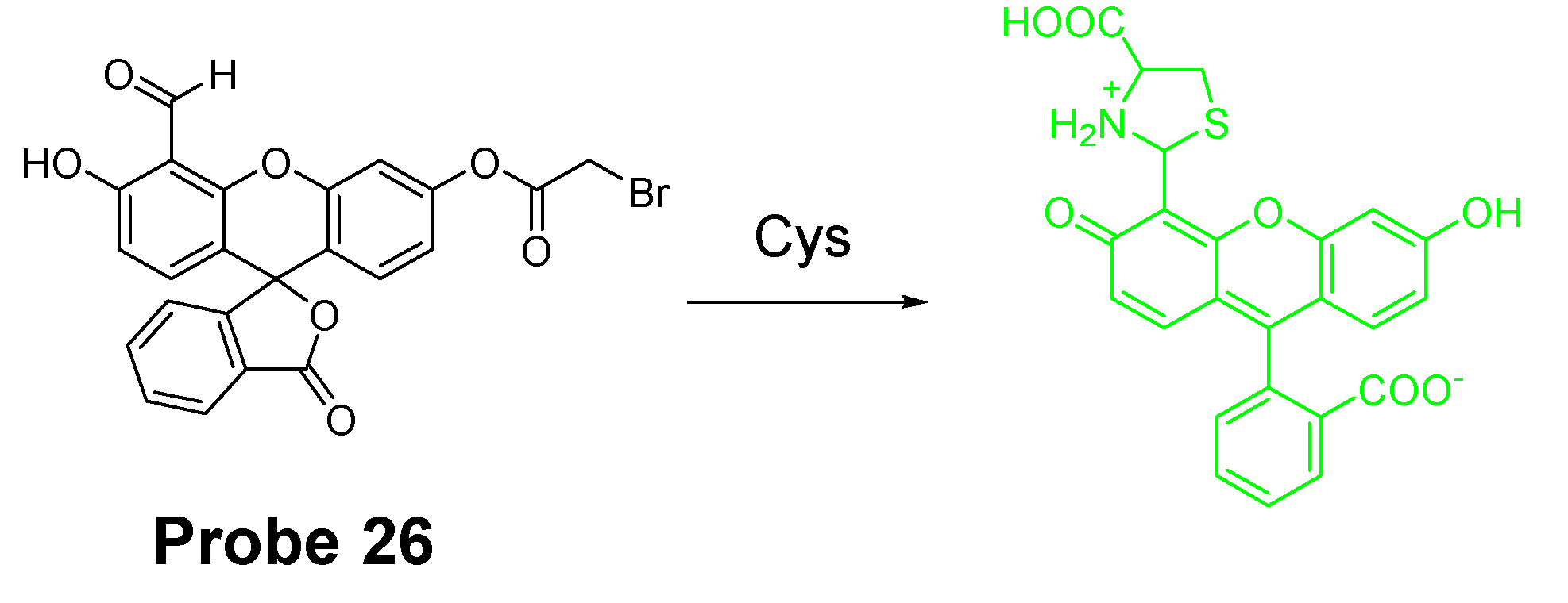
| Probe 26 | cyclization of Cys/Hcy with bromoacetylfluorescein monoaldehyde | Whole cells (Cys selective) | Cys (0.51 μM) | 114 |
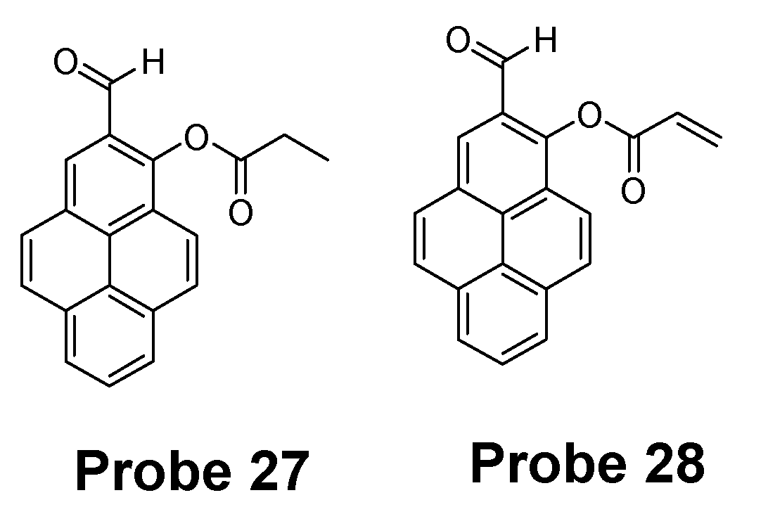
| Probes 27, 28 | cyclization | Whole cells (Hcy selective) | Hcy (P-Hcy-1, 1.94 × 10⁻⁶ M; P-Hcy-2, 1.44 × 10⁻⁷ M) | 43 |
| Probe 29 | Cys-induced SNAr substitution−rearrangement reaction | Whole cells (Cys selective) | Cys (2.12 × 10⁻⁷ M) | 117 |
| Probe 30 | Cys-induced SNAr substitution−rearrangement reaction | Whole cells (Cys selective) | Cys (5.52 × 10⁻⁷ M) | 118 |
| Probe 31 | Cys-induced SNAr substitution−rearrangement reaction | Whole cells (Cys/Hcy selective) | Cys (2.00 × 10⁻⁸ M) Hcy (1.02 × 10⁻⁸ M) | 120 |
| Probe 32 | Cys-induced SNAr substitution−rearrangement reaction | Whole cells (Cys selective) | Cys (22 nM) | 121 |
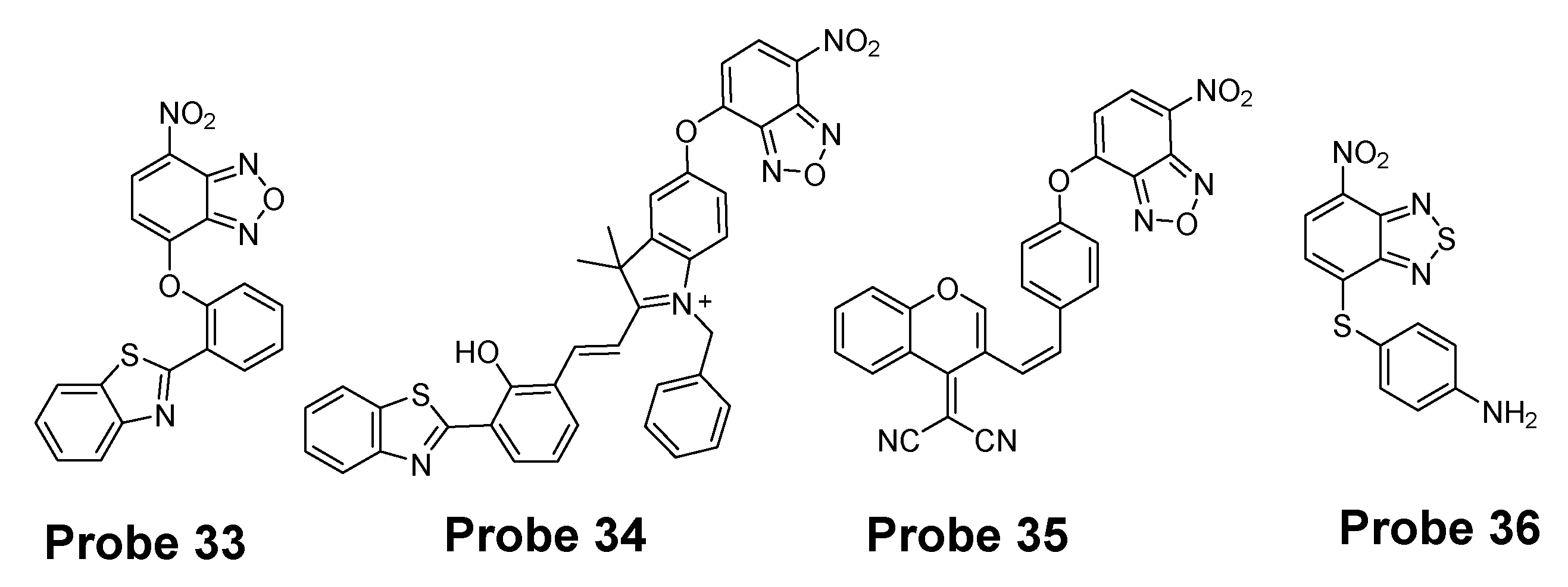
| Probe 33 | Cys-induced SNAr substitution−rearrangement reaction | Whole cells, discrimination of Cys and GSH | Cys (0.08 μM) GSH (0.06 μM) | 124 |
| Probe 34 | Cys-induced SNAr substitution−rearrangement reaction | Whole cells, discrimination of Cys/Hcy, GSH, and H2S | GSH (4.30 μM) Cys (4.25 μM) Hcy (5.11 μM) H2S (6.74 μM) | 125 |
| Probe 35 | Cys-induced SNAr substitution−rearrangement reaction | Whole cells, discriminate Cys/Hcy from GSH | Cys (2.1 × 10⁻⁸ M) Hcy 1.7 × 10⁻⁸ M; GSH (2.6 ×10⁻⁸ M) | 126 |
| Probe 36 | Cys-induced SNAr substitution−rearrangement reaction | Whole cells, (Cys/Hcy selective) | Cys and Hcy (0.1 μM) | 127 |
| Probe 37 | Cys-induced SNAr substitution−rearrangement reaction | Whole cells, (Cys selectvie) | Cys (0.3 μM) | 128 |
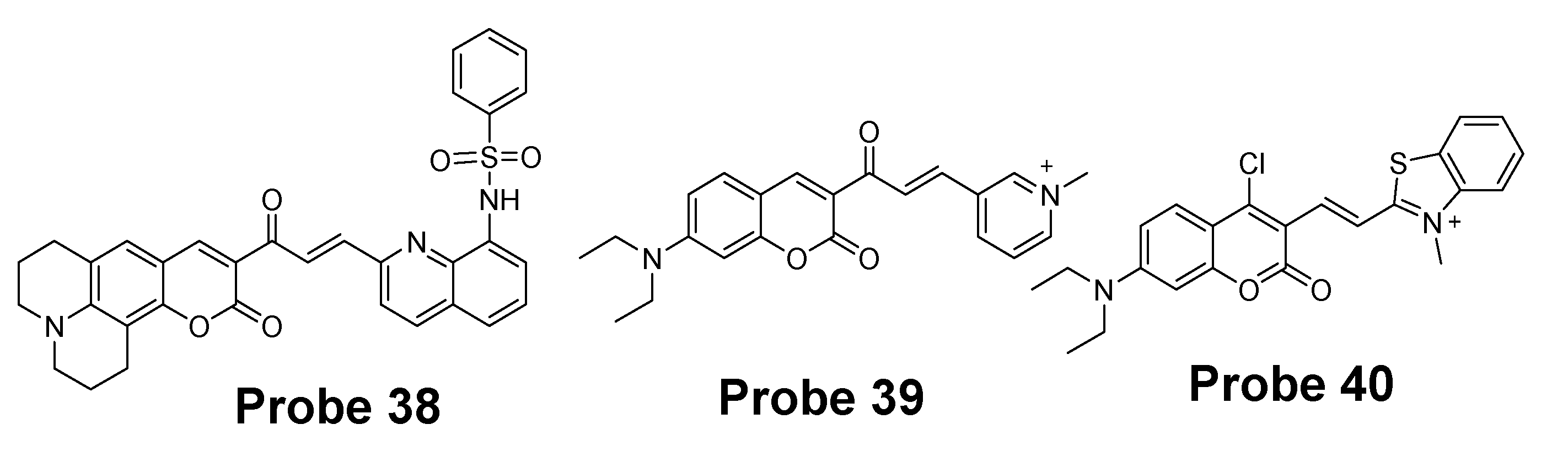
| Probe 38 | Michael addition reaction in combination with a steric hinderance factor. | Whole cells (Cys selective) | Cys (10−7 M) | 129 |
| Probe 39 | Michael addition assisted by an electrostatic attraction. | Whole cells (Cys selective) | Cys (25 nM) | 130 |
| Probe 40 | SNAr substitution−rearrangement reaction | Whole cells simultaneously detects Cys and GSH | Cys (>0.4 μM) GSH (0.05 μM) | 102 |
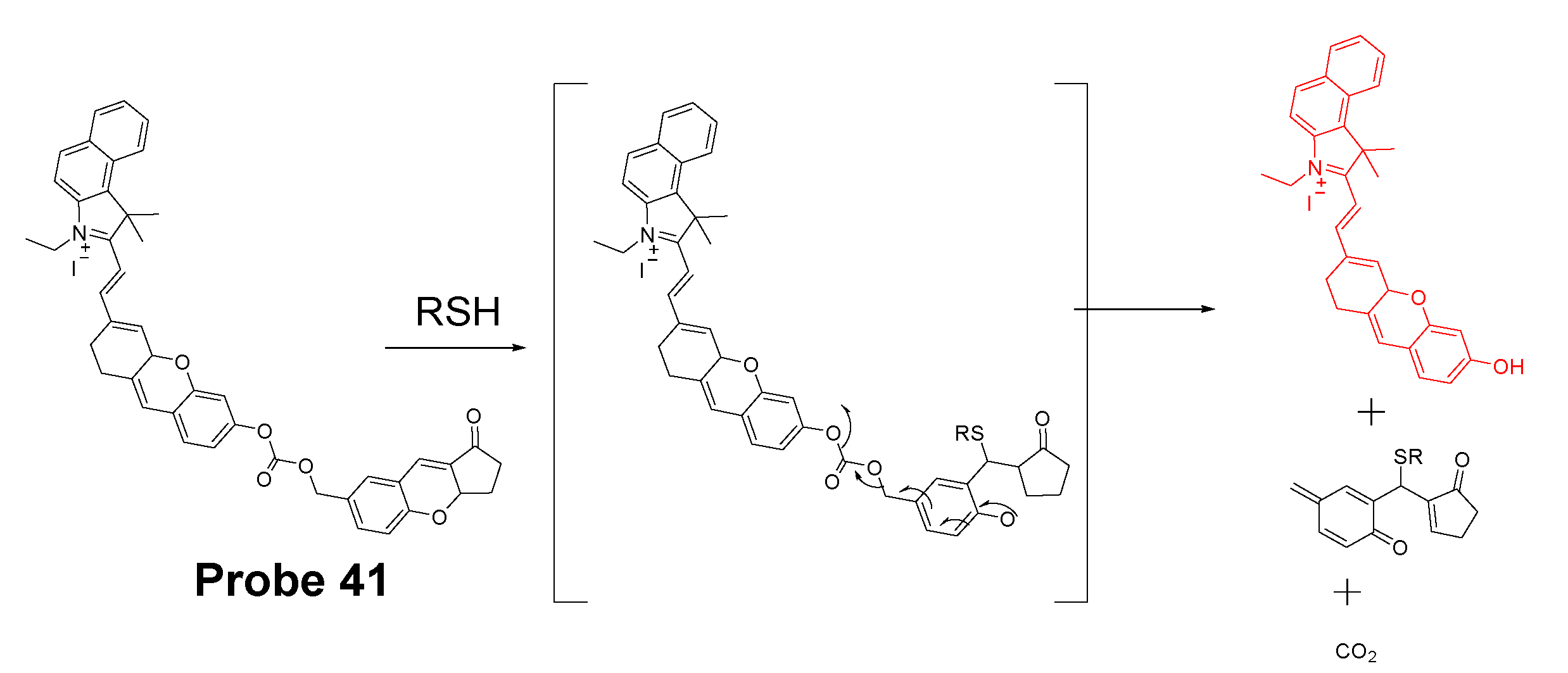
| Probe 41 | Michael addition followed by self-immolative reaction | Mitochondrial thiols | GSH (0.59 μM) Cys (0.39 μM) Hcy (0.54 μM) | 137 |
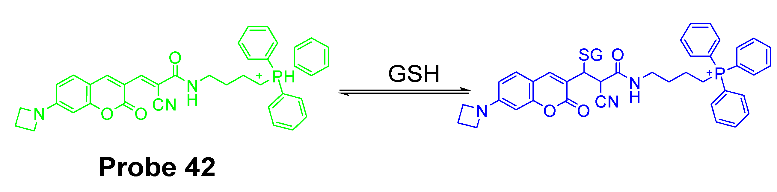
| Probe 42 | Michael addition | Mitochondrial GSH | NA | 138 |
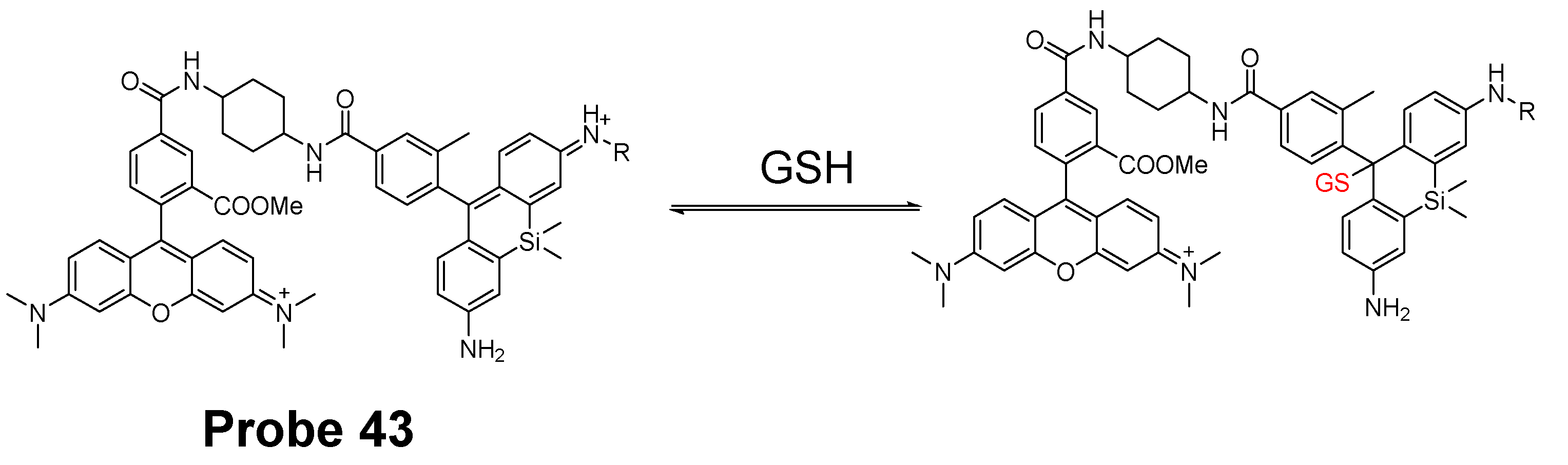
| Probe 43 | Michael addition | Mitochondrial GSH | NA | 139 |
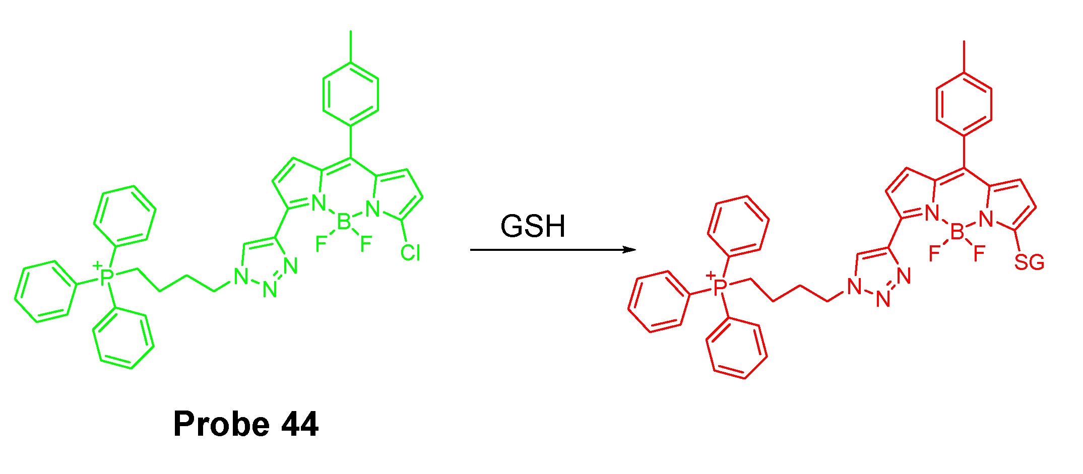
| Probe 44 | SNAr reaction | Mitochondrial GSH | GSH (1.1 μM) | 140 |
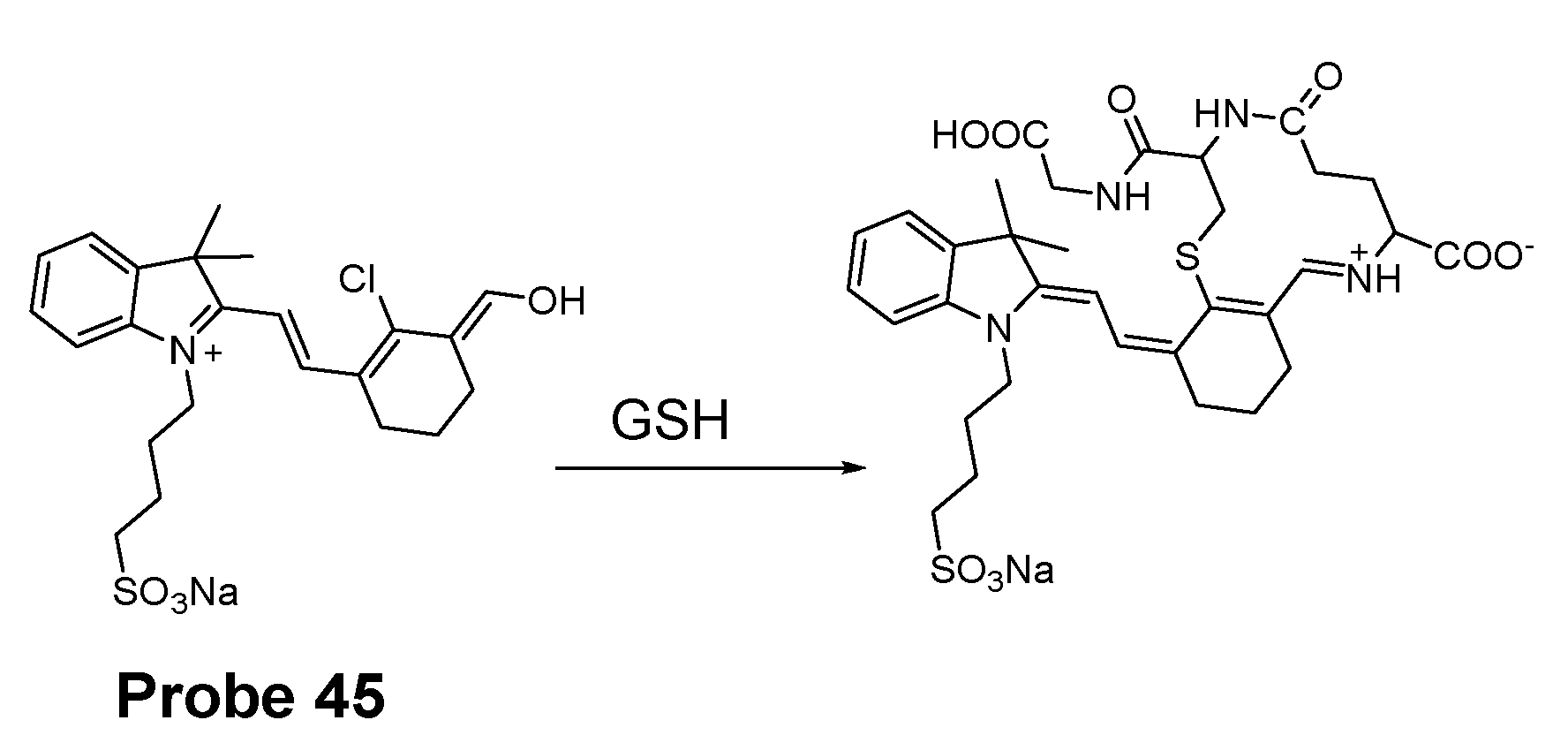
| Probe 45 | SNAr reaction | Mitochondrial GSH | GSH (24.16 μM) | 141 |
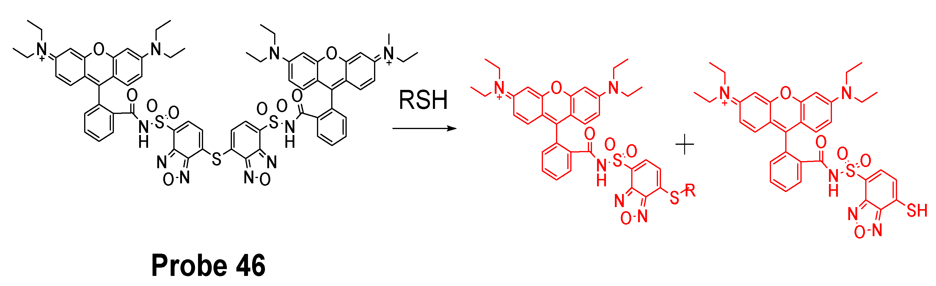
| Probe 46 | Thiol–sulfide exchange reaction | Mitochondrial thiols | NA | 22 |
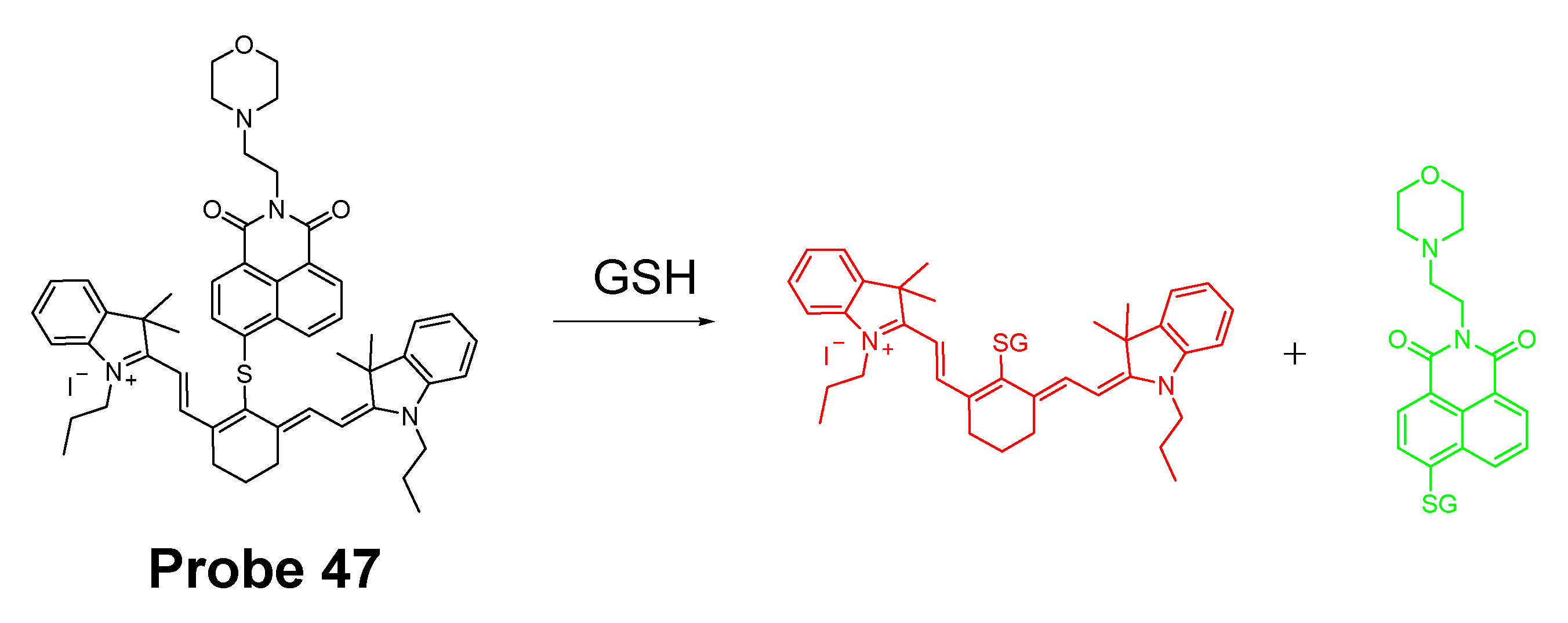
| Probe 47 | SNAr reaction | Mitochondrial and lysosomal GSH | GSH (1 nM) | 143 |
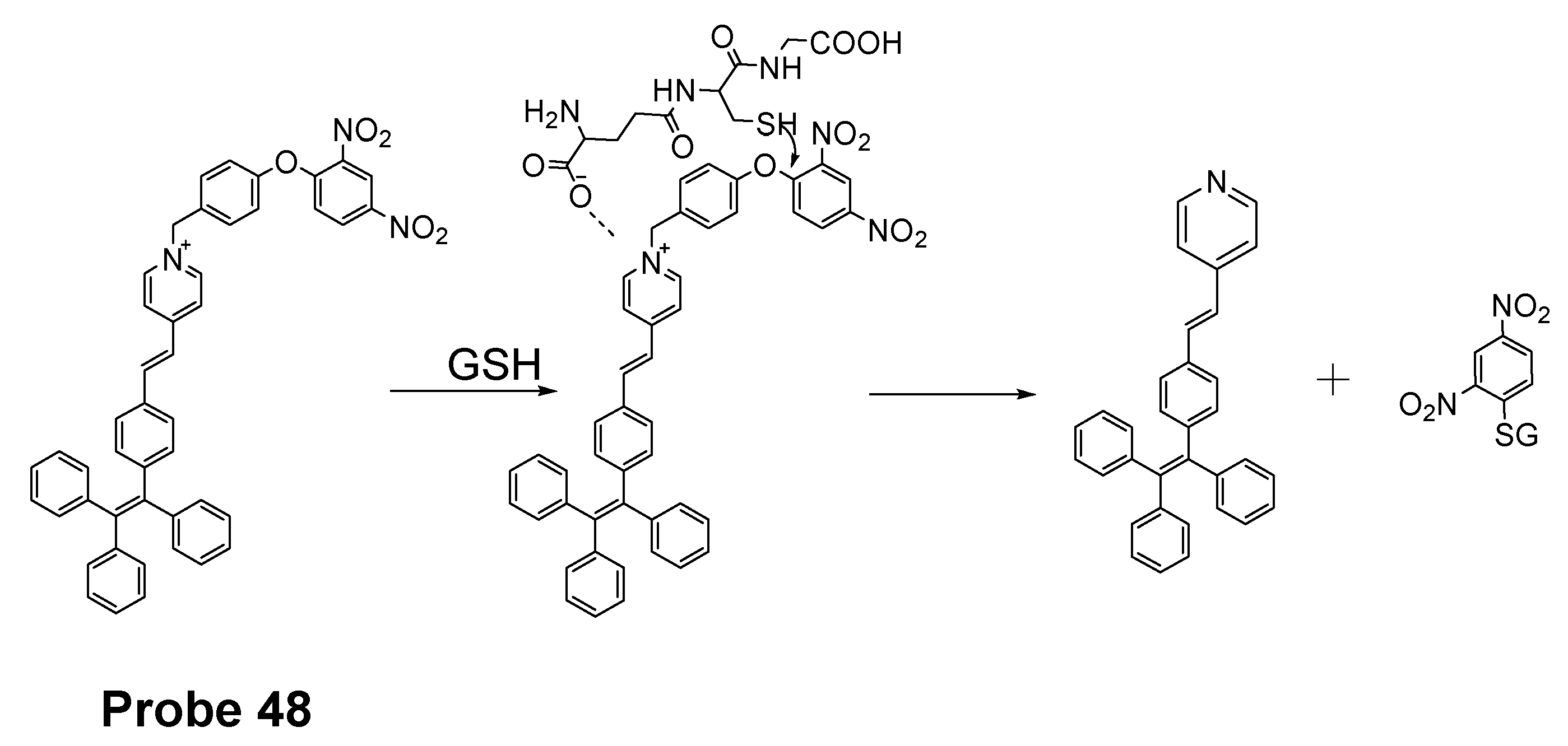
| Probe 48 | SNAr reaction | Mitochondrial thiols | GSH (0.61 μM). | 35 |
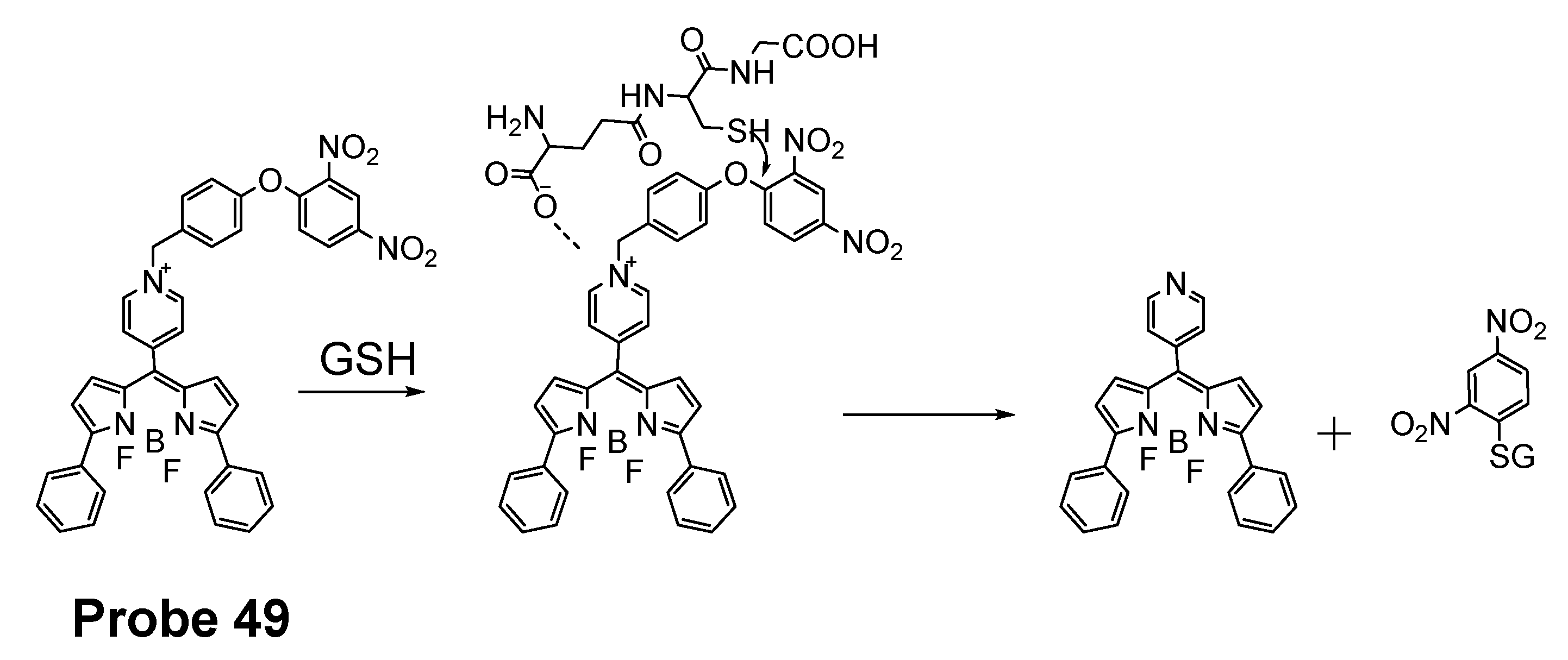
| Probe 49 | SNAr reaction | Mitochondrial GSH | GSH (109 nM) | 160 |
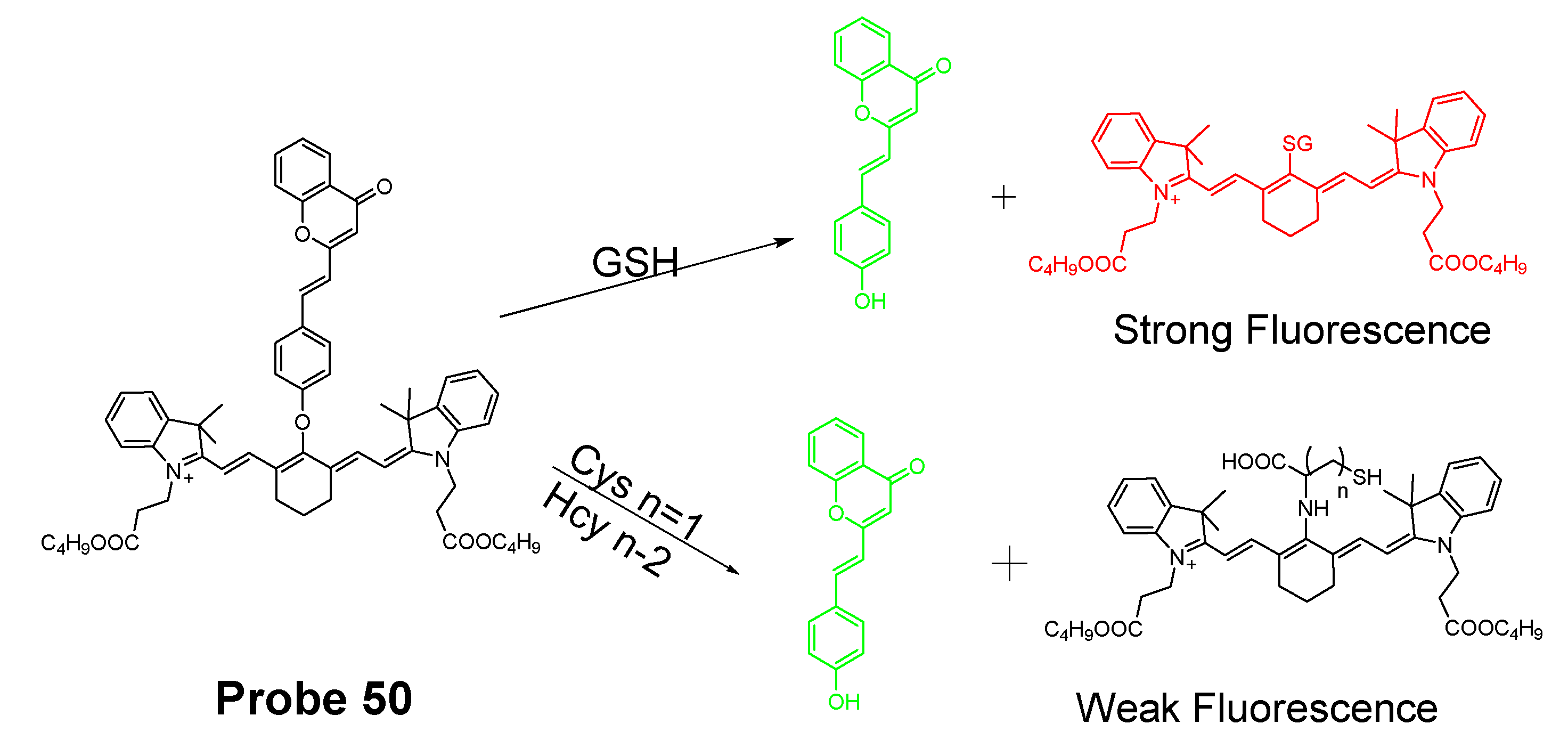
| Probe 50 | SNAr reaction | Mitochondrial GSH | GSH (24 nM, visible) and (32 nM, NIR) | 1 |
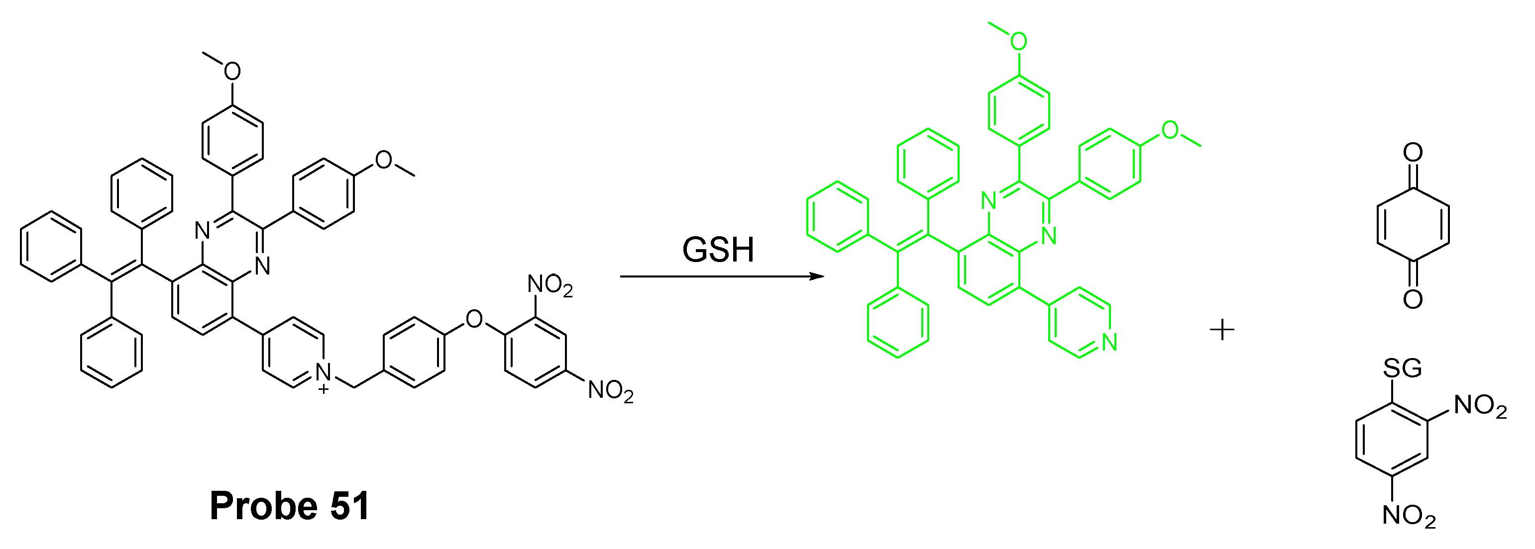
| Probe 51 | Cleavage of the dinitrophenyl ether | Mitochondrial GSH | GSH (434 nM) | 161 |
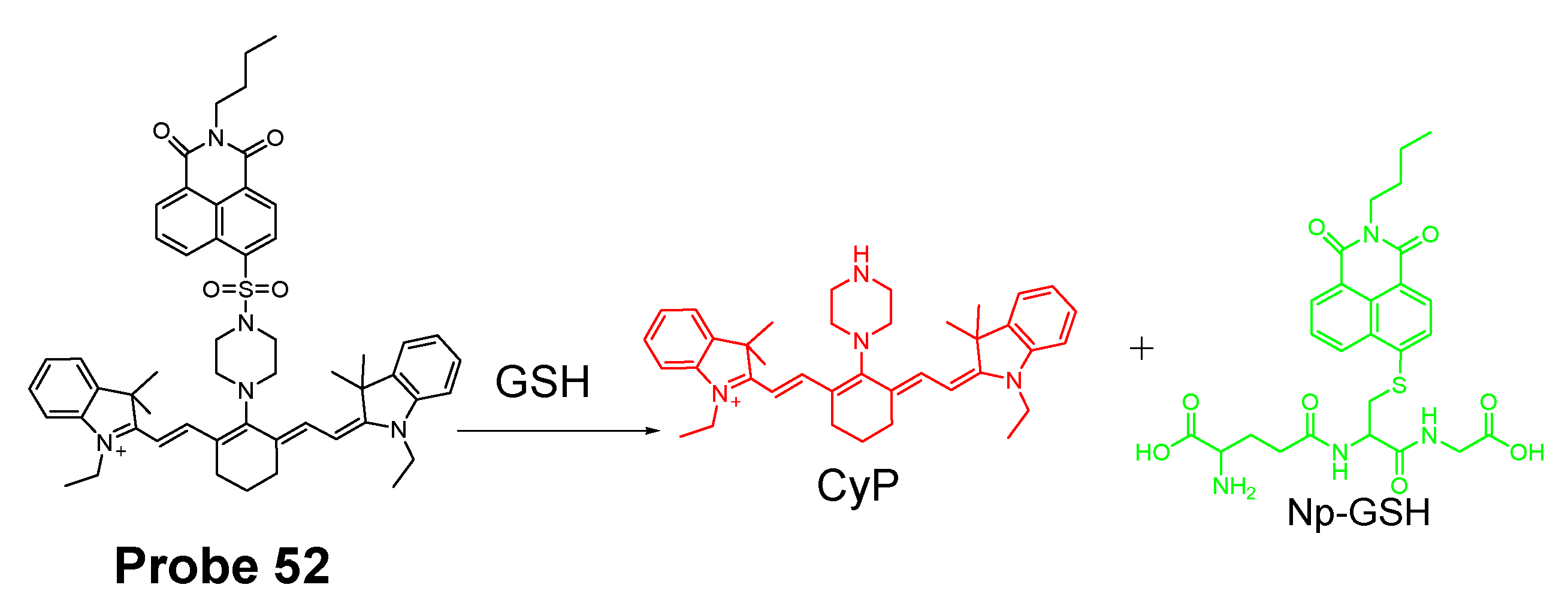
| Probe 52 | cleavage of sulfonamide | Mitochondrial GSH | GSH (1.53 × 10⁻⁷ M, visible channel) and (1.71 × 10⁻⁷ M, NIR channel) | 162 |
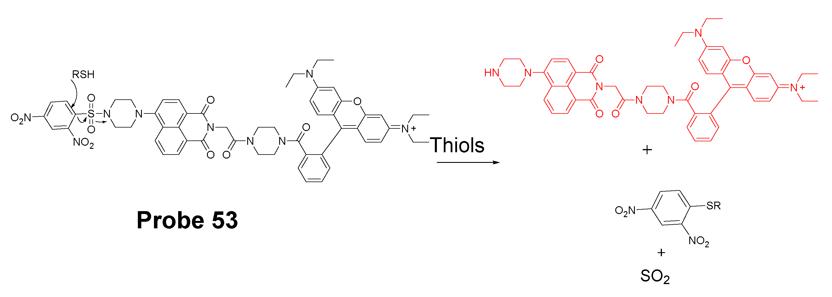
| Probe 53 | cleavage of sulfonamide | Mitochondrial thiols | GSH (0.89 μM); Cys (0.47 μM), Hcy (2.4 μM) | 164 |
| Probe 54 | cleavage of sulfonate ester | Mitochondrial thiols | Hcy (87 nM) Cys (147 nM) GSH (129 nM) | 167 |
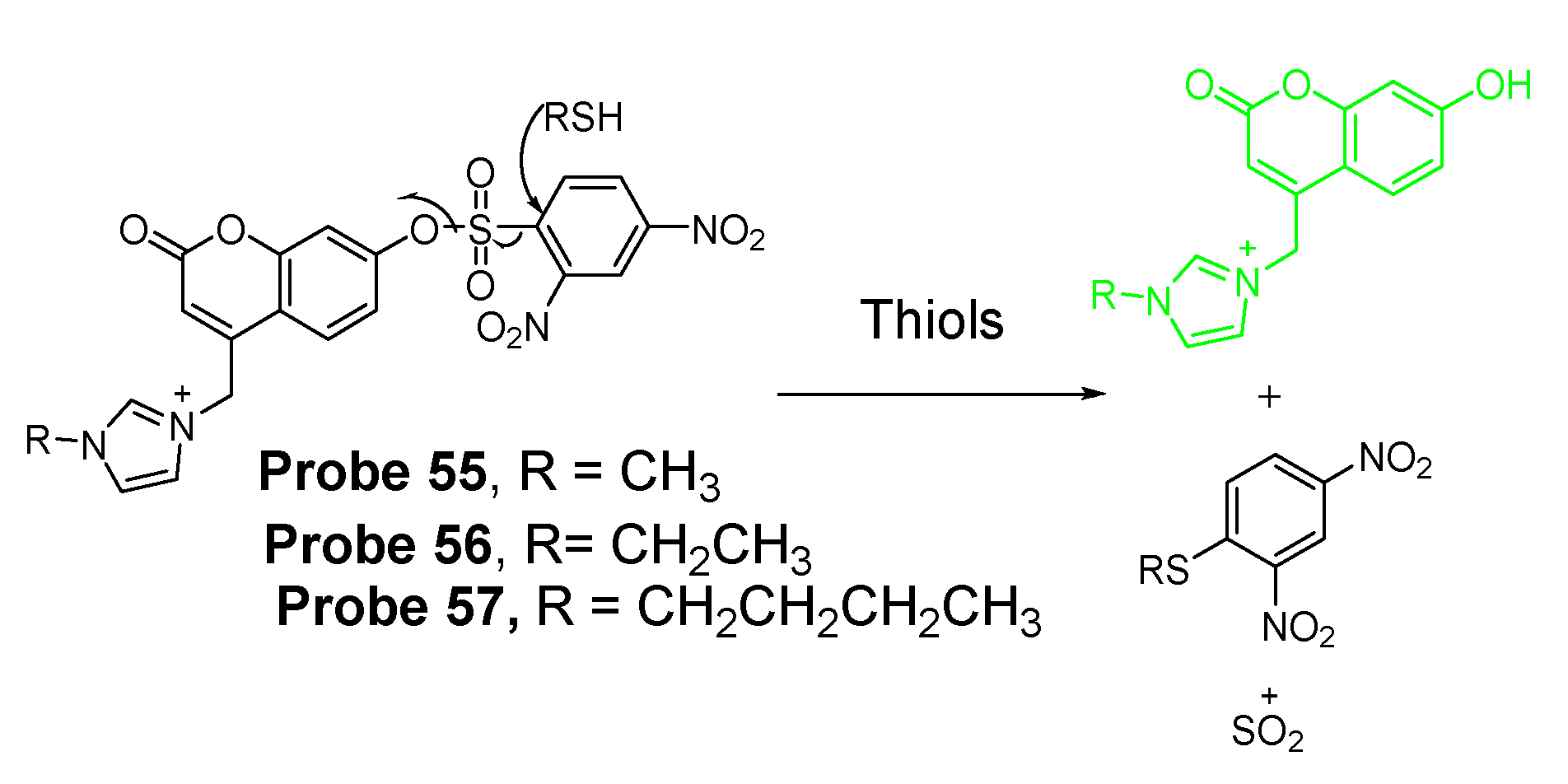
| Probes 55, 56, 57 | cleavage of sulfonate ester | Mitochondrial thiols | GSH (Probe 55, 31.4 nM; Probe 56, 29.2 nM; Probe 57, 29.6 nM) | 171 |
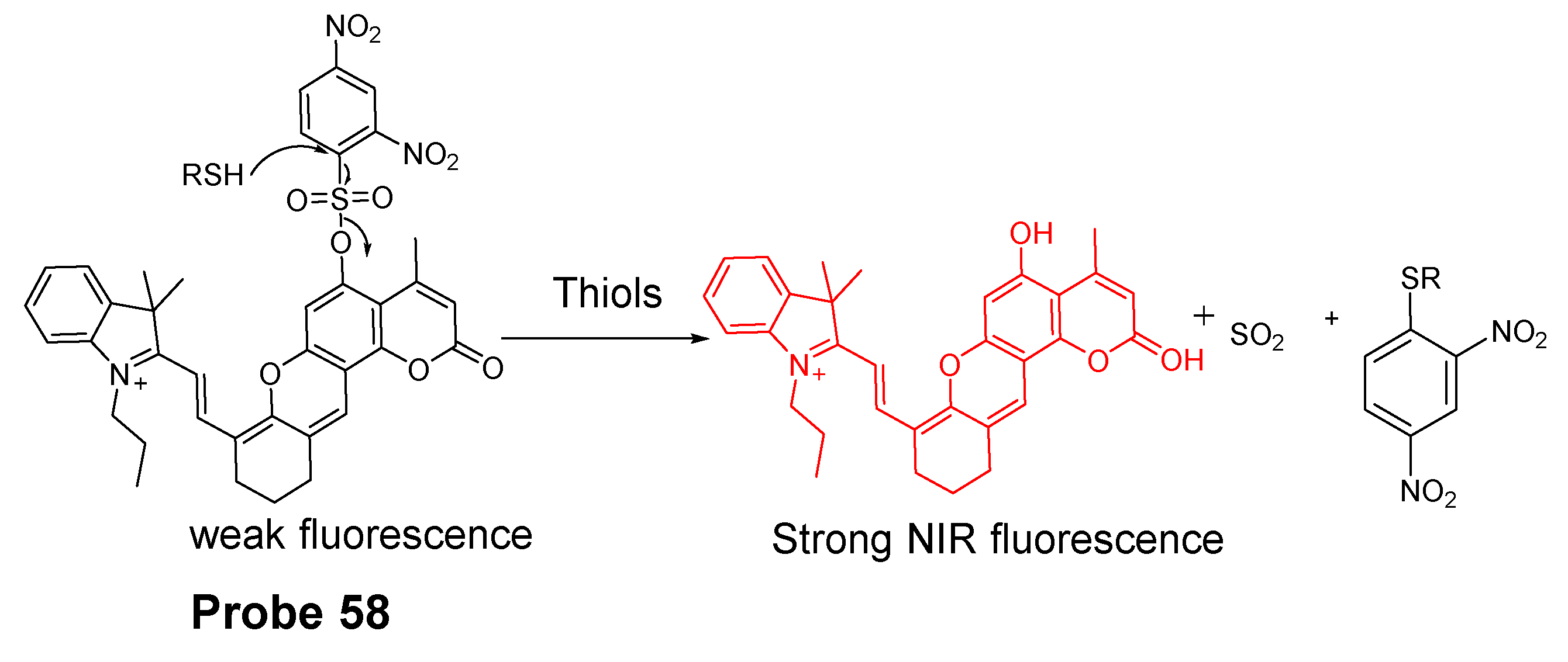
| Probe 58 | cleavage of sulfonate ester | Mitochondrial thiols | GSH (0.11μM) Cys (0.08 μM) Hcy (0.20 μM) | 172 |
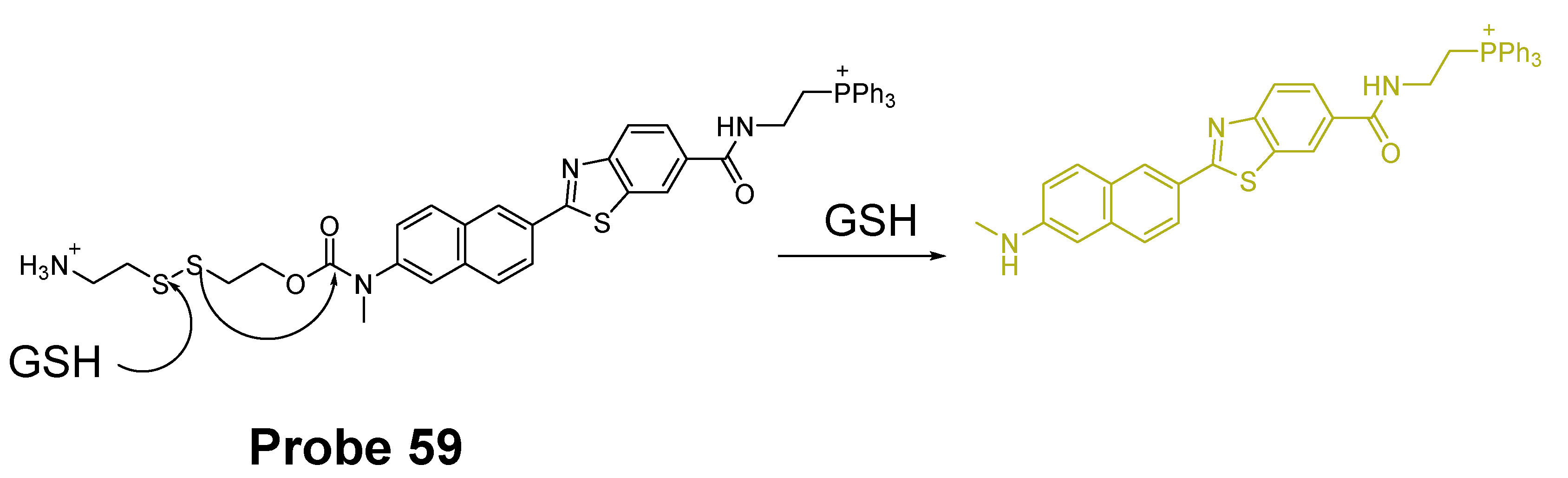
| Probe 59 | cleavage of disulfide bond | Mitochondrial thiols | NA | 173 |
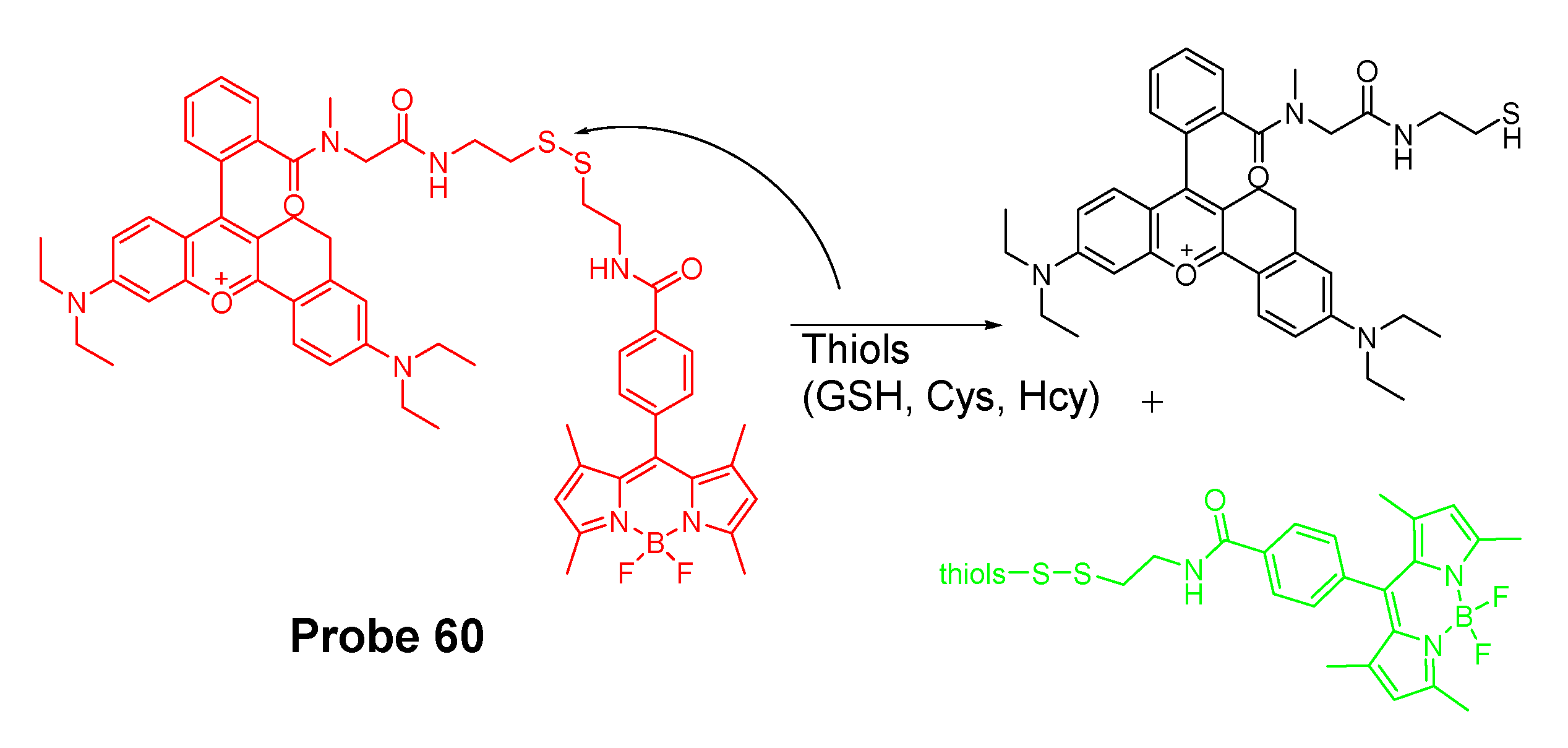
| Probe 60 | cleavage of disulfide bond | Mitochondrial thiols | GSH (0.26 μM) | 174 |
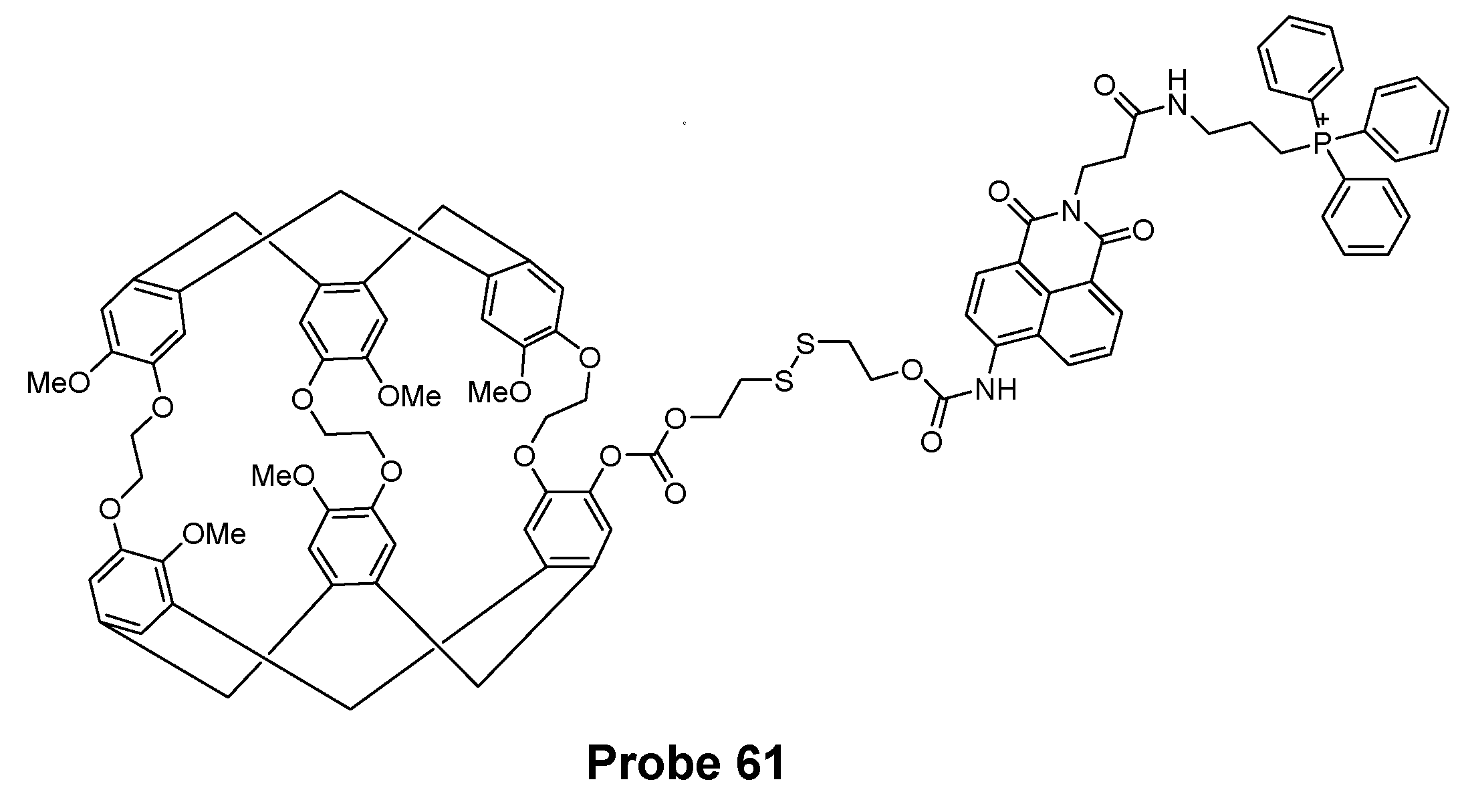
| Probe 61 | cleavage of disulfide bond | Mitochondrial thiols | GSH (10−10 M, using Hyper-CEST NMR) | 178 |
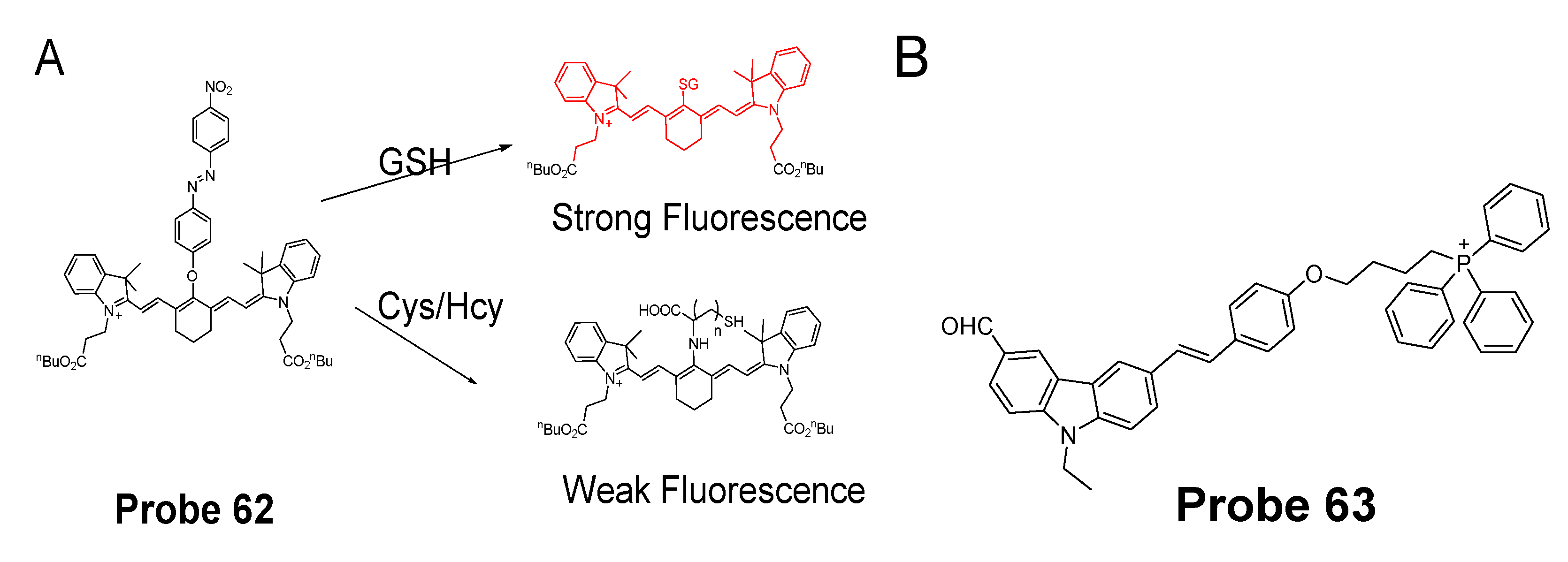
| Probe 62 | 1, 6-conjugate addition and subsequent elimination reaction | Mitochondrial GSH | GSH (26 nM) | 179 |
| Probe 63 | Others | Mitochondrial thiols | Cys (0.2 μM) | 180 |
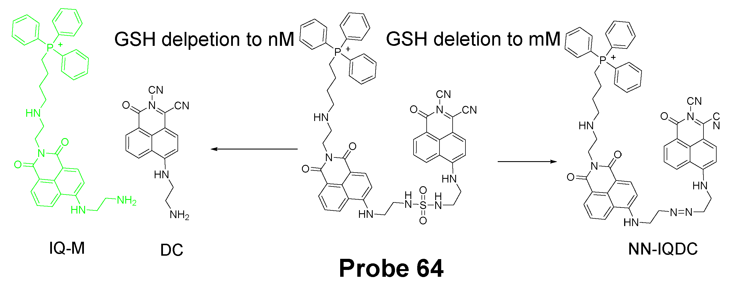
| Probe 64 | cleavage of sulfonamide for ultratrace change of GSH | Mitochondrial GSH | GSH (2.02 nM) | 181 |
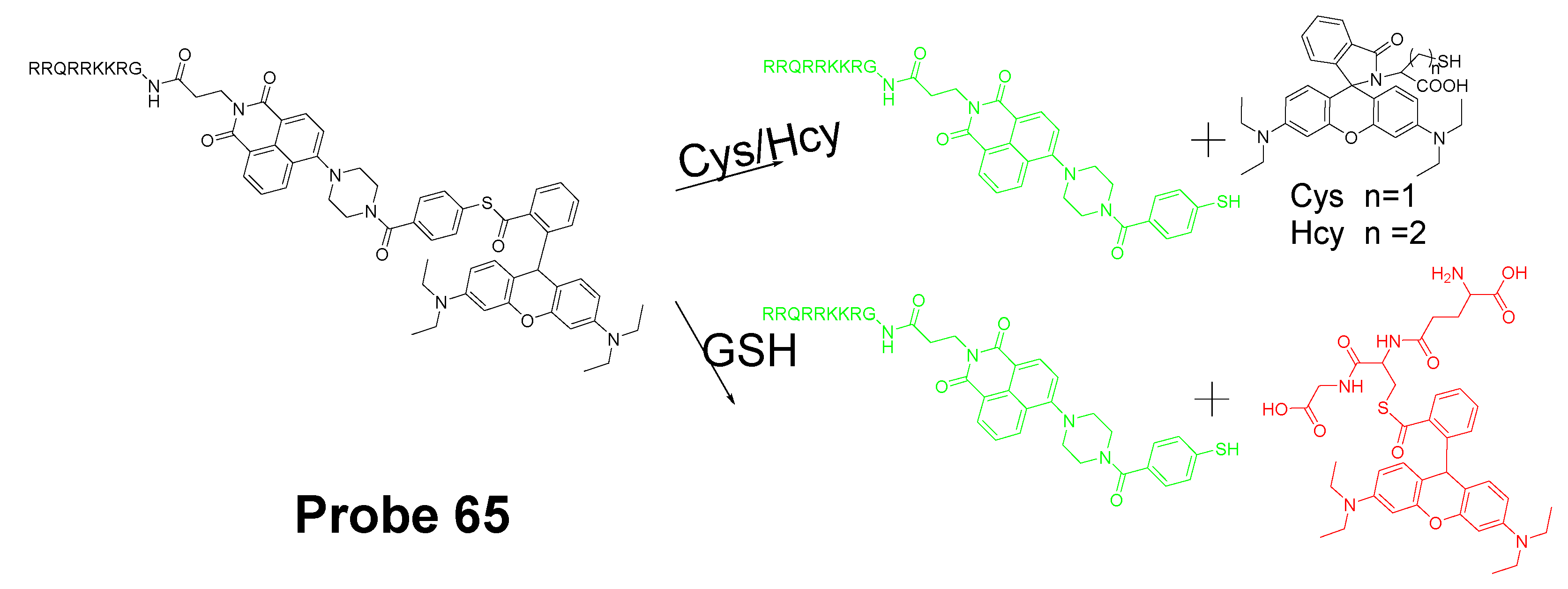
| Probe 65 | Others | Mitochondrial GSH | GSH (5.15 μM) Cys (0.865 μM) Hcy (6.51 μM) | 135 |
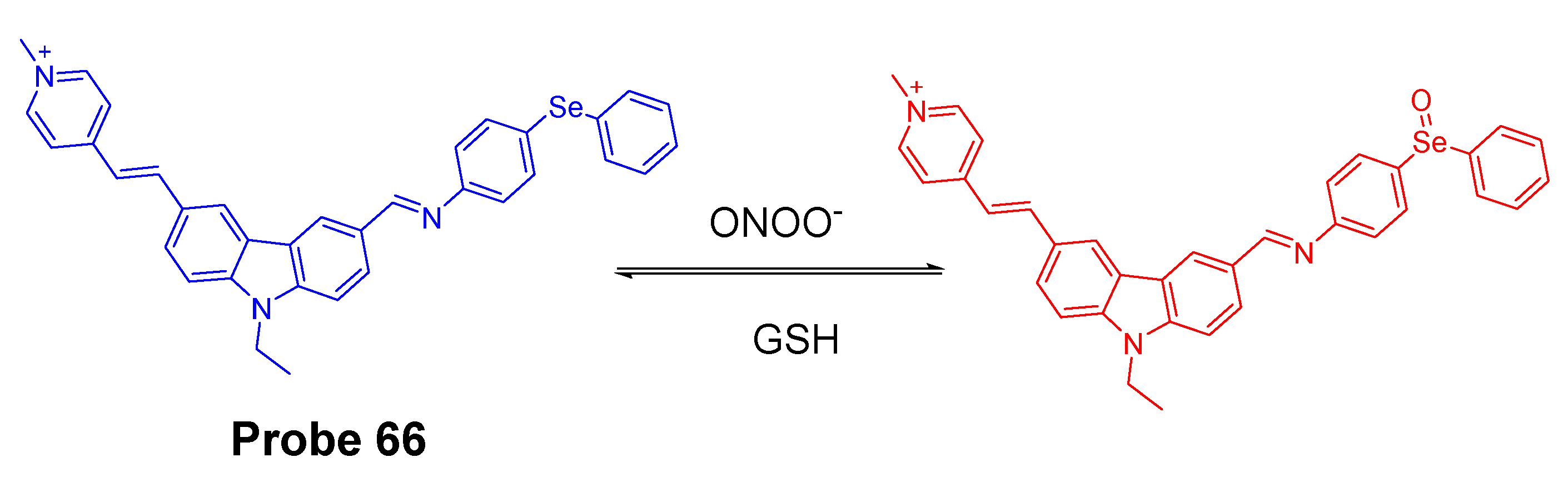
| Probe 66 | Others | Mitochondrial ONOO−/GSH levels | NA | 186 |
| Probe 67 | Others | Mitochondrial redox potential | NA | 187 |
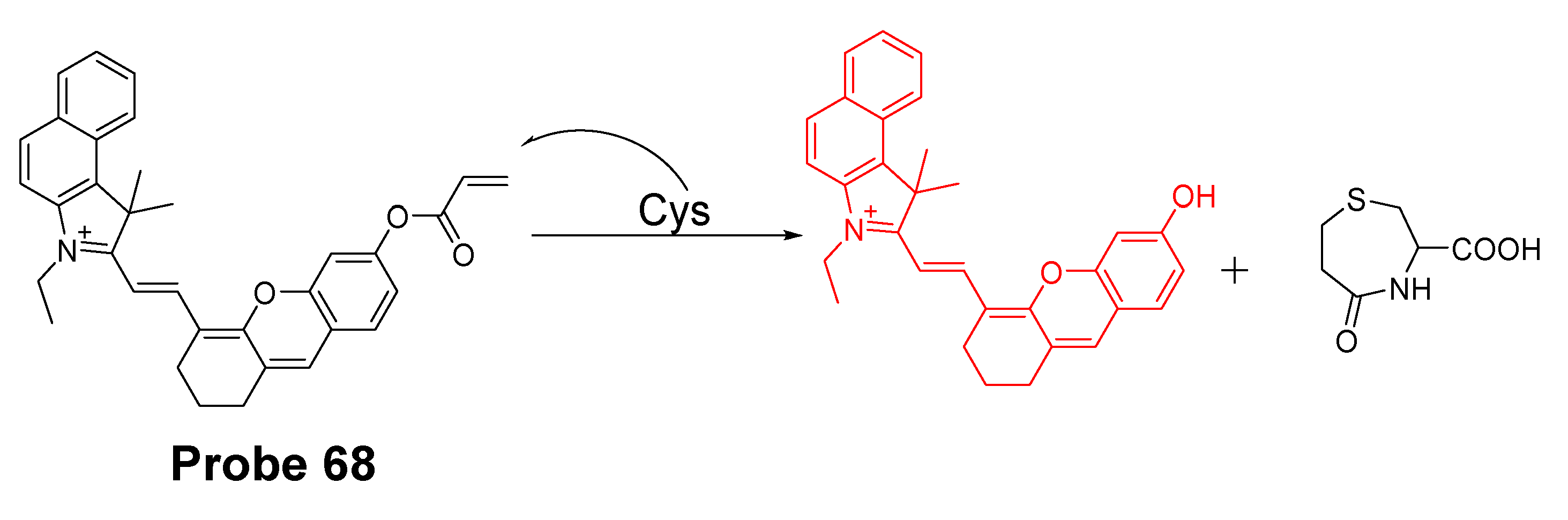
| Probe 68 | cyclization of Cys with acrylates or aldehydes | Mitochondrial Cys | Cys (14.5 nM) | 188 |
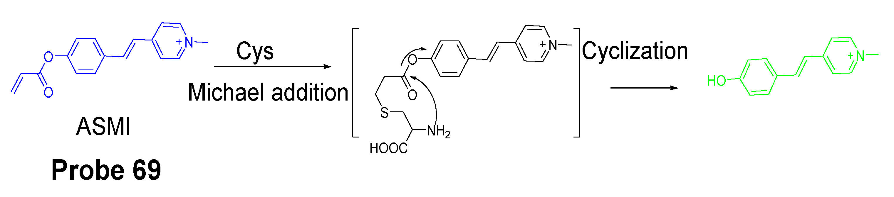
| Probe 69 | cyclization of Cys with acrylates or aldehydes | Mitochondrial Cys | NA | 112 |
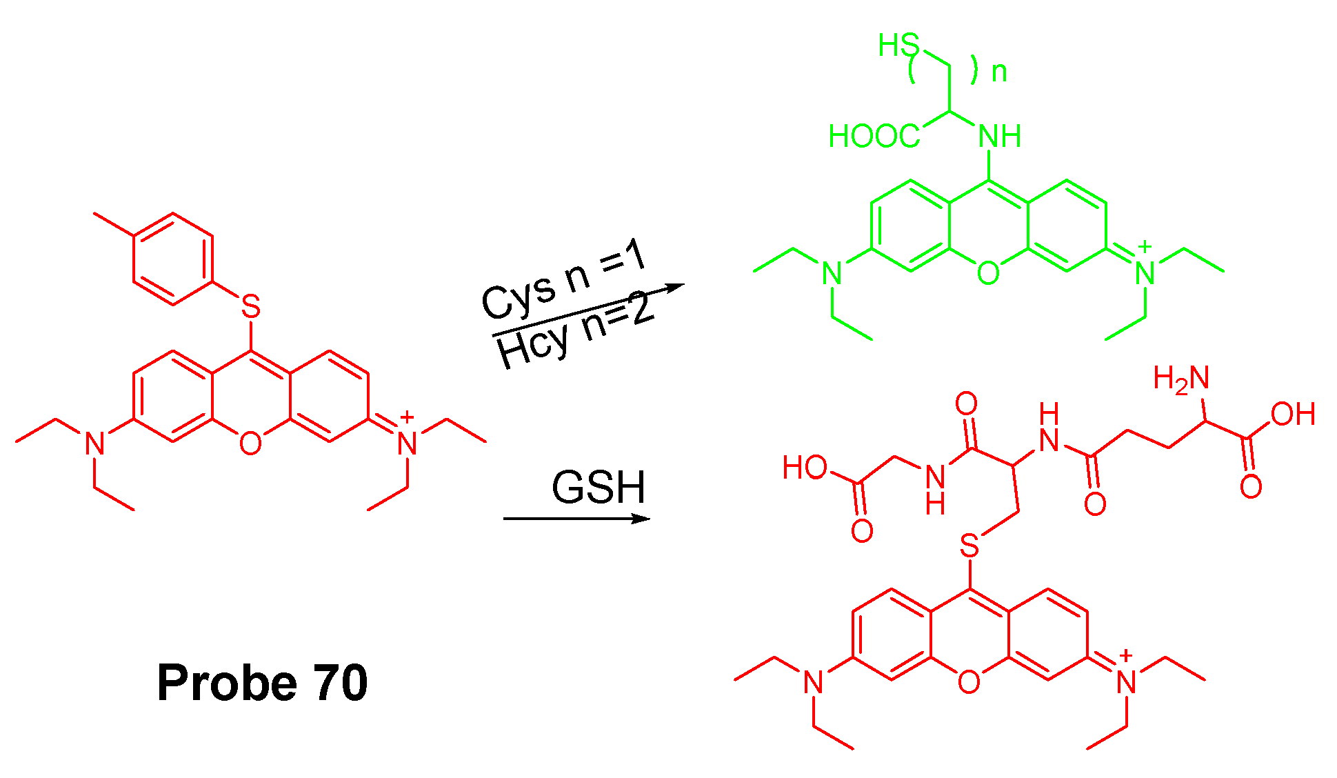
| Probe 70 | Cys-induced SNAr substitution−rearrangement reaction | Mitochondrial Cys/Hcy | Cys (22 nM) Hcy (23 nM) | 192 |
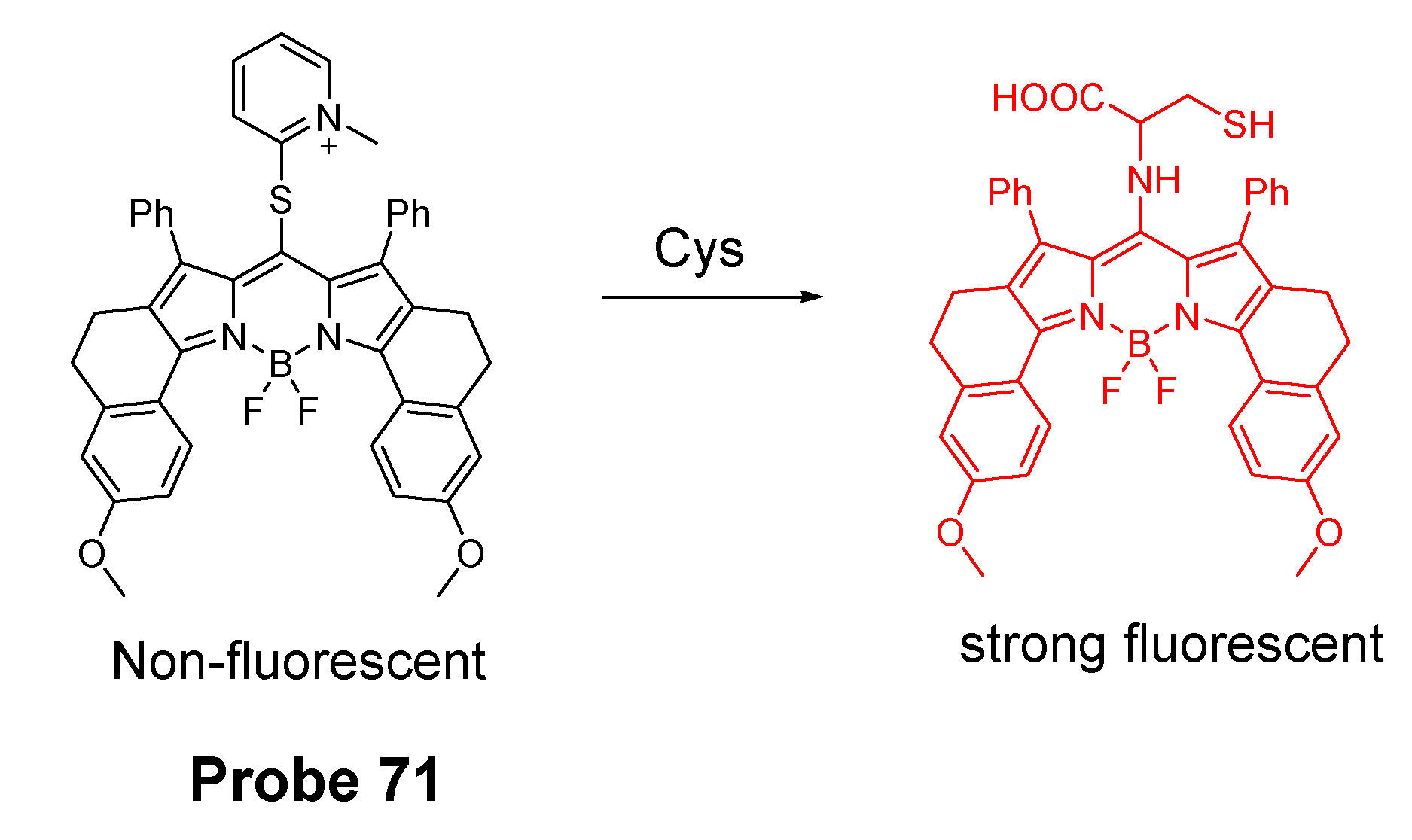
| Probe 71 | SNAr substitution−rearrangement reaction | Mitochondrial Cys | Cys (72 nM) | 193 |
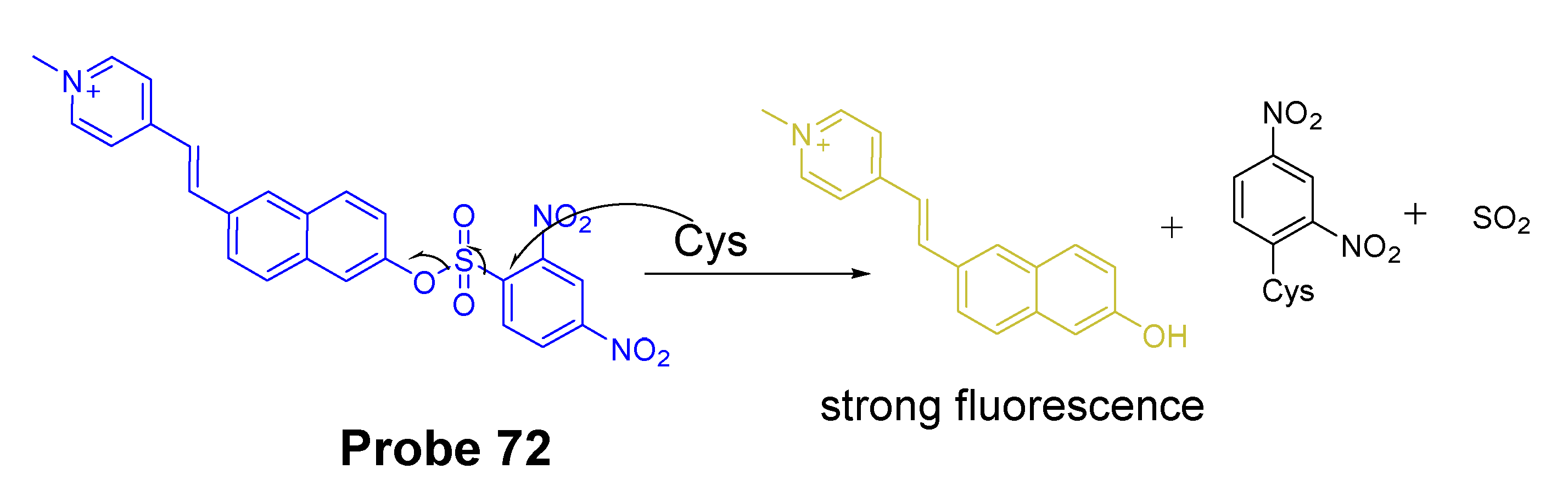
| Probe 72 | cleavage of sulfonamide | Mitochondrial Cys | Cys (0.29 μM) | 194 |
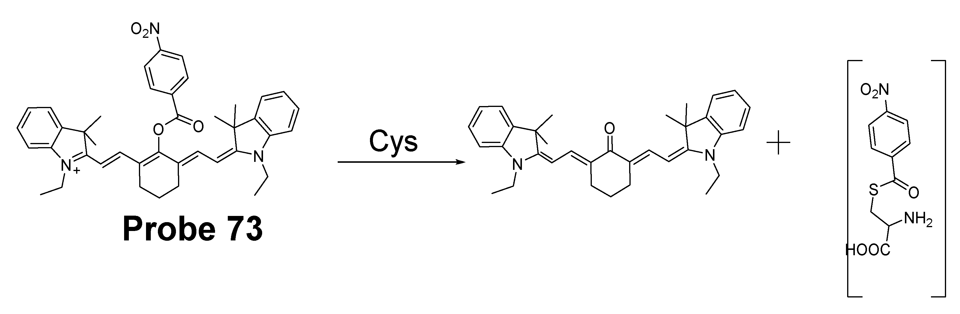
| Probe 73 | Others | Mitochondrial Cys | Cys (0.2 μM) | 195 |
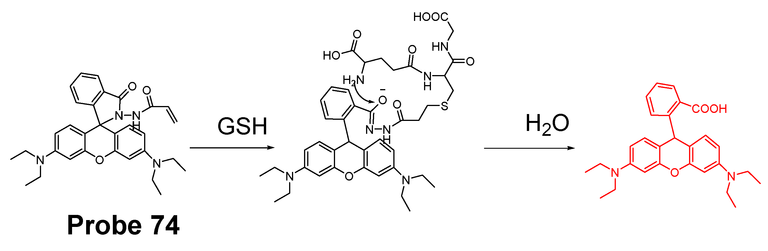
| Probe 74 | Michael addition | Lysosomal GSH | GSH (190 nM) | 203 |
| Probe 75 | SNAr reactions | Lysosomal thiols | GSH (16 nM) Cys (27 nM) Hcy (33 nM) | 204 |
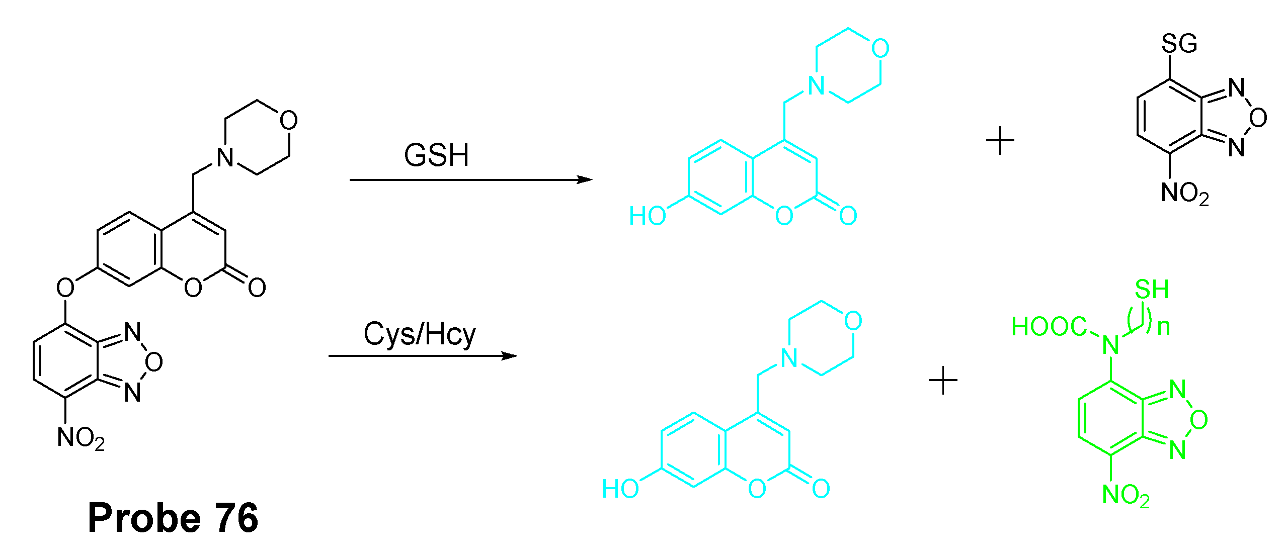
| Probe 76 | SNAr reactions | Lysosomal thiols | GSH (3.9 × 10⁻⁸ M); Cys (3.3 × 10⁻⁸ M); Hcy (5.2 × 10 −8 M) | 205 |
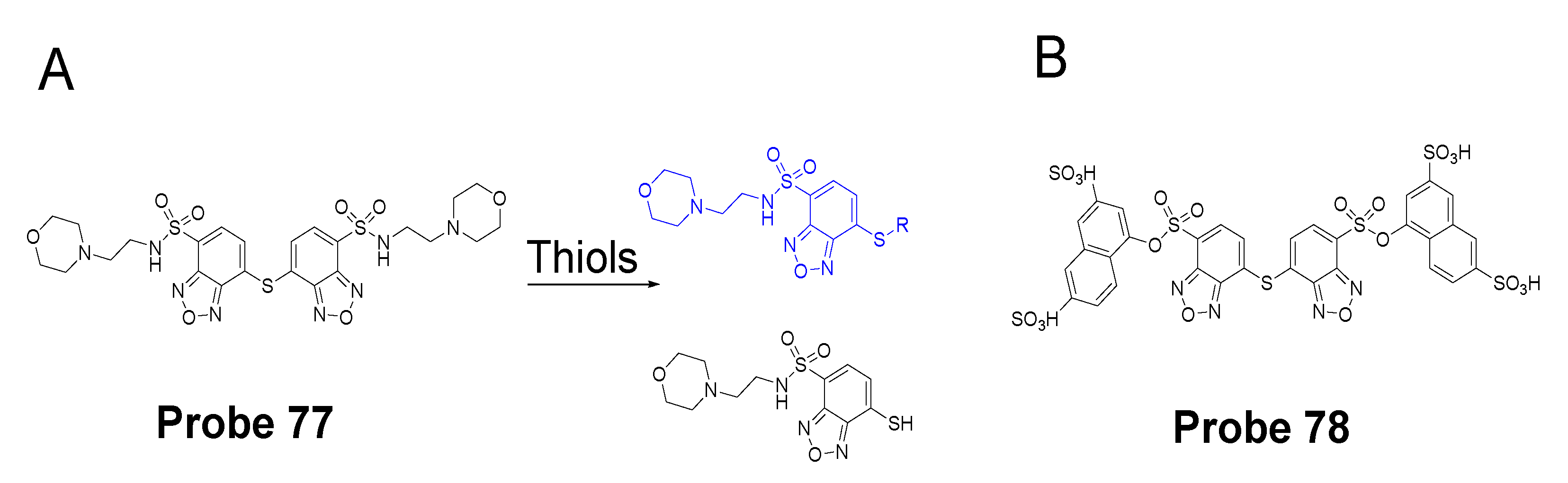
| Probe 77 | Thiol–sulfide exchange reaction | Lysosomal thiols | NA | 206 |
| Probe 78 | Thiol–sulfide exchange reaction | Lysosomal thiols | NA | 207 |
| Probe 79 | cleavage of sulfonamide | Lysosomal GSH | NA | 208 |
| Probe 80 | cleavage of sulfonamide | Lysosomal thiols | GSH (2.41 × 10⁻⁶ M); Cys (2.6 × 10⁻⁷ M); Hcy (4.87 × 10⁻⁶ M) | 165 |
| Probe 81 | cleavage of sulfonate | Lysosomal thiols | GSH (62 nM); Cys (146 nM); Hcy (115 nM) | 209 |
| Probe 82 | cleavage of disulfide | Lysosomal GSH | GSH (0.15 μM) | 210 |
| Probe 83 | others | Lysosomal GSH | GSH (1.03 μM) | 211 |
| Probes 84, 85 | Cys-induced SNAr substitution followed by a intramolecular rearrangement | Lysosomal Cys | Cys (46 nM for Lyso-S and 76 nM for Lyso-D) | 212 |
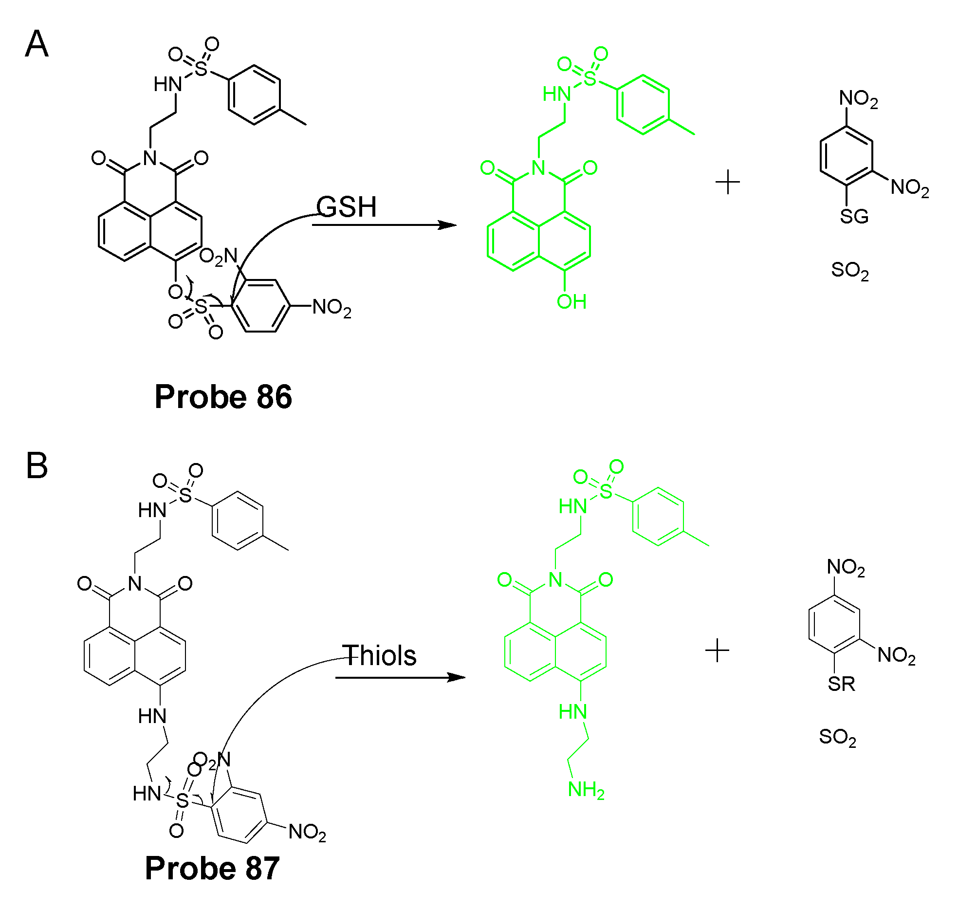
| Probe 86 | cleavage of sulfonate | Endoplasmic reticulum GSH | NA | 215 |
| Probe 87 | cleavage of sulfonamide | Endoplasmic reticulum thiols | GSH (4.70 × 10⁻⁶ M); Cys (1.67 × 10⁻⁷ M); Hcy (9.62 × 10⁻⁷ M) | 214 |
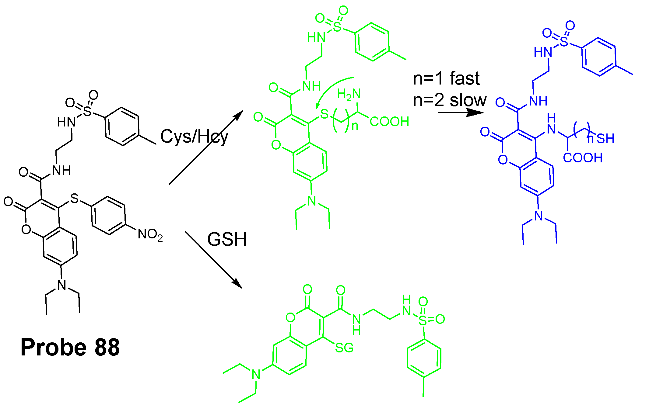
| Probe 88 | SNAr substitution | Endoplasmic reticulum thiols | GSH (23 nM), Cys (14 nM), Hcy (16 nM) | 216 |
Publisher’s Note: MDPI stays neutral with regard to jurisdictional claims in published maps and institutional affiliations. |
© 2021 by the authors. Licensee MDPI, Basel, Switzerland. This article is an open access article distributed under the terms and conditions of the Creative Commons Attribution (CC BY) license (https://creativecommons.org/licenses/by/4.0/).
Share and Cite
Wang, S.; Huang, Y.; Guan, X. Fluorescent Probes for Live Cell Thiol Detection. Molecules 2021, 26, 3575. https://doi.org/10.3390/molecules26123575
Wang S, Huang Y, Guan X. Fluorescent Probes for Live Cell Thiol Detection. Molecules. 2021; 26(12):3575. https://doi.org/10.3390/molecules26123575
Chicago/Turabian StyleWang, Shenggang, Yue Huang, and Xiangming Guan. 2021. "Fluorescent Probes for Live Cell Thiol Detection" Molecules 26, no. 12: 3575. https://doi.org/10.3390/molecules26123575
APA StyleWang, S., Huang, Y., & Guan, X. (2021). Fluorescent Probes for Live Cell Thiol Detection. Molecules, 26(12), 3575. https://doi.org/10.3390/molecules26123575