
Synthesis, Anticancer Screening of Some Novel Trimethoxy Quinazolines and VEGFR2, EGFR Tyrosine Kinase Inhibitors Assay; Molecular Docking Studies

 , , , , and
Abstract
1. Introduction
2. Results and Discussion
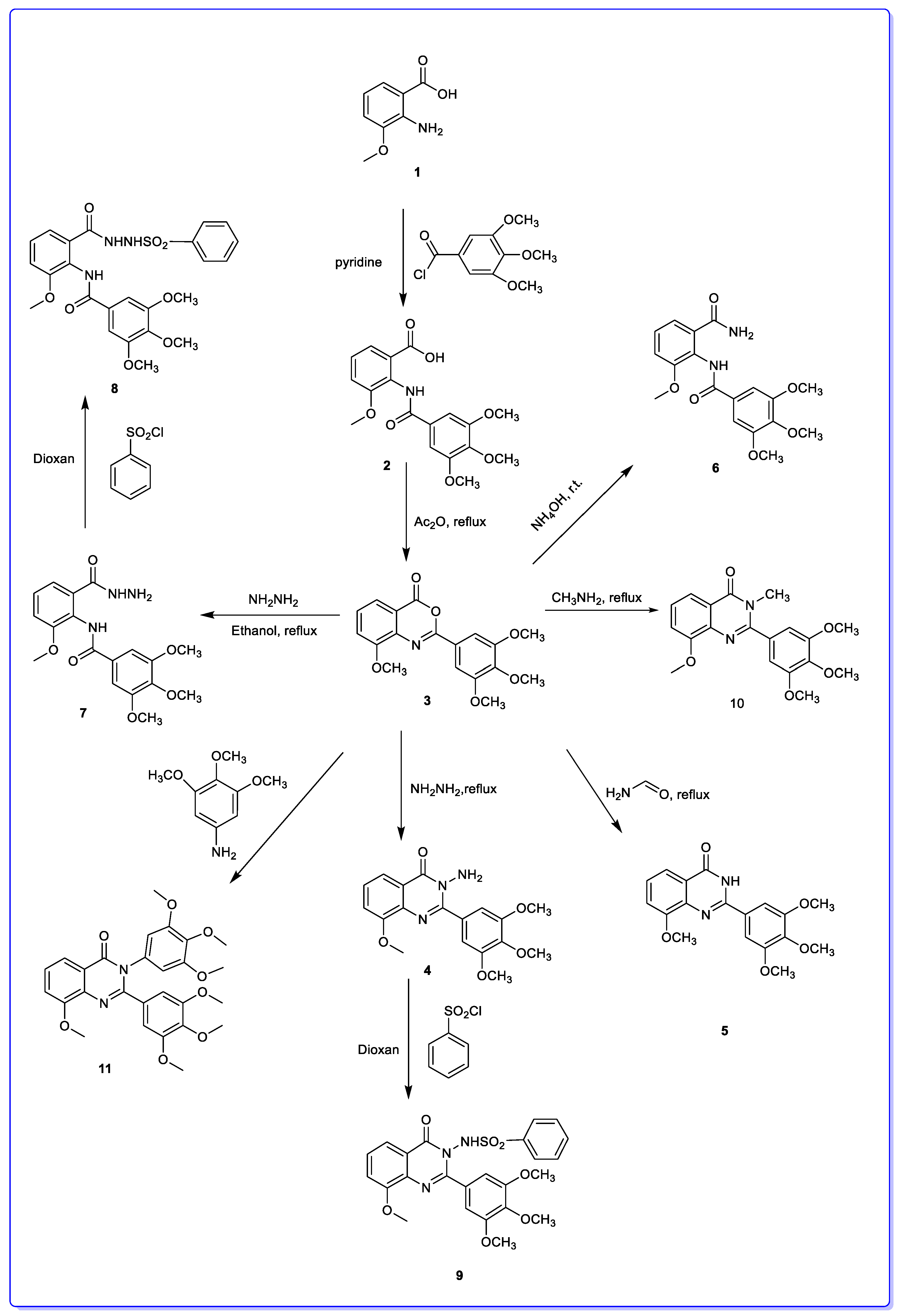
2.1. Chemistry
2.2. VEGFR2 and EGFR Inhibitory Activity
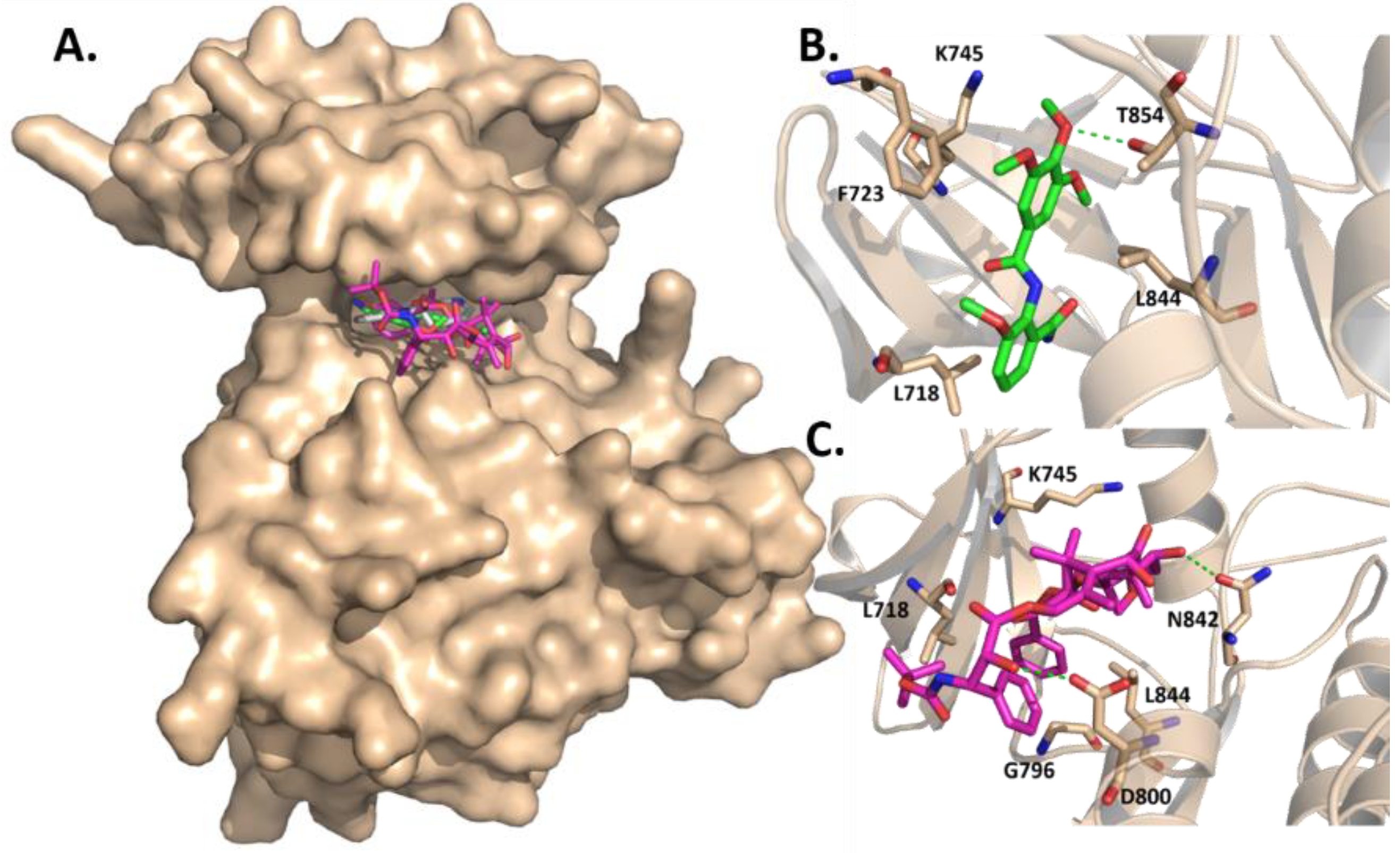
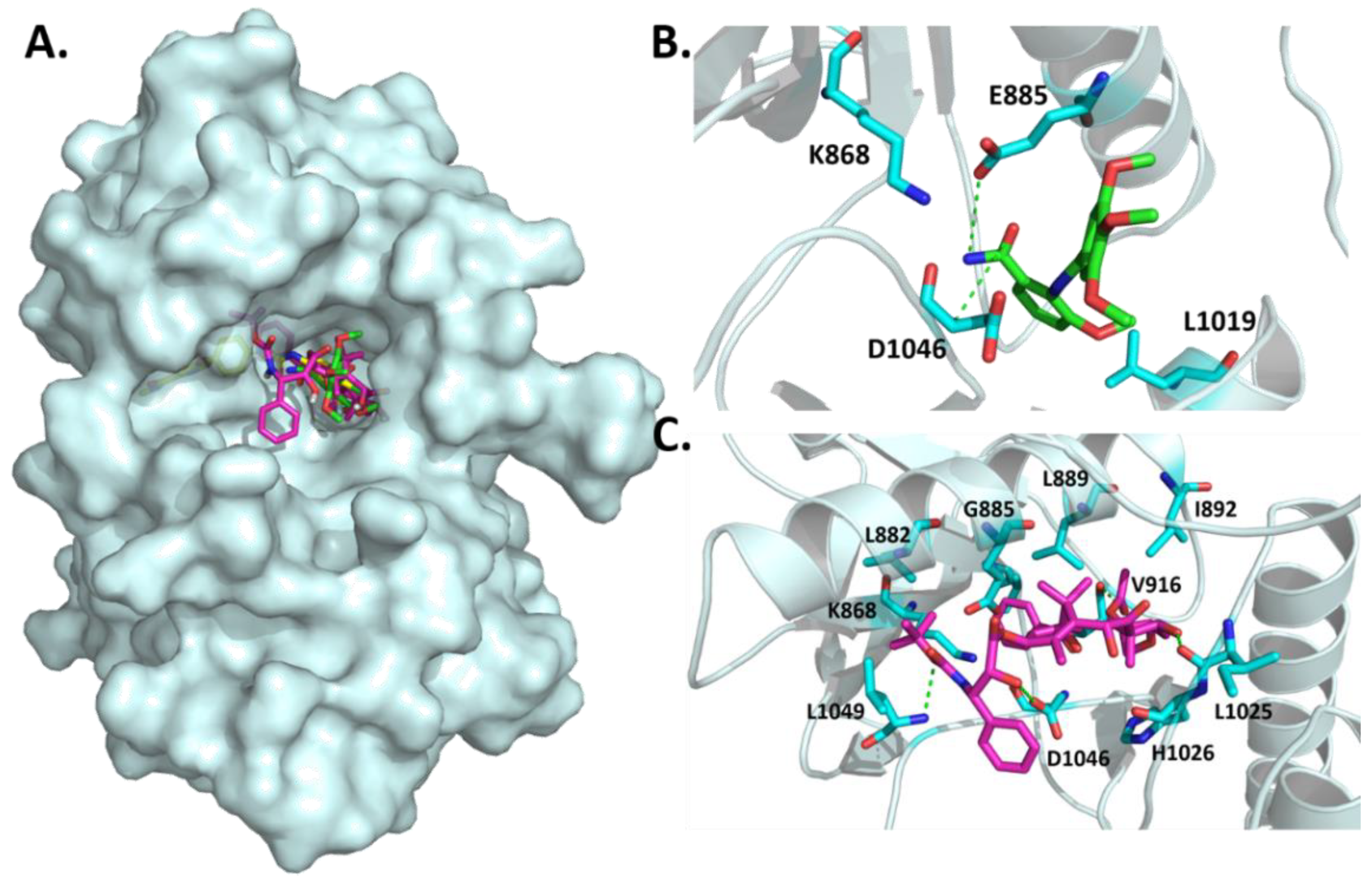
2.3. Molecular Docking Analysis
3. Materials and Methods
3.1. Chemistry
3.2. Molecular Docking
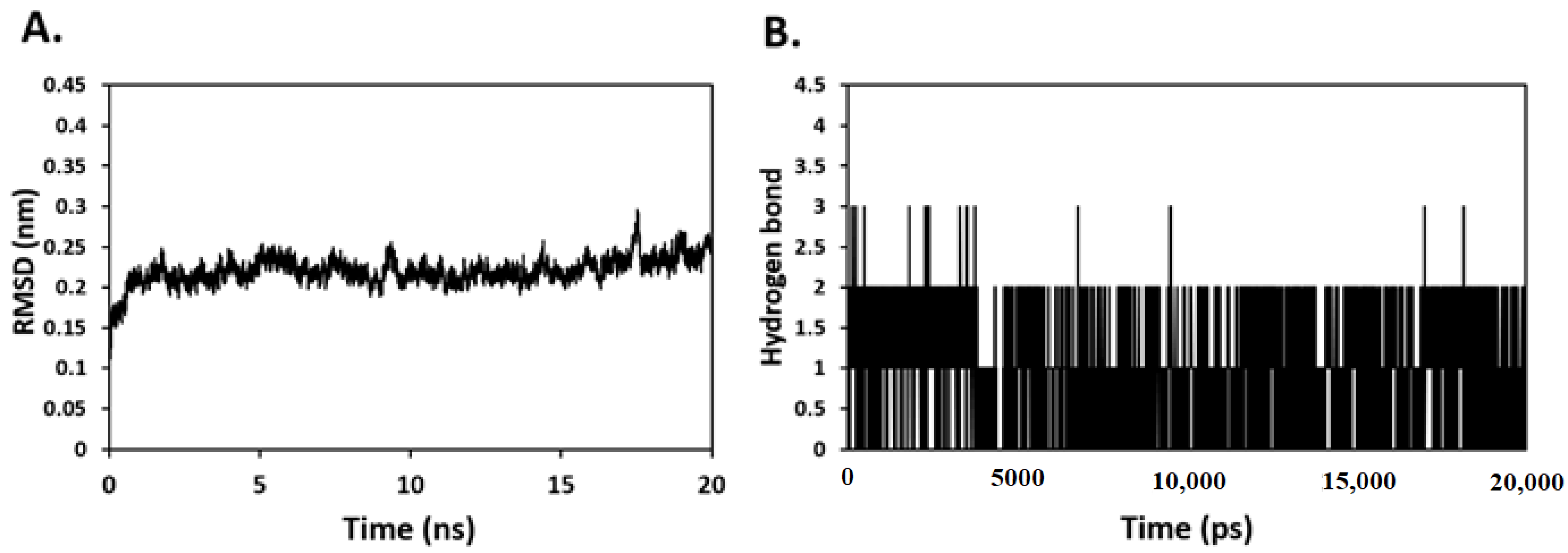
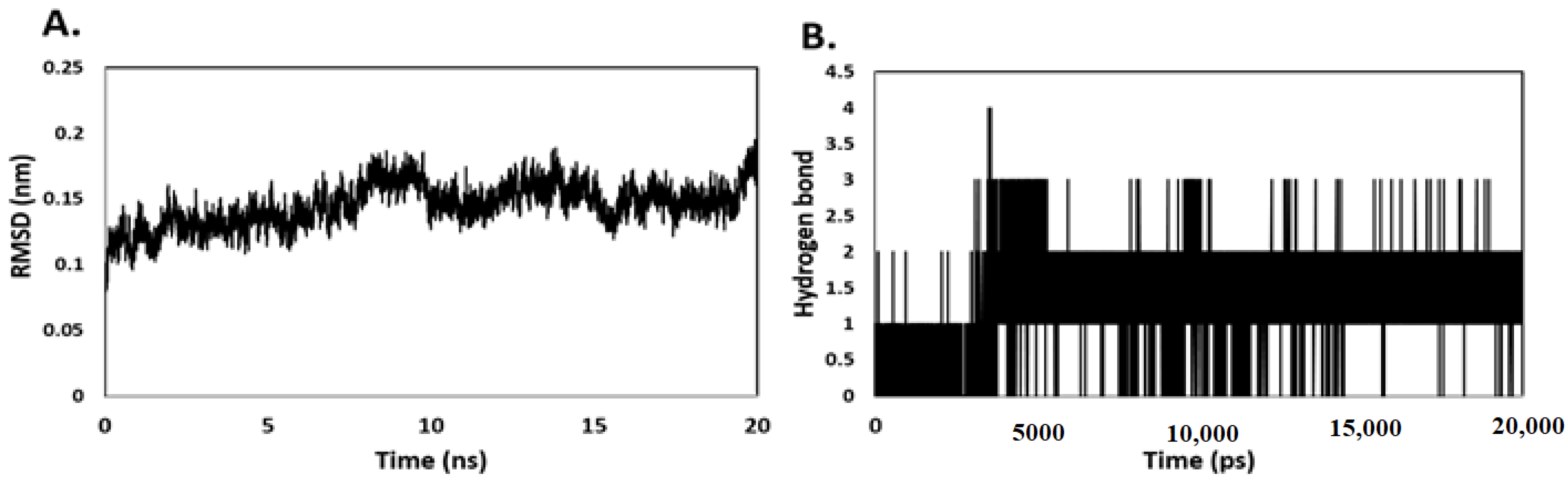
3.3. Molecular Dynamic Simulation
3.4. Antitumor Screening
4. Conclusions
Author Contributions
Funding
Institutional Review Board Statement
Informed Consent Statement
Data Availability Statement
Acknowledgments
Conflicts of Interest
Sample Availability
References
- Bavetsias, V.; Jakman, A.L.; Marriott, J.H.; Kimbell, R.; Gibson, W.; Boyle, F.T.; Bisset, G.M. Folate-based inhibitors of thymidylate synthase: Synthesis and antitumor activity of gamma-linked sterically hindered dipeptide analogues of 2-desamino-2-methyl-N10-propargyl-5,8-dideazafolic acid (ICI 198583). J. Med. Chem. 1997, 40, 1495–1510. [Google Scholar] [CrossRef] [PubMed]
- Bavetsias, V.; Marriott, J.H.; Melin, C.; Kimbell, R.; Matusiak, Z.S.; Boyle, F.T.; Jakman, A.L. Design and synthesis of Cyclopenta[g]quinazoline-based antifolates as inhibitors of thymidylate synthase and potential antitumor agents. J. Med. Chem. 2000, 43, 1910–1926. [Google Scholar] [CrossRef] [PubMed]
- El-Azab, A.S.; Abdel-Aziz, A.A.; Bua, S.; Nocentini, A.; El-Gendy, M.A.; Mohamed, M.A.; Shawer, T.Z.; AlSaif, N.A.; Supuran, C.T. Synthesis of benzensulfonamides linked to quinazoline scaffolds as novel carbonic anhydrase inhibitors. Bioorg. Chem. 2019, 89, 78–90. [Google Scholar] [CrossRef] [PubMed]
- de Castro Barbosa, M.L.; Lima, L.M.; Tesch, R.; Sant’Anna CM, R.; Totzke, F.; Kubbutat, M.H.; Schächtele, C.; Laufer, S.A.; Barreiro, E.J. Novel 2-chloro-4-anilino-quinazoline derivatives as EGFR and VEGFR-2 dual inhibitors. Eur. J. Med. Chem. 2014, 71, 1–14. [Google Scholar] [CrossRef]
- El-Azab, A.S.; Al-Dhfyan, A.; Abdel-Aziz, A.A.; Abou-Zeid, L.A.; Alkahtani, H.M.; Al-Obaid, A.M.; Al-Gendy, M.A. Synthesis, anticancer and apoptosis-inducing activities of quinazoline-isatin conjugates: Epidermal growth factor receptor-tyrosine kinase assay and molecular docking studies. J. Enzyme Inhib. Med. Chem. 2017, 32, 935–944. [Google Scholar] [CrossRef]
- El-Azab, A.S.; Abdel-Aziz, A.A.; Ghabbour, H.A.; Al-Gendy, M.A. Synthesis, in vitro antitumour activity, and molecular docking study of novel 2-substituted mercapto- 3-(3,4,5-trimethoxybenzyl)-4(3H)-quinazolinone analogues. J. Enzyme Inhib. Med. Chem. 2017, 32, 1229–1239. [Google Scholar] [CrossRef]
- Mohamed, M.A.; Ayyad, R.R.; Shawer, T.Z.; Alaa, A.-M.; El-Azab, A.S. Synthesis and antitumor evaluation of trimethoxyanilides based on 4 (3H)-quinazolinone scaffolds. Eur. J. Med. Chem. 2016, 112, 106–113. [Google Scholar] [CrossRef]
- Al-Suwaidan, I.A.; Alanazi, A.M.; El-Azab, A.S.; Al-Obaid, A.M.; El-Tahir, K.E.; Maarouf, A.R.; Abu El-Enin, M.A.; Abdel-Aziz, A.A. Molecular design, synthesis and biological evaluation of cyclic imides bearing benzenesulfonamide fragment as potential COX-2 inhibitors. Part 2. Bioorg. Med. Chem. Lett. 2013, 23, 2601–2605. [Google Scholar] [CrossRef]
- Alaa, A.M.; Abou-Zeid, L.A.; ElTahir, E.H.K.; Mohamed, M.A.; El-Enin, M.A.A.; El-Azab, A.S. Design, synthesis of 2, 3-disubstitued 4 (3H)-quinazolinone derivatives as anti-inflammatory and analgesic agents: COX-1/2 inhibitory activities and molecular docking studies. Bioorg. Med. Chem. 2016, 24, 3818–3828. [Google Scholar]
- Abdel-Aziz, A.A.-M.; Abou-Zeid, L.A.; El-Tahir, K.E.H.; Ayyad, R.R.; Magda, A.-A.; El-Azab, A.S. Synthesis, anti-inflammatory, analgesic, COX-1/2 inhibitory activities and molecular docking studies of substituted 2-mercapto-4 (3H)-quinazolinones. Eur. J. Med. Chem. 2016, 121, 410–421. [Google Scholar] [CrossRef]
- Alafeefy, A.M.; El-Azab, A.S.; Mohamed, M.A.; Bakhat, M.A.; Abdel-Hamid, S. Synthesis of some new substituted iodoquinazoline derivatives and their antimicrobial screening J. Saudi Chem. Soc. 2011, 15, 319–325. [Google Scholar] [CrossRef]
- Alanazi, A.M.; Alaa, A.-M.; Al-Suwaidan, I.A.; Abdel-Hamide, S.G.; Shawer, T.Z.; El-Azab, A.S. Design, synthesis and biological evaluation of some novel substituted quinazolines as antitumor agents. Eur. J. Med. Chem. 2014, 79, 446–454. [Google Scholar] [CrossRef] [PubMed]
- Alanazi, A.M.; Al-Suwaidan, I.A.; Alaa, A.-M.; Mohamed, M.A.; El_Morsy, A.M.; El-Azab, A.S. Design synthesis and biological evaluation of some novel substituted 2-mercapto-3-phenethylquinazolines as antitumor agents. Med. Chem. Res. 2013, 22, 5566–5577. [Google Scholar] [CrossRef]
- Al-Obaid, A.; Abdel-Hamide, S.; El-Kashef, H.; Abdel-Aziz, A.; El-Azab, A.; Al-Khamees, H.; El-Subbagh, H. Synthesis, in vitro antitumor activity and molecular modelling study of certain 2-thieno-4 (3H)-quinazolinone analogs. Eur. J. Med. Chem. 2009, 44, 2379–2391. [Google Scholar] [CrossRef]
- Al-Suwaidan, I.A.; Abdel-Aziz, A.A.-M.; Shawer, T.Z.; Ayyad, R.R.; Alanazi, A.M.; El-Morsy, A.M.; Mohamed, M.A.; Abdel-Aziz, N.I.; El-Sayed, M.A.-A.; El-Azab, A.S. Synthesis, antitumor activity and molecular docking study of some novel 3-benzyl-4(3H) quinazolinone analogues. J. Enzyme Inhib. Med. Chem. 2016, 31, 78–89. [Google Scholar] [CrossRef]
- Al-Suwaidan, I.A.; Alanazi, A.M.; Alaa, A.-M.; Mohamed, M.A.; El-Azab, A.S. Design, synthesis and biological evaluation of 2-mercapto-3-phenethylquinazoline bearing anilide fragments as potential antitumor agents: Molecular docking study. Bioorg. Med. Chem. Lett. 2013, 23, 3935–3941. [Google Scholar] [CrossRef] [PubMed]
- Aziza, M.; Nassar, M.; AbdelHamide, S.; El-Hakim, A.; El-Azab, A. Synthesis and antimicrobial activities of some new 3-heteroaryl-quinazolin-4-ones. Indian J. Heterocycl. Chem. 1996, 6, 25–30. [Google Scholar]
- El-Azab, A.S. Synthesis of some new substituted 2-mercaptoquinazoline analogs as potential antimicrobial agents. Phosph. Sulfur Silicon Related Elem. 2007, 182, 333–348. [Google Scholar] [CrossRef]
- El-Azab, A.S.; Abdel-Hamide, S.G.; Sayed-Ahmed, M.M.; Hassan, G.S.; El-Hadiyah, T.M.; Al-Shabanah, O.A.; Al-Deeb, O.A.; El-Subbagh, H. Novel 4(3H)-Quinazolinone Analogues: Synthesis and Anticonvulsant Activity, Med. Chem. Res. 2013, 22, 2815–2827. [Google Scholar]
- El-Azab, A.S.; Al-Omar, M.A.; Alaa, A.-M.; Abdel-Aziz, N.I.; Magda, A.-A.; Aleisa, A.M.; Sayed-Ahmed, M.M.; Abdel-Hamide, S.G. Design, synthesis and biological evaluation of novel quinazoline derivatives as potential antitumor agents: Molecular docking study. Eur. J. Med. Chem. 2010, 45, 4188–4198. [Google Scholar] [CrossRef]
- El-Azab, A.S.; ElTahir, K.E. Synthesis and anticonvulsant evaluation of some new 2,3,8-trisubstituted-4 (3H)-quinazoline derivatives. Bioorg. Med. Chem. Lett. 2012, 22, 327–333. [Google Scholar] [CrossRef]
- El-Azab, A.S.; ElTahir, K.E. Design and synthesis of novel 7-aminoquinazoline derivatives: Antitumor and anticonvulsant activities. Bioorg. Med. Chem. Lett. 2012, 22, 1879–1885. [Google Scholar] [CrossRef] [PubMed]
- El-Azab, A.S.; ElTahir, K.E.; Attia, S.M. Synthesis and anticonvulsant evaluation of some novel 4 (3H)-quinazolinones. Mon. Chem.-Chem. Month. 2011, 142, 837–848. [Google Scholar] [CrossRef]
- Alanazi, A.M.; Abdel-Aziz, A.A.; Shawer, T.Z.; Ayyad, R.R.; Al-Obaid, A.M.; Al-Agamy, M.H.; Maarouf, A.R.; El-Azab, A.S. Synthesis, antitumor and antimicrobial activity of some new 6-methyl-3-phenyl-4(3H)-quinazolinone analogues: In silico studies. J. Enzyme Inhib. Med. Chem. 2016, 31, 721–735. [Google Scholar] [CrossRef] [PubMed]
- Larroque, A.L.; Peori, B.; Williams, C.; Fang, Y.Q.; Qiu, Q.; Rachid, Z.; Jean-Claude, B.J. Synthesis of Water Soluble Bis-triazenoquinazolines: An Unusual Predicted Mode of Binding to the Epidermal Growth Factor Receptor Tyrosine Kinase. J. Chem. Biol. Drug Des. 2008, 71, 374–379. [Google Scholar] [CrossRef]
- Amin, K.M.; Georgey, H.H.; Awadallah, F.M. EGFR tyrosine kinase targeted compounds: Synthesis, docking study, and in vitro antitumor activity of some new quinazoline and benzo[d]isothiazole derivatives. J. Med. Chem. Res. 2011, 20, 1042–1053. [Google Scholar] [CrossRef]
- Mphahlele, M.J.; Maluleka, M.M.; Mmonw, M.M. Synthesis of Heterocycle-Appended 4-Aminoquinazolines with Antiproliferative Properties and Potential to Inhibit Tyrosine Kinases. In Chemistry for a Clean and Healthy Planet; Springer: Cham, Germany, 2019. [Google Scholar]
- Mohamed, M.A.; Alafeefy, A.M.; Abdel Hamid, S.G. Synthesis of Some New Quinazoline Derivatives as Potential Anti-Infective Agents. Life Sci. J. 2014, 11, 108–112. [Google Scholar]
- Grever, M.R.; Schepartz, S.A.; Chabner, B.A. The National Cancer Institute: Cancer drug discovery and development program. Semin. Oncol. 1992, 19, 622–638. [Google Scholar]
- Monks, A.; Scudiero, D.; Skehan, P.; Shoemaker, R.; Paull, K.; Vistica, D.; Hose, C.; Langley, J.; Cronise, P.; Vaigro-Wolff, A.; et al. Feasibility of a high-flux anticancer drug screen using a diverse panel of cultured human tumor cell lines. J. Natl. Cancer Inst. 1991, 83, 757–766. [Google Scholar] [CrossRef]
- Boyd, M.R.; Paull, K. Some practical considerations and applications of the national cancer institute in vitro anticancer drug discovery screen. Drug Dev. Res. 1995, 34, 91–109. [Google Scholar] [CrossRef]
- Shoemaker, R.H. The NCI60 human tumour cell line anticancer drug screen. Nat. Rev. Cancer 2006, 6, 813–823. [Google Scholar] [CrossRef]
- Alley, M.C.; Scudiero, D.A.; Monks, A.; Hursey, M.L.; Czerwinski, M.J.; Fine, D.L.; Abbott, B.J.; Mayo, J.G.; Shoemaker, R.H.; Boyd, M.R. Feasibility of drug screening with panels of human tumor cell lines using a microculture tetrazolium assay. Cancer Res. 1988, 48, 589–601. [Google Scholar] [PubMed]
- Engelhardt, H.; Böse, D.; Petronczki, M.; Scharn, D.; Bader, G.; Baum, A.; Bergner, A.; Chong, E.; Döbel, S.; Egger, G.; et al. Start selective and rigidify: The discovery path toward a next generation of EGFR tyrosine kinase inhibitors. J. Med. Chem. 2019, 62, 10272–10293. [Google Scholar] [CrossRef]
- Bersanelli, M.; Minari, R.; Bordi, P.; Gnetti, L.; Bozzetti, C.; Squadrilli, A.; Lagrasta, C.; Bottarelli, L.; Osipova, G.; Capelletto, E.; et al. L718Q Mutation as New Mechanism of Acquired Resistance to AZD9291 in EGFR -Mutated NSCLC. J. Thorac. Oncol. 2016, 11, e121–e123. [Google Scholar] [CrossRef]
- McTigue, M.; Murray, B.W.; Chen, J.H.; Deng, Y.L.; Solowiej, J.; Kania, R.S. Molecular conformations, interactions, and properties associated with drug efficiency and clinical performance among VEGFR TK inhibitors. Proc. Natl. Acad. Sci. USA 2012, 109, 18281–18289. [Google Scholar] [CrossRef] [PubMed]
- Delano, W.L. Pymol: An open-source molecular graphics tool. CCP4 Newsl. Protein Crystallogr. 2002, 40, 82–92. [Google Scholar]
- Oleg, T.; Olson, A.J. AutoDock Vina: Improving the speed and accuracy of docking with a new scoring function, efficient optimization, and multithreading. J. Comput. Chem. 2010, 31, 455–461. [Google Scholar]
- Hess, B.; Kutzner, C.; van der Spoel, D.; Lindahl, E. GROMACS 4: Algorithms for Highly Efficient, Load-Balanced, and Scalable Molecular Simulation. J. Chem. Theory Comput 2008, 4, 435–447. [Google Scholar] [CrossRef]
- Kaminski, G.A.; Friesner, R.A.; Tirado-Rives, J.; Jorgensen, W.L. Evaluation and Reparameterization of the OPLS-AA Force Field for Proteins via Comparison with Accurate Quantum Chemical Calculations on Peptides. J. Phys. Chem. B 2001, 105, 6474–6487. [Google Scholar] [CrossRef]
- Zoete, V.; Cuendet, M.A.; Grosdidier, A.; Michielin, O. SwissParam: A fast force field generation tool for small organic molecules. J. Comput. Chem. 2011, 32, 2359–2368. [Google Scholar] [CrossRef] [PubMed]
- Alamri, M.A. Pharmacoinformatics and molecular dynamic simulation studies to identify potential small-molecule inhibitors of WNK-SPAK/OSR1 signaling that mimic the RFQV motifs of WNK kinases. Arab. J. Chem. 2020, 13, 5107–5117. [Google Scholar] [CrossRef]


| Compound | Results | |||
|---|---|---|---|---|
| VEGFR2 | EGFR | |||
| Compound No. | Code | MW | IC50nM | IC50nM |
| 6 | M6 | 360 | 98.1 ± 2.93 | 106 ± 2.22 |
| Reference | Docetaxel | 807.88 | 89.3 ± 2.67 | 56.1 ± 1.17 |
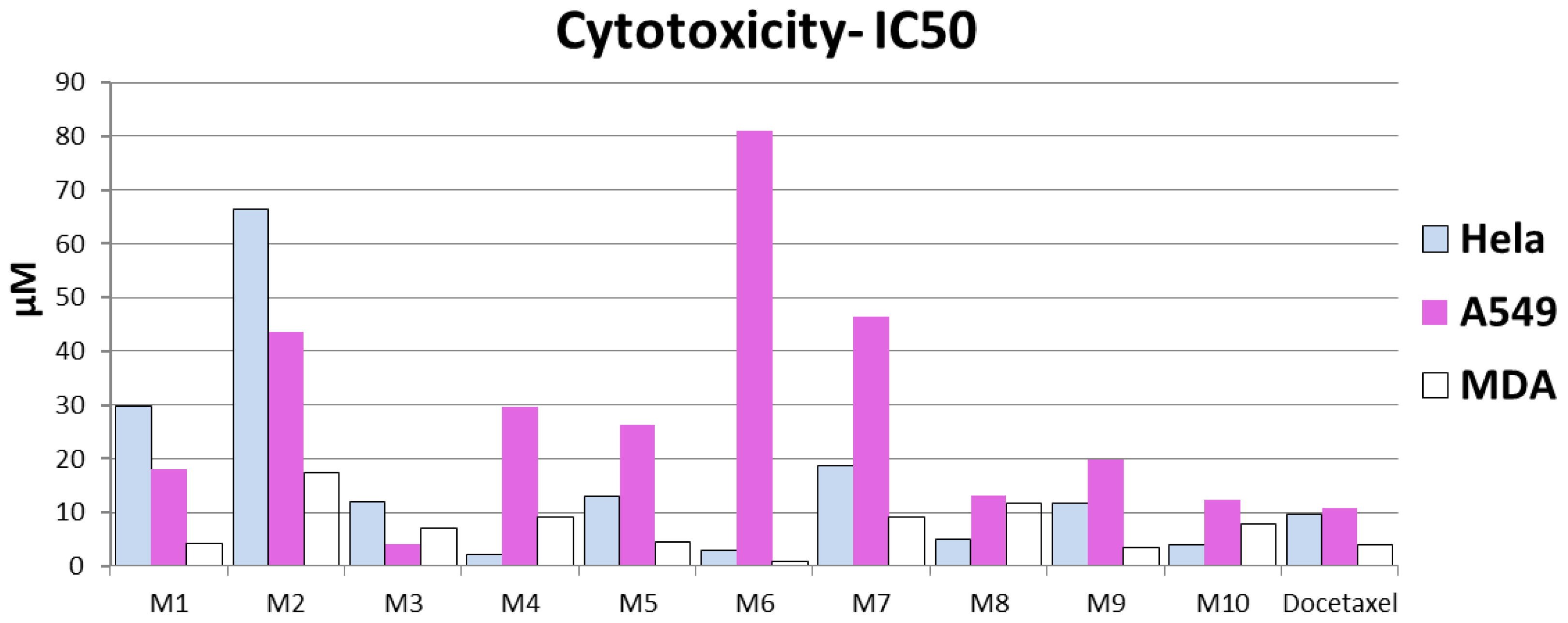
| Compound No. | Group M | Cytotoxicity IC50 µM | |||
|---|---|---|---|---|---|
| Code | MW | Hela | A549 | MDA | |
| 4 | M-1 | 357 | 29.8 ± 0.78 | 18 ± 0.5 | 4.12 ± 0.1 |
| 3 | M-2 | 343 | 66.5 ± 1.74 | 43.6 ± 1.1 | 17.4 ± 0.5 |
| 11 | M-3 | 506 | 12.0 ± 0.31 | 4.03 ± 0.1 | 7.06 ± 0.2 |
| 2 | M-4 | 361 | 2.13 ± 0.06 | 29.7 ± 0.8 | 9.24 ± 0.2 |
| 5 | M-5 | 342 | 12.9 ± 0.34 | 26.3 ± 0.7 | 4.39 ± 0.1 |
| 6 | M-6 | 360 | 2.8 ± 0.07 | 81 ± 2.1 | 0.79 ± 0.04 |
| 7 | M-7 | 375 | 18.7 ± 0.49 | 46.3 ± 1.2 | 9.0 ± 0.2 |
| 8 | M-8 | 515 | 4.94 ± 0.13 | 13 ± 0.3 | 11.7 ± 0.3 |
| 9 | M-9 | 497 | 11.7 ± 0.31 | 19.9 ± 0.5 | 3.42 ± 0.1 |
| 10 | M-10 | 356 | 3.98 ± 0.1 | 12.3 ± 0.3 | 7.77 ± 0.2 |
| Ref. | Docetaxel | 807.8 | 9.65 ± 0.2 | 10.8 ± 0.23 | 3.98 ± 0.08 |
Publisher’s Note: MDPI stays neutral with regard to jurisdictional claims in published maps and institutional affiliations. |
© 2021 by the authors. Licensee MDPI, Basel, Switzerland. This article is an open access article distributed under the terms and conditions of the Creative Commons Attribution (CC BY) license (https://creativecommons.org/licenses/by/4.0/).
Share and Cite
Altamimi, A.S.; El-Azab, A.S.; Abdelhamid, S.G.; Alamri, M.A.; Bayoumi, A.H.; Alqahtani, S.M.; Alabbas, A.B.; Altharawi, A.I.; Alossaimi, M.A.; Mohamed, M.A. Synthesis, Anticancer Screening of Some Novel Trimethoxy Quinazolines and VEGFR2, EGFR Tyrosine Kinase Inhibitors Assay; Molecular Docking Studies. Molecules 2021, 26, 2992. https://doi.org/10.3390/molecules26102992
Altamimi AS, El-Azab AS, Abdelhamid SG, Alamri MA, Bayoumi AH, Alqahtani SM, Alabbas AB, Altharawi AI, Alossaimi MA, Mohamed MA. Synthesis, Anticancer Screening of Some Novel Trimethoxy Quinazolines and VEGFR2, EGFR Tyrosine Kinase Inhibitors Assay; Molecular Docking Studies. Molecules. 2021; 26(10):2992. https://doi.org/10.3390/molecules26102992
Chicago/Turabian StyleAltamimi, Abdulmalik S., Adel S. El-Azab, Sami G. Abdelhamid, Mubarak A. Alamri, Ashraf H. Bayoumi, Safar M. Alqahtani, Alhumaidi B. Alabbas, Ali I. Altharawi, Manal A. Alossaimi, and Menshawy A. Mohamed. 2021. "Synthesis, Anticancer Screening of Some Novel Trimethoxy Quinazolines and VEGFR2, EGFR Tyrosine Kinase Inhibitors Assay; Molecular Docking Studies" Molecules 26, no. 10: 2992. https://doi.org/10.3390/molecules26102992
APA StyleAltamimi, A. S., El-Azab, A. S., Abdelhamid, S. G., Alamri, M. A., Bayoumi, A. H., Alqahtani, S. M., Alabbas, A. B., Altharawi, A. I., Alossaimi, M. A., & Mohamed, M. A. (2021). Synthesis, Anticancer Screening of Some Novel Trimethoxy Quinazolines and VEGFR2, EGFR Tyrosine Kinase Inhibitors Assay; Molecular Docking Studies. Molecules, 26(10), 2992. https://doi.org/10.3390/molecules26102992