Article

Synthesis of Galectin Inhibitors by Regioselective 3′-O-Sulfation of Vanillin Lactosides Obtained under Phase Transfer Catalysis

1
Department of Chemistry, University of Québec à Montréal, P.O. Box 8888, Succ. Centre-Ville, Montréal, QC H3C 3P8, Canada
2
INRS-Institut Armand-Frappier, Université du Québec, 531 boul. des Prairies, Laval, QC H7V 1B7, Canada
*
Author to whom correspondence should be addressed.
Molecules 2021, 26(1), 115; https://doi.org/10.3390/molecules26010115
Submission received: 2 November 2020 / Revised: 11 December 2020 / Accepted: 25 December 2020 / Published: 29 December 2020

Abstract

:
Vanillin-based lactoside derivatives were synthetized using phase-transfer catalyzed reactions from per-O-acetylated lactosyl bromide. The aldehyde group of the vanillin moiety was then modified to generate a series of related analogs having variable functionalities in the para- position of the aromatic residue. The corresponding unprotected lactosides, obtained by Zemplén transesterification, were regioselectively 3′-O-sulfated using tin chemistry activation followed by treatment with sulfur trioxide-trimethylamine complex (Men3N-SO3). Additional derivatives were also prepared from the vanillin’s aldehyde using a Knoevenagel reaction to provide extended α, β-unsaturated carboxylic acid which was next reduced to the saturated counterpart.

Graphical Abstract

1. Introduction

Cellular communications are frequently governed by molecular interactions involving cell surface glycoconjugates overlay expressed as glycosaminoglycans (GAGs), glycoproteins, and glycolipids. Of particular interest are the family of S-type lectins represented by galectins (Gal) that are characterized by their carbohydrate recognition domains (CRDs) having affinity for glycoconjugates with exposed β-d-galactopyranoside residues in common [1,2,3]. So far, 15 family members have been identified in mammals. In spite of their similar characteristic sugar binding recognition patterns, they are distinctly divided into three categories depending on their molecular architectures. They are classified as: a) Prototype dimers (Gal-1, -2, -5, -7, -10, -11, -13, -14, -15); b) tandem repeat (Gal-4, -6, -8, -9, -12); and c) monomeric chimera type capable of oligomerization (Gal-3) [4]. Given that they are expressed intracellularly together with being present extracellularly by secretion and that they are critically implicated in a plethora of physiological functions, including cancer, inflammation, and immune responses, discovery of specific inhibitors has become of keen therapeutic interest, albeit a major challenge in medicinal chemistry [4,5,6,7,8,9].
Consequently, the design of highly selective sugar-based inhibitors against each of the galectins has been the subject of intense research activities. The field is however only dominated by a few research groups [4,5,6,7,8,9] amongst which the team of Leffler/Nilsson being clearly dominating with a number of successful glycomimetics, some of which reaching clinical phases (thiodigalactosides, TD139, GB1107) [10,11,12,13]. Essentially based on β-d-galactopyranoside/lactoside/N-acetyllactosamine lead scaffolds [14], the incorporation of pharmacophores have provided the most successful candidates when appended at either the anomeric position [9,15,16,17,18,19,20,21,22] or on position -3 of the galactopyranosides [10,11,12,13] residue or O-3′ in the case of lactosides [4,23]. Appealingly, introduction of a negative sulfate group at O-3/O-3′ has also afforded potent ligands owing to the presence of charged amino acids within the CRD [15,23,24,25,26,27]. In addition to the above chemical modifications, an additional and quite successful synthetic strategy has been the discovery that multivalent galactosides/lactosides in the form of glycodendrimers [28,29,30,31,32,33,34], glycopolymers [35,36], liposomes/dendrimersomes [37,38,39,40], and protein conjugate with appended TD139 [41] could similarly provide gains in both affinity and selectivity. This was predominantly observed for the chimeric galectin-3 that can oligomerize upon binding to multivalent receptors due to its collagenous peptide tail [29].
In this paper, we aimed to combine both modifications to a lactopyranoside scaffold by incorporating aromatic aglycons simultaneously to an O-3′ sulfate group (Figure 1). This combined choice was dictated by the fact that a few galectin members have been shown to bind preferentially to sulfated glycans. This was particularly through for Gal-1 [23,24], -3 [15], -4 [25,26], and -8 [27]. In the latter case, recent modeling and X-ray experiments demonstrated that the beneficial interactions were due to favorable electrostatic interactions with arginine residues (Arg-45 and Arg-59) [4,27]. Furthermore, even though several hydrophobic aglycons have been advantageous in the binding events using affinity measurements by ITC, we choose the natural vanillin as aglycon because of its well-established lack of toxicity, the presence of a large amount of vanillin glucoside (glucovanillin) in foodstuffs [42], and the well-known antioxidant properties of phenolic glycosides.

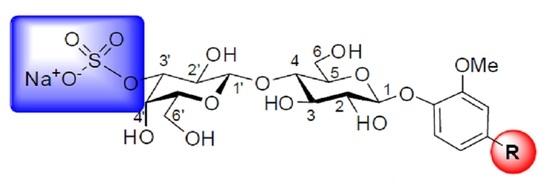
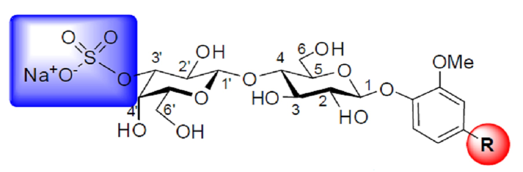
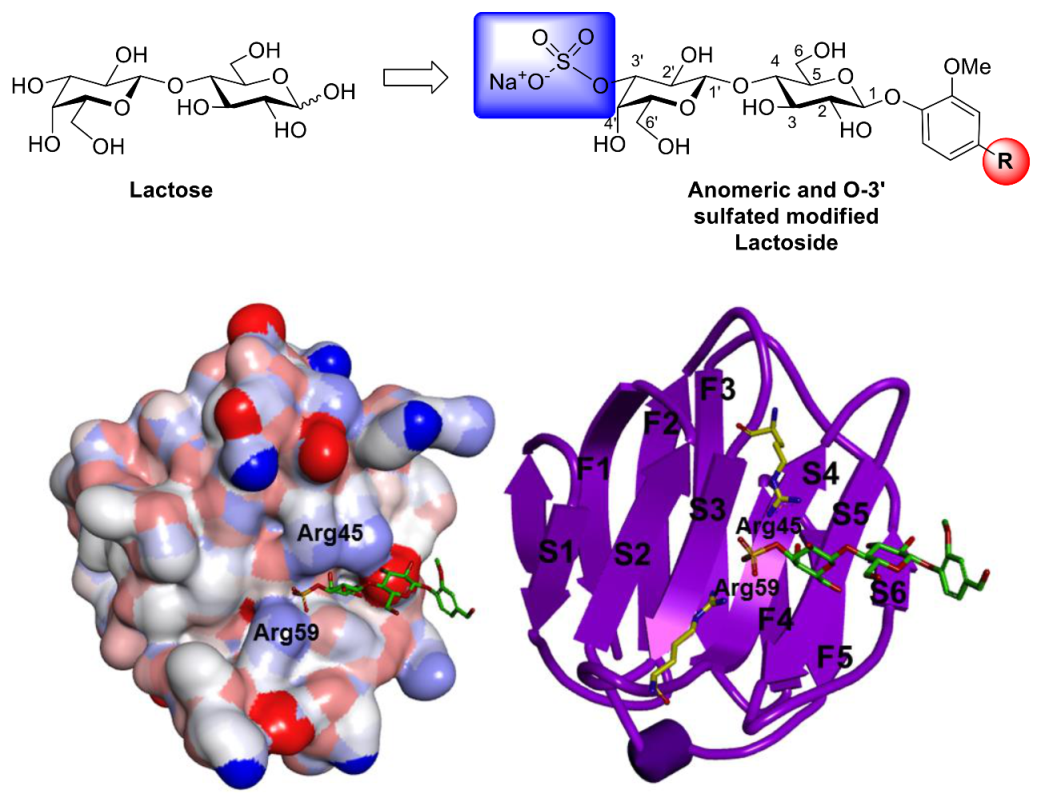
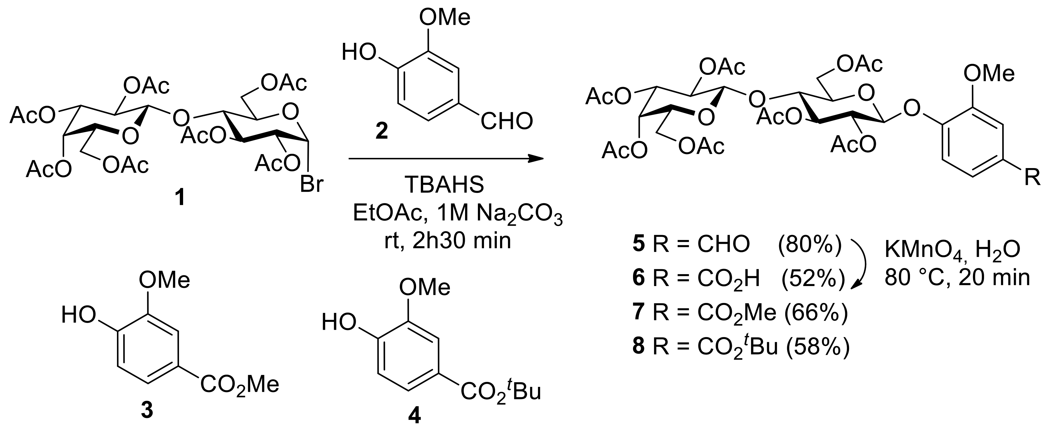
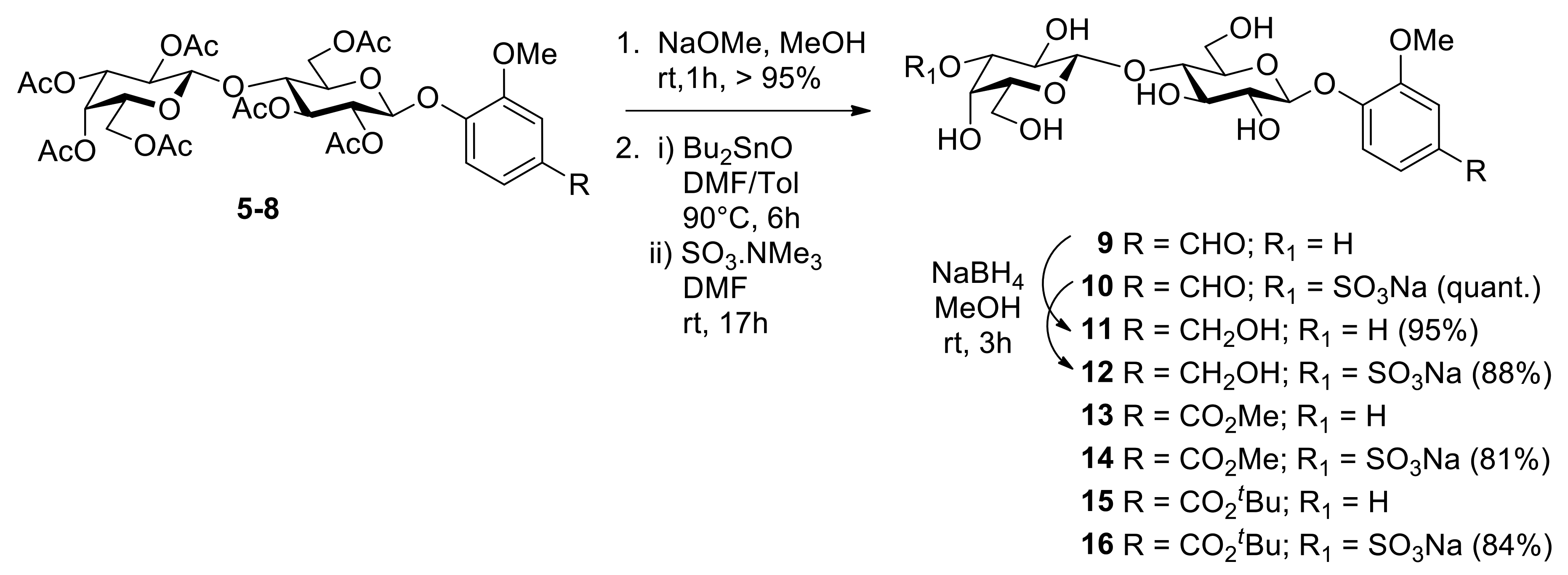
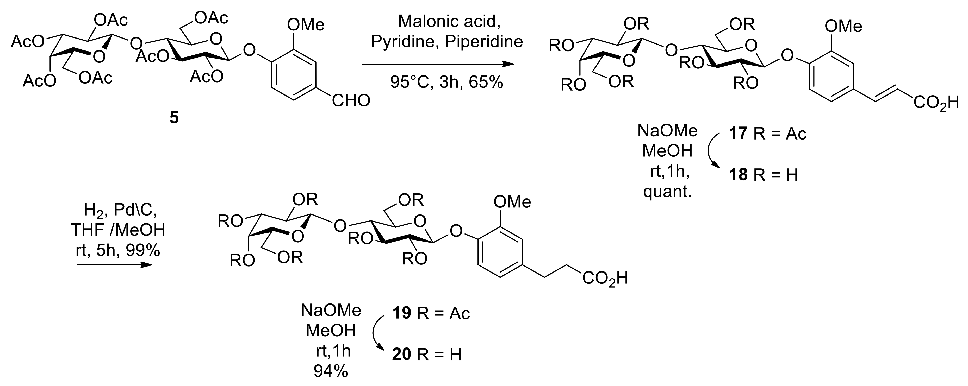
2. Results and Discussion

Over the years, Phase Transfer Catalysis (PTC) has emerged as an efficient and practical methodology for stereoselective glycosidation [43,44] and related anomeric substitutions. Under our previously optimized conditions for phenolic glycosides (TBAHS, EtOAc, 1M Na2CO3), we treated peracetylated lactosyl bromide 1 [43] with vanillin derivatives 2, 3, and 4 to afford lactosides 5, 7, and 8 in 80, 66, and 58% yields, respectively (Scheme 1). The major by-product of the reactions was the usual 2-acetoxy-lactal (1H-NMR of H1 at δ 6.61 ppm) resulting from the HBr SN2 elimination process [43,44]. The disappearance of the anomeric doublet of the bromide 1 at δ 6.52 ppm with a J1,2 coupling constant of 4.0 Hz was replaced by a new doublet at δ 5.10 ppm with a distinctive trans coupling constant of J1,2 of 7.4 Hz in compound 5. The same held for methyl ester 7 (H1: δ 5.05, J1,2 = 7.4 Hz) and tert-butyl ester 8 (H1: δ 5.03, J1,2 = 7.5 Hz). As previously demonstrated [42,43,44], these PTC conditions afforded complete stereoselectivity in favor of anomeric inversion from the α-bromide 1 to β-glycosides 5, 7, and 8 exclusively. Since we also wanted to explore the role played by the carboxylic function in the para-position of the vanillin residue, coupled to the fact that we could not hydrolyze the acetate protecting groups in derivative 7 and 8 without losing the esters (vide infra), we opted for the oxidation of the native vanillin’s aldehyde in 5 using permanganate treatment (KMnO4, H2O, 80 °C, 20 min) which afforded acid 6 in 52% yield, showing disappearance of the aldehyde proton at δ 9.91 ppm. All compounds were fully characterized by NMR spectroscopy (1H, 13C), mass spectrometry, and the datasets agreed with literature data when known (see experimental section).
The peracetylated intermediates 5, 7, 8 were next de-O-acetylated under the classical Zemplén conditions (NaOMe, MeOH) in essentially quantitative yields in all cases to give free lactosides 9, 13, and 15 (Scheme 2). Following sequential treatment of the unprotected lactosides with dibutyltin oxide (Bu2SnO, DMF, PhMe, 90° C, 6h) and regioselective sulfation [45] using sulfur trioxide-trimethylamine complex (Me3N.SO3), the 3′-O-sulfated lactosides 10, 14, 16 (Scheme 2) were obtained in good to excellent yields (81%-quantitative). The regioselectivity of this transformation is well-known and has been explained through the formation of a cyclic stannylene complex at the unique cis-3′,4′-dihydroxyl groups of the galactoside moiety [46,47]. The aldehyde functions of vanillin lactoside 9 and 3′-O-sulfated lactoside 10 were reduced using NaBH4 in MeOH (rt, 3h) to give vanillin lactoside analogs 11 and 12 in excellent 95% and 88% yields, respectively. The position of the sulfate groups were readily confirmed on the basis of the H-3′ downfield shift from ~3.66 ppm to ~4.28 ppm (SI) together with the characteristic 13C-NMR chemical shift displacement of the 3′-carbon, which usually appears ~7 ppm downfield (~ δ 80 ppm) from the unsubstituted precursors at ~δ 73 ppm [47]. Dept 135 13C-NMR analysis was required to ensure that the regioselective sulfation was unequivocally performed at the C-3’ position in order to show the absence of signals attributed to C-6 and C-6’s modifications (usually at δ 59–62 ppm [47]. NMR COSY and HSQC experiments were also used to unambiguously correlate the sulfation process and regioselectivity.
A Knoevenagel-Doebner condensation was also used (malonic acid, C5H5N, Piperidine, 95° C, 3h) for the synthesis of two carbons homologated vanillin lactosides 1720 (Scheme 3), analogously used before for a galactoside derivative [48]. Thus, aldehyde 5 provided peracetylated α, β-unsaturated analog 17 in 65% yield, which upon catalytic hydrogenation (H2, Pd-C, THF-MeOH, rt, 5 h) gave the expected α, β-saturated lactoside 19 essentially quantitatively.
The 1H-NMR spectra of 17 clearly showed the two trans proton signals corresponding to the unsaturated protons H-14 and H-13 at δ 6.33 and 7.68 ppm with a typical coupling constant J13-14 of 15.9 Hz. The palladium-catalyzed reduction leading to 19 showed the disappearance of these two proton signals and the appearance of new H-14 and H-13 protons at 2.62 and 2.89 ppm, respectively (see SI). Zemplén deprotection was used to afford the de-O-acetylated lactosides 18 and 20 (Scheme 3).

3. Materials and Methods

3.1. General Synthetic Methods

All reactions in organic medium were performed in standard oven dried glassware under an inert atmosphere of nitrogen using freshly distilled solvents. Solvents and reagents were deoxygenated, when necessary by purging with nitrogen. All reagents were used as supplied without prior purification unless otherwise stated, and obtained from Sigma-Aldrich Chemical Co. Ltd.(St. Louis, MO, USA) Reactions were monitored by analytical thin-layer chromatography (TLC) using silica gel 60 F254 precoated plates (E. Merck (Darmstadt, Germany)) and compounds were visualized with a 254 nm UV lamp, a mixture of iodine/silica gel and/or mixture of ceric ammonium molybdate solution (100 mL H2SO4, 900 mL H2O, 25 g (NH4)6Mo7O24H2O, 10 g Ce(SO4)2), and subsequent spots development by gentle warming with a heat-gun. Purifications were performed by silica gel flash column chromatography using Silica (60 Å, 40–63 µm) with the indicated eluent. NMR spectroscopy was used to record 1H-NMR and 13C-NMR spectra at 300 or 600 MHz and at 75 or 150 MHz, respectively, on Bruker (300 MHz) and Bruker Avance III HD 600 MHz spectrometers (Billerica, MA, USA). Proton and carbon chemical shifts (δ) are reported in ppm relative to the chemical shift of residual CHCl3, which was set at 7.26 ppm (1H) and 77.16 ppm (13C). Coupling constants (J) are reported in Hertz (Hz), and the following abbreviations are used for peak multiplicities: Singlet (s), doublet (d), doublet of doublets (dd), doublet of doublet with equal coupling constants (tap), triplet (t), multiplet (m). Analysis and assignments were made using COSY (Correlated SpectroscopY) and HSQC (Heteronuclear Single Quantum Coherence) experiments. High-resolution mass spectrometry (HRMS) data were measured with a LC-MS-TOF (Liquid Chromatography-Mass Spectrometry-Time of Flight; Agilent Technologies) in positive and/or negative electrospray mode(s) at the analytical platform of UQAM.

3.2. General Synthetic Procedure A: Phase-Transfer Catalysis (PTC) Reaction

PTC reactions were performed following the previously established protocols [42,43,44,45,46] or under the slightly modified procedure as follows: To a solution of peracetylated lactosyl bromide 1 [49] (1 equiv.) in ethyl acetate (6 mL) was added the corresponding aromatic alcohol (2.5 equiv.), tetrabutylammonium hydrogen sulfate (TBAHS, 1.1 equiv.) and 1M Na2CO3 (1.3 equiv.). The mixture was stirred at room temperature for 2h30 min and then washed successively with water and brine. The organic layer was dried over Na2SO4 and concentrated under reduced pressure. Purification by silica gel column chromatography (Hex/AcOEt) afforded the corresponding compounds 5, 7, and 8 as white powders (yield 58–80%).

3.3. General Synthetic Procedure B: Zemplén Transesterification Reaction

To a solution of lactosides 5, 6, 7, 8, 17 and 19 in dry methanol was added a solution of sodium methoxide (1 M in MeOH, 0.1 equiv.). After stirring at room temperature for 1–2 h, the reaction was completed and then neutralized by addition of ion-exchange resin (Amberlite IR 120 H+). The solution was filtered and evaporated in vacuo to afford the de-O-acetylated lactosides as white powders (yield 95%-quant.)

3.4. General Synthetic Procedure C: Preparation of 3′-O-sulfated Lactosides

A mixture of deacetylated lactosides (1 equiv.) and dibutyltin oxide (Bu2SnO, 1.15 equiv.) in DMF/toluene (6 mL/3 mL) was stirred at 90 °C for 6 h. The solution was then concentrated and sulfur trioxide-trimethylamine complex (Me3N.SO3) (1.3 equiv.) and dry DMF (6 mL) were added. After stirring at room temperature for 17 h, the reaction was quenched with water and evaporated under vacuum. The residue was purified through a column of DOWEX Marathon C (Na+) and eluted with H2O to obtain the pure 3′-O-sulfated lactosides as white powder after lyophilization (yields 82%-quantitative).

3.5. 2,3,4,6-Tetra-O-acetyl-β-d-galactopyranosyl-(1-4)-2,3,6-tri-O-acetyl-α-d-glucopyranosyl bromide (1) (Acetobromolactose)

To a solution of per-O-acetylated lactose [49] (14.2 g, 21 mmoL) in anhydrous CH2Cl2 (63 mL) was added hydrobromic acid (33% in AcOH, 47.9 mL). The reaction mixture was stirred at room temperature for 1 h, then neutralized with saturated aqueous NaHCO3 and washed with brine. The organic layer was dried over Na2SO4 and concentrated under reduced pressure to give lactosyl bromide 1 (13.6 g, 93%) as a white solid. Its spectroscopic data agreed well with those of the literature [49].

3.6. 3-Methoxy-4-(2,3,6,2′,3′,4′,6′-hepta-O-acetyl-β-D-lactopyranosyloxy)benzaldehyde (5) (4-formyl-2-methoxyphenyl 2,3,6,2′,3′,4′,6′-hepta-O-acetyl-β-D-lactopyranoside)

Following the general procedure A, compound 5 was obtained as a white powder; yield: 176 mg (80%). 1H-NMR (300 MHz, CDCl3): δ (ppm) 9.91 (s, 1H, CHO), 7.38–7.46 (m, 2H, Harom), 7.19 (d, 1H, J = 8.0 Hz,Harom), 5.38 (d,1H, J = 2.8 Hz, H-4′), 5.28 (dd, 2H, J = 13.9,J = 8.0 Hz, H-3 et H-2), 5.17(dd, 1H, J = 14.7, J = 6.9 Hz, H-2′), 5.10 (d, 1H, J = 7.4 Hz, H-1), 4.99 (dd,1H,J = 10.4, J = 3.4 Hz, H-3′), 4.52–4.57 (m, 2H, H-1′, H-6a), 4.04–4.24(m, 3H, H-6b, H-6′ab), 3.91–3.96 (m, 2H, H-4 et H-5′), 3.90 (s, 3H, OMe), 3.76–3.81 (m, 1H, H-5), 1.94–2.22 (21H, 7OAc). 13C-NMR (75 MHz, CDCl3): (75 MHz, CDCl3): δ (ppm) 190.9 (CHO), 170.3, 170.2, 170.1, 170.0, 169.7, 169.5, 169.0 (CO), 151.1, 150.8, 132.6, 125.3, 117.7, 110.6 (Carom), 101.1 (C-1′), 99.3 (C-1), 76.0 (C-4), 72.9 (C-5), 72.4 (C-3), 71.2 (C-2), 70.9 (C-3′), 70.7 (C-5′), 69.0 (C-2′), 66.5 (C-4′), 61.8 (C-6′), 60.8 (C-6), 56.0 (OMe), 20.8, 20.7, 20.6, 20.5.

3.7. 3-Methoxy-4-(2,3,6,2′,3′,4′,6′-hepta-O-acetyl-β-d-lactopyranosyloxy)benzoic Acid (6)

A solution of compound 5 (50 mg, 0.063 mmol) in 1 mL of water and in the presence of KMnO4 (12.04 mg, 0.076 mmol), which was added drip under agitation and refluxed of 70–80 °C for about 20 min. The residue is washed with hot water, filtered, concentrated, and then acidified to give the compound 6 was obtained as a white powder; yield: 26.5mg (52%), 1H-NMR (300 MHz, CDCl3): δ (ppm) 7.67 (d, 1H, J = 8.4 Hz, Harom), 7.61 (s, 1H, Harom),7.19 (d, 1H, J = 8.0 Hz, Harom),5.36 (d,1H, J = 2.8 Hz, H-4′), 5.26(ddd,2H, J = 13.9,J = 8.0 Hz, H-3 et H-2), 5.12(dd, 1H, J = 10.3, J = 7.9 Hz, H-2′), 5.05 (d, 1H, J = 7.3 Hz, H-1), 4.97 (dd,1H,J = 10.4, J = 3.4 Hz, H-3′), 4.50–4.54 (m, 2H, H-1′,H-6a), 4.03–4.21 (m, 3H, H-6b, H-6′ab), 3.89–3.91 (m, 2H, H-4 et H-5′), 3.87 (s, 3H, OMe), 3.69–3.81 (m, 1H, H-5), 1.81–2.34 (21H, 7OAc). 13C-NMR (75 MHz, CDCl3): (75 MHz, CDCl3): 170.4, 170.3, 170.1, 170.1, 169.8, 169.6 (CO), 169.1 (COOH), 150.5, 150.0, 125.3, 123.7, 117.7, 113.8 (Carom), 101.1 (C-1′), 99.4 (C-1), 76.1 (C-4), 72.9 (C-5), 72.4 (C-3), 71.3 (C-2), 70.9 (C-3′), 70.7(C-5′), 69.1(C-2′), 66.6 (C-4′), 61.8 (C-6′), 60.8 (C-6), 56.1 (OMe), 20.8, 20.6, 20.5.

3.8. Methyl 3-methoxy-4-(2,3,6,2′,3′,4′,6′-hepta-O-acetyl-β-d-lactopyranosyloxy)benzoate (7)

Following the general procedure A, compound 7 was obtained as a white powder; yield: 573 mg (66 %): mp 85–90 °C; [α] 20D = −33.0 (c 0.25, DCM). 1H-NMR (300 MHz, CDCl3): δ ppm 7.76–7.50 (m, 2H, Ar), 7.10 (d, 1H, J = 8.3 Hz, Ar), 5.38–5.37 (m, 1H, H-4′), 5.37–5.28 (m, 1H, H-3), 5.25–5.20 (m, 1H, H-2), 5.17–5.12 (m, 1H, H-2′), 5.05 (d, 1H, J = 7.4 Hz, H-1), 4.99–4.96 (m, 1H, H-3′), 4.52–4.51 (m, 2H, H-1′,6a), 4.20–4.08 (m, 3H, H-6b, 6′ab), 3.94–3.88 (m, 2H, H-4, 5′), 3.92 (s, 3H, CO2Me), 3.88 (s, 3H, OMe), 3.80–3.75 (m, 1H, H-5), 2.18 (s, 3H), 2.18–1.99 (21H, 7OAc). 13C-NMR (75 MHz, CDCl3): δ (ppm) 170.3, 170.2, 170.1, 170.0, 169.7, 169.5, 169.1, 166.5, 150.0, 149.8, 126.0, 122.9, 117.9, 113.4, 101.1 (C-1′), 99.6 (C-1), 76.1 (C-4), 72.9 (C-5), 72.4 (C-3), 71.3 (C-2), 70.9 (C-3′), 70.7 (C-5′), 69.0 (C-2′), 66.6 (C-4′), 61.9 (C-6), 60.8 (C-6′), 56.1 (OMe), 52.2 (CO2Me), 20.8, 20.6, 20.5. ESI-HRMS: m/z calcd for C35H44O21, 800.2375; found 818.2675 [M + NH4]+.

3.9. Tert-butyl 3-methoxy-4-(2,3,6,2′,3′,4′,6′-hepta-O-acetyl-β-d-lactopyranosyloxy)benzoate (8)

Following the general procedure A, compound 8 was obtained as a white powder; yield: 771 mg (58%): mp 98 °C; [α] 20D = −21.5 (c 0.25, DCM). 1H-NMR (300 MHz, CDCl3): 7.59–7.51 (m, 2H, Harom), 7.19 (d, 1H, J = 8.0 Hz,Harom),5.38 (d, 1H, J = 2.7 Hz, H-4′), 5.31 (dd, 1H, J = 9.0Hz, H-3), 5.22 (dd, 1H, J = 10.4, J =7.8 Hz, H-2), 5.15 (dd, 1H, J = 10.4, J =7.8 Hz, H-2′), 5.03 (d, 1H, J = 7.5 Hz, H-1), 4.99 (dd, 1H,J = 10.4, J = 3.4 Hz, H-3′), 4.50–4.54 (m, 2H, H-1′,H-6a),4.24–4.05 (m, 3H, H-6b, H-6′ab), 3.88–3.94 (m, 2H, H-4 et H-5′), 3.87 (s, 3H, OMe), 3.81–3.69 (m, 1H, H-5), 2.21–1.94 (21H, 7OAc), 1.60 (s, 9H, 3CH3). 13C-NMR (75 MHz, CDCl3): δ (ppm) 170. 170.2, 170.1, 170.0, 169.7, 169.5, 169.0, 165.2, 149.9, 149.4, 128.0, 122.6, 118.0, 113.4, 101.1 (C-1′), 99.8 (C-1), 81.1 (C-CH3), 76.1 (C-4), 72.9 (C-5), 72.5 (C-3), 71.3 (C-2), 70.9 (C-3′), 70.7 (C-5′), 69.0 (C-2′), 66.6 (C-4′), 61.8 (C-6), 60.8 (C-6′), 56.0 (OMe), 28.1 (3CH3), 20.7, 20.6, 20.5. ESI-HRMS: m/z calcd for C38H50O21: 842.2845; found 865.2652 [M + Na+].

3.10. 3-Methoxy-4-(β-d-lactopyranosyloxy)benzaldehyde (9), (4-Formyl-2-methoxyphenyl β-d-lactopyranoside)

Following the general procedure B, compound 9 was obtained as a white powder; yield: (232 mg, 92 %); mp 215–220 °C. 1H-NMR (300 MHz, D2O): δ (ppm) 9.73 (s, 1H, CHO), 7.55 (d, 1H, J = 6.7 Hz, Harom), 7.50 (s, 1H, Harom), 7.26 (d, 1H, J = 8.3 Hz, Harom), 5.25 (d,1H, J = 7.5 Hz, H-1), 4.44 (d, 1H, J = 7.6 Hz, H-1′), 3.92–3.96(m, 1H, H-4′), 3.89 (s, 3H, OMe), 3.62–2.84 (m, 9H, H-5′, H-5, H-2, H-3, H-6ab, H-6′ab et H-4), 3.61 (d, 1H, J = 3.1 Hz, H-3′), 3.47 (dd, 1H, J = 9.0 Hz, H-2′). 13C-NMR (75 MHz, DMSO-d6): δ(ppm) 192.0 (CHO), 152.0, 149.7, 131.0, 125.8, 114.8, 110.8 (Carom), 104.3 (C-1′), 99.2 (C-1), 80.4 (C-4), 76.0 (C-5), 75.4 (C-5′), 75.4 (C-2′), 73.7 (C-3′), 73.2 (C-3), 71.0 (C-2), 68.6 (C-4′), 60.9 (C-6′), 60.4 (C-6), 56.0 (OMe).

3.11. Methyl-3-methoxy-4-(3′-O-sulfo-β-d-lactopyranosyloxy)benzaldehyde, Sodium Salt (10)

Following the general procedure C, compound 10 was obtained as a white powder; yield: (132 mg, quant.): mp 218–232 °C; [α]20D = −60.2 (c 0.25, MeOH). 1H-NMR (300 MHz, D2O): δ (ppm) 9.66 (s, 1H, CHO), 7.47 (d, 2H, J = 8.4 Hz, Harom), 7.40 (t, 1H, J = 5.4 Hz, Harom), 7.19 (d, 1H, J = 8.7 Hz, Harom), 5.19 (d, 1H, J = 7.9 Hz, H-1′),4.51 (d, 1H, J = 7.9 Hz, H-1′) 4.26 (d, 2H, J = 18.4, 8.4, 3.2 Hz, H-3′et H-4′), 3.89–3.98 (m, 2H, H-6), 3.80–3.85 (m, 4H, OMe, H-6′), 3.68–3.78 (m, 5H, H-2′,H-4, H-3 et H-5, H-5′), 3.58–3.68 (m, 1H, H-2). 13C-NMR (75 MHz, DMSO-d6): δ (ppm) 194.7 (CHO), 151.0, 148.8, 130.9, 126.7, 114.7, 111.2 (Carom), 102.5 (C-1′), 99.3 (C-1), 79.9 (C-3′), 77.6 (C-4), 75.0, 74.9 et 74.0 (C-3,5,5’), 72.3 (C-2), 66.7 (C-2′), 69.0 (C-4′), 60.9 (C-6′), 59.7 (C-6), 55.7 (OMe). ESI-HRMS: m/z calcd for C20H28O16S: 556.1093; found 579.0985 [M + Na+].

3.12. 3-Methoxy-4-(β-d-lactopyranosyloxy)benzylic alcohol (11) (4-hydroxymethyl-2-methoxyphenyl β-d-lactopyranoside)

The reduction of compound 9 by NaBH4 (1.2 equiv.) in methanol by agitation at room temperature; compound 11 was obtained as a white powder (46 mg, 95%): mp 202 °C; [α]20D = −71.5 (c 0.25, MeOH). 1H-NMR (300 MHz, D2O): δ (ppm) 7.04 (d, 1H, J = 8.0 Hz,Harom), 6.98 (s, 1H, Harom), 6.86 (d, 1H, J = 8.2 Hz, Harom), 5.03 (d, 1H, J = 7.5 Hz, H-1), 4.47 (s, 2H, CH2OH), 4.37(d, 1H, J = 7.6 Hz, H-1′), 3.82–3.87 (m, 1H, H-5 et H-2), 3.77 (s, 3H, OMe), 3.75–3.57 (m, 8H, H-5′, H-4′, H-2′,H-3, H-6ab, H-6′ab et), 3.55 (d, 1H, J = 2.6 Hz, H-3′), 3.47 (dd, 1H, J = 9.0 Hz, H-4). 13C-NMR (75 MHz, D2O): δ (ppm) 152.0, 148.6, 144.7, 135.8, 120.2, 116.0, 112.0 (Carom), 102.9 (C-1′), 100.2 (C-1), 77.9 (C-4), 75.3 (C-5), 74.8 (C-5′), 74.1 (C-3′), 72.4 (C-3 et C-2′), 70.9 (C-2), 68.5 (CH2), 63.4 (C-4′), 61.0 (C-6′), 59.7 (C-6), 55.7 (OMe). ESI-HRMS: m/z calcd for C20H30O13: 478.1686; found 501.1562 [M + Na+].

3.13. Methyl-3-methoxy-4-(3′-O-sulfo-β-d-lactopyranosyloxy)benzylic Alcohol, Sodium Salt (12)

The reduction of compound 10 by NaBH4 (1.2 equiv.) in methanol gave the compound 12 after 3 h of agitation at room temperature; compound 12 was obtained as a white powder (48 mg, 88%): mp 264 °C. 1H-NMR (300 MHz, D2O): δ (ppm) 7.09 (d, 2H, J = 8.2 Hz, Harom), 7.02 (s, 1H, Harom), 6.91 (d, 1H, J = 8.2 Hz, Harom), 5.07(d, 1H, J = 7.3 Hz, H-1′),4.51(s, 2H, CH2-OH), 4.42 (d, 1H, J = 7.9 Hz, H-1′) 4.15 (d, 1H, J = 5.6 Hz, H-3′), 3.86–3.92 (m, 2H, H-6ab, et H-6′b), 3.81 (sl, 4H, OMe, H-6′a), 3.49–3.74 (m, 8H, H-2′,H-4, H-4′,H-3 et H-5, H-5′, H-2). ESI-HRMS: m/z calcd for C20H30O16S: 558.1255; found 557.1196 [M−H]-.

3.14. Methyl 3-methoxy-4-(β-d-lactopyranosyloxy)benzoate (13)

Following the general procedure B, compound 13 was obtained as a white powder; yield: 283 mg (quant.): mp 85–90 °C; Rf 0.26 (CH2Cl2/MeOH: 75/25). 1H-NMR (300 MHz, D2O+DMSO-d6): δ 7.76–7.74 (m, 2H, Ar), 7.31 (d, 1H, J = 9.2 Hz, Ar), 5.32 (d, 1H, J = 7.7 Hz, H-1), 4.53 (d, 1H, J = 7.4 Hz, H-1′), 4.07–3.90 (m, 9H, H-4′,6′,OMe and CO2Me), 3.90–3.69 (m, 6H, H-3,4,5,5′,6), 3.66 (m, 2H, H-2,3′), 3.55 (m, 1H, H-2′). 13C-NMR (75 MHz, D2O+DMSO-d6): δ 157.3 (Cq Ar), 150.2 (Cq Ar), 148.5 (Cq Ar), 123.7, 114.8, 112.8, 103.4 (C-1′), 99.3 (C-1), 78.8, 75.6, 75.1, and 74.5 (C-3,4,5,5′), 72.9 (C-3′), 72.7 (C-2), 70.9 (C-2′), 68.5 (C-4′), 61.0 (C-6′), 60.0 (C-6), 56.1 (OMe), 52.7 (CO2Me). ESI-HRMS: m/z calcd for C21H30O14, 506.1636; found 529.1521 [M + Na]+.

3.15. Methyl 3-methoxy-4-(3′-O-sulfo-β-d-lactopyranosyloxy)benzoate, Sodium Salt (14)

Following the general procedure C, compound 14 was obtained as a white powder; yield: 45 mg (81%): mp 244–252 °C; [α]D-58.8 (c 0.25, MeOH); Rf 0.12 (CH2Cl2/MeOH: 75/25). 1H-NMR (300 MHz, MeOD): δ 7.65 (m, 2H, Ar), 7.26 (d, 1H, Ar), 5.13 (d, 1H, J = 7.7 Hz, H-1), 4.56 (d, 1H, J = 7.8 Hz, H-1′), 4.37–4.18 (m, 2H, H-3′,4′), 3.98–3.87 (m, 8H, H-6, OMe and CO2Me), 3.85–3.77 (m, 2H, H-6′), 3.75 (m, 1H, H-2′), 3.73–3.71 (m, 1H, H-4), 3.70–3.61 (m, 3H, H-3,5,5′), 3.61–3.57 (m, 1H, H-2). 13C-NMR (75 MHz, MeOD): δ 156.8 (Cq Ar), 150.6 (Cq Ar), 149.1 (Cq Ar), 123.1, 115.0, 112.7, 103.5 (C-1′), 100.2 (C-1), 80.3 (C-3′), 79.0 (C-4), 75.4, and 75.3 and 74.7 (C-3,5,5’), 73.0 (C-2), 69.5 (C-2′), 67.1 (C-4′), 61.0 (C-6′), 60.1 (C-6), 55.3 (OMe), 51.2 (CO2Me). ESI-HRMS: m/z calcd for C21H30O17S, 586.1204; found 585.1152 [M − H]-.

3.16. Tert-butyl 3-methoxy-4-(β-d-lactopyranosyloxy)benzoate (15)

Following the general procedure B, compound 15 was obtained as a white powder; yield: 122 mg (92 %): mp 206.3 °C; [α]20D = −66.0 (c 0.25, MeOH). 1H-NMR (300 MHz, CD3OD-d4): δ (ppm) 7.52–7.62 (m, 2H, HArom), 7.19 (d, 1H, J = 8.3 Hz,HArom), 5.09(d, 1H, J = 7.2 Hz, H-1), 4.42(d, 1H, J = 7.4 Hz, H-1′), 3.90 (sl, 4H, H-3′, et OMe), 3.49–3.85 (m, 11H, H-2, 2′, 3, 4, 4′, 5, 5′,6, 6′), 1.60 (s, 9H, 3CH3); 13C-NMR (75 MHz, CD3OD-d4): δ (ppm) 166.0( CO-C(CH3), 150.1, 148.7, 125.8, 123.1, 114.7, 112.5, 103.4 (C-1′), 100.1 (C-1), 81.3 (C-CH3), 78.4, 75.6, 75.2, 74.6 (C-3,4,5,5′), 73.2 (C-3′), 72.8 (C-2), 71.1 (C-2′), 68.8 (C-4′), 61.1 (C-6′), 60.0 (C-6), 55.4 (OMe), 27.1 (CH3); ESI-HRMS: m/z calcd for C24H36O14: 548.2105; found 571.1992 [M + Na+].

3.17. Tert-butyl 3-methoxy-4-(3′-O-sulfo-β-d-lactopyranosyloxy)benzoate, Sodium Salt (16)

Following the general procedure C, compound 16 was obtained as a white powder; yield: 50 mg (84%): mp 218 °C; [α]20D = −104.8 (c 0.25, MeOH). 1H-NMR (300 MHz, D2O): δ (ppm) 7.34 (d, 1H, J = 9.0 Hz, Harom), 7.19 (s, 1H, Harom), 6.94 (d, 1H,J = 8.8 Hz, Harom), 5.00 (d, 1H, J = 7.6 Hz, H-1′), 4.54 (d, 1H, J = 7.7 Hz, H-1′) 4.28 (ddd, 2H,J = 22.6, 14.7, 4.7 Hz,H-3′et H-4′), 3.78–4.02 (m, 3H, H-6 et H-5), 3.68–3.78 (m, 7H, OMe, H-6′, H-2′,H-4), 3.54–3.68 (m, 3H, H-2, H-3 et H-5′), 1.44 (s, 9H, 3CH3). 13C-NMR (75 MHz, D2O): δ (ppm) 166.7 (COO(CH3)3), 149.3, 147.9, 125.8, 123.4, 114.5, 112.5 (Carom), 102.6 (C-1′), 99.9 (C-1), 80.1 (C-3′), 78.0 (C-4), 74.1, 74.9, 74.1 (C-3, 5, 5’), 72.41 (C-2), 66.87 (C-2′), 69.13 (C-4′),60.96 (C-6′), 59.96 (C-6), 49.15 (C-(CH3)3), 27.49 (CH3). ESI-HRMS (neg.): m/z calcd for C24H36O17S: 628.1673; found 627.1611 [M − H]-.

3.18. 4-E-[(2-Carboxy)ethenyl]-2-methoxyphenyl 2,3,6,2′,3′,4′,6′-hepta-O-acetyl-β-d-lactopyranoside (17)

A solution of 2 (50 mg, 0.063 mmol), malonic acid (40 mg, 0.381mmol) in mixture of pyridine (0.2 mL) and piperidine (0.01mL) was stirred at 95 °C. After 3 h, the reaction was completed and the mixture was cooled and acidified to PH 1–2 by slow addition of 6M aqueous HCl. The organic phase was extracted twice with dichloromethane, then washed with water to neutralised pH 7 and dried with sodium sulfate. The residue was purified by silica gel column chromatography (MeOH/DCM: 1/9) afforded the corresponding compound 17 as pale-yellow powder, yield: 256 mg (65%): mp 120–125 °C; [α]20D = −20.7 (c 0.25, DCM). 1H-NMR (300 MHz, CDCl3): δ (ppm) 7.68 (d, 1H, J = 15.9 Hz, Ha), 7.06 (s, 3H, 3Harom), 6.33 (d, 1H, J = 15.9 Hz,Hb), 5.34 (d,1H, J = 3.0 Hz, H-4′), 5.19–5.30(m,2H, H-3 et H-2), 5.08–5.15(m, 1H, H-2′), 4.90–5.01(m, 2H, H-1et H-3′), 4.49–4.52 (m, 2H, H-1′,H-6a), 4.00–4.21 (m, 3H, H-6b, H-6′ab), 3.85–3.91 (m, 2H, H-4 et H-5′), 3.83 (s, 3H, OMe), 3.65–3.77 (m, 1H, H-5), 1.90–2.20 (21H, 7OAc). 13C-NMR (75 MHz, CDCl3): δ (ppm) 171.7 (COOH), 170.4, 170.3, 170.1, 170.1, 169.8, 169.6, 169.1 (CO), 150.6, 148.1, 130.4, 122.0, 119.0, 111.4 (Carom), 146.2 (C-Ha), 116.5 (C-Hb), 101.1 (C-1′), 99.8 (C-1), 76.1 (C-4), 72.9 (C-5), 72.5(C-3), 71.3 (C-2), 70.9 (C-3′), 70.7 (C-5′), 69.0 (C-2′), 66.6 (C-4′), 61.8 (C-6′), 60.8 (C-6), 56.0 (OMe), 20.8, 20.6, 20.5. ESI-HRMS: m/z calcd for C36H44O21: 812.2375; found 835.2267 [M + Na+].

3.19. 4-E-[(2-Carboxy)ethenyl]-2-methoxyphenyl β-d-lactopyranoside (18)

Following the general procedure B, compound 18 was obtained as a white powder; yield: 95 mg (quant.): mp 190–206 °C; [α]20D = −201.5 (c 0.25, MeOH); 1H-NMR (300 MHz, D2O): δ (ppm) 7.22 (d, 1H, J = 16.2 Hz, Ha), 7.18 (s, 1H, Harom), 7.06 (dd, 2H, J = 16.8, J =8.5 Hz, 2Harom), 6.34 (d, 1H, J = 16.0 Hz,Hb), 5.03 (d, 1H, J = 7.5 Hz, H-1), 4.37 (d, 1H, J = 7.5 Hz, H-1′), 3.80–3.88(m, 5H, H-5, H-2, OMe), 3.78–3.53 (m, 9H, H-5′, H-4′, H-3, H-6ab, H-6′ab, H-2′et H-3′), 3.46 (dd, 1H, J = 9.0 Hz, H-4). 13C-NMR (75 MHz, D2O): δ (ppm) 175.70 (COOH), 148.6, 146.3, 130.6, 123.2, 121.6, 111.2 (Carom), 140.2 (C-Ha), 115.6 (C-Hb) 102.9(C-1′), 99.9 (C-1), 77.8 (C-4), 75.3 (C-5), 74.9 (C-3), 74.0 (C-2), 72.4 (C-3′ et C-2′), 70.9 (C-5′), 68.5 (C-4′), 61.0 (C-6′), 59.8 (C-6), 56.8 (OMe), 20.8, 20.6, 20.5. ESI-HRMS: m/z calcd for C22H30O14: 518.1636; found 541.1522 [M + Na+].

3.20. 4-(2-Carboxyethyl)-2-methoxyphenyl 2, 3, 6, 2′,3′,4′,6′-hepta-O-acetyl-β-d-lactopyranoside (19)

To a solution of 17 (200 mg, 0.246 mmol) in THF/MeOH (4/2mL), Pd/C (50 mg) was added under hydrogen atmosphere and stirred for 5 h. The catalyst was removed by filtration through celite and the solvent evaporated under reduced to afford a pale-yellow powder; yield: 200 mg (99%): mp 101 °C; [α]20D = −18.1 (c 0.25, DCM); 1H-NMR (300 MHz, CDCl3): δ (ppm) 6.99 (d, 1H, J = 8.1 Hz, Harom), 6.74 (d, 1H, J = 1.6 Hz,Harom), 6.69 (dd, 1H, J = 8.2, J = 1.7 Hz,Harom), 5.35 (d, 1H, J = 2.9 Hz, H-4′), 5.27 (t, 1H, H-3), 5.04–5.21 (m, 2H, H-2 et H-2′), 4.97 (dd, 1H, J = 10.4, J = 3.4 Hz, H-1), 4.89 (d, 1H, J = 7.7 Hz, H-3′), 4.47–4.53 (m, 2H, H-1′,H-6a), 4.00–4.21 (m, 3H, H-6b, H-6′ab), 3.83–3.95 (m, 2H, H-4, H-5′), 3.77 (s, 3H, OMe), 3.61–3.72 (m, 1H, H-5), 2.90 (t, 2H, J = 7.6 Hz, CH2a), 2.64 (t, 2H, J = 7.7 Hz, CH2b), 1.90–2.20 (21H, 7OAc). 13C-NMR (75 MHz, CDCl3): δ (ppm) 177.8 (COOH), 170.4, 170.1, 170.0, 169.8, 169.7, 169.1 (CO), 150.5, 144.5, 137.1, 120.2, 120.0, 112.9 (Carom), 101.0 (C-1′), 100.5 (C-1), 76.2 (C-4), 72.7 (C-5), 72.6 (C-3), 71.5 (C-2), 70.9 (C-3′), 70.7 (C-5′), 69.1 (C-2′), 66.6 (C-4′), 61.9 (C-6′), 60.8 (C-6), 56.0 (OMe), 30.6 (C-Ha), 36.0 (C-Hb), 20.8, 20.6, 20.6, 20.5. ESI-HRMS: m/z calcd for C36H46O21: 814.2532; found 837.2382 [M + Na+].

3.21. 4-(2-Carboxyethyl)-2-methoxyphenyl β-d-lactopyranoside (20)

Following the general procedure B, compound 19 was obtained as a white powder; yield: 51 mg (94%): mp 190–222 °C; [α]20D = −55.8 (c 0.25, methanol); 1H-NMR (300 MHz, D2O): δ (ppm) 6.96 (d, 1H, J = 8.3, Harom), 6.86 (s, 1H, Harom), 6.72 (d, 1H, J =8.3 Hz, Harom), 4.94 (d, 1H, J = 7.6 Hz, H-1), 4.36 (d, 1H, J = 7.6 Hz, H-1′), 3.81 (d, 2H, H-5, H-2), 3.73 (sl, 2H,OMe, H-5′), 3.60–3.72 (m, 5H, H-3, H-6ab, H-6′ab), 3.60–3.50 (m, 3H, H-4′, H-2′, H-3′), 3.40–3.50 (m, 1H, H-4), 2.73 (t, 1H, J = 7.4 Hz, Ha), 2.40 (t, 1H, J = 7.4 Hz,Hb); 13C-NMR (75 MHz, D2O): δ (ppm) 175.7 (COOH), 148.6, 146.3, 130.6, 123.2, 121.6, 111.2 (Carom), 140.2 (C-Ha), 115.6 (C-Hb), 102.9 (C-1′), 99.9 (C-1), 77.8 (C-4), 75.3 (C-5), 74.9 (C-3), 74.0 (C-2), 72.4 (C-3′, C-2′), 70.9 (C-5′), 68.5 (C-4′), 61.0 (C-6′), 59.8 (C-6), 56.8 (OMe), 20.8, 20.6, 20.5. ESI-HRMS: m/z calcd for C22H32O14: 520.1792; found 543.1674 [M + Na+].

3.22. Docking Manipulation for Providing Figure 1

DS Biovia Discovery Studio 2020 (https://www.3ds.com) was used for the docking experiments. Galectin-8 N-terminal crystallographic data were obtained from the Protein Data Bank as PDB 3AP6 in which lactose 3′-O-sulfate had been co-crystallized. The crystallographic data of p-nitrophenyl lactoside were next obtained from https://pubchem.ncbi.nlm.nih.gov/ as accession no CID 11812612. The two sugar derivatives were superimposed using the tool of the DS software by the tether method and the aglycon (PNP) was next modified into vanillin.

4. Conclusions

A series of extended aromatic lactosides harboring vanillin pharmacophores at the anomeric position together with their corresponding O-3′-sulfated analogs were efficiently prepared using PTC and tin acetal-catalyzed stereo-(β-anomer) and regioselective (O-3′-position) transformations, respectively. The para-position of the aromatic moiety was further adjusted to afford additional modifications, which would allow evaluating the detailed role of this area upon binding to various galectin family members, especially those (Gal-1, -3, and -8) shown to be particularly sensitive to the presence of sulfation. As previously seen in the case of E. coli FimH inhibitors, the design of glycomimetics with optimized aglycons permitted to generate drug-like mannosides of real therapeutic interests [50]. In the present case, the numerous challenges raised for the identification of potent and selective galectin ligands of therapeutic interest are even more exacerbated by the 15 family members having their own physiological function and subtle structural differences. Moreover, the fact that galectins possess extended and shallow binding sites capable of accommodating longer oligosaccharides offers several opportunities in the design of improved ligands. When the actual double modification strategy will be coupled to the third one involving multivalent presentation, further improvement in affinity and selectivity would be achievable, as recently seen when the TD139 clinical phase 2 candidate [10,11,12,13] was coupled to a multivalent protein scaffold [41]. Preliminary data with Gal-3 have been obtained for compounds 13 and 14, which clearly indicated their potential [15]. Analogous modifications using the N-acetyllactosamine scaffold are in preparation and await comparative studies.

Supplementary Materials

The following are available online. 1H-NMR and 13C-NMR of compounds 520 are included.

Author Contributions

K.B. and Y.C. performed the experiments, analyzed the NMR data. K.B. wrote the first draft of the paper. R.R. conceived the paper, designed the thematic, wrote and finalized the paper. All authors have read and agreed to the published version of the manuscript.

Funding

This work was supported by grants from the Natural Science and Engineering Research Council of Canada (NSERC) to R. Roy including a Canadian Research Chair and the Fonds du Québec–Nature et Technologies to R.R.

Data Availability Statement

Data sharing not applicable.

Acknowledgments

We are thankful to L. Ohlund and S.K. Das from UQAM and the Drug Discovery Platform of the Research Institute of the McGill University Health Center, Qc Canada, respectively for mass spectrometry analysis.

Conflicts of Interest

The authors declare no conflict of interest. The founding sponsors had no role in the design of the study; in the collection, analyses, or interpretation of data; in the writing of the manuscript, and in the decision to publish the results

Sample Availability

Samples of the compounds are not available from the authors.

References

  1. Solís, D.; Bovin, N.V.; Davis, A.P.; Jiménez-Barbero, J.; Romero, A.; Roy, R.; Smetana Jr, K.; Gabius, H.-J. A guide into glycosciences: How chemistry and biochemistry cooperate to crack the sugar code. Biochim. Biophys. Acta 2015, 1850, 186–235. [Google Scholar]
  2. Johannes, L.; Jacob, R.; Leffler, H. Galectins at a glance. J. Cell Sci. 2018, 131, 1–8. [Google Scholar] [CrossRef] [PubMed] [Green Version]
  3. Girotti, M.R.; Salatino, M.; Dalotto-Moreno, T.; Rabinovich, G.A. Sweetening the hallmarks of cancer: Galectins as multifunctional mediators of tumor progression. J. Exp. Med. 2020, 217, 1–14. [Google Scholar] [CrossRef] [PubMed]
  4. Roy, R.; Murphy, P.V.; Gabius, H.-J. Multivalent Carbohydrate-Lectin Interactions: How Synthetic Chemistry Enables Insights into Nanometric Recognition. Molecules 2016, 21, 629. [Google Scholar] [CrossRef] [PubMed] [Green Version]
  5. Hockl, P.F.; Wolosiuk, A.; Pérez-Sáeza, J.M.; Bordoni, A.V.; Crocia, D.O.; Toum-Terrones, Y.; Soler-Illia, G.J.A.A.; Rabinovich, G.A. Glyco-nano-oncology: Novel therapeutic opportunities by combining small and sweet. Pharmacol. Res. 2016, 109, 45–54. [Google Scholar] [CrossRef]
  6. Öberga, C.T.; Leffler, H.; Nilsson, U.J. Inhibition of Galectins with Small Molecules. Chimia 2011, 65, 18–23. [Google Scholar] [CrossRef]
  7. Pieters, R.J. Inhibition and Detection of Galectins. ChemBioChem 2006, 7, 721–728. [Google Scholar] [CrossRef]
  8. Bertuzzi, S.; Quintana, J.I.; Ardá, A.; Gimeno, A.; Jiménez-Barbero, J. Targeting galectins with glycomimetics. Front. Chem. 2020, 8, 593. [Google Scholar] [CrossRef]
  9. Rauthu, S.R.; Shiao, T.C.; André, S.; Miller, M.C.; Madej, É.; Mayo, K.; Gabius, H.-J.; Roy, R. Defining the potential of aglycone modifications for affinity/selectivity enhancement against medically relevant lectins: Synthesis, activity screening, and HSQC-based NMR analysis. ChemBiochem 2015, 16, 126–139. [Google Scholar] [CrossRef]
  10. Zetterberg, F.R.; Peterson, K.; Johnsson, R.E.; Brimert, T.; Håkansson, M.; Logan, D.T.; Leffler, H.; Nilsson, U.J. Monosaccharide Derivatives with Low-Nanomolar Lectin Affinity and High Selectivity Based on Combined Fluorine–Amide, Phenyl–Arginine, Sulfur–p, and Halogen Bond Interactions. ChemMedChem 2018, 13, 133–137. [Google Scholar] [CrossRef] [Green Version]
  11. Peterson, K.; Collins, P.M.; Huang, X.; Kahl-Knutsson, B.; Essén, S.; Zetterberg, F.R.; Oredsson, S.; Leffler, H.; Blanchard, H.; Nilsson, U.J. Aromatic heterocycle galectin-1 interactions for selective single-digit nM affinity ligands. RSC Adv. 2018, 8, 24913–24922. [Google Scholar] [CrossRef] [Green Version]
  12. Vuong, L.; Kouverianou, E.; Rooney, C.M.; McHugh, B.J.; Howie, S.E.M.; Gregory, C.D.; Forbes, S.J.; Henderson, N.C.; Zetterberg, F.R.; Nilsson, U.J.; et al. An Orally Active Galectin-3 Antagonist Inhibits Lung Adenocarcinoma Growth and Augments Response to PD-L1 Blockade. Cancer Res. 2019, 79, 1480–1492. [Google Scholar] [CrossRef] [PubMed] [Green Version]
  13. Bratteby, K.; Torkelsson, E.; TampioL’Estrade, E.; Peterson, K.; Shalgunov, V.; Xiong, M.; Leffler, H.; Zetterberg, F.R.; Olsson, T.G.; Gillings, N.; et al. In Vivo Veritas: 18F-Radiolabeled Glycomimetics Allow Insights into the Pharmacological Fate of Galectin-3 Inhibitors. J. Med. Chem. 2020, 63, 747–755. [Google Scholar] [CrossRef] [PubMed]
  14. Ahmed, H.; Allen, H.J.; Sharma, A.; Matta, K.L. Human Splenic Galaptin: Carbohydrate-Binding Specificity and Characterization of the Combining Site? Biochemistry 1990, 29, 5315–5319. [Google Scholar] [CrossRef]
  15. Roy, R.; Cao, Y.; Kaltner, H.; Kottari, N.; Shiao, T.C.; Belkhadem, K.; André, S.; Manning, J.C.; Murphy, P.V.; Gabius, H.-J. Teaming up synthetic chemistry and histochemistry for activity screening in galectin-directed inhibitor design. Histochem. Cell Biol. 2017, 147, 285–301. [Google Scholar] [CrossRef]
  16. Sirois, S.; Giguère, D.; Roy, R. A first QSAR model for galectin-3 glycomimetic inhibitors based on 3D docked structures. Med. Chem. 2006, 2, 481–489. [Google Scholar] [CrossRef]
  17. Giguère, D.; Bonin, M.-A.; Cloutier, P.; Patnam, R.; St-Pierre, C.; Sato, S.; Roy, R. Syntheis of Stable and Selective Inhibitors of Human Galectin-1 and -3. Bioorg. Med. Chem. 2008, 16, 7811–7823. [Google Scholar]
  18. Giguère, D.; André, S.; Bonin, M.-A.; Bellefleur, M.A.; Provencal, A.; Cloutier, P.; Roy, R.; Gabius, H.-J. Inhibitory potential of chemical substitutions at bioinspired sites of β-D-galactopyranoside on glycoprotein/cell surface binding of two classes of medically relevant lectins. Bioorg. Med. Chem. 2011, 19, 3280–3287. [Google Scholar]
  19. Giguère, D.; Patnam, R.; Bellefleur, M.A.; St-Pierre, C.; Sato, S.; Roy, R. Carbohydrate triazoles and isoxazoles as inhibitors of galectins-1 and -3. Chem. Commun. 2006, 2379–2381. [Google Scholar] [CrossRef]
  20. André, S.; Giguère, D.; Dam, T.K.; Brewer, C.F.; Gabius, H.-J.; Roy, R. O/S-Glycosides with aglyconic extensions at the anomeric position: Synthesis and novel HTS screening for inhibitory activity on medically relevant galactoside-specific lectins in assays of increasing biorelevance. New J. Chem. 2010, 34, 2229–2240. [Google Scholar] [CrossRef]
  21. Ruiz, F.M.; Gilles, U.; Ludwig, A.-K.; Sehad, C.; Shiao, T.C.; Caballero, G.G.; Kaltner, H.; Lindner, I.; Roy, R.; Reusch, D.; et al. Chicken GRIFIN: Structural characterization in crystals and in solution. Biochimie 2018, 146, 127–138. [Google Scholar] [CrossRef] [PubMed]
  22. Dahlqvist, A.; Leffler, H.; Nilsson, U.J. C1-Galactopyranosyl Heterocycle Structure Guides Selectivity: Triazoles Prefer Galectin-1 and Oxazoles Prefer Galectin-3. ACS Omega 2019, 4, 7047–7053. [Google Scholar] [CrossRef]
  23. Giguère, D.; Sato, S.; St-Pierre, C.; Sirois, S.; Roy, R. Aryl O- and S-galactosides and lactosides as specific inhibitors of human galectins-1 and -3: Role of electrostatic potential at O-3. Bioorg. Med. Chem. Lett. 2006, 16, 1668–1672. [Google Scholar] [CrossRef] [PubMed]
  24. Allen, H.J.; Ahmed, H.; Matta, L. Binding of synthetic sulfated ligands by human splenic galectin 1, a β-galactoside-binding lectin. Glycoconjugate J. 1998, 15, 691–695. [Google Scholar] [CrossRef] [PubMed]
  25. Ideo, H.; Seko, A.; Yamashita, K. Galectin-4 Binds to Sulfated Glycosphingolipids and Carcinoembryonic Antigen in Patches on the Cell Surface of Human Colon Adenocarcinoma Cells. J. Biol. Chem. 2005, 280, 4730–4737. [Google Scholar] [CrossRef] [PubMed] [Green Version]
  26. Bum-Erdene, K.; Leffler, H.; Nilsson, U.J.; Blanchard, H. Structural characterisation of human galectin-4 N-terminal carbohydrate recognition domain in complex with glycerol, lactose, 3′-sulfo-lactose, and 2′-fucosyllactose. Sci. Rep. 2016, 6, 20289. [Google Scholar] [CrossRef] [PubMed] [Green Version]
  27. Ideo, H.; Matsuzaka, T.; Nonaka, T.; Seko, A.; Yamashita, K. Galectin-8-N-domain Recognition Mechanism for Sialylated and Sulfated Glycans. J. Biol. Chem. 2011, 286, 11346–11355. [Google Scholar] [CrossRef] [Green Version]
  28. André, S.; Cejas Ortega, P.J.; Alamino Perez, M.; Roy, R.; Gabius, H.-J. Lactose-containing Starburst Dendrimers: Influence of Dendrimer Generation and Binding-Site Orientation of Receptors (Plant/animal Lectins and Immunoglobulins) on Binding Properties. Glycobiol. 1999, 9, 1253–1261. [Google Scholar] [CrossRef] [Green Version]
  29. Ahmad, N.; Gabius, H.-J.; André, S.; Kaltner, H.; Sabesan, S.; Roy, R.; Liu, B.; Macaluso, F.; Brewer, C.F. Galectin-3 precipitates as a pentamer with synthetic multivalent carbohydrates and forms heterogeneous cross-linked complexes. J. Biol. Chem. 2004, 279, 10841–10847. [Google Scholar] [CrossRef] [Green Version]
  30. Abbassi, L.; Chabre, Y.M.; Kottari, N.; Arnold, A.A.; André, S.; Josserand, J.; Gabius, H.-J.; Roy, R. Multifaceted Glycodendrimers With Programmable Bioactivity Through Convergent, Divergent, and Accelerated Approaches Using Polyfunctional Cyclotriphosphazenes. Polym.Chem. 2015, 6, 7666–7683. [Google Scholar] [CrossRef]
  31. Chabre, Y.M.; Roy, R. Design and Creativity in Multivalent Neoglycoconjugate Synthesis. Adv. Carbohydr. Chem. Biochem. 2010, 63, 165–393. [Google Scholar] [PubMed]
  32. André, S.; Pieters, R.J.; Vrasidas, I.; Kaltner, H.; Kuwabara, I.; Liu, F.-T.; Liskamp, R.M.J.; Gabius, H.-J. Wedgelike Glycodendrimers as Inhibitors of Binding of Mammalian Galectins to Glycoproteins, Lactose Maxiclusters, and Cell Surface Glycoconjugates. ChemBioChem 2001, 2, 822–830. [Google Scholar] [CrossRef]
  33. Ennist, J.H.; Termuehlen, H.R.; Bernhard, S.P.; Fricke, M.S.; Cloninger, M.J. Chemoenzymatic Synthesis of Galectin Binding Glycopolymers. Bioconjugate Chem. 2018, 29, 4030–4039. [Google Scholar] [CrossRef] [PubMed]
  34. Cousin, J.M.; Cloninger, M.J. Glycodendrimers: Tools to explore multivalent galectin-1 interactions. Beilstein J. Org. Chem. 2015, 11, 739–747. [Google Scholar] [CrossRef] [Green Version]
  35. Tavares, M.R.; Bláhová, M.; Sedláková, L.; Elling, L.; Pelantová, H.; Konefał, R.; Etrych, T.; Křen, V.; Bojarová, P.; Chytil, P. High-Affinity N-(2-Hydroxypropyl)methacrylamide Copolymers with Tailored N-Acetyllactosamine Presentation Discriminate between Galectins. Biomacromolecules 2020, 21, 641–652. [Google Scholar] [CrossRef]
  36. Filipová, M.; Bojarová, P.; Tavares, M.R.; Bumba, L.; Elling, L.; Chytil, P.; Gunár, K.; Křen, V.; Etrych, T.; Janoušková, O. Glycopolymers for Efficient Inhibition of Galectin-3: In Vitro Proof of Efficacy Using Suppression of T Lymphocyte Apoptosis and Tumor Cell Migration. Biomacromolecules 2020, 21, 3122–3133. [Google Scholar]
  37. Percec, V.; Leowanawat, P.; Sun, H.-J.; Kulikov, O.; Nusbaum, C.D.; Tran, T.M.; Bertin, A.; Wilson, D.A.; Peterca, M.; Zhang, S.; et al. Modular Synthesis of Amphiphilic Janus Glycodendrimers and their Self-Assembly into Glycodendrimersomes and Other Complex Architectures with Bioactivity to Biomedically Relevant Lectins. J. Am. Chem. Soc. 2013, 135, 9055–9077. [Google Scholar] [CrossRef]
  38. Zhang, S.; Moussodia, R.-O.; Sun, H.-J.; Leowanawat, P.; Muncan, A.; Nusbaum, C.D.; Chelling, K.M.; Heiney, P.A.; Klein, M.L.; André, S.; et al. Mimics of Biological Membranes with Programmable Glycan Ligands Self-Assembled from Amphiphilic Janus Glycodendrimers. Angew. Chem Int. Ed. 2014, 53, 10899–10903. [Google Scholar] [CrossRef]
  39. Zhang, S.; Moussodia, R.-O.; Murzeau, C.; Sun, H.-J.; Klein, M.L.; Vértesy, S.; André, S.; Roy, R.; Gabius, H.-J.; Percec, V. Dissecting molecular aspects of cell interactions using glycodendrimersomes with programmable glycan presentation and engineered human lectins. Angew. Chem. Int. Ed. 2015, 54, 4036–4040. [Google Scholar] [CrossRef]
  40. Freichel, T.; Laaf, D.; Hoffmann, M.; Konietzny, P.B.; Heine, V.; Wawrzinek, R.; Rademacher, C.; Snyder, N.L.; Elling, L.; Hartmann, L. Effects of linker and liposome anchoring on lactose-functionalized glycomacromolecules as multivalent ligands for binding galectin-3. RSC Adv. 2019, 9, 23484–23497. [Google Scholar] [CrossRef] [Green Version]
  41. Zhang, H.; Laaf, D.; Elling, L.; Pieters, R.J. Thiodigalactoside−Bovine Serum Albumin Conjugates as High-Potency Inhibitors of Galectin-3: An Outstanding Example of Multivalent Presentation of Small Molecule Inhibitors. Bioconjugate Chem. 2018, 29, 1266–1275. [Google Scholar] [CrossRef] [PubMed] [Green Version]
  42. Deseo, M.A.; Elkins, A.; Rochfort, S.; Kitchen, B. Antioxidant activity and polyphenol composition of sugarcane molasses extract. Food Chem. 2020, 314, 126180. [Google Scholar] [CrossRef] [PubMed]
  43. Roy, R. Phase transfer catalysis in carbohydrate chemistry. In Phase Transfer Catalysis; Sasson, Y., Neumann, R., Eds.; Chapman and Hall: Heidelberg, Germany, 1997; pp. 244–275. [Google Scholar]
  44. Roy, R.; Tropper, F.D.; Cao, S.; Kim, J.M. Anomeric Group Transformations Under PTC. ACS Symp. Ser. 1997, 659, 163–180. [Google Scholar]
  45. Wang, L.-X.; Pavlova, N.V.; Yang, M.; Li, S.-C.; Li, Y.-T.; Lee, Y.C. Synthesis of aryl 3’-sulfo-β-lactosides as fluorogenic and chromogenic substrates for ceramide glycanases. Carbohydr. Res. 1998, 306, 341–348. [Google Scholar] [CrossRef]
  46. Grindley, T.B. Applications of tin-containing intermediates to carbohydrate chemistry. Adv. Carbohydr. Chem. Biochem. 1998, 53, 17–142. [Google Scholar] [PubMed]
  47. David, S.; Hanessian, S. Regioselective manipulation of hydroxyl groups via organotin derivatives. Tetrahedron 1985, 41, 643–663. [Google Scholar] [CrossRef]
  48. de Andrade, S.F.; Figueiredo, R.C.; de Souza Filho, J.D.; Alves, R.J. An Efficient Synthesis of D-Galactose-Based Multivalent Neoglycoconjugates. J. Braz. Chem. Soc. 2012, 6, 1062–1069. [Google Scholar] [CrossRef] [Green Version]
  49. Shiao, T.C.; Giguère, D.; Galanos, N.; Roy, R. Efficient Synthesis of Hepta-O-acetyl-β-lactosyl azide via Phase Transfer Catalysis. In Carbohydrate Chemistry: Proven Synthetic Methods; Kovac, P., Ed.; CRC Press: Boca Raton, FL, USA; Taylor & Francis Group: Abingdon, UK, 2014; Volume 2, pp. 253–256. [Google Scholar]
  50. Mousavifar, L.; Touaibia, M.; Roy, R. Development of Mannopyranoside Therapeutics Against Adherent-invasive E. coli Infections. Acc. Chem. Res. 2018, 51, 2937–2948. [Google Scholar] [CrossRef]
Figure 1. Top panel: Synthetic strategy for simultaneous modifications of lactose at the anomeric position with aromatic vanillin aglycon together with incorporating an anionic sulfate group at O-3′. Lower panel: Left panel: Predicted Connelly surface of the Gal-8 N-terminal (PDB 3AP6) with docked compound 12 that was superimposed to the original O-3′-sulfated lactose. Right panel: Schematic representation of Gal-8 N-terminal illustrating the beta strands and Arg45 and Arg59 (S4 section) are clearly in electrostatic contact with the sulfate group [4,27].
Molecules 26 00115 g001
Scheme 1. Phase Transfer Catalysis (PTC)-catalyzed synthetic steps leading to vanillin-based lactosides 58 using peracetylated lactosyl bromide 1 and vanillin derivatives 24.
Molecules 26 00115 sch001
Scheme 2. Tin acetal catalyzed regioselective 3′-O-sulfation of vanillin derivatives 58.
Molecules 26 00115 sch002
Scheme 3. Knoevenagel–Doebner condensation reaction for the synthesis of α, β-unsaturated lactosides 1720.
Molecules 26 00115 sch003
Publisher’s Note: MDPI stays neutral with regard to jurisdictional claims in published maps and institutional affiliations.

