Article

Stereoisomers of Colourless Carotenoids from the Marine Microalga Dunaliella salina

School of Science, Faculty of Engineering and Science, University of Greenwich, Central Avenue, Chatham Maritime, Kent ME4 4TB, UK
*
Author to whom correspondence should be addressed.
Molecules 2020, 25(8), 1880; https://doi.org/10.3390/molecules25081880
Submission received: 31 March 2020 / Revised: 15 April 2020 / Accepted: 16 April 2020 / Published: 18 April 2020
(This article belongs to the Special Issue Bioactive Compounds Isolated from Microalgae)

Abstract

:
Carotenoids comprise a diverse range of naturally occurring stereoisomers, which differ in their physico-chemical properties. Their biosynthesis begins with phytoene, which is a rarity among carotenoids because it is colourless. Phytoene is sought after as a skin protectant against harmful UV range B (290–320 nm) and C (100–290 nm) light, and as a natural skin-whitening agent and is synthesized from geranylgeranyl diphosphate. Geranylgeranyl diphosphate is catalysed by phytoene synthase and phytoene desaturase to phytoene and phytofluene, respectively. The subsequent steps involve desaturation, isomerisation and cyclisation reactions to form α- and β-carotene stereoisomers, via all-trans lycopene. The marine microalga Dunaliella salina is the richest source of β-carotene, but it can accumulate phytoene and phytofluene as well. In the present study, different analytical tools including High-Performance Liquid Chromatography (HPLC), Ultra-Performance Convergence Chromatography (UPC2-MS) and Nuclear Magnetic Resonance (NMR) were used to characterize and quantify the phytoene isomeric configurations in D. salina in order to explore both the feasibility of D. salina as a cell factory for phytoene production and to gain new insight into the carotenoid synthesis pathway in D. salina. D. salina, similar to tomato, produced predominantly 15-cis phytoene isomer (>98%) and a trace amount of all-trans phytoene (<2%). High light stress, red light stress, or use of a phytoene desaturase inhibitor or a mitotic disrupter herbicide led to the accumulation of 15-cis phytoene but not all-trans phytoene. 9-cis phytoene was not detected in any of the extracts of D. salina biomass. Our main findings suggest that 15-cis phytoene is the most abundant isomer in D. salina and that it is subject to a series of isomerisation and desaturation reactions to form all-trans and 9-cis β-carotene.

Graphical Abstract

1. Introduction

Carotenoids are lipophilic compounds with a common C40 backbone of isoprenoid units and are naturally synthesized by photosynthetic organisms and some non-photosynthetic bacteria and fungi [1,2]. The high number of conjugated double bonds in carotenoid molecules contributes to their strong antioxidant capacity, and therefore they can potentially protect humans against ageing and diseases that are caused by harmful free radicals. The colourless carotenoids, phytoene ((6E,10E,14E,16E,18E,22E,26E)-2,6,10,14,19,23,27,31-octamethyldotriaconta-2,6,10,14,16,18,22,26,30-nonaene) and phytofluene ((6E,10E,12E,14E,16E,18E,22E,26E)-2,6,10,14,19,23,27,31-octamethyldotriaconta-2,6,10,12,14,16,18,22,26,30-decaene) are of particular interest: phytoene, the progenitor in the carotenoid synthesis pathway, is able to absorb both hazardous UV range B (290–320 nm) and C light (100–290 nm) and phytofluene, to absorb UV range A (315–400 nm). They may protect the skin against erythema, premature skin aging and skin cancer [3]. Phytoene has also been reported to be anti-inflammatory, hepato-protective and to prevent several other types of cancers [3,4]. Ultimately, their colourless features make phytoene and phytofluene valuable as food additives and for cosmetic products [4,5]. Phytoene and phytofluene are found in higher plants such as bell pepper, apricot, melon, orange and tomato [4]. They have also been detected in numerous cyanobacteria and microalgae. However, apart from tomatoes, the amount found in most of the organisms is small [4,6].
It is generally considered that carotenoid biosynthesis in photosynthetic plants and algae takes place in the chloroplast with some specific steps in the cytoplasm [7]. The pathway involves a series of desaturation and isomerization steps to form a diverse range of stereoisomers (Figure 1). The first committed step in carotenoid biosynthesis is the chain-elongating condensation reaction between two molecules of geranylgeranyl diphosphate (C20) to form 15-cis phytoene (C40). This step is catalysed by the enzyme phytoene synthase (PSY) and is followed by a two-step desaturation reaction catalysed by 15-cis-phytoene desaturase (PDS) with 9,15-di-cis-phytofluene (equation 1), and 9,9’,15-tri-cis-ζ-carotene (equation 2) as end products (R = C53H80O2 plastoquinone), and plastoquinone as hydrogen acceptor [8,9,10,11].
C40H64 + 2 R Molecules 25 01880 i001 C40H62 + 2 RH2
C40H62 + 2 R Molecules 25 01880 i001 C40H60 + 2 RH2
The subsequent steps involve further desaturation and isomerization reactions catalysed by ζ-carotene desaturase (ZDS) and carotenoid isomerase (CrtISO) respectively, to form all-trans lycopene [8,9,12]. All-trans lycopene is cyclized into either β-carotene or α-carotene catalysed by different groups of lycopene cyclases (LYC). Xanthophyll products, which include zeaxanthin, antheraxanthin, violaxanthin, lutein and their derivatives, are formed via the hydroxylation of β-carotene or α-carotene [7].
The halotolerant green microalga Dunaliella salina is one of the richest sources of carotenoids, especially all-trans- and 9-cis β-carotene. However, it will also accumulate phytoene and phytofluene in the presence of a phytoene desaturase (PDS) inhibitor [13,14]. D. salina is easy to culture, as it requires light and a few other nutrients (nitrogen, phosphate and salts) to grow. For these reasons, it is considered a promising species to produce carotenoids [15].
Bleaching herbicides such as norflurazon are known to boost phytoene accumulation in the cells of D. salina and other higher plants, by inhibiting PDS, which prevents the conversion of phytoene to phytofluene [14,16,17]. However, little information has been provided on the isomer composition of the accumulated phytoene and available data to date are contradictory. Ebenezer [18] for example showed by using Nuclear Magnetic Resonance (NMR) methods that the main phytoene isomer, which accumulated in norflurazon-treated D. bardawil cultures (Dunaliella bardawil is a variant of Dunaliella salina), was the 15-cis isomer, whilst Werman et al. [6] and Ben-Amotz et al. [13] using High Performance Liquid Chromatography (HPLC) techniques, reported that the phytoene-rich D. bardawil powder used in their study contained the 9-cis phytoene isomer together with all-trans phytoene. This leads to an unresolved point in regard to the carotenoid biosynthetic pathway of D. salina: whether the high quantity of 9-cis β-carotene derives directly from 9-cis phytoene, as proposed in [13], or from the isomerization of all-trans β-carotene, as proposed by Davidi et al. [19] and by Xu and Harvey [14,20]. Resolving the identity of the phytoene isomers produced by D. salina could provide new insight into the carotenoid synthesis pathway and clarify this point.
The ratio of absorption peak heights at defined wavelengths, which defines fine structure, is useful for distinguishing carotenoids and their isomers and HPLC coupled with UV-Vis detection appears to be the most common method adopted for their study [21]. However, most studies have examined phytoene extracted from tomato. In ripening tomato, all-trans phytoene usually has a higher fine structure than 15-cis phytoene [22]. NMR analysis is necessary to discriminate at a structural level between different isomers. The values of specific chemical shifts for the cis- and trans- isomeric forms of phytoene have been assigned in previous studies [18,23,24].
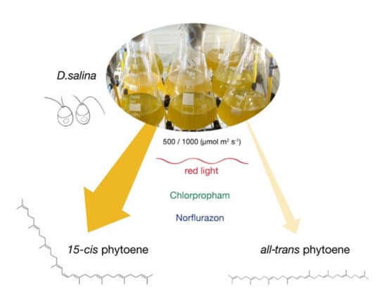
The main objective of this work was to resolve the phytoene isomers in D. salina and to gain new insight into the carotenoid synthesis pathway of D. salina. Methods of sample preparation from microalgal biomass and NMR methods were developed to characterize the phytoene isomers. The effects of cultivation conditions, light wavelength and light intensity, and herbicide treatment with two different classes of herbicides, chlorpropham and norflurazon, on carotenoid synthesis and phytoene isomer accumulation were also investigated.

2. Results

2.1. Identification of Phytoene Isomers in D. salina with HPLC

In the present study, HPLC analysis was carried out to identify the phytoene isomers in D. salina cultures. Method parameters were tuned to reduce the shifts in retention time of the phytoene peaks among runs. The phytoene standard purchased from Sigma is composed of a mixture of 6 isomers (Figure 2). Peak 3 of the standard matched with the phytoene isomer from tomato extract, which is widely considered as 15-cis phytoene. Peak 3 is also the closest peak to that from D. salina extracts, suggesting that phytoene accumulated in D. salina is most likely to be 15-cis phytoene as well.
A flow rate of 0.5 mL/min under isocratic solvent conditions (MeOH/MTBE- 88/10) was used to elute stereo-mutated tomato extract and phytoene standard to reveal the details of the elution peaks (Figure 3). The tomato extract stereo-mutated showed the presence of a second peak eluting after the 15-cis isomer, which was identified as all-trans phytoene, in accordance with previous work by Melendez-Martinez et al. [22]. This peak matched with peak 5 of the phytoene standard and peak 2 of D. salina extract shown previously in Figure 2, suggesting they are all-trans phytoene isomers.
Although both 15-cis and all-trans phytoene isomers gave absorption maxima at 275, 285 and 297 nm, they had slightly different absorption spectra (Figure 4). 15-cis phytoene showed a loss in fine structure in comparison with the all-trans phytoene in all the extracts, which agrees with the work by Melendez-Martinez et al. [22] and Britton et al. [25].

2.2. Phytoene Accumulation in D. salina Treated by Chlorpropham or Norflurazon

Use of either chlorpropham or norflurazon leads to over production of phytoene in D. salina [13,14]. Here, we treated the cultures with 20 µM chlorpropham or 5 µM norflurazon and found that the percentage abundance of the 15-cis phytoene in total phytoene was 99.80 ±0.08, 99.80 ± 0.07 and 99.78 ± 0.07 for the control cultures, chlorpropham-treated cultures and norflurazon-treated cultures respectively. 15-cis phytoene was therefore the predominant isomer that accumulated with herbicide treatment (Figure 5).

2.3. Analysis of Phytoene with UPC2-MS

The presence of the 15-cis phytoene in the norflurazon- and chlorpropham-treated D. salina extracts was evaluated with UPC2 analysis coupled with mass spectrometry, using 15-cis phytoene extracted from tomato as a standard. Figure 6 shows the UPC2 chromatograms of phytoene extracted from tomato and D. salina cultures treated with either chlorpropham or norflurazon. The chromatograms could be superimposed on one another (RT chlorpropham extract = 2.50 min; RT norflurazon extract = 2.49 min; RT tomato = 2.49 min). Mass accuracy analysis confirmed the presence of phytoene in all the extracts (Figure 7). Protonated ions [M + H]+ corresponding to phytoene were all within 10 ppm.

2.4. NMR Analysis of Phytoene Extracts from D. salina

In the present study, NMR analysis was carried out to prove that the main peak from D. salina detected with HPLC was 15-cis phytoene. Phytoene in a carotenoid-lipophilic mixture extracted from tomato was analysed with NMR, and chemical shifts values were assigned and compared to those identified in the mixture extracted from D. salina biomass. Figure 8a shows the results of 2D-HSCQ obtained from the tomato extract. Proton Chemical shifts of the conjugated central part C14, C15, C15′, C14′ were as follow: δ h14,14′ = 6.31 ppm, δ h15, 15′ = 6.100. Carbon chemical shifts were H-C (14, 14′) 120.00, H-C (15, 15′) 123.18. Chemical shifts values assigned were in accordance with the literature [18,23,24]. 2D-HSCQ NMR spectra of the alga biomass are shown in Figure 8b,c. The spectra of the tomato and the alga mixtures are superimposable. Proton chemical shifts were δ h14,14′ = 6.31 ppm and δ h15, 15′ = 6.10, and δ h14,14′ = 6.31 ppm and δ h15, 15′ = 6.10 ppm for the chlorpropham and norflurazon treated alga respectively. Carbon chemical shifts were H-C (14, 14′) 120.42, H-C (15, 15′) 123.60 for both the treated algae. These results show that in the presence of either chlorpropham or norflurazon, the alga Dunaliella salina accumulates 15-cis phytoene as the main isomer.

2.5. Colourless Phytofluene Isomers Produced by D. salina

Phytofluene isomeric forms were detected at the wavelength of 350 nm in HPLC-diode array analysis (Figure 9). Two phytofluene peaks were detected in tomato extracts. Peak 1 from tomato extracts matched with peak 1 of D. salina extracts (RT: (a) 16.177; (b) 16.171; (c) 16.212 and (d) 16.186) and corresponded to the major phytofluene in both tomato and D. salina. Phytofluene was over-accumulated in chlorpropham-treated D. salina cultures but not in norflurazon-treated D. salina cultures (Figure 9a). The second phytofluene isomeric form in tomato extracts appeared in trace amount and could only be detected in the extracts of chlorpropham treated D. salina cultures (RT: (a) 23.714 and RT (b) 23.674), but not in the norflurazon treated cultures or the control cultures.

2.6. Phytoene and Phytofluene Production in D. salina under Different Light Conditions

Previously, we have shown that light wavelengths affect the synthesis of carotenoids in D. salina. We found that the red light increased the cellular content of all major carotenoids and total carotenoids [14]. Figure 10 shows the concentration of phytoene isomers in D. salina cultures grown under red or blue light at different light intensities (200, 500 or 1000 µmol·m−2·s−1) for 48 h. The major phytoene isomer 15-cis phytoene in D. salina cultures changed with light wavelengths and light intensities, while no significant difference was found in the concentration of all-trans phytoene under all light conditions.
Concentrations of 15-cis phytoene in the cultures increased with light intensity under both red and blue light, and generally showed higher concentrations under red than blue light. The ratio of 15-cis/all-trans phytoene increased with light intensity from 2.2 ± 0.1 to 5.9 ± 0.1 under red light and increased from 1.4 ± 0.2 to 3.9 ± 0.4 under blue light. A Pearson correlation analysis was carried out between the two phytoene isomers, and no significant correlation was found (p = 0.34).
Phytofluene was measured at 355 nm using HPLC. Only one phytofluene isomer was detected in the carotenoid extracts from D. salina biomass, and its RT corresponded to that for phytofluene from tomato extracts. The concentration of phytofluene increased with light intensity under both red and blue light, with concentrations under red light more than double the concentrations under blue light (Figure 11).

3. Discussion

Phytoene demand has increased recently due to its potential health benefits. In higher plants and fruits, such as tomato, phytoene is found predominantly in the 15-cis form. However, little work has been done to characterize the phytoene isomers in microalgae, such as D. salina, which is a rich source of natural carotenoids, especially β-carotene with a high ratio of the valuable 9-cis β-carotene. This work characterized phytoene isomers in D. salina and compared the production of phytoene in D. salina cultures treated with the two different classes of herbicides norflurazon and chlorpropham. After both treatments, the concentration of 15-cis phytoene increased. Moreover, the colourless phytofluene isomers over-accumulated in chlorpropham, but not norflurazon-treated D. salina cultures.
Results of HPLC analysis, UPC2 analysis and NMR analysis each showed that 15-cis phytoene was the main phytoene isomer that accumulated in D. salina (>98% of total phytoene). Furthermore, the 15-cis phytoene increased with the treatment of both the herbicides norflurazon and chlorpropham and with increasing light intensity. Identification of 15-cis phytoene with only trace amounts of all-trans phytoene is in concord with findings for ripe tomato and higher plants [8,22] and with results obtained by Ebenezer et al. for D. bardawil [18], but not with the results obtained by Ben-Amotz et al. [13] and Werman et al. [6], who showed that both 9-cis phytoene and all-trans phytoene accumulated in D. bardawil treated with herbicides.
15-cis phytofluene has been considered by many to be the predominant phytofluene isomer (for a review see [4]). However, Koschmieder et al. [26] compared synthetic 15-cis phytofluene and all-trans phytofluene with phytofluene isomers from tomato and norflurazon-treated Dunaliella bardawil, and on the basis of RT obtained after HPLC analysis as well as absorption maxima at 331nm, 348 nm and 366 nm, concluded that phytofluene in tomato extract was predominately 9,15-di-cis phytofluene, which also exists in Dunaliella bardawil in a small proportion. They also determined that the main phytofluene isomer in norflurazon treated D. bardawil was 9-cis phytofluene but did not present the isomeric configurations in untreated D. bardawil cultures. In contrast, our results showed that the main phytofluene isomer in both untreated and norflurazon in chlorpropham treated D. salina was the same as in tomato extracts, which is 9,15-di-cis phytofluene. The 9-cis phytofluene shown by Koschmieder et al. [26] is likely not a phytofluene peak because norflurazon inhibits PDS function and blocks the desaturation of phytoene to subsequent phytofluene products.
The fact that only 15-cis phytoene accumulated in D. salina when treated with phytoene desaturase inhibitor, while no accumulation of all-trans phytoene was found, is consistent with 15-cis phytoene as the most likely precursor of phytofluene. All-trans phytoene present in D. salina is likely to be formed by the isomerisation of the predominant 15-cis phytoene and this reaction step along with the conversion of 15-cis phytoene to phytofluene might be fast enough to occur prior to the interference of the chlorpropham herbicide action. As a second hypothesis, all-trans phytoene may also be formed directly from the precursor of geranylgeranyl pyrophosphate as suggested by Gregonis and Rilling [27]. An early study on a green microalga Scenedesmus obliquus also supports the idea that 15-cis phytoene is converted via 15-cis phytofluene and 15-cis ζ-carotene into all-trans-gz-carotene and trans-bicyclic carotenoids such as α-carotene and β-carotene [28]. Our results show that 15-cis phytoene is the main stereoisomer produced by D. salina and that it is over-accumulated with red light and with the use of herbicides. Our results also support the recent finding that 9-cis β-carotene is formed by isomerisation of all-trans β-carotene with the function of a 9-cis/all-trans β-carotene isomerase [19,20], rather than synthesised from the precursor of 9-cis phytoene.

4. Materials and Methods

4.1. Algal Cultivation

D. salina strain CCAP 19/41 (PLY DF15) was obtained from the Marine Biological Association, Plymouth, UK (MBA). Algae were cultured as described in previous work [14]. Inoculum was prepared by adding 10 mL of the culture stock of D. salina to 250 mL of fresh media until the culture reached the exponential phase. Algae were cultured in 500 mL Modified Johnsons Medium containing 1.5 M NaCl and 10 mM NaHCO3 in an illuminated incubator (Varicon Aqua, Worcester, UK) under white light of 500 µmol·m−2·s−1 at 25 °C. To study the effect of light wavelength, algae were cultivated in Algem photobioreactor under red or blue light of 500 µmol·m−2·s−1 at 25 °C. To study the effect of herbicides, cultures were treated with either 5 µM of norflurazon or 20 µM of chlorpropham for 48 h before carotenoid extraction. All the experiments were performed in triplicate.

4.2. Standards and Solvents

Phytoene standard (mixture of isomers (E/Z), 95% purity) was purchased from Sigma–Aldrich (Merck KGaA, Darmstadt, Germany). Methanol (MeOH) and Methyl tert Butyl Ether (MtBE), both HPLC grade, were purchased from Fischer Scientific UK Ltd. (Loughborough, Leicestershire, UK).

4.3. Carotenoids Extraction

The carotenoids were extracted from wet alga biomass as follows: the samples were centrifuged at 3000× g for 5 min at 5 °C; 10 mL MeOH-MtBE (80:20) were added to the pellets and sonicated and vortexed for 20 s; extracts were clarified at the centrifuge then filtered (0.20 µm filter) into amber HPLC vials before HPLC analysis.
15-cis phytoene was also extracted from ripe tomatoes (purchased from Waitrose and Partners, UK) to be used as control material because it favourably accumulates the compound at high concentration.

4.4. HPLC Analysis

High-Performance Liquid Chromatography equipped with Diode-Array Detection (HPLC-DAD; Agilent Technologies 1200 series, Agilent, Santa Clara, CA, United States), on-line degasser, a quaternary pump system, a YMC30 250 × 4.9 mm I.D S-5µ column (YMC, Europe GmbH) was used to resolve the phytoene and the phytofluene from the carotenoid extracts. The column temperature was set at 15 °C, the gradient solvent system was MeOH (A): MtBE (B) running at 80% A for the first 10 min at a flow rate of 1 mL/min, then increased to 100% A for the next 10 min at a flow rate of 0.5 mL/min before going back to the initial conditions at 20 min. Total run time was 45 min. The absorbance at four wavelengths (276, 282, 293 and 350) was monitored. All data were acquired and analysed with Chemstation for LC System software. Phytoene extracted from ripe tomatoes was stereo-mutated similarly to Melendez-Martinez et al. [29]. The sample extracted was heated at 75 °C for a total of 240 min and it was then injected in the HPLC system for analysis. Methods parameters were as follow: the column was set at 15 °C, the isocratic solvent system was MeOH (A): MtBE (B) running at 88% A at a flow rate of 0.5 mL/min.

4.5. UPC2 Analysis

Supercritical, or near-critical, CO2 was used as the primary eluent in the mobile phase with a MeOH cosolvent gradient to resolve the phytoene from the total-tomato and D. salina extracts using a seven-minute gradient. Supercritical fluid chromatography is very powerful when it comes to identifying compounds based on their retention time, as it allows shorter run times than HPLC and therefore a decrease in shifts in the retention time among runs. The UPC2 (Waters Limited, Herts, UK) was equipped with a binary solvent delivery pump, auto-sampler, column oven, photodiode array detector and back-pressure regulator. Mobile phase A was CO2 and mobile phase B was MeOH with 0.1% formic acid (v/v). The gradient was held at 5% B for 0.5 min, then increased to 50% B at 4.9 min, held at 50% for 0.1 min, ramped to 5% B in 0.1 min, and equilibrated for 1.9 min to give a total run time of 7 min. The binary solvent flow rate was 1.0 mL/min, the isocratic make-up solvent flow was 0.3 mL/min, and the UPC2 was operated with a back-pressure of 2000 psi. The column oven was maintained at a temperature of 50 °C and an Acquity UPC2 HSS C18 SB (1.8 µm, 3.0 mm × 100 mm) column was used. A sample injection volume of 3.0 µL was used with all samples. The UPC2 was fitted with an Acquity PDA detector (190–800 nm) and operated in the wavelength range 210–600 nm with a 1.2 nm resolution. In addition, the absorbance at four wavelengths (276 nm, 282 nm, 293 nm and 450 nm) was monitored with a resolution of 1.2 nm.

4.6. Mass Spectrometry Analysis

All mass spectrometry experiments were collected on a Waters Synapt G2 Q-ToF mass spectrometer (Manchester, UK). The instrument was operated in positive ion mode electrospray with a capillary voltage of 2.0 kV and sampling cone voltage was 30 V. Nitrogen at a flow rate of 650 L/h was used as the desolvation gas with a constant desolvation temperature of 350 °C, a cone gas flow rate of 50 L/h, and a source temperature of 130 °C. Data were acquired over the m/z range 50–800. An integral LockSpray unit infusing Leucine-Enkephalin peptide into the electrospray sample stream was used to collect reference scans. Scans were performed every 10 s with the reference calibrant introduced at a flow rate of 10 µL/min using the fluidics system of the instrument. Single point lock-mass correction was used for the protonated pseudo-molecular ion at m/z 556.2771 (+ve). All data were acquired and analysed with Waters MassLynx v4.1 software (Manchester, UK).

4.7. NMR Sample Preparation

Fresh tomato samples and harvested D. salina cultures were freeze-dried and a total of 20 g of dried tomato powder and dried algae samples was used for the carotenoid extraction. The procedure described above was modified as follow: MtBE was chosen as solvent for extraction due to its lower polarity index (2.4), hence its higher selectivity for only carotenoids (and lipids) in comparison to the MeOH and ethanol solvents; moreover, its high volatility value allowed shorter evaporation times and minimisation of sample losses. The extracts were injected in the HPLC system to confirm the presence of phytoene. The MtBE extracts were evaporated for one and a half hour, flushed with nitrogen and suspended with deuterated chloroform prior to analysis.

4.8. NMR Conditions

The NMR experiments were carried out with a JEOL (Tokyo, Japan) 500 MHz. All the spectra were recorded at ambient temperature. Proton and carbon spectra for carotenoid analysis were referenced to the TMS signal (δ = 0.00 ppm). 1D Proton parameters: 1H 45° flip angle of 6.32 µs, spectral width 9.38 kHz, 65K data points, 64 scans and relaxation delay 2s. 1D Carbon parameters: 13C 30° flip angle of 3.56 µs, spectral width of 39.31 kHz, 65k data points, 1024 scans and relaxation delay 2s. 2D 1H-13C pulse field gradient heteronuclear single quantum correlation spectroscopy (HSQC): pulse of 12.64 µs for 1H, pulse of 10.7 µs for 13C, decoupling, J_constant of 135 Hz, spectral width of 6.26 and 31.45 kHz for the proton and carbon dimensions, respectively; relaxation delay 2s, 2048 total scan and 1024 and 256 data point in f2 and f1, respectively. Data were acquired and analysed with MNova software (12.0.3, Mestrelab Research S.L., Santiago de Compostela, Spain).

5. Conclusions

The present work used different analytical tools to identify and characterize the isomeric composition of phytoene, an increasingly sought after colourless carotenoid and the precursor of all other carotenoids in the carotenoid biosynthetic pathway, in order to evaluate its production from D. salina and help understand the synthesis of high content of 9-cis β-carotene in D. salina. Results show 15-cis phytoene is the major isomer in D. salina (>98% of total phytoene) while all-trans phytoene presents in trace amount. 9-cis phytoene has not been detected in any of the D. salina extracts.

6. Patents

WO2019097219-Production of Dunaliella (2019). Harvey PJ, Xu Y.; WO/2018/141978 Algal strains (2018). Schroeder D, Harvey PJ, Xu Y.

Author Contributions

Conceptualization, L.M., Y.X. and P.H.; methodology, L.M and Y.X.; validation, Y.X. and P.H..; investigation, L.M. and Y.X.; data curation, L.M and Y.X; writing—original draft preparation, L.M. and Y.X.; writing—review and editing, Y.X. and P.H.; visualization, L.M. and Y.X..; supervision, Y.X., P.H.; project administration, P.H.; funding acquisition, P.H. All authors have read and agreed to the published version of the manuscript.

Funding

LM was funded by a VC Scholarship, University of Greenwich. YZ and PH were funded by the University of Greenwich and by the Interreg 2 Seas programme 2014-2020 cofunded by the European Regional Development Fund under subsidy contract No ValgOrize 2S05017.

Acknowledgments

We are grateful to Iain Goodall, Pink Darren, Victoria Elmes of the University of Greenwich for their technical assistance.

Conflicts of Interest

The authors declare no conflict of interest.

References

  1. Tanaka, Y.; Sasaki, N.; Ohmiya, A. Biosynthesis of plant pigments: Anthocyanins, betalains and carotenoids. Plant J. 2008, 54, 733–749. [Google Scholar] [CrossRef]
  2. Khoo, H.E.; Prasad, K.N.; Kong, K.W.; Jiang, Y.; Ismail, A. Carotenoids and their isomers: Color pigments in fruits and vegetables. Molecules 2011, 16, 1710–1738. [Google Scholar] [CrossRef] [PubMed]
  3. Engelmann, N.J.; Clinton, S.K.; Erdman, J.W., Jr. Nutritional aspects of phytoene and phytofluene, carotenoid precursors to lycopene. Adv. Nutr. 2011, 2, 51–61. [Google Scholar] [CrossRef] [PubMed] [Green Version]
  4. Meléndez-Martínez, A.J.; Mapelli-Brahm, P.; Benítez-González, A.; Stinco, C.M. A comprehensive review on the colorless carotenoids phytoene and phytofluene. Arch. Biochem. Biophys. 2015, 572, 188–200. [Google Scholar] [CrossRef] [PubMed] [Green Version]
  5. Srinivasan, R.; Babu, S.; Gothandam, K.M. Accumulation of phytoene, a colorless carotenoid by inhibition of phytoene desaturase (PDS) gene in Dunaliella salina V-101. Bioresour. Technol. 2017, 242, 311–318. [Google Scholar] [CrossRef] [PubMed]
  6. Werman, M.J.; Mokady, S.; Ben-Amotz, A. Bioavailability of the isomer mixture of phytoene and phytofluene-rich alga Dunaliella bardawil in rat plasma and tissues. J. Nutr. Biochem. 2002, 13, 585–591. [Google Scholar] [CrossRef]
  7. Takaichi, S. Carotenoids in algae: Distributions, biosynthesis and functions. Marine drugs. 2011, 9, 1101–1118. [Google Scholar] [CrossRef] [PubMed]
  8. Alagoz, Y.; Nayak, P.; Dhami, N.; Cazzonelli, C.I. Cis-carotene biosynthesis, evolution and regulation in plants: The emergence of novel signaling metabolites. Arch. Biochem. Biophys. 2018, 654, 172–184. [Google Scholar] [CrossRef]
  9. Enfissi, E.M.; Nogueira, M.; Bramley, P.M.; Fraser, P.D. The regulation of carotenoid formation in tomato fruit. Plant J. 2017, 89, 774–788. [Google Scholar] [CrossRef] [Green Version]
  10. McQuinn, R.P.; Wong, B.; Giovannoni, J.J. AtPDS overexpression in tomato: Exposing unique patterns of carotenoid self-regulation and an alternative strategy for the enhancement of fruit carotenoid content. Plant Biotechnol. J. 2018, 16, 482–494. [Google Scholar] [CrossRef] [Green Version]
  11. Welsch, R.; Wüst, F.; Bär, C.; Al-Babili, S.; Beyer, P. A third phytoene synthase is devoted to abiotic stress-induced abscisic acid formation in rice and defines functional diversification of phytoene synthase genes. Plant Physiol. 2008, 147, 367–380. [Google Scholar] [CrossRef] [PubMed] [Green Version]
  12. Giuliano, G.; Giliberto, L.; Rosati, C. Carotenoid isomerase: A tale of light and isomers. Trends Plant Sci. 2002, 7, 427–429. [Google Scholar] [CrossRef]
  13. Ben-Amotz, A.; Lers, A.; Avron, M. Stereoisomers of β-carotene and phytoene in the alga Dunaliella bardawil. Plant Physiol. 1988, 86, 1286–1291. [Google Scholar] [CrossRef] [PubMed] [Green Version]
  14. Xu, Y.; Harvey, P.J. Carotenoid Production by Dunaliella salina under Red Light. Antioxidants 2019, 8, 123. [Google Scholar] [CrossRef] [Green Version]
  15. Lamers, P.P.; Janssen, M.; De Vos, R.C.; Bino, R.J.; Wijffels, R.H. Exploring and exploiting carotenoid accumulation in Dunaliella salina for cell-factory applications. Trends Biotechnol. 2008, 26, 631–638. [Google Scholar] [CrossRef]
  16. Breitenbach, J.; Zhu, C.; Sandmann, G. Bleaching herbicide norflurazon inhibits phytoene desaturase by competition with the cofactors. J. Agric. Food Chem. 2001, 49, 5270–5272. [Google Scholar] [CrossRef]
  17. Simkin, A.J.; Breitenbach, J.; Kuntz, M.; Sandmann, G. In vitro and in situ inhibition of carotenoid biosynthesis in Capsicum annuum by bleaching herbicides. J. Agric. Food Chem. 2000, 48, 4676–4680. [Google Scholar] [CrossRef]
  18. Ebenezer, W.J.; Pattenden, G. Cis-Stereoisomers of β-carotene and its congeners in the alga Dunaliella bardawil, and their biogenetic interrelationships. J. Chem. Soc. Perkin Trans. 1 1993, 1869–1873. [Google Scholar] [CrossRef]
  19. Davidi, L.; Pick, U. Novel 9-cis/all-trans β-carotene isomerases from plastidic oil bodies in Dunaliella bardawil catalyze the conversion of all-trans to 9-cis β-carotene. Plant Cell Rep. 2017, 36, 807–814. [Google Scholar] [CrossRef]
  20. Xu, Y.; Harvey, P.J. Red Light Control of β-Carotene Isomerisation to 9-cis β-Carotene and Carotenoid Accumulation in Dunaliella salina. Antioxidants 2019, 8, 148. [Google Scholar] [CrossRef] [Green Version]
  21. Gupta, P.; Sreelakshmi, Y.; Sharma, R. A rapid and sensitive method for determination of carotenoids in plant tissues by high performance liquid chromatography. Plant Methods 2015, 11, 5. [Google Scholar] [CrossRef] [PubMed] [Green Version]
  22. Melendez-Martinez, A.J.; Stinco, C.M.; Liu, C.; Wang, X.D. A simple HPLC method for the comprehensive analysis of cis/trans (Z/E) geometrical isomers of carotenoids for nutritional studies. Food Chem. 2013, 138, 1341–1350. [Google Scholar] [CrossRef] [PubMed]
  23. Granger, P.; Maudinas, B.; Herber, R.; Villoutreix, J. 1H and 13C NMR spectra of cis and trans phytoene isomers. J. Magn. Reson. 1973, 10, 43–50. [Google Scholar] [CrossRef]
  24. Tiziani, S.; Schwartz, S.J.; Vodovotz, Y. Profiling of carotenoids in tomato juice by one-and two-dimensional NMR. J. Agric. Food Chem. 2006, 54, 6094–6100. [Google Scholar] [CrossRef]
  25. Britton, G. Structure and properties of carotenoids in relation to function. Faseb J. 1995, 9, 1551–1558. [Google Scholar] [CrossRef]
  26. Koschmieder, J.; Fehling-Kaschek, M.; Schaub, P.; Ghisla, S.; Brausemann, A.; Timmer, J.; Beyer, P. Plant-type phytoene desaturase: Functional evaluation of structural implications. PLoS ONE 2017, 12, e0187628. [Google Scholar] [CrossRef] [Green Version]
  27. Gregonis, D.E.; Rilling, H.C. Stereochemistry of trans-phytoene synthesis. Lycopersene as a carotene precursor and a mechanism for the synthesis of cis-and trans-phytoene. Biochemistry 1974, 13, 1538–1542. [Google Scholar] [CrossRef]
  28. Powls, R.; Britton, G. The roles of isomers of phytoene, phytofluene and ζ-carotene in carotenoid biosynthesis by a mutant strain of Scenedesmus obliquus. Arch. Microbiol. 1977, 115, 175–179. [Google Scholar] [CrossRef]
  29. Meléndez-Martínez, A.J.; Paulino, M.; Stinco, C.M.; Mapelli-Brahm, P.; Wang, X.D. Study of the time-course of cis/trans (Z/E) isomerization of lycopene, phytoene, and phytofluene from tomato. J. Agric. Food Chem. 2014, 62, 12399–12406. [Google Scholar] [CrossRef]
Sample Availability: Samples of tomato fruits and phytoene standard are available from the authors. Dunaliella salina strain CCAP 19/41 (PLY DF15) was obtained from the Marine Biological Association, UK.
Figure 1. A generally acknowledged biosynthetic pathway of carotenoids in plants and algae [8,9,10]. PSY, phytoene synthase; PDS, phytoene desaturase; Z-ISO, ζ-carotene isomerase; ZDS, ζ-carotene desaturase; CRTISO, carotenoid isomerase; εLCY, lycopene ε-cyclase; βLCY, lycopene β-cyclase; Chemical structures of 15-cis phytoene and 9,15-di-cis-phytofluene (ACD/ChemSketch Software) are also shown in the figure.
Molecules 25 01880 g001
Figure 2. HPLC analysis of phytoene standard purchased from Sigma, phytoene extracts from ripe tomatoes and phytoene extracts from D. salina cultures.
Molecules 25 01880 g002
Figure 3. HPLC chromatograms of stereo-mutated phytoene extracts from ripe tomatoes compared to phytoene standard mixture, only showing the region of interest.
Molecules 25 01880 g003
Figure 4. Absorption spectra of (a) 15-cis and (b) all-trans phytoene analysed by HPLC-diode array in MeOH/MTBE (8/2) solvent with maximum absorption at 275, 285 and 297 nm.
Molecules 25 01880 g004
Figure 5. HPLC chromatograms of phytoene extracts from D. salina cultures treated with 20 µM of chlorpropham or 5 µM of norflurazon, only showing the region of interest in the chromatogram.
Molecules 25 01880 g005
Figure 6. UPC2 chromatogram, showing phytoene extracted from ripe tomatoes (peak 1), Norflurazon (peak 2) and chlorpropham (peak 3) treated D. salina cultures.
Molecules 25 01880 g006
Figure 7. Region of mass spectra showing the molecular mass of phytoene derived from (a) ripe tomatoes (545.5081), (b) chlorpropham-treated D. salina (545.5073) and (c) norflurazon-treated D. salina (545.5080), in positive ionisation mode [M + H]+.
Molecules 25 01880 g007
Figure 8. 1H–13C-NMR spectra displayed as heteronuclear single quantum correlation spectra (HSQC) to show the chemical shifts values (ppm) for the carbon atoms C14, C15, C15′ and C14′ of phytoene extracted from (a) tomato, (b) chlorpropham and (c) norflurazon treated D. salina extracts. Numbers above the 1H-NMR spectra indicate the carbon number of phytoene associated with the resonance.
Molecules 25 01880 g008aMolecules 25 01880 g008b
Figure 9. (a) HPLC analysis of phytofluene isomers extracted from ripe tomatoes, control D. salina cultures with no herbicide, D. salina cultures treated with 20 µM chlorpropham, and D. salina cultures treated with 5 µM norflurazon, only showing the region of interest in the chromatogram and (b) absorption spectrum of phytofluene in Methanol/MTBE (8/2) obtained from UPC2.
Molecules 25 01880 g009
Figure 10. Phytoene isomeric composition in D. salina cultivated under (a), (b) red or (c), (d) blue light at different light intensities (200, 500 or 1000 µmol·m−2·s−1) for 48 h.
Molecules 25 01880 g010
Figure 11. Phytofluene concentration in D. salina under red light or blue light at different light intensities (200, 500 or 1000 µmol·m−2·s−1) for 48 h.
Molecules 25 01880 g011

