Article

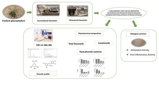
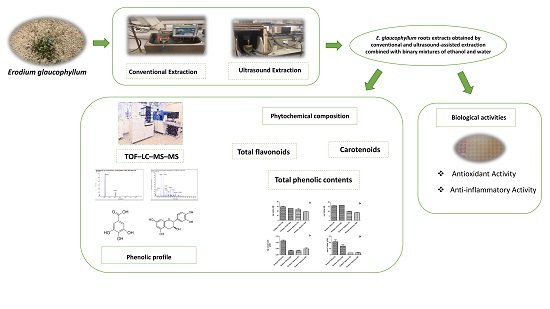
Ultrasonically-Assisted and Conventional Extraction from Erodium Glaucophyllum Roots Using Ethanol:Water Mixtures: Phenolic Characterization, Antioxidant, and Anti-Inflammatory Activities

1
Nutrition and Food Science Area, Preventive Medicine and Public Health, Food Science, Toxicology and Forensic Medicine Department, Faculty of Pharmacy, Universitat de València, Avda, Vicent Andrés Estellés, s/n, Burjassot, 46100 València, Spain
2
Department of Biotechnology, Institute of Agrochemistry and Food Technology, Spanish National Research Council (IATA-CSIC), Av. Agustin Escardino 7, 46980 València, Spain
3
Range Ecology Laboratory, Institute of Arid Regions (IRA), University of Gabès, Medenine 4100, Tunisia
4
Centro Tecnológico de la Carne de Galicia, Adva. Galicia n°4, Parque Tecnológico de Galicia, San Cibrao das Viñas, 32900 Ourense, Spain
5
Grupo de Análisis y Simulación de Procesos Agroalimentarios (ASPA), Departamento de Tecnología de Alimentos, Universitat Politècnica de València, Cami de Vera s/n, 46022 Valencia, Spain
*
Authors to whom correspondence should be addressed.
Molecules 2020, 25(7), 1759; https://doi.org/10.3390/molecules25071759
Submission received: 6 March 2020 / Revised: 1 April 2020 / Accepted: 8 April 2020 / Published: 10 April 2020

Abstract

:
The paper presents experimental results concerning the ultrasonically-assisted extraction of bioactive compounds from Erodium glaucophyllum roots. A comparison with conventional methodology is presented, and thereby the phytochemical composition and the antioxidant and anti-inflammatory activities of extracts are evaluated. The phenolic profile of Erodium extracts was analyzed by TOF–LC–MS–MS. The identification of phenolic compounds revealed that the major component was (+)-gallocatechin in the aqueous extracts obtained for the different extraction methodologies. The highest quantity of phenolic compounds and antioxidant capacity was found in the hydroethanolic extract obtained by conventional extraction (29.22–25.50 mg GAE/g DM; 21.174 mM Trolox equivalent). The highest content of carotenoids, varying from 0.035 to 0.114 mg/g dry matter, was reached by ultrasonic-assisted extraction. Furthermore, Erodium extracts showed a potent inhibition of the inflammatory reaction by means of the inhibition of tumor necrosis factor-alpha (TNF-α). The extracts obtained when ultrasound extraction was combined with ethanol:water (50:50, v/v) presented the greatest inhibition (92%).

Graphical Abstract

1. Introduction

Erodium glaucophyllum belongs to a genus of flowering plants which have been used from ancient times in traditional medicine due to their health attributes. Some of these beneficial properties have been linked to their high content of antioxidant bioactive components, such as vitamin C, polyphenols and carotenoids, among others. For instance, due to the interesting phytochemical profile of E. glaucophyllum, several attempts have been made, especially in North Africa, to evaluate its potential use in the areas of agri-food, pharmaceutics, and cosmetics [1,2].
In this vein, Munekata et al. [1] and Al-snafi [3] reviewed the phytochemical profile of Erodium sp. showing the potential of Erodium cicutarium as a source of vitamin C and phenolic compounds (eg., tannins, catechins, gallic and ellagic acids), with antibacterial and antiviral, antioxidant, and anti-inflammatory properties. Moreover, they found a significant quantity of sugars, amino acids, and vitamin K, among others. In addition, the biochemical composition and the biological activities of Erodium glaucophyllum have also been evaluated. Indeed, Bouaziz et al. [4] observed that the aerial part of Erodium glaucophyllum presented the highest levels of total phenols and flavonoids after evaluating the phytochemical composition of 25 plants from southern Tunisia, as well as powerful antioxidant activity.
Therefore, there is a need to develop fast, reliable, and efficient methods with which to obtain the extracts. Conventional solid–liquid extraction techniques, involving the use of maceration or Soxhlet, have significant drawbacks, mainly high energy consumption and the use of polluting solvents [5]. Currently, the use of ecological, sustainable extraction methods for natural products is a live issue in the multidisciplinary areas of applied chemistry, biology, and technology.
Ultrasonic-assisted extraction (UAE) is a promising strategy for the purposes of recovering bioactive molecules [6,7,8]. Literature reports that efficient UAE mainly contributes to the shortening of the extraction times, the use of milder temperatures, and the reduction of solvents; all these aspects not only positively impact on the industrial productivity but also have environmental benefits linked to energy and solvent reduction. In addition, UAE could modify the profile of the extracted compounds compared to conventional extraction since it may facilitate the release of molecules strongly attached to the solid matrix. The effects of UAE are mainly attributed to cavitation phenomena, thus promoting stirring, and also to the temperature increase associated with the ultrasound process; this both promotes bioactive diffusion and solubility, and subsequently the rate extraction yield, and shortens the extraction time [9,10,11,12]. However, it should be noted that when extreme temperatures are reached, the degradation of thermolabile compounds could be promoted [13]. The thermal effect linked to UAE in a liquid medium is frequently not considered during experimentation, which leads to a misunderstanding of the phenomena involved due to the fact that most of the observed effects on the kinetics, yields, or extract composition are brought about by the temperature increase in the medium. In addition, the UAE is often compared to static maceration which evidences a marked effect of ultrasound on fluid turbulence, although similar findings could be reached using efficient mechanical agitators. Therefore, in order to assess the feasibility of UAE compared to other efficient approaches, experimental setups should incorporate accurate temperature control systems and fluid stirrers. However, these aspects are sometimes not taken into account in the available literature.
The process of inflammation is explained according to the production of nitric oxide (NO) from L-arginine by the inducible NO synthase (iNOS). In quick reactions with superoxide, NO gives rise to peroxynitrite. Peroxynitrite is a reactive oxidant and toxic substance that causes tissue damage during inflammatory diseases [14]. Since the critical etiology and exacerbating mechanisms are yet to be identified, it is difficult to find a side-effect free “miracle” drug for chronic inflammatory disorders [15]. Therefore, the demand for natural, safe anti-inflammatory agents has increased. Citing the example of currently used steroidal and non-steroidal anti-inflammatory drugs to treat acute inflammatory disorders, these conventional drugs have not been successful in curing chronic inflammatory disorders, such as chronic rheumatoid arthritis and atopic dermatitis [16].
Plants are a potential source of natural bioactive molecules. They are the subject of rigorous scientific studies for their possible use as alternative medicines, especially for the treatment of anti-inflammatory diseases. Among the several groups of natural products present in plants, their anti-inflammatory activity is mainly attributed to the phenolic compounds. Khlifi et al. [17] showed a good correlation between the phenolic content in plant extracts, in particular that of tannins and flavonoids, with the anti-inflammatory activity. Another study confirmed this correlation, indicating that the flavonoids and sterols identified in several plants are responsible for the anti-inflammatory activity [18].
Despite the great importance and significant benefits that North African countries (e.g., Tunisia) could gain from E. glaucophyllum, to the best of our knowledge, the literature evaluating the phytochemical profile of E. glaucophyllum is scarce. Evaluating the phytochemical profile is a crucial step prior to using a plant. Moreover, there is no information about the best extraction method with which to extract the phytochemical compounds from E. glaucophyllum and the potential implications of the anti-inflammatory response. Therefore, this study analyzed the antioxidant profile of E. glaucophyllum extracts obtained after conventional or UAE combined with binary mixtures of ethanol and water. Moreover, the anti-inflammatory response of the extracts was evaluated and compared.

2. Results and Discussion

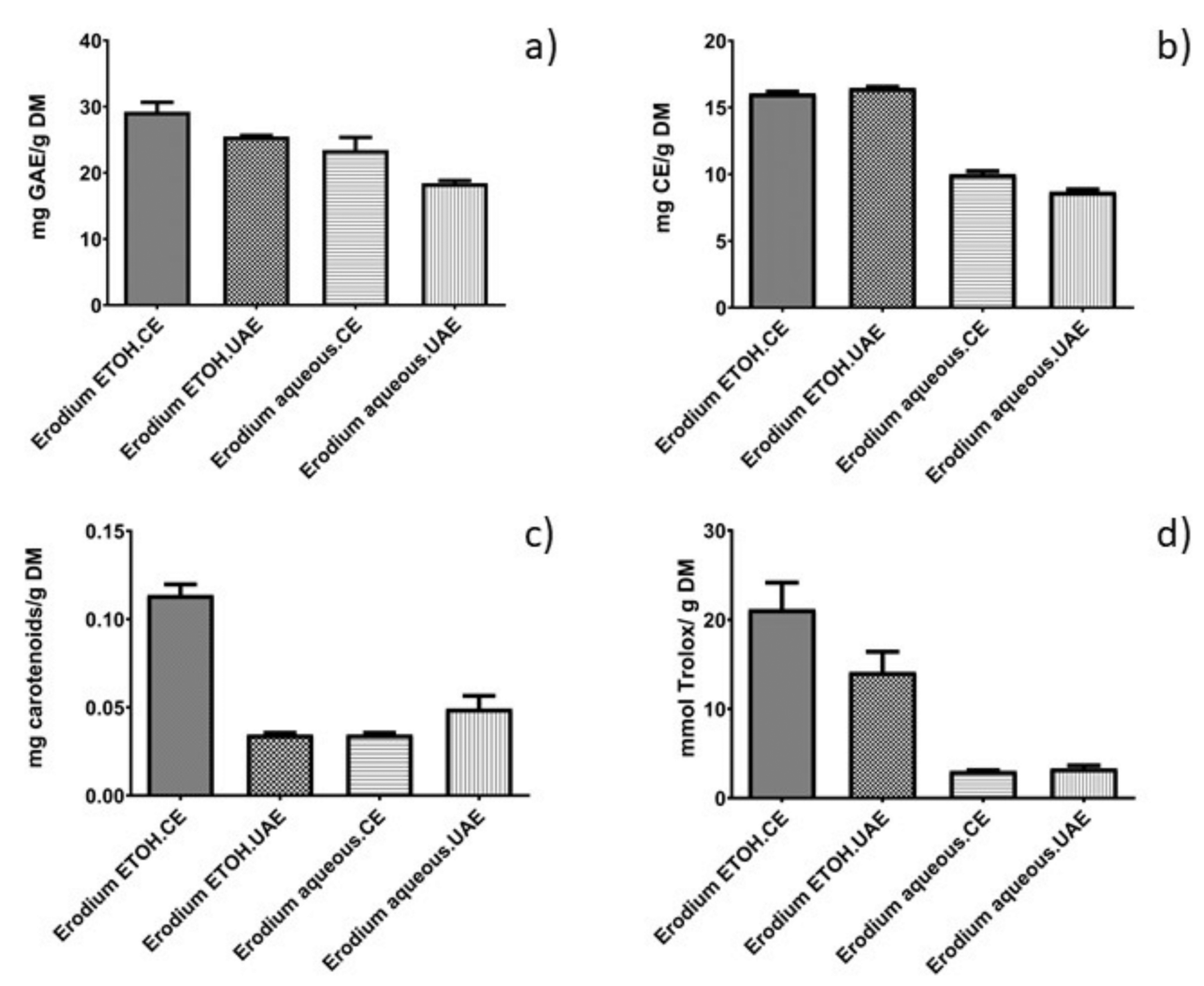
2.1. Assessment of Total Phenolic Compounds, Total Flavonoids, Phenolic Profile, Carotenoid Content, and Antioxidant Capacity

Firstly, the contents of the total phenolic compounds (TPC), total flavonoids, carotenoids, and the total antioxidant capacity of the E. glaucophyllum hydroethanolic extracts obtained after conventional extraction (CE) or ultrasound-assisted extraction (UAE) were evaluated (Figure 1).
Overall, the total phenolic contents (TPC) were similar to the results previously reported by Bouaziz et al. [4] when they evaluated the TPC composition of aerial parts of Erodium glaucophyllum (22.25 mg pyrogallol equivalents/g dry matter (DM)). In this study, the authors compared the levels of the phenolic compounds of 25 plants and found that the extract of the aerial part of E. glaucophyllum had the highest content.
Moreover, the ANOVA analysis showed that both treatment and solvent had a significant impact (p < 0.05) on TPC Erodium extracts. In fact, the extracts obtained after CE exhibited a higher TPC than the UAE treatments (Figure 1). These results are in close agreement with those previously reported by Zhang et al. [19] and Qiao et al. [20] who also observed a significant effect of ultrasound on the degradation of gallic acid and phenolic acids, especially when ultrasound was applied at high energy levels. The authors attributed this phenomenon to the changes in the physicochemical properties of the liquid phase under ultrasonic irradiation, commonly having significant effects on i) the number of the cavities or ii) the pressure/temperature pulse generated, which are attributed to the collapse of the cavity. This fact could be attributed to the instability of these phenolic compounds under UAE conditions (temperature, time, power, solvent). For a detailed description about the UAE mechanism see [21,22,23]. In addition, it could be also considered that under these particular conditions, the optimized CE used was more efficient than UAE to release the phenolic compounds from the solid matrix.
The combination of ethanol:water (50:50, v/v) permitted TPC values to be higher (29.223–25.504 mg GAE/g dry matter (DM)) than aqueous-assisted extraction (23.454–18.445 mg GAE/g DM). Previous studies have shown that polyphenols are compounds of moderate polarity, which tend to be extracted with solvents with medium polarity; thus, ethanol is a more convenient solvent than water with which to extract TPC. Similar findings were also reported by Wei et al. [24] after using solvents of increasing polarities for the characterization of medicinal plants and Carbonell-Capella et al. [25] when comparing combinations of ethanol:water vs. water to extract TPC from Stevia rebaudiana Bertoni leaves. Owczarek et al. [26] showed that a polyphenol-rich extract exhibits an inhibitory effect on the insulin-like growth factors that participate in several cellular processes (proliferation, differentiation, and apoptosis).
Like the results obtained for TPC, the ANOVA analysis showed that the total flavonoid content was markedly superior (p < 0.05) in hydroethanolic extracts (≈16 mg catechin equivalents (CE)/g DM) than in aqueous extracts (≈9 mg CE/g DM). These values were significantly higher than those reported by Bouaziz et al. [3] when they evaluated the total flavonoid content of Erodium glaucophyllum (2.03 mg rutin equivalents/g DM). However, the negative effects of ultrasound were only shown when water was used as a solvent. On the other hand, Galvan D’Alessandro et al. [27] tested the yield of phenolic compounds in black chokeberry extracts obtained following ultrasound extraction, varying the ethanol:water percentages. The authors noted that the presence of ethanol in the extraction solvent increased the yield of polyphenols, and a 50% extraction of ethanol was 2 times higher than the extraction yield with pure water. Solvents with alcoholic percentages have been commonly used to extract phenolic compounds (anthocyanins, flavonoids, tannins, etc.) from plant matrices [28,29].
In order to evaluate the influence of the treatment on the phenolic profile, a triple TOF–LC–MS–MS analysis was carried out (Table 1). As can be seen in Table 1, the profile of individual phenolic compounds obtained from conventional and ultrasound extracts were different. For instance, the analysis of the aqueous extract of Erodium obtained following a CE identified 5 flavonoids, 3 phenolic acids, and 4 polyphenols corresponding to the group of “other polyphenols,” while 10 flavonoids, 5 phenolic acids, 1 phenol, 2 polyphenols corresponding to the group of “other polyphenols,” and 1 stilbenoid were identified after UAE.
The predominant compound for both extracts was (+)-gallocatechin, and the intensity of the peak was higher in the extracts obtained by CE (Table 1). Moreover, our results coincide with those found by Roseiro et al. [30], showing that the phenolic profile of the carob (Ceratonia siliqua L.) extracts obtained using CE presented chromatograms with peaks more intense than those obtained after UAE.
In another study, the UAE of blackberry juice promoted the degradation of the juice’s color, which was mainly due to the degradation of carotenoid and anthocyanin pigments [31]. According to the authors, the degradation under UAE can be caused by cavitation, which governs various reactions: the modification of diffusion rates, the quickening of chemical reactions, and the degradation of enzymes and microorganisms [32]. Da Porto et al. [33] compared the extraction of polyphenols from grape (Vitis vinifera L.) seeds using two different methods: CE and UAE. The authors observed that UAE promoted a decrease in the contents of phenolic compounds, such as cinnamic acids, and the degradation of certain compounds [34]. Zhang et al. [19] also observed the same effect of UAE on the degradation of phenolic compounds. This result agrees with the TPC content (Figure 1) which, overall, showed a smaller quantity of phenolic compounds in UAE extracts. Finally, it can be also hypothesized that in the phenolic extraction of polyphenols the UAE was less efficient than the optimized CE employed in this work using mechanical stirrers.
It is difficult to compare the results obtained in our study for Erodium as the literature evaluating its phenolic profile is scarce, and there are no studies available reporting the impact of the extraction conditions used in our study. Zbigniew, Sroka, and Mażol [35] studied the phenolic profile of Erodium cicutarium by analysis with GC–MS and their antioxidant effect. The main phenolic compounds identified were tannins, gallic acid, and catechin. The presence of these phenolic compounds in the aqueous Erodium glaucophyllum extracts confers antibacterial, antiviral, and antifungal activities [36]. Prakash et al. [37] described the biological activities of catechin-based molecules, such as epicatechins. These metabolites extracted from plants demonstrated various biological functions, such as anti-proliferative, anti-inflammatory, antioxidant, antimicrobial, and cardio-protective activities.
Carotenoid content is of interest due to their potential use not only as natural pigments but also as antioxidants. For instance, the content varied from 0.114 to 0.035 mg/g dry matter, with the highest quantity of carotenoids obtained after UAE, combined with the mixture ethanol:water (50:50, v/v), while the conventional aqueous extraction presented the lowest carotenoid yields. These results can be explained by the fact that cavitation linked to UAE facilitated the penetration of the solvent through the cell membrane into the plant matrix and, as a consequence, more pigments were extracted than during CE [25,38]. In addition, the increase of turbulence caused by UAE may also facilitate the diffusion from the solid surface to the bulk solvent. Carbonell-Capella et al. [25] also observed that the hydroalcoholic extracts increased the extraction of carotenoids compared to the aqueous extracts.
In regards to total antioxidant capacity, some previous studies have suggested that phenolic compounds are mainly responsible for the antioxidant activity of plants [39]. This study revealed that the antioxidant activity expressed as the Trolox equivalent antioxidant capacity (TEAC) of Erodium hydroethanolic extracts (21.17 mM Trolox equivalent (TE)) was significantly higher than that of the aqueous extracts (3.04 mM TE). Since it is known that the TEAC value quantifies the antioxidant activity, a higher TEAC value characterizes a greater activity of our extracts.
The experimental results revealed that the plant under study clearly exhibited a higher antioxidant activity than some common vegetables and fruits (nutritional plants). Similarly, Bouaziz et al. [4] confirmed that the antioxidant activity of Erodium glaucophyllum was dependent on the solvent used, observing that the methanolic extract of Erodium glaucophyllum exhibited the highest free radical scavenging activity (DPPH) antiradical power (IC50 = 0.44 μg/mL) of 100 plant extracts [4].

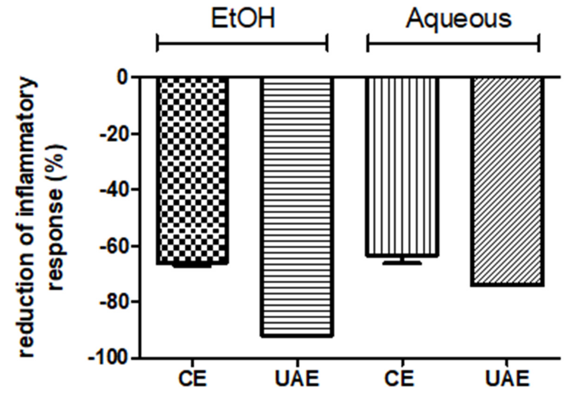
2.2. Anti-Inflammatory Analysis

Finally, the potential of Erodium extracts for anti-inflammatory response was determined by measuring the inhibition of tumor necrosis factor-alpha (TNF-α)-induced pro-inflammatory gene expression in HT-29 human colon cells (Figure 2).
As can be seen in Figure 2, both treatment and solvent had a significant effect on the anti-inflammatory response of the extracts obtained. The ANOVA analysis showed significant differences between the extracts obtained following different extraction methods and by using different extraction solvents.
As far as UAE is concerned, the extracts obtained when the UAE was combined with ethanol:water (50:50, v/v) presented the greatest inhibition (92%) of the inflammatory response followed by the aqueous extract (72%). Similar behavior was also found in the case of the conventional extraction, obtaining the highest inhibition percentage (65%) after using the hydroethanolic mixture compared to the aqueous extraction (63%).
The significant anti-inflammatory activity of Erodium glaucophyllum could be essentially attributed to the presence of phenolic compounds. Along these lines, the antibacterial activity of Geraniaceae genera was evaluated in a previous study, and the authors observed an anti-inflammatory potential of the extracts obtained due to their ability to modulate pro-inflammatory cytokine expression [40]. Moreover, some compounds from Erodium have been involved in inhibiting the formation of 5-lipoxygenase, which is associated with the inflammatory response [41].
Choi et al. [42] described the potent effect of phenolic compounds and terpenoids as anti-inflammatory agents. For this reason, numerous plants rich in potent antioxidant compounds have been exploited in the treatment of inflammatory diseases. These results agree closely with those previously reported by Asif and Khodadadi [43], who pointed out the important role of flavonoids in anti-inflammatory activity. As was previously indicated, several studies indicated that Erodium extracts had important levels of phenolic compounds [4]. As shown in , the Erodium extracts contain isorhamnetin 3-O-glucoside, which is a quercetin derivative. A recent study showed that this compound has a significant inhibitory effect on the synthesis of inflammatory mediators (12-HHT, TXB2, and PGE2), comparable to aspirin [44].

3. Materials and Methods

3.1. Chemicals and Reagents

Aluminum chloride solution, 2,2′-azinobis-(3-ethylbenzothiazoline-6-sulfonic acid) diammonium salt, ABTS radical (≥99% purity), (+)-catechin, Folin–Ciocalteu reagent, 6-hydroxy-2,5,7,8-tetramethylchroman-2-carboxylic acid, gallic acid, HPLC-grade solvents (acetonitrile, methanol, and formic acid), Trolox® (97% purity), resveratrol, and potassium persulfate were purchased from Sigma–Aldrich (St. Louis, MO, USA). Disodium hydrogen phosphate, potassium dihydrogen phosphate, sodium bicarbonate, sodium carbonate, chelex® dipotassium peroxodisulfate, and absolute ethanol were obtained from Merck (Barcelona, Spain), whereas chloride acid (37%) was supplied by Merck and Co., Inc. (Whitehouse Station, NJ, USA).

3.2. Plant Material

Erodium glaucophyllum L. was collected from the south east of Tunisia (Boughrara), in the flowering stage (April 2015). After the separation of the root bark, the plant material was washed and air-dried at room temperature (25 °C) to a constant weight. Afterwards, it was ground (Blixer 2, Robot Coupe, France) to a uniform powder and particles of under 0.05 mm were used. Finally, the powder was vacuum packaged and stored at 4 ± 1 °C in the dark until needed. The authenticity of the plant materials was confirmed by the evaluation of the morphological structure of the samples made by experts in the Department of Plant Biology of the University of Valencia (Valencia, Spain).

3.3. Extraction Experiments: Ultrasound-Assisted (UAE) and Conventional (CE) Extraction

The experiments were performed according to the set-up previously established by Khemakhem et al. [11]. UAE experiments were conducted using an ultrasonic probe system (UP400S, Dr. Hielscher, Teltow, Germany). The ultrasonic emitter, with a diameter of 2.2 cm, was placed in a jacketed vessel and immersed 1 cm in the solvent. The extraction temperature was controlled by re-circulating an ethylene glycol solution (40%) through a cooling coil made with a copper tube (4 mm diameter), which was immersed in the solvent, and also through the jacket of the vessel. In order to re-circulate the ethylene glycol solution, a peristaltic pump (302 S, Watson–Marlow, Postfach, Germany) was connected to a cooling reservoir (refrigerated Circ, Model 1190S, USA) and to a process controller (E5CK, Omron, Hoofddorp, Netherlands) to perform an ON–OFF type control. The temperature of the solvent was monitored using a Pt100 sensor, which was also wired to the controller.
Extraction experiments were carried out using distilled water (100%, v/v) or a hydroethanolic solution (50%, v/v) as solvents. A ratio of 2% (w/v, weight of dry root/volume of water) and a total volume of 100 mL were used for each experiment. The experiments were performed at a constant temperature of 40 ± 1 °C, supplying 100% of the total power of the system (300 W) in every case. The extraction time was fixed at 10 min according to the optimal conditions previously established by other authors [11]. Once the sample was taken, the same volume of water was introduced to keep the ratio of dry roots/volume of solvent constant. Each extract was filtered (0.45 μm) prior to the analytical determinations and the extraction experiments were performed in triplicate.
The characterization of the ultrasonic field was determined by a calorimetric method, used to estimate the actual ultrasonic power released into the medium. To achieve this purpose, the temperature of the solvent was measured every second for the first 3 min of the ultrasonic application without the sample and without controlling the temperature. Then, the ultrasonic power applied (P [W]) was calculated using the rise in temperature, as expressed in Equation (1):
P = m C p d T d t ,
where m (kg) is the solvent mass, Cp (J/kg °C) the specific heat of the solvent, and dT/dt is the slope of the logged temperature–time curve.
The ultrasonic power was measured, in triplicate at least, using two type-K thermocouples wired to a data logger. In this case, the effective ultrasonic power measured was 32.2 ± 1.5 W, by providing 300 W of electrical energy to the transducer.
CE experiments were carried out in the same experimental conditions, but replacing the ultrasonic probe by a mechanical stirrer (RZR 1, Heidolph, Germany) provided with an axial flow impeller working at 540 rpm. The same control temperature system as in the UAE experiments was used, but with the control loop working in heating mode (set point 40 °C).

3.4. Total Polyphenol Content and TOF–LC–MS–MS Analysis

The total amount of polyphenols was measured spectrophotometrically by the Folin–Ciocalteu method, based on a colorimetric oxidation/reduction reaction of phenols [45] with some modifications to prevent the interferences that are common in the traditional Folin-Ciocalteau method. Two hundred microliters of diluted extract and 1 mL of Folin–Ciocalteu reagent (diluted 1:10 with water) were mixed. Then, 0.8 mL of Na2CO3 (75 g/L) was added. The sample was incubated for 10 min at 50 °C and then cooled to room temperature. For the control sample, 0.2 mL of distilled water was taken. The absorbance was measured at 750 nm by the UV/Vis spectrophotometer (Libra S32, Biochrom, France). Gallic acid was used for the calibration curve. The results were expressed as mg gallic acid equivalents (GAE)/g dry matter (DM). The analyses were performed in triplicate and the average deviation was calculated.
In order to determine total flavonoids, the method of Sakanata et al. [46] was used. Two hundred and fifty microliters of extract (appropriately diluted) were mixed with 1.25 mL of distilled water and 75 μL of a sodium nitrite solution (5%, w/v), and left to rest for 6 min. Then, 10% aluminum chloride solution (150 μL) was added and kept for 5 min under resting conditions. Afterwards, 500 µL of sodium hydroxide (1 M) were added and the mixture was brought to 2.5 mL using distilled water and mixed. The absorbance was measured at 510 nm in a UV/Vis spectrophotometer. (+)-Catechin was used to prepare a calibration curve, and the results were expressed as mg catechin equivalents (CE)/g DM.
The identification of the major phenolic compounds present in the Erodium glaucophyllum extracts was carried out using a TripleTOF™ 5600 (AB SCIEX) LC–MS–MS system equipped with Agilent 1260 Infinity (Agilent, Waldbronn, Germany) according to the method previously published of Abdelkebir et al. [47]. Chromatographic separation was performed using a Waters UPLC C18 column 1.7 µm (2.1 × 50 mm) Acquity UPLC BEH.C18 from Waters (Cerdanyola del Vallès, Spain). The mobile phase was composed of water (0.1% formic acid, A) and methanol (0.1% formic acid, B). The gradient elution of the mobile phase was as follows: from 0 to 13 min, 0%–90% B (v/v); 13–15 min, 100% (v/v) B; 15.1–22 min, 90% (v/v) A. The flow rate used was 0.4 ml/min. The injection volume was 5 μL.
The MS acquisition was performed in negative mode, over a mass range of 80–1200 m/z. An automated calibration was carried out using an external calibration delivery system (CDS), which infuses a calibration solution prior to sample introduction. The MS was done using an IDA acquisition method with the survey scan type (TOF–MS) and the dependent scan type (product ion) using −50V of collision energy. The MS parameters were: ion spray voltage, −4500 V; declustering potential (DP) 90 V; collision energy (CE) −50 V; temperature 400 °C with curtain gas (CU) 25 psi; ion source gas 1 (GC1) 50 psi, and ion source gas 2 (GS2) 50 psi. IDA MS–MS was performed using the following criteria: ions that exceeded 100 CPS, ion tolerance 50 mDa, collision energy fixed at 25 V, and dynamic background subtract activated. Data acquisition and processing were carried out using the software analyst PeakView1.1, with the application XIC Manager and Formula Finder.

3.5. Total Carotenoids

Total carotenoid extraction was carried out in accordance with Lee and Castle [48]. An aliquot of extract (2 mL) was homogenized with 5 mL of extracting solvent (hexane/acetone/ethanol, 50:25:25, v/v) and centrifuged for 5 min at 4000 rpm at 5 °C. The top layer of hexane containing the color was recovered and transferred to a 25 mL volumetric flask and the volume was then adjusted to 25 mL with hexane. Total carotenoid determination was carried out on an aliquot of the hexane extract by measuring the absorbance at 450 nm. Total carotenoids were calculated according to Ritter and Purcell [49] using an extinction coefficient of β-carotene, E1% = 2505.

3.6. Trolox Equivalent Antioxidant Capacity (TEAC)

The antioxidant activity of Erodium extracts was determined using the TEAC method [50]. Trolox (5 mM) was prepared in ethanol for use as a stock standard. ABTS radical (ABTS·+) was generated by mixing 25 mL ABTS and 440 μL of potassium persulfate (140 mM). Then, the mixture was allowed to stand in the dark at room temperature for 16 h before use. The solution was diluted with ethanol until an absorbance of 0.70 ± 0.02 was reached at 734 nm. Once the radical was formed, 2 mL of ABTS•+ were mixed with 100 μL of extract (appropriately diluted) and the absorbance was measured at 734 nm for 30 min in a Perkin Elmer UV/Vis Lambda 2 spectrophotometer (Perkin-Elmer, Oberlingen, Germany) in accordance with Barba et al. [51]. The results, obtained from triplicate analyses, were expressed as mM Trolox equivalents (TE).

3.7. Anti-Inflammatory Activity

The human colon tumorigenic cell line HT-29 clone #16 (kindly provided by Prof. G. Perez-Martinez, laboratory Lactic Acid Bacteria and Probiotics, IATA-CSIC, Valencia, Spain) contains a secreted alkaline phosphatase (SEAP) reporter gene under the control of the NF-κB promotor after stable transfection with the plasmid pNiFty2-SEAP (Invivogen), in order to detect pro-inflammatory gene expression induced by the transcription factor NF-κB, a master regulator in inflammatory signaling. The cell line was routinely maintained in Dulbecco’s Modified Eagle Medium (DMEM) high glucose supplemented with 10% fetal bovine serum (FBS), 1 mM sodium pyruvate, antibiotics (100 U/mL penicillin, 100 µg/mL streptomycin), and zeocin (200 µg/mL). Cells were cultured at 37 °C in a humidified atmosphere containing 5% CO2.
For each experiment, the cells were detached using 0.25% Trypsin–EDTA solution (Gibco, Life Technologies) resuspended in complete cell culture medium and seeded at 70,000 cells/well in 96-well plates. The cells were grown 24 hours before the experiment and then stimulated simultaneously with TNF-α at final concentration of 10 ng/mL (Immunotools, Friesoythe, Germany) and 10 µL of each extract in a final volume of 100 µL. After 24 hours of stimulation, SEAP (secreted alkaline phosphatase) activity in the cell culture supernatant was quantified using p-nitrophenyl phosphate as phosphatase substrate, according to the manufacturer’s instructions (Thermo Scientific, Ref.: 37620). The yellow-colored reaction products were detected using a microplate reader (Multiskan Ascent) at 414 nm.

3.8. Statistical Analyses

An analysis of variance (ANOVA) was used in order to analyze the influence of the extraction solvent and mode (UAE and CE methods) on the bioactive compound content, antioxidant capacity and anti-inflammatory parameters. A Tukey’s Multiple Comparison Test was applied to indicate the samples between which differences existed. A multiple regression analysis was performed to study the influence of different factors on a given parameter. [52]. All statistical analyses were performed using the Statgraphics® Centurion XV (Statpoint Technologies, Inc., USA) software.

4. Conclusions

From the results obtained in this study, it can be concluded that the solvent (ethanol:water) and extraction methodologies (conventional and ultrasonically-assisted) led to relevant differences in the quantity of bioactive compounds extracted. This study confirmed that the phenolic compounds of Erodium glaucophyllum had a higher affinity for the hydroethanolic mixture (50:50, v/v, ethanol:water) than aqueous media. However, significant differences were also observed between conventional and ultrasonically-assisted extracts in terms of total phenolics, flavonoids, and carotenoids. We have demonstrated that the different Erodium glaucophyllum extracts are endowed with a potent anti-inflammatory activity and that the hydroethanolic extract obtained by ultrasound extraction has the highest percentage of TNF-α inhibition. The potential of Erodium extracts as anti-inflammatory compounds has been shown, although further studies should be conducted to elucidate which specific compounds or group of compounds are mainly responsible for the anti-inflammatory activities. For this purpose, it would be necessary to isolate and purify each specific compound from Erodium and evaluate its activity. Finally, further research is necessary in order to bring the extraction of Erodium glaucophyllum to an industrial level. Thereby, the performance of conventional and ultrasonic-assisted extraction has to be compared at continuous reactors, instead of the batch-type used in this work, and testing of higher ratios of dried material/solvent, as well as the energy and economic analysis of both strategies, has to be conducted.

Author Contributions

J.V.G.-P., F.J.B., and M.C.C. designed the study; R.A., G.P.M., C.B. conducted the experiments and analyzed the data with J.V.G.P., F.J.B., and M.C.C.; R.A., G.P.-M., C.B., J.V.G.P., J.M.L., F.J.B., C.A. and M.C.C. wrote the first draft. All authors critically reviewed the manuscript and approved the final manuscript.

Funding

This research received no external funding.

Acknowledgments

R.A. would like to acknowledge the Republic of Tunisia, the Ministry of Higher Education and Scientific Research, and the University of Gabes for the fellowship at the Universitat de València, as well as her PhD supervisor M. Neffati. Moreover, R.A. would like to thank COST Action CA15136 for her STSM fellowship.

Conflicts of Interest

The authors have no conflict of interest to disclose.

References

  1. Munekata, P.E.S.; Alcántara, C.; Collado, M.C.; Garcia-Perez, J.V.; Saraiva, J.A.; Lopes, R.P.; Barba, F.J.; do Prado Silva, L.; Sant’Ana, A.S.; Fierro, E.M.; et al. Ethnopharmacology, phytochemistry and biological activity of Erodium species: A review. Food Res. Int. 2019, 126, 108659. [Google Scholar] [CrossRef] [PubMed]
  2. Muthu, C.; Ayyanar, M.; Raja, N.; Ignacimuthu, S. Medicinal plants used by traditional healers in Kancheepuram District of Tamil Nadu, India. J. Ethnobiol. Ethnomed. 2006, 2, 43. [Google Scholar] [CrossRef] [Green Version]
  3. Al-snafi, A.E. A review on Erodium cicutarium: A potential medicinal plant. Indo Am. J. Pharm. Sci. 2017, 4, 110–116. [Google Scholar]
  4. Bouaziz, M.; Dhouib, A.; Loukil, S.; Boukhris, M.; Sayadi, S. Polyphenols content, antioxidant and antimicrobial activities of extracts of some wild plants collected from the south of Tunisia. Afr. J. Biotechnol. 2009, 8, 7017–7027. [Google Scholar]
  5. Ameer, K.; Shahbaz, H.M.; Kwon, J.H. Green extraction methods for polyphenols from plant matrices and their byproducts: A review. Compr. Rev. Food Sci. Food Saf. 2017, 16, 295–315. [Google Scholar] [CrossRef] [Green Version]
  6. Žlabur, J.Š.; Žutić, I.; Radman, S.; Pleša, M.; Brnćić, M.; Barba, F.J.; Rocchetti, G.; Lucini, L.; Lorenzo, J.M.; Domínguez, R.; et al. Effect of different green extraction methods and solvents on bioactive components of chamomile (Matricaria chamomilla L.) flowers. Molecules 2020, 25, 810. [Google Scholar] [CrossRef] [PubMed] [Green Version]
  7. Roselló-Soto, E.; Martí-Quijal, F.J.; Cilla, A.; Munekata, P.E.S.; Lorenzo, J.M.; Remize, F.; Barba, F.J. Influence of temperature, solvent and pH on the selective extraction of phenolic compounds from tiger nuts by-products: Triple-TOF-LC-MS-MS characterization. Molecules 2019, 24, 797. [Google Scholar] [CrossRef] [Green Version]
  8. Marić, M.; Grassino, A.N.; Zhu, Z.; Barba, F.J.; Brnćić, M.; Rimac Brnćić, S. An overview of the traditional and innovative approaches for pectin extraction from plant food wastes and by-products: Ultrasound-, microwaves-, and enzyme-assisted extraction. Trends Food Sci. Technol. 2018, 76, 28–37. [Google Scholar] [CrossRef]
  9. Barba, F.J.; Grimi, N.; Vorobiev, E. Evaluating the potential of cell disruption technologies for green selective extraction of antioxidant compounds from Stevia rebaudiana Bertoni leaves. J. Food Eng. 2015, 149, 222–228. [Google Scholar] [CrossRef]
  10. Roselló-Soto, E.; Galanakis, C.M.; Brnčić, M.; Orlien, V.; Trujillo, F.J.; Mawson, R.; Knoerzer, K.; Tiwari, B.K.; Barba, F.J. Clean recovery of antioxidant compounds from plant foods, by-products and algae assisted by ultrasounds processing. Modeling approaches to optimize processing conditions. Trends Food Sci. Technol. 2015, 42, 134–149. [Google Scholar] [CrossRef]
  11. Khemakhem, I.; Ahmad-Qasem, M.H.; Catalán, E.B.; Micol, V.; García-Pérez, J.V.; Ayadi, M.A.; Bouaziz, M. Kinetic improvement of olive leaves’ bioactive compounds extraction by using power ultrasound in a wide temperature range. Ultrason. Sonochem. 2017, 34, 466–473. [Google Scholar] [CrossRef] [PubMed]
  12. Ahmad-Qasem, M.H.; Cánovas, J.; Barrajón-Catalán, E.; Micol, V.; Cárcel, J.A.; García-Pérez, J.V. Kinetic and compositional study of phenolic extraction from olive leaves (var. Serrana) by using power ultrasound. Innov. Food Sci. Emerg. Technol. 2013, 17, 120–129. [Google Scholar] [CrossRef]
  13. Šic Žlabur, J.; Colnar, D.; Voća, S.; Lorenzo, J.M.; Munekata, P.E.S.; Barba, F.J.; Dobrićević, N.; Galić, A.; Dujmić, F.; Pliestić, S.; et al. Effect of ultrasound pre-treatment and drying method on specialized metabolites of honeyberry fruits (Lonicera caerulea var. kamtschatica). Ultrason. Sonochem. 2019, 56, 372–377. [Google Scholar] [CrossRef] [PubMed]
  14. Szabó, C. Multiple pathways of peroxynitrite cytotoxicity. Toxicol. Lett. 2003, 140–141, 105–112. [Google Scholar] [CrossRef]
  15. Kim, H.P.; Son, K.H.; Chang, H.W.; Kang, S.S. Anti-inflammatory plant flavonoids and cellular action mechanisms. J. Pharmacol. Sci. 2004, 96, 229–245. [Google Scholar] [CrossRef] [PubMed] [Green Version]
  16. Ait El Cadi, M.; Makram, S.; Ansar, M.; Khabbal, Y.; Alaoui, K.; Faouzi, M.A.; Cherrah, Y.; Taoufik, J. Activité anti-inflammatoire des extraits aqueux et éthanolique de Zygophyllum gaetulum. Ann. Pharm. Fr. 2012, 70, 113–116. [Google Scholar] [CrossRef] [PubMed]
  17. Khlifi, D.; Sghaier, R.M.; Amouri, S.; Laouini, D.; Hamdi, M.; Bouajila, J. Composition and anti-oxidant, anti-cancer and anti-inflammatory activities of Artemisia herba-alba, Ruta chalpensis L. and Peganum harmala L. Food Chem. Toxicol. 2013, 55, 202–208. [Google Scholar] [CrossRef]
  18. Al-Said, M.S.; Tariq, M.; Al-Yahya, M.A.; Rafatullah, S.; Ginnawi, O.T.; Ageel, A.M. Studies on Ruta chalepensis, an ancient medicinal herb still used in traditional medicine. J. Ethnopharmacol. 1990, 28, 305–312. [Google Scholar] [CrossRef]
  19. Zhang, Q.A.; Shen, H.; Fan, X.H.; Shen, H.; Wang, X.; Song, Y. Changes of gallic acid mediated by ultrasound in a model extraction solution. Ultrason. Sonochem. 2015, 22, 149–154. [Google Scholar] [CrossRef]
  20. Qiao, L.; Ye, X.; Sun, Y.; Ying, J.; Shen, Y.; Chen, J. Sonochemical effects on free phenolic acids under ultrasound treatment in a model system. Ultrason. Sonochem. 2013, 20, 1017–1025. [Google Scholar] [CrossRef]
  21. Misra, N.N.; Martynenko, A.; Chemat, F.; Paniwnyk, L.; Barba, F.J.; Jambrak, A.R. Thermodynamics, transport phenomena, and electrochemistry of external field-assisted nonthermal food technologies. Crit. Rev. Food Sci. Nutr. 2018, 58, 1832–1863. [Google Scholar] [CrossRef] [PubMed] [Green Version]
  22. Chemat, F.; Rombaut, N.; Meullemiestre, A.; Turk, M.; Perino, S.; Fabiano-Tixier, A.-S.; Abert-Vian, M. Review of Green Food Processing techniques. Preservation, transformation, and extraction. Innov. Food Sci. Emerg. Technol. 2017, 41, 357–377. [Google Scholar] [CrossRef]
  23. Putnik, P.; Bursać Kovacević, D.; Režek Jambrak, A.; Barba, F.J.; Cravotto, G.; Binello, A.; Lorenzo, J.M.; Shpigelman, A. Innovative “green” and novel strategies for the extraction of bioactive added value compounds from citruswastes—A review. Molecules 2017, 22, 680. [Google Scholar] [CrossRef] [PubMed] [Green Version]
  24. Wei, S.D.; Zhou, H.C.; Lin, Y.M. Antioxidant activities of extract and fractions from the hypocotyls of the mangrove plant Kandelia candel. Int. J. Mol. Sci. 2010, 11, 4080–4093. [Google Scholar] [CrossRef] [PubMed] [Green Version]
  25. Carbonell-Capella, J.M.; Šic Žlabur, J.; Rimac Brnčić, S.; Barba, F.J.; Grimi, N.; Koubaa, M.; Brnčić, M.; Vorobiev, E. Electrotechnologies, microwaves, and ultrasounds combined with binary mixtures of ethanol and water to extract steviol glycosides and antioxidant compounds from Stevia rebaudiana leaves. J. Food Process. Preserv. 2017, 41, e13179. [Google Scholar] [CrossRef]
  26. Owczarek, K.; Chojnacka, K.; Lewandowska, U. Overview of polyphenols and polyphenol-rich extracts as modulators of IGF- 1, IGF-1R, and IGFBP expression in cancer diseases. J. Funct. Foods 2019, 52, 389–407. [Google Scholar]
  27. Galvan D’Alessandro, L.; Kriaa, K.; Nikov, I.; Dimitrov, K. Ultrasound assisted extraction of polyphenols from black chokeberry. Sep. Purif. Technol. 2012, 93, 42–47. [Google Scholar] [CrossRef]
  28. Sun, Y.; Liu, Z.; Wang, J. Ultrasound-assisted extraction of five isoflavones from Iris tectorum Maxim. Sep. Purif. Technol. 2011, 78, 49–54. [Google Scholar] [CrossRef]
  29. Spigno, G.; Tramelli, L.; De Faveri, D.M. Effects of extraction time, temperature and solvent on concentration and antioxidant activity of grape marc phenolics. J. Food Eng. 2007, 81, 200–208. [Google Scholar] [CrossRef]
  30. Roseiro, L.B.; Duarte, L.C.; Oliveira, D.L.; Roque, R.; Bernardo-Gil, M.G.; Martins, A.I.; Sepúlveda, C.; Almeida, J.; Meireles, M.; Gírio, F.M.; et al. Supercritical, ultrasound and conventional extracts from carob (Ceratonia siliqua L.) biomass: Effect on the phenolic profile and antiproliferative activity. Ind. Crops Prod. 2013, 47, 132–138. [Google Scholar] [CrossRef]
  31. Tiwari, B.K.; O’Donnell, C.P.; Cullen, P.J. Effect of sonication on retention of anthocyanins in blackberry juice. J. Food Eng. 2009, 93, 166–171. [Google Scholar] [CrossRef]
  32. Sala, F.J.; Burgos, J.; Condón, S.; Lopez, P.; Raso, J. Effect of heat and ultrasound on microorganisms and enzymes. In New Methods Food Preserv; Gould, G.W., Ed.; Springer: Boston, MA, USA, 1995; pp. 176–204. [Google Scholar]
  33. Da Porto, C.; Porretto, E.; Decorti, D. Comparison of ultrasound-assisted extraction with conventional extraction methods of oil and polyphenols from grape (Vitis vinifera L.) seeds. Ultrason. Sonochem. 2013, 20, 1076–1080. [Google Scholar] [CrossRef] [PubMed]
  34. Rana Kidak, N.H.I. Ultrasonic destruction of phenol and substituted phenols: A review of current research. Ultrason. Sonochem. 2006, 13, 195–199. [Google Scholar] [CrossRef] [PubMed]
  35. Sroka, Z.; Bodalska, H.R.; Mażol, I. Antioxidative effect of extracts from Erodium cicutarium L. Z. Nat. C 1994, 49, 881–884. [Google Scholar] [CrossRef]
  36. Daglia, M. Polyphenols as antimicrobial agents. Curr. Opin. Biotechnol. 2012, 23, 174–181. [Google Scholar] [CrossRef]
  37. Prakash, M.; Basavaraj, B.V.; Murthy, K.N.C. Biological functions of epicatechin: Plant cell to human cell health. J. Funct. Foods 2019, 52, 14–24. [Google Scholar] [CrossRef]
  38. Macías-Sánchez, M.D.; Mantell, C.; Rodríguez, M.; de la Ossa, E.; Lubián, L.M.; Montero, O. Comparison of supercritical fluid and ultrasound-assisted extraction of carotenoids and chlorophyll a from Dunaliella salina. Talanta 2009, 77, 948–952. [Google Scholar] [CrossRef]
  39. Kaur, C.; Kapoor, H.C. Antioxidants in fruits and vegetables—The millennium’s health. Int. J. Food Sci. Technol. 2001, 36, 703–725. [Google Scholar] [CrossRef]
  40. Shim, J.U.; Oh, P.S.; Lim, K.T. Anti-inflammatory activity of ethanol extract from Geranium sibiricum Linne. J. Ethnopharmacol. 2009, 126, 90–95. [Google Scholar] [CrossRef]
  41. Gohar, A.A.; Lahloub, M.F.; Niwa, M. Antibacterial polyphenol from Erodium glaucophyllum. Z. Nat. Sect. C J. Biosci. 2003, 58, 670–674. [Google Scholar] [CrossRef]
  42. Choi, M.S.; Lee, S.H.; Cho, H.S.; Kim, Y.; Yun, Y.P.; Jung, H.Y.; Jung, J.K.; Lee, B.C.; Pyo, H.B.; Hong, J.T. Inhibitory effect of obovatol on nitric oxide production and activation of NF-κB/MAP kinases in lipopolysaccharide-treated RAW 264.7cells. Eur. J. Pharmacol. 2007, 556, 181–189. [Google Scholar] [CrossRef] [PubMed]
  43. Asif, M.; Khodadadi, E. Medicinal uses and chemistry of flavonoid contents of some common edible tropical plants. J. Paramed. Sci. 2013, 4, 119–138. [Google Scholar]
  44. Lesjak, M.; Beara, I.; Pinta, D.; Majki, T.; Bekvalac, K.; Or, D.; Mimica-duki, N. Antioxidant and anti-infl ammatory activities of quercetin and its derivatives. J. Funct. Foods 2018, 40, 68–75. [Google Scholar] [CrossRef]
  45. Singleton, V.L.; Orthofer, R.; Lamuela-Raventós, R.M. Analysis of total phenols and other oxidation substrates and antioxidants by means of Folin-ciocalteu reagent. In Methods in Enzymology; Packer, L., Ed.; Oxidants and Antioxidants Part A; Academic Press: Cambridge, MA, USA, 1999; Volume 299, pp. 152–178. [Google Scholar]
  46. Sakanaka, S.; Tachibana, Y.; Okada, Y. Preparation and antioxidant properties of extracts of Japanese persimmon leaf tea (kakinoha-cha). Food Chem. 2005, 89, 569–575. [Google Scholar] [CrossRef]
  47. Abdelkebir, R.; Alcántara, C.; Falcó, I.; Sánchez, G.; Garcia-Perez, J.V.; Neffati, M.; Lorenzo, J.M.; Barba, F.J.; Collado, M.C. Effect of ultrasound technology combined with binary mixtures of ethanol and water on antibacterial and antiviral activities of Erodium glaucophyllum extracts. Innov. Food Sci. Emerg. Technol. 2019, 52, 189–196. [Google Scholar] [CrossRef]
  48. Lee, H.S.; Castle, W.S. Seasonal changes of carotenoid pigments and color in Hamlin, Earlygold, and Budd Blood orange juices. J. Agric. Food Chem. 2001, 49, 877–882. [Google Scholar] [CrossRef]
  49. De Ritter, E.; Purcell, A.E. Carotenoid analytical methods. In Carotenoids as Colorants and Vitamin A Precursors; Academic Press: Cambridge, MA, USA, 1981; pp. 815–923. [Google Scholar]
  50. Re, R.; Pellegrini, N.; Proteggente, A.; Pannala, A.; Yang, M.; Rice-Evans, C. Antioxidant activity applying an improved ABTS radical cation decolorization assay. Free Radic. Biol. Med. 1999, 26, 1231–1237. [Google Scholar] [CrossRef]
  51. Barba, F.J.; Criado, M.N.; Belda-Galbis, C.M.; Esteve, M.J.; Rodrigo, D. Stevia rebaudiana Bertoni as a natural antioxidant/antimicrobial for high pressure processed fruit extract: Processing parameter optimization. Food Chem. 2014, 148, 261–267. [Google Scholar] [CrossRef]
  52. Metsalu, T.; Vilo, J. ClustVis: A web tool for visualizing clustering of multivariate data using Principal Component Analysis and heatmap. Nucleic Acids Res. 2015, 43, W566–W570. [Google Scholar] [CrossRef]
Sample Availability: Samples of the compounds are available from the authors.
Figure 1. (a) The total phenolic compounds expressed as mg gallic acid equivalents (GAE)/g dry matter (DM), (b) the flavonoid content expressed as mg catechin equivalents (CE)/g dry matter (DM), (c) total carotenoid content expressed as mg carotenoids/g dry matter (DM), and (d) antioxidant capacity (TEAC) expressed as mM Trolox equivalents/g dry matter (DM); determined in Erodium glaucophyllum in aqueous extract and ethanol (ETOH) extract (50:50, v/v, ethanol:water). UAE: extracts obtained by ultrasound-assisted extraction; CE: extracts obtained by conventional extraction.
Molecules 25 01759 g001
Figure 2. The effect of Erodium glaucophyllum extracts on TNF-α-induced pro-inflammatory response. The diagram shows the reduction of secreted alkaline phosphatase activity (SEAP) activity (%) in the cell line reporter (HT-29 clone #16) with respect to TNF-α (0%). EtOH: hydroethanolic extract; aqueous: extracts with water. UAE: extracts obtained by ultrasound-assisted extraction; CE: extracts obtained by conventional extraction.
Molecules 25 01759 g002
Table 1. Specific polyphenols (peak intensity) identified in Erodium glaucophyllum aqueous extracts confirmed by TOF–LC–MS–MS obtained by conventional extraction (CE) and ultrasound-assisted extraction (UAE).
GroupCompound Name ScoreFormulaThresholdExpected m/zCE
(Intensity Average)
UAE
(Intensity Average)
Flavonoids(+)-Catechin 91%C15H14O650289.0718141,164 ± 29,05975,051 ± 4815
(+)-Catechin 3-O-glucose92%C21H24O1150451.1246-50,611 ± 5769
Dihydroquercetin 86%C15H12O750303.051-33,681 ± 2987
(+)-Gallocatechin 93%C15H14O750305.0667382,755 ± 33,378323,730.5 ± 28,429
Hydroxytyrosol 1-O-glucoside 90%C14H20O950331.1035-28,168 ± 72
Isorhamnetin 3-O-glucoside 89%C22H22O1250477.1039-8504 ± 0
Myricetin 3-O-galactoside 90%C21H20O1350479.0831-11,651 ± 1373
Procyanidin dimer B793%C30H26O1250577.135742,121 ± 10,649-
Procyanidin trimer T3 73%C45H38O1850865.1985-10,731 ± 1930
Prodelphinidin dimer B3 80%C30H26O1350593.1301235,192 ± 24,370193,398 ± 11,667
Quercetin 3-O-glucuronide 91%C21H18O1350477.067583,703 ± 682560,377 ± 3596
Phenolic acidsGallic acid 90%C7H6O550169.014220,921 ± 175517,788.5 ± 1623
Gallic acid 4-O-glucoside 92%C13H16O1050331.0671-60,362 ± 11,976
2,6-Dihydroxybenzoic acid 94%C7H6O450153.0193-5827 ± 957
Ellagic acid 97%C14H6O850300.99914,692 ± 41796479 ± 1558
Sinapoyl glucose 90%C17H22O1050385.114242,036 ± 51,397178,689 ± 1122
PhenolsPyrogallol 86%C6H6O350125.0244-20,505 ± 120
Other polyphenols3,4-Dihydroxyphenylglycol 80%C8H10O450169.05128176 ± 216-
Oleoside 11-methylester 93%C17H24O1150403.1246-11,485 ± 3661
Oleuropein 85%C25H32O1350539.177-116,572 ± 33,774
Resorcinol91%C6H6O250109.02938290 ± 470-
Rosmadial88%C20H24O550343.154922,938 ± 2316-
4-Vinylphenol 84%C8H8O50119.05034643 ± 619-
StilbenoidsResveratrol 85%C14H12O350227.0714-2746 ± 174

