Article

Yellow Emission Obtained by Combination of Broadband Emission and Multi-Peak Emission in Garnet Structure Na2YMg2V3O12: Dy3+ Phosphor

Beijing Key Laboratory of Materials Utilization of Nonmetallic Minerals and Solid Wastes, National Laboratory of Mineral Materials, School of Materials Science and Technology, China University of Geosciences, Beijing 100083, China
*
Authors to whom correspondence should be addressed.
Molecules 2020, 25(3), 542; https://doi.org/10.3390/molecules25030542
Submission received: 26 December 2019 / Revised: 20 January 2020 / Accepted: 24 January 2020 / Published: 27 January 2020
(This article belongs to the Section Inorganic Chemistry)

Abstract

:
The fabrication and luminescent performance of novel phosphors Na2YMg2V3O12:Dy3+ were investigated by a conventional solid-state reaction method. Under near-UV light, the Na2YMg2V3O12 host self-activated and released a broad emission band (400–700 nm, with a peak at 524 nm) ascribable to charge transfer in the (VO4)3− groups. Meanwhile, the Na2YMg2V3O12:Dy3+ phosphors emitted bright yellow light within both the broad emission band of the (VO4)3- groups and the sharp peaks of the Dy3+ ions at 490, 582, and 663 nm at a quenching concentration of 0.03 mol. The emission of the as-prepared Na2YMg2V3O12:Dy3+ phosphors remained stable at high temperatures. The obtained phosphors, commercial Y2O3:Eu3+ red phosphors, and BaMgAl10O17:Eu2+ blue phosphors were packed into a white light-emitting diode (WLED) device with a near-UV chip. The designed WLED emitted bright white light with good chromaticity coordinates (0.331, 0.361), satisfactory color rendering index (80.2), and proper correlation to a color temperature (7364 K). These results indicate the potential utility of Na2YMg2V3O12:Dy3+ phosphor as a yellow-emitting phosphor in solid-state illumination.

Graphical Abstract

1. Introduction

For several years, phosphor-converted white light-emitting diodes (pc-WLEDs) have been recognized as the most promising replacements of traditional incandescent and fluorescence lamps [1,2,3,4,5]. These solid-state light sources deliver high luminescence efficiency and an excellent operation lifetime (>10,000 h) while conserving energy and lowering the pollution risk. Most commercial pc-WLEDs are obtained by two methods [6,7]. One method generates white light by combining yellow phosphors with blue LED chips, such as the yellow phosphor YAG: Ce3+ [8,9]. However, the absence of the red-emitting component reduces the quality of the white light, yielding poor color reproduction and a low color rendering index (Ra) [10]. The other method stimulates blue, green, and red (RGB) phosphors by violet or ultraviolet light LEDs [11]. Although this method improves the Ra and can tune the correlated color temperature (CCT), the emission efficiency is limited by reabsorption among the different phosphors [1,12]. The different thermal stabilities and ageing rates of the various phosphors also restrict their applications in WLEDs [13]. Therefore, high-performance single-phased phosphors that can be excited by ultraviolet (UV) or near-UV (n-UV) light are urgently needed [14,15,16]. A single-host white-emitting phosphor must usually have a broad emission peak or multiple emission peaks in the visible region. Therefore, searching for new broadband emission or multi-peak emission phosphors is significant for improving the color stability and service life of WLEDs excited by UV or n-UV light [17,18].
In recent decades, the rare earth luminescent materials have been used in many fields, such as lighting, photosynthesis enhancement, and photodynamic activation for cancer treatments [19,20,21]. Broad emission peaks or multiple emission peaks in the visible region facilitate white light emission with appropriate Ra and CCT values [22,23,24]. Vanadate composed of VO43− tetrahedrons is an important non-rare earth ion-doped luminescent material displaying broadband emission, excellent luminous efficiency, and good chemical stability [25,26]. For example, A3(VO4)2 (A = Mg, Sr, Ba) phosphors yield intense charge transfer (CT) absorption bands in the n-UV region and broad emission bands covering 400–700 nm. Further studies have reported that when doped with rare earth ions, vanadate is a good host material that enhances the emission efficiency of phosphors [27,28]. Guo et al. reported that Eu3+-activated Ba2BiV3O11 phosphors are promising candidates for red-emitting phosphors in WLEDs, as they efficiently convert UV light from 394 nm onwards into red light [29]. Bright orange-red emission has been obtained by doping Sm3+ in NaSrVO4 phosphor under n-UV light excitation [30]. Song et al. [17] studied self-activated Na2YMg2V3O12 vanadate phosphors, and reported a broad emission band of 400–700 nm centered at 520 nm. However, the red emission intensity was relatively low, below the requirements of white light emission. Dy3+ doping should broaden the emission band of Na2YMg2V3O12 phosphors.
In this work, a series of yellow-emitting Na2YMg2V3O12:Dy3+ phosphors was prepared by a conventional high-temperature solid-state method. The phase purities, micromorphologies, luminescence properties, and decay times of the as-prepared Na2YMg2V3O12:Dy3+ phosphors were studied in detail. The suitability of the yellow-emitting phosphors for indoor illumination was demonstrated in a WLED device incorporating the developed phosphors.

2. Results and Discussion

The phase compositions and crystal structures of the as-prepared powder samples were characterized at room temperature. The XRD patterns of Na2YMg2V3O12:xDy3+ (x = 0, 0.005, 0.01, 0.03, 0.05, 0.07) samples exhibited main peaks at 17.5°, 20.3°, 28.8°, 32.3°, 33.9°, 35.5°, 36.9°, 51.0°, 53.2°, and 55.4° (Figure 1a), corresponding, respectively, to the (2 1 1), (2 2 0), (4 0 0), (4 2 0), (3 3 2), (4 2 2), (4 3 1), (4 4 4), (6 4 0), and (6 4 2) facets of a single garnet structure with a cubic Ia3d (No. 230) space group. All diffraction peaks of the Na2YMg2V3O12:Dy3+ samples were well matched with the standard profile (PDF No.49-0412), confirming that doping with Y3+ ions did not significantly affect the crystalline structure of Na2YMg2V3O12.
Figure 1b shows the spatial structure of the unit cell of the garnet-structured Na2YMg2V3O12. As implied, the A sites were occupied by alkaline metal ions Na+ and rare earth ions Y3+, which were coordinated with eight oxygen O2− ions to form a dodecahedron with D2 symmetry (without an inverse center). The alkaline-earth metal Mg2+ ions located in the octahedral sites B bonded with six oxygen atoms, and the metal ion V5+ (in VO43−) occupied the Td sites and were surrounded by four O2− ions. As Y3+ and Dy3+ have similar cationic radii and the same valence, the Y3+ ions in the host lattice were easily replaced by Dy3+ ions with no structural transformation. The XRD patterns of Na2YMg2V3O12:Dy3+ match those of the standard card, further confirming that the Dy3+ ions doped in the Na2YMg2V3O12 host had replaced the Y3+ sites.
The microscopic morphology, particle size, and grain shape of a phosphor are important factors in applications. Field emission scanning electron microscopy (FESEM) images of the Na2YMg2V3O12:0.03Dy3+ sample confirmed that all particles were irregular oblate spheres with an average particle size of 1 μm (Figure 1c). The spherical morphology was similar to that of commercial YAG: Ce3+ phosphor, which possesses the same garnet structure. The average particle size of the prepared phosphor was also similar to that of commercial phosphors. This size may enhance the dispersion and transparency of phosphors in the glue when packaging with the WLEDs.
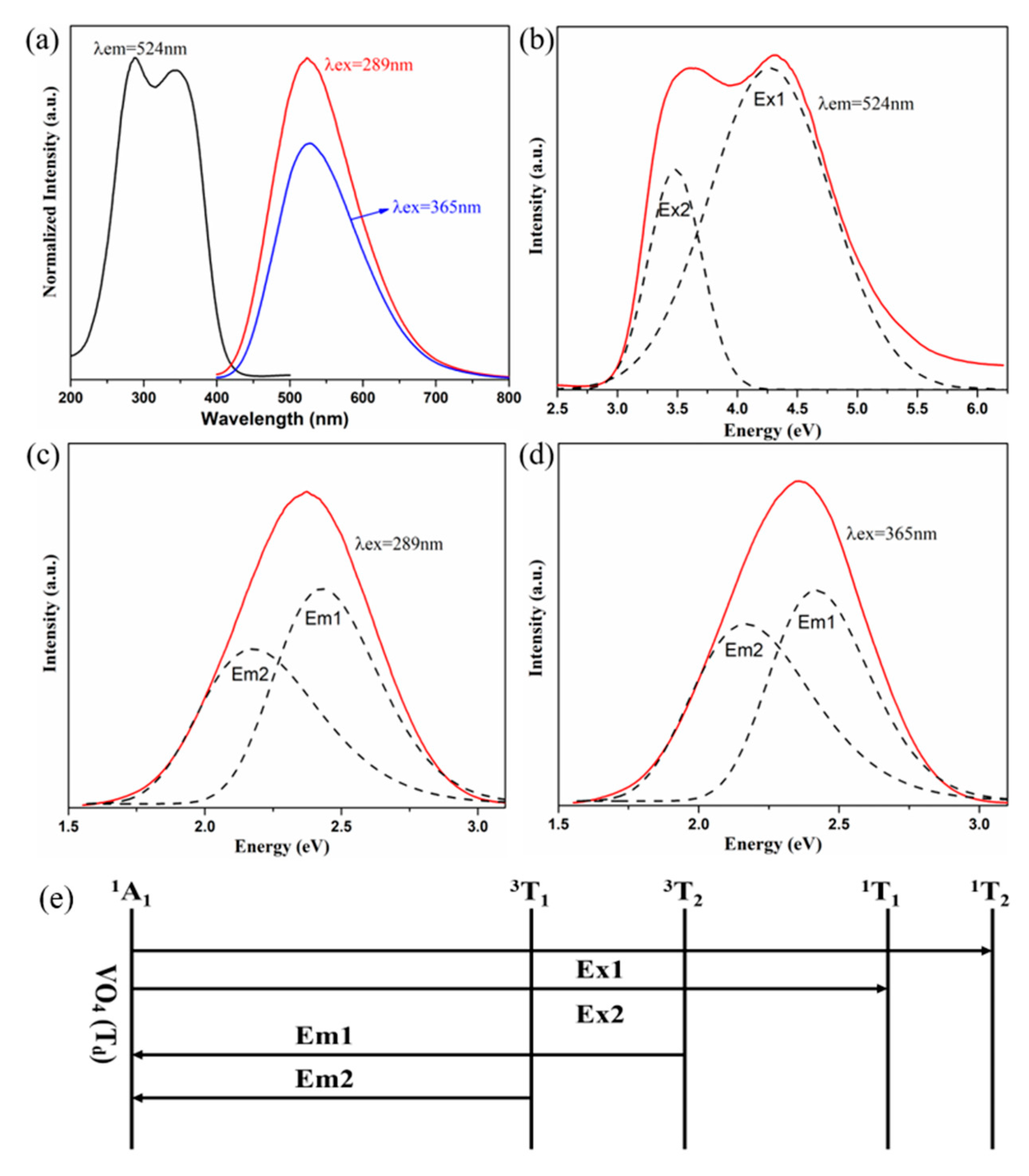
The photoluminescence (PL) and PL emission (PLE) spectra of the undoped Na2YMg2V3O12 sample are presented in Figure 2a–c. Na2YMg2V3O12 shows a broad absorption band of 250–400 nm, matching the absorption of near-UV chips in WLEDs. When excited at 289 nm and 365 nm, the as-prepared particles also emitted a broad emission band, ranging from 400 to 700 nm with a maximum at 524 nm. This emission was attributed to the CT of an electron from the 2p orbital of oxygen to the vacant 3d orbital of V5+ in the tetrahedral (VO4)3− groups [31,32]. The emission band centered at 524 nm was decomposed into two sub-bands by Gaussian peak separation, one centered at 289 nm (4.30 eV), the other at 365 nm (3.41 eV) [33]. As shown in Figure 2d, the (VO4)3− group has a ground state 1A1 and excited states 1T1, 1T2, 3T1, and 3T2. The decomposed emission sub-bands were attributed to 3T21A1 (Em1 = 512 nm (2.43 eV)) and 3T11A1 (Em2 = 571 nm (2.18 eV)) transitions of the (VO4)3− groups, respectively. The excitation band was also composed of two sub-bands, which were assigned to the 1A11T2 (Ex1 = 4.30 eV) and 1A11T1 (Ex2 = 3.41 eV) transitions of the (VO4)3− groups.
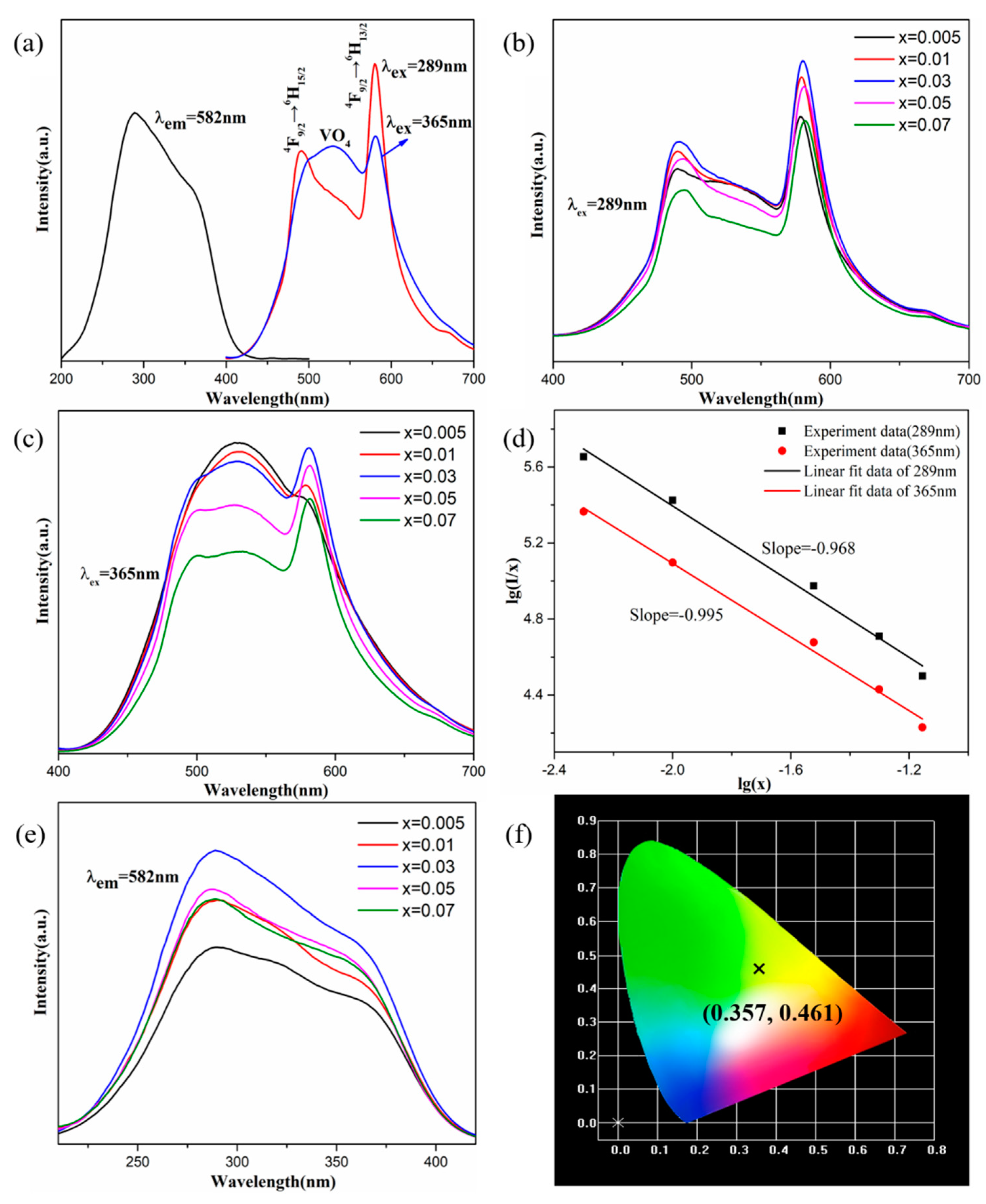
Figure 3a shows the PLE (λem = 582 nm) and PL (λex = 289 and 365 nm) spectra of the Na2YMg2V3O12:0.03Dy3+ phosphors at room temperature. The broad emission band at 524 nm was assigned to the CT transitions of the (VO4)3− groups, and the emission peaks at 490, 582, and 663 nm were, respectively, attributed to 4F9/26H15/2, 4F9/26H13/2, and 4F9/26H11/2 transitions of Dy3+. Under excitation at 289 nm and 365 nm, the intensity ratios of the (VO4)3− and Dy3+ emissions changed because the excitation pathways of Dy3+ luminescence depend on the excitation wavelength. When excited at 289 nm and 365 nm, the Dy3+ emission was mainly caused by Dy-O CT and by energy transfer from the absorption of V-O CT, respectively [12,34]. Monitoring the phosphor emission under 582 nm, the broad excitation band from 250 to 400 nm (which peaks at two sites: 289 nm and 365 nm) resembles the excitation spectrum of non-doped Na2YMg2V3O12. This may have resulted from the energy transfer behavior from Na2YMg2V3O12 to Dy3+ ions, which completely overlaps the excitation spectrum of Na2YMg2V3O12 to Dy3+. The broad excitation spectrum indicates that the Na2YMg2V3O12:Dy3+ sample can be efficiently excited under n-UV light, and can be well matched with n-UV LED chips.
The PL spectra of Na2YMg2V3O12:xDy3+ (x = 0, 0.005, 0.01, 0.03, 0.05, 0.07) samples with different doping concentrations are shown in Figure 3b,c. As the Dy3+ concentration increased, the intensities of the emission peaks increased to a maximum at x = 0.03, and then decreased under the concentration quenching effect [35]. To investigate the cause of concentration quenching, the interaction type between two excitations was calculated by the following formula:
I x = k 1 + β x Q / 3
where k and β are constants, I is the emission intensity, and Q represents the interaction type. When Q is 3, 6, 8, and 10, the interactions are exchange, dipole–dipole, dipole–quadrupole, and quadrupole–quadrupole interactions, respectively. The Q value was obtained by linear fitting of the relationship between lg(I/x) and lg(x). When the phosphors were excited at 289 and 365 nm, the slopes (−Q/3) were determined as −0.995 and −0.968, respectively (Figure 3d). Both Q values were close to the theoretical value of 3.0, indicating that at higher concentrations, the intensity of the Na2YMg2V3O12:Dy3+ phosphors was quenched by exchange interactions. The excitation spectra of Na2YMg2V3O12:xDy3+ monitored at 582 nm were also optimized at x = 0.03 (Figure 3e). The CIE (International Commission on illumination) chromaticity coordinates of the Na2YMg2V3O12:0.03Dy3+ sample were determined as (0.357, 0.461) (Figure 3f). The yellow light emission was the combination of the self-activated emission of the Na2YMg2V3O12 host with the dominant 4f–4f transitions of the Dy3+ ion [36].
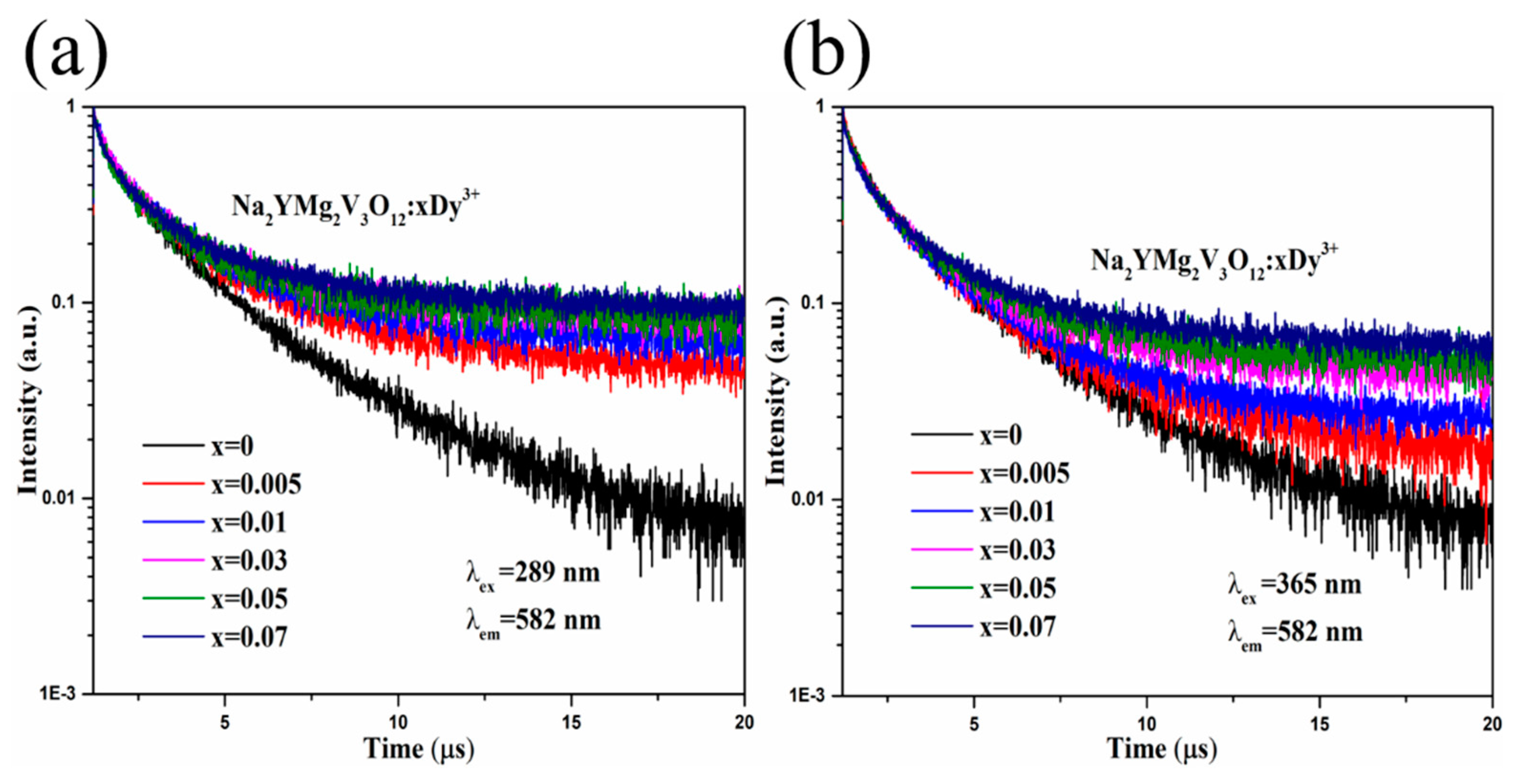
To understand the behaviors of the synthesized compounds, the Na2YMg2V3O12:xDy3+ phosphors were excited at 289 and 365 nm, and their PL decay curves were recorded at 582 nm. The results are shown in Figure 4. The decay curves of Na2YMg2V3O12:xDy3+ were well fitted to the following exponential function [37]:
I t = A e t τ + I 0
where It and I0 are the emission intensities at time t and the initial time, respectively, and A is a constant. τ determines the decay time. The average lifetimes of the Na2YMg2V3O12:xDy3+ phosphors with x = 0, 0.005, 0.01, 0.03, 0.05, and 0.07 were determined as 1.60, 1.58, 1.54, 1.48, 1.42, and 1.44 μs, respectively, at λex = 289 nm, and as 1.53, 1.52, 1.49, 1.50, 1.41, and 1.40 μs, respectively, at λex =365 nm. The PL lifetimes of the Na2YMg2V3O12:xDy3+ were similar under both excitation wavelengths, possibly reflecting the similar energy transfer behaviors between the vanadate host and Dy3+.
High thermal resistance of phosphors is very important for practical applications in solid-state lighting, as it ensures high optical performance of the WLED device. The thermal quenching performance of Na2YMg2V3O12:0.03Dy3+ phosphor was assessed from the temperature-dependent emission spectra under excitation at 289 and 365 nm. As shown in Figure 5a,c, the emission intensity reduced smoothly as the temperature increased, because the probability of non-radiative transitions increases at higher temperatures. As shown in the insets of Figure 5a,c, the PL integral intensities at 100 °C were 61.6% (λex = 289 nm) and 61.48% (λex = 365 nm) of their room temperature intensities. However, the emission positions in the temperature-dependent emission spectra were relatively robust to temperature changes.
To further investigate the thermal stability of this phosphor, the activation energy (ΔE) of Na2YMg2V3O12:0.03Dy3+ was calculated by the Arrhenius equation [38]:
I ( T ) = I 0 1 + c e Δ E k T
where I0 is the emission intensity of the phosphor at room temperature, I(T) is the temperature-dependent intensity, c is a constant, and k is the Boltzmann constant (8.629 × 10⁻⁵ eV K−1). From the slopes of the ln[I0/I(T) − 1] versus 1/kT plots (Figure 5b,d), which were well fitted to Equation (3), the ΔEs were determined as 0.21 and 0.26 eV under excitation at 289 and 365 nm, respectively. Table 1 compares the CIE chromaticity coordinates, CCT and lifetimes of Na2YMg2V3O12:0.03Dy3+, and other Dy3+-doped phosphors [39,40,41]. The obtained Na2YMg2V3O12:Dy3+ phosphors presented relatively high thermal stability and are potentially applicable to WLEDs.
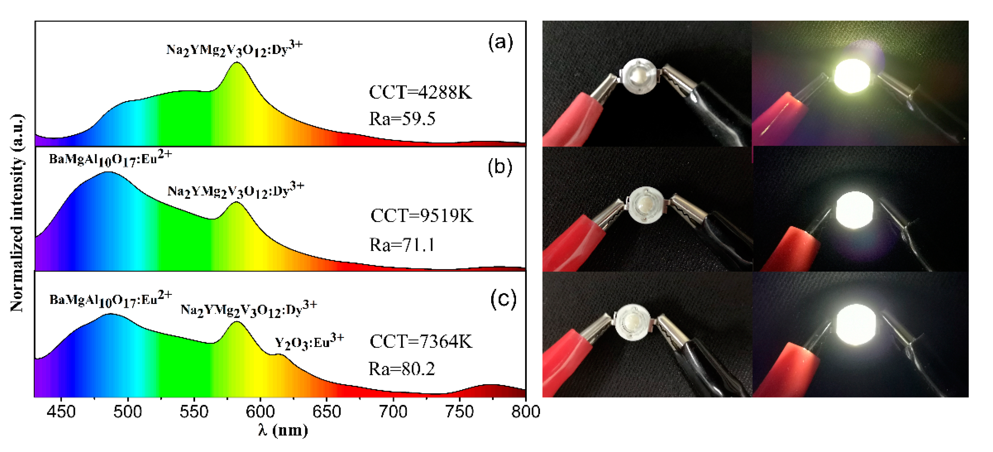
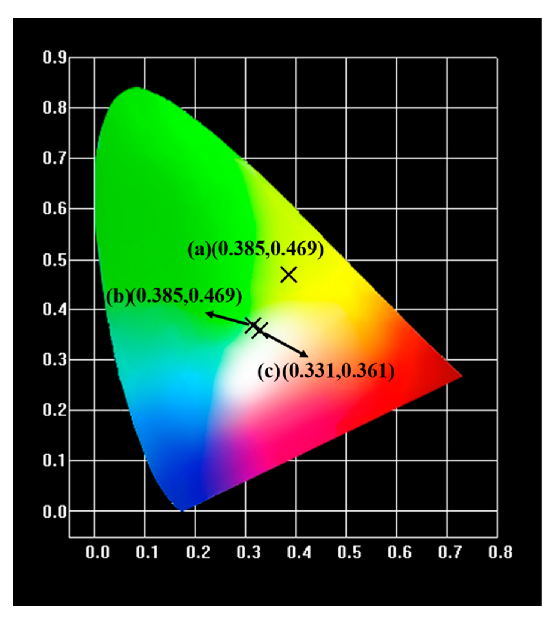
To further prove the feasibility of the as-prepared phosphors in solid-state illumination, we designed and packaged WLED devices based on an n-UV chip (365 nm) and the Na2YMg2V3O12:Dy3+ phosphors. To compensate for the color combination imbalance and improve the Ra of the LEDs, we added small amounts of commercial Y2O3:Eu3+ red phosphors and BaMgAl10O17:Eu2+ blue phosphors, thereby fabricating a warm white-emitting LED. Figure 6 shows the electroluminescence spectra and photographs of the as-fabricated LED devices. Obviously, after adding the red and blue phosphors, the emission light of the LED device changed from yellow to white. The CIE coordinates, Ra value, and CCT of the white light generated from the LED device (Figure 6c) were (0.331, 0.361), 80.2, and 7364 K, respectively. The CIE chromaticity coordinates of the LED device are also given in Figure 7. The fabricated device yielded a warm white light. The results demonstrate that the as-prepared phosphors are promising yellow-emitting phosphors for indoor solid-state illumination.

3. Materials and Methods

The Na2YMg2V3O12:xDy3+ (x = 0, 0.005, 0.01, 0.03, 0.05, 0.07) phosphors were prepared through a solid state reaction method. The analytical reagent Mg(OH)2 (average particle size, d50~3.798 μm), NaHCO3 (d50~5.638 μm), NH4VO3 (d50~160.3 μm), and high pure rare earth oxides Y2O3 (99.99%, d50~ 3.869 μm) and Dy2O3 (99.9% d50~3.990 μm) were used as raw materials. It is noteworthy that 5 mol% excess NaHCO3 was needed to compensate for the volatilization loss. The raw materials were mixed thoroughly in agate mortar for 30 min and then put into a crucible with a lid. These mixed chemicals were preheated in a muffle furnace at 500 °C for 6 h, and then heated at 800 °C for 6 h in air. After cooling to room temperature naturally, the samples were ground into powders for measurement.
The LED devices were fabricated with the as-prepared Na2YMg2V3O12:0.03Dy3+ phosphor, commercial Y2O3:Eu3+ red phosphors, BaMgAl10O17:Eu2+ blue phosphors, and an InGaN chip with a dominant emission at 365 nm (Shenzhen Looking Long Technology Co., Shenzhen, China). First, the phosphors were thoroughly mixed with organic silica gel. The weight ratio of total phosphors to organic silica gel is about 1:5. The silica gel used to package LED chips needs excellent light transmittance [46], and the light transmittance of the silica gel we chose is 96% (thickness of 1mm). Then, the surfaces of the InGaN chips were coated with the mixture with an approximate thickness of 0.5 mm. Finally, the chips were dried at 135 °C for 2h and the LED devices was obtained.
The powder X-ray diffractometer (XRD-6000, SHIMADZU, Kyoto, Japan) with Cu Kα radiation (λ = 0.15406 nm) was used to measure the phase composition of the as-prepared samples with a 40 kV operating voltage and 30 mA current. The microscopic morphology of the as-synthesized sample was investigated by a field-emission scanning electron microscope (SEM, Model Zeiss Supra-55, Heidenheim, Germany). The fluorescence spectrophotometer (F-4600, HITACHI, Tokyo, Japan) equipped with a 150 W Xe lamp as an excitation source was utilized to measure the photoluminescence (PL) and photoluminescence excitation (PLE) spectra at room temperature under 400 V of operating voltage (Xe lamp). The photoluminescence spectrum of the selected phosphor, which is temperature-dependent, was examined using a computer-controlled electric furnace spectrophotometer (TAP02, Orient KOJI, Tianjin, China). The phosphor powder was heated with a heating rate of 50 °C/min, and held at each test temperature for 3 min. The PL decay curves were obtained through a spectrofluorometer (TBX-PS; HORIBA Jobin Yvon, Paris, France) monitoring at 593nm under excitations of 289 nm and 365 nm, respectively. The electroluminescence spectra, CCT, and Ra of the packed LED devices were measured using a UV-vis-near IR spectrophotocolorimeter (PMS-80, Everfine, Hangzhou, China).

4. Conclusions

In summary, a series of vanadate phosphors Na2YMg2V3O12:Dy3+ was synthesized by the conventional solid-state reaction method at 800 °C for 6 h. In the XRD analysis, the as-prepared phosphors were found to crystallize in a single garnet structure with a cubic Ia3d (230) space group. When excited by near-UV light, the Na2YMg2V3O12 host was self-activated and emitted a broad emission band of 400–700 nm with a peak at 524 nm. This emission was ascribed to CT in the (VO4)3− groups. Meanwhile, the Na2YMg2V3O12:Dy3+ phosphors showed both the broadband luminescence of the (VO4)3− groups and the sharp peak emissions of Dy3+ ions, and emitted intense yellow light. The phosphors were also excited by light at 289 and 365 nm, and the optimum Dy3+ concentration was around 0.03 mol. The temperature-dependent emission spectra indicated high thermal stability of the Na2YMg2V3O12:Dy3+ phosphors. Finally, a WLED device based on n-UV chip, Na2YMg2V3O12:0.03Dy3+, Y2O3:Eu3+, and BaMgAl10O17:Eu2+ presented an intense white light with CIE coordinates, color rendering index, and CCT of (0.331, 0.361), 80.2, and 7364 K, respectively. These results suggest the suitability of Na2YMg2V3O12:Dy3+ phosphor as a yellow-emitting phosphor in WLEDs.

Author Contributions

Data curation, X.H.; Formal analysis, C.H. and M.F.; Investigation, F.L. and Y.L.; Methodology, X.M.; Writing—original draft, W.Z.; Writing—review & editing, X.W. and Z.H. All authors have read and agree to the published version of the manuscript.

Funding

Supported by the Fundamental Research Funds for the Central Universities (Grant No. 2652018325, 2652018321, 2652018320) and the National Key R&D Program of China (Grant No. 2018YFC190503).

Conflicts of Interest

The authors declare no conflict of interest.

References

  1. Du, P.; Yu, J.S. Photoluminescence, cathodoluminescence and thermal stability of Sm3+-activated Sr3La(VO4)3 red-emitting phosphors. Luminescence 2017, 32, 1504–1510. [Google Scholar] [CrossRef]
  2. He, C.; Ji, H.; Huang, Z.; Wang, T.; Zhang, X.; Liu, Y.; Fang, M.; Wu, X.; Zhang, J.; Min, X. Red-shifted emission in Y3MgSiAl3O12: Ce3+ garnet phosphor for blue light-pumped white light-emitting diodes. J. Phys. Chem. C 2018, 122, 15659–15665. [Google Scholar] [CrossRef] [Green Version]
  3. Min, X.; Fang, M.; Huang, Z.; Liu, Y.G.; Tang, C.; Wu, X. Synthesis and optical properties of Pr3+-doped LaMgAl11O19—A novel blue converting yellow phosphor for white light emitting diodes. Ceram. Int. 2015, 41, 4238–4242. [Google Scholar] [CrossRef]
  4. Xie, R.-J.; Hirosaki, N. Silicon-based oxynitride and nitride phosphors for white LEDs—A review. Sci. Technol. Adv. Mater. 2007, 8, 588. [Google Scholar] [CrossRef]
  5. Xie, R.-J.; Hirosaki, N.; Suehiro, T.; Xu, F.-F.; Mitomo, M. A simple, efficient synthetic route to Sr2Si5N8: Eu2+-based red phosphors for white light-emitting diodes. Chem. Mater. 2006, 18, 5578–5583. [Google Scholar] [CrossRef]
  6. Chiang, C.-H.; Fang, Y.-C.; Lin, H.-Y.; Chu, S.-Y. Photoluminescence properties and thermal stability of samarium-doped barium phosphate phosphors. Ceram. Int. 2017, 43, 4353–4356. [Google Scholar] [CrossRef]
  7. Cao, Y.; Ding, J.; Ding, X.; Wang, X.; Wang, Y. Tunable white light of multi-cation-site Na2BaCa(PO4)2: Eu, Mn phosphor: Synthesis, structure and PL/CL properties. J. Mater. Chem. C 2017, 5, 1184–1194. [Google Scholar] [CrossRef]
  8. Dang, P.; Liang, S.; Li, G.; Wei, Y.; Cheng, Z.; Lian, H.; Shang, M.; Al Kheraif, A.A.; Lin, J. Full Color Luminescence Tuning in Bi3+/Eu3+-Doped LiCa3MgV3O12 Garnet Phosphors Based on Local Lattice Distortion and Multiple Energy Transfers. Inorg. Chem. 2018, 57, 9251–9259. [Google Scholar] [CrossRef]
  9. Ji, H.; Cho, Y.; Wang, L.; Hirosaki, N.; Molokeev, M.S.; Huang, Z.; Xie, R.-J. Phase formation of (Y,Ce)2BaAl4SiO12 yellow microcrystal-glass phosphor for blue LED pumped white lighting. Ceram. Int. 2017, 43, 6425–6429. [Google Scholar] [CrossRef]
  10. Lee, S.H.; Du, P.; Bharat, L.K.; Yu, J.S. Ultraviolet radiation excited strong red-emitting LaAlO3: Eu3+ nanophosphors: Synthesis and luminescent properties. Ceram. Int. 2017, 43, 4599–4605. [Google Scholar] [CrossRef]
  11. Qiao, J.; Xia, Z.; Zhang, Z.; Hu, B.; Liu, Q. Near UV-pumped yellow-emitting Sr9MgLi(PO4)7:Eu2+ phosphor for white-light LEDs. Sci. China Mater. 2018, 61, 985–992. [Google Scholar] [CrossRef] [Green Version]
  12. Liu, S.; Liu, S.; Wang, J.; Sun, P.; Zhong, Y.; Jeong, J.H.; Deng, B.; Yu, R. Preparation and investigation of Dy3+-doped Ca9LiGd2/3(PO4)7 single-phase full-color phosphor. Mater. Res. Bull. 2018, 108, 275–280. [Google Scholar] [CrossRef]
  13. Xia, Z.; Meijerink, A. Ce3+-Doped garnet phosphors: Composition modification, luminescence properties and applications. Chem. Soc. Rev. 2017, 46, 275–299. [Google Scholar] [CrossRef] [PubMed] [Green Version]
  14. Huang, C.-H.; Liu, W.-R.; Chen, T.-M. Single-phased white-light phosphors Ca9Gd(PO4)7: Eu2+, Mn2+ under near-ultraviolet excitation. J. Phys. Chem. C 2010, 114, 18698–18701. [Google Scholar] [CrossRef]
  15. Miao, S.; Xia, Z.; Zhang, J.; Liu, Q. Increased Eu2+ content and codoping Mn2+ induced tunable full-color emitting phosphor Ba1.55Ca0.45SiO4: Eu2+, Mn2+. Inorg. Chem. 2014, 53, 10386–10393. [Google Scholar] [CrossRef]
  16. Min, X.; Fang, M.; Huang, Z.; Liu, Y.; Tang, C.; Wu, X. Luminescent properties of white-light-emitting phosphor LaMgAl11O19:Dy3+. Mater. Lett. 2014, 125, 140–142. [Google Scholar] [CrossRef]
  17. Song, D.; Guo, C.; Li, T. Luminescence of the self-activated vanadate phosphors Na2LnMg2V3O12 (Ln=Y, Gd). Ceram. Int. 2015, 41, 6518–6524. [Google Scholar] [CrossRef]
  18. Zhang, Q.; Hu, Y.; Ju, G.; Zhang, S.; Xue, F. Photoluminescence of a novel Na3Y(VO4)2:Eu3+ red phosphor for near ultraviolet light emitting diodes application. J. Mater. Sci. Mater. Electron. 2016, 28, 2529–2537. [Google Scholar] [CrossRef]
  19. Chen, W.; Zhang, J. Using nanoparticles to enable simultaneous radiation and photodynamic therapies for cancer treatment. J. Nanosci. Nanotechnol. 2006, 6, 1159–1166. [Google Scholar] [CrossRef]
  20. Liu, Y.; Chen, W.; Wang, S.; Joly, A.G. Investigation of water-soluble x-ray luminescence nanoparticles for photodynamic activation. Appl. Phys. Lett. 2008, 92, 043901. [Google Scholar] [CrossRef]
  21. Zou, X.; Yao, M.; Ma, L.; Hossu, M.; Han, X.; Juzenas, P.; Chen, W. X-ray-induced nanoparticle-based photodynamic therapy of cancer. Nanomedicine 2014, 9, 2339–2351. [Google Scholar] [CrossRef] [PubMed]
  22. Hussain, S.K.; Giang, T.T.H.; Yu, J.S. UV excitation band induced novel Na3Gd(VO4)2:RE3+ (RE3+ = Eu3+ or Dy3+ or Sm3+) double vanadate phosphors for solid-state lightning applications. J. Alloys Compd. 2018, 739, 218–226. [Google Scholar] [CrossRef]
  23. Huang, X.; Guo, H. LiCa3MgV3O12:Sm3+: A new high-efficiency white-emitting phosphor. Ceram. Int. 2018, 44, 10340–10344. [Google Scholar] [CrossRef]
  24. Duke John David, A.; Muhammad, G.S.; Sivakumar, V. Synthesis and photoluminescence properties of Sm3+ substituted glaserite-type orthovanadates K3Y[VO4]2 with monoclinic structure. J. Lumin. 2016, 177, 104–110. [Google Scholar] [CrossRef]
  25. Kang, F.; Yang, X.; Peng, M.; Wondraczek, L.; Ma, Z.; Zhang, Q.; Qiu, J. Red photoluminescence from Bi3+ and the influence of the oxygen-vacancy perturbation in ScVO4: A combined experimental and theoretical study. J. Phys. Chem. C 2014, 118, 7515–7522. [Google Scholar] [CrossRef]
  26. Yu, M.; Lin, J.; Fang, J. Silica Spheres Coated with YVO4: Eu3+ Layers via sol–gel process: A simple method to obtain spherical core–shell phosphors. Chem. Mater. 2005, 17, 1783–1791. [Google Scholar] [CrossRef]
  27. Li, K.; Lian, H.; Shang, M.; Lin, J. A novel greenish yellow-orange red Ba3Y4O9: Bi3+, Eu3+ phosphor with efficient energy transfer for UV-LEDs. Dalton Trans. 2015, 44, 20542–20550. [Google Scholar] [CrossRef]
  28. Jing, L.; Liu, X.; Li, Y.; Wang, Y. Green-to-red tunable luminescence of Eu3+-doped K3Y(VO4)2 phosphors. J. Mater. Sci. 2016, 51, 903–910. [Google Scholar] [CrossRef]
  29. Zhao, J.; Guo, C.; Li, T.; Su, X.; Zhang, N.; Chen, J. Synthesis, electronic structure and photoluminescence properties of Ba2BiV3O11: Eu3+ red phosphor. Dyes Pigments 2016, 132, 159–166. [Google Scholar] [CrossRef]
  30. Biswas, P.; Kumar, V.; Agarwal, G.; Ntwaeaborwa, O.; Swart, H. NaSrVO4: Sm3+—An n-UV convertible phosphor to fill the quantum efficiency gap for LED applications. Ceram. Int. 2016, 42, 2317–2323. [Google Scholar] [CrossRef]
  31. Nakajima, T.; Isobe, M.; Tsuchiya, T.; Ueda, Y.; Manabe, T. Correlation between luminescence quantum efficiency and structural properties of vanadate phosphors with chained, dimerized, and isolated VO4 tetrahedra. J. Phys. Chem. C 2010, 114, 5160–5167. [Google Scholar] [CrossRef]
  32. Huang, Y.; Yu, Y.M.; Tsuboi, T.; Seo, H.J. Novel yellow-emitting phosphors of Ca5M4(VO4)6 (M=Mg, Zn) with isolated VO4 tetrahedra. Opt. Express 2012, 20, 4360–4368. [Google Scholar] [CrossRef] [PubMed]
  33. Wang, Y.; Townsend, P.D. Potential problems in collection and data processing of luminescence signals. J. Lumin. 2013, 142, 202–211. [Google Scholar] [CrossRef]
  34. Zhang, G.; Zhao, L.; Fan, F.; Bai, Y.; Ouyang, B.; Chen, W.; Li, Y.; Huang, L. Near UV-pumped yellow-emitting Ca3TeO6:Dy3+ phosphor for white light-emitting diodes. Spectrochim. Acta A Mol. Biomol. Spectrosc. 2019, 223, 117343. [Google Scholar] [CrossRef] [PubMed]
  35. Zheng, T.; Ye, S.; Wang, D.; Li, K.; Wang, Y.; Liu, Z.; Wang, H. Surface modification of SrAl2O4: Eu2+, Dy3+ and Sr4Al14O25: Eu2+, Dy3+ long lasting phosphors and their application in water-borne paint. J. Chin. Ceram. Soc 2015, 2, 17–23. [Google Scholar]
  36. Li, K.; Zhang, Y.; Li, X.; Shang, M.; Lian, H.; Lin, J. Host-sensitized luminescence in LaNbO4: Ln3+ (Ln3+ = Eu3+/Tb3+/Dy3+) with different emission colors. Phys. Chem. Chem. Phys. 2015, 17, 4283–4292. [Google Scholar] [CrossRef] [PubMed]
  37. Xia, Z.; Zhou, J.; Mao, Z. Near UV-pumped green-emitting Na3(Y,Sc)Si3O9: Eu2+ phosphor for white-emitting diodes. J. Mater. Chem. C 2013, 1, 5917–5924. [Google Scholar] [CrossRef]
  38. Xia, Z.; Zhang, Y.; Molokeev, M.S.; Atuchin, V.V. Structural and luminescence properties of yellow-emitting NaScSi2O6: Eu2+ phosphors: Eu2+ site preference analysis and generation of red emission by codoping Mn2+ for white-light-emitting diode applications. J. Phys. Chem. C 2013, 117, 20847–20854. [Google Scholar] [CrossRef]
  39. Lakshmi Devi, L.; Jayasankar, C.K. Spectroscopic investigations on high efficiency deep red-emitting Ca2SiO4:Eu3+ phosphors synthesized from agricultural waste. Ceram. Int. 2018, 44, 14063–14069. [Google Scholar] [CrossRef]
  40. Nakano, H.; Kamimoto, K.; Yokoyama, N.; Fukuda, K. The Effect of Heat Treatment on the Emission Color of P-Doped Ca2SiO4 Phosphor. Materials 2017, 10, 1000. [Google Scholar] [CrossRef] [Green Version]
  41. Rojas-Hernandez, R.E.; Rubio-Marcos, F.; Serrano, A.; Salas, E.; Hussainova, I.; Fernandez, J.F. Towards Blue Long-Lasting Luminescence of Eu/Nd-Doped Calcium-Aluminate Nanostructured Platelets via the Molten Salt Route. Nanomaterials 2019, 9, 1473. [Google Scholar] [CrossRef] [PubMed] [Green Version]
  42. Li, P.; Yang, Z.; Wang, Z.; Guo, Q. White-light-emitting diodes of UV-based Sr3Y2(BO3)4:Dy3+ and luminescent properties. Mater. Lett. 2008, 62, 1455–1457. [Google Scholar] [CrossRef]
  43. Song, M.; Liu, Y.; Liu, Y.; Wang, L.; Zhang, N.; Wang, X.; Huang, Z.; Ji, C. Sol-gel synthesis and luminescent properties of a novel KBaY(MoO4)3:Dy3+ phosphor for white light emission. J. Lumin. 2019, 211, 218–226. [Google Scholar] [CrossRef]
  44. Rao, B.V.; Jang, K.; Lee, H.S.; Yi, S.-S.; Jeong, J.-H. Synthesis and photoluminescence characterization of RE3+ (=Eu3+, Dy3+)-activated Ca3La(VO4)3 phosphors for white light-emitting diodes. J. Alloys Compd. 2010, 496, 251–255. [Google Scholar] [CrossRef]
  45. Ratnam, B.V.; Jayasimhadri, M.; Jang, K.; Sueb Lee, H.; Yi, S.-S.; Jeong, J.-H. White Light Emission from NaCaPO4:Dy3+ Phosphor for Ultraviolet-Based White Light-Emitting Diodes. J. Am. Ceram. Soc. 2010, 93, 3857–3861. [Google Scholar] [CrossRef]
  46. Rojas-Hernandez, R.E.; Barradas, N.P.; Alves, E.; Santos, L.F.; Almeida, R.M. Up-conversion emission of aluminosilicate and titania films doped with Er3+/Yb3+ by ion implantation and sol-gel solution doping. Surf. Coat. Technol. 2018, 355, 162–168. [Google Scholar] [CrossRef]
Sample Availability: Samples of the compounds are available from the authors.
Figure 1. (a) XRD patterns of Na2YMg2V3O12:Dy3+ phosphors and the standard profile (Pdf NO. 49-0412), (b) schematic of the crystal structure of Na2YMg2V3O12, and (c) FESEM micrograph of the Na2YMg2V3O12:0.03Dy3+ phosphor.
Molecules 25 00542 g001
Figure 2. (a) Photoluminescence (PL) and PL emission (PLE) spectra of the Na2YMg2V3O12 samples, (b), (c), and (d) PLE and PL spectra of the samples after Gaussian peak separation. (e) Schematic of the excitation and emission processes of (VO4)3− tetrahedrons in vanadate phosphor.
Molecules 25 00542 g002
Figure 3. (a) PLE and PL spectra of the Na2YMg2V3O12:0.03Dy3+ sample. Emission spectra at (b) λex = 289 nm and (c) λex = 365 nm for different concentrations of Dy3+ in Na2YMg2V3O12:xDy3+. (d) Linear fitting data of lg(I/x) versus lg(x) for the Na2YMg2V3O12:xDy3+ phosphors. (e) Excitation spectra (λem = 582 nm) of Na2YMg2V3O12:xDy3+. (f) CIE chromaticity coordinates of the Na2YMg2V3O12:0.03Dy3+ sample.
Molecules 25 00542 g003
Figure 4. Decay curves of Na2YMg2V3O12:xDy3+ with different concentrations of Dy3 excited at (a) λex = 289 nm and (b) λex = 365 nm, (λem = 582 nm).
Molecules 25 00542 g004
Figure 5. (a,c): PL spectra of the Na2YMg2V3O12:0.03Dy3+ phosphor at different temperatures (25–175 °C) excited at λex = 289 and 365 nm, respectively. Insets show the PL intensities of Na2YMg2V3O12:0.03Dy3+ as functions of temperature. (b,d): Linear fitting curves of ln[I0/I(T) − 1] versus 1/kT for the Na2YMg2V3O12:0.03Dy3+ phosphor excited at 582 and 365 nm, respectively.
Molecules 25 00542 g005
Figure 6. Electroluminescence (EL) spectra (left) and photographs (right) of (a) Na2YMg2V3O12:0.03Dy3+, (b) Na2YMg2V3O12:0.03Dy3+ with BaMgAl10O17:Eu2+, and (c) Na2YMg2V3O12:0.03Dy3+ with BaMgAl10O17:Eu2+ and Y2O3:Eu3+. The samples were incorporated into 365 nm InGaN LED chips with an injunction current.
Molecules 25 00542 g006
Figure 7. CIE coordinates of LEDs fabricated with (a) Na2YMg2V3O12:0.03Dy3+, (b) Na2YMg2V3O12:0.03Dy3+ and BaMgAl10O17:Eu2+, and (c) Na2YMg2V3O12:0.03Dy3+, BaMgAl10O17:Eu2+, and Y2O3:Eu3+ phosphors.
Molecules 25 00542 g007
Table 1. Comparison of CIE chromaticity coordinates (x, y), correlated color temperature (CCT) (K), and lifetimes (μs) of Dy3+-doped phosphors.
Sample(x, y)CCTLifetimesReference
Na2YMg2V3O12: Dy3+(0.357, 0.461)42881.50Present work
Sr3Y2(BO3)4: Dy3+(0.300, 0.314)5896-[42]
KBaY(MoO4)3: Dy3+(0.431, 0.457)39880.125[43]
Na3Gd(VO4)2:Dy3+(0.664, 0.335)-0.234[22]
Ca3TeO6:Dy3+(0.417, 0.460)37300.506[35]
NaLa(PO3)4: Dy3+(0.292, 0.336)-0.78[44]
NaCaPO4:Dy3+(0.32, 0.37)59620.604[45]