
Alginate-Mediated Synthesis of Hetero-Shaped Silver Nanoparticles and Their Hydrogen Peroxide Sensing Ability

Abstract
1. Introduction
2. Results
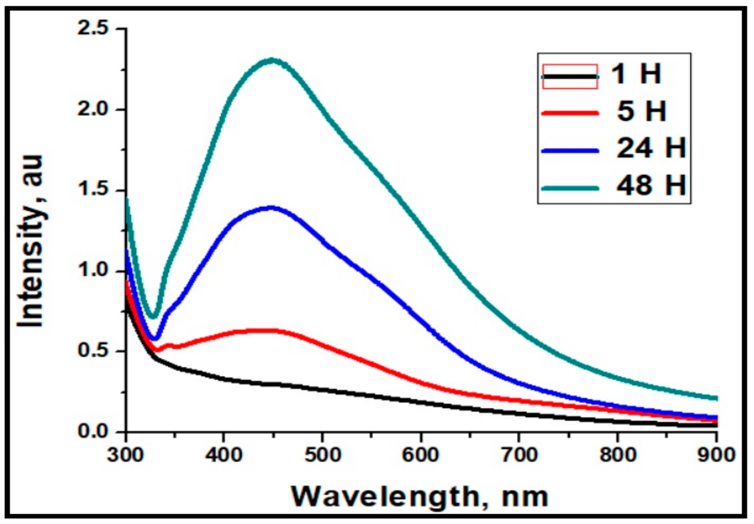
2.1. UV-Visible Spectra Analysis of Ag-NPs
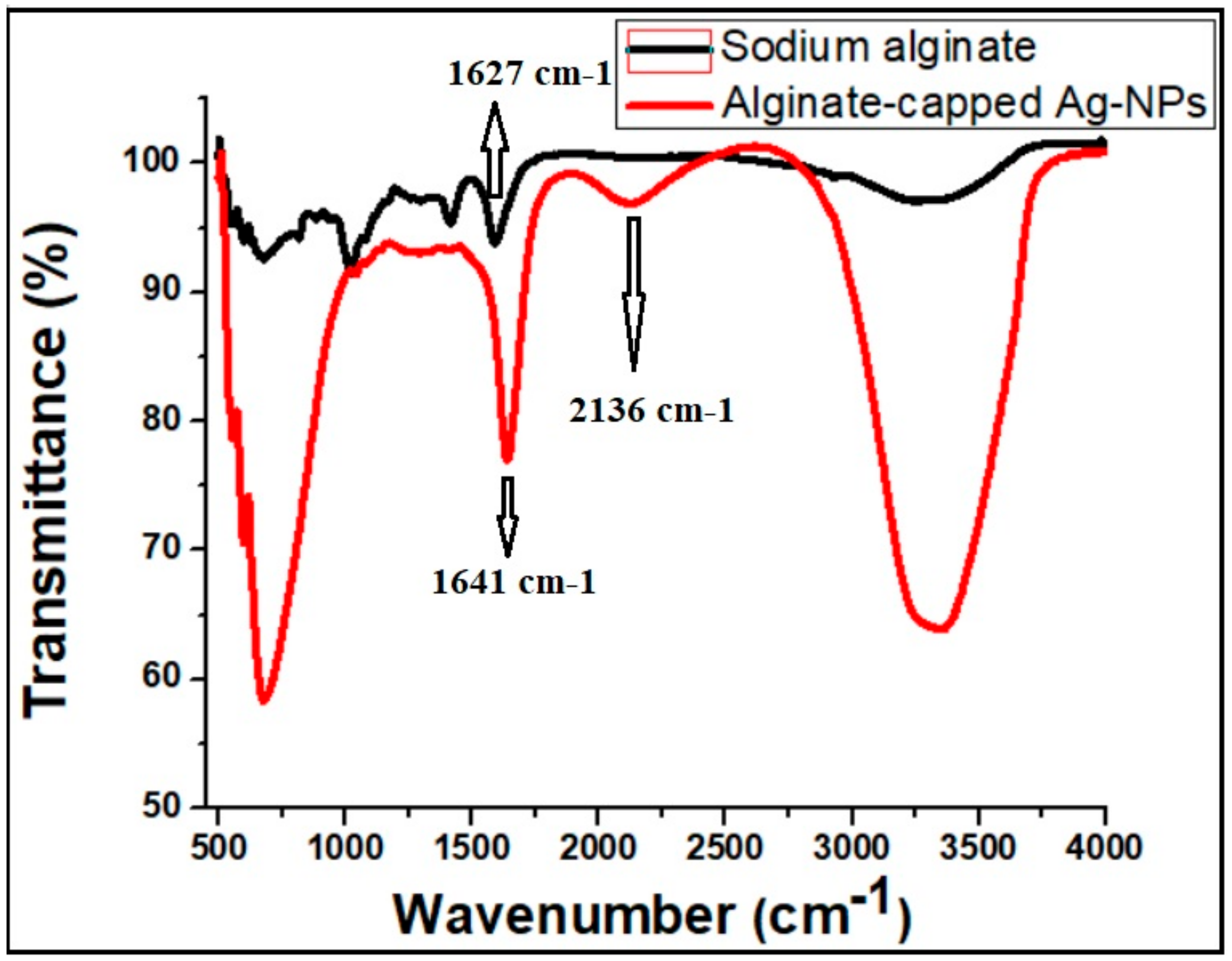
2.2. FTIR Analysis of Ag-NPs
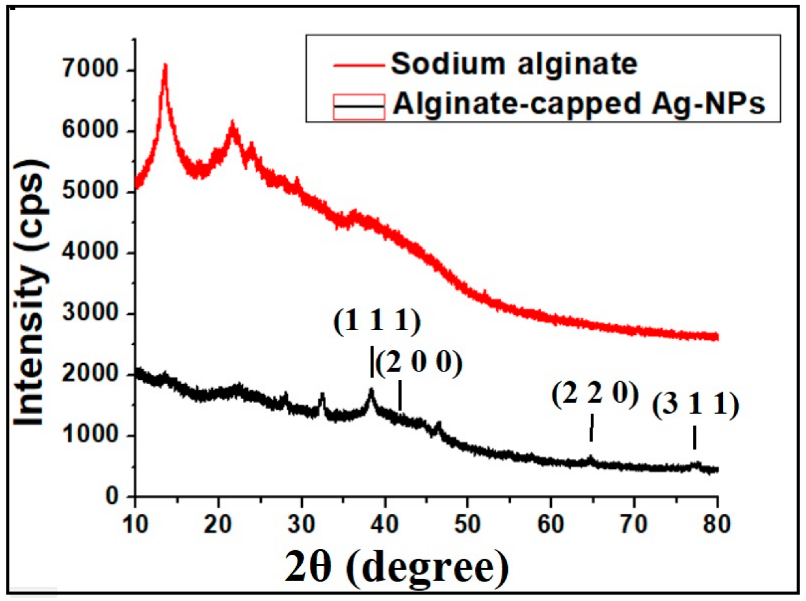
2.3. XRD Analysis
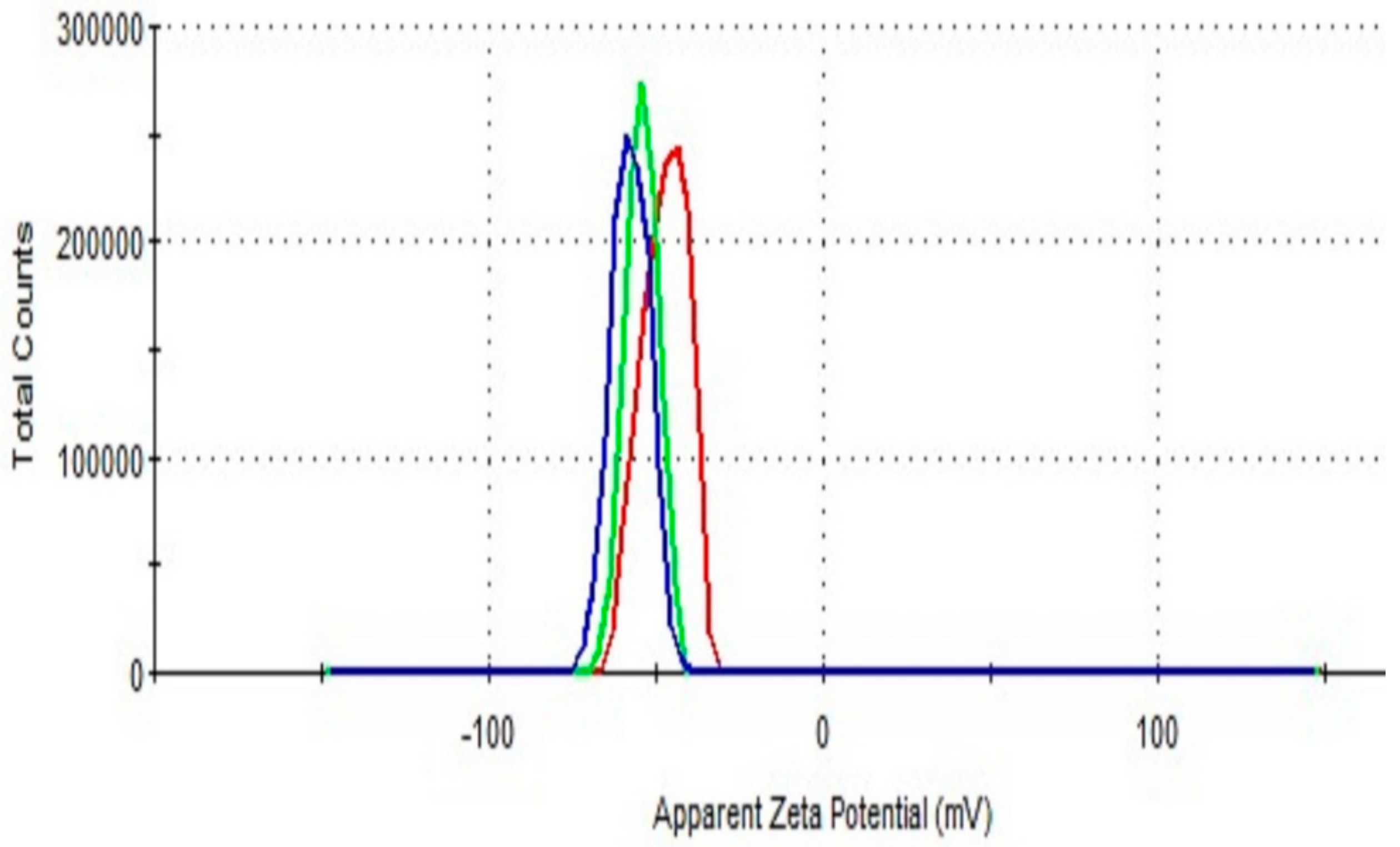
2.4. Zeta Potential Measurements
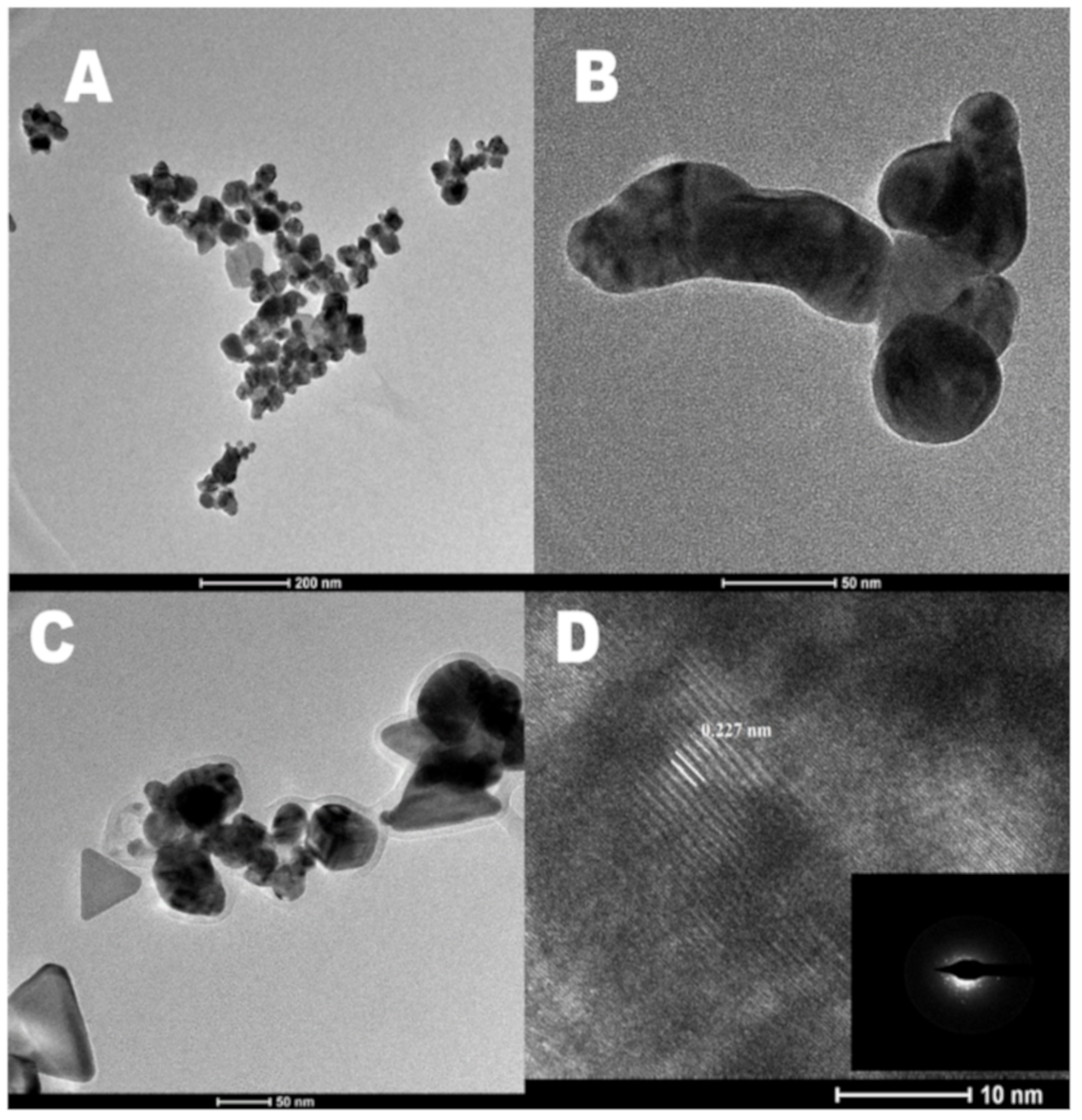
2.5. TEM and HRTEM Measurements
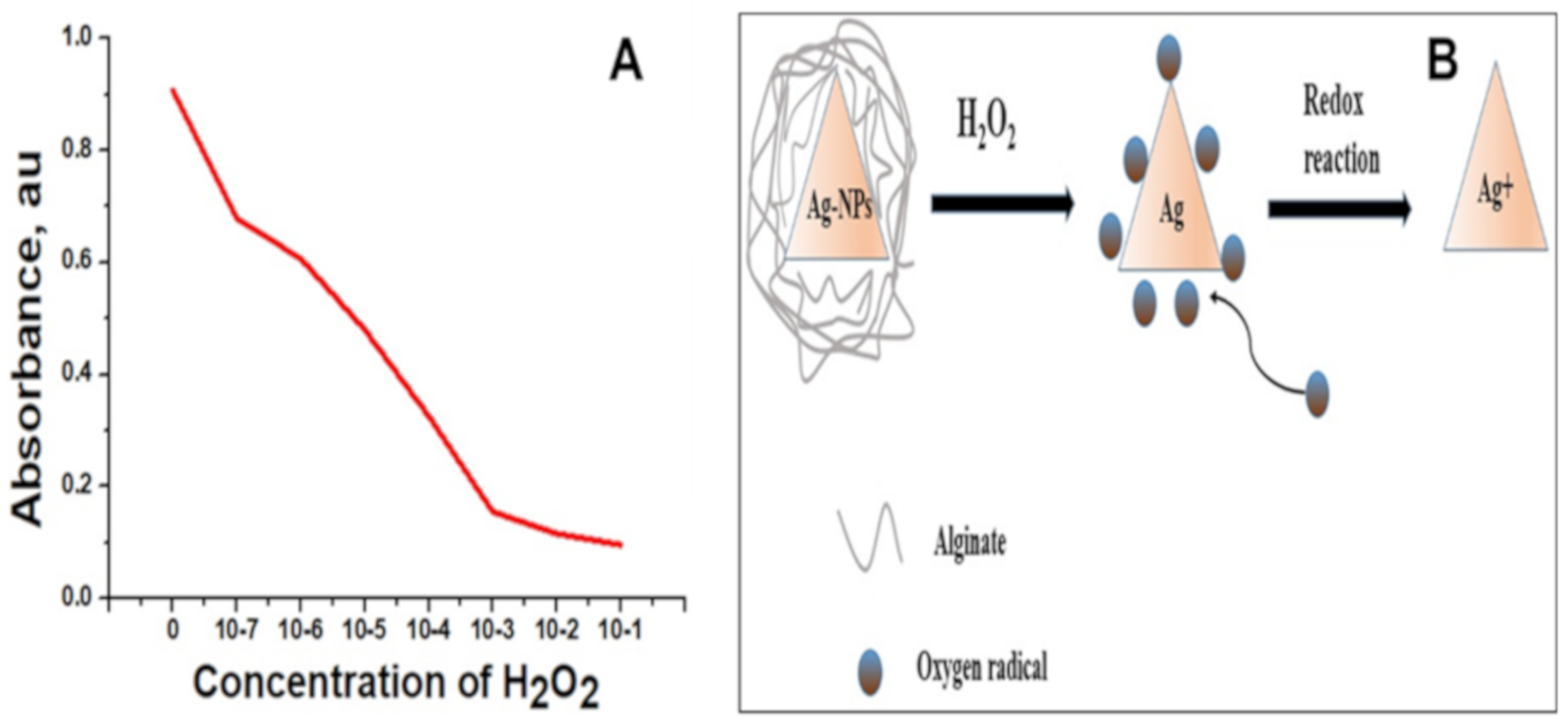
2.6. Hydrogen Peroxide Sensing Measurements
3. Materials and Methods
3.1. Materials
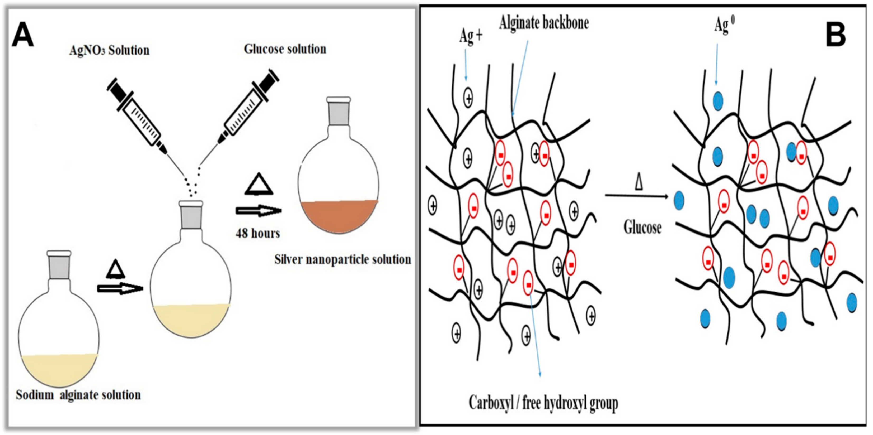
3.2. Method
3.3. Characterizations
4. Conclusions
Author Contributions
Funding
Acknowledgments
Conflicts of Interest
References
- Darroudi, M.; Ahmad, M.B.; Zamiri, R.; Zak, A.K.; Abdullah, A.H.; Ibrahim, N.A. Time-Dependent Effect in Green Synthesis of Silver Nanoparticles. Int. J. Nanomed. 2011, 6, 677–681. [Google Scholar] [CrossRef] [PubMed]
- Raveendran, P.; Fu, J.; Wallen, S.L. Completely “green” Synthesis and Stabilization of Metal Nanoparticles. J. Am. Chem. Soc. 2003, 125, 13940–13941. [Google Scholar] [CrossRef] [PubMed]
- Haque, M.N.; Kwon, S.; Cho, D. Formation and Stability Study of Silver Nano-Particles in Aqueous and Organic Medium. Korean J. Chem. Eng. 2017, 34, 2072–2078. [Google Scholar] [CrossRef]
- Kumar, S.S.D.; Rajendran, N.K.; Houreld, N.N.; Abrahamse, H. Recent Advances on Silver Nanoparticle and Biopolymer-Based Biomaterials for Wound Healing Applications. Int. J. Biol. Macromol. 2018, 115, 165–175. [Google Scholar] [CrossRef] [PubMed]
- Emam, H.E.; Manian, A.P.; Široká, B.; Duelli, H.; Redl, B.; Pipal, A.; Bechtold, T. Treatments to Impart Antimicrobial Activity to Clothing and Household Cellulosic-textiles–why “Nano”-Silver? J. Clean. Prod. 2013, 39, 17–23. [Google Scholar] [CrossRef]
- Majeed, S.; Aripin, F.H.B.; Shoeb, N.S.B.; Danish, M.; Ibrahim, M.M.; Hashim, R. Bioengineered Silver Nanoparticles Capped with Bovine Serum Albumin and its Anticancer and Apoptotic Activity against Breast, Bone and Intestinal Colon Cancer Cell Lines. Mater. Sci. Eng. C. 2019, 102, 254–263. [Google Scholar] [CrossRef]
- Sana, S.S.; Dogiparthi, L.K. Green Synthesis of Silver Nanoparticles Using Givotia Moluccana Leaf Extract and Evaluation of their Antimicrobial Activity. Mater. Lett. 2018, 226, 47–51. [Google Scholar] [CrossRef]
- Khan, M.J.; Shameli, K.; Sazili, A.Q.; Selamat, J.; Kumari, S. Rapid Green Synthesis and Characterization of Silver Nanoparticles Arbitrated by Curcumin in an Alkaline Medium. Molecules 2019, 24, 719. [Google Scholar] [CrossRef]
- Chiguvare, H.; Oyedeji, O.; Matewu, R.; Aremu, O.; Oyemitan, I.; Oyedeji, A.; Nkeh-Chungag, B.; Songca, S.; Mohan, S.; Oluwafemi, O. Synthesis of Silver Nanoparticles Using Buchu Plant Extracts and Their Analgesic Properties. Molecules 2016, 21, 774. [Google Scholar] [CrossRef]
- Zahran, M.K.; Ahmed, H.B.; El-Rafie, M.H. Alginate Mediate for Synthesis Controllable Sized AgNPs. Carbohyd. Polym. 2014, 111, 10–17. [Google Scholar] [CrossRef]
- Ranoszek-Soliwoda, K.; Tomaszewska, E.; Socha, E.; Krzyczmonik, P.; Ignaczak, A.; Orlowski, P.; Krzyzowska, M.; Celichowski, G.; Grobelny, J. The Role of Tannic Acid and Sodium Citrate in the Synthesis of Silver Nanoparticles. J. Nanopart. Res. 2017, 19, 273. [Google Scholar] [CrossRef] [PubMed]
- Dukes, K.D.; Christensen, K.A.; Chumanov, G. Core–Shell Silver Nanoparticles for Optical Labeling of Cells. Anal. Biochem. 2014, 458, 43–48. [Google Scholar] [CrossRef] [PubMed]
- Taira, N.; Ino, K.; Robert, J.; Shiku, H. Electrochemical Printing of Calcium Alginate/Gelatin Hydrogel. Electrochimica Acta 2018, 281, 429–436. [Google Scholar] [CrossRef]
- Zhang, R.; Lei, L.; Song, Q.; Li, X. Calcium Ion Cross-Linking Alginate/Dexamethasone Sodium Phosphate Hybrid Hydrogel for Extended Drug Release. Colloids Surf. B: Biointerfaces 2019, 175, 569–575. [Google Scholar] [CrossRef] [PubMed]
- Shao, Y.; Wu, C.; Wu, T.; Yuan, C.; Chen, S.; Ding, T.; Ye, X.; Hu, Y. Green Synthesis of Sodium Alginate-Silver Nanoparticles and their Antibacterial Activity. Int. J. Biol. Macromol. 2018, 111, 1281–1292. [Google Scholar] [CrossRef] [PubMed]
- Zhao, X.; Li, Q.; Ma, X.; Quan, F.; Wang, J.; Xia, Y. The Preparation of Alginate–AgNPs Composite Fiber with Green Approach and its Antibacterial Activity. J. Ind. Eng. Chem. 2015, 24, 188–195. [Google Scholar] [CrossRef]
- Aktürk, A.; Taygun, M.E.; Güler, F.K.; Goller, G.; Küçükbayrak, S. Fabrication of Antibacterial Polyvinylalcohol Nanocomposite Mats with Soluble Starch Coated Silver Nanoparticles. Colloid Surf. A. Physicochem. Eng. Asp. 2019, 562, 255–262. [Google Scholar] [CrossRef]
- Luo, L.J.; Lin, T.Y.; Yao, C.H.; Kuo, P.Y.; Matsusaki, M.; Harroun, S.G.; Huang, C.C.; Lai, J.Y. Dual-Functional Gelatin-Capped Silver Nanoparticles for Antibacterial and Antiangiogenic Treatment of Bacterial Keratitis. J. Colloid Interface Sci. 2019, 536, 112–126. [Google Scholar] [CrossRef]
- Beisl, S.; Monteiro, S.; Santos, R.; Figueiredo, A.S.; Sánchez-Loredo, M.G.; Lemos, M.A.; De Pinho, M.N. Synthesis and Bactericide Activity of Nanofiltration Composite Membranes–Cellulose Acetate/Silver Nanoparticles and Cellulose Acetate/Silver Ion Exchanged Zeolites. Water Res. 2019, 149, 225–231. [Google Scholar] [CrossRef]
- Ahn, E.Y.; Jin, H.; Park, Y. Assessing the Antioxidant, Cytotoxic, Apoptotic and Wound Healing Properties of Silver Nanoparticles Green-Synthesized by Plant Extracts. Mater. Sci. Eng. C. 2019, 101, 204–216. [Google Scholar] [CrossRef]
- Lin, G.; Lewandowska, M. Plasmon-Enhanced Fluorescence Provided by Silver Nanoprisms for Sensitive Detection of Sulfide. Sens. Actuat. B. Chem. 2019, 292, 241–246. [Google Scholar] [CrossRef]
- Fernández-Arias, M.; Zimbone, M.; Boutinguiza, M.; Del Val, J.; Riveiro, A.; Privitera, V.; Grimaldi, M.G.; Pou, J. Synthesis and Deposition of Ag Nanoparticles by Combining Laser Ablation and Electrophoretic Deposition Techniques. Coatings 2019, 9, 571. [Google Scholar] [CrossRef]
- Sharma, S.; Sanpui, P.; Chattopadhyay, A.; Ghosh, S.S. Fabrication of Antibacterial Silver Nanoparticle—Sodium Alginate–Chitosan Composite Films. RSC. Adv. 2012, 2, 5837–5843. [Google Scholar] [CrossRef]
- Malyukin, Y.; Seminko, V.; Maksimchuk, P.; Okrushko, E.; Sedyh, O.; Zorenko, Y. Hydrogen Peroxide Sensing Using Ce3+ Luminescence of Cerium Oxide (CeO2-x) Nanoparticles. Opt. Mater. 2018, 85, 303–307. [Google Scholar] [CrossRef]
- Liu, J.; Lu, L.; Li, A.; Tang, J.; Wang, S.; Xu, S.; Wang, L. Simultaneous Detection of Hydrogen Peroxide and Glucose in Human Serum with Upconversion Luminescence. Biosens. Bioelectron. 2015, 68, 204–209. [Google Scholar] [CrossRef]
- Parnell, C.M.; Watanabe, F.; Nasini, U.B.; Berry, B.C.; Mitchell, T.; Shaikh, A.U.; Ghosh, A. Electrochemical Sensing of Hydrogen Peroxide Using a Cobalt (III) Complex Supported on Carbonaceous Nanomaterials. J. Electroanal Chem. 2015, 740, 37–44. [Google Scholar] [CrossRef]
- Li, L.; Li, B.; Liu, H.; Li, M.; Wang, B. Photoelectrochemical Sensing of Hydrogen Peroxide Using TiO2 Nanotube Arrays Decorated with RGO/CdS. J. Alloy. Compd. 2020, 815, 152241. [Google Scholar] [CrossRef]
- Ahmad, M.B.; Lim, J.J.; Shameli, K.; Ibrahim, N.A.; Tay, M.Y. Synthesis of Silver Nanoparticles in Chitosan, Gelatin and Chitosan/Gelatin Bionanocomposites by a Chemical Reducing Agent and their Characterization. Molecules 2011, 16, 7237–7248. [Google Scholar] [CrossRef]
- Agnihotri, S.; Mukherji, S.; Mukherji, S. Size-Controlled Silver Nanoparticles Synthesized over the Range 5–100 nm Using the Same Protocol and their Antibacterial Efficacy. RSC. Adv. 2014, 4, 3974–3983. [Google Scholar] [CrossRef]
- Mihaylov, M.; Chakarova, K.; Hadjiivanov, K.; Marie, O.; Daturi, M. FTIR Spectroscopy Study of CO Adsorption on Pt− Na− Mordenite. Langmuir 2015, 21, 11821–11828. [Google Scholar] [CrossRef]
- Shameli, K.; Ahmad, M.B.; Zargar, M.; Yunus, W.M.Z.W.; Ibrahim, N.A.; Shabanzadeh, P.; Ghaffari, M.M. Synthesis and Characterization of Silver/Montmorillonite/Chitosan Bionanocomposites by Chemical Reduction Method and their Antibacterial Activity. Int. J. Nanomed. 2011, 6, 271–284. [Google Scholar] [CrossRef]
- Raveendran, P.; Fu, J.; Wallen, S.L. A Simple and “Green” Method for the Synthesis of Au, Ag, and Au–Ag Alloy Nanoparticles. Green Chem. 2006, 8, 34–38. [Google Scholar] [CrossRef]
- Mao, S.; Zhang, T.; Sun, W.; Ren, X. The Depolymerization of Sodium Alginate by Oxidative Degradation. Pharm. Dev. Technol. 2012, 17, 763–769. [Google Scholar] [CrossRef]
- Pal, T.; Maity, D.S.; Ganguly, A. Silver—Gelatin Method for Determination of Inorganic Peroxides in Alkaline Solution. Talanta 1988, 35, 658–660. [Google Scholar] [CrossRef]
- Mohan, S.; Oluwafemi, O.S.; George, S.C.; Jayachandran, V.P.; Lewu, F.B.; Songca, S.P.; Kalarikkal, N.; Thomas, S. Completely Green Synthesis of Dextrose Reduced Silver Nanoparticles, its Antimicrobial and Sensing Properties. Carbohyd. Polym. 2014, 106, 469–474. [Google Scholar] [CrossRef]
- Endo, T.; Yanagida, Y.; Hatsuzawa, T. Quantitative Determination of Hydrogen Peroxide Using Polymer Coated Ag Nanoparticles. Measurement 2008, 41, 1045–1053. [Google Scholar] [CrossRef]
Sample Availability: Samples of the alginate-capped Ag-NPs are available from the authors. |


© 2020 by the authors. Licensee MDPI, Basel, Switzerland. This article is an open access article distributed under the terms and conditions of the Creative Commons Attribution (CC BY) license (http://creativecommons.org/licenses/by/4.0/).
Share and Cite
Bhagyaraj, S.; Krupa, I. Alginate-Mediated Synthesis of Hetero-Shaped Silver Nanoparticles and Their Hydrogen Peroxide Sensing Ability. Molecules 2020, 25, 435. https://doi.org/10.3390/molecules25030435
Bhagyaraj S, Krupa I. Alginate-Mediated Synthesis of Hetero-Shaped Silver Nanoparticles and Their Hydrogen Peroxide Sensing Ability. Molecules. 2020; 25(3):435. https://doi.org/10.3390/molecules25030435
Chicago/Turabian StyleBhagyaraj, Sneha, and Igor Krupa. 2020. "Alginate-Mediated Synthesis of Hetero-Shaped Silver Nanoparticles and Their Hydrogen Peroxide Sensing Ability" Molecules 25, no. 3: 435. https://doi.org/10.3390/molecules25030435
APA StyleBhagyaraj, S., & Krupa, I. (2020). Alginate-Mediated Synthesis of Hetero-Shaped Silver Nanoparticles and Their Hydrogen Peroxide Sensing Ability. Molecules, 25(3), 435. https://doi.org/10.3390/molecules25030435