Review

Plant Xyloglucan Xyloglucosyl Transferases and the Cell Wall Structure: Subtle but Significant

1
Institute of Chemistry, Centre for Glycomics, Slovak Academy of Sciences, Dúbravská cesta 9, SK-84538 Bratislava, Slovakia
2
Faculty of Natural Sciences, Department of Physical and Theoretical Chemistry, Comenius University, Mlynská Dolina, SK-84215 Bratislava, Slovakia
3
School of Life Science, Huaiyin Normal University, Huai’an 223300, China
4
School of Agriculture, Food and Wine, University of Adelaide, Glen Osmond, SA 5064, Australia
*
Author to whom correspondence should be addressed.
Molecules 2020, 25(23), 5619; https://doi.org/10.3390/molecules25235619
Submission received: 25 October 2020 / Revised: 25 November 2020 / Accepted: 26 November 2020 / Published: 29 November 2020
(This article belongs to the Special Issue Targeting Carbohydrate–Protein Interactions)

Abstract

:
Plant xyloglucan xyloglucosyl transferases or xyloglucan endo-transglycosylases (XET; EC 2.4.1.207) catalogued in the glycoside hydrolase family 16 constitute cell wall-modifying enzymes that play a fundamental role in the cell wall expansion and re-modelling. Over the past thirty years, it has been established that XET enzymes catalyse homo-transglycosylation reactions with xyloglucan (XG)-derived substrates and hetero-transglycosylation reactions with neutral and charged donor and acceptor substrates other than XG-derived. This broad specificity in XET isoforms is credited to a high degree of structural and catalytic plasticity that has evolved ubiquitously in algal, moss, fern, basic Angiosperm, monocot, and eudicot enzymes. These XET isoforms constitute gene families that are differentially expressed in tissues in time- and space-dependent manners during plant growth and development, and in response to biotic and abiotic stresses. Here, we discuss the current state of knowledge of broad specific plant XET enzymes and how their inherently carbohydrate-based transglycosylation reactions tightly link with structural diversity that underlies the complexity of plant cell walls and their mechanics. Based on this knowledge, we conclude that multi- or poly-specific XET enzymes are widespread in plants to allow for modifications of the cell wall structure in muro, a feature that implements the multifaceted roles in plant cells.

Table of Contents
  • Plant cell walls and structure, and key components
  • Roles of xyloglucan xyloglucosyl transferases in cell wall formation and re-modelling
    • Catalysis, and remarks on nomenclature and classification
    • Enzyme activity assay methods
  • Substrate specificity of plant xyloglucan xyloglucosyl transferases
    • Homo-transglycosylation reactions
    • Hetero-transglycosylation reactions with neutral donor and acceptor substrates
    • Hetero-transglycosylation reactions with charged (ionic) acceptor substrates
  • Implications of transglycosylation reactions catalysed by xyloglucan xyloglucosyl transferases in the cell wall structure, function and dynamics

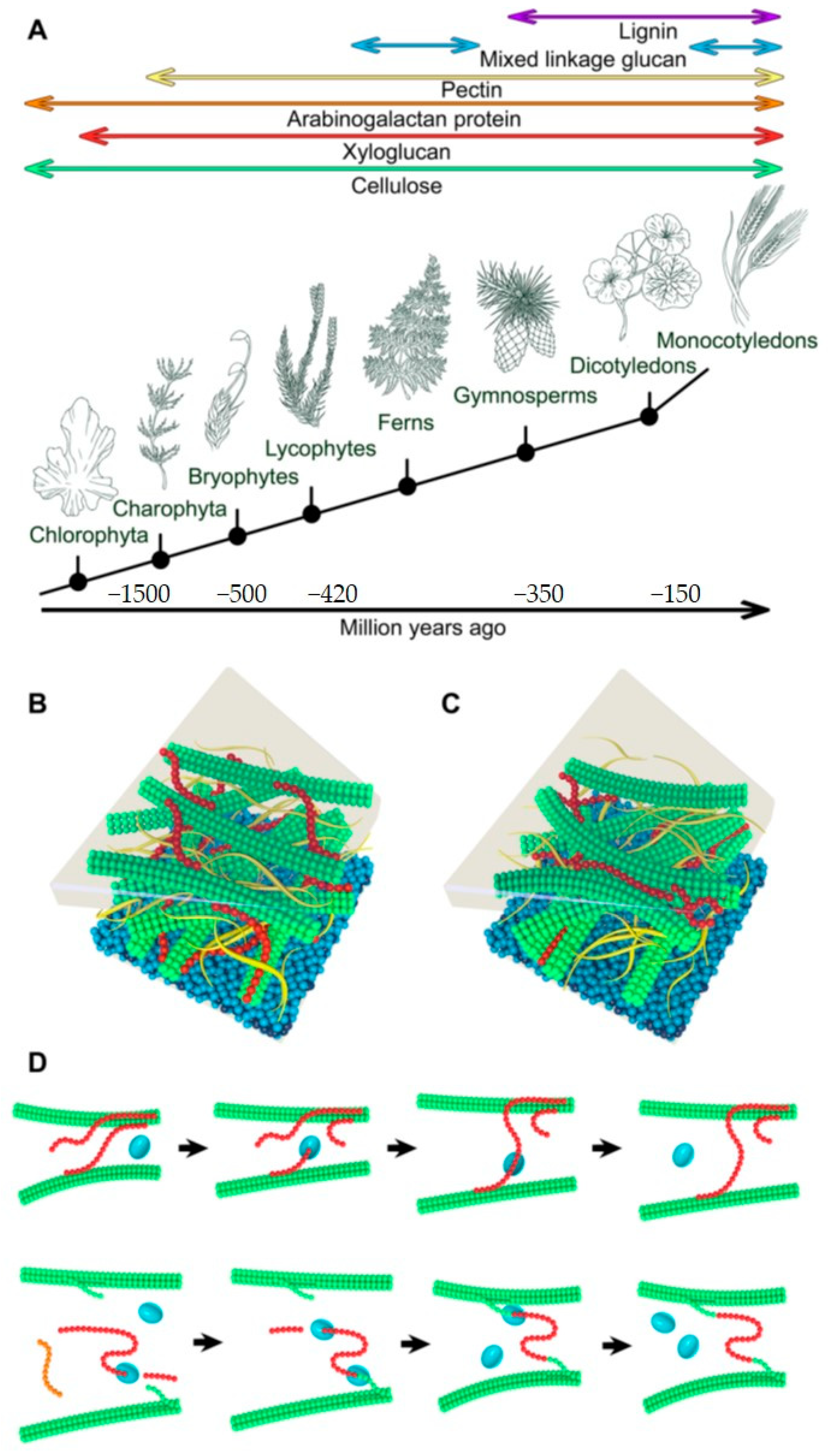
1. Plant Cell Walls and Structure and Key Components

The presence of polysaccharide-rich cell walls (CWs) is a characteristic feature of plants and fungi. Throughout the evolution of plants, CWs have conformed to multiple roles, including mechanical support, diffusion and growth regulation, defence against biotic and abiotic stresses, and cell-to-cell communication. CWs are highly complex structural entities largely composed of organic polymeric molecules interlinked by covalent and non-covalent linkages. Properties of CWs depend on the composition and chemical linkages of individual adjoined components, and their structure [1,2,3,4,5,6].
All land plants classified in Embryophytes evolved from Charophytes green algae (Figure 1A) contain CWs assumed to be one of the most decisive factors that allowed for terrestrialisation [7,8,9,10]. Despite certain common components, the composition of CWs varies in species [11,12] and tissues [13,14,15,16,17]. The structure of plant CWs could also be affected by growth conditions [18] and some common distributions of main structural polysaccharides are observed that depend on the evolutionary history of a plant including algae [10,19,20,21,22]. However, certain polysaccharides could have evolved independently several times [16], and this is observed in (1,3;1,4)-β-d-glucans (mix-linkage glucans, MLGs) in Pteridophytes (ferns, whisk ferns, horsetails) [23,24,25] and Poales (grasses) [26] (Figure 1A). CWs of grasses differ from those of other higher plants in a lower content of xyloglucans (XGs) and pectins but in a higher content of heteroxylans [13].
Plants are known to construct two types of CWs, termed primary cell walls (PCWs) and secondary cell walls (SCWs) that differ in composition, structure, and function. The dynamic PCW structures comprise of the networks of cellulose micro-fibrils tethered by cross-linking glycans [27] that are embedded in the matrix of pectin substances [28,29] and glycoproteins. Glycans or hemicelluloses include variously substituted XGs, which are the major form of cross-linking glycans in dicotyledonous plants, while xylan(s)—Xyl(s), arabinoxylan(s)—AraXyl(s), glucuronoarabinoxylan(s), mannan(s)—Man(s), galactomannan(s)—GalMan(s), glucomannan(s)—GlcMan(s) and galactoglucomannan(s)—GalGlcMan(s), and MLGs largely replace XGs in monocots and lower plants [1,30]. Pectins [31] composed of the homogalacturonan (HG) part are cross-linked via Ca2+ bridges that causes the gelling effect, while the ‘hairy’ regions of pectins are made of rhamnogalacturonan I (RG I) and rhamnogalacturonan II (RG II) [32,33,34,35]. Conversely, SCWs that are more typical for woody and vascular tissues after their growth ceases are more rigid compared to PCWs since they contain more of other cross-linking glycans than pectins, and are reinforced by lignin, a hydrophobic phenylpropanoid polymer [36,37,38].
Cellulose, as the most abundant organic compound on Earth [39], consists of the repeating glucopyranose moieties linked through (1,4)-β-d-linkages that form micro-fibrils tightly bound via hydrogen bonds—these supra-molecular structures form the backbone of PCWs [40]. Cellulose is found not only in CWs of green algae and higher plants but also in Rhodophyta (red algae), Phaeophyceae (brown algae), Oomycetes (fungus-like microorganisms), Ameobozoa, animals, and in some Procaryotes (Cyanobacteria) [22].
The biosynthesis of XGs [41] seems to be reserved for plants including certain green algae [42]. XG was not detected in red and brown algae [22], and this absence is also supported by the lack of XG modifying enzymes in these organisms [43]. XG consists of repeating (1,4)-β-d-linked glucopyranosyl moieties [44], which contain the C-6 carbon branching by α-d-xylopyranosyl residues. Xylosyl moieties could be further substituted by galactopyranosyl residues on C-2 carbons (β-d-Galp-(1,2)-α-d-Xylp) and the galactosyl moieties could carry the fucopyranosyl branching (α-l-Fucp-(1,2)-β-d-Galp-(1,2)-α-d-Xylp). In certain instances, the arabinopyranosyl [45,46] and galacturonate [47,48] substituents are found, which suggests that the structure of XG differs from plant to plant, but also between the parts of the same plant [49,50,51]. The XG backbone is synthesised by a XG:glucan synthase encoded by members of the C subfamily of cellulose synthase-like (CSL) genes [41,52], but only when both UDP-glucose and UDP-xylose are present [53,54], meaning that the activity of another enzyme XG:xylosyltransferase is required to produce XG [53]. Most of XG residues are substituted with d-Gal by XG:galactosyltransferases and further modified with l-fucose by XG:fucosyltransferases [53,54], although more work is required to clarify all aspect of XG biosynthesis. A recent study with the C subfamily CSL synthases and their genes found a quintuple mutant with disruptions in five C subfamily CSL genes that had no detectable XG, and did not display the significant alteration of gene expression at the whole genome level [41].
The similarity between the structure of XG and cellulose underlies the conformational homology of these polysaccharides, which results in their strong noncovalent associations [54]. Networks of cellulose and XGs were for a long time considered to support the structure of PCWs, which also trigger their flexibility and strength [59,60,61,62,63]. The most recognised PCW model of dicotyledons [56] is based on linear micro-fibrils of cellulose, each consisting of 32 cellulosic micro-fibrils inter-connected through hydrogen bonds. These para-crystalline micro-fibrils are interwoven and bridged to polymeric XGs. According to this model (Figure 1B), the function of pectin is attributed to a gelling material that pervades the space between the cellulosic and XG structures. The advances in the field of microscopic techniques allowed for the development of advanced CW models based on so-called hot spots of cellulose micro-fibrils, where these micro-fibrils composed of eight or sixteen cellulosic subunits [64], come to close contacts with each other (Figure 1C). These hot spots of cellulosic micro-fibrils are isolated from each other by a thin layer of XGs, which according to this model are isolated in PCWs [57]. Here, pectins serve as the filling material between the cellulosic and XGs structures, and take over the major role of the XGs, compared to the first model. Various pectins interact both with cellulose and XGs, thus ensuring the flexibility and strength of CWs [65]. The processes of hot spots formation remain unknown, although one possibility is that they are formed spontaneously during the cellulose deposition into CWs, or that they could be formed enzymatically [66].
While there are numerous studies focused on the role of XGs in CW modifications during plant growth and development, little information is available as to how XGs participate in the CW formation. Recent work focused on the regeneration of CWs in wild-type Arabidopsis thaliana and a double mutant xxt1 xxt2 lacking any detectable XGs, suggested that the formation of cellulosic networks is XG-independent [67].
As already noted, CWs show a remarkable diversity that underlies the function of each cell, which is directly linked to the basic and subtle structure of polymers, their quantity, ratios, and underpins mutual interactions. The syntheses of CW polysaccharides occur due to the cooperative activities of prodigious numbers of biosynthetic glycosyl transferases (GTs) or synthases [41,68,69,70,71,72,73,74,75,76,77,78,79,80,81,82,83,84], localised mostly in the Golgi apparatus [85]. The products of these GTs and synthases are transported to CWs by secretory vesicles, although the exception includes the superfamilies of cellulose synthases [86,87,88,89,90] and callose synthases [91] locating in the plasma membrane [92]. In the latter instances, polysaccharides synthesised by these enzymes could be exported directly to CWs. However, the structural polysaccharides that are observed in CWs are often heterogeneous. This is assumed to be achieved through a wide range of available activated sugar donors required for GTs activities, which could give rise to a variety of glycosidic linkages using different sugar isomers [16].
Life processes in plants are underscored by CWs structure and re-organisation, which involves disintegration, elongation and expansion. In addition to GTs, these processes are governed by hydrolases and lyases, and non-catalytic expansin proteins [93,94,95,96]. As in the case of GTs, there is a vast number of enzymes that could modify structural polysaccharides in muro by cleaving bonds, esterifying or de-esterifying saccharide moieties [97,98], and incorporating new material into CWs or re-constructing CW polymers by cross-linking. The last two processes are secured by xyloglucan xyloglucosyl transferase also known as xyloglucan endo-transglycosylase (XET) enzymes (Figure 1D), which could either loosen or enhance the packing of cellulosic micro-fibrils and other polysaccharides in CWs [58,99].
Xyloglucan xyloglucosyl transferases or XET enzymes (EC 2.4.1.207) as one of the key glycosidic bond-formation enzymes participating in plant CW expansion, reconstruction, and re-modelling [100,101,102,103,104] were independently discovered by three groups [105,106,107]. According to the Enzyme Commission [108], these enzymes are also named as endo-xyloglucan transferases.

2. Roles of Xyloglucan Xyloglucosyl Transferases in Cell Wall Formation and Re-Modelling

Catalysis, and remarks on the nomenclature and classification—The nomenclature of xyloglucan xyloglucosyl transferases or XET enzymes is defined by the International Union of Biochemistry and Molecular Biology (IUBMB)/International Union of Pure and Applied Chemistry (IUPAC) Biochemical Nomenclature Committee [108] that is also implemented in the Kyoto Encyclopaedia of Genes and Genomes Enzyme Database (KEGG). These enzymes are classified amongst transferases by the Enzyme Commission (EC) and listed under the primary identifier EC 2.4.1.207 in IUBMB/IUPAC and the BRENDA collection of enzyme functional data [109]. The differences in the used nomenclature are based on whether the authors consider the transfer of ‘glycosyl’ (xyloglucan endo-transglycosylase) or ‘glucosyl’ (xyloglucan endo-transglucosylase) groups [110,111,112,113,114,115,116,117], although given that XETs primarily transfer XG fragments, the usage of the first name should be preferred. The fundamental feature of the catalysis mediated by XETs is the breaking of a bond between 1,4-β-d-linked glucosyl residues of XGs and the transfer of an XG fragment onto O-4 of the non-reducing terminal end of the glucose moiety of the acceptor, which can be XG or its oligosaccharide (XG-OS). This constitutes a so-called ping-pong bi bi reaction mechanism rather than a sequential one [118,119]. It is of note that the definition by the Enzyme Commission contains a strict note ‘does not use cello-oligosaccharides as either donor or acceptor’, although, in the light of the current knowledge, this specification is obsolete [58,99,120,121,122,123,124,125].
A more objective view on the XET enzyme nomenclature and classification is given by the Carbohydrate-Active enZYmes Database (CAZy; CAZypedia Consortium 2018) [126], which is based on protein tertiary structures and substrate specificities or activities. According to CAZy, XETs are classified in a glycoside hydrolase (GH16 family) and not in a glycoside transferase (GT) group. According to this classification, the latter group contains enzymes which utilise activated sugar donors. In accordance with tertiary structures of XET enzymes, the first steps of both transglycosylation and hydrolytic reactions are binding and cleavage of donor substrates. The difference occurs in the second step, in which the fragment with the original non-reducing end of the substrate is transferred to an acceptor, which in the case of a typical transglycosylase is another saccharide, while, in the case of a hydrolase, it is a water molecule (Figure 2A) [127]. This second step of the reaction has the key importance for the nomenclature and classification of transglycosylases as transferases. Commonly, most hydrolases could also transglycosylate, but this only occurs under high substrate concentrations [128], when in the later stages the products accumulate and shift the chemical equilibrium of the reaction towards transglycosylation reactions, due to a specific response of the biocatalyst. Contrary to this, ‘true’ transglycosylases, which include XETs, catalyse primarily the transfer on a saccharide from the beginning of the reaction. While hydrolytic reactions catalysed by hydrolases reflect the increased concentrations of reducing groups in the reaction system, this is not the case for ‘true’ transglycosylases. Furthermore, during reactions with endo-transglycosylases, no dramatic decrease in the viscosity of the polymeric substrates is observed at the early stages of reactions, contrary to endo-hydrolases. Besides XETs, other transglycosylases or enzymes with potential transglycosylase activities were described in plants as endo-transglycosylases/hydrolases recognising (1,4)-β-d-mannan-derived polysaccharides [129,130], xylan endo-transglycosylases functionalising heteroxylan polysaccharides [131,132,133], MLG: xyloglucan endotransglucosylases [134,135] recognising MLGs, and the hetero-trans-β-glucanase (HTG) that functionalise cellulose [96,136], although designating these enzymes as such hides the fact that the latter enzymes are broad specific or poly-specific XET enzymes.
XET enzymes are classified in GH family 16 (GH16) in CAZy. A GH16 family is a large group, which was according to specific features in tertiary structures further sub-divided into 23 subfamilies [137]. Subfamily GH16_20 includes XETs and xyloglucan endohydrolases (XEHs, EC 3.2.1.151) [138,139] with predominantly hydrolytic activities towards XGs. This group of enzymes contains the products of XTH (xyloglucan transglycosylase/hydrolase) genes encoding both types of XG-modifying enzymes which display close similarity in their tertiary structures [140].
Currently, the best characterised XET enzyme is the PttXET16A isoform from hybrid aspen, Populus tremulus x tremuloides [138,140]. After its expression in a recombinant host achieved in high yields in Pichia [102], the crystal structure of PttXET16A [127] (Protein Data Bank—PDB accessions 1UN1 and 1UMZ) revealed that the enzyme folds into two antiparallel β-sheets, which form a β-sandwich consisting of convex and concave regions. The catalytic machinery, formed by two glutamic acid residues Glu85 and Glu89, with an aspartate Asp87, is located approximately mid-way in the convex region. The C-terminal end is elongated compared to other XTH family members [138,140] and located near the convex region of the β-sheet, forming an α-helix and another β-strand on the concave side of the molecule; this part of the structure is stabilised by two disulphide bridges. PttXET16A is N-glycosylated at Asn93 with two N-acetylglucosaminyl and mannosyl moieties that are stabilised by hydrogen bonds. The structures of other plant XET enzymes are yet to be determined, however, the structural features of barley and nasturtium XETs (Figure 2B, Figure 3B, and Figure 4B) and HTG were defined through homology modelling, an approach that could introduce local approximations in structural features of modelled XETs [121,123,125,136,142].
The mechanism of transglycosylation catalysed by XET enzymes proceeds in two stages that incorporate two transition states (Figure 2A). The first step is the deprotonation of the carboxyl acid residue acting as the nucleophile that attacks the anomeric carbon forming the glycosyl-enzyme intermediate complex with acidic assistance provided by the acidic carboxylate. In PttXET16A, the nucleophile attacking the anomeric carbon is Glu85, while Glu89 acts as an acid/base, which protonates the released saccharide and subsequently de-protonates the glycosyl acceptor. The Asp87 residue located mid-way between both catalysts Glu85 and Glu89 controls the protonation state of the catalytic machinery and operates through hydrogen bonding interactions. The nucleophile must be de-protonated during the donor substrate attack, while Asp87 and Glu89 are protonated and donate the proton to a leaving saccharide. During the later stages of the transfer reaction, the glycosyl-enzyme intermediate complex dissociates after the nucleophile attack on the anomeric carbon, and a new glycosidic bond is formed [127,138].
Interactions between residues in PttXET16A and dimeric XG nonasaccharide were obtained using molecular dynamics simulations [140]. The substrate was modelled in active site in a way, in which one of the XG nonasaccharide dimers occupied the donor site creating a stable intermediate with the enzyme, while the second XG nonasaccharide occupied the acceptor site. It was important to observe that the XG nonasaccharide of the reducing-end glucose moiety changed its conformation into a boat at the beginning of the simulation and kept this conformation during the whole simulation time. This was the case not only for PttXET16A, but also for XEHs [140].
Attention was paid to structural differences, which determine whether the XET/XEH enzymes act as transglycosylases (PDB accessions 1UN1, 1UMZ) or hydrolases (TmNXG1; PDB accession 2UWA) [138,139,140,143]; here, the information of their primary and crystal structures was compared including the TmNXG1-DELTAYNIIG mutant (PDB accession 2VH9). Minor differences in structures lead to stronger binding of a donor substrate combined with larger loop flexibility at the acceptor binding site, and the higher conformational flexibility of specific residues underlined the hydrolytic preference [140,143] (Figure 2B). This was also reflected by the phylogenetic analyses, which showed that the distribution of XTH gene products was segregated into three groups, with XEHs belonging to the XTH III clade, while XETs were placed in both XTH I and XTH II clades (Figure 2C, Supplementary Data Set S1) [121,123,138,144]. Based on these structural and phylogeny insights, it was postulated that GH16 hydrolases could have evolved from XET transglycosylases [138,143], although the research including a gradual cross-genome survey and the whole GH16 family phylogeny continues [125,145].
Enzyme activity assays methods—Preferred methods of XET activity assays are based on the use of radiochemically or fluorescently labelled acceptor oligosaccharides and an unlabelled donor polysaccharide. Alternatively, radiolabelled donor and unlabelled acceptor substrates could be used [96]. Products then contain labelled components that are incorporated into products, and remaining unincorporated donors or acceptors are removed through washing on a filter-paper [146], through a gel [120,147], size-exclusion chromatography [58,107,123], or ethanol precipitation [148]. Labelled oligosaccharides could also be used in high-throughput activity assays [149] and for visualisations of XET activities in vivo [150,151,152,153]. Despite an undeniable advantage of the latter technique for screening of XET activities, it is important to consider ongoing reactions amongst polysaccharides in muro. For the selection of activity assays, it is crucial to consider the choice of fluorescent tags [154,155] and the removal of unreacted donors or acceptors [99,123]. Other XET activity assays include viscometry and colorimetry [156].

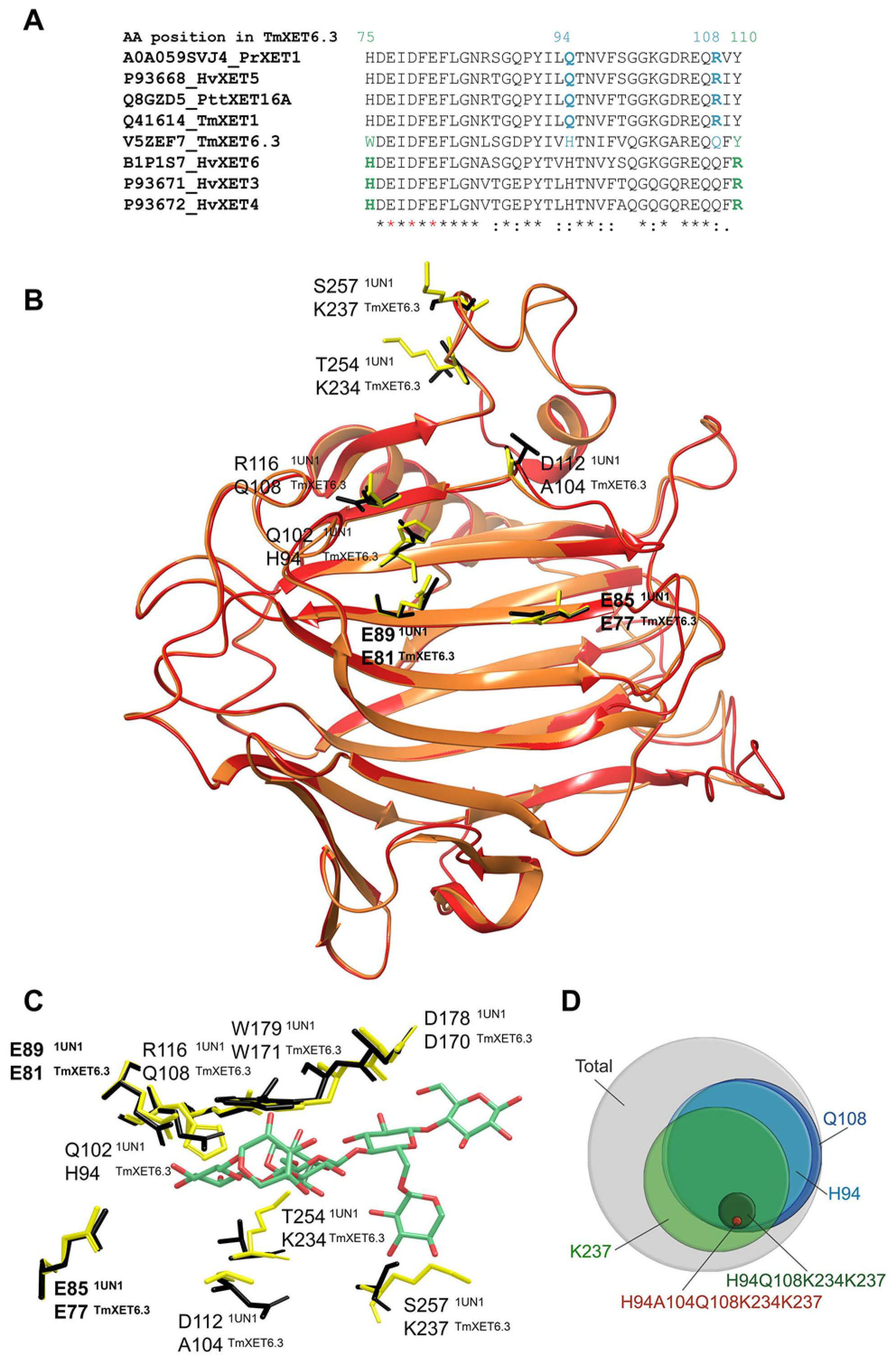
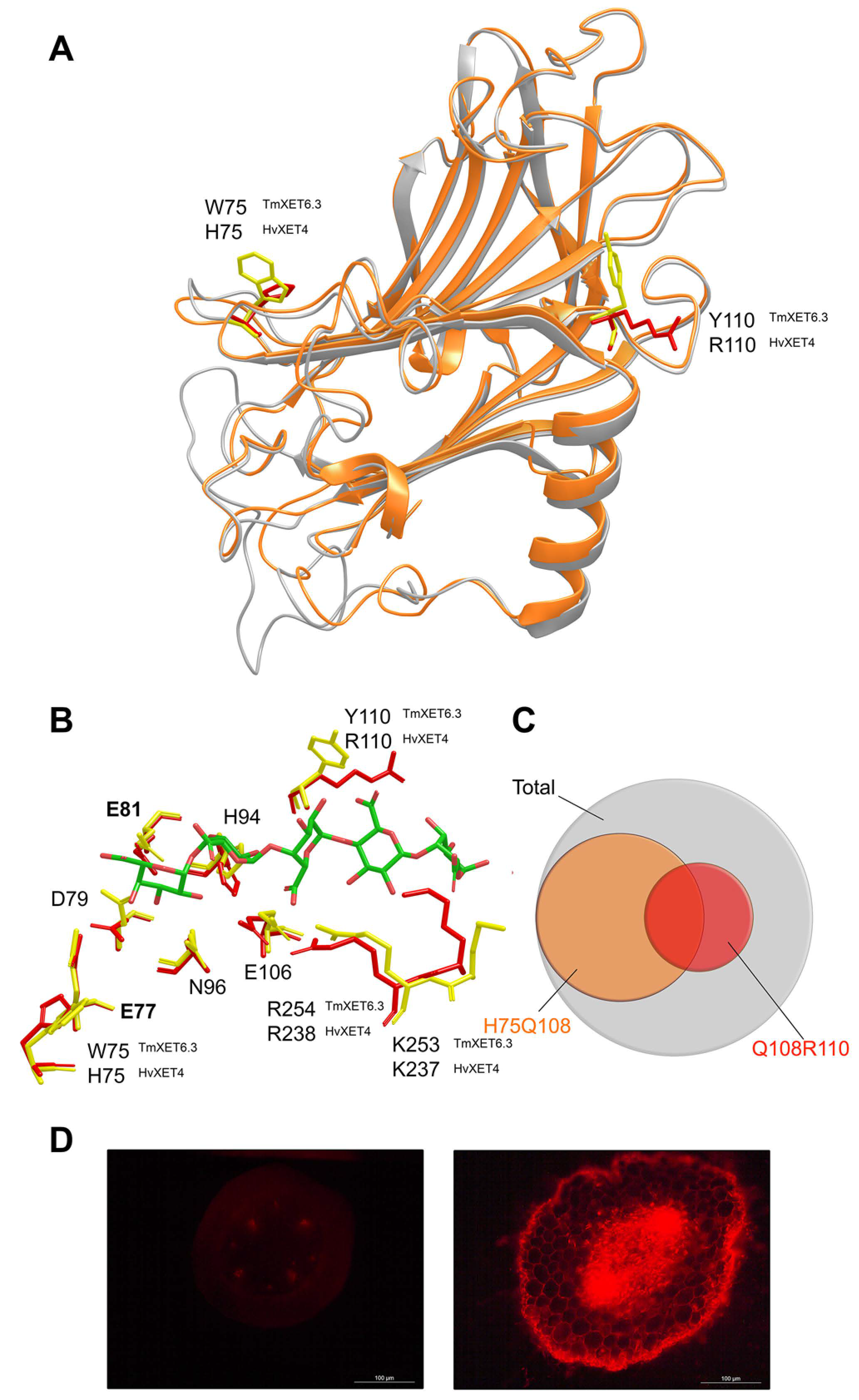
3. Substrate Specificity of Plant Xyloglucan Xyloglucosyl Transferases

Two major subtypes of reactions catalysed by XET enzymes are known that reflect the chemical nature of substrates and structural characteristics of enzymes that recognise these saccharide substrates. These reactions are termed homo-transglycosylation reactions with XG-derived donors and acceptors, and hetero-transglycosylation reactions that utilise neutral donor and neutral or ionic acceptor saccharides other than XG-derived. The concise summary of characterised XET enzymes and their isoforms is summarised in Table 1.
Homo-transglycosylation reactions with XG-derived donor and acceptor substrates—The PttXET16A enzyme from hybrid aspen Populus tremulus x tremuloides (EC 2.4.1.207; defined by the IUBMB/IUPAC Biochemical Nomenclature Committee) is considered to be an XET enzyme that utilises XG-derived donors and acceptors, meaning that it is XG-specific with a narrow substrate specificity [127,138]. A similar specificity was demonstrated in Pinus radiata PrXTH1 using computational approaches [157]. Both enzymes belong to the I/II cluster of XTH gene products [138,157]. Hetero-transglycosylation activities in PttXET16A were not detected with cellooligosaccharides (Cello-OS) [138], although in silico data with PrXTH1 indicated the weak binding of cellooctaose [157]. Based on evolutionary analyses of the GH16 family, spanning monocots, eudicots, and a basal Angiosperm [125], the PttXET16A enzyme clustered in the proximity of the barley HvXET5 isoform that catalysed these reactions at high rates [58].
Hetero-transglycosylation reactions with neutral donor and acceptor substrates—The first evidence for the existence of XETs that catalysed hetero-transglycosylation reactions, i.e., to mediate the transfer between structurally distinct saccharides, comes from the work of Ait Mohand and Farkaš [120]. These authors described the glycosyl transfer from XG to Cello-OS, laminarioligosaccharides (La-OS), and from the soluble carboxymethyl cellulose (CMC) and HEC cellulose derivates to XG-OS. These enzyme activities were detected in crude protein extracts prepared from germinating nasturtium seeds in polyacrylamide gel after isoelectric focussing [147]. Before this evidence, the substrate promiscuity in XETs from Poaceae was predicted based on molecular modelling of the GH16 family [158].
Hetero-transglycosylation activities of XETs with Cello-OS and cellulose conflict with the IUBMB/IUPAC Biochemical Nomenclature and Classification System [108], and recent research of these enzymes complicates this situation further. The key question arises if enzymes catalysing hetero-transglycosylating reactions could still be classified as XETs and, if so, what is the allowable limit for the ratio of the activity with the XG/XG-OS substrate pair and structurally different substrates. Another problem arises from the fact that currently the substrate specificity of only a few XETs in near-homogenous forms was defined, and therefore a given substrate specificity could unequivocally be assigned to a handful of XET enzymes. The latter criterion of near-homogeneity has not been always followed, whereby undefined crude protein extracts were used for substrate specificity definitions of XET enzymes.
The first XET in a near-homogenous form with a defined primary structure catalysing hetero-transglycosylation reactions was the HvXET5 isoform from barley (Hordeum vulgare L.) [58]. Except for XG and XG-OS, this enzyme in vitro mediated the formation of covalent linkages between XGs, CMC, HEC, and MLGs (donor substrates) and XG-OS or Cello-OS (acceptor substrates). The efficiency of the covalent bond formation with HEC was comparable to that with XG (44%), whereas it was lower but significant with MLGs (0.2%), and the formation of the hybrid saccharide products was defined by mass spectrometry. The substrate specificity was also defined for another near-homogenous barley HvXET6 isoform, where the efficiencies of transfer between XG and Cello-OS with degrees of polymerisation (DPs 2–6) for HvXET5 and HvXET6 were similar [121].
HEC and Cello-OS substrates functioned as respective donors and acceptors also for partially purified XET isoforms from parsley roots (Petroselinum crispum) [148]. The same was observed for XETs isolated from parsley stems and leaves, and from stems, leaves, and roots from nasturtium [123]. The efficiencies of these activities did not exceed 5% for those with HEC as the donor, and 0.5% for Cello-OS as an acceptor compared to homo-transglycosylation reactions with the XG/XG-OS pair. In comparison, 23% efficiency was reported for a crude nasturtium protein extract from germinating seeds with the HEC/XG-OS substrate pair [120].
Other XET enzyme recognising cellulose as the donor substrate is AtXTH3 from Arabidopsis thaliana, which in near-homogenous form catalysed hetero-transglycosylation reactions between cellulose and Cello-OS, cellulose and XG-OS, in addition to the homo-transglycosylation reaction with the XG/XG-OS pair, which was the dominant reaction. Notably, AtXTH3 also produced cello-oligomers with higher DPs than the original aminopyridyl cellohexaose substrate, characterised by mass spectrometry, and insoluble cellulose-like material in the absence of other substrates [122]. Despite differences in the ability to bind cellulose-derived substrates and the yields of transglycosylation products, HvXET5 [58] and AtXTH3 [122] clustered within the same phylogenetic group of XTH I, as presumably XG-specific PttXET16-34 and PrXTH1 [138,157], although they segregated to different sub-clades (Figure 2C).
Further XETs with known primary structures and the ability to utilise besides XG also cellulose or MLGs as donors, are three acidic EfXTH-A, EfXTH-H, and EfXTH-I isoforms from Equisetum fluviatile [117], although the homogeneity of these enzymes was not demonstrated. Homo-transglycosylation activity with the XG/XG-OS pair was the dominant reaction for all isoforms and the efficiencies with cellulose and MLGs varied. EfXTH-A displayed the comparable transfer of MLG fragments to XG-OS, as was the case of HvXET5 (0.2–0.3%) [58], while the activity with cellulose was incomparably higher with HvXET5. EfXTH-H and EfXTH-I showed equivalent hetero-transglycosylation activities with both MLGs and cellulose donors that were around an order of magnitude higher than that of EfXTH-A [117]. Conversely, HTG and MLG: xyloglucan endotransglycosylase from Equisetum preferred cellulose and MLGs as donors with XG-OS [117]. Such enzymes could only be found in various Equisetum species and charophytic algae [134], and their predicted function is to re-model hemicelluloses in horsetails shoots but not in a horsetail callus [135]. HTG belongs to the GH16_20 subfamily [137] and was characterised at the structural level [136].
Homology modelling of HTG suggested that three amino acid residues Pro10, Ser34, and Leu245 were responsible for the evolution of their substrate specificity and that Pro10 and Ser34 participated in the binding of the donor and Leu245 in the binding of the acceptor substrates [136]. It is notable that, in other XETs, Pro10 is substituted by Trp and Ser34 by Gly [117] and that enzymes with high reaction rates with cellulose donors, such as HvXET5 [58] and AtXTH3 [122] also contain P10W and S34G mutations.
It was further suggested that, in HTG, the R246L mutation could underlie differences in the binding of Cello-OS [136], although TmXET6.3 [123], EfXTHs [117], and HvXET3, HvXET4 and HvXET6 [125] that also bind Cello-OS, have Arg in equivalent positions, whilst AtXTH3 [122] contains the Lys residue.
The broader acceptor substrate specificity of XETs from nasturtium germinating seeds [120] initiated in-depth studies of their isoforms [159] and the development of suitable methods for the detection of hetero-transglycosylation activities [123,149]. Additional activities of nasturtium XET isoforms with the XG-OS, Cello-OS, and La-OS acceptors [120] were defined with the oligosaccharides derived from MLG (MLG-OS), xylan (Xyl-OS), glucomannan (GlcMan-OS), galactomannan (GalMan-OS), and surprisingly with pustulan (Pu-OS) [123,149], which is found mostly in fungal CWs [3]. However, the data showed that the incorporation of Pu-OS in vivo was directed to the plasma membrane rather than to nasturtium CWs [160], but that this activity was also mediated by recombinant TmXET6.3 that locates to germinating seeds [123]. This TmXET6.3 was solely responsible for all additionally found hetero-transglycosylation activities [159]. Some of these activities were found in parsley and comparably to nasturtium they located to germinating seeds [123]. Crude protein extracts from nasturtium germinating seeds also catalysed hetero-transglycosylation reactions with XGs and HEC [120] and glucuronoxylan and GalMan [149]. However, besides XG and HEC, recombinant TmXET6.3 in a near-homogenous form could not utilise the above-listed donor polysaccharides [123], suggesting that this broad substrate specificity detected in crude protein extracts could belong to other nasturtium XET isoforms; this observation further emphasises the need for using near-homogenous XETs to unequivocally define substrate specificity.
TmXET6.3 belongs to the group of XTHs II (Figure 2C, Supplementary Data Set S1), and in its recombinant and a near-homogenous form it displayed the hetero-transglycosylation activities with neutral acceptors, such as those derived from arabinoxylan (AraXyl-OS), arabinan (Ara-OS), arabinogalactan (AraGal-OS), mannan (Man-OS), glucomannan (GlcMan-OS), and galactomannans (GalMan-OS) in combination with donor XG or HEC [123]. Reaction rates differed significantly in the following order: MLG-OS > Cello-OS > Pu-OS > AraXyl-OS > La-OS > Xyl-OS GlcMan-OS > Ara-OS, where tiny activities were observed with AraGal-OS, Man-OS, and GalMan-OS, and negatively charged oligogalacturonates did not serve as acceptors. Other factors that affected TmXET6.3 activities were DPs of Cello-OS acceptors and for MLGs the positions of (1,3)-β or (1,4)-β bonds [123].
Comparative bioinformatics of TmXET1 [161], PttXET16A [127], HvXET5 [58], PrXTH1 [157], and TmXET6.3 [123] (Figure 3A), including structural analyses of TmXET6.3 suggested that the efficiencies of hetero-transglycosylation reactions of TmXET6.3 could be modified via a single (H94Q, Q108R, K237T) or multiple mutations (H94Q/A104D/Q108R, K234T/K237T) (Figure 3B). The key residue that underlined this activity in TmXET6.3 was Q108 that interacted with saccharide moieties near the catalytic site, while K237, located at the additional β-sheet of the binding site sited near the C-terminus, restrained the acceptor to the position crucial for the reaction (Figure 3C). The effects of single Q108R and K237T mutations were less effective than when both were implemented together. The only exception in the observed suppression of hetero-transglycosylating activities were the reactions with Cello-OS that were significantly increased [123]. These mutational analyses inspired the large-scale bioinformatics analyses of residue positions in available UniProtKB XET entries (3394). Here, the presence of H94, Q108, and K237 occurs in 1%, 4%, and 10% respective cases, H94/Q108 in 11% cases, H94/Q108/K237 in 25% instances, H94/Q108/K234/K237 in 2% cases, and H94/A104/Q108/K234/K237 in 0.1% instances (Figure 3D). It was concluded that the enzymes with a predisposition to catalyse hetero-transglycosylation reactions could be more widespread in plants than previously thought.
Comparisons of primary structures of TmXET6.3 and barley HvXET3, HvXET4 [142,154] and HvXET6 [121,142] isoforms (Figure 3A) indicated that all barley enzymes should transfer fragments of XG or HEC to the same wide spectrum of oligosaccharides as it was found in TmXET6.3 because they contained suitable residues H94, Q108, and except for HvXET6 also K237 in positions crucial for the acceptor substrate specificity. This comparison showed that a full grasp of the structural basis of hetero-transglycosylation required the examinations of roles of lysine residues disposed around Arg238 (numbering of TmXET6.3). Further assessments of activities confirmed the assumptions on the hetero-transglycosylation activities of HvXETs towards neutral acceptors. However, these assessments also led to the discovery of hetero-transglycosylation activities with negatively charged oligosaccharide acceptors that have never been observed before [125].
Hetero-transglycosylation reactions with a charged (ionic) acceptor substrate—These activities were investigated with recombinant HvXET isoenzymes in near-homogenous forms [121,144,154]. As mentioned above, all tested neutral oligosaccharides served as convenient acceptor substrates for HvXET3, HvXET4, and HvXE6 isoforms although hetero-transglycosylation efficiencies differed between isoforms [125]. Unlike TmXET6.3, both HvXET3 and HvXET4 could transfer fragments of XG or HEC to the penta-galacturonic acid ([α(1-4)GalAp]5), which represents a fragment of the linear part of pectin, known as homogalacturonan. While the highest hetero-transglycosylation activity in HvXET3 was observed with the XG/[α(1-4)GalAp]5 substrate pair, the highest activity with the HEC donor was observed with Cello-OS. HvXET6 could also transfer XG fragments to [α(1-4)GalAp]5 but with lower efficiency, whilst Cello-OS were the preferred acceptors with the XG donor [125]. These findings were confirmed by homology modelling of barley isoforms and suggested that their substrate promiscuity resulted from minor changes in protein sequences and thus structural arrangements [123,125]. The molecular models of HvXET3 and HvXET4 superposed with TmXET6.3 (Figure 4A) and containing docked XG heptasaccharide (XXXG) donor and [α(1-4)GalAp]5 acceptor substrates at the −4 to +5 subsites pointed to those critical interactions between residues and saccharide moieties of [α(1-4)GalAp]5 (Figure 4B) [125]. To evaluate the statistical distribution of XETs with the potential to catalyse hetero-transglycosylation reactions using the XG/[α(1-4)GalAp]5 substrate pair, a total of 3394 UniProtKB XET entries were examined, where about 44% of XETs were identified to be able to do so (Figure 4C) [125]. Finally, the activities of hetero-transglycosylating XETs [153] were detected in vivo in CWs of barley roots where they incorporated fluorescently-labelled [α(1-4)GalAp]5) [125]. The latter activity was also detected in nasturtium but solely in roots and not stems (Figure 4D).
The information on enzyme–substrate interactions combined with comparative sequence analyses of barley isoforms and TmXET6.3 (Figure 3A) allowed the engineering of mutants in TmXET6.3 that could catalyse hetero-transglycosylation reaction with the XG/[α(1-4)GalAp]5 substrate pair, while wild-type TmXET6.3 lacked this activity. Single mutations of W75H and Y110R led to the binding of this [α(1-4)GalAp]5 ionic acceptor and the implementation of the double mutation W75H/Y110R increased this activity. Although the XG-specific enzymes have in the position corresponding to 75 in TmXET6.3 the His residue (Figure 3A), these enzymes could not utilise the [α(1-4)GalAp]5 acceptor due to the lack of Q108 that is also required for the binding of different oligo- or polysaccharides. As it is the case of hetero-transglycosylation reactions with neutral acceptors, the detailed examination of the function of lysine residues near Arg238 could bring new information on acceptor binding in TmXET6.3 and in barley isoforms.
In summary, it seems that there are only a few key residues in XETs, a combination of which leads to particular substrate specificity, and a certain homo- and hetero-transglycosylation ratio that underpins the utilisation of a variety of individual donor and acceptor substrates. Despite the unquestionable success of identifying these residues in XETs [123,125], it is imperative to solve more of the tertiary structures of these enzymes, in addition to unravelling more of XET protein sequences from a variety of organisms. It would be useful to focus on XETs, which exert a range of substrate specificities, e.g., on barley and Equisetum isoforms, as this information could contribute to the progress in the XTH field.

4. Implications of Transglycosylation Reactions Catalysed by Xyloglucan Xyloglucosyl Transferases in the Cell Wall Structure, Function and Dynamics

From the phylogeny point of view, it seems that the first XET enzymes have appeared along with XGs in CWs of green algae and were not found in red or brown algae [22,43,162]. However, it is not possible to establish whether other transglycosylases with related substrate specificities, e.g., recognising cellulose as the donor substrate, existed before the emergence of XET enzymes in red or brown algae, as it is the case of HTG that functionalises cellulose [136]; exploring these evolutionary events would open exciting avenues in the XET field.
The expression of XTH genes is independently regulated by various factors, e.g., hormones and environmental changes. As a consequence, XETs evolved in multiple isoforms, which fulfill various roles during different stages of plant growth and development and in response to biotic and abiotic stresses [111,114,159,163,164,165,166,167,168,169,170,171]. Elevated expression of XTH genes can also be observed during fruit ripening that leads to CW loosening but also stiffening, thus allowing for these CWs to be conditioned for future modifications and degradation by other enzymes [172,173]. XTH genes further play an irreplaceable role in saccharide metabolism connected to plant growth since their elevated expression during cell elongation affects properties of CWs that underlie sizes and shapes of plant tissues [104]. It is also assumed that the XTH gene products play key roles in the re-structuralisation of CWs [102]. Tissue localisation and suggested function of selected XET, XTH and other GH16 family enzymes examined in this work are defined in Supplementary Data Set S1.
As it was mentioned before, XETs are involved in the depolymerisation of plant structural polysaccharides such as XGs and cellulose, and in the incorporation of newly synthetised components within these polymers and their cross-linking [99,100,101,127,174,175,176,177]. XET enzymes primarily work on XG molecules associated with cellulose micro-fibrils in a way that does not affect the integrity of CWs [1,58] (Figure 1D). The discovery of XET isoforms that link effectively not only XG molecules but also those between XGs and cellulose or between celluloses themselves, such as HvXET5 [58] and AtXTH3 [122], or between XG or MLG and cellulose catalysed by HvXET5 [58] and HTG [96,124,136,177], or between XG and a whole spectrum of structurally distinct neutral saccharides such as TmXET6.3 [123], and between XG and pectin fragments by barley HvXET3, HvXET4 and HvXET6 isoforms [125], indicates that plants have developed sophisticated and variable mechanisms to influence firmness, porosity, and flexibility of plant CWs [58], and thus the ability to modify the properties of CWs that otherwise would lack adaptability to the actual needs. These specific XET isoforms are expressed only in plants in a defined developmental stage or in a certain plant organ or tissue, or exclusively during the limited stages of plant development, as it can be gleaned from the data regarding the localisation of these enzymes based on their activities or gene expression [123,125,135,162,178,179,180]. The significance of these hetero-transglycosylation reactions by broad specific XET enzymes may be related to the structural roles of individual polysaccharides in PCW and SCW. The flexibility and dynamic properties of CWs that are required to fulfill multiple roles during plant growth and development result from complex interactions between the major CW polysaccharides in muro such as cellulose, (glucurono-arabino) xylans or GlcMan, MLG, XG and, pectin polysaccharides, structural proteins and polyphenolic compounds, inorganic molecules, and expansin proteins [125].
The fundamental complexities between the major structural polysaccharides in monocot and dicot plant CWs have been outlined [66,82,181,182,183], although the significance of homo- and hetero-transglycosylation reactions for the CW structure and function needs to be further explored, e.g., by mutant knockout plants lacking enzymes that catalyse these reactions. This multipronged research of XET enzymes is required to understand the fine structure, processes, and inter-connections between structural components of CWs, and how these relationships assist with better understanding the roles of XETs in plant cells from macro to atomic levels.

Supplementary Materials

The following are available online. Supplementary Data Set S1 contains the list of sequences of the GH16 family and their properties, such as tissue localisation and suggested function (sheet 1), and the FASTA alignment file (sheet 2) used for the generation of the unrooted phylogenetic tree (Figure 2C).

Author Contributions

Conceptualisation, M.H.; writing—original draft preparation B.S.; additional editing—E.S.; co-writing, editing and review—M.H.; figure, table and supplementary data generation—B.S., fluorescent labelling by [α(1-4)GalAp]5 and formal analysis—B.S. and E.S.; structural graphics preparation—B.S and S.K. All authors have read and agreed to the published version of the manuscript.

Funding

This work was supported by the 2/0137/20 grant from VEGA (Slovakia) to S.K., the APP0075 DoktoGrant (Slovakia) to B.S. and the DP120100900 grant from the Australian Research Council (Australia) to M.H.

Acknowledgments

M.H. acknowledges funding from the University of Adelaide (Australia) and the Huaiyin Normal University (China).

Conflicts of Interest

The authors declare no conflict of interest. The funders had no role in the design of the study; in the collection, analyses, or interpretation of data; in the writing of the manuscript, or in the decision to publish the results.

Abbreviations

[α(1-4)GalAp]5, penta-galacturonic acid; Ara-OS, arabinan oligosaccharide(s)s; AraGal-OS, arabinogalactan oligosaccharide(s); AraXyl-OS, arabinoxylan oligosaccharide(s); AraXyl(s), arabinoxylan(s); CW(s), cell wall(s); CAZy, Carbohydrate-Active enZYmes; CSL, cellulose synthase-like; Cello-OS, cellooligosaccharide(s); CMC, carboxymethyl cellulose; DP(s), degree(s) of polymerisation; GalGlcMan; galactoglucomannan; GalMan(s), galactomannan(s); GalMan-OS, galactomannan oligosaccharide(s); GH(s), glycoside hydrolase(s); GlcMan-OS, glucomannan oligosaccharide(s); GlcMan(s), glucomannan(s); GT(s), glycosyl transferase(s); HEC, hydroxyethyl cellulose; HG, homogalacturonan; HTG, hetero-trans-β-glucanase; IUBMB, International Union of Biochemistry and Molecular Biology; IUPAC, International Union of Pure and Applied Chemistry; KEGG, Kyoto Encyclopaedia of Genes and Genomes; La-OS, laminarioligosaccharide(s); Man(s), mannan(s); Man-OS, mannan oligosaccharide(s); MLG(s), (1,3;1,4)-β-d-glucan(s) or mix-linkage glucans; MLG-OS, mix-linkage oligosaccharide(s); PCW(s), primary cell wall(s); PDB, Protein Data Bank; Pu-OS pustulan oligosaccharide(s); RGI/II, rhamnogalacturonan I/II; SCW(s), secondary cell wall(s); UniProtKB, Universal Protein KnowledgeBase; XEH(s), xyloglucan endohydrolase(s); XET(s), xyloglucan endo-transglycosylase(s); XG(s); xyloglucan(s); XG-OS, xyloglucan oligosaccharide(s); XTH, xyloglucan transglycosylase/hydrolase; Xyl(s), xylan(s); Xyl-OS, xylan oligosaccharide(s).

References

  1. Fry, S.C. Primary cell wall metabolism: Tracking the careers of wall polymers in living plant cells. New Phytol. 2004, 161, 641–675. [Google Scholar] [CrossRef]
  2. Cosgrove, D. Growth of the plant cell wall. Nat. Rev. Mol. Cell Biol. 2005, 6, 850–861. [Google Scholar] [CrossRef]
  3. Cabib, E.; Farkaš, V.; Kosík, O.; Blanco, N.; Arroyo, J.; McPhie, P. Assembly of the yeast cell wall. Crh1p and Crh2p act as transglycosylases in vivo and in vitro. J. Biol. Chem. 2008, 283, 29859–29872. [Google Scholar] [CrossRef] [PubMed] [Green Version]
  4. Farrokhi, N.; Burton, R.A.; Brownfield, L.; Hrmova, M.; Wilson, S.M.; Bacic, A.; Fincher, G.B. Plant cell wall biosynthesis: Genetic, biochemical and functional genomics approaches to the identification of key genes. Plant Biotech. J. 2009, 4, 145–167. [Google Scholar] [CrossRef] [PubMed]
  5. Blanco, N.; Sanz, A.B.; Rodriguez-Pena, J.M.; Nombela, C.; Farkaš, V.; Hurtado-Guerrero, R.; Arroyo, J. Structural and functional analysis of yeast Crh1 and Crh2 transglycosylases. FEBS J. 2015, 282, 715–731. [Google Scholar] [CrossRef] [PubMed] [Green Version]
  6. Cosgrove, D.J. Plant cell wall extensibility: Connecting plant cell growth with cell wall structure, mechanics, and the action of wall modifying enzymes. J. Exp. Bot. 2015, 67, 463–476. [Google Scholar] [CrossRef] [PubMed]
  7. Graham, L.E.; Cook, M.E.; Busse, J.S. The origin of plants: Body plan changes contributing to a major evolutionary radiation. PNAS 2000, 97, 4535–4540. [Google Scholar] [CrossRef] [Green Version]
  8. Niklas, K.J. The cell walls that bind the tree of life. BioScience 2004, 54, 831–841. [Google Scholar] [CrossRef] [Green Version]
  9. Niklas, K.J.; Kutschera, U. The evolution of the land plant life cycle. New Phytol. 2010, 185, 27–41. [Google Scholar] [CrossRef]
  10. Sørensen, I.; Pettolino, F.A.; Bacic, A.; Ralph, J.; Lu, F.; O’Neill, M.A.; Fei, Z.; Rose, J.K.C.; Domozych, D.S.; Willats, W.G.T. The charophycean green algae provide insights into the early origins of plant cell walls. Plant J. 2011, 68, 201–211. [Google Scholar] [CrossRef]
  11. Popper, Z.A.; Fry, S.C. Primary cell wall composition of bryophytes and charophytes. Ann. Bot. 2003, 91, 1–12. [Google Scholar] [CrossRef] [PubMed] [Green Version]
  12. Popper, Z.A.; Fry, S.C. Primary cell wall composition of the pteridophytes and spermatophytes. New Phytol. 2004, 164, 165–174. [Google Scholar] [CrossRef]
  13. Fincher, G.B. Revolutionary times in our understanding of cell wall biosynthesis and remodeling in the grasses. Plant Physiol. 2009, 149, 7–37. [Google Scholar] [CrossRef] [PubMed] [Green Version]
  14. Burton, R.A.; Gidley, M.J.; Fincher, G.B. Heterogeneity in the chemistry, structure and function of plant cell walls. Nat. Chem. Biol. 2010, 6, 724–732. [Google Scholar] [CrossRef]
  15. Sørensen, I.; Domozych, D.; Willats, W.G.T. How have plant cell walls evolved? Plant Physiol. 2010, 153, 366–372. [Google Scholar] [CrossRef] [Green Version]
  16. Fangel, J.U.; Ulvskov, P.; Knox, J.P.; Mikkelsen, M.D.; Harholt, J.; Popper, Z.A.; Willats, W.G.T. Cell wall evolution and diversity. Front. Plant Sci. 2012, 3, 153. [Google Scholar] [CrossRef] [Green Version]
  17. Kozlova, L.V.; Nazipova, A.R.; Gorshkov, O.V.; Petrova, A.A.; Gorshkova, T.A. Elongating maize root: Zone-specific combinations of polysaccharides from type I and tape II primary cell walls. Sci. Rep. 2020, 10, 10956. [Google Scholar] [CrossRef]
  18. Iraki, N.M.; Bressan, R.A.; Hasegawa, P.M.; Carpita, N.C. Alteration of the physical and chemical structure of the primary cell wall of growth-limited plant cells adapted to osmotic stress. Plant Physiol. 1989, 91, 39–47. [Google Scholar] [CrossRef] [Green Version]
  19. Popper, Z.A. Evolution and diversity of green plant cell walls. Curr. Opin. Plant Biol. 2008, 11, 286–292. [Google Scholar] [CrossRef]
  20. Sarkar, P.; Bosneaga, E.; Auer, M. Plant cell walls throughout evolution: Towards a molecular understanding of their design principles. J. Exp. Bot. 2009, 60, 3615–3635. [Google Scholar] [CrossRef] [Green Version]
  21. Popper, Z.A.; Tuohy, M.G. Beyond the green: Understanding the evolutionary puzzle of plant and algal cell walls. Plant Physiol. 2010, 153, 373–383. [Google Scholar] [CrossRef] [PubMed] [Green Version]
  22. Popper, Z.A.; Michel, G.; Hervé, C.; Domozych, D.S.; Willats, W.G.; Tuohy, M.G.; Kloareg, B.; Stengel, D.B. Evolution and diversity of plant cell walls: From algae to flowering plants. Annu. Rev. Plant Biol. 2011, 62, 567–590. [Google Scholar] [CrossRef] [PubMed] [Green Version]
  23. Fry, S.C.; Nesselrode, B.H.W.A.; Miller, J.G.; Mewburn, B.R. Mixed-linkage (1→3,1→4)-β-D-glucan is a major hemicellulose of Equisetum (horsetail) cell walls. New Phytol. 2008, 179, 104–115. [Google Scholar] [CrossRef] [PubMed]
  24. Sørensen, I.; Pettolino, F.A.; Wilson, S.M.; Doblin, M.S.; Johansen, B.; Bacic, A.; Willats, W.G. Mixed-linkage (1→ 3),(1→4)-β-D-glucan is not unique to the Poales and is an abundant component of Equisetum arvense cell walls. Plant J. 2008, 54, 510–521. [Google Scholar] [CrossRef]
  25. Xue, X.; Fry, S.C. Evolution of mixed-linkage (1→3, 1→4)-β-D-glucan (MLG) and xyloglucan in Equisetum (horsetails) and other monilophytes. Ann. Bot. 2012, 109, 873–886. [Google Scholar] [CrossRef] [Green Version]
  26. Burton, R.A.; Fincher, G.B. (1,3;1,4)-β-D-glucans in cell walls of the Poaceae, lower plants, and fungi: A tale of two linkages. Mol. Plant 2009, 2, 873–882. [Google Scholar] [CrossRef] [Green Version]
  27. Scheller, H.V.; Ulvskov, P. Hemicelluloses. Annu. Rev. Plant Biol. 2010, 61, 263–289. [Google Scholar] [CrossRef]
  28. Popper, Z.A.; Fry, S.C. Widespread occurrence of a covalent linkage between xyloglucan and acidic polysaccharides in suspension-cultured angiosperm cells. Ann. Bot. 2005, 96, 91–99. [Google Scholar] [CrossRef] [Green Version]
  29. Marcus, S.E.; Verhertbruggen, Y.; Hervé, C.; Ordaz-Ortiz, J.J.; Farkaš, V.; Pedersen, H.L.; Willats, W.G.T.; Knox, J.P. Pectic homogalacturonan masks abundant sets of xyloglucan epitopes in plant cell walls. BMC Plant Biol. 2008, 8, 60. [Google Scholar] [CrossRef] [Green Version]
  30. McCann, M.C.; Knox, J.P. Plant cell wall biology: Polysaccharides in architectural and developmental contexts. Ann. Plant Rew. Online 2018, 343–366. [Google Scholar] [CrossRef]
  31. Harholt, J.; Suttangkakul, A.; Scheller, V.H. Biosynthesis of pectin. Plant Physiol. 2010, 153, 384–395. [Google Scholar] [CrossRef] [PubMed] [Green Version]
  32. Ridley, B.L.; O’Neill, M.A.; Mohnen, D. Pectins: Structure, biosynthesis, and oligogalacturonide-related signalling. Phytochemistry 2001, 57, 929–967. [Google Scholar] [CrossRef]
  33. Vincken, J.P.; Schols, H.A.; Oomen, R.J.; Mccann, M.C.; Ulvskov, P.; Voragen, A.G.J.; Visser, R.G.F. If homogalacturonan were a side chain of rhamnogalacturonan I. Implications for cell wall architecture. Plant Physiol. 2003, 132, 1781–1789. [Google Scholar] [CrossRef] [PubMed] [Green Version]
  34. Mohnen, D. Pectin structure and biosynthesis. Curr. Opin. Plant. Biol. 2008, 11, 266–277. [Google Scholar] [CrossRef]
  35. Caffall, K.H.; Mohnen, D. The structure, function, and biosynthesis of plant cell wall pectic polysaccharides. Carbohydr. Res. 2009, 344, 1879–1900. [Google Scholar] [CrossRef]
  36. Boerjan, W.; Ralph, J.; Baucher, M. Lignin biosynthesis. Annu. Rev. Plant. Biol. 2003, 54, 519–546. [Google Scholar] [CrossRef]
  37. Weng, J.K.; Chapple, C. The origin and evolution of lignin biosynthesis. New Phytol. 2010, 187, 273–285. [Google Scholar] [CrossRef]
  38. Mnich, E.; Bjarnholt, N.; Eudes, A.; Harholt, J.; Holland, C.; Jørgensen, B.; Larsen, F.H.; Liu, M.; Manat, R.; Meyer, A.S.; et al. Phenolic cross-links: Building and de-constructing the plant cell wall. Nat. Prod. Rep. 2020, 37, 919–961. [Google Scholar] [CrossRef]
  39. Horton, D. Preface. The two most abundant organic substances on Earth, cellulose and starch. Adv. Carbohydr. Chem. Biochem. 2010, 64, xi–xiii. [Google Scholar] [CrossRef]
  40. Heinze, T. Cellulose: Structure and properties. In Cellulose Chemistry and Properties: Fibers, Nanocelluloses and Advanced Materials. Advances in Polymer Science; Rojas, O., Ed.; Springer: Cham, Switzerland, 2015; Volume 271. [Google Scholar] [CrossRef]
  41. Kim, S.J.; Chandrasekar, B.; Rea, A.C.; Danhof, L.; Zemelis-Durfee, S.; Thrower, N.; Shepard, Z.S.; Pauly, M.; Brandizzi, F.; Keegstra, K. The synthesis of xyloglucan, an abundant plant cell wall polysaccharide, requires CSLC function. Proc. Natl. Acad. Sci. USA 2020, 117, 20316–20324. [Google Scholar] [CrossRef]
  42. Ikegaya, H.; Hayashi, T.; Kaku, T.; Iwata, K.; Sonobe, S.; Shimmen, T. Presence of xyloglucan-like polysaccharide in Spirogyra and possible involvement in cell–cell attachment. Phycol. Res. 2008, 56, 216–222. [Google Scholar] [CrossRef]
  43. Van Sandt, V.S.; Stieperaere, H.; Guisez, Y.; Verbelen, J.P.; Vissenberg, K. XET activity is found near sites of growth and cell elongation in bryophytes and some green algae: New insights into the evolution of primary cell wall elongation. Ann. Bot. 2007, 99, 39–51. [Google Scholar] [CrossRef] [PubMed] [Green Version]
  44. Fry, S.C.; Aldington, S.; Hetherington, P.R.; Aitken, J. Oligosaccharides as signals and substrates in the plant cell wall. Plant Physiol. 1993, 103, 1–5. [Google Scholar] [CrossRef] [PubMed]
  45. York, W.S.; Van Halbeek, H.; Darvill, A.G.; Albersheim, P. Structural analysis of xyloglucan oligosaccharides by 1H-n.m.r. spectroscopy and fast-atom-bombardment mass spectrometry. Carbohydr Res. 1990, 200, 9–31. [Google Scholar] [CrossRef]
  46. York, W.S.; Harvey, L.K.; Guillen, R.; Albersheim, P.; Darvill, A.G. Structural analysis of tamarind seed xyloglucan oligosaccharides using beta-galactosidase digestion and spectroscopic methods. Carbohydr. Res. 1993, 248, 285–301. [Google Scholar] [CrossRef]
  47. Peña, M.J.; Darvill, A.G.; Eberhard, S.; York, W.S.; O’Neill, M.A. Moss and liverwort xyloglucans contain galacturonic acid and are structurally distinct from the xyloglucans synthesized by hornworts and vascular plants. Glycobiology 2008, 18, 891–904. [Google Scholar] [CrossRef]
  48. Peña, M.J.; Kong, Y.; York, W.S.; O’Neill, M.A. A galacturonic acid-containing xyloglucan is involved in Arabidopsis root hair tip growth. Plant Cell 2012, 24, 4511–4524. [Google Scholar] [CrossRef] [Green Version]
  49. Hsieh, Y.S.Y.; Harris, P.J. Xyloglucans of monocotyledons have diverse structures. Mol. Plant 2009, 2, 943–965. [Google Scholar] [CrossRef]
  50. Hsieh, Y.S.Y.; Paxton, M.; Ade, C.P.; Harris, P.J. Structural diversity, functions and biosynthesis of xyloglucans in angiosperm cell walls. NZ J. Forest Sci. 2009, 39, 187–196. [Google Scholar] [CrossRef] [Green Version]
  51. Zheng, Y.; Wang, X.; Chen, Y.; Wagner, E.; Cosgrove, D.J. Xyloglucan in the primary cell wall: Assessment by FESEM, selective enzyme digestions and nanogold affinity tags. Plant J. 2018, 93, 211–226. [Google Scholar] [CrossRef] [Green Version]
  52. Cocuron, J.C.; Lerouxel, O.; Drakakaki, G.; Alonso, A.P.; Liepman, A.H.; Keegstra, K.; Raikhel, N.; Wilkerson, C.G. A gene from the cellulose synthase-like C family encodes a β-1,4 glucan synthase. Proc. Natl. Acad. Sci. USA 2007, 104, 8550–8555. [Google Scholar] [CrossRef] [PubMed] [Green Version]
  53. Pauly, M.; Keegstra, K. Biosynthesis of the plant cell wall matrix polysaccharide xyloglucan. Annu. Rev. Plant Biol. 2016, 67, 235–259. [Google Scholar] [CrossRef] [PubMed]
  54. Hayashi, T. Xyloglucans in the primary cell wall. Ann. Rev. Plant Physiol. Plant Mol. Biol. 1989, 40, 139–168. [Google Scholar] [CrossRef]
  55. Zhang, Y.; Xiao, G.; Wang, X.; Zhang, X.; Friml, J. Evolution of fast root gravitropism in seed plants. Nat. Commun. 2019, 10, 3480. [Google Scholar] [CrossRef] [PubMed] [Green Version]
  56. Albersheim, P.; Darvill, A.; Roberts, K.; Sederoff, R.; Staehelin, A. Plant Cell Walls, from Chemistry to Biology. Garland Science; Garland Science: New York, NY, USA, 2011; pp. 227–272. ISBN 978-0815319962. [Google Scholar]
  57. Park, Y.B.; Cosgrove, D.J. A revised architecture of primary cell walls based on biomechanical changes induced by substrate-specific endoglucanases. Plant Physiol. 2012, 158, 1933–1943. [Google Scholar] [CrossRef] [PubMed] [Green Version]
  58. Hrmova, M.; Farkaš, V.; Lahnstein, J.; Fincher, G.B. A barley xyloglucan xyloglucosyl transferase covalently links xyloglucan, cellulosic substrates, and (1,3;1,4)-β-D-glucans. J. Biol. Chem. 2007, 283, 27344. [Google Scholar] [CrossRef] [PubMed] [Green Version]
  59. Keegstra, K.; Talmadge, K.W.; Bauer, W.D.; Albersheim, P. The structure of plant cell walls. III. A model of the walls of suspension-cultured sycamore cells based on the interconnections of the macromolecular components. Plant Physiol. 1973, 51, 188–196. [Google Scholar] [CrossRef] [Green Version]
  60. Carpita, N.C.; Gibeaut, D.M. Structural models of primary cell walls in flowering plants: Consistency of molecular structure with the physical properties of the walls during growth. Plant J. 1993, 3, 1–30. [Google Scholar] [CrossRef]
  61. Pauly, M.; Albersheim, P.; Darvill, A.; York, W.S. Molecular domains of the cellulose/xyloglucan network in the cell walls of higher plants. Plant J. 1999, 20, 629–639. [Google Scholar] [CrossRef]
  62. Chanliaud, E.; De Silva, J.; Strongitharm, B.; Jeronimidis, G.; Gidley, M.D. Mechanical effects of plant cell wall enzymes on cellulose/xyloglucan composites. Plant J. 2004, 38, 27–37. [Google Scholar] [CrossRef]
  63. Chanliaud, E.; Burrows, K.M.; Jeronimidis, G.; Gidley, M.J. Mechanical properties of primary plant cell wall analogues. Planta 2002, 215, 989–996. Available online: https://www.jstor.org/stable/23387052 (accessed on 25 November 2020). [CrossRef] [PubMed]
  64. Cosgrove, D.J. Re-constructing our models of cellulose and primary cell wall assembly. Curr. Opin. Plant Biol. 2014, 22, 122–131. [Google Scholar] [CrossRef] [Green Version]
  65. Park, Y.B.; Cosgrove, D.J. Xyloglucan and its interactions with other components of the growing cell wall. Plant Cell Physiol. 2015, 56, 180–194. [Google Scholar] [CrossRef] [PubMed] [Green Version]
  66. Cosgrove, D.J. Diffuse growth of plant cell walls. Plant Physiol. 2018, 176, 16–27. [Google Scholar] [CrossRef] [PubMed] [Green Version]
  67. Kuki, H.; Yokoyama, R.; Kuroha, T.; Nishitani, K. Xyloglucan is not essential for the formation and integrity of the cellulose network in the primary cell wall regenerated from Arabidopsis protoplasts. Plants 2020, 9, 629. [Google Scholar] [CrossRef]
  68. Scheible, W.R.; Pauly, M. Glycosyltransferases and cell wall biosynthesis: Novel players and insights. Curr. Opin. Plant. Biol. 2004, 7, 285–295. [Google Scholar] [CrossRef]
  69. Burton, R.A.; Wilson, S.M.; Hrmova, M.; Harvey, A.J.; Shirley, N.J.; Medhurst, A.; Stone, B.A.; Newbigin, E.J.; Bacic, A.; Fincher, G.B. Cellulose synthase-like CslF genes mediate the synthesis of cell wall (1,3;1,4)-β-D-glucans. Science 2006, 311, 1940–1942. [Google Scholar] [CrossRef]
  70. Qu, Y.; Egelund, J.; Gilson, P.R.; Houghton, F.; Gleeson, P.A.; Schultz, C.J.; Bacic, A. Identification of a novel group of putative Arabidopsis thaliana β-(1,3)-galactosyltransferases. Plant Mol. Biol. 2008, 68, 43–59. [Google Scholar] [CrossRef]
  71. Doblin, M.S.; Pettolino, F.A.; Wilson, S.M.; Campbell, R.; Burton, R.A.; Fincher, G.B.; Newbigin, E.; Bacic, A. A barley cellulose synthase-like CSLH gene mediates (1,3;1,4)-β-D-glucan synthesis in transgenic Arabidopsis. Proc. Natl. Acad. Sci. USA 2009, 106, 5996–6001. [Google Scholar] [CrossRef] [Green Version]
  72. Wu, A.-M.; Hörnblad, E.; Voxeur, A.; Gerber, L.; Rihouey, C.; Lerouge, P.; Marchant, A. Analysis of the Arabidopsis IRX9/IRX9-L and IRX14/IRX14-L pairs of glycosyltransferase genes reveals critical contributions to biosynthesis of the hemicellulose glucuronoxylan. Plant Physiol. 2010, 153, 542–554. [Google Scholar] [CrossRef] [Green Version]
  73. Anders, N.; Wilkinson, M.D.; Lovegrove, A.; Freeman, J.; Tryfona, T.; Pellny, T.K.; Weimar, T.; Mortimer, J.C.; Stott, K.; Baker, J.M.; et al. Glycosyl transferases in family 61 mediate arabinofuranosyl transfer ontoxylan in grasses. Proc. Natl. Acad. Sci. USA 2012, 17, 989–993. [Google Scholar] [CrossRef] [PubMed] [Green Version]
  74. Hansen, S.F.; Harholt, J.; Oikawa, A.; Scheller, H.V. Plant glycosyl transferases beyond CAZy: A perspective on DUF families. Front. Plant Sci. 2012, 3, 59. [Google Scholar] [CrossRef] [PubMed] [Green Version]
  75. Harholt, J.; Sørensen, I.; Fangel, J.U.; Roberts, A.; Willats, W.G.T.; Scheller, H.V.; Larsen, B.; Petersen, B.L.; Banks, J.A.; Ulvskov, P. The glycosyltransferase repertoire of the spikemoss Selaginella moellendorffii and a comparative study of the cell wall structure. PLoS ONE 2012, 7, e35846. [Google Scholar] [CrossRef] [PubMed]
  76. Zabotina, O.A.; Avci, U.; Cavalier, D.; Pattathil, S.; Chou, Y.-H.; Eberhard, S.; Danhof, L.; Keegstra, K.; Hahn, M.G. Mutations in multiple XXT genes of Arabidopsis reveal the complexity of xyloglucan biosynthesis. Plant Physiol. 2012, 159, 1367–1384. [Google Scholar] [CrossRef] [Green Version]
  77. Stonebloom, S.; Ebert, B.; Xiong, G.; Pattathil, S.; Birdseye, D.; Lao, J.; Pauly, M.; Hahn, M.G.; Heazlewood, J.I.; Scheller, H.V. A DUF-246 family glycosyltransferase-like gene affects male fertility and the biosynthesis of pectic arabinogalactans. BMC Plant Biol. 2016, 16, 90. [Google Scholar] [CrossRef] [Green Version]
  78. Zeng, W.; Lampugnani, E.R.; Picard, K.L.; Song, L.L.; Wu, A.M.; Farion, I.M.; Zhao, J.; Ford, K.; Doblin, M.S.; Bacic, A. Asparagus IRX9, IRX10, and IRX14A are components of an active xylan backbone synthase complex that forms in the Golgi apparatus. Plant Physiol. 2016, 171, 93–109. [Google Scholar] [CrossRef] [Green Version]
  79. Takenaka, Y.; Kato, K.; Ogawa-Ohnishi, M.; Tsuruhama, K.; Kajiura, H.; Yagyu, K.; Tekeda, A.; Takeda, Y.; Kunieda, T.; Hara-Nishimura, I.; et al. Pectin RG-I rhamnosyl-transferases represent a novel plant-specific glycosyltransferase family. Nat. Plants 2018, 4, 669–676. [Google Scholar] [CrossRef]
  80. Voiniciuc, C.; Engle, K.A.; Günl, M.; Dieluweit, S.; Schmidt, M.H.; Yang, J.Y.; Moremen, K.W.; Mohnen, D.; Usadel, B. Identification of key enzymes for pectin synthesis in seed mucilage. Plant Physiol. 2018, 178, 1045–1064. [Google Scholar] [CrossRef] [Green Version]
  81. Amos, R.A.; Mohnen, D. Critical review of plant cell wall matrix polysaccharide glycosyltransferase activities verified by heterologous protein expression. Front. Plant Sci. 2019, 10, 915. [Google Scholar] [CrossRef] [Green Version]
  82. Penning, B.W.; McCann, M.C.; Carpita, N.C. Evolution of the cell wall gene families of grasses. Front. Plant Sci. 2019, 10, 1205. [Google Scholar] [CrossRef] [Green Version]
  83. Yang, J.; Bak, G.; Burgin, T.; Barnes, W.J.; Mayes, H.B.; Peña, M.J.; Urbanowicz, B.R.; Nielsen, E. Biochemical and genetic analysis Identify CSLD3 as a beta-1,4-glucan synthase that functions during plant cell wall synthesis. Plant Cell 2020, 32, 1749–1767. [Google Scholar] [CrossRef] [PubMed] [Green Version]
  84. Wachananawat, B.; Kuroha, T.; Takenaka, Y.; Kajiura, H.; Naramoto, S.; Yokoyama, R.; Ishizaki, K.; Nishitani, K.; Ishimizu, T. Diversity of pectin rhamnogalacturonan I rhamnosyltransferases in glycosyltransferase family 106. Front. Plant Sci. 2020, 11, 997. [Google Scholar] [CrossRef] [PubMed]
  85. Dhugga, K.S. Biosynthesis of non-cellulosic polysaccharides of plant cell walls. Phytochemistry 2012, 74, 8–19. [Google Scholar] [CrossRef] [PubMed]
  86. Roberts, A.W.; Roberts, E.M.; Delmer, D.P. Cellulose synthase (CesA) genes in the green alga Mesotaenium caldariorum. Eukaryot. Cell 2002, 1, 847–855. [Google Scholar] [CrossRef] [PubMed] [Green Version]
  87. Roberts, A.W.; Roberts, E. Evolution of the Cellulose Synthase (CesA) Gene Family: Insights from Green Algae and Seedless Plants. In Cellulose: Molecular and structural Biology; Brown, R.M., Saxena, I.M., Eds.; Springer: Dordrecht, The Netherlands, 2007; pp. 17–34. [Google Scholar] [CrossRef]
  88. Yin, Y.; Huang, J.; Xu, Y. The cellulose synthase superfamily in fully sequenced plants and algae. BMC Plant Biol. 2009, 9, 99. [Google Scholar] [CrossRef] [Green Version]
  89. Endler, A.; Persson, S. Cellulose synthases and synthesis in Arabidopsis. Mol. Plant 2011, 4, 199–211. [Google Scholar] [CrossRef] [Green Version]
  90. Little, A.; Schwerdt, J.G.; Shirley, N.J.; Khor, S.F.; Neumann, K.; O’Donovan, L.A.; Lahnstein, J.; Collins, H.M.; Henderson, M.; Fincher, J.B.; et al. Revised phylogeny of the cellulose synthase gene superfamily: Insights into cell wall evolution. Plant Physiol. 2018, 177, 1124–1141. [Google Scholar] [CrossRef] [Green Version]
  91. Abercrombie, J.M.; O’Meara, B.C.; Moffatt, A.R.; Williams, J.H. Developmental evolution of flowering plant pollen tube cell walls: Callose synthase (CalS) gene expression patterns. EvoDevo 2011, 2, 14. [Google Scholar] [CrossRef] [Green Version]
  92. Park, S.; Szumlanski, A.L.; Gu, F.; Guo, F.; Nielsen, E. A role for CSLD3 during cell-wall synthesis in a pical plasma membranes of tip-growing root-hair cells. Nat. Cell Biol. 2011, 13, 973–980. [Google Scholar] [CrossRef]
  93. Cosgrove, D.J. Loosening of plant cell walls by expansins. Nature 2000, 407, 321–326. [Google Scholar] [CrossRef]
  94. Lv, L.M.; Zuo, D.Y.; Wang, X.F.; Cheng, H.L.; Zhang, Y.P.; Wang, Q.L.; Song, G.L.; Ma, Z.Y. Genome-wide identification of the expansin gene family reveals that expansin genes are involved in fibre cell growth in cotton. BMC Plant Biol. 2020, 20, 223. [Google Scholar] [CrossRef] [PubMed]
  95. Liu, W.; Lyu, T.; Xu, L.; Hu, Z.; Xiong, X.; Liu, T.; Cao, J. Complex molecular evolution and expression of expansin gene families in three basic diploid species of Brassica. Int. J. Mol. Sci. 2020, 21, 3424. [Google Scholar] [CrossRef] [PubMed]
  96. Herburger, K.; Franková, L.; Picmanova, M.; Loh, J.W.; Valenzuela-Ortega, M.; Meulewaeter, F.; Hudson, A.D.; French, C.E.; Fry, S.C. Hetero-trans-β-Glucanase produces cellulose–xyloglucan covalent bonds in the cell walls of structural plant tissues and is stimulated by expansin. Mol. Plant 2020, 13, 1047–1062. [Google Scholar] [CrossRef] [PubMed]
  97. Willats, W.G.T.; Orfila, C.; Limberg, G.; Buchholt, H.C.; van Alebeek, G.-J.W.M.; Voragen, A.G.J.; Marcus, S.E.; Christensen, T.M.I.E.; Mikkelsen, J.D.; Murray, B.S.; et al. Modulation of the degree and pattern of methyl-esterification of pectic homogalacturonan in plant cell walls: Implications for pectin methylesterase action, matrix properties and cell adhesion. J. Biol. Chem. 2001, 276, 19404–19413. [Google Scholar] [CrossRef] [PubMed] [Green Version]
  98. Wolf, S.; Mouille, G.; Pelloux, J. Homogalacturonan methyl-esterification and plant development. Mol. Plant 2009, 2, 851–860. [Google Scholar] [CrossRef]
  99. Franková, L.; Fry, S.C. Biochemistry and physiological roles of enzymes that ‘cut and paste’ plant cell-wall polysaccharides. J. Exp. Bot. 2013, 64, 3519–3550. [Google Scholar] [CrossRef] [Green Version]
  100. Thompson, J.E.; Smith, R.C.; Fry, S.C. Xyloglucan undergoes interpolymeric transglycosylation during binding to the plant cell wall in vivo: Evidence from 13C/3H dual labelling and isopycnic centrifugation in caesium trifluoroacetate. Biochem. J. 1997, 327, 699–708. [Google Scholar] [CrossRef] [Green Version]
  101. Campbell, P.; Braam, J. Xyloglucan endotransglycosylases: Diversity of genes, enzymes and potential wall-modifying functions. Trends Plant Sci. 1999, 4, 361–366. [Google Scholar] [CrossRef]
  102. Bourquin, V.; Nishikubo, N.; Abe, H.; Brumer, H.; Denman, S.; Eklund, M.; Christiernin, M.; Teeri, T.T.; Sundberg, B.; Mellerowicz, E.J. Xyloglucan endotransglycosylases have a function during the formation of secondary cell walls of vascular tissues. Plant Cell 2002, 14, 3073–3088. [Google Scholar] [CrossRef] [Green Version]
  103. Nishitani, K. Division of roles among members of the XTH gene family in plants. Plant Biosyst. 2005, 139, 98–101. [Google Scholar] [CrossRef]
  104. Nishikubo, N.; Takahashi, J.; Roos, A.A.; DerbaMaceluch, M.; Piens, K.; Brumer, H.; Teeri, T.T.; Stalbrand, H.; Mellerowicz, E.J. Xyloglucan endotransglycosylase-mediated xyloglucan rearrangements in developing wood of hybrid aspen. Plant Physiol. 2011, 155, 399–413. [Google Scholar] [CrossRef] [PubMed] [Green Version]
  105. Farkaš, V.; Sulová, Z.; Stratilová, E.; Hanna, R.; Maclachlan, G. Cleavage of xyloglucan by nasturtium seed xyloglucanase and transglycosylation to xyloglucan subunit oligosaccharides. Arch. Biochem. Biophys. 1992, 298, 365–370. [Google Scholar] [CrossRef]
  106. Fry, S.; Smith, R.; Renwick, K.; Martin, D.; Hodge, S.; Matthews, K. Xyloglucan endotransglycosylase, a new wall-loosening enzyme activity from plants. Biochem. J. 1992, 282, 821–828. [Google Scholar] [CrossRef] [PubMed] [Green Version]
  107. Nishitani, K.; Tominaga, R. Endo-xyloglucan transferase, a novel class of glycosyltransferase that catalyzes transfer of a segment of xyloglucan molecule to another xyloglucan molecule. J. Biol. Chem. 1992, 267, 21058–21064. [Google Scholar] [PubMed]
  108. Webb, E.C. Enzyme Nomenclature 1992: Recommendations of the Nomenclature Committee of the International Union of Biochemistry and Molecular Biology on the Nomenclature and Classification of Enzymes; Academic Press Inc: Harcourt Brace, Jovanovich Publishers: San Diego, CA, USA, 1992; ISBN 0-12-227164-5. [Google Scholar]
  109. Placzek, S.; Schomburg, I.; Chang, A.; Jeske, L.; Ulbrich, M.; Tillack, J.; Schomburg, D. BRENDA in 2017: New perspectives and new tools in BRENDA. Nucleic Acids Res. 2017, 45, D380–D388. [Google Scholar] [CrossRef]
  110. Rose, J.K.C.; Braam, J.; Fry, S.C.; Nishitani, K. The XTH family of enzymes involved in xyloglucan endotransglucosylation and endohydrolysis: Current perspectives and a new unifying nomenclature. Plant Cell Physiol. 2002, 43, 1421–1435. [Google Scholar] [CrossRef] [Green Version]
  111. Atkinson, R.G.; Johnston, S.L.; Yauk, Y.K.; Sharma, N.N.; Schröder, R. Analysis of xyloglucan endotransglucosylase/hydrolase (XTH) gene families in kiwifruit and apple. Postharvest Biol. Technol. 2009, 51, 149–157. [Google Scholar] [CrossRef]
  112. Maris, A.; Suslov, D.; Fry, S.C.; Verbelen, J.P.; Vissenberg, K. Enzymic characterization of two recombinant xyloglucan endotransglucosylase/hydrolase (XTH) proteins of Arabidopsis and their effect on root growth and cell wall extension. J. Exp. Bot. 2009, 60, 3959–3972. [Google Scholar] [CrossRef] [Green Version]
  113. Yokoyama, R.; Uwagaki, Y.; Sasaki, H.; Harada, T.; Hiwatashi, Y.; Hasebe, M.; Nishitani, K. Biological implications of the occurrence of 32 members of the XTH (xyloglucan endotransglucosylase/hydrolase) family of proteins in the bryophyte Physcomitrella patens. Plant J. 2010, 64, 645–656. [Google Scholar] [CrossRef]
  114. Muñoz-Bertomeu, J.; Miedes, E.; Lorences, E.P. Expression of xyloglucan endotransglucosylase/hydrolase (XTH) genes and XET activity in ethylene treated apple and tomato fruits. J. Plant Physiol. 2013, 170, 1194–1201. [Google Scholar] [CrossRef] [Green Version]
  115. Shi, Y.Z.; Zhu, X.F.; Miller, J.G.; Gregson, T.; Zheng, S.J.; Fry, S.C. Distinct catalytic capacities of two aluminium-repressed Arabidopsis thaliana xyloglucan endotransglucosylase/hydrolases, XTH15 and XTH31, heterologously produced in Pichia. Phytochemistry 2015, 112, 160–169. [Google Scholar] [CrossRef] [PubMed]
  116. Fu, M.M.; Liu, C.; Wu, F. Genome-wide identification, characterization and expression analysis of xyloglucan endotransglucosylase/hydrolase genes family in barley (Hordeum vulgare). Molecules 2019, 24, 1935. [Google Scholar] [CrossRef] [PubMed] [Green Version]
  117. Holland, C.; Simmons, T.J.; Meulewaeter, F.; Hudson, A.; Fry, S.C. Three highly acidic Equisetum XTHs differ from hetero-trans-β-glucanase in donor substrate specificity and are predominantly xyloglucan homo-transglucosylases. J. Plant Physiol. 2020, 251, 153210. [Google Scholar] [CrossRef] [PubMed]
  118. Baran, R.; Sulová, Z.; Stratilová, E.; Farkaš, V. Ping-Pong character of nasturtium-seed xyloglucan endotransglycosylase (XET) Reaction. Gen. Physiol. Biophys. 2000, 19, 427–440. [Google Scholar] [PubMed]
  119. Saura-Valls, M.; Faure, R.; Ragas, S.; Piens, K.; Brumer, H.; Teeri, T.T.; Cottaz, S.; Driguez, H.; Planas, A. Kinetic analysis using low-molecular mass xyloglucan oligosaccharides defines the catalytic mechanism of a Populus xyloglucan endotransglycosylase. Biochem. J. 2006, 395, 99–106. [Google Scholar] [CrossRef] [PubMed]
  120. Ait Mohand, F.; Farkaš, V. Screening for hetero-transglycosylating activities in extracts from nasturtium (Tropaeolum majus). Carbohydr. Res. 2006, 34, 577–581. [Google Scholar] [CrossRef]
  121. Hrmova, M.; Farkaš, V.; Harvey, A.J.; Lahnstein, J.; Wischmann, B.; Kaewthai, N.; Ezcurra, I.; Teeri, T.T.; Fincher, G.B. Substrate specificity and catalytic mechanism of a xyloglucan xyloglucosyl transferase HvXET6 from barley (Hordeum vulgare L.). FEBS J. 2009, 276, 437–456. [Google Scholar] [CrossRef]
  122. Shinohara, N.; Sunagawa, N.; Tamura, S.; Yokoyama, R.; Ueda, M.; Igarashi, K.; Nishitani, K. The plant cell-wall enzyme AtXTH3 catalyses covalent cross-linking between cellulose and cello-oligosaccharide. Nat. Sci. Rep. 2017, 7, 46099–46108. [Google Scholar] [CrossRef]
  123. Stratilová, B.; Firáková, Z.; Klaudiny, J.; Šesták, S.; Kozmon, S.; Strouhalová, D.; Garajová, S.; Ait-Mohand, F.; Horváthová, Á.; Farkaš, V.; et al. Engineering the acceptor substrate specificity in the xyloglucan endotransglycosylase TmXET6.3 from nasturtium seeds (Tropaeolum majus L.). Plant Mol. Biol. 2019, 100, 181–197. [Google Scholar] [CrossRef]
  124. Herburger, K.; Franková, L.; Sanhueza, D.; Roig-Sanchez, S.; Meulewaeter, F.; Hudson, A.; Thomson, A.; Laromaine, A.; Budtova, T.; Fry, S.C. Enzymically attaching oligosaccharide-linked ‘cargoes’ to cellulose and other commercial polysaccharides via stable covalent bonds. Int. J. Biol. Macromol. 2020, 164, 4359–4369. [Google Scholar] [CrossRef]
  125. Stratilová, B.; Šesták, S.; Mravec, J.; Garajová, S.; Pakanová, Z.; Vadinová, K.; Kučerová, D.; Kozmon, S.; Schwerdt, J.G.; Shirley, N.; et al. Another building block in the plant cell wall: Barley xyloglucan xyloglucosyl transferases link covalently xyloglucan and anionic oligosaccharides derived from pectin. Plant J. 2020, 104, 752–767. [Google Scholar] [CrossRef] [PubMed]
  126. Lombard, V.; Golaconda Ramulu, H.; Drula, E.; Coutinho, P.M.; Henrissat, B. The carbohydrate-active enzymes database (CAZy) in 2013. Nucleic Acids Res. 2014, 42, D490–D495. [Google Scholar] [CrossRef] [PubMed] [Green Version]
  127. Johansson, P.; Brumer, H.; Baumann, M.J.; Kallas, A.M.; Henriksson, H.; Denman, S.E.; Teeri, T.T.T.; Jones, T.A. Crystal structures of a poplar xyloglucan endotransglycosylase reveal details of transglycosylation acceptor binding. Plant Cell 2004, 16, 874–886. [Google Scholar] [CrossRef] [PubMed] [Green Version]
  128. Hrmova, M.; MacGregor, E.A.; Biely, P.; Stewart, R.J.; Fincher, G.B. Substrate binding and catalytic mechanism of a barley β-D-glucosidase/(1,4)-β-D-glucan exohydrolase. J. Biol. Chem. 1998, 273, 11134–11143. [Google Scholar] [CrossRef] [Green Version]
  129. Schröder, R.; Wegrzyn, T.F.; Sharma, N.N.; Atkinson, R.G. LeMAN4 endo-beta-mannanase from ripe tomato fruit can act as a mannan transglycosylase or hydrolase. Planta 2006, 224, 1091–1102. [Google Scholar] [CrossRef]
  130. Schröder, R.; Atkinson, R.G.; Redgwell, R.J. Re-interpreting the role of endo-β-mannanases as mannan endotransglycosylase/hydrolases in the plant cell wall. Ann. Bot. 2009, 104, 197–204. [Google Scholar] [CrossRef] [Green Version]
  131. Franková, L.; Fry, S.C. Phylogenetic variation in glycosidases and glycanases acting on plant cell wall polysaccharides, and the detection of transglycosidase and trans-β-xylanase activities. Plant J. 2011, 67, 662–681. [Google Scholar] [CrossRef]
  132. Johnston, S.; Prakash, R.; Chen, N.J.; Kumagai, M.H.; Turano, H.M.; Cooney, J.M.; Atkinson, R.G.; Paull, R.E.; Cheetamun, R.; Bacic, A.; et al. An enzyme activity capable of endotransglycosylation of heteroxylan polysaccharides is present in plant primary cell walls. Planta 2013, 237, 173–187. [Google Scholar] [CrossRef]
  133. Derba-Maceluch, M.; Awano, T.; Takahashi, J.; Lucenius, J.; Ratke, C.; Kontro, I.; Busse-Wicher, M.; Kosík, O.; Tanaka, R.; Winzéll, A.; et al. Suppression of xylan endotransglycosylase PtxtXyn10A affects cellulose microfibril angle in secondary wall in aspen wood. New Phytol. 2015, 205, 666–681. [Google Scholar] [CrossRef] [Green Version]
  134. Fry, S.C.; Mohler, K.E.; Nesselrode, B.H.W.A.; Franková, L. Mixed-linkage β-glucan:xyloglucan endotransglucosylase, a novel wall-remodelling enzyme from Equisetum (horsetails) and charophytic algae. Plant J. 2008, 55, 240–252. [Google Scholar] [CrossRef]
  135. Mohler, K.E.; Simmons, T.J.; Fry, S.C. Mixed-linkage glucan:xyloglucan endotransglucosylase (MXE) re-models hemicelluloses in Equisetum shoots but not in barley shoots or Equisetum callus. New Phytol. 2013, 197, 111–122. [Google Scholar] [CrossRef] [PubMed]
  136. Simmons, T.J.; Mohler, K.E.; Holland, C.; Goubet, F.; Franková, L.; Houston, D.R.; Hudson, A.D.; Meulewaeter, F.; Fry, S.C. Hetero-trans-β-glucanase, an enzyme unique to Equisetum plants, functionalizes cellulose. Plant J. 2015, 83, 753–769. [Google Scholar] [CrossRef] [PubMed] [Green Version]
  137. Viborg, A.H.; Terrapon, N.; Lombard, V.; Michel, G.; Gurvan, M.; Czjzek, M.; Henrissat, B.; Brumer, H. A subfamily roadmap for functional glycogenomics of the evolutionarily diverse Glycoside Hydrolase Family 16 (GH16). J. Biol. Chem. 2019, 294, 15973–15986. [Google Scholar] [CrossRef] [PubMed] [Green Version]
  138. Baumann, M.J.; Eklöf, J.M.; Michel, G.; Kallas, A.M.; Teeri, T.T.; Czjzek, M.; Brumer, H. Structural evidence for the evolution of xyloglucanase activity from xyloglucan endo-transglycosylases: Biological implications for cell wall metabolism. Plant Cell 2007, 19, 1947–1963. [Google Scholar] [CrossRef] [PubMed] [Green Version]
  139. Eklöf, J.M.; Brumer, H. The XTH gene family: An update on enzyme structure, function, and phylogeny in xyloglucan remodeling. Plant Physiol. 2010, 153, 456–466. [Google Scholar] [CrossRef] [PubMed] [Green Version]
  140. Mark, P.; Baumann, M.J.; Eklöf, J.M.; Gullfot, F.; Michel, G.; Kallas, A.M.; Teeri, T.T.; Brumer, H.; Czjek, M. Analysis of nasturtium TmNXG1 complexes by crystallography and molecular dynamics provides detailed insight into substrate recognition by family GH16 xyloglucan endo-transglycosylases and endo-hydrolases. Proteins 2009, 75, 820–836. [Google Scholar] [CrossRef] [PubMed]
  141. Kumar, S.; Stecher, G.; Tamura, K. MEGA7: Molecular evolutionary genetics analysis version 7.0 for bigger datasets. Mol. Biol. Evol. 2016, 33, 1870–1874. [Google Scholar] [CrossRef] [Green Version]
  142. Vaaje-Kolstad, G.; Farkaš, V.; Fincher, G.B.; Hrmova, M. Barley xyloglucan xyloglucosyl transferases bind xyloglucan-derived oligosaccharides in their acceptor-binding regions in multiple conformational states. Arch. Biochem. Biophys. 2010, 496, 61–68. [Google Scholar] [CrossRef]
  143. McGregor, N.; Yin, V.; Tung, C.C.; Van Petegem, F.; Brumer, H. Crystallographic insight into the evolutionary origins of xyloglucan endotransglycosylases and endohydrolases. Plant J. 2017, 89, 651–670. [Google Scholar] [CrossRef] [Green Version]
  144. Kaewthai, N.; Harvey, A.J.; Hrmova, M.; Brumer, H.; Ezcurra, I.; Teeri, T.T.; Fincher, G.B. Heterologous expression of diverse barley XTH genes in the yeast Pichia pastoris. Plant Biotechnol. 2010, 27, 251–258. [Google Scholar] [CrossRef] [Green Version]
  145. Behar, H.; Graham, S.W.; Brumer, H. Comprehensive cross-genome survey and phylogeny of glycoside hydrolase family 16 members reveals the evolutionary origin of EG 16 and XTH proteins in plant lineages. Plant J. 2018, 95, 1114–1128. [Google Scholar] [CrossRef] [PubMed] [Green Version]
  146. Fry, S.C. Novel ’dot-blot’ assays for glycosyltransferases and glycosylhydrolases: Optimization for xyloglucan endotransglycosylase (XET) activity. Plant J. 1997, 11, 1141–1150. [Google Scholar] [CrossRef]
  147. Farkaš, V.; Ait-Mohand, F.; Stratilová, E. Sensitive detection of transglycosylating activity of xyloglucan endotransglycosylase/hydrolase (XTH) after isoelectric focusing in polyacrylamide gels. Plant Physiol. Biochem. 2005, 43, 431–435. [Google Scholar] [CrossRef] [PubMed]
  148. Garajová, S.; Flodrová, D.; Ait-Mohand, F.; Farkaš, V.; Stratilová, E. Characterization of two partially purified xyloglucan endotransglycosylases from parsley (Petroselinum crispum) roots. Biologia 2008, 63, 313–319. [Google Scholar] [CrossRef]
  149. Kosík, O.; Auburn, R.P.; Russell, S.; Stratilová, E.; Garajová, S.; Hrmova, M.; Farkaš, V. Polysaccharide microarrays for high-throughput screening of transglycosylase activities in plant extracts. Glycoconj. J. 2010, 27, 79–87. [Google Scholar] [CrossRef]
  150. Vissenberg, K.; Martinez-Vilchez, I.M.; Verbelen, J.P.; Miller, J.G.; Fry, S.C. In vivo colocalization of xyloglucan endotransglycosylase activity and its donor substrate in the elongation zone of Arabidopsis roots. Plant Cell 2000, 12, 1229–1237. [Google Scholar] [CrossRef] [Green Version]
  151. Nishikubo, N.; Awano, T.; Banasiak, A.; Bourquin, V.; Ibatullin, F.; Funada, R.; Brumer, H.; Teeri, T.T.; Hayashi, T.; Sundberg, B.; et al. Xyloglucan endo-transglycosylase (XET) functions in gelatinous layers of tension wood fibers in poplar—A glimpse into the mechanism of the balancing act of trees. Plant Cell Phys. 2007, 48, 843–855. [Google Scholar] [CrossRef] [Green Version]
  152. Ibatullin, F.M.; Banasiak, A.; Baumann, M.J.; Greffe, L.; Takahashi, J.; Mellerowicz, E.J.; Brumer, H.A. Real-time fluorogenic assay for the visualization of glycoside hydrolase activity in planta. Plant Phys. 2009, 151, 1741–1750. [Google Scholar] [CrossRef] [Green Version]
  153. Mravec, J.; Kračun, S.K.; Rydahl, M.G.; Westereng, B.; Miart, F.; Clausen, M.H.; Fangel, J.U.; Daugaard, M.; Van Cutsem, P.; De Fine Licht, H.H.; et al. Tracking developmentally regulated post-synthetic processing of homogalacturonan and chitin using reciprocal oligosaccharide probes. Development 2014, 141, 4841–4850. [Google Scholar] [CrossRef] [Green Version]
  154. Vaaje-Kolstad, G.; Farkaš, V.; Hrmova, M.; Fincher, G.B. Xyloglucan xyloglucosyl transferases from barley (Hordeum vulgare L.) bind oligomeric and polymeric xyloglucan molecules in their acceptor binding sites. Biochim. Biophys. Acta 2010, 1800, 674–684. [Google Scholar] [CrossRef]
  155. Kosík, O.; Garajová, S.; Matulová, M.; Řehulka, P.; Stratilová, E.; Farkaš, V. Effect of the label of oligosaccharide acceptors on the kinetic parameters of nasturtium seed xyloglucan endotransglycosylase (XET). Carbohydr. Res. 2011, 346, 357–361. [Google Scholar] [CrossRef] [PubMed]
  156. Sulová, Z.; Lednická, M.; Farkaš, V. A colorimetric assay for xyloglucan endotransglycosylase from germinating seeds. Anal. Biochem. 1995, 229, 80–85. [Google Scholar] [CrossRef] [PubMed]
  157. Morales-Quintana, L.; Carrasco-Orellana, C.; Beltrán, D.; Moya-León, M.A.; Herrera, R. Molecular insights of a xyloglucan endo-transglycosylase/hydrolase of radiata pine (PrXTH1) expressed in response to inclination: Kinetics and computational study. Plant Physiol. Biochem. 2019, 136, 155–161. [Google Scholar] [CrossRef] [PubMed]
  158. Strohmeier, M.; Hrmova, M.; Fischer, M.; Harvey, A.J.; Fincher, J.B.; Pleiss, J. Molecular modeling of family GH16 glycoside hydrolases: Potential roles for xyloglucan transglucosylases/hydrolases in cell wall modification in the Poaceae. Protein Sci. 2004, 13, 3200–3213. [Google Scholar] [CrossRef] [PubMed] [Green Version]
  159. Stratilová, E.; Ait-Mohand, F.; Řehulka, P.; Garajová, S.; Flodrová, D.; Řehulková, H.; Farkaš, V. Xyloglucan endotransglycosylases (XETs) from germinating nasturtium (Tropaeolum majus) seeds: Isolation and characterization of the major form. Plant Physiol. Biochem. 2010, 48, 207–215. [Google Scholar] [CrossRef]
  160. Zemková, Z.; Garajová, S.; Flodrová, D.; Řehulka, P.; Zelko, I.; Vadkertiová, R.; Farkaš, V.; Stratilová, E. Incorporation of β-(1,6)-linked glucooligosaccharides (pustulooligosaccharides) into plant cell wall structures. Chem. Pap. 2012, 66, 14–820. [Google Scholar] [CrossRef]
  161. Rose, J.K.C.; Brummell, D.A.; Bennett, A.B. Two divergent xyloglucan endotransglycosylases exhibit mutually exclusive patterns of expression in nasturtium. Plant Physiol. 1996, 110, 493–499. [Google Scholar] [CrossRef] [Green Version]
  162. Herburger, K.; Ryan, L.M.; Popper, Z.A.; Holzinger, A. Localisation and substrate specificities of transglycanases in charophyte algae relate to development and morphology. J. Cell Sci. 2018, 131. [Google Scholar] [CrossRef] [Green Version]
  163. Schünmann, P.H.D.; Smith, R.C.; Lang, V.; Matthews, R.; Chandler, P.M. Expression of XET-related genes and its relation to elongation in leaves of barley (Hordeum vulgare L.). Plant Cell Environ. 1997, 20, 1439–1450. [Google Scholar] [CrossRef]
  164. Schröder, R.; Atkinson, R.G.; Langenkamper, G.; Redgwell, R.J. Biochemical and molecular characterisation of xyloglucan endotransglycosylase from ripe kiwifruit. Planta 1998, 204, 242–251. [Google Scholar] [CrossRef]
  165. Catala, C.; Rose, J.K.C.; York, W.S.; Albersheim, P.; Darvill, A.G.; Bennett, A.B. Characterization of a tomato xyloglucan endotransglycosylase gene that is down-regulated by auxin in etiolated hypocotyls. Plant Physiol. 2001, 127, 1180–1192. [Google Scholar] [CrossRef] [PubMed]
  166. Matsui, A.; Yokoyama, R.; Seki, M.; Ito, T.; Shinozaki, K.; Takahashi, T.; Komeda, Y.; Nishitani, K. AtXTH27 plays an essential role in cell wall modification during the development of tracheary elements. Plant J. 2005, 42, 525–534. [Google Scholar] [CrossRef] [PubMed]
  167. Maris, A.; Kaewthai, N.; Eklöf, J.M.; Miller, J.G.; Brumer, H.; Fry, S.C.; Verbelen, J.P.; Vissenberg, K. Differences in enzymatic properties of XTH proteins of Arabidopsis thaliana. J. Exp. Bot. 2011, 62, 261–271. [Google Scholar] [CrossRef] [Green Version]
  168. Nardi, C.F.; Villarreal, N.M.; Opazo, M.C.; Martinez, G.A.; Moya León, M.A.; Civello, P.M. Expression of FaXTH1 and FaXTH2 genes in strawberry fruit. Cloning of promoter regions and effect of plant growth regulators. Sci. Hortic. 2014, 165, 111–122. [Google Scholar] [CrossRef]
  169. Han, Y.; Zhu, Q.; Zhang, Z.; Meng, K.; Hou, Y.; Ban, Q.; Suo, J.; Rao, J. Analysis of xyloglucan endotransglycosylase/hydrolase (XTH) genes and diverse roles of isoenzymes during persimmon fruit development and postharvest softening. PLoS ONE 2015, 10, e0123668. [Google Scholar] [CrossRef] [PubMed] [Green Version]
  170. Han, Y.; Ban, Q.; Hou, Y.; Meng, K.; Suo, J.; Rao, J. Isolation and characterization of two persimmon xyloglucan endotransglycosylase/hydrolase (XTH) genes that have divergent functions in cell wall modification and fruit postharvest softening. Front. Plant Sci. 2016, 7, 624. [Google Scholar] [CrossRef] [Green Version]
  171. Kushwah, S.; Banasiak, A.; Nishikubo, N.; Derba-Maceluch, M.; Majda, M.; Endo, S.; Kumar, V.; Gomez, L.; Gorzsas, A.; McQueen-Mason, S.; et al. Arabidopsis XTH4 and XTH9 contribute to wood cell expansion and secondary wall formation. Plant Physiol. 2020, 182, 1946–1965. [Google Scholar] [CrossRef] [Green Version]
  172. Redgwell, R.J.; Fry, S.C. Xyloglucan endotransglycosylase activity increases during kiwifruit (Actinidia deliciosa) ripening. Plant Physiol. 1993, 103, 1399–1406. [Google Scholar] [CrossRef] [Green Version]
  173. Rose, J.K.C.; Bennett, A.B. Cooperative disassembly of the cellulose-xyloglucan network of plant cell walls: Parallels between cell expansion and fruit ripening. Trends Plant Sci. 1999, 4, 176–183. [Google Scholar] [CrossRef]
  174. Oh, M.H.; Romanow, W.G.; Smith, R.C.; Zamski, E.; Sasse, J.; Clouse, S.D. Soybean BRU1 encodes a functional xyloglucan endotransglycosylase that is highly expressed in inner epicotyl tissues during brassinosteroid-promoted elongation. Plant Cell Physiol. 1998, 39, 124–130. [Google Scholar] [CrossRef] [Green Version]
  175. Campbell, P.; Braam, J. In vitro activities of four xyloglucan endotransglycosylases from Arabidopsis. Plant J. 1999, 18, 371–382. [Google Scholar] [CrossRef] [PubMed]
  176. Thompson, J.E.; Fry, S.C. Restructuring of wall-bound xyloglucan by transglycosylation in living plant cells. Plant J. 2001, 26, 23–34. [Google Scholar] [CrossRef] [PubMed]
  177. Simmons, T.J.; Fry, S.C. Bonds broken and formed during the mixed-linkage glucan:xyloglucan endotransglucosylase reaction catalysed by Equisetum hetero-trans-β-glucanase. Biochem. J. 2017, 474, 1055–1070. [Google Scholar] [CrossRef] [PubMed] [Green Version]
  178. Tucker, M.R.; Lou, H.; Aubert, M.K.; Wilkinson, L.G.; Little, A.; Houston, K.; Pinto, S.C.; Shirley, N.J. Exploring the role of cell wall-related genes and polysaccharides during plant development. Plants 2018, 7, 42. [Google Scholar] [CrossRef] [PubMed] [Green Version]
  179. Li, Q.; Li, H.; Yin, C.; Wang, X.; Jiang, Q.; Zhang, R.; Ge, F.; Chen, Y.; Yang, L. Genome-wide identification and characterization of xyloglucan endotransglycosylase/ hydrolase in Ananas comosus during Development. Genes 2019, 10, 537. [Google Scholar] [CrossRef] [PubMed] [Green Version]
  180. Li, M.; Xie, F.; He, Q.; Li, J.; Liu, J.; Sun, B.; Luo, Y.; Zhang, Y.; Chen, Q.; Zhang, F.; et al. Expression analysis of XTH in stem swelling of stem mustard and selection of reference genes. Genes 2020, 11, 113. [Google Scholar] [CrossRef] [Green Version]
  181. Carpita, N.; McCann, M. The cell wall. In Biochemistry and Molecular Biology of Plants; Buchanan, B.B., Wilhelm, G., Jones, R.L., Eds.; American Society of Plant Physiologists: Rockville, IL, USA, 2000; pp. 52–108. ISBN 978-0-470-71421-8. [Google Scholar]
  182. Thompson, D.S. How do cell walls regulate plant growth? J. Exp. Bot. 2005, 56, 2275–2285. [Google Scholar] [CrossRef] [Green Version]
  183. Bulone, V.; Schwerdt, J.G.; Fincher, G.B. Co-evolution of enzymes involved in plant cell wall metabolism in the grasses. Front. Plant Sci. 2009, 10, 1009. [Google Scholar] [CrossRef] [Green Version]
Figure 1. Polysaccharides form the structural foundation of plant CWs. (A) distinct polysaccharides emerge in plant CWs in phyla during the evolutionary history of plants [22,55]; (B,C) intuitive CW structural models by Albersheim et al. [56] (B), and Park and Cosgrove [57] (C); (D) mechanisms of action of XET enzymes leading to the loosening of CWs through homo-transglycosylation reactions (top panel) [1], and cross-linking cellulose and XGs through hetero-transglycosylation reactions (bottom panel) that could enhance tighter packing of cellulose micro-fibrils and other polysaccharides [58]. Cellulose micro-fibrils are in green and XGs in red in (BD); pectins are in yellow in (B,C); XETs are in cyan in (D); the plasma membrane is in blue in (B,C).
Molecules 25 05619 g001
Figure 2. The GH16_20 subfamily of the XTH enzymes. (A) reaction mechanism [138] leading to transglycosylation or hydrolytic reactions; (B) superposition of the crystal structure of poplar PttXET16A transglycosylase (PDB accession 1UN1; red) and the nasturtium TmNXG1 hydrolase (green) [138,140] points to structural differences that underlie their distinct activities; differences in selected signatures that underlie these activities are indicated by sequence alignments, and some of these residues shown in the inset; (C) unrooted phylogenetic tree of the GH16 subfamily (MEGA v7.0.26; [141]) shows clustering of entries into three subgroups, where subgroups I and II consist of transglycosylases and subgroup III of hydrolases [121,123,125].
Molecules 25 05619 g002
Figure 3. Structural basis of hetero-transglycosylation reactions catalysed by plant XETs with neutral acceptor substrates. (A) sequence alignment of specific and nonspecific XETs to indicate target residues destined for mutagenesis in TmXET6.3 to suppress the hetero-transglycosylation activity with neutral acceptors (blue, mutations H94Q and Q108R) and evoke the hetero-transglycosylation activity with the charged [α(1-4)GalAp]5 acceptor (green, mutations W75H, Y110R). Red asterisks mark catalytic residues [125]; (B) superposition of poplar PttXET16A (PDB accession 1UN1; red) and the TmXET6.3 model [123] orange; points to differences in residues between the two structures, which are visualised in yellow (PttXET16A) and black (TmXET6;3) sticks; (C) interactions of the XXXG acceptor (cpk green) with the residues of PttXET16A (yellow sticks) and TmXET6.3 (black sticks); (D) Venn diagram of the occurrence of nonspecific XETs (with detailed residue configurations) based on the analysis of 3394 UniProtKB entries [123].
Molecules 25 05619 g003
Figure 4. Structural basis of hetero-transglycosylation reactions catalysed by plant XETs with the negatively charged homogalacturonan [α(1-4)GalAp]5 acceptor substrate. (A) 3D models of TmXET6.3 (orange) and HvXET4 (gray) indicate target amino acid residues destined for mutagenesis in TmXET6.3, to evoke the binding of the [α(1-4)GalAp]5 acceptor and catalyse hetero-transglycosylation reactions with XG or HEC as donors, similarly to HvXET4 [125]; (B) interactions of [α(1-4)GalAp]5 (cpk green) with the residues of HvXET4 (yellow sticks), where the corresponding residues in TmXET6.3 are indicated in red sticks; (C) Venn diagram of the occurrence of nonspecific XETs (with detailed residue configurations) with the potential to link XG or cellulose to ([α(1-4)GalAp]5) based on the analysis of 3394 UniProtKB entries [125]; (D) incorporation of fluorescently labelled [α(1-4)GalAp]5 into the stem (left) or root (right) cells of the nasturtium seedling. The pronounced labelling of root epidermal and vascular bundle CWs are visible, while no or little incorporation of [α(1-4)GalAp]5 is seen in stem cells. Labelling was performed as described [125]. Scale-bar lengths are indicated.
Molecules 25 05619 g004
Table 1. Substrate specificities and other properties of selected plant XET and XTH enzymes.
EnzymePlant SourceEnzyme Purity aDonor bAcceptor bAssay Method c
Reference
HvXET3Hordeum vulgare L.+XG, HECXG-OS, MLG-OS, Cello-OS, Pu-OS, AraXyl-OS, La-OS, Xyl-OS, GlcMan-OS, Ara-OS, [α(1-4)GalAp]5R, F
[125,144,154]
HvXET4Hordeum vulgare L.+XG, HECXG-OS, MLG-OS, Cello-OS, Pu-OS, AraXyl-OS, La-OS, Xyl-OS, GlcMan-OS, Ara-OS, [α(1-4)GalAp]5R, F
[125,144,154]
HvXET5Hordeum vulgare L.+XG, CMC, HEC, MLGXG-OS, Cello-OSR, F
[58]
HvXET6Hordeum vulgare L.+XG, CMC, HEC, MLGXG-OS, MLG-OS, Cello-OS, Pu-OS, AraXyl-OS, La-OS, Xyl-OS, GlcMan-OS, Ara-OS, [α(1-4)GalAp]5R, F
[121,125,144,154]
TmXET6.3Tropaeolum majus L.+/−XG, HECXG-OS, MLG-OS, Cello-OS, Pu-OS, AraXyl-OS, La-OS, Xyl-OS, GlcMan-OS, Ara-OSF
[123,159]
PttXET16APopulus tremulus x tremuloides L.+XGXG-OSC
[127,138]
PrXTH1Pinus radiata L.XGXG-OS, Cello-OSC
[157]
EfXTH-AEquisetum fluviatile L.XG, cellulose, MLGXG-OSR
[117]
EfXTH-HEquisetum fluviatile L.XG, cellulose, MLGXG-OSR
[117]
EfXTH-IEquisetum fluviatile L.XG, cellulose, MLGXG-OSR
[117]
EfHTGEquisetum fluviatile L.+/−XG, cellulose, MLGXG-OSR, F
[136]
a Enzyme purity: (+) near-homogenous; (−) not demonstrated; (+/−) purity demonstrated for native but not for recombinant EfHTG and TmXET6.3, expressed in Pichia. b Abbreviations for donor and acceptor substrates defined in [123,125]. c Assay method: F—fluorimetric; C—colorimetric; R—radiometric.
Publisher’s Note: MDPI stays neutral with regard to jurisdictional claims in published maps and institutional affiliations.

