Review

The Strong Anti-Kinetoplastid Properties of Bee Propolis: Composition and Identification of the Active Agents and Their Biochemical Targets

1
School of Health and Life Sciences, Teesside University, Middlesbrough TS1 3BX, UK
2
Phytochemistry Research Group, Department of Chemistry, University of Agriculture, Makurdi 2373, Nigeria
3
Strathclyde Institute of Pharmacy and Biomedical Sciences, University of Strathclyde, Glasgow G1 1XQ, UK
4
Institute of Infection, Immunity and Inflammation, College of Medical, Veterinary and Life Sciences, University of Glasgow, Glasgow G12 8TA, UK
*
Author to whom correspondence should be addressed.
Molecules 2020, 25(21), 5155; https://doi.org/10.3390/molecules25215155
Submission received: 30 September 2020 / Revised: 1 November 2020 / Accepted: 3 November 2020 / Published: 5 November 2020

Abstract

:
The kinetoplastids are protozoa characterized by the presence of a distinctive organelle, called the kinetoplast, which contains a large amount of DNA (kinetoplast DNA (kDNA)) inside their single mitochondrion. Kinetoplastids of medical and veterinary importance include Trypanosoma spp. (the causative agents of human and animal African Trypanosomiasis and of Chagas disease) and Leishmania spp. (the causative agents of the various forms of leishmaniasis). These neglected diseases affect millions of people across the globe, but drug treatment is hampered by the challenges of toxicity and drug resistance, among others. Propolis (a natural product made by bees) and compounds isolated from it are now being investigated as novel treatments of kinetoplastid infections. The anti-kinetoplastid efficacy of propolis is probably a consequence of its reported activity against kinetoplastid parasites of bees. This article presents a review of the reported anti-kinetoplastid potential of propolis, highlighting its anti-kinetoplastid activity in vitro and in vivo regardless of geographical origin. The mode of action of propolis depends on the organism it is acting on and includes growth inhibition, immunomodulation, macrophage activation, perturbation of the cell membrane architecture, phospholipid disturbances, and mitochondrial targets. This gives ample scope for further investigations toward the rational development of sustainable anti-kinetoplastid drugs.

1. Introduction

Kinetoplastids are a diverse group of flagellated protozoa, whose common feature is the presence of a structure of mitochondrial DNA located at the base of the flagellum, called the kinetoplast. Trypanosoma and Leishmania species are the kinetoplastids known to cause disease in humans, as well as in livestock and/or companion animals such as dogs. The most common human diseases caused by these parasitic protozoa are Human African Trypanosomiasis (HAT or sleeping sickness), caused by Trypanosoma brucei subspecies T. b. rhodesiense and T. b. gambiense, American Trypanosomiasis (Chagas disease), caused by T. cruzi, and several forms of leishmaniasis (cutaneous, mucocutaneous, and visceral) caused by an estimated 20 different Leishmania species. They are designated neglected tropical diseases (NTDs) by the World Health Organization (WHO) [1]. Over one billion people from tropical and subtropical regions of the world are at risk of this group of vector-borne kinetoplastid diseases [2,3].
Vaccines have not yet been developed and are unlikely to be developed in the foreseeable future, while interest in drug discovery and development for these diseases is low, primarily because the populations (mostly poor and low-income) affected by these diseases do not represent a profitable market for the pharmaceutical industry [2]. This task is made even more daunting by differing clinical manifestations of the various forms of leishmaniasis and trypanosomiasis, thus requiring different pharmaceutical and pharmacokinetic requirements for each drug to be used against each form of the infection [4,5].
Chemotherapeutic options currently in use do not give optimal results due to high toxicity, damaging side effects, long periods of treatment, and drug resistance [2,5,6,7,8]. However, WHO’s collaboration with the pharmaceutical industry and other stakeholders to accelerate research and development of new treatments for NTDs [9] has kindled interest in research toward developing drugs with minimal side effects and higher efficacy [4].
The extensive and continuous use of natural products in folk medicine is evidence that they contain bioactive molecules that can be developed into drugs. They provide a rich source of molecules with structural and chemical diversity that can serve as drugs or scaffolds for the development of new drugs [10,11]; as a result, there is a rapidly growing interest in natural product-based drug approaches [12].
Owing to its long use in traditional medicine for treatment of infectious diseases and its reported antimicrobial activities [13,14], the chemical compositions and properties of propolis from diverse locations and floral origins are now being intensely investigated. Many scientific studies on propolis samples from different botanical sources, geographical regions, and seasons of collection have reported its pharmacological activities with results that point to its therapeutic potential against diseases caused by kinetoplastids [14,15,16,17,18,19,20,21,22,23,24]. This review contributes to this growing body of knowledge by bringing together the chemistry of propolis and its pharmacological activities, including its mode of action against the kinetoplastids.

2. The Chemistry of Propolis

Bees use propolis to smoothen the inner walls of their hives or as gum to seal holes or cracks in the hives to keep intruders away. Propolis is also thought to protect the hive from bacterial, viral, and fungal infections [25,26]. Propolis color depends on its age and botanical source, and it could be yellow, green, red, dark brown, or transparent [27]. Both honeybees (Apis mellifera L.) and stingless bees (Tetragonisca angustula Illiger) produce propolis.
Bees collect plant resins and sticky exudates from flora in the area around the hive, usually from cracks in the bark or from leaf buds [28]. Then, they add salivary enzymes to the resins and mix it with beeswax, thereby forming propolis. Thus, the chemical composition of propolis varies with the geographical region, season, surrounding flora, and bee species [13,14,27,29,30,31,32]. Over 500 chemical compounds have been identified from various samples of propolis from different regions and seasons [27,33].

2.1. General Composition
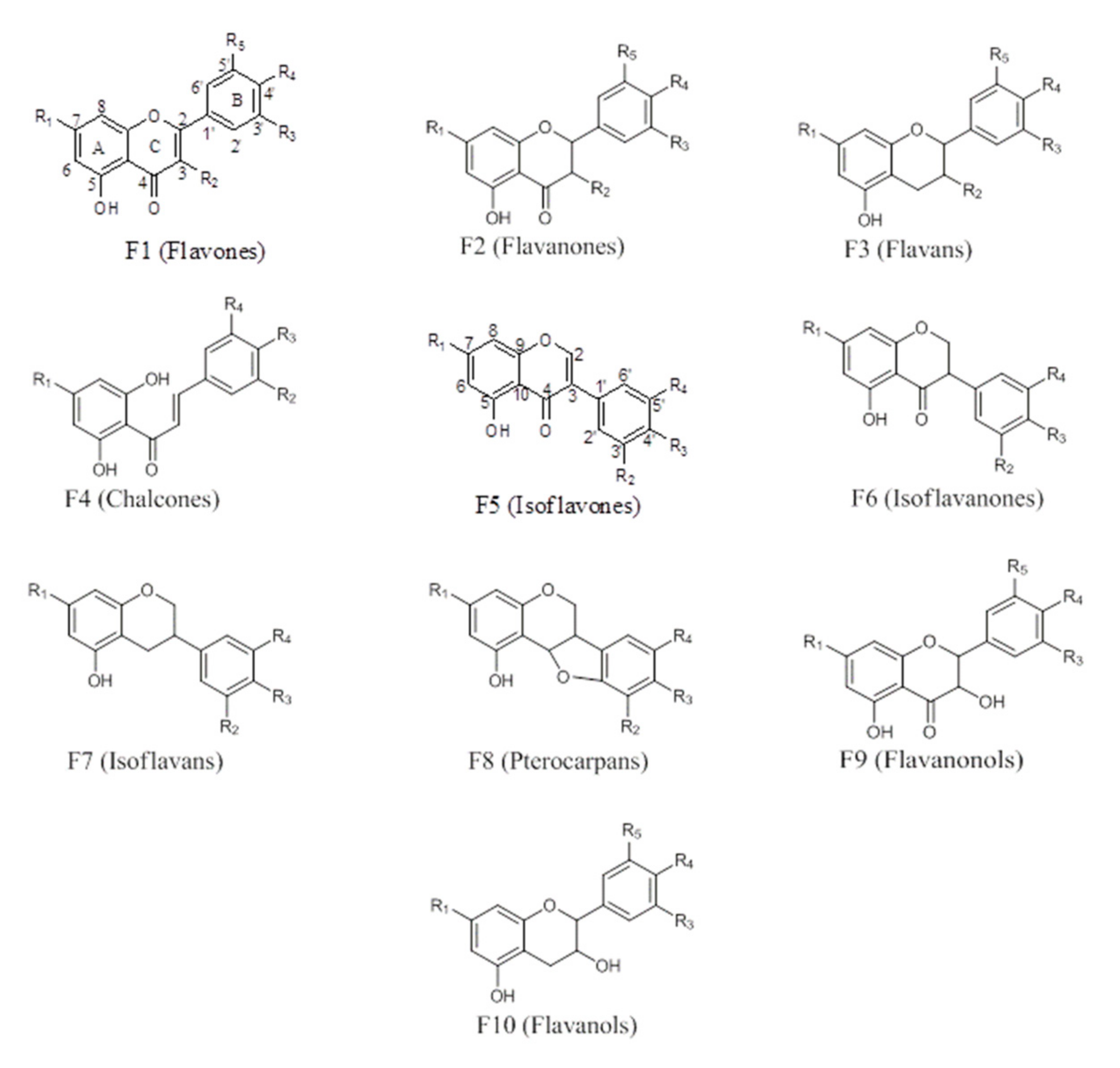
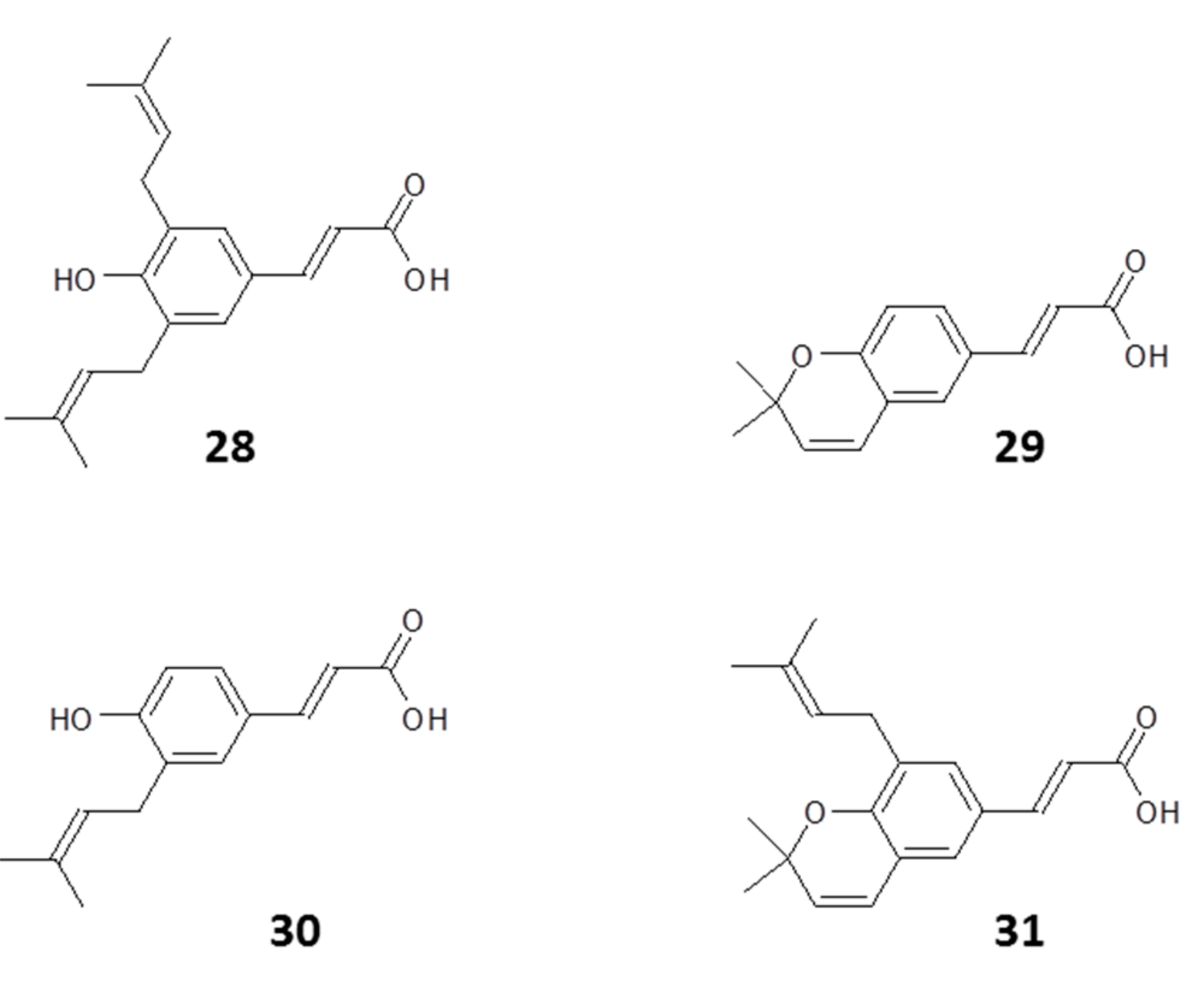
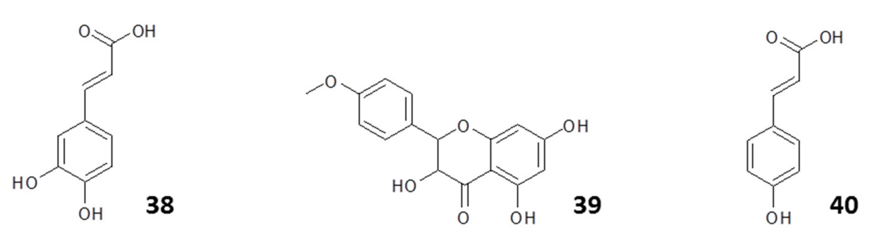
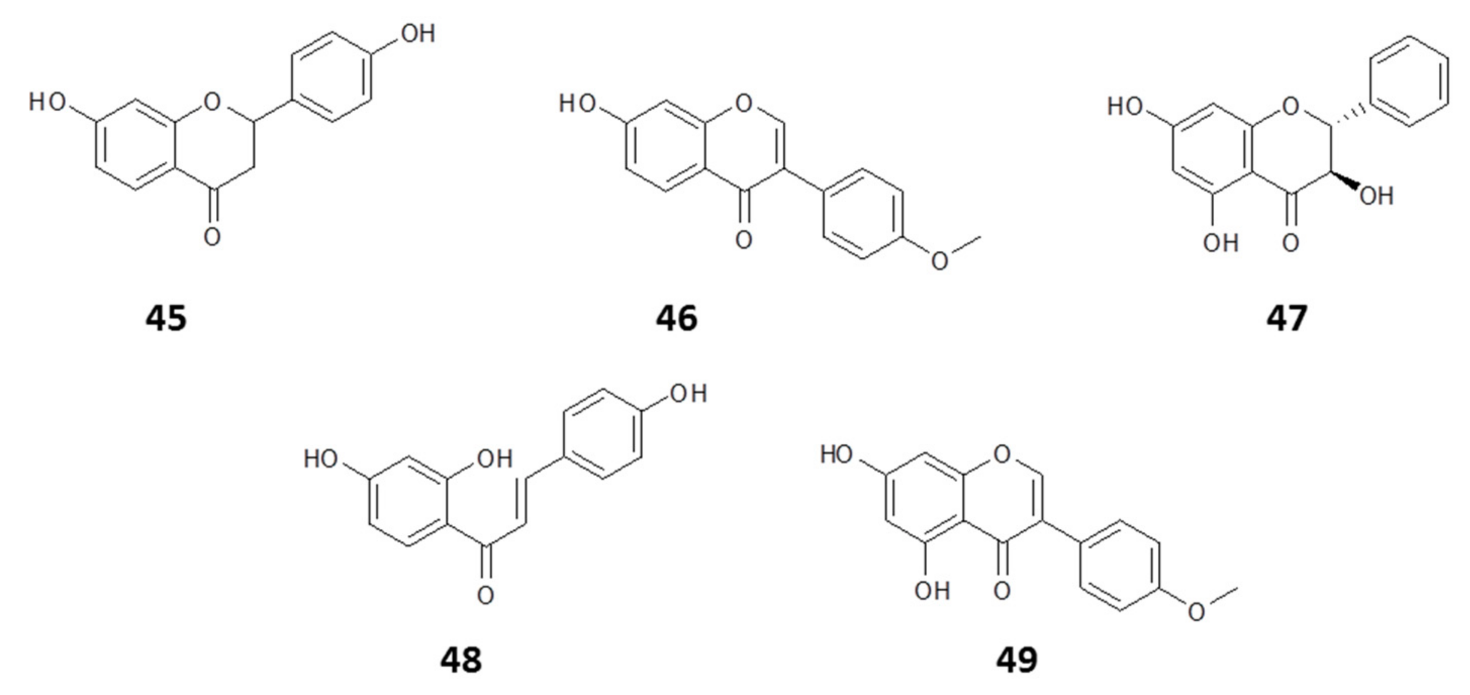
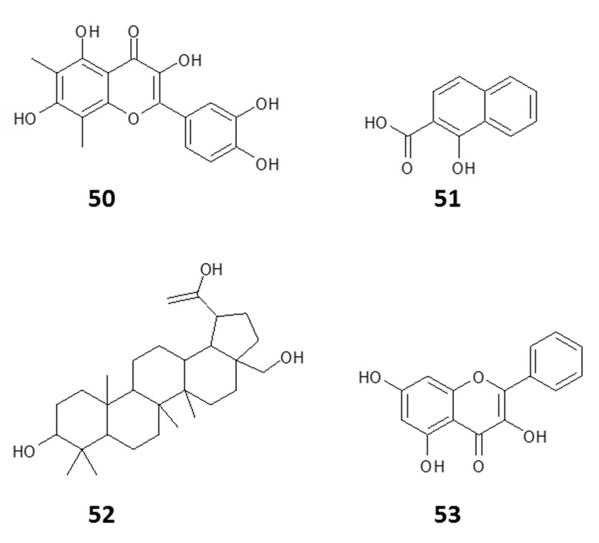
The compounds most commonly isolated and identified from propolis are polyphenols, representing a diverse class of compounds. They include simple flavonoids, phenyl propanoids, phenols, benzoquinones, phenolic acids, acetophenones, phenylacetic acids, hydroxycinnamic acids, phenylpropenes, coumarins and isocoumarins, chromones, naphtoquinones, xanthones, stilbenes, anthraquinones, flavonoids, lignans, neolignans, lignins, and condensed tannins [27]. Although there are several reports on the chemical composition of propolis [27,34,35,36] many of these reports are based on hyphenated techniques such as GC–MS, HPLC–MS, and LC–MS/MS for the identification of constituents. Since these are dereplication techniques, this review is on compounds reportedly isolated and characterized using NMR and MS techniques in the period 2015–2020.

2.1.1. Flavonoids

The majority of phenolic compounds isolated from propolis are flavonoids (Figure 1). The variation between the compounds is mostly in the degree of saturation (Structure F2) or unsaturation (Structure F1) of ring C, the absence of the carbonyl group at C-4 (Structure F3), or the opening of ring C (Structure F4). Those with an unsaturated ring C containing a ketone at C-4 are known as flavones, while those with saturated ring C and C-4 ketone are flavanones. An –OH substitution at C-3 leads to flavanols (with an unsaturated ring C; F10) or flavanonols (with a saturated ring C; F9); compounds with the open C ring (F4) are chalcones. The compounds usually have a 5-OH substitution, and ring B can either be unsubstituted or substituted with –OH, –OCH3, or other substituents such as isoprenyl or sugars. These substituents could be at C-3, C-6, C-8, C-6′, or C-2′, and the –OH at position C-5 could be absent. Substitution of ring B to position C-3 instead of C2 produces the isoflavones (F5), isoflavanones (F6), and other moieties such as isoflavans (Structure F7) and pterocarpans (Structure F8). Many flavonoids have been identified in propolis; recent examples include isosativan, (2′-hydroxy-7,4′-dimethoxyisoflavan), liquiritigenin (45), isoliquiritigenin, formononetin (46), vestitol (21), neovestitol, medicarpin, 7-O-neovestitol, pinobanksin (47), pinocembrin, chrysin, and pinobanksin-3-O-acetate, astrapterocarpan, 3,8-dihydroxy-9-methoxy-pterocarpan, broussonin B, 8-prenylnaringenin (20), and gerontoxanthone H (12) [37,38,39].

2.1.2. Phenyl Propanoids

The next set of abundant compounds in propolis are phenyl propanoids (Structure F11; Figure 2). Here, the aromatic rings can also be substituted with –OH or –OCH3, and there could be phenethyl or benzyl substituents. Drupanin, 2,2-dimethylchromene-6-propenoic acid, artepillin C, baccharin, 7-methoxy-3-hydroxy-2,2-dimethyl-8-prenylchromane-6-propenoic acid, 2,2-dimethyl-8-hexylchromene-6-propenoic acid, and 3-hydroxy-2,2-dimethyl-8-prenylchromane-6-propenoic acid [40] are some examples of phenyl propanoids recently reported from propolis.

2.1.3. Other Constituents

Other compounds that have been identified in propolis include aliphatic hydrocarbons, stilbenes, diterpenes, triterpenes [41], benzoic acid and its derivatives, benzaldehyde derivatives, cinnamyl alcohol, cinnamic acid and its derivatives, nicotinic acid, pantothenic acid, amino acids, carbohydrates, vitamins, and enzymes (glucose-6-phosphatase, acid phosphatase, adenosine triphosphatase and succinic dehydrogenase) [13]. Dereplication studies using HPLC–DAD–ESI-MS/MS identified the presence of pyrrolizidine alkaloids 7-(3-methoxy-2-methylbutyryl)-9-echimidinylretronecine and caffeoylquinic acid-O-arabinoside [42], but studies that report the isolation and identification of alkaloids from propolis using MS and NMR are rare. Trace elements such as Al, Ca, Fe, K, Mg, P, Zn, Cr, Ni, and Cu and possible toxic metals (As, Cd, and Pb) have also been reported [43,44,45].

2.2. Composition Based on Geographical Origin

Since the vegetation of different geographical regions varies, in addition to variations within the same region, and the phytochemicals in plants vary from season to season, the chemical composition of a propolis sample is determined primarily by its botanical source, the season, and the collection preferences of the bee species [13,14,29,30]. Thus, according to its botanical source, propolis may be classified into various chemotypes [30,46,47]; however, the assigned classifications unfortunately vary among authors as more types of propolis of different plant origins are being identified and characterized. However, the poplar type and Brazilian green propolis are the most widely available commercially and widely studied because of their medicinal properties. Poplar-type propolis is predominantly found in temperate regions and has been found to contain poplar bud phenolics [48]. Plant resins from the genus Populus (poplars) are the principal source of “poplar-type” propolis [49], found in parts of Europe, North America, New Zealand, temperate regions of Asia, and some regions of China. They typically contain aromatic (phenolic) acids and their esters, flavonoids, chalcones, dihydrochalcones, terpenoids, acyclic hydrocarbons, esters, alcohols, aldehydes, amino acids, aromatic hydrocarbons, fatty acids, ketones, sterols, sugars, and alcohols [50,51].
There is no clear geographic delineation for the classification of propolis from tropical regions such as Africa [14], because of the diversity of the tropical flora. Bees collecting propolis in tropical regions have a wider variety of plant sources; hence, there is little uniformity in the botanical source and, consequently, the phytochemicals. The compounds reported from tropical propolis include diterpenes, lignans, prenylated derivatives of p-coumaric acid, acetophenone, caffeic acid phenethyl ester, terpenoids, stilbenes, benzophenones, phenolic lipids, flavonoids, and diterpenic acids [13,27,30,38,52].
Over 148 compounds have been isolated from propolis from Africa (Table 1), South America (Table 2), Asia (Table 3), and Australia (Oceania) (Table 4) from different chemical classes. The majority of the compounds are flavonoids and their prenylated derivatives, whose isolation has been reported from propolis originating from every continent (except Antarctica). Flavonoids are abundant in the leaves, flowers, and fruits of the plants [53,54] from which bees collect resins and sticky exudates to make their propolis.
Triterpenoids are widely distributed in African propolis [15,16,55,57,58] but have also been reported in Bolivia [62,63], Brazil [61], Indonesia [28], and Thailand [67]. The most common triterpenes isolated from propolis are cycloartanes [15,28,58,61,62,63].
Other phytochemicals isolated from propolis within the period under review were not as widely distributed across all regions as the flavonoids and triterpenoids. However, studies indicate surprising similarity between propolis from Nigeria, Thailand, and Brazil, countries that could hardly be further apart, in their composition of triterpenoids, xanthones, and their prenylated derivatives [14,15,61,67]. Isolation of diterpenes is reported most from mainland Australia and the islands [71,72]. Australian propolis is unique, in that it contains stilbenes [73,74], which have not been reported in propolis from any other region. None of the studies reviewed reported the isolation of alkaloids from propolis. This could be due to the fact that alkaloids are much more abundant in roots and, since bees collect plant resins and sticky exudates from cracks in the bark or leaf buds of plants to make propolis, it is unlikely that alkaloids would be present in propolis.
Some of the compounds isolated from propolis are reported to have shown medicinal properties. For example, some flavonoids isolated from Nigerian propolis (astrapterocarpan, 3,8-dihydroxy-9-methoxy-pterocarpan, vesticarpan, medicarpin, vestitol, broussonin B, and 8-prenylnaringenin (20)) [39] or Bolivian propolis (3-prenyl-p-coumaric acid, kaempferol 3-methyl ether, and kaempferol 7-O-methyl ether) [63], were reported to have antioxidant properties.

3. Evidence for Propolis Protection against Bee Infections

Propolis is widely believed to be an important part of the bees’ defenses against infection of themselves and of their hive. The composition of propolis is principally dependent on the vegetation in the vicinity of the hive and on the bee species. Although the observation that some bee species collect only a small quantity of propolis leaves a question mark on the absolute requirement of propolis for bees, the notion that it protects bees from infection is backed up by a growing body of literature [76,77,78,79,80].
There is evidence showing a strong positive correlation between the amounts of propolis collected by bees and their heath condition, including their ability to produce viable broods (Simone-Finstrom and Spivak, 2010). Bees that collected larger quantities of propolis were reported to be healthier, producing viable broods and displaying superior hygienic behavior compared with the ones that collected less [76,77,81]. It was also found that bees usually respond to pathogens by collecting more propolis to ward off infections, while the immunity of the colony against infection is improved by the propolis envelop [78,82,83]. In addition, the microbiome of the bee colony is stabilized by propolis [84]. Colonies respond to Ascophaera apis (chalkbrood) by increasing resin collection for propolis, with hives with more propolis decreasing infection intensity [82]. However, the success of such a response depends on the type of vegetation in the vicinity of the hive and the chemical composition of its exudates.
It was shown that the ethanolic extracts of propolis were highly effective against Paenibacillus larvae, both in vitro and when field-tested in hives. It was not toxic to the bees when mixed with sugar syrup (oral administration), showing that propolis and its constituents are not toxic to bees. Propolis from Brazil also displayed significantly superior effects against P. larvae than propolis from Minnesota, United States of America (USA) [85,86], confirming that the activity of propolis depends on the vegetation around the hive.
Individual components isolated from propolis are also active against bee pathogens. Flavonoids and caffeates isolated from propolis displayed anti-P. larvae activity in vitro [87]. Another indication that certain chemical constituents in propolis offer an increased protective effect against P. larvae is that propolis from colonies free of P. larvae was reported to contain significantly more ferulic acid and coniferyl benzoate than propolis from colonies infected by this pathogen [88].
The protective efficacy of propolis for bees infested with Varroa destructor mites, a common pest of beehives, was recently directly confirmed. For instance, Argentinian propolis was found to be very effective against Varroa [89]. Furthermore, ethanolic extracts of German propolis were highly toxic to Varroa destructor, with a 10% w/v solution being lethal at 5 s contact [90], and Pusceddu et al. observed that raw propolis highly significantly increased the lifespan of Varroa-infected bees, almost completely reversing Varroa-associated mortality [91]. Moreover, the addition of natural propolis to hives reduced the titer of Varroa-transmitted deformed wing virus (DWV) [92] and Varroa-infected colonies specifically increased resin foraging [93]. It is not yet clear which chemical agents in propolis reduce the impact of Varroa infestation, but it was reported that the total polyphenolic content of propolis correlated with levels of Varroa infection in experimental hives in Sardinia [91]. Caffeic acid and pentenyl caffeates were found to be more abundant in propolis from Varroa-susceptible colonies [94], but this association requires further investigation in order to be confirmed as causal.
Propolis is also effective against bee infections caused by fungi. For instance, it was recently found that propolis fed to bees led to a significant reduction in Nosema ceranae infection [95]. Some acyl esters of flavonoids recently purified from propolis were characterized against two other honeybee pathogens: the fungus Ascosphaera apis, the causative agent of chalkbrood disease, and Paenibacillus larvae bacteria, which cause the disease American foulbrood. Pinobanksin 3-butyrate was identified as the most active chemical constituent against A. apis, while pinobanksin 3-octanoate was the most active agent against P. larvae [80].
It is becoming increasingly clear that the collection of propolis with strong antiprotozoal agents by bees is done purposefully. For instance, the microbiome of Scottish honeybees was reported to possess a high level of Lotmaria passim genetic material [96]. L. passim and Crithidia mellificae are trypanosomatids that are widespread in bee populations and are linked to colony losses that presently constitute a significant threat to honeybees; [96,97,98,99] characterized C. mellificae, together with L. passim, isolated from the honeybee Apis mellifera. However, the degree of pathogenicity of these trypanosomatid infections remains unclear but there is growing evidence for this. For instance, Gómez-Moracho et al. recently provided direct experimental evidence of the detrimental effects of the two trypanosomatids L. passim and Crithidia mellificae on honeybees in which honeybees inoculated with either L. passim or C. mellificae died faster than control bees [100]
Protozoal infections are spread within bee colonies through feces [101]. Therefore, bees may deliberately collect propolis that is active against trypanosomatids which they use for coating the surfaces within the hive to prevent disease transmission. It is, therefore, perhaps not surprising that propolis extract or purified chemical constituents are active against other trypanosomatids or kinetoplastids.

4. Propolis as an Anti-Kinetoplastid Agent

Currently there is a great interest in natural products-based drug discovery as a viable strategy for the treatment of diseases caused by the kinetoplastids. Among the most promising sources for such materials, propolis is being actively investigated based on its strong antiprotozoal activity [13,14]. Different types of propolis collected from diverse geographical locations all over the world have been attributed distinct pharmacological activities with promising results against various parasites belonging to the order kinetoplastida: Trypanosoma spp., Leishmania spp., and Crithidia fasciculata, a kinetoplastid model organism that is a close relative of C. mellificae (a bee pathogen) [102].

4.1. Antitrypanosomal Activity of Propolis

Several published papers have described the activity of propolis extracts and isolated components against a number of protozoan parasites. One of such reported biological properties, notwithstanding the quite distinct origins and compositions, is its in vitro and in vivo antitrypanosomal activity, reported by several authors [15,16,26,59,103,104,105,106,107,108,109,110,111,112].

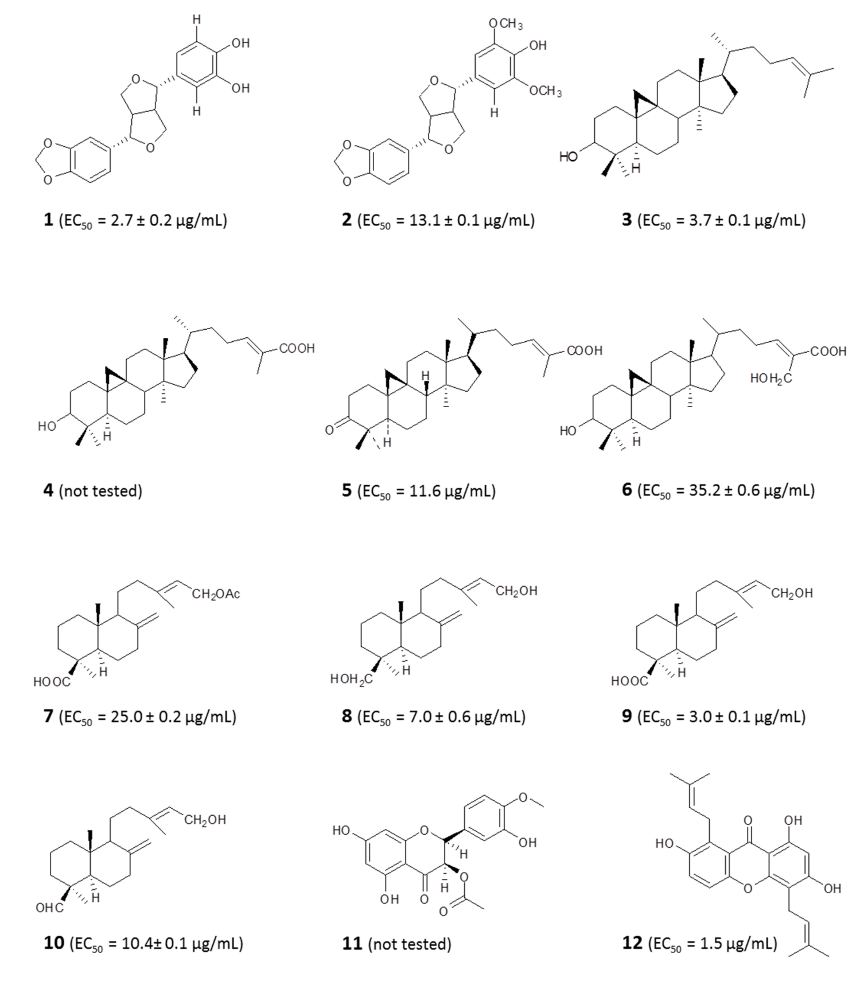
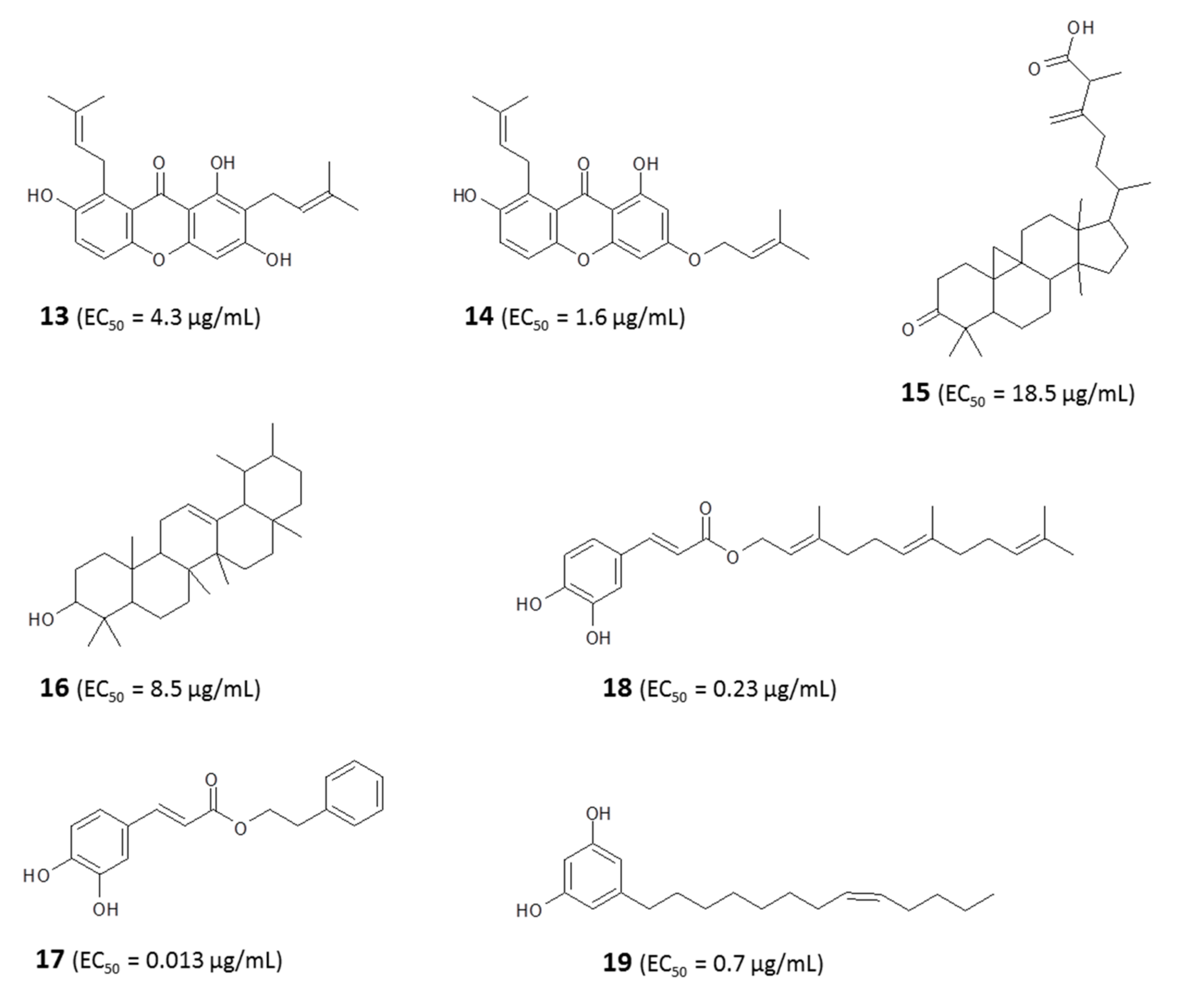
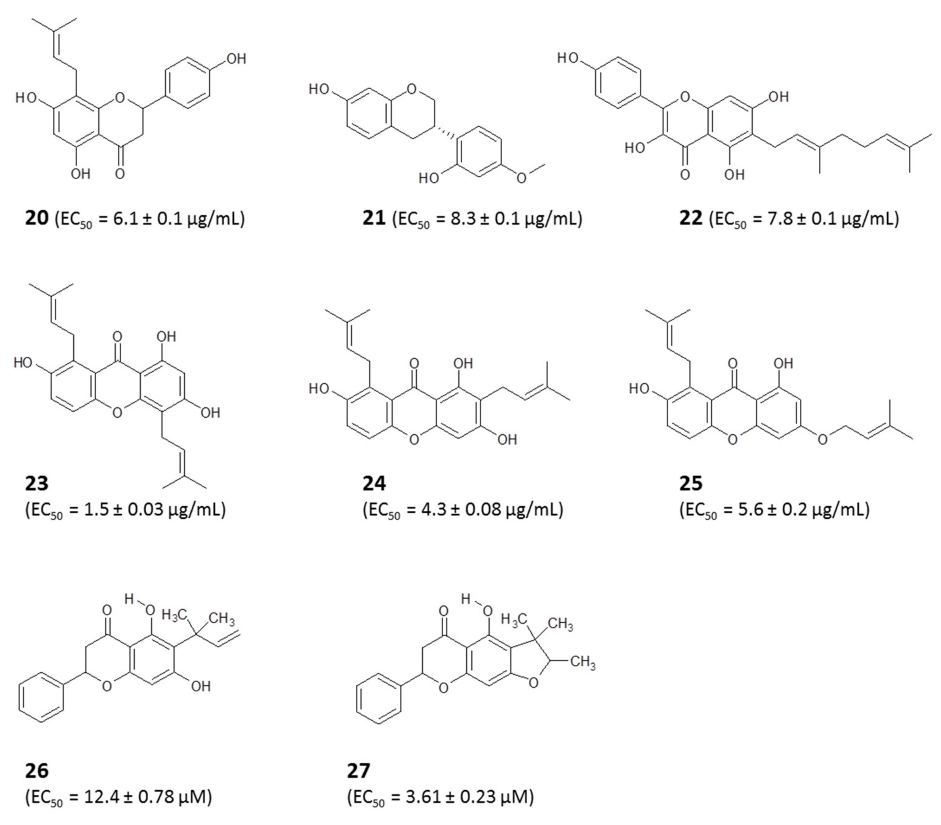
4.1.1. Identification of Bioactive Antitrypanosomal Compounds in Propolis Extracts

Libyan propolis has yielded lignans (demethylpiperitol (1) and 5′-methoxypiperitol (2)), cycloartane triterpenes (cycloartenol (3), mangeferolic acid, (4) mangiferonic acid (5), and 27-hydroxymangiferonic acid (6)), diterpenes (acetylisocupressic acid (7), agathadiol (8), isocupressic acid (9), and isoagatholal (10)), and a flavanone (taxifolin-3-acetyl-4′methyl ether (11)) with various levels of antitrypanosomal activity [59]. Studies of propolis samples from Nigeria also reported xanthones (Gerontoxanthone H (12), 6-deoxy-γ-mangostin (13), 1,7-dihydro-3-O-(3-methylbut-2-enyl)-8(3-methylbut-2-enyl) xanthone) (14), triterpenes (mangiferonic acid, ambonic acid (15), α-amyrin (16), and isoflavonoids with activity against Trypanosoma brucei brucei [14,15]. Two caffeic acid derivatives (β-phenethyl caffeate (17) and 2,2-dimethylallyl caffeate (18)) isolated from Fijian propolis were earlier reported to also have anti-Trypanosoma activity [18] (for structures, see Figure 3). Dereplication studies of European and Brazilian propolis samples associated their activity against Trypanosoma spp. with butyl and propionyl esters of pinobanksin, derivatives of benzopyran, caffeic acid, cinnamic acid, and flavonoids, where structures were not characterized by NMR [32,112].

4.1.2. Metabolomic Profiling Revealed a Possible Mode of Action of Propolis In Vitro

We recently reported the activities of Libyan propolis against T. b. brucei and the isolation of an alkyl resorcinol from the extract. A fraction containing a cardol identified as bilobol (19) (Figure 3) exhibited a strong antitrypanosomal activity (50% effective concentration (EC50) = 0.7 μg/mL) and had no significant effect on a human cell line (human foreskin fibroblasts (HFF)), demonstrating excellent selectivity. Metabolomic profiling revealed the mechanism of action of the cardol-rich fraction. We observed a significant disturbance in the metabolism of choline phospholipids [59]. This suggests that (this component of) Libyan propolis might be targeting the cell membrane of trypanosomes, acting selectively on one class of phospholipids, rather like a surfactant, extracting lipid from the cell membrane, resulting in the leakage of high-energy phosphates. This mode of action of the cardol-rich fraction of Libyan propolis might be comparable to that of miltefosine, a well-established antileishmanial drug reported for selectively perturbing microbial membrane fluidity [113]. There are also reports suggesting that alkyl resorcinols disrupt cellular membrane phospholipid metabolism by inhibiting phospholipase C1 [114,115].

4.1.3. Propolis Is Active against Drug-Sensitive and -Resistant Strains of T. brucei

Propolis is active against trypanosome various Trypanosoma strains including those that are highly resistant to current first-line drugs, as shown by our previous studies [14,15,112]. We determined the activities of compounds isolated from the ethanolic extracts of propolis collected from two regions in Nigeria against a panel of T. brucei strains including (i) T. brucei Lister 427 wild type (WT), which is the standard drug-sensitive control; (ii) an aquaglyceroporin2/3-null (AQP2/3-KO) strain, from which the TbAQP2/AQP3 locus was deleted [116], coding for the critical drug transporter HAPT1 [117,118] and, consequently, resistant to pentamidine and melarsoprol [119]; (iii) a multidrug-resistant strain, B48, adapted from Lister 427WT by deletion of the TbAT1/P2 drug transporter [120] and subsequent adaptation to very high concentrations of pentamidine in vitro [121], making the strain highly resistant to all diamidine- and melaminophenyl arsenical-based drugs.
The crude extracts all contained complex mixtures of natural compounds, of which 8-prenylnaringenin (20) was the most active of the purified compounds at 6.1 ± 0.1 µg/mL, and vestitol (21) and macarangin (22) displayed similar activities. Importantly, none of the diamidine- and arsenical-resistant strains were co-resistant to either the crude ethanolic extracts or the isolated compounds [15] (for structures, see Figure 4). This significant finding suggests that propolis may be a potential solution to the present challenge of drug resistance facing chemotherapy of human [122] and veterinary [123] trypanosomiasis.
A profiling of 12 additional propolis samples, collected from eight regions in Nigeria, identified three xanthones, 1,3,7-trihydroxy-4,8-di-(3-methylbut-2-enyl) xanthone (23), 1,3,7-trihydroxy-2,8-di-(3-methylbut-2-enyl) xanthone (24), and a xanthone that was previously undescribed (1,7-dihydroxy-3-O-(3-methylbut-2-enyl),(3-methylbut-2-enyl)xanthone) (25) (Figure 4), as well as three triterpenes, mangiferonic acid (5), a mixture of α-amyrin (16) with mangiferonic acid (1:3), and ambonic acid (15). These compounds all displayed trypanocidal activities against wild-type and resistant strains of T. b. brucei with EC50 values below 25 µg/mL but only the xanthones displayed high activity, i.e., EC50 values <5 μg/mL; xanthone (23) was the most active, with an EC50 of 1.5 ± 0.03 μg/mL. Interestingly, the compound displayed even higher activity against the AQP2 knockout strain (0.8 ± 0.02 µg/mL, p < 0.001), which was >30-fold resistant to pentamidine [14]. Similarly, and very recently, a bioassay-guided fractionation of Tanzanian and Zambian propolis samples led to the isolation of two novel flavanones with antitrypanosomal activities. The compounds were identified as 6-(1,1-dimethylallyl)pinocembrin (26) from the Zambian propolis sample and 5-hydroxy-4″,4″-dimethyl-5″-methyl-5″-H-dihydrofurano [2″,3″,6,7]flavanone (27) obtained from the Tanzanian propolis sample [124].

4.1.4. Propolis Contains Antitrypanosomal Activities Regardless of Geographical Location

Propolis samples collected from different locations within a country may possess different antitrypanosomal efficacies, due to differences in vegetation and/or bee species. For instance, ethanolic extracts obtained from 12 propolis samples collected from various regions in Libya showed a wide range of activity against T. brucei (EC50 value 1.67 μg/mL–39.38 μg/mL) [16]. Similarly, the antitrypanosomal activity of 35 propolis samples collected from different parts of Europe displayed varying activities against wild-type (WT) T. brucei and T. congolense, including the multidrug resistant strain T. brucei B48. Four of these samples showed high activity, while 23 had an intermediate activity (5–10 μg/mL) against WT and B48 T. brucei [112]. For the purpose of comparative analysis, C. fasciculata was also included and tested in parallel with these Trypanosoma species.
Interestingly, there was a very good overall correlation between the activities of each of the samples against the various kinetoplastid species, particularly between the Trypanosoma species/strains. This is very important because “African” trypanosomiasis is caused by multiple Trypanosoma species including T. congolense, T. vivax, T. b. brucei, T. b. gambiense, and T. b. rhodesiense; moreover, African trypanosomes that have adapted to non-tsetse fly transmission, including T. evansi, T. equiperdum, and T. vivax, have spread far beyond the African continent [123,125]. Few if any current drugs are effective against all these species. Another highly significant finding in the report was the very good correlation observed between the activity against drug-sensitive and drug-resistant strains, with the activities of the propolis samples against the highly resistant strain B48 on average performing even better than against the parental strains. This report, therefore, confirmed that cross-resistance with the current available trypanocidal drugs is less likely for propolis-derived compounds. Mechanistically, this is due to drug resistance in African trypanosomes being mostly linked to loss of drug transporters [118], which would not be the import mechanism for the structurally very different propolis-derived natural compounds.

4.1.5. Propolis Is Active In Vitro and In Vivo Against Trypanosomes

There are also several reports in the literature showing that propolis has in vitro activity against T. cruzi, the causative agent of American trypanosomiasis [126]. Marcucci et al. purified four bioactive phenolic compounds from Brazilian propolis: 3,5-diprenyl-4-hydroxycinnamic acid (28), 2,2-dimethyl-6-carboxyethenyl-8-prenyl-2H-1-benzopyran (29), 3-prenyl-4-hydroxycinnamic acid (30), and 2,2-dimethyl-6-carboxyethenyl-2H-1-benzopyran (31) (structures in Figure 5). All four phenolic compounds showed activity against T. cruzi Y strain trypomastigote (bloodstream) forms after 24 h exposure. The 24 h EC50 values of 0.72–2.64 mg/mL are high but reflect an incubation time of 24 h at 4 °C, chosen to test the feasibility of decontaminating donated blood batches in a blood bank [105]. Dantas et al. also observed anti-T. cruzi activity when assessing the in vitro antitrypanosomal effects of ethanolic and supercritical extracts of green, brown, and red, propolis from different regions of Brazil against Y strain epimastigote (insect) forms, albeit at high concentrations. Brazilian red propolis appeared to be the most active in these tests [127].
Extracts of Brazilian green propolis were used as oral treatment for acute infections of T. cruzi in mice (25–300 mg/kg body weight/day for 10 days). The mice showed reduced parasitemia and increased survival with no observable toxicity [128]. Similarly, rats infected with T. b. brucei and treated orally with Nigerian red propolis extracts (600 and 400 mg/kg for 5 days) had significantly reduced parasitemia, with higher red cell counts, packed-cell volume, and weight gain than untreated (control) mice [129]. It is important to note that these therapeutic effects were achieved with the crude extracts rather than purified active compounds, which would be expected to have stronger beneficial effects.
Chagas disease features T. cruzi parasites in the bloodstream (trypomastigotes) and inside mammalian host cells (amastigotes) and, crucially, propolis is active against both life-cycle forms. Treatment of heart muscle cells and macrophages infected with amastigotes with the ethanolic propolis extracts dose- and time-dependently reduced parasite loads, and 100 μg/mL of the extract fully lysed trypomastigotes within 24 h [103].

4.1.6. Direct Antiparasitic Efficacy and Sites of Action of Propolis in T. cruzi

One of the cellular sites of propolis action in T. cruzi is the mitochondrion. Treatment of T. cruzi-infected skeletal muscle cells with the ethanolic fraction of a Bulgarian propolis caused a decrease in the proliferation of intracellular amastigotes, swelling of the parasite’s mitochondrion, and concentric membrane structures appearing in the mitochondrial matrix. It also inflicted ultrastructural changes in the mitochondrion–kinetoplast complex of trypomastigotes and in the reservosomes of epimastigotes, characterized by distinct changes in their electron density and morphology. Reservosomes are large membrane-bound organelles located at the posterior end of the epimastigotes of T. cruzi, but absent in trypomastigote and amastigote forms [130]. The presence of electrolucent rod-shaped inclusions was also observed [106].
Most studies report that amastigotes derived from cell culture were more susceptible to treatment with ethanolic propolis fractions than trypomastigotes [131] and epimastigotes [132], and it may be that different compounds in these complex extracts are active on the diverse forms. Investigations by [128] to determine the cellular target of the ethanolic extract of Brazilian green propolis on various life-cycle stages of T. cruzi found different effects on epimastigotes (alterations in the ultrastructure of the mitochondrion, reservosomes, and Golgi complex) and trypomastigotes (loss of integrity and functionality of plasma membrane) [128].

4.1.7. Indirect Antiparasite Efficacy of Propolis via Immune Modulation in T. cruzi Infection

Apart from the direct antiparasite efficacy, propolis also interferes with the basic functions of the immune cells. Orally administered ethanolic extracts (50 mg/kg body weight) of Bulgarian propolis to T. cruzi-infected mice decreased parasitemia with no observable hepatic or renal damage. The treatment also decreased the spleen mass, including modulation of inflammatory reactions such as preferential expansion of CD8+ cells [107].
This immunomodulatory mechanism of action is likely associated with an increased resistance to infection, because activated CD4+ cells are known to increase the production of cytokines including IL-2 and IFN-γ, which are associated with differentiation and activation of the CD8+ T cells, resulting in an increased immune response.

4.2. Anti-Leishmania Effects of Propolis

Leishmaniasis affects over 12 million people and is endemic in 88 countries across the tropics and subtropical regions of the world. Like trypanosomiasis, almost all chemotherapeutic options for leishmaniasis have unacceptable side effects, and there are as yet no vaccines for human use. There is currently an intense search for alternative safe anti-Leishmania chemotherapy from propolis. Several studies have shown that propolis obtained from diverse origins possesses antileishmanial activity due to the presence of flavonoids [16,17,18,19,20,21,22,23,24].

4.2.1. Identification of Active Antileishmanial Compounds in Propolis Extracts

In fact, Ecuadorian propolis high in flavonoids including (5,7,4′-trihydroxyflavanone (32), 5,4′-dihydroxy-7-methoxyflavanone (33), 3,5,4′-trihydroxy-7,3′-dimethoxyflavanone (34), 5,4′-dihydroxy-7,3′-dimethoxyflavanone (35), 3,5,3′,4′-tetrahydroxy-6,7-dimethoxy flavone (36), and 3,5,4′-trihydroxy-7,3′-dimethoxy flavone (37)) was found to possess much better antileishmanial activity than Ecuadorian propolis samples rich in triterpenic alcohols and acetyl triterpenes and inhibited the growth of L. amazonensis amastigotes and promastigotes [63]; structures are shown in Figure 6.
One of the common factors usually evaluated when investigating the pharmacological activities of propolis is isolating the active component and testing it against a pathogen. However, studies have shown that a synergistic effect offered by several chemical constituents of the mixture may exist and should not be ignored when evaluating its biological activities. In some experiments to study the effect of propolis extracts on Leishmania, the active principles that contributed to the inhibition of the proliferation of the promastigote forms of Leishmania (viannia) braziliensis were caffeic acid (38), aromadendrine-4′-methyl ether (dihydrokaemferide) (39), p-coumaric acid (40), 3,5-diprenyl-p-coumaric (artepillin C) (28), and 3-prenyl-p-coumaric (30) acid (Figure 7). These compounds together reduced the lesions caused by the infection [23]. Importantly, extracts of Bulgarian propolis, rich in flavonoids, displayed activity against several old- and new-world Leishmania species, L. amazonensis, L. braziliensis, L. chagasi, and L. major [24], indicating a broad spectrum of antileishmanial activity. This is considered to be essential for the development of antileishmanial drugs for the international market, although it could be envisaged that propolis-derived treatments could be developed to fill a more local need.
Similarly, Nina et al. [65] assayed Bolivian propolis extracts and their active compound against promastigotes of L. braziliensis and L. amazonensis. They found that propolis rich in phenolic compounds displayed superior antibacterial and antileishmanial activity than those containing mostly triterpenes. The methanol extracts showed leishmanicidal activity against promastigotes of both species with MIC100 values in a tight range of 7.8 to 12.1 μg/mL depending on the Leishmania species and the geographical origin of the propolis, further reinforcing the activity of propolis against multiple Leishmania species.
It thus appears that, as for trypanocidal activity, propolis from different countries are also active against Leishmania, despite a great variation in chemical constituents, although the level of antileishmanial activity does depend on the propolis constitution. Extracts of 35 propolis samples collected from different parts of Europe were assessed for antileishmanial activity against wild-type and miltefosine-APC12-resistant strains (C12R×, resistance factor (RF) >600-fold) of L. mexicana promastigotes. All the samples showed a high or moderate level of activity against the wild-type strain (EC50 0.35–5.67 μg/mL) and the miltefosine-APC12-resistant strain (0.28–1.55 μg/mL); the best activity was again noticed in propolis from Bulgaria [112]. Interestingly, in most cases, the propolis samples were more active against the resistant strain (RF was as low as 0.23, i.e., >4-fold more sensitive). Although very preliminary, this study offers the suggestion that propolis could offer a solution to the current issue of drug resistance in Leishmania chemotherapy, at least for miltefosine. Clearly, much more research into this important possibility is required, including the mechanism by which the resistant strains would become particularly sensitized to (which?) specific constituents of European propolis.

4.2.2. Effects of Propolis on Infected Macrophages

Propolis is capable of killing Leishmania in macrophages, thereby reducing parasitemia load. For instance, Santana et al. reported effects of brown propolis from the semiarid region of Piauí, Brazil, against both promastigotes and intramacrophage amastigotes, with the dichloromethane fraction being the most active [133]. A survey of Cuban propolis samples particularly highlighted the activity of a yellow propolis rich in acetyl triterpenes against intramacrophage L. infantum, as well as against T. cruzi and T. brucei [134]. However, that study also found that the selectivity of the yellow propolis samples over MRC-5 human fibroblasts was quite low. In contrast, [135] found that ethanolic extracts of green and red propolis against L. braziliensis promastigote-infected BALB/c mouse-derived macrophages reduced the L. braziliensis load without observable toxicity to the macrophages. Nonetheless, the red propolis showed a stronger parasite reduction than the green propolis extract and, at a concentration of 100 μg/mL, showed almost the same effect as the standard drug amphotericin B. Ethanolic extracts of Brazilian red propolis were also active against promastigotes and extracellular amastigote forms of L. amazonensis in infected macrophages in a time- and dose-dependent manner, with low toxicity to noninfected macrophage controls [136]. This extract was previously reported to be rich in benzophenones and prenylated compounds [105,137].

4.2.3. Propolis in Animal Models of Leishmaniasis

The above studies all describe the antileishmanial activity of propolis in vitro and sometimes only on the promastigote (insect) form. However, a very recent study showed that propolis was as active as one of the available standard drugs when tested against cutaneous leishmaniasis in an in vivo model of L. major infection. Tavakoli et al. observed that the ethanolic extract of Iranian propolis inhibited the growth of promastigote forms of L. major, as well as the standard drug Glucantime (meglumine antimoniate) at concentrations >37.5 μg/mL in vitro (p > 0.05). More importantly, in a mouse model of cutaneous leishmaniasis (L. major), treatment with 4% ethanolic propolis extract (4 g extract plus 96 g vaseline–oserin) reduced the size of skin lesions with similar efficacy as Glucantime, the standard antileishmanial treatment in much of the world [138].
Some of the in vivo benefits of propolis may be through direct action on the parasite. However, a water extract of green propolis was able to prevent the progression of L. infantum-induced lesions in the liver during infection, even better than some of the commercially available drugs such as Glucantime, by reducing the parasite-induced lesions and secondary chronic inflammatory processes in the liver [139]. Da Silva et al. similarly showed that propolis treatment reduced leishmaniasis-associated liver inflammation, reporting decreases in the levels of liver N-acetyl-β-glucosaminidase and myeloperoxidase activity and of proinflammatory cytokines, as well as lower collagen fiber deposition, and plasma aspartate [140]. In contrast, the levels of anti-inflammatory cytokine were increased, and hepatosplenomegaly was at least partially reversed [139,140]. Propolis also decreases the side effects of meglumine antimoniate in the host [139].

4.2.4. Synergy of Propolis and Mainstream Antileishmanials

Propolis also exhibits synergistic leishmanicidal effect when combined with standard drugs such as Glucantime or amphotericin B. Ayres et al. reported that a gel prepared from Brazilian red propolis reduced the amount of exudate from leishmanial skin lesions, particularly when combined with Glucantime [141]. Very recently, Jihene et al. assessed the antileishmanial effect of an essential oil from Tunisian propolis and its combination with amphotericin B against clinical isolates of L. infantum and L. major [142]. The essential oil showed good activity against promastigote forms of L. infantum and L. major (EC50 = 5.29 μg/mL and 3.67 μg/mL, respectively) and against the amastigote forms (EC50 = 7.38 μg/mL and 4.96 μg/mL, respectively), with low cytotoxicity. The very similar activity against the promastigote and amastigote forms is important for the evaluation of other studies that tested only against the easy-to-culture promastigote stage. A synergistic efficacy was observed when the essential oil was combined with amphotericin B (fractional inhibitory concentration (FIC) = 0.37). The active principles were further identified as α-pinene (41) (36.7%), α-cedrol (42) (6.7%), totarol (43) (6.6%), and dehydroabietane (44) (5.2%). The authors attributed the antileishmanial efficacy of the essential oil mostly to α-pinene, which has a reported moderate activity against promastigotes and amastigotes [143], synergistically with that of the minor but more potent components, especially α-cedrol (EC50 = 1.5 µM) [142] and totarol (EC50 = 12.2 µM) to L. donovani promastigotes [144]. However, that putative synergism remains to be experimentally tested. The mechanism of action of the propolis essential oil was proposed to be activation of macrophages by hyperproduction of NO, and this could play a role, but would not explain the effects against promastigotes, which was in fact higher than against intramacrophage amastigotes.

4.2.5. Direct Antiparasite and Indirect Effects of Propolis on Intramacrophage Amastigotes via Immunomodulation

Upregulation of the macrophage microbicidal activities is one of the reported modes of action of propolis in Leishmania infection, and it is becoming increasingly clear that immunomodulation is a major mechanism of action of propolis in Leishmania infection. Brazilian propolis extracts with high concentrations of phenolic compounds (flavonoids, benzopyrans, and aromatic acids), di- and triterpenes, and essential oils showed a direct inhibitory effect on promastigote forms of L. braziliensis, with a concentration of 100 µg/mL of propolis extract as effective as 250 µg/mL Glucantime. Interestingly, the preincubation of macrophages with just 5 µg/mL or 10 µg/mL propolis extract induced them to take up more promastigotes but resulted in a strong reduction in recovered promastigotes after 5 days [17], demonstrating an increased proficiency of the macrophages to kill the parasites internalized. The authors linked this observation to an observed increase in the level of TNF-α in mice pretreated with propolis extracts, coupled with the downregulation of IL-12 during the infection [17]. Orsatti et al. investigated immunomodulation in mice treated for 3 days with ethanolic extracts of propolis and reported an increase in the expression of Toll-like receptors (TLR)-2 and TLR-4 in macrophages, as well as an increase in the production of proinflammatory cytokines IL-1β and IL-6, indicators of activation of the innate immune response [143].
Moreover, dry, alcoholic, and glycolic propolis extracts at various concentrations (10, 50, or 100 µg/mL) showed, again, a dose-dependent effect on the viability of promastigotes of L. braziliensis in culture, as well as reduced parasite loads in macrophages. There were reduced levels of superoxide and nitric oxide in activated macrophages infected with L. braziliensis, as well as increased activity of superoxide dismutase (SOD), following treatment by the alcoholic and glycolic extracts; these are all antioxidant responses. However, the inflammatory profile of macrophages was significantly modified by the dry propolis extract via upmodulating TNF-α, while downmodulating the production of IL-10 and TGF-β [145], changes that lead to a greater activation of the cells.
These data put together suggest that propolis extracts or its constituents are well tolerated by macrophages and can increase the mechanisms of macrophage activation, resulting in the neutralization of Leishmania.

4.2.6. Nanotechnology in Delivering Propolis Therapy

Several technological advances have been made with regard to the use of propolis in leishmaniasis drug discovery. Propolis has been loaded onto polymeric nanoparticles for targeted drug delivery [146,147]. Some of the most accepted drug delivery vehicles with pharmaceutical applications are polymeric nanoparticles and liposomes. This is due to the advantages of target delivery, nontoxicity, biocompatibility, biodegradability, controlled drug release, and stability during storage, all leading to an increased therapeutic efficacy [148].
Correspondingly, [149] assessed the efficacy of polymeric nanoparticles loaded with an ethanolic extract of Brazilian red propolis for antileishmanial therapies in a multiple-constituent co-delivery system. Using a nanoprecipitation method, polymeric nanoparticles (poly-ε-caprolactone and pluronic) were loaded with red propolis extract and were characterized for leishmanicidal activity. The red propolis nanoparticles were stable without any aggregation phenomenon observed during a 1 month period, while exhibiting antileishmanial activity with an EC50 value of 31–47 μg/mL against L. braziliensis promastigotes in vitro. Analysis of the propolis extract identified several flavonoids as the potential active compounds, specifically liquiritigenin (45), formononetin (46), pinobanksin (47), isoliquiritigenin (48), and biochanin A (49) (structures in Figure 8).
In summary, it appears that the major antileishmanial chemical constituents of propolis are specific flavonoids and certain metabolites of caffeic acid [150,151], whereas the most reported mode of action of propolis against Leishmania is immunomodulation through the activation of macrophages, although it is clear that direct antileishmanial effects also importantly contribute given the in vitro observations.

4.3. Effects on Crithidia fasciculata

Crithidia, Leishmania, and Trypanosoma are members of the order Kinetoplastida, and Crithidia fasciculata is a very close relative of C. mellificae, a parasite of honeybees, and of Crithidia bombi, the bumble bee pathogen. C. mellificae has been reported to be significantly responsible for the winter mortality often observed in beehives across Western European [152]. Considering that these pathogens are closely related genetically and consequently possess comparable metabolism and life cycle, Crithidia fasciculata was adopted as an accessible and well-researched model organism for the study of these important bee infections.
Accordingly, our lab successfully developed strategies for the screening of propolis extracts and fractions on C. fasciculata [16] and used this approach to chemically characterize various propolis samples. The strong anti-kinetoplastid activity of propolis extracts seems to be a virtually constant feature in the literature reports and our own experience, which strongly indicates that bees deliberately collect propolis to protect themselves against invasion of their hive by pathogens, including Crithidia species, coating the hive in an antimicrobial substance. Investigation of the activity of propolis on C. fasciculata by screening of ethanolic extracts of 12 Libyan propolis samples showed that all the extracts were active but exhibited a range of EC50, with the most active extract having an EC50 of 6.5 μg/mL. This activity was correlated strongly with dimethylquercetin (50) and a derivative of hydroxynaphthoic acid (51) in an orthogonal partial least squares (OPLS) model of the anticrithidial activity [16] (structures in Figure 9).
Extracts of propolis from Papua New Guinea inhibited the growth of C. fasciculata [153]. Chemical profiling of the extract conducted using negative ion spray ESI (LC–MS) revealed a high concentration of triterpenes in the active (ethanolic) fraction, indicating that the observed activity was likely due to the inhibitory action of triterpenes on the viability of C. fasciculata. Nine compounds were subsequently purified from the ethanolic fraction and their structural elucidation revealed eight cycloartane-type triterpenes and a pentacyclic triterpene (20-hydroxybetulin (52, Figure 9)), which on further testing gave the best activity against C. fasciculata [153].
To assess the effect of geographical location of the propolis samples on the activity against C. fasciculata and, by extension, the possible effects on bee pathogens caused by the trypanosomatids, the anticrithidial activity of extracts from 35 propolis samples from different parts of Europe was investigated. Moderate-to-high levels of anticrithidial activity were observed for all 35 samples, with EC50 values in the range of 2.5–22.7 µg/mL. OPLS modeling of the chemical constituents correlated the highest activity with pinobanksin (47) and a methyl ether of galangin (53, Figure 9) [112].
A higher activity against C. fasciculata and T. brucei was observed in a comparative study of propolis efficacy against the kinetoplastids with ethanolic extracts of Nigerian propolis samples. The triterpenoids mangiferonic acid (5, EC50 = 11.6 μg/mL), ambonic acid (15, EC50 = 18.5 μg/mL), and α-amyrin (16, EC50 = 8.5 μg/mL), and the xanthones gerontoxanthone H (12, EC50 = 1.2 μg/mL), 6-deoxy-γ-mangostin (13, EC50 = 4.3 μg/mL) and 1,7-dihydro-3-O-(3-methylbut-2-enyl)-8-(3-methylbut-2-enyl) xanthone (14, EC50 = 1.6 μg/mL) were isolated from the samples. The crude extract samples were found to have higher antitrypanosomal activity than most of the isolated compounds. EC50 values of the most active crude samples were 1.2 and 4.2 µg/mL for C. fasciculata and T. brucei, respectively [14]. Other triterpenoids, diterpenes, lignans, flavonoids, etc. isolated from propolis samples collected from different geographical regions (Table 1) are reported to have different levels of anti-kinetoplastid activity. This shows that, regardless of geographical location, propolis contains anti-kinetoplastid compounds. Overall, the consistently observed activity against C. fasciculata gives support to the hypothesis that bees collect propolis specifically to protect them from infections caused by pathogens, particularly those caused by species of Crithidia, well-known bee pathogens that are quite closely related to the human pathogens Leishmania and Trypanosoma [102], and the closely related L. passim which has been found to be abundant in bees [98,99]. This view was further strengthened by the surprisingly excellent correlation observed between the EC50 values of Nigerian propolis fractions against T. brucei and C. fasciculata [14]. This, however, further strengthens the case for the development of drugs against other trypanosomatids such as Leishmania and Trypanosoma species from propolis samples.

5. Conclusions

Propolis samples possess a wide range of chemical constituents, which largely depend on the geographical location where it was collected, in addition to seasonal variations stemming from the vegetation in the locality. The compounds present in propolis, particularly the different types of flavonoids, appear to be responsible for the observed broad spectrum of biological activities, with some individual compounds in the samples showing activity against different organisms, particularly the kinetoplastids. The consistently observed high levels of antiprotozoal activity of propolis extracts, especially against the kinetoplastids, together with the recent findings of Regan et al. [96] regarding the presence of DNA of several protozoan parasite species in the bee metagenome, indicate that these pathogens may be exerting more pressure on the health of bee colonies than heretofore known. Therefore, there remains a lot to be understood regarding the role of propolis in bee health, but it now looks certain that the near-universal presence of anti-kinetoplastid activity in bee propolis is not incidental. Thus, propolis is a source of natural compounds, preselected by evolution, against important neglected diseases such as leishmaniasis, sleeping sickness, and Chagas disease. The broad anti-kinetoplastid activity of propolis components reviewed here, together with the generally low toxicity to macrophages and experimental animals, beneficial immunomodulation, and our recent findings that the main bioactive metabolites (flavonoids) present in propolis are well absorbed and tolerated by the human body [154], gives ample scope for further investigations toward the rational development of anti-kinetoplastid drugs that will replace the existing ones, which have many undesirable side effects and often suffer from drug resistance after decades of use [155]. However, questions remain with regard to the efficacy of propolis components (such as flavonoids) in vivo since, although these compounds are often well absorbed, they are also rapidly metabolized particularly to glucuronides and sulfates. This problem has been extensively addressed in previous papers and reviews, and the following points are of importance [156,157,158,159,160]:
(i)
In some cases, the biological activity of flavonoids is not improved or sometimes increased by conjugation.
(ii)
At higher doses and in samples containing a mixture of flavonoids, there may be incomplete conjugation of particular flavonoids.
(iii)
It is possible that flavonoid metabolites can become deconjugated.
Given the generally low toxicity of propolis and the high toxicity of many of the existing antiprotozoal drugs, it may be possible to optimize the efficacy of propolis treatments by giving a high dosage. In addition, if crude extracts were to be used as treatment, it would be important to set a standard, perhaps on the basis of the concentrations of the key components in the extracts.
The mode of action of propolis depends on the organism it is acting on, and ranges from direct effects on growth and/or viability of the pathogen to immunomodulation via macrophage activation or cytokine changes, perturbation of the cell membrane architecture through phospholipid disturbances, and mitochondrial targets. Given the complexity and variability of propolis, mechanism-of-action studies are particularly fraught and, frankly, lagging. The mechanism of any activity of “propolis” could only be defined if “propolis” itself is perfectly defined and standardized. Although there has been excellent analytic work to identify the constituents of specific propolis samples, each of these have been very different. Logically, then, mechanistic studies can only be performed with individual compounds shown to be present and even dominant in some types of propolis. However, this potentially loses synergy (or, conversely, antagonism) between components in complex propolis samples: a well-understood conundrum for ethnopharmacologists and phytochemists. Meanwhile, identification of specific cellular targets makes limited sense until a genuine lead compound is chosen from among the large number of compounds that show promise. The criteria for the lead compound need to be agreed upon but must surely include high efficacy, low toxicity, metabolic stability, good absorption/bioavailability, and either abundant cheap availability from a natural source or easy synthesis.
In theory, it is possible to reconstitute a “standardized propolis” of known composition for mechanistic studies, be they cellular or in vivo, although the optimal composition of such propolis might be a cause for some debate. However, as a potential treatment, this is highly unlikely to be commercially viable. It is therefore incumbent on the scientific community, at this point in time, to start selecting a limited panel of propolis-derived compounds with particular promise against a specific infectious agent, e.g., Trypanosoma cruzi, and take these as screening “hits” for further development. This will require a multidisciplinary consortium approach including medicinal chemistry for the development of structure–activity relationships (SAR), toxicology, pharmacokinetics/dynamics, biochemical parasitology for direct action studies, and immunology for the indirect effects of propolis. The screening of propolis fractions from various locations (with different vegetation) and the identification of the active compounds are of course by no means complete, but the development of new treatments from the knowledge accrued so far does not need to wait until the full catalog has been hoisted onto library shelves. Clearly, a good number of active compounds with apparent selectivity have been identified, and a “round table” of experts in medicinal chemistry, drug metabolism, drug delivery systems, and parasitology should be able to triage and select potential drug candidates from among them, as well as cost-effective ways to narrow the field further with standardized tests such as in vivo stability. This approach will give the field the clearest way forward toward genuine preclinical lead compounds for mechanistic, SAR, and extensive in vivo evaluations. It is only at this stage that serious partnerships with private sector pharmaceuticals or with multinational not-for-profit organization such as the Drugs for Neglected Diseases initiative (DNDi) become possible. A bundling of resources and coordination in efforts between research groups will be needed to start moving in the direction that all of us, according to the rationales stated in the introductions of our published papers, aspire to.

Author Contributions

Conceptualization, H.P.D.K., J.O.I., G.U.E., and D.G.W.; writing—original draft preparation, G.U.E., N.I., J.O.I., H.P.D.K., D.G.W., and W.S.; writing—review and editing, H.P.D.K., J.O.I., G.U.E., N.I., W.S., and D.G.W. All authors read and agreed to the published version of the manuscript.

Funding

This research received no external funding.

Conflicts of Interest

The authors declare no conflict of interest.

References

  1. Alvar, J.; Vélez, I.D.; Bern, C.; Herrero, M.; Desjeux, P.; Cano, J.; Jannin, J.; den Boer, M.; WHO Leishmaniasis Control Team. Leishmaniasis worldwide and global estimates of its incidence. PLoS ONE 2012, 7, e35671. [Google Scholar] [CrossRef] [PubMed]
  2. Alcântara, L.M.; Ferreira, T.C.; Gadelha, F.R.; Miguel, D.C. Challenges in drug discovery targeting TriTryp diseases with an emphasis on leishmaniasis. Int. J. Parasitol. Drugs Drug Resist. 2018, 8, 430–439. [Google Scholar] [CrossRef] [PubMed]
  3. Rao, S.P.; Barrett, M.P.; Dranoff, G.; Faraday, C.J.; Gimpelewicz, C.R.; Hailu, A.; Jones, C.L.; Kelly, J.M.; Lazdins-Helds, J.K.; Mäser, P. Drug discovery for kinetoplastid diseases: Future directions. ACS Infect. Dis. 2018, 5, 152–157. [Google Scholar] [CrossRef] [PubMed] [Green Version]
  4. Johnston, K.L.; Ford, L.; Taylor, M.J. Overcoming the challenges of drug discovery for neglected tropical diseases: The A·WOL experience. J. Biomol. Screen. 2014, 19, 335–343. [Google Scholar] [CrossRef] [PubMed] [Green Version]
  5. Croft, S.L.; Seifert, K.; Yardley, V. Current scenario of drug development for leishmaniasis. Indian J. Med. Res. 2006, 123, 399–410. [Google Scholar] [PubMed]
  6. Ebiloma, G.U.; Igoli, J.O.; Katsoulis, E.; Donachie, A.-M.; Eze, A.; Gray, A.I.; de Koning, H.P. Bioassay-guided isolation of active principles from Nigerian medicinal plants identifies new trypanocides with low toxicity and no cross-resistance to diamidines and arsenicals. J. Ethnopharmacol. 2017, 202, 256–264. [Google Scholar] [CrossRef] [Green Version]
  7. Ebiloma, G.U.; Katsoulis, E.; Igoli, J.O.; Gray, A.I.; De Koning, H.P. Multi-target mode of action of a Clerodane-type diterpenoid from Polyalthia longifolia targeting African trypanosomes. Sci. Rep. 2018, 8, 4613. [Google Scholar] [CrossRef] [Green Version]
  8. Stuart, K.; Brun, R.; Croft, S.; Fairlamb, A.; Gürtler, R.E.; McKerrow, J.; Reed, S.; Tarleton, R. Kinetoplastids: Related protozoan pathogens, different diseases. J. Clin. Investig. 2008, 118, 1301–1310. [Google Scholar] [CrossRef] [Green Version]
  9. World Health Organization. WHO Roadmap Inspires Unprecedented Support to Defeat Neglected Tropical Diseases. World Health Organization: Geneva, Switzerland. Available online: http://www.who.int/neglected_diseases/London_meeting_follow_up/en/ (accessed on 6 June 2020).
  10. Ichoron, N.; Tyoer, S.; James, E.J.; Igoli, J.O. A survey of medicinal plants used as traditional medicine in Ukum and Ogbadibo Local Government Areas of Benue state, Nigeria. Plants Environ. 2019, 1, 5–11. [Google Scholar]
  11. Gray, A.I.; Igoli, J.O.; Edrada-Ebel, R. Natural products isolation in modern drug discovery programs. In Natural Products Isolation; Springer: Berlin, Germany, 2012; pp. 515–534. [Google Scholar]
  12. Brahmachari, G. Therapeutics from natural products against neglected tropical diseases: An overview. In Discovery and Development of Therapeutics from Natural Products against Neglected Tropical Diseases; Elsevier: Amsterdam, The Netherlands, 2019; pp. 1–6. [Google Scholar]
  13. Anjum, S.I.; Ullah, A.; Khan, K.A.; Attaullah, M.; Khan, H.; Ali, H.; Bashir, M.A.; Tahir, M.; Ansari, M.J.; Ghramh, H.A. Composition and functional properties of propolis (bee glue): A review. Saudi J. Biol. Sci. 2019, 26, 1695–1703. [Google Scholar] [CrossRef]
  14. Omar, R.; Igoli, J.O.; Zhang, T.; Gray, A.I.; Ebiloma, G.U.; Clements, C.J.; Fearnley, J.; Ebel, R.E.; Paget, T.; De Koning, H.P. The chemical characterization of Nigerian propolis samples and their activity against Trypanosoma brucei. Sci. Rep. 2017, 7, 1–10. [Google Scholar] [CrossRef] [PubMed] [Green Version]
  15. Omar, R.M.; Igoli, J.; Gray, A.I.; Ebiloma, G.U.; Clements, C.; Fearnley, J.; Edrada Ebel, R.A.; Zhang, T.; De Koning, H.P.; Watson, D.G. Chemical characterisation of Nigerian red propolis and its biological activity against Trypanosoma brucei. Phytochem. Anal. 2016, 27, 107–115. [Google Scholar] [CrossRef] [PubMed] [Green Version]
  16. Siheri, W.; Zhang, T.; Ebiloma, G.U.; Biddau, M.; Woods, N.; Hussain, M.Y.; Clements, C.J.; Fearnley, J.; Ebel, R.E.; Paget, T. Chemical and antimicrobial profiling of propolis from different regions within Libya. PLoS ONE 2016, 11, e0155355. [Google Scholar] [CrossRef] [PubMed] [Green Version]
  17. Santos da Silva, S.; da Silva Thomé, G.; Cataneo, A.H.D.; Miranda, M.M.; Felipe, I.; Andrade, C.G.T.D.J.; Watanabe, M.A.E.; Piana, G.M.; Sforcin, J.M.; Pavanelli, W.R. Brazilian propolis antileishmanial and immunomodulatory effects. Evid. Based Complement. Alternat. Med. 2013, 2013, 673058. [Google Scholar]
  18. Otoguro, K.; Iwatsuki, M.; Ishiyama, A.; Namatame, M.; Nishihara-Tsukashima, A.; Kiyohara, H.; Hashimoto, T.; Asakawa, Y.; Ōmura, S.; Yamada, H. In vitro antitrypanosomal activity of some phenolic compounds from propolis and lactones from Fijian Kawa (Piper methysticum). J. Nat. Med. 2012, 66, 558–561. [Google Scholar] [CrossRef]
  19. Amarante, M.K.; Watanabe, M.A.E.; Conchon-Costa, I.; Fiori, L.L.; Oda, J.M.M.; Búfalo, M.C.; Sforcin, J.M. The effect of propolis on CCL5 and IFN-γ expression by peripheral blood mononuclear cells from leishmaniasis patients. J. Pharm. Pharmacol. 2012, 64, 154–160. [Google Scholar] [CrossRef]
  20. Fidalgo, L.M.; Ramos, I.S.; Parra, M.G.; Cuesta-Rubio, O.; Hernández, I.M.; Fernández, M.C.; Piccinelli, A.L.; Rastrelli, L. Activity of Cuban propolis extracts on Leishmania amazonensis and Trichomonas vaginalis. Nat. Prod. Commun. 2011, 6, 973–976. [Google Scholar] [CrossRef] [Green Version]
  21. Ozbilge, H.; Kaya, E.G.; Albayrak, S.; Silici, S. Anti-leishmanial activities of ethanolic extract of Kayseri propolis. Afr. J. Microbiol. Res. 2010, 4, 556–560. [Google Scholar]
  22. Duran, G.; Duran, N.; Culha, G.; Ozcan, B.; Oztas, H.; Ozer, B. In vitro antileishmanial activity of Adana propolis samples on Leishmania tropica: A preliminary study. Parasitol. Res. 2008, 102, 1217–1225. [Google Scholar] [CrossRef]
  23. Pontin, K.; Da Silva Filho, A.A.; Santos, F.F.; e Silva, M.L.A.; Cunha, W.R.; Nanayakkara, N.D.; Bastos, J.K.; de Albuquerque, S. In vitro and in vivo antileishmanial activities of a Brazilian green propolis extract. Parasitol. Res. 2008, 103, 487–492. [Google Scholar] [CrossRef]
  24. de Carvalho Machado, G.M.; Leon, L.L.; de Castro, S.L. Activity of Brazilian and Bulgarian propolis against different species of Leishmania. Mem. Inst. Oswaldo Cruz 2007, 102, 73–77. [Google Scholar] [CrossRef] [Green Version]
  25. Ahangari, Z.; Naseri, M.; Vatandoost, F. Propolis: Chemical composition and its applications in endodontics. Iran. Endodont. J. 2018, 13, 285. [Google Scholar]
  26. Falcão, S.I.; Vale, N.; Cos, P.; Gomes, P.; Freire, C.; Maes, L.; Vilas-Boas, M. In vitro evaluation of Portuguese propolis and floral sources for antiprotozoal, antibacterial and antifungal activity. Phytother. Res. 2014, 28, 437–443. [Google Scholar] [CrossRef]
  27. Šturm, L.; Ulrih, N.P. Advances in the propolis chemical composition between 2013 and 2018: A review. eFood 2019, 1, 24–37. [Google Scholar] [CrossRef] [Green Version]
  28. Pujirahayu, N.; Suzuki, T.; Katayama, T. Cycloartane-type triterpenes and botanical origin of propolis of stingless Indonesian bee Tetragonula sapiens. Plants 2019, 8, 57. [Google Scholar] [CrossRef] [Green Version]
  29. Santos, L.M.; Fonseca, M.S.; Sokolonski, A.R.; Deegan, K.R.; Araújo, R.P.; Umsza-Guez, M.A.; Barbosa, J.D.; Portela, R.D.; Machado, B.A. Propolis: Types, composition, biological activities, and veterinary product patent prospecting. J. Sci. Food Agric. 2020, 100, 1369–1382. [Google Scholar] [CrossRef] [PubMed]
  30. Blicharska, N.; Seidel, V. Chemical diversity and biological activity of African propolis. In Progress in the Chemistry of Organic Natural Products 109; Springer: Berlin, Germany, 2019; pp. 415–450. [Google Scholar]
  31. Betances-Salcedo, E.; Revilla, I.; Vivar-Quintana, A.M.; González-Martín, M.I. Flavonoid and antioxidant capacity of propolis prediction using near infrared spectroscopy. Sensors 2017, 17, 1647. [Google Scholar] [CrossRef] [PubMed] [Green Version]
  32. da Silva Cunha, I.B.; Salomão, K.; Shimizu, M.; Bankova, V.S.; Custódio, A.R.; de Castro, S.L.; Marcucci, M.C. Antitrypanosomal activity of Brazilian propolis from Apis mellifera. Chem. Pharm. Bull. 2004, 52, 602–604. [Google Scholar] [CrossRef] [PubMed] [Green Version]
  33. Righi, A.A.; Negri, G.; Salatino, A. Comparative chemistry of propolis from eight Brazilian localities. Evid. Based Complement. Alternat. Med. 2013, 2013, 267878. [Google Scholar] [CrossRef] [Green Version]
  34. Huang, S.; Zhang, C.-P.; Wang, K.; Li, G.Q.; Hu, F.-L. Recent advances in the chemical composition of propolis. Molecules 2014, 19, 19610–19632. [Google Scholar] [CrossRef] [Green Version]
  35. Bankova, V.; Popova, M.; Trusheva, B. Propolis volatile compounds: Chemical diversity and biological activity: A review. Chem. Cent. J. 2014, 8, 28. [Google Scholar] [CrossRef] [Green Version]
  36. Sforcin, J.M.; Bankova, V. Propolis: Is there a potential for the development of new drugs? J. Ethnopharmacol. 2011, 133, 253–260. [Google Scholar] [CrossRef]
  37. Sami, B.; Okoro, H.; Igoli, N.P.; Igoli, J.O. Isolation of isosativan from Nigerian red propolis. Trop. J. Nat. Prod. Res. 2020, 4, 77–79. [Google Scholar] [CrossRef]
  38. Falcão, S.I.; Lopes, M.; Vilas-Boas, M. A first approach to the chemical composition and antioxidant potential of Guinea-Bissau propolis. Nat. Prod. Commun. 2019, 14, 1–5. [Google Scholar] [CrossRef]
  39. Alaribe, C.S.; Esposito, T.; Sansone, F.; Sunday, A.; Pagano, I.; Piccinelli, A.L.; Celano, R.; Cuesta Rubio, O.; Coker, H.A.; Nabavi, S.M. Nigerian propolis: Chemical composition, antioxidant activity and α-amylase and α-glucosidase inhibition. Nat. Prod. Res. 2019, 1–5. [Google Scholar] [CrossRef]
  40. Do Nascimento Araujo, C.; Sivero Mayworm, M.A.; Yatsuda, R.; Negri, G.; Faria Salatino, M.L.; Salatino, A.; Timenetsky, J.; Campos, G.B. Chemical composition and antimycoplasma activity of a brown propolis from southern Brazil. J. Food Sci. Technol. 2020, 57, 4228–4235. [Google Scholar] [CrossRef]
  41. Al-Ghamdi, A.A.; Bayaqoob, N.I.; Rushdi, A.I.; Alattal, Y.; Simoneit, B.R.; El-Mubarak, A.H.; Al-Mutlaq, K.F. Chemical compositions and characteristics of organic compounds in propolis from Yemen. Saudi J. Biol. Sci. 2017, 24, 1094–1103. [Google Scholar] [CrossRef]
  42. Bonamigo, T.; Campos, J.F.; Alfredo, T.M.; Balestieri, J.B.P.; Cardoso, C.A.L.; Paredes-Gamero, E.J.; de Picoli Souza, K.; dos Santos, E.L. Antioxidant, cytotoxic, and toxic activities of propolis from two native bees in Brazil: Scaptotrigona depilis and Melipona quadrifasciata anthidioides. Oxid. Med. Cell. Longev. 2017, 2017, 1038153. [Google Scholar] [CrossRef] [Green Version]
  43. Hodel, K.V.; Machado, B.A.; Santos, N.R.; Costa, R.G.; Menezes-Filho, J.A.; Umsza-Guez, M.A. Metal content of nutritional and toxic value in different types of Brazilian propolis. Sci. World J. 2020, 2020, 395496. [Google Scholar] [CrossRef]
  44. Souza, E.; Zaluski, R.; Veiga, N.; Orsi, R. Effects of seasonal variations and collection methods on the mineral composition of propolis from Apis mellifera Linnaeus beehives. Braz. J. Biol. 2016, 76, 396–401. [Google Scholar] [CrossRef] [PubMed] [Green Version]
  45. González-Martín, M.I.; Escuredo, O.; Revilla, I.; Vivar-Quintana, A.M.; Coello, M.C.; Riocerezo, C.P.; Moncada, G.W. Determination of the mineral composition and toxic element contents of propolis by near infrared spectroscopy. Sensors 2015, 15, 27854–27868. [Google Scholar] [CrossRef]
  46. da Silva, M.V.; de Moura, N.G., Jr.; Motoyama, A.B.; Ferreira, V.M. A review of the potential therapeutic and cosmetic use of propolis in topical formulations. J. Appl. Pharmacol. Sci. 2019, 10, 131–141. [Google Scholar]
  47. Popova, M.; Giannopoulou, E.; Skalicka-Woźniak, K.; Graikou, K.; Widelski, J.; Bankova, V.; Kalofonos, H.; Sivolapenko, G.; Gaweł-Bęben, K.; Antosiewicz, B. Characterization and biological evaluation of propolis from Poland. Molecules 2017, 22, 1159. [Google Scholar] [CrossRef] [PubMed]
  48. Falcão, S.I.; Vale, N.; Gomes, P.; Domingues, M.R.; Freire, C.; Cardoso, S.M.; Vilas-Boas, M. Phenolic profiling of Portuguese propolis by LC–MS spectrometry: Uncommon propolis rich in flavonoid glycosides. Phytochem. Anal. 2013, 24, 309–318. [Google Scholar] [CrossRef] [PubMed] [Green Version]
  49. Wagh, V.D. Propolis: A wonder bees product and its pharmacological potentials. Adv. Pharmacol. Pharm. Sci. 2013, 2013, 308249. [Google Scholar] [CrossRef] [Green Version]
  50. De Groot, A.C.; Popova, M.P.; Bankova, V.S. An Update on the Constituents of Poplar-Type Propolis; Acdegroot Publishing: Wapserveen, The Netherlands, 2014. [Google Scholar]
  51. Saleh, K.; Zhang, T.; Fearnley, J.; Watson, D.G. A comparison of the constituents of propolis from different regions of the United Kingdom by liquid chromatography-high resolution mass spectrometry using a metabolomics approach. Curr. Metabolom. 2015, 3, 42. [Google Scholar] [CrossRef]
  52. Kardar, M.; Zhang, T.; Coxon, G.; Watson, D.; Fearnley, J.; Seidel, V. Characterisation of triterpenes and new phenolic lipids in Cameroonian propolis. Phytochemistry 2014, 106, 156–163. [Google Scholar] [CrossRef] [Green Version]
  53. Panche, A.N.; Diwan, A.D.; Chandra, S.R. Flavonoids: An overview. J. Nutr. Sci. 2016, 5, e47. [Google Scholar] [CrossRef] [Green Version]
  54. Iwashina, T. The structure and distribution of the flavonoids in plants. J. Plant Res. 2000, 113, 287. [Google Scholar] [CrossRef]
  55. Mahamat, A.A.; Nyemb, J.N.; Gade, I.S.; Ngenge, A.T.; Talla, E.; Céline, H.; Sophie, L.; Mbafor, J.T. A New fatty acid and some triterpenoids from propolis of Nkambe (North-West Region, Cameroon) and evaluation of the antiradical scavenging activity of their extracts. Open Chem. 2020, 18, 239–243. [Google Scholar] [CrossRef]
  56. Talla, E.; Tamfu, A.N.; Gade, I.S.; Yanda, L.; Mbafor, J.T.; Laurent, S.; Elst, L.V.; Popova, M.; Bankova, V. New mono-ether of glycerol and triterpenes with DPPH radical scavenging activity from Cameroonian propolis. Nat. Prod. Res. 2017, 31, 1379–1389. [Google Scholar] [CrossRef] [PubMed]
  57. Tamfu, A.N.; Sawalda, M.; Fotsing, M.T.; Kouipou, R.M.T.; Talla, E.; Chi, G.F.; Epanda, J.J.E.; Mbafor, J.T.; Baig, T.A.; Jabeen, A. A new isoflavonol and other constituents from Cameroonian propolis and evaluation of their anti-inflammatory, antifungal and antioxidant potential. Saudi J. Biol. Sci. 2020, 27, 1659–1666. [Google Scholar] [CrossRef] [PubMed]
  58. Siheri, W.; Ebiloma, G.U.; Igoli, J.O.; Gray, A.I.; Biddau, M.; Akrachalanont, P.; Alenezi, S.; Alwashih, M.A.; Edrada-Ebel, R.; Muller, S. Isolation of a novel flavanonol and an alkylresorcinol with highly potent anti-trypanosomal activity from Libyan propolis. Molecules 2019, 24, 1041. [Google Scholar] [CrossRef] [Green Version]
  59. Segueni, N.; Zellagui, A.; Moussaoui, F.; Lahouel, M.; Rhouati, S. Flavonoids from Algerian propolis. Arab. J. Chem. 2016, 9, S425–S428. [Google Scholar] [CrossRef] [Green Version]
  60. Sokeng, S.D.; Talla, E.; Sakava, P.; Fokam Tagne, M.A.; Henoumont, C.; Sophie, L.; Mbafor, J.T.; Tchuenguem Fohouo, F.-N. Anti-inflammatory and analgesic effect of arachic acid ethyl ester isolated from propolis. BioMed Res. Int. 2020, 2020, 8797284. [Google Scholar] [CrossRef]
  61. Ccana-Ccapatinta, G.V.; Mejía, J.A.A.; Tanimoto, M.H.; Groppo, M.; de Carvalho, J.C.A.S.; Bastos, J.K. Dalbergia ecastaphyllum (L.) Taub. and Symphonia globulifera Lf: The botanical sources of isoflavonoids and benzophenones in Brazilian red propolis. Molecules 2020, 25, 2060. [Google Scholar] [CrossRef]
  62. Nina, N.; Lima, B.; Feresin, G.E.; Giménez, A.; Salamanca Capusiri, E.; Schmeda-Hirschmann, G. Antibacterial and leishmanicidal activity of Bolivian propolis. Lett. Appl. Microbiol. 2016, 62, 290–296. [Google Scholar] [CrossRef]
  63. Nina, N.; Quispe, C.; Jiménez-Aspee, F.; Theoduloz, C.; Giménez, A.; Schmeda-Hirschmann, G. Chemical profiling and antioxidant activity of Bolivian propolis. J. Sci. Food Agric. 2016, 96, 2142–2153. [Google Scholar] [CrossRef]
  64. Mitsui, T.; Hotta, S.; Tazawa, S.; Arai, Y.; Kato, K.; Ichihara, K. Chemical constituents of Brazilian propolis from the state of Bahia and their growth inhibitory activities against cancer cells. Biosci. Biotechnol. Biochem. 2018, 82, 417–421. [Google Scholar] [CrossRef] [Green Version]
  65. Cuesta-Rubio, O.; Fernández, M.C.; Hernández, I.M.; Jaramillo, C.G.J.; González, V.H.; Porto, R.M.D.O.; Delange, D.M.; Fidalgo, L.M.; Piccinelli, A.L.; Campone, L. Chemical profile and anti-leishmanial activity of three Ecuadorian propolis samples from Quito, Guayaquil and Cotacachi regions. Fitoterapia 2017, 120, 177–183. [Google Scholar] [CrossRef]
  66. Nina, N.; Quispe, C.; Jiménez-Aspee, F.; Theoduloz, C.; Feresín, G.E.; Lima, B.; Leiva, E.; Schmeda-Hirschmann, G. Antibacterial activity, antioxidant effect and chemical composition of propolis from the Región del Maule, Central Chile. Molecules 2015, 20, 18144–18167. [Google Scholar] [CrossRef] [PubMed] [Green Version]
  67. Sanpa, S.; Popova, M.; Bankova, V.; Tunkasiri, T.; Eitssayeam, S.; Chantawannakul, P. Antibacterial compounds from propolis of Tetragonula laeviceps and Tetrigona melanoleuca (Hymenoptera: Apidae) from Thailand. PLoS ONE 2015, 10, e0126886. [Google Scholar] [CrossRef]
  68. Zhao, L.; Yu, M.; Sun, M.; Xue, X.; Wang, T.; Cao, W.; Sun, L. Rapid determination of major compounds in the ethanol extract of geopropolis from Malaysian stingless bees, Heterotrigona itama, by UHPLC-Q-TOF/MS and NMR. Molecules 2017, 22, 1935. [Google Scholar] [CrossRef] [Green Version]
  69. Ismail, T.N.N.T.; Sulaiman, S.A.; Ponnuraj, K.T.; Man, C.N.; Hassan, N.B. Chemical constituents of Malaysian Apis mellifera propolis. Sains Malays. 2018, 47, 117–122. [Google Scholar] [CrossRef]
  70. Ishizu, E.; Honda, S.; Ohta, T.; Vongsak, B.; Kumazawa, S. Component analysis and antiangiogenic activity of Thailand stingless bee propolis. Makara J. Technol. 2019, 23, 78–83. [Google Scholar] [CrossRef] [Green Version]
  71. Georgieva, K.; Trusheva, B.; Uzunova, V.; Stoyanova, T.; Valcheva, V.; Popova, M.; Tzoneva, R.; Bankova, V. New cycloartane triterpenes from bioactive extract of propolis from Pitcairn Island. Fitoterapia 2018, 128, 233–241. [Google Scholar] [CrossRef]
  72. Aminimoghadamfarouj, N.; Nematollahi, A. Propolis diterpenes as a remarkable bio-source for drug discovery development: A review. Int. J. Mol. Sci. 2017, 18, 1290. [Google Scholar] [CrossRef] [Green Version]
  73. Trusheva, B.; Stancheva, K.; Gajbhiye, N.; Dimitrova, R.; Popova, M.; Saraf, R.; Bankova, V. Two new prenylated stilbenes with an irregular sesquiterpenyl side chain from propolis from Fiji Islands. Rec. Nat. Prod. 2016, 10, 465. [Google Scholar]
  74. Duke, C.C.; Tran, V.H.; Duke, R.K.; Abu-Mellal, A.; Plunkett, G.T.; King, D.I.; Hamid, K.; Wilson, K.L.; Barrett, R.L.; Bruhl, J.J. A sedge plant as the source of Kangaroo Island propolis rich in prenylated p-coumarate ester and stilbenes. Phytochemistry 2017, 134, 87–97. [Google Scholar] [CrossRef] [Green Version]
  75. Nishimura, E.; Murakami, S.; Suzuki, K.; Amano, K.; Tanaka, R.; Shinada, T. Structure determination of monomeric phloroglucinol derivatives with a cinnamoyl group isolated from propolis of the stingless bee, Tetragonula carbonaria. Asian J. Org. Chem. 2016, 5, 855–859. [Google Scholar] [CrossRef]
  76. Simone-Finstrom, M.; Spivak, M. Propolis and bee health: The natural history and significance of resin use by honey bees. Apidologie 2010, 41, 295–311. [Google Scholar] [CrossRef] [Green Version]
  77. Nicodemo, D.; De Jong, D.; Couto, R.; Malheiros, B. Honey bee lines selected for high propolis production also have superior hygienic behavior and increased honey and pollen stores. Genet. Mol. Res. 2013, 12, 6931–6938. [Google Scholar] [CrossRef]
  78. Borba, R.S.; Klyczek, K.K.; Mogen, K.L.; Spivak, M. Seasonal benefits of a natural propolis envelope to honey bee immunity and colony health. J. Exp. Biol. 2015, 218, 3689–3699. [Google Scholar] [CrossRef] [Green Version]
  79. Simone-Finstrom, M.; Borba, R.S.; Wilson, M.; Spivak, M. Propolis counteracts some threats to honey bee health. Insects 2017, 8, 46. [Google Scholar] [CrossRef] [Green Version]
  80. Wilson, M.B.; Pawlus, A.D.; Brinkman, D.; Gardner, G.; Hegeman, A.D.; Spivak, M.; Cohen, J.D. 3-Acyl dihydroflavonols from poplar resins collected by honey bees are active against the bee pathogens Paenibacillus larvae and Ascosphaera apis. Phytochemistry 2017, 138, 83–92. [Google Scholar] [CrossRef] [PubMed] [Green Version]
  81. Nicodemo, D.; Malheiros, E.B.; De Jong, D.; Couto, R.H.N. Increased brood viability and longer lifespan of honeybees selected for propolis production. Apidologie 2014, 45, 269–275. [Google Scholar] [CrossRef] [Green Version]
  82. Simone-Finstrom, M.D.; Spivak, M. Increased resin collection after parasite challenge: A case of self-medication in honey bees? PLoS ONE 2012, 7, e34601. [Google Scholar] [CrossRef] [Green Version]
  83. Borba, R.S.; Spivak, M. Propolis envelope in Apis mellifera colonies supports honey bees against the pathogen, Paenibacillus larvae. Sci. Rep. 2017, 7, 11429. [Google Scholar] [CrossRef] [PubMed] [Green Version]
  84. Saelao, P.; Borba, R.S.; Ricigliano, V.; Spivak, M.; Simone-Finstrom, M. Honeybee microbiome is stabilized in the presence of propolis. Biol. Lett. 2020, 16, 20200003. [Google Scholar] [CrossRef]
  85. Antúnez, K.; Harriet, J.; Gende, L.; Maggi, M.; Eguaras, M.; Zunino, P. Efficacy of natural propolis extract in the control of American Foulbrood. Vet. Microbiol. 2008, 131, 324–331. [Google Scholar] [CrossRef]
  86. Bastos, E.M.A.; Simone, M.; Jorge, D.M.; Soares, A.E.E.; Spivak, M. In vitro study of the antimicrobial activity of Brazilian propolis against Paenibacillus larvae. J. Invertebr. Pathol. 2008, 97, 273–281. [Google Scholar] [CrossRef]
  87. Bilikova, K.; Popova, M.; Trusheva, B.; Bankova, V. New anti-Paenibacillus larvae substances purified from propolis. Apidologie 2013, 44, 278–285. [Google Scholar] [CrossRef] [Green Version]
  88. Popova, M.; Antonova, D.; Bankova, V. Chemical composition of propolis and American foulbrood: Is there any relationship? Bulg. Chem. Commun. 2017, 49, 171–175. [Google Scholar]
  89. Damiani, N.; Fernández, N.J.; Maldonado, L.M.; Álvarez, A.R.; Eguaras, M.J.; Marcangeli, J.A. Bioactivity of propolis from different geographical origins on Varroa destructor (Acari: Varroidae). Parasitol. Res. 2010, 107, 31–37. [Google Scholar] [CrossRef]
  90. Garedew, A.; Lamprecht, I.; Schmolz, E.; Schricker, B. The varroacidal action of propolis: A laboratory assay. Apidologie 2002, 33, 41–50. [Google Scholar] [CrossRef] [Green Version]
  91. Pusceddu, M.; Floris, I.; Mura, A.; Theodorou, P.; Cirotto, G.; Piluzza, G.; Bullitta, S.; Angioni, A.; Satta, A. The effects of raw propolis on Varroa-infested honey bee (Apis mellifera) workers. Parasitol. Res. 2018, 117, 3527–3535. [Google Scholar] [CrossRef]
  92. Drescher, N.; Klein, A.-M.; Neumann, P.; Yañez, O.; Leonhardt, S.D. Inside honeybee hives: Impact of natural propolis on the ectoparasitic mite Varroa destructor and viruses. Insects 2017, 8, 15. [Google Scholar] [CrossRef] [Green Version]
  93. Pusceddu, M.; Piluzza, G.; Theodorou, P.; Buffa, F.; Ruiu, L.; Bullitta, S.; Floris, I.; Satta, A. Resin foraging dynamics in Varroa destructor-infested hives: A case of medication of kin? Insect Sci. 2019, 26, 297–310. [Google Scholar] [CrossRef]
  94. Popova, M.; Reyes, M.; Le Conte, Y.; Bankova, V. Propolis chemical composition and honeybee resistance against Varroa destructor. Nat. Prod. Res. 2014, 28, 788–794. [Google Scholar] [CrossRef]
  95. Mura, A.; Pusceddu, M.; Theodorou, P.; Angioni, A.; Floris, I.; Paxton, R.J.; Satta, A. Propolis consumption reduces Nosema ceranae infection of European honey bees (Apis mellifera). Insects 2020, 11, 124. [Google Scholar] [CrossRef] [Green Version]
  96. Regan, T.; Barnett, M.W.; Laetsch, D.R.; Bush, S.J.; Wragg, D.; Budge, G.E.; Highet, F.; Dainat, B.; de Miranda, J.R.; Watson, M. Characterisation of the British honey bee metagenome. Nat. Commun. 2018, 9, 1–13. [Google Scholar] [CrossRef] [PubMed] [Green Version]
  97. Ravoet, J.; Schwarz, R.S.; Descamps, T.; Yañez, O.; Tozkar, C.O.; Martin-Hernandez, R.; Bartolomé, C.; De Smet, L.; Higes, M.; Wenseleers, T. Differential diagnosis of the honey bee trypanosomatids Crithidia mellificae and Lotmaria passim. J. Invertebr. Pathol. 2015, 130, 21–27. [Google Scholar] [CrossRef]
  98. Castelli, L.; Branchiccela, B.; Invernizzi, C.; Tomasco, I.; Basualdo, M.; Rodriguez, M.; Zunino, P.; Antúnez, K. Detection of Lotmaria passim in Africanized and European honey bees from Uruguay, Argentina and Chile. J. Invertebr. Pathol. 2019, 160, 95–97. [Google Scholar] [CrossRef]
  99. Schwarz, R.S.; Bauchan, G.R.; Murphy, C.A.; Ravoet, J.; de Graaf, D.C.; Evans, J.D. Characterization of two species of Trypanosomatidae from the honey bee Apis mellifera: Crithidia mellificae Langridge and McGhee, and Lotmaria passim n. gen., n. sp. J. Eukaryot. Microbiol. 2015, 62, 567–583. [Google Scholar] [CrossRef]
  100. Gómez-Moracho, T.; Buendía-Abad, M.; Benito, M.; García-Palencia, P.; Barrios, L.; Bartolomé, C.; Maside, X.; Meana, A.; Jiménez-Antón, M.D.; Olías-Molero, A.I. Experimental evidence of harmful effects of Crithidia mellificae and Lotmaria passim on honey bees. Int. J. Parasitol. 2020, in press. [Google Scholar]
  101. Ruiz-González, M.X.; Brown, M.J. Honey bee and bumblebee trypanosomatids: Specificity and potential for transmission. Ecol. Entomol. 2006, 31, 616–622. [Google Scholar] [CrossRef]
  102. Runckel, C.; DeRisi, J.; Flenniken, M.L. A draft genome of the honey bee trypanosomatid parasite Crithidia mellificae. PLoS ONE 2014, 9, e95057. [Google Scholar] [CrossRef] [PubMed] [Green Version]
  103. Higashi, K.; de Castro, S. Propolis extracts are effective against Trypanosoma cruzi and have an impact on its interaction with host cells. J. Ethnopharmacol. 1994, 43, 149–155. [Google Scholar] [CrossRef]
  104. de Castro, S.; Higashi, K. Effect of different formulations of propolis on mice infected with Trypanosoma cruzi. J. Ethnopharmacol. 1995, 46, 55–58. [Google Scholar] [CrossRef]
  105. Marcucci, M.C.; Ferreres, F.; Garcıa-Viguera, C.; Bankova, V.; de Castro, S.; Dantas, A.; Valente, P.; Paulino, N. Phenolic compounds from Brazilian propolis with pharmacological activities. J. Ethnopharmacol. 2001, 74, 105–112. [Google Scholar] [CrossRef]
  106. Dantas, A.P.; Salomão, K.; Barbosa, H.S.; de Castro, S.L. The effect of Bulgarian propolis against Trypanosoma cruzi and during its interaction with host cells. Mem. Inst. Oswaldo Cruz 2006, 101, 207–211. [Google Scholar] [CrossRef] [Green Version]
  107. Dantas, A.P.; Olivieri, B.P.; Gomes, F.H.; de Castro, S.L. Treatment of Trypanosoma cruzi-infected mice with propolis promotes changes in the immune response. J. Ethnopharmacol. 2006, 103, 187–193. [Google Scholar] [CrossRef]
  108. Salomão, K.; De Souza, E.; Barbosa, H.; de Castro, S. Propolis and derivatives of megazol: In vitro and in vivo activity on Trypanosoma cruzi, mechanism of action and selectivity. Int. J. Infect. Dis. 2010, 14, e441–e442. [Google Scholar] [CrossRef] [Green Version]
  109. Almutairi, S.; Eapen, B.; Chundi, S.M.; Akhalil, A.; Siheri, W.; Clements, C.; Fearnley, J.; Watson, D.G.; Edrada-Ebel, R. New anti-trypanosomal active prenylated compounds from African propolis. Phytochem. Lett. 2014, 10, 35–39. [Google Scholar] [CrossRef]
  110. Almutairi, S.; Edrada-Ebel, R.; Fearnley, J.; Igoli, J.O.; Alotaibi, W.; Clements, C.J.; Gray, A.I.; Watson, D.G. Isolation of diterpenes and flavonoids from a new type of propolis from Saudi Arabia. Phytochem. Lett. 2014, 10, 160–163. [Google Scholar] [CrossRef]
  111. Siheri, W.; Igoli, J.O.; Gray, A.I.; Nasciemento, T.G.; Zhang, T.; Fearnley, J.; Clements, C.J.; Carter, K.C.; Carruthers, J.; Edrada-Ebel, R. The isolation of antiprotozoal compounds from Libyan propolis. Phytother. Res. 2014, 28, 1756–1760. [Google Scholar] [CrossRef] [PubMed] [Green Version]
  112. Alotaibi, A.; Ebiloma, G.U.; Williams, R.; Alenezi, S.; Donachie, A.-M.; Guillaume, S.; Igoli, J.O.; Fearnley, J.; De Koning, H.P.; Watson, D.G. European propolis is highly active against trypanosomatids including Crithidia fasciculata. Sci. Rep. 2019, 9, 1–10. [Google Scholar] [CrossRef] [Green Version]
  113. Rakotomanga, M.; Blanc, S.; Gaudin, K.; Chaminade, P.; Loiseau, P. Miltefosine affects lipid metabolism in Leishmania donovani promastigotes. Antimicrob. Agents Chemother. 2007, 51, 1425–1430. [Google Scholar] [CrossRef] [Green Version]
  114. Lee, J.S.; Cho, Y.S.; Park, E.J.; Kim, J.; Oh, W.K.; Lee, H.S.; Ahn, J.S. Phospholipase Cγ1 inhibitory principles from the sarcotestas of Ginkgo biloba. J. Nat. Prod. 1998, 61, 867–871. [Google Scholar] [CrossRef] [PubMed]
  115. Kozubek, A.; Tyman, J.H. Resorcinolic lipids, the natural non-isoprenoid phenolic amphiphiles and their biological activity. Chem. Rev. 1999, 99, 1–26. [Google Scholar] [CrossRef]
  116. Baker, N.; Glover, L.; Munday, J.C.; Andrés, D.A.; Barrett, M.P.; De Koning, H.P.; Horn, D. Aquaglyceroporin 2 controls susceptibility to melarsoprol and pentamidine in African trypanosomes. Proc. Natl. Acad. Sci. USA 2012, 109, 10996–11001. [Google Scholar] [CrossRef] [Green Version]
  117. Munday, J.C.; Eze, A.A.; Baker, N.; Glover, L.; Clucas, C.; Aguinaga Andrés, D.; Natto, M.J.; Teka, I.A.; McDonald, J.; Lee, R.S.; et al. Trypanosoma brucei aquaglyceroporin 2 is a high-affinity transporter for pentamidine and melaminophenyl arsenic drugs and the main genetic determinant of resistance to these drugs. J. Antimicrob. Chemother. 2014, 69, 651–663. [Google Scholar] [CrossRef]
  118. Munday, J.C.; Settimo, L.; De Koning, H.P. Transport proteins determine drug sensitivity and resistance in a protozoan parasite, Trypanosoma brucei. Front. Pharmacol. 2015, 6, 32. [Google Scholar] [CrossRef] [PubMed]
  119. Alghamdi, A.H.; Munday, J.C.; Campagnaro, G.; Gurvič, D.; Svensson, F.; Okpara, C.E.; Kumar, A.; Quintana, J.F.; Martin Abril, M.E.; Milic, P.; et al. Positively selected modifications in the pore of TbAQP2 allow pentamidine to enter Trypanosoma brucei. eLife 2020, 9, e56416. [Google Scholar] [CrossRef]
  120. Matovu, E.; Stewart, M.L.; Geiser, F.; Brun, R.; Mäser, P.; Wallace, L.J.; Burchmore, R.J.; Enyaru, J.C.; Barrett, M.P.; Kaminsky, R. Mechanisms of arsenical and diamidine uptake and resistance in Trypanosoma brucei. Eukaryot. Cell 2003, 2, 1003–1008. [Google Scholar] [CrossRef] [Green Version]
  121. Bridges, D.J.; Gould, M.K.; Nerima, B.; Mäser, P.; Burchmore, R.J.; De Koning, H.P. Loss of the high-affinity pentamidine transporter is responsible for high levels of cross-resistance between arsenical and diamidine drugs in African trypanosomes. Mol. Pharmacol. 2007, 71, 1098–1108. [Google Scholar] [CrossRef] [PubMed] [Green Version]
  122. De Koning, H.P. The drugs of sleeping sickness: Their mechanisms of action and resistance, and a brief history. Trop. Med. Infect. Dis. 2020, 5, 14. [Google Scholar] [CrossRef] [Green Version]
  123. Giordani, F.; Morrison, L.J.; Rowan, T.G.; De Koning, H.P.; Barrett, M.P. The animal trypanosomiases and their chemotherapy: A review. Parasitology 2016, 143, 1862–1889. [Google Scholar] [CrossRef] [PubMed]
  124. Alenezi, S.S.; Natto, M.J.; Igoli, J.O.; Gray, A.I.; Fearnley, F.; Fearnley, H.; De Koning, H.P.; Watson, D.G. Novel flavanones with anti-trypanosomal activity isolated from Zambian and Tanzanian propolis samples. Int. J. Parasitol. Drugs Drug Resist. 2020. [Google Scholar] [CrossRef]
  125. Aregawi, W.G.; Agga, G.E.; Abdi, R.D.; Büscher, P. Systematic review and meta-analysis on the global distribution, host range, and prevalence of Trypanosoma evansi. Parasit. Vectors 2019, 12, 67. [Google Scholar] [CrossRef] [Green Version]
  126. Rassi, A., Jr.; Rassi, A.; Marin-Neto, J.A. Chagas disease. Lancet 2010, 375, 1388–1402. [Google Scholar] [CrossRef]
  127. Dantas Silva, R.P.; Machado, B.A.S.; de Abreu Barreto, G.; Costa, S.S.; Andrade, L.N.; Amaral, R.G.; Carvalho, A.A.; Padilha, F.F.; Barbosa, J.D.V.; Umsza-Guez, M.A. Antioxidant, antimicrobial, antiparasitic, and cytotoxic properties of various Brazilian propolis extracts. PLoS ONE 2017, 12, e0172585. [Google Scholar] [CrossRef]
  128. Salomao, K.; de Souza, E.M.; Henriques-Pons, A.; Barbosa, H.S.; de Castro, S.L. Brazilian green propolis: Effects in vitro and in vivo on Trypanosoma cruzi. Evid. Based Complement. Alternat. Med. 2011, 2011, 185918. [Google Scholar] [CrossRef] [PubMed]
  129. Nweze, N.E.; Okoro, H.O.; Al Robaian, M.; Omar, R.M.; Tor-Anyiin, T.A.; Watson, D.G.; Igoli, J.O. Effects of Nigerian red propolis in rats infected with Trypanosoma brucei brucei. Comp. Clin. Pathol. 2017, 26, 1129–1133. [Google Scholar] [CrossRef]
  130. Figueiredo, R.; Rosa, D.; Gomes, Y.; Nakasawa, M.; Soares, M. Reservosome: An endocytic compartment in epimastigote forms of the protozoan Trypanosoma cruzi (Kinetoplastida: Trypanosomatidae). Correlation between endocytosis of nutrients and cell differentiation. Parasitology 2004, 129, 431. [Google Scholar] [CrossRef] [PubMed]
  131. Salomão, K.; Dantas, A.P.; Borba, C.M.; Campos, L.; Machado, D.; Aquino Neto, F.; de Castro, S. Chemical composition and microbicidal activity of extracts from Brazilian and Bulgarian propolis. Lett. Appl. Microbiol. 2004, 38, 87–92. [Google Scholar] [CrossRef] [PubMed]
  132. Prytzyk, E.; Dantas, A.P.; Salomão, K.; Pereira, A.S.; Bankova, V.S.; de Castro, S.L.; Neto, F.R.A. Flavonoids and trypanocidal activity of Bulgarian propolis. J. Ethnopharmacol. 2003, 88, 189–193. [Google Scholar] [CrossRef]
  133. Santana, L.C.; Carneiro, S.M.P.; Caland-Neto, L.B.; Arcanjo, D.D.; Moita-Neto, J.M.; Citó, A.M.; Carvalho, F.A.A. Brazilian brown propolis elicits antileishmanial effect against promastigote and amastigote forms of Leishmania amazonensis. Nat. Prod. Res. 2014, 28, 340–343. [Google Scholar] [CrossRef]
  134. Monzote, L.; Cuesta-Rubio, O.; Campo Fernandez, M.; Márquez Hernandez, I.; Fraga, J.; Pérez, K.; Kerstens, M.; Maes, L.; Cos, P. In vitro antimicrobial assessment of Cuban propolis extracts. Mem. Inst. Oswaldo Cruz 2012, 107, 978–984. [Google Scholar] [CrossRef] [Green Version]
  135. Devequi-Nunes, D.; Machado, B.A.S.; de Abreu Barreto, G.; Rebouças Silva, J.; da Silva, D.F.; da Rocha, J.L.C.; Brandão, H.N.; Borges, V.M.; Umsza-Guez, M.A. Chemical characterization and biological activity of six different extracts of propolis through conventional methods and supercritical extraction. PLoS ONE 2018, 13, e0207676. [Google Scholar] [CrossRef]
  136. Ayres, D.C.; Marcucci, M.C.; Giorgio, S. Effects of Brazilian propolis on Leishmania amazonensis. Mem. Inst. Oswaldo Cruz 2007, 102, 215–220. [Google Scholar] [CrossRef]
  137. Trusheva, B.; Popova, M.; Bankova, V.; Simova, S.; Marcucci, M.C.; Miorin, P.L.; da Rocha Pasin, F.; Tsvetkova, I. Bioactive constituents of Brazilian red propolis. Evid. Based Complement. Alternat. Med. 2006, 3, 249–254. [Google Scholar] [CrossRef] [PubMed]
  138. Tavakoli, P.; Shaddel, M.; Yakhchali, M.; Raoufi, N.; Shamsi, H.; Dastgheib, M. Antileishmanial effects of propolis against Leishmania major in vitro and in vivo. Ann. Milit. Health Sci. Res. 2020, 18, e100630. [Google Scholar] [CrossRef] [Green Version]
  139. Ferreira, F.M.; Castro, R.A.; Batista, M.A.; Rossi, F.M.; Silveira-Lemos, D.; Frézard, F.; Moura, S.A.; Rezende, S.A. Association of water extract of green propolis and liposomal meglumine antimoniate in the treatment of experimental visceral leishmaniasis. Parasitol. Res. 2014, 113, 533–543. [Google Scholar] [CrossRef]
  140. da Silva, S.S.; Mizokami, S.S.; Fanti, J.R.; Miranda, M.M.; Kawakami, N.Y.; Teixeira, F.H.; Araújo, E.J.; Panis, C.; Watanabe, M.A.; Sforcin, J.M. Propolis reduces Leishmania amazonensis-induced inflammation in the liver of BALB/c mice. Parasitol. Res. 2016, 115, 1557–1566. [Google Scholar] [CrossRef]
  141. Ayres, D.C.; Fedele, T.A.; Marcucci, M.C.; Giorgio, S. Potential utility of hyperbaric oxygen therapy and propolis in enhancing the leishmanicidal activity of glucantime. Rev. Inst. Med. Trop. Sao Paulo 2011, 53, 329–334. [Google Scholar] [CrossRef] [Green Version]
  142. Jihene, A.; Rym, E.; Ines, K.J.; Majdi, H.; Olfa, T.; Abderrabba, M. Antileishmanial potential of propolis essential oil and its synergistic combination with Amphotericin B. Nat. Prod. Commun. 2020, 15, 1–8. [Google Scholar] [CrossRef]
  143. Essid, R.; Rahali, F.Z.; Msaada, K.; Sghair, I.; Hammami, M.; Bouratbine, A.; Aoun, K.; Limam, F. Antileishmanial and cytotoxic potential of essential oils from medicinal plants in Northern Tunisia. Ind. Crops Prod. 2015, 77, 795–802. [Google Scholar] [CrossRef]
  144. Orsatti, C.; Missima, F.; Pagliarone, A.; Bachiega, T.F.; Búfalo, M.; Araújo, J., Jr.; Sforcin, J. Propolis immunomodulatory action in vivo on Toll-like receptors 2 and 4 expression and on pro-inflammatory cytokines production in mice. Phytother. Res. 2010, 24, 1141–1146. [Google Scholar] [CrossRef]
  145. Rebouças-Silva, J.; Celes, F.S.; Lima, J.B.; Barud, H.S.; de Oliveira, C.I.; Berretta, A.A.; Borges, V.M. Parasite killing of Leishmania (V) braziliensis by standardized propolis extracts. Evid. Based Complement. Alternat. Med. 2017, 2017, 6067172. [Google Scholar] [CrossRef] [Green Version]
  146. Ribeiro, T.G.; Chávez-Fumagalli, M.A.; Valadares, D.G.; França, J.R.; Rodrigues, L.B.; Duarte, M.C.; Lage, P.S.; Andrade, P.H.; Lage, D.P.; Arruda, L.V. Novel targeting using nanoparticles: An approach to the development of an effective anti-leishmanial drug-delivery system. Int. J. Nanomed. 2014, 9, 877. [Google Scholar]
  147. Ahmad, A.; Syed, F.; Shah, A.; Khan, Z.; Tahir, K.; Khan, A.U.; Yuan, Q. Silver and gold nanoparticles from Sargentodoxa cuneata: Synthesis, characterization and antileishmanial activity. RSC Adv. 2015, 5, 73793–73806. [Google Scholar] [CrossRef]
  148. Hadinoto, K.; Sundaresan, A.; Cheow, W.S. Lipid–polymer hybrid nanoparticles as a new generation therapeutic delivery platform: A review. Eur. J. Pharm. Biopharm. 2013, 85, 427–443. [Google Scholar] [CrossRef]
  149. Do Nascimento, T.G.; Da Silva, P.F.; Azevedo, L.F.; Da Rocha, L.G.; de Moraes Porto, I.C.C.; Moura, T.F.A.L.; Basílio-Júnior, I.D.; Grillo, L.A.M.; Dornelas, C.B.; da Silva Fonseca, E.J. Polymeric Nanoparticles of Brazilian red propolis extract: Preparation, characterization, antioxidant and leishmanicidal activity. Nanoscale Res. Lett. 2016, 11, 301. [Google Scholar] [CrossRef] [Green Version]
  150. Wilson, M.B.; Spivak, M.; Hegeman, A.D.; Rendahl, A.; Cohen, J.D. Metabolomics reveals the origins of antimicrobial plant resins collected by honey bees. PLoS ONE 2013, 8, e77512. [Google Scholar] [CrossRef] [Green Version]
  151. Bankova, V.; Popova, M.; Trusheva, B. The phytochemistry of the honeybee. Phytochemistry 2018, 155, 1–11. [Google Scholar] [CrossRef] [PubMed]
  152. Ravoet, J.; Maharramov, J.; Meeus, I.; De Smet, L.; Wenseleers, T.; Smagghe, G.; De Graaf, D.C. Comprehensive bee pathogen screening in Belgium reveals Crithidia mellificae as a new contributory factor to winter mortality. PLoS ONE 2013, 8, e72443. [Google Scholar] [CrossRef] [Green Version]
  153. Alenezi, S.; Manal, N.; Natto, J.; De Konıng, H.P.; Igoli, J.; Watson, D. Chemical profiling of Papua New Guinea propolis and assay its antiprotozoal activity. J. Apither. Nat. 2018, 1, 35. [Google Scholar]
  154. Al Rofaidi, M.; Alotaibi, A.; Alqarni, A.; Alghamdi, A.; Fearnley, J.; Watson, D.G. A preliminary study of the absorption and metabolism of temperate propolis by human subjects. J. Food Nutr. Metab. 2020, 3, 2–6. [Google Scholar]
  155. De Koning, H.P. Drug resistance in protozoan parasites. Emerg. Topics Life Sci. 2017, 1, 627–632. [Google Scholar]
  156. Beekmann, K.; Actis-Goretta, L.; van Bladeren, P.J.; Dionisi, F.; Destaillats, F.; Rietjens, I.M. A state-of-the-art overview of the effect of metabolic conjugation on the biological activity of flavonoids. Food Funct. 2012, 3, 1008–1018. [Google Scholar] [CrossRef]
  157. Cassidy, A.; Minihane, A.-M. The role of metabolism (and the microbiome) in defining the clinical efficacy of dietary flavonoids. Am. J. Clin. Nutr. 2017, 105, 10–22. [Google Scholar] [CrossRef] [PubMed] [Green Version]
  158. Velderrain-Rodríguez, G.; Palafox-Carlos, H.; Wall-Medrano, A.; Ayala-Zavala, J.; Chen, C.O.; Robles-Sánchez, M.; Astiazaran-García, H.; Alvarez-Parrilla, E.; González-Aguilar, G. Phenolic compounds: Their journey after intake. Food Funct. 2014, 5, 189–197. [Google Scholar] [CrossRef]
  159. Cazarolli, L.H.; Zanatta, L.; Alberton, E.H.; Bonorino Figueiredo, M.S.R.; Folador, P.; Damazio, R.G.; Pizzolatti, M.G.; Barreto Silva, F.R.M. Flavonoids: Prospective drug candidates. Mini Rev. Med. Chem. 2008, 8, 1429–1440. [Google Scholar] [CrossRef]
  160. Silberberg, M.; Morand, C.; Mathevon, T.; Besson, C.; Manach, C.; Scalbert, A.; Remesy, C. The bioavailability of polyphenols is highly governed by the capacity of the intestine and of the liver to secrete conjugated metabolites. Eur. J. Nutr. 2006, 45, 88–96. [Google Scholar] [CrossRef] [PubMed]
Figure 1. Flavonoid moieties. R1, R2, R3, R4 = H, OH, OCH3, O-sugar, prenyl, isoprenyl, etc.
Molecules 25 05155 g001
Figure 2. Phenyl propanoids. R1, R2, R3, R4 = H, OH, OCH3, O-sugar, prenyl, isoprenyl, phenethyl, benzyl, etc.
Molecules 25 05155 g002
Figure 3. Structures of some compounds with activity against Trypanosoma brucei, isolated from propolis. The 50% effective concentrations (EC50) against the parasites in culture are indicated where available. Reference for EC50 values: Omar et al., 2016, 2017; Siheri et al., 2019 [14,15,59].
Molecules 25 05155 g003aMolecules 25 05155 g003b
Figure 4. Antitrypanosomal compounds from Nigerian propolis. EC50 values are from Omar et al., 2016, 2017 [14,15].
Molecules 25 05155 g004
Figure 5. Structures of some phenolic compounds from Brazilian propolis active against T. cruzi. [105].
Molecules 25 05155 g005
Figure 6. Structures of some flavonoids identified in the active fraction of propolis from Ecuador with antileishmanial activity [63].
Molecules 25 05155 g006
Figure 7. Structures of some compounds isolated from propolis with synergetic activity against Leishmania (Viannia) braziliensis [23].
Molecules 25 05155 g007
Figure 8. Structures of some flavonoids with antileishmanial activity isolated from Brazilian propolis and loaded into polymeric nanoparticles [149].
Molecules 25 05155 g008
Figure 9. Compounds identified in the ethanolic extracts of Libyan propolis samples with anticrithidial activity [16,153].
Molecules 25 05155 g009
Table 1. Some compounds isolated from African propolis between 2015 and 2020.
NameClass of CompoundCountryReference
Lupenone TriterpenoidCameroon[55]
α-Amyrin (16)TriterpenoidCameroon/Nigeria[15,55]
β-AmyrinTriterpenoidCameroon[55]
Methyl-3β,27-dihydroxycycloart-24-en-26-oateTriterpenoidCameroon[56]
Oleanolic acidTriterpenoid Cameroon[57]
β-Amyrin acetateTriterpenoid Cameroon[57]
LupeolTriterpenoidCameroon[57]
Betulinic acidTriterpenoidCameroon[57]
Lupeol acetateTriterpenoidCameroon[57]
CycloartanolCycloartane triterpeneLibya[58]
Mangiferolic acid (4)Cycloartane triterpeneLibya/Nigeria[15,58]
Mangiferonic acid (5)Cycloartane triterpeneLibya/Nigeria[15,58]
Ambolic acidCycloartane triterpeneLibya[58]
27-Hydroxymangiferonic acid (6)Cycloartane triterpeneLibya[58]
Ambonic acid (15)Cycloartane triterpeneNigeria[15]
13-EpitorulosolDiterpeneLibya[58]
Acetylisocupressic acid (7)DiterpeneLibya[58]
Agathadiol (8)DiterpeneLibya[58]
Isocupressic acid (9)DiterpeneLibya[58]
Isoagatholal (10)DiterpeneLibya[58]
2-Hydroxy-8-prenylbiochanin AFlavonoid Cameroon[57]
Taxifolin-3-acetyl-4′methyl ether (11)FlavanoidLibya[58]
3,8-dihydroxy-9-methoxy-pterocarpanFlavonoid Nigeria[39]
AstrapterocarpanFlavonoid Nigeria[39]
VesticarpanFlavonoid Nigeria[39]
Vestitol (21)FlavonoidNigeria[15,39]
Broussonin BFlavonoidNigeria[39]
Calycosin Flavonoid Nigeria[15]
Liquiritigenin (45)FlavonoidNigeria[15]
PinocembrinFlavonoidNigeria[15]
Isosativan, (2′-hydroxy-7,4′-dimethoxyisoflavan)FlavonoidNigeria[37]
MedicarpinFlavanoidNigeria[39]
Pectolinarigenin FlavonoidAlgeria[59]
6,7-Dihydroxy-7,4′-dimethoxyflavone (Ladanein)Flavonoid Algeria [59]
8-Prenylnaringenin (20)Prenylated flavonoidNigeria[15]
6-PrenylnaringeninPrenylated flavonoidNigeria[15]
Propolin DPrenylated flavonoidNigeria[15]
MacaranginPrenylated flavonoidNigeria[15]
Gerontoxanthone H (12)XanthoneNigeria[15]
6-Deoxy-γ-mangostin (13)XanthoneNigeria[15]
1,7-Dihydro-3-O-(3-methylbut-2-enyl)-8(3-methylbut-2-enyl) xanthone (14)XanthoneNigeria[15]
Demethylpiperitol (1)LignanLibya[59]
5′-Methoxypiperitol (2)LignanLibya[59]
RiverinolBenzofuran Nigeria[15]
Triacontyl ρ-coumarateCoumarinCameroon[57]
Arachic/arachidic acid ethyl ester (PEN4)AlkylphenolCameroon[60]
CardolAlkylresorcinolLibya/Cameroon[55,58]
1′-O-Eicosanyl glycerolAcylglycerolCameroon[56]
Oleic acid Fatty acid Nigeria[37]
Propyl stearateFatty acid ester Nigeria[37]
Hexatriacontanoic acidFatty acidCameroon[55]
2′,3′-DihydroxypropyltetraeicosanoateFatty acidCameroon[57]
Table 2. Some compounds isolated from South American propolis between 2015 and 2020.
NameClass of Compound Country Reference
β-Amyrin Triterpenoid Brazil[61]
Glutinol Triterpenoid Brazil[61]
Cycloart-24-en-3β-olTriterpenoid Bolivia [62,63]
Cycloart-24-en-3β,26-diolTriterpenoid Bolivia [62,63]
24(E)-Cycloart-24-en-26-ol-3-oneCycloartane triterpeneBolivia [62,63]
Cycloart-24-en-3-oneCycloartane triterpeneBolivia [62,63]
LupeolPentacyclic triterpeneBolivia [62,63]
CycloartenoneCycloartane triterpeneBolivia [62,63]
Liquiritigenin (45)Flavonoid Brazil[61]
Isoliquiritigenin (48)Flavonoid Brazil[61]
Formononetin (46)Flavonoid Brazil[61]
Vestitol (21)Flavonoid Brazil[61]
Neovestitol Flavonoid Brazil[61]
Medicarpin Flavonoid Brazil[61]
7-O-Neovestitol FlavonoidBrazil[61]
3-O-MethylquercetinFlavonoidBrazil[64]
3,6,4′-TrimethoxychrysinFlavonoidBrazil[64]
3,6-DimethoxyapigeninFlavonoidBrazil[64]
6-MethoxykaempferolFlavonoidBrazil[64]
6-MethoxyapigeninFlavonoidBrazil[64]
5,7,4′-Trihydroxyflavanone (Naringenin)FlavonoidEcuador [65]
5,4′-Dihydroxy-7-methoxyflavanone (Sakuranetin)FlavonoidEcuador[65]
3,5,4′-Trihydroxy-7,3′-dimethoxyflavanoneFlavonoidEcuador [65]
5,4′-Dihydroxy-7,3′-dimethoxyflavanonFlavonoidEcuador[65]
3,5,3′,4′-Tetrahydroxy-6,7-dimethoxy flavone (Eupatolitin)FlavonoidEcuador [65]
3,5,4′-Trihydroxy-7,3′-dimethoxy flavone (Rhamnazin)FlavonoidEcuador[65]
Pinocembrin Flavonoid Chile[66]
ChrysinFlavonoidChile[66]
Kaempferol 3-methyl etherFlavonoid Bolivia [62,63]
Kaempferol 7-O-methyl etherFlavonoidBolivia [62,63]
2-PhenoxychromoneBenzopyran derivativeBrazil[64]
Cinnamic acidPhenyl propanoidBolivia [62,63]
3-Prenyl-p-coumaric acid (Drupanin)CoumarinBolivia [62,63]
Benzyl benzoateBenzyl esterBolivia [62,63]
Guttiferone EPolyprenylated benzophenoneBrazil[61]
Oblongifolin BPolyprenylated benzophenoneBrazil[61]
(E)-3-Hydroxy-1,7-diphenylhept-1-ene-5-acetate DiarylheptanoidChile[66]
(E)-5-Hydroxy-1,7-diphenylhept-1-ene-3-acetateDiarylheptanoidChile[66]
Table 3. Some compounds isolated from Asian propolis between 2015 and 2020.
NameClass of CompoundCountryReference
Mangiferolic acid Cycloartane triterpenoidIndonesia [28]
CycloartenolCycloartane triterpenoidIndonesia [28]
Mangferonic acid (5)Cycloartane triterpenoidIndonesia [28]
Ambonic acid (15)Cycloartane triterpenoidIndonesia [28]
Ambolic acidCycloartane triterpenoidIndonesia [28]
3-O-Acetyl ursolic acidTriterpenoidThailand [67]
Ocotillone ITriterpenoidThailand [67]
Ocotillone IITriterpenoidThailand [67]
Ursolic aldehydeTriterpenoidThailand [67]
Oleanolic aldehyde TriterpenoidThailand [67]
20-Hydroxy-24-dammaren-3-oneTriterpenoidMalaysia[68]
DipterocarpolTriterpenoidThailand [67]
CabralealactoneTriterpenoid Thailand [67]
IsocabralealactoneTriterpenoid Thailand [67]
β-Panasinsene Sesquiterpene Malaysia[69]
α-MangostinPrenylated xanthoneThailand [70]
γ-MangostinPrenylated xanthoneThailand [70]
Cochinchinone TPrenylated xanthoneThailand [70]
β-MangostinPrenylated xanthoneThailand [70]
GartaninPrenylated xanthoneThailand [70]
8-DeoxygartaninPrenylated xanthoneThailand [70]
9-HydroxycalabaxanthonePrenylated xanthoneThailand [70]
MangostanolPrenylated xanthoneThailand [70]
MangostaninXanthoneThailand [67]
Garcinone BXanthone Thailand [67]
MethylpinoresinolLignanThailand [67]
Table 4. Some compounds isolated from Australian propolis between 2015 and 2020.
NameClass of CompoundCountryReference
3-Oxo-cycloart-24E-en-21,26-diol-21,26-diacetateTriterpenoidPitcairn Island [71]
3-Oxo-cycloart-24E-en-21,26-diolTriterpenoidPitcairn Island [71]
3-Oxo-cycloart-24E-en-21,26-diol-21-acetateTriterpenoidPitcairn Island [71]
3-Oxo-cycloart-24E-en-21,26-diol-26-acetateTriterpenoidPitcairn Island [71]
3-Oxo-cycloart-24-en-26-alTriterpenoidPitcairn Island [71]
7,8,18-Trihydroxyserrulat-14-eneDiterpeneAustralia [72]
5,18-Epoxyserrulat-14-en-7,8-dioneDiterpeneAustralia [72]
(18RS)-5,18-Epoxyserrulat-14-en-8,18-diolDiterpeneAustralia [72]
AbietinalDiterpene Pitcairn Island [71]
GlyasperinFlavonoid Fiji Islands[73]
(E)-4-(3-Methyl-2-buten-1-yl)-3,4′,5-trihydroxy-3′-methoxystilbeneStilbeneKangaroo Island[74]
(E)-2-(3-Methyl-2-buten-1-yl)-3,4′,5-trihydroxystilbene (2-prenylresveratrol)StilbeneKangaroo Island[74]
(E)-2,4-Bis(3-methyl-2-buten-1-yl)-3,3′,4′,5-tetrahydroxystilbeneStilbeneKangaroo Island[74]
(E)-2-(3-Methyl-2-buten-1-yl)-3-(3-methyl-2-butenyloxy)-3′,4′,5-trihydroxystilbeneStilbeneKangaroo Island[74]
(E)-2,6-Bis(3-methyl-2-buten-1-yl)-3,3′,5,5′-tetrahydroxystilbeneStilbeneKangaroo Island[74]
(E)-2,6-Bis-(3-methyl-2-buten-1-yl)-3,4′,5-trihydroxy-3′-methoxystilbeneStilbeneKangaroo Island[74]
Tetragocarbone APhenolAustralia [75]
Tetragocarbone BPhenolAustralia [75]
Solomonin BStilbene Fiji Islands[73]
Solomonin CStilbeneFiji Islands[73]
Publisher’s Note: MDPI stays neutral with regard to jurisdictional claims in published maps and institutional affiliations.

