
The Chemical Composition of Brazilian Green Propolis and Its Protective Effects on Mouse Aortic Endothelial Cells against Inflammatory Injury


Abstract
1. Introduction
2. Results
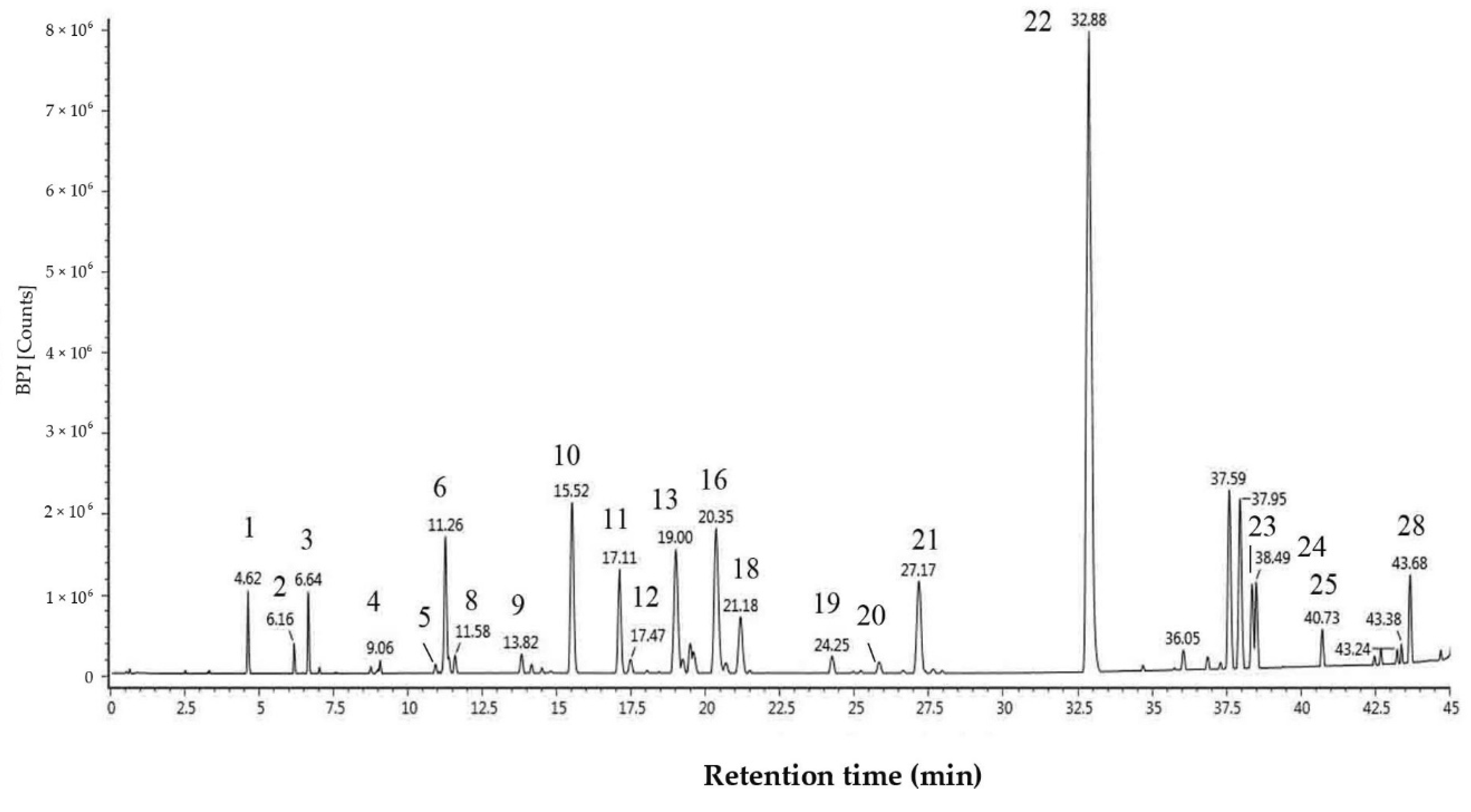
2.1. Chemical Composition
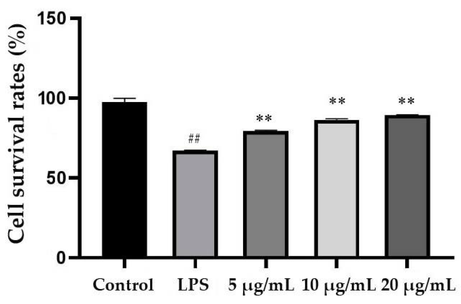
2.2. Effect of EEP-B on MACE Proliferation
2.3. Effect of EEP-B on the Levels of IL-6 and TNF-α
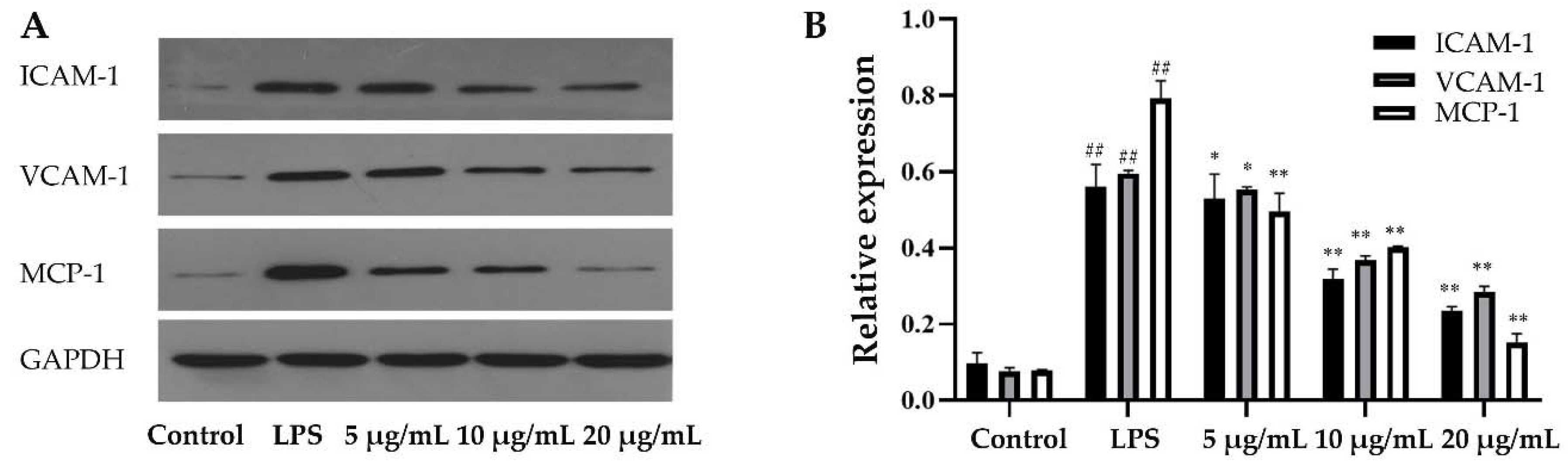
2.4. Effect of Propolis on the Levels of MCP-1, ICAM-1 and VCAM-1
3. Discussion
4. Materials and Methods
4.1. Preparation of Experimental Samples
4.2. Chemical Analysis of EEP-B
4.3. Cell Culture
4.4. Cell Viability Assay
4.5. ELISA Analysis of IL-6 and TNF-α Expression
4.6. Western Blot Analysis of ICAM-l, VCAM-1 and MCP-1 Expression
4.7. Statistical Analysis
5. Conclusions
Author Contributions
Funding
Conflicts of Interest
References
- Wioleta, P.; Renata, N.; Urszula, G.D.; Lemieszek, M.K.; Rzeski, W. LC-ESI-MS/MS Identification of Biologically Active Phenolic Compounds in Mistletoe Berry Extracts from Different Host Trees. Molecules 2017, 22, 624. [Google Scholar] [CrossRef]
- Oruç, H.H.; Sorucu, A.; Ünal, H.H.; Aydin, L. Effects of season and altitude on biological active certain phenolic compounds levels and partial standardization of propolis. Ank. Üniv. Vet. Fak. Derg. 2017, 64, 13–20. [Google Scholar]
- Valencia, D.; Alday, E.; Robles-Zepeda, R.; Garibay-Escobar, A.; Galvez-Ruiz, J.C.; Salas-Reyes, M.; Jiménez-Estrada, M.; Velazquez-Contreras, E.; Hernandez, J.; Velazquez, C. Seasonal effect on chemical composition and biological activities of Sonoran propolis. Food Chem. 2012, 131, 645–651. [Google Scholar] [CrossRef]
- Ristivojević, P.; Trifković, J.; Andrić, F.; Milojković-Opsenicab, D. Poplar-type Propolis: Chemical Composition, Botanical Origin and Biological Activity. Nat. Prod. Commun. 2015, 10, 1869–1876. [Google Scholar] [CrossRef]
- Lemos, M.; Barros, M.P.D.; Sousa, J.P.B.; Barros, M.P.D.; Sousa, J.P.B.; Filho, A.A.D.S.; Bastos, J.K.; Andrade, S.F.D. Baccharis dracunculifolia, the main botanical source of Brazilian green propolis, displays antiulcer activity. J. Pharm. Pharmacol. 2010, 59, 603–608. [Google Scholar] [CrossRef]
- Regueira-Neto, M.D.S.; Tintino, S.R.; Rolón, M.; Coronal, C.; Vega, M.C.; Balbino, V.D.Q.; Coutinho, H.D.D.M. Antitrypanosomal, antileishmanial and cytotoxic activities of Brazilian red propolis and plant resin of Dalbergia ecastophyllum (L) Taub. Food Chem. Toxicol. 2018, 2018, S0278691518302370. [Google Scholar]
- Cunha, M.G.D.; Rosalen, P.L.; Franchin, M.; de Alencar, S.M.; Ikegaki, M.; Ransom, T.; Beutler, J.A. Antiproliferative Constituents of Geopropolis from the Bee Melipona scutellaris. Planta Med. 2015, 82, 190–194. [Google Scholar] [CrossRef]
- Popova, M.; Dimitrova, R.; Al-Lawati, H.T.; Tsvetkova, I.; Najdenski, H.; Bankova, V. Omani propolis: Chemical profiling, antibacterial activity and new propolis plant sources. Chem. Cent. J. 2013, 7, 158. [Google Scholar] [CrossRef][Green Version]
- Park, Y.K.; Paredes-Guzman, J.F.; Aguiar, C.L.; Alencar, S.M.; Fujiwara, F.Y. Chemical constituents in Baccharis dracunculifolia as the main botanical origin of southeastern Brazilian propolis. J. Agric. Food Chem. 2004, 52, 1100–1103. [Google Scholar] [CrossRef]
- Chang, R.; Piló-Veloso, D.; Morais, S.A.L.; Nascimento, E.A. Analysis of a Brazilian green propolis from Baccharis dracunculifolia by HPLC-APCI-MS and GC-MS. Rev. Bras. De Farmacogn. 2008, 18, 549–556. [Google Scholar] [CrossRef]
- Rodrigues, D.M.; De Souza, M.C.D.; Arruda, C.; Pereira, R.A.S. The Role of Baccharis dracunculifolia and its Chemical Profile on Green Propolis Production by Apis mellifera. J. Chem. Ecol. 2019, 46, 150–162. [Google Scholar] [CrossRef] [PubMed]
- Matsuda, A.H.; Almeida-Muradian, L.B.D. Validated method for the quantification of artepillin-C in Brazilian propolis. Phytochem. Anal. 2008, 19, 179–183. [Google Scholar] [CrossRef] [PubMed]
- Barth, O.M.; Freitas, A.D.S.D.; Matsuda, A.H.; Almeida-Muradian, L.B.D. Botanical origin and Artepillin-C content of Brazilian propolis samples. Grana Palynol. 2013, 52, 129–135. [Google Scholar] [CrossRef]
- Cunha, M.G.D.; Franchin, M.; de Carvalho Galvão, L.C.; de Ruiz, A.L.; de Carvalho, J.E.; Ikegaki, M.; de Alencar, S.M.; Koo, H.; Rosalen, P.L. Antimicrobial and antiproliferative activities of stingless bee Melipona scutellaris geopropolis. BMC Complement. Altern. Med. 2013, 13, 23. [Google Scholar] [CrossRef] [PubMed]
- Hatano, A.; Nonaka, T.; Yoshino, M.; Ahn, M.R.; Tazawa, S.; Araki, Y.; Kumazawa, S. Antioxidant Activity and Phenolic Constituents of Red Propolis from Shandong. China. Food Sci. Technol. Res. 2012, 18, 577–584. [Google Scholar] [CrossRef]
- Funakoshi-Tago, M.; Okamoto, K.; Izumi, R.; Tago, K.; Yanagisawa, K.; Narukawa, Y.; Kiuchi, F.; Kasahara, T.; Tamura, H. Anti-inflammatory activity of flavonoids in Nepalese propolis is attributed to inhibition of the IL-33 signaling pathway. Int. Immunopharmacol. 2015, 25, 189–198. [Google Scholar] [CrossRef]
- Machado, J.L.; Assunçao, A.K.; da Silva, M.C.; Reis, A.S.; Costa, G.C.; Arruda, D.D.; Rocha, B.A.; Vaz, M.M.; Paes, A.M.; Guerra, R.N.; et al. Brazilian Green Propolis: Anti-Inflammatory Property by an Immunomodulatory Activity. Evid. Based Complement. Altern. Med. 2012, 2012, 157652. [Google Scholar] [CrossRef]
- Lima, L.D.C.; Andrade, S.P.; Campos, P.P.; Barcelos, L.S.; Soriani, F.M.; Moura, S.A.L.; Ferreira, M.A.N.D. Brazilian green propolis modulates inflammation, angiogenesis and fibrogenesis in intraperitoneal implant in mice. BMC Complement. Altern. Med. 2014, 14, 177. [Google Scholar] [CrossRef]
- Franchin, M.; Freires, I.A.; Lazarini, J.G.; Dias, N.B.; Cunha, M.G.D.; Colón, D.F.; Alencar, S.M.D.; Rosalen, P.L. The use of Brazilian propolis for discovery and development of novel anti-inflammatory drugs. Eur. J Med. Chem. 2018, 153, 49–55. [Google Scholar] [CrossRef]
- Fang, Y.; Sang, H.; Yuan, N.; Sun, H.; Yao, S.; Wang, J.; Qin, S. Ethanolic extract of propolis inhibits atherosclerosis in Apo E-knockout mice. Lipids Health Dis. 2013, 12, 123. [Google Scholar] [CrossRef]
- Messerli, S.M.; Ahn, M.R.; Kunimasa, K.; Yanagihara, M.; Tatefuji, T.; Hashimoto, K.; Mautner, V.; Uto, Y.; Hori, H.; Kumazawa, S.; et al. Artepillin C (ARC) in Brazilian green propolis selectively blocks oncogenic PAK1 signaling and suppresses the growth of NF tumors in mice. Phytother. Res. 2010, 23, 423–427. [Google Scholar] [CrossRef]
- Veiga, R.S.; Mendonça, S.D.; Mendes, P.B.; Paulino, N.; Mimica, M.J.; Netto, A.A.L.; Lira, I.S.; López, B.G.C.; Negrão, V.; Marcucci, M.C. Artepillin C and phenolic compounds responsible for antimicrobial and antioxidant activity of green propolis and Baccharis dracunculifolia DC. J. Appl. Microbiol. 2017, 122, 911–920. [Google Scholar] [CrossRef] [PubMed]
- Bueno-Silva, B.; Alencar, S.M.; Koo, H.; Ikegaki, M.; Silva, G.V.J.; Napimoga, M.H.; Rosalen, P.L. Anti-Inflammatory and Antimicrobial Evaluation of Neovestitol and Vestitol Isolated from Brazilian Red Propolis. J. Agric. Food Chem. 2013, 61, 4546–4550. [Google Scholar] [CrossRef] [PubMed]
- RMarkiewicz-Żukowska, R.; Car, H.; Naliwajko, S.K.; Sawicka, D.; Szynaka, B.; Chyczewski, L.; Isidorov, V.; Borawska, M.H. Ethanolic extract of propolis, chrysin, CAPE inhibit human astroglia cells. Adv. Med. Sci. 2012, 57, 208–216. [Google Scholar]
- Ohkura, N.; Takata, Y.; Ando, K.; Kanai, S.; Watanabe, E.; Nohira, T.; Atsumi, G. Propolis and its constituent chrysin inhibit plasminogen activator inhibitor 1 production induced by tumour necrosis factor-α and lipopolysaccharide. J. Apic. Res. 2015, 51, 179–184. [Google Scholar] [CrossRef]
- Szliszka, E.; Mertas, A.; Czuba, Z.P.; Król, W. Inhibition of Inflammatory Response by Artepillin C in Activated RAW264.7 Macrophages. Evid. Based Complement. Altern. Med. 2013, 2013, 735176. [Google Scholar] [CrossRef]
- Tennant, M.; Mcgeachie, J.K. Blood vessel structure and function: A brief update on recent advances. Aust. N. Z. J. Surg. 1990, 60, 747–753. [Google Scholar] [CrossRef] [PubMed]
- Watson, T.; Goon, P.K.Y.; Lip, G.Y.H. Endothelial progenitor cells, endothelial dysfunction, inflammation, and oxidative stress in hypertension. Antioxid. Redox Signal 2008, 10, 1079–1088. [Google Scholar] [CrossRef]
- Woo, M.S.; Park, J.S.; Choi, I.Y.; Kim, W.K. Inhibition of MMP-3 or -9 suppresses lipopolysaccharide-induced expression of proinflammatory cytokines and iNOS in microglia. J. Neurochem. 2008, 106, 770–780. [Google Scholar] [CrossRef]
- Lu, W.Q.; Qiu, Y.; Li, T.J.; Xia, T.; Sun, L.N.; Chen, W.S. Timosaponin B-II inhibits pro-inflammatory cytokine induction by lipopolysaccharide in BV2 cells. Arch. Pharmacal Res. 2009, 32, 1301–1308. [Google Scholar] [CrossRef]
- Falcao, S.I.; Vale, N.; Gomes, P.; Domingues, M.R.; Freire, C.; Cardoso, S.M.; Vilas-Boas, M. Phenolic Profiling of Portuguese Propolis by LC-MS Spectrometry: Uncommon Propolis Rich in Flavonoid Glycosides. Phytochem. Anal. 2013, 24, 309–318. [Google Scholar] [CrossRef] [PubMed]
- Gardana, C.; Scaglianti, M.; Pietta, P.; Simonetti, P. Analysis of the polyphenolic fraction of propolis from different sources by liquid chromatography-tandem mass spectrometry. J. Pharm. Biomed. Anal. 2007, 45, 390–399. [Google Scholar] [CrossRef] [PubMed]
- Midorikawa, K.; Banskota, A.H.; Tezuka, Y.; Nagaoka, T.; Matsushige, K.; Message, D.; Huertas, A.A.G.; Kadota, S. Liquid chromatography-mass spectrometry analysis of propolis. Phytochem. Anal. 2001, 12, 366–373. [Google Scholar] [CrossRef]
- Hayashi, K.; Komura, S.; Isaji, N.; Ohishi, N.; Yagi, K. Isolation of antioxidative compounds from Brazilian propolis: 3,4-dihydroxy-5-prenylcinnamic acid, a novel potent antioxidant. Chem. Pharm. Bull. 1999, 47, 1521–1524. [Google Scholar] [CrossRef][Green Version]
- Park, Y.K.; Alencar, S.M.; Aguiar, C.L. Botanical origin and chemical composition of Brazilian propolis. J. Agric. Food Chem. 2002, 50, 2502–2506. [Google Scholar] [CrossRef] [PubMed]
- Silva, B.B.; Rosalen, P.L.; Cury, J.A.; Ikegaki, M.; Souza, V.C.; Esteves, A.; Alencar, S.M. Chemical composition and botanical origin of red propolis, a new type of Brazilian propolis. Evid. Based Complement. Altern. Med. 2008, 5, 313–316. [Google Scholar] [CrossRef]
- Zhang, C.P.; Shen, X.G.; Chen, J.W.; Jiang, X.S.; Wang, K.; Hu, F.L. Artepillin C, is it a good marker for quality control of Brazilian green propolis? Nat. Prod. Res. 2017, 31, 2441–2444. [Google Scholar] [CrossRef]
- Xu, X.; Pu, R.; Li, Y.; Wu, Z.; Li, C.; Miao, X.; Yang, W. Chemical Compositions of Propolis from China and the United States and their Antimicrobial Activities Against Penicillium notatum. Molecules 2019, 24, 3576. [Google Scholar] [CrossRef]
- Szliszka, E.; Kucharska, A.Z.; Sokół-Łętowska, A.; Mertas, A.; Czuba, Z.P.; Król, W. Chemical Composition and Anti-Inflammatory Effect of Ethanolic Extract of Brazilian Green Propolis on Activated J774A.1 Macrophages. Evid. Based Complement. Altern. Med. 2013, 2013, 976415. [Google Scholar] [CrossRef]
- Righi, A.A.; Negri, G.; Salatino, A. Comparative chemistry of propolis from eight Brazilian localities. Evid. Based Complement. Altern. Med. 2013, 2013, 267878. [Google Scholar] [CrossRef]
- Spagnoli, L.G.; Bonanno, E.; Sangiorgi, G.; Mauriello, A. Role of Inflammation in Atherosclerosis. J. Nucl. Med. 2007, 48, 1800–1815. [Google Scholar] [CrossRef] [PubMed]
- Saxena, M.; Srivastava, N.; Banerjee, M. Association of IL-6, TNF-α and IL-10 gene polymorphisms with type 2 diabetes mellitus. Mol. Biol. Rep. 2013, 40, 6271–6279. [Google Scholar] [CrossRef] [PubMed]
- Ebaid, H.; Bashandy, S.A.E.; Alhazza, I.M.; Hassan, I.; Al-Tamimi, J. Efficacy of a Methanolic Extract of Adansonia digitata Leaf in Alleviating Hyperglycemia, Hyperlipidemia, and Oxidative Stress of Diabetic Rats. Biomed Res. Int. 2019, 2019, 2835152. [Google Scholar] [CrossRef] [PubMed]
- Nerstedt, A.; Johansson, A.; Andersson, C.X.; Cansby, E.; Smith, U.; Mahlapuu, M. AMP-activated protein kinase inhibits IL-6-stimulated inflammatory response in human liver cells by suppressing phosphorylation of signal transducer and activator of transcription 3 (STAT3). Diabetologia 2010, 53, 2406–2416. [Google Scholar] [CrossRef]
- Nishida, M.; Moriyama, T.; Ishii, K.; Takashima, S.; Yoshizaki, K.; Sugita, Y.; Yamauchi-Takihara, K. Effects of IL-6, adiponectin, CRP and metabolic syndrome on subclinical atherosclerosis. Clin. Chim. Acta 2007, 384, 99–104. [Google Scholar] [CrossRef]
- Jiang, Y.; Jiang, L.L.I.; Maimaitirexiati, X.M.Z.Y.; Zhang, Y.; Wu, L. Irbesartan attenuates TNF-α-induced ICAM-1, VCAM-1, and E-selectin expression through suppression of NF-κB pathway in HUVECs. Eur. Rev. Med. Pharm. Sci. 2015, 19, 3295–3302. [Google Scholar]
- Wu, S.; Xu, H.; Peng, J.; Wang, C.; Jin, Y.; Liu, K.; Sun, H.; Qin, J. Potent anti-inflammatory effect of dioscin mediated by suppression of TNF-α-induced VCAM-1, ICAM-1and EL expression via the NF-κB pathway. Biochimie 2015, 110, 62–72. [Google Scholar] [CrossRef]
- Chang, C.C.; Chu, C.F.; Wang, C.N.; Wu, H.T.; Bi, K.W.; Pang, J.H.S.; Huang, S.T. The anti-atherosclerotic effect of tanshinone IIA is associated with the inhibition of TNF-α-induced VCAM-1, ICAM-1 and CX3CL1 expression. Phytomed. Int. J. Phytother. Phytopharm. 2014, 21, 207–216. [Google Scholar] [CrossRef]
- Xuan, H.; Yuan, W.; Chang, H.; Liu, M.; Hu, F. Anti-inflammatory effects of Chinese propolis in lipopolysaccharide-stimulated human umbilical vein endothelial cells by suppressing autophagy and MAPK/NF-κB signaling pathway. Inflammopharmacology 2018, 27, 561–571. [Google Scholar] [CrossRef]
- Bueno-Silva, B.; Franchin, M.; Alves, C.D.F.; Denny, C.; Colón, D.F.; Cunha, T.M.; Alencar, S.M.; Napimoga, M.H.; Rosalen, P.L. Main pathways of action of brazilian red propolis on the modulation of neutrophils migration in the inflammatory process. Phytomedicine 2016, 23, 1583–1590. [Google Scholar] [CrossRef]
- Öğretmen, F.; Inanan, B.E.; Öztürk, M. Protective effects of propolis on cryopreservation of common carp (Cyprinus carpio) sperm. Cryobiology 2014, 68, 107–112. [Google Scholar] [CrossRef] [PubMed]
- Bueno-Silva, B.; Rosalen, P.L.; Alencar, S.M.; Maye, M.P.A. Anti-inflammatory mechanisms of neovestitol from Brazilian red propolis in LPS-activated macrophages. J. Funct. Foods 2017, 36, 440–447. [Google Scholar] [CrossRef]
- Daleprane, J.B.; Freitas, V.D.S.; Pacheco, A.; Rudnicki, M.; Faine, L.A.; Dörr, F.A.; Ikegaki, M.; Salazar, L.A.; Ong, T.P.; Abdalla, D.S.P. Anti-atherogenic and anti-angiogenic activities of polyphenols from propolis. J. Nutr. Biochem. 2012, 23, 557–566. [Google Scholar] [CrossRef] [PubMed]
- Nader, M.A.; El-Agamy, D.S.; Suddek, G.M. Protective effects of propolis and thymoquinone on development of atherosclerosis in cholesterol-fed rabbits. Arch. Pharmacal Res. 2010, 33, 637–643. [Google Scholar] [CrossRef] [PubMed]
- Hofmann, N.; Lachnit, N.; Streppel, M.; Witter, B.; Neiss, W.F.; Guntinas-Lichius, O.; Angelov, D.N. Increased expression of ICAM-1, VCAM-1, MCP-1, and MIP-1α by spinal perivascular macrophages during experimental allergic encephalomyelitis in rats. BMC Immunol. 2002, 3, 11. [Google Scholar] [CrossRef]
- Fan, J.; Li, X.; Zhong, L.; Tong, H.; Di, J.; Liu, F.; Zhao, H.H.; Bai, S.L. MCP-1, ICAM-1 and VCAM-1 are present in early aneurysmal dilatation in experimental rats. Folia Histochem. Et Cytobiol. 2010, 48, 455–461. [Google Scholar] [CrossRef]
Sample Availability: Samples of the compounds are not available from the authors. |


| No | tR (min) | λmax (nm) | Selected Ion | Formula | Measured Mass | Calculated Mass | Mass Error | MS/MS Fragmentation | Compound Name | Relative Area (%) |
|---|---|---|---|---|---|---|---|---|---|---|
| 1 | 4.62 | 237,310 | [M−H] | C9H8O3 | 163.0400 | 163.0395 | 0.5 | 163.0400, 119.0502, 93.0368 | p-Coumaric acid a,c | 1.31% |
| 2 | 6.18 | 324 | [M−H] | C25H24O12 | 515.1191 | 515.1190 | 0.1 | 353.0868, 91.0554, 179.0342, 135.0444 | Dicaffeoylquinic acid isomer a,d | 0.48% |
| 3 | 6.64 | 324 | [M−H] | C25H24O12 | 515.1193 | 515.1190 | 0.3 | 353.0871, 191.0555, 179.0343, 135.0445 | Dicaffeoylquinic acid a,d | 1.31% |
| 4 | 9.06 | 253,372 | [M−H] | C15H10O7 | 301.0365 | 301.0348 | 1.7 | 243.02886, 151.00304 | Quercetin a,c | 0.29% |
| 5 | 10.96 | 292 | [M−H] | C15H12O5 | 271.0613 | 271.0606 | 0.7 | 271.0613, 253.0504 | Pinobanksin a,c | 0.19% |
| 6 | 11.26 | 292 | [M−H] | C16H14O6 | 301.0714 | 301.0712 | 301.0714, 283.0603, 268.0367, 152.0107 | 4′-methoxy Pinobanksin a,e | 4.10% | |
| 7 | 11.40 | 265,365 | [M−H] | C15H10O6 | 285.0400 | 285.0399 | 0.1 | 285.0400, 227.03402 | Kaempferol a,b | 0.30% |
| 8 | 11.58 | 251,365 | [M−H] | C16H11O7 | 315.0505 | 315.0505 | 0 | 315.0505, 300.0266, 243.0292 | Quercetin-methyl ether a | 0.43% |
| 9 | 13.80 | 326 | [M−H] | C14H16O4 | 247.0971 | 247.0970 | 0.1 | 247.0971, 203.10683 | 5-isoprenyl caffeic acid a,f | 0.53% |
| 10 | 15.52 | 315 | [M−H] | C14H16O3 | 231.1025 | 231.1021 | 0.4 | 187.1121, 132.0575 | 3-isoprenyl-p-Coumaric acid a,e | 6.19% |
| 11 | 17.11 | 313 | [M−H] | C19H24O4 | 315.1597 | 315.1596 | 0.1 | 315.1597, 271.1692, 253.1591, 198.1043 | 3-hydroxy-2,2-dimethy-8-prenylchromane-6-propenoic a,e | 3.39% |
| 12 | 17.47 | 324 | [M−H] | C14H16O4 | 247.0971 | 247.0970 | 0.1 | 247.0971, 179.0341, 161.0236, 135.0443 | Caffeic acid isoprenyl ester a,c | 0.46% |
| 13 | 19.00 | 286 | [M−H] | C15H12O4 | 255.0662 | 255.0657 | 0.5 | 213.0552, 151.0031, 107.0134 | Pinocembrin a,c | 5.56% |
| 14 | 19.24 | 318 | [M−H] | C19H24O4 | 315.1597 | 315.1596 | 0.1 | 271.1675, 253.1589, 198.1041 | 3-hydroxy-2,2-dimethy-8-preylchromane-6-propenoic isomer a,e | 0.45% |
| 15 | 19.65 | 265,361 | [M−H] | C15H10O5 | 269.0456 | 269.0450 | 0.6 | 211.03914, 145.0288, 117.0340 | Galangin a,b | 0.53% |
| 16 | 20.35 | 265,365 | [M−H] | C16H11O6 | 299.0557 | 299.0556 | 0.1 | 284.0320, 151.0029, 107.0132 | Kaempferide a,c | 7.06% |
| 17 | 20.68 | 292 | [M−H] | C17H14O6 | 313.0745 | 313.0712 | 3.3 | 253.0505, 119.0498 | Pinobanksinr-3-O-acetate a,c | 0.37% |
| 18 | 21.18 | 269, 363 | [M−H] | C17H14O7 | 329.0665 | 329.0661 | 0.4 | 314.0424, 299.0180, 271.02431 | Quercetin-dimethyl ether a,c | 2.52% |
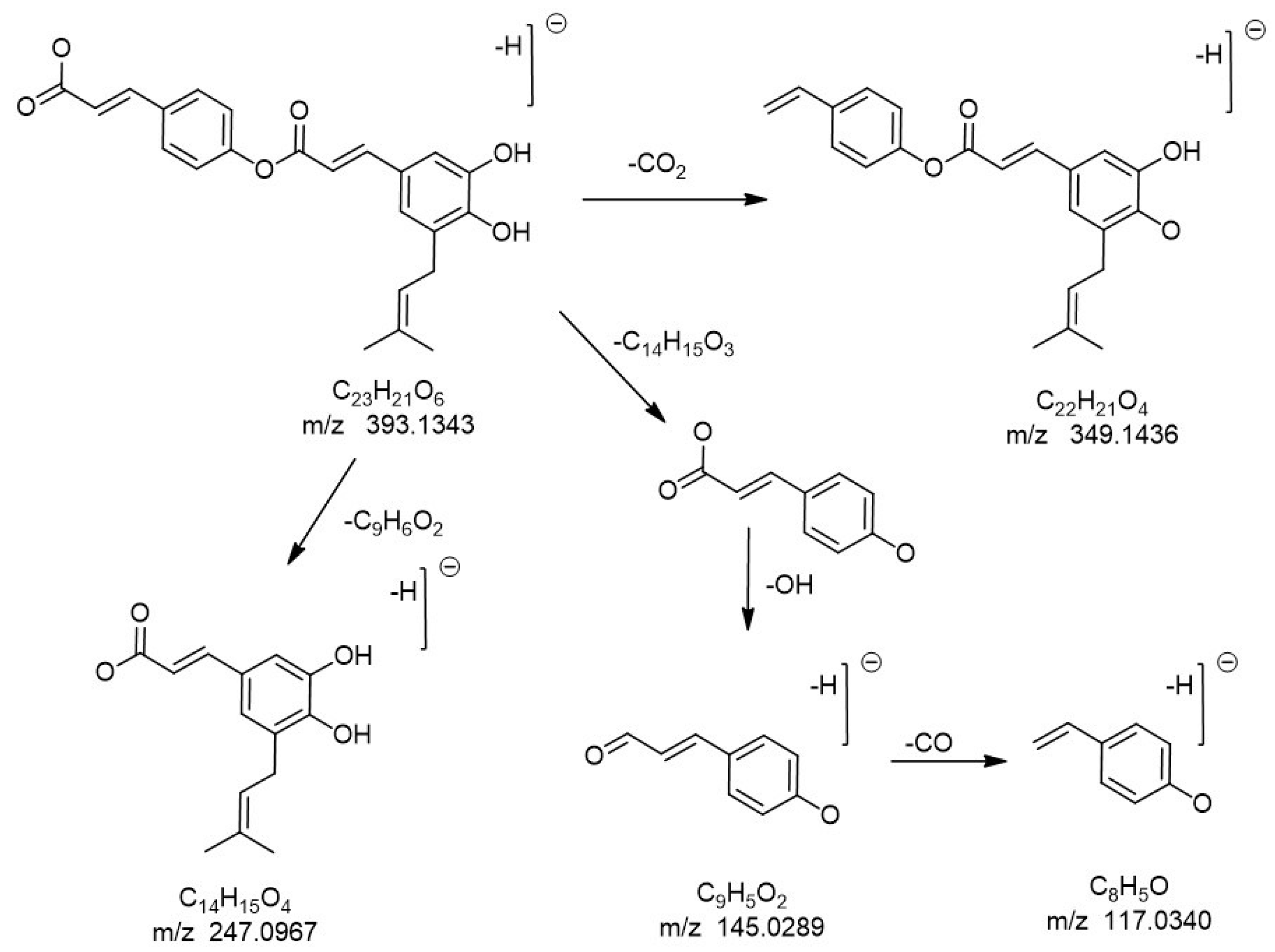
| 19 | 24.25 | 319 | [M−H] | C23H22O6 | 393.1343 | 393.1338 | 0.5 | 349.1436, 247.0967, 163.0392, 145.0289, 117.0340 | 5-isoprenyl caffeic acid-p-coumaric acid ester a | 0.62% |
| 20 | 25.84 | 318 | [M−H] | C19H24O4 | 315.1600 | 315.1596 | 0.5 | 245.1170, 201.1279, 146.0731 | 3-hydroxy-2,2-dimethy-8-preylchromane-6-propenoic isomer a,e | 0.43% |
| 21 | 27.17 | 287 | [M−H] | C19H24O3 | 299.1647 | 299.1647 | 0 | 255.1748, 187.1117, 161.0601 | Diisoprenyl -p-Coumaric acid isomer a,e | 4.49% |
| 22 | 32.88 | 314 | [M−H] | C19H24O3 | 299.1652 | 299.1647 | 0.5 | 255.1750, 201.1238, 145.0652 | Artepillin C (3,5-diisopentenyl-4-hydroxycinnamic acid) a,e | 35.68% |
| 23 | 38.36 | 279 | [M−H] | C23H24O4 | 363.1588 | 363.1596 | −0.8 | 231.1021, 187.1122, 149.0621 | 3-Prenyl-4-(dihydrocinnamoyloxy)-cinnamic acid a,d | 2.44% |
| 24 | 38.51 | 308 | [M−H] | C28H32O5 | 447.2175 | 447.2171 | 0.4 | 297.1492, 253.1592, 198.1041, 149.0603, 105.0704 | (E)-3-[2,3-dihydro-2-(1-methylethenyl)-7-prenyl-5-benzofuranyl]-2-propenoic acid a,d | 2.27% |
| 25 | 40.73 | - | [M−H] | C30H48O4 | 471.3485 | 471.3474 | 1.1 | 517.3542, 471.3485, 407.3318 | triterpenes | 0.94% |
| 26 | 42.48 | - | [M−H] | C30H46O4 | 469.3323 | 469.3318 | 0.5 | 515.3377, 469.3323, 423.3260 | triterpenes | 0.17% |
| 27 | 43.25 | - | [M−H] | C30H48O4 | 471.3480 | 471.3474 | 0.6 | 517.3436, 471.3481, 339.1994 | triterpenes | 0.24% |
| 28 | 43.68 | [M−H] | C30H46O4 | 469.3322 | 469.3318 | 0.4 | 515.3377, 469.3322, 407.3314 | triterpenes | 2.26% |
© 2020 by the authors. Licensee MDPI, Basel, Switzerland. This article is an open access article distributed under the terms and conditions of the Creative Commons Attribution (CC BY) license (http://creativecommons.org/licenses/by/4.0/).
Share and Cite
Xu, X.; Yang, B.; Wang, D.; Zhu, Y.; Miao, X.; Yang, W. The Chemical Composition of Brazilian Green Propolis and Its Protective Effects on Mouse Aortic Endothelial Cells against Inflammatory Injury. Molecules 2020, 25, 4612. https://doi.org/10.3390/molecules25204612
Xu X, Yang B, Wang D, Zhu Y, Miao X, Yang W. The Chemical Composition of Brazilian Green Propolis and Its Protective Effects on Mouse Aortic Endothelial Cells against Inflammatory Injury. Molecules. 2020; 25(20):4612. https://doi.org/10.3390/molecules25204612
Chicago/Turabian StyleXu, Xiaolan, Bo Yang, Danfeng Wang, Yuxuan Zhu, Xiaoqing Miao, and Wenchao Yang. 2020. "The Chemical Composition of Brazilian Green Propolis and Its Protective Effects on Mouse Aortic Endothelial Cells against Inflammatory Injury" Molecules 25, no. 20: 4612. https://doi.org/10.3390/molecules25204612
APA StyleXu, X., Yang, B., Wang, D., Zhu, Y., Miao, X., & Yang, W. (2020). The Chemical Composition of Brazilian Green Propolis and Its Protective Effects on Mouse Aortic Endothelial Cells against Inflammatory Injury. Molecules, 25(20), 4612. https://doi.org/10.3390/molecules25204612