Article

Crystallographic and NMR Investigation of Ergometrine and Methylergometrine, Two Alkaloids from Claviceps Purpurea

1
Department of Pharmaceutical Sciences, University of Milan, via L. Mangiagalli, 25, 20133 Milano, Italy
2
Department of Medical Biotechnology and Translational Medicine, University of Milan, Via Saldini 50, 20133 Milano, Italy
3
Chemical-Pharmaceutical Consulting and IP Management, Viale Giovanni da Cermenate 58, 20141 Milano, Italy
*
Author to whom correspondence should be addressed.
Molecules 2020, 25(2), 331; https://doi.org/10.3390/molecules25020331
Submission received: 11 December 2019 / Revised: 9 January 2020 / Accepted: 10 January 2020 / Published: 14 January 2020

Abstract

:
Ergometrine and methylergometrine are two alkaloids that are used as maleate salts for the prevention and control of postpartum hemorrhage. Although the two molecules have been known for a long time, few and discordant crystallographic and NMR spectroscopic data are available in the literature. With the aim of providing more conclusive data, we performed a careful NMR study for the complete assignment of the 1H, 13C, and 15N NMR signals of ergometrine, methylergometrine, and their maleate salts. This information allowed for a better definition of their conformational equilibria. In addition, the stereochemistry and the intermolecular interactions in the solid state of the two maleate salts were deeply investigated by means of single-crystal X-ray diffraction, showing the capability of these derivatives to act as both hydrogen-bond donors and acceptors, and evidencing a correlation between the number of intermolecular interactions and their different solubility.

Graphical Abstract

1. Introduction

The fungus Claviceps purpurea (commonly known as ergot) is a plant pathogen that produces a family of toxic alkaloids endowed with pharmacological properties [1,2]. These ergot alkaloids are derivatives of the tetracyclic ergoline 1 (Figure 1) and can be chemically classified by the nature of the substituent at the 8-position.
From a therapeutic point of view, the d-lysergic acid amides are in a prominent position among the ergot alkaloids. The d-lysergic acid 2 carries a carboxyl group at the 8β position (cis with respect to the H-5) and a 9–10 double bond (Figure 2). The presence of the double bond is responsible for the easy and spontaneous isomerization of the carboxyl substituent at the C-8 stereocenter, leading to mixtures of d-lysergic and d-isolysergic acid 3. In the tetracyclic moiety of compounds 2 and 3, two stereocenters are present, namely at the 5 and 8 positions. The stereocenter at the 5 position presents an R configuration, while the substituent at the 8 position can be either in the α-configuration (trans with respect to the H-5) or in the β-configuration (cis with respect to the H-5). The α-isomer is usually indicated by the prefix iso- or by the ending –inine. The β-isomers are usually endowed with a significant biological activity.
Among the d-lysergic derivatives, the natural amide ergometrine (ergonovine) 4 and the semisynthetic methylergometrine 5 (methylergonovine) (Figure 2) are endowed with uterotonic activity, and for this reason, their maleate salts are administered in the third stage of labor for the prevention of postpartum hemorrhage [3,4].
Although ergometrine 4 was isolated in 1932 [5] and the first synthesis of methylergometrine 5, starting from the lysergic azide, was published in 1943 [6], only a few spectroscopic and crystallographic studies are reported in the literature. In two 2017 Chinese patents [7,8], the 1H NMR data of the ergometrine 4 are reported but not assigned; in 1974 [9], the assigned 13C NMR (60 MHz) data were reported. In the case of ergometrine maleate, the XRD and the single-crystal X-ray diffraction [10] are reported, and only the 1H NMR (400 MHz) characterization is available [11]. Methylergometrine 5 was analyzed by 1H NMR (values not assigned) in a 2014 WO patent [12]; the corresponding maleate was characterized by XRD, IR, and DSC in a 2015 Chinese patent [13] and by synchrotron powder diffraction data [14]. These incomplete (and sometimes contradictory) NMR and crystallographic analyses prompted us to gather new and more conclusive data for the two amides, 4 and 5. Considering the easy epimerization of these compounds, we decided to expand our investigations to their maleate salts, 6 and 7, as well. Indeed, ergometrine 4 and methylergometrine 5 are always obtained as mixtures of epimers, while the corresponding maleate salts can be obtained as pure β-isomers by simple crystallization.
In a continuing effort to fully characterize molecules that exert important therapeutic properties [15,16,17], a complete NMR (1H, 13C, and 15N) characterization of compounds 4, 5, 6, and 7 was carried out. Moreover, a detailed crystallographic investigation was performed on 6 and 7 in order to explore their conformational features. In addition, we carefully analyzed the hydrogen-bonding interactions of these molecules, as the earlier report did not account for their supramolecular aggregation.

2. Results and Discussion

Ergometrine 4 and methylergometrine 5 were obtained by treatment of the corresponding commercially available maleates, 6 and 7, with sodium hydrogen carbonate as described in the Materials and Methods section.

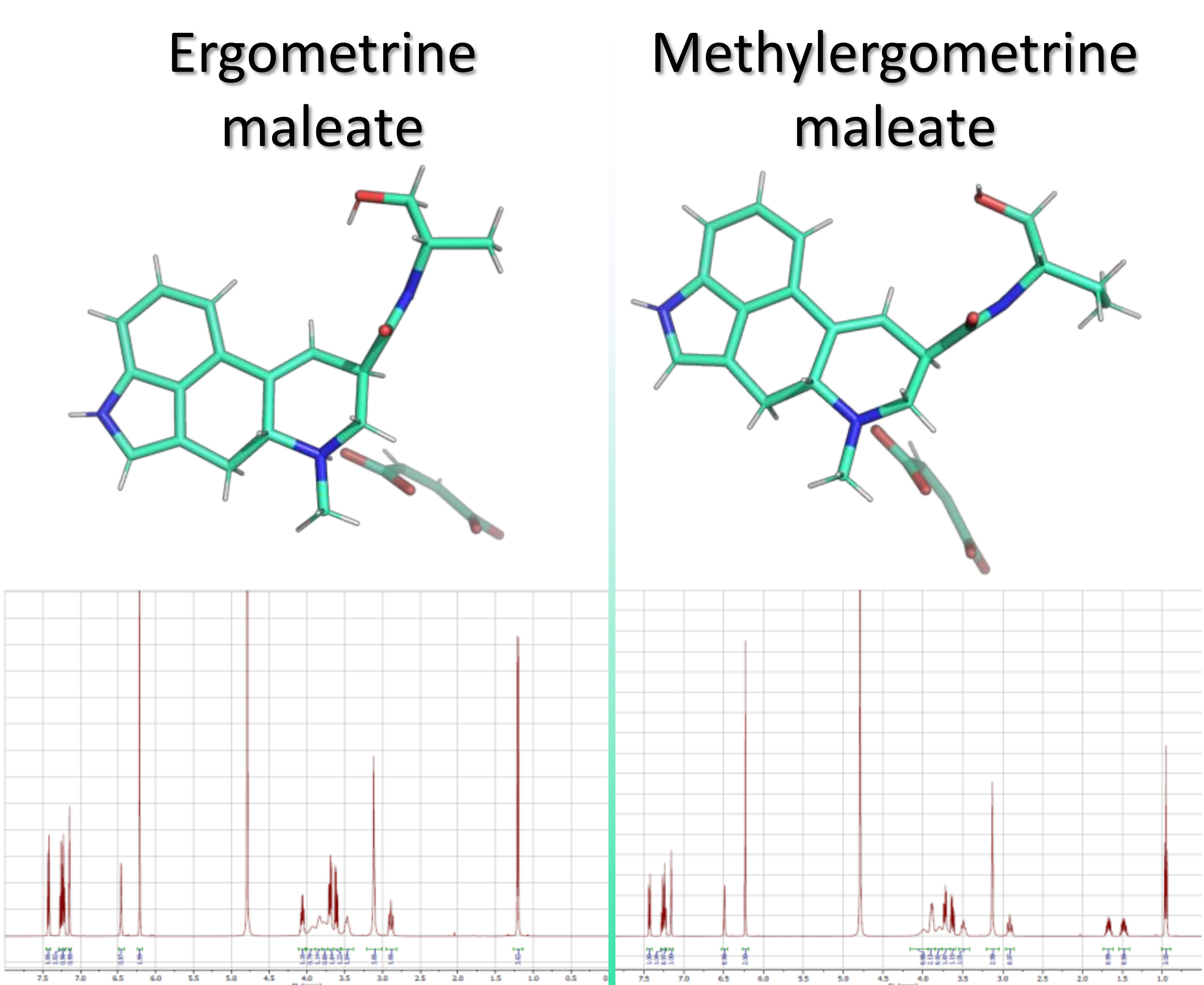
2.1. NMR Spectroscopy

The NMR study was carried out on 4 and 5 and on their maleate salts 6 and 7 (the clinically administered derivatives). Compounds 4 and 5 were dissolved in DMSO-d6, while compounds 6 and 7 were dissolved in D2O. Although in both solvents the 1H NMR spectra of 6 and 7 present broadened signals and a slight concentration dependence of chemical shifts, D2O was chosen in these cases due to more resolved NMR spectra. 1H, 13C, and 15N resonances (Table 1, Table 2 and Table 3) were unambiguously assigned combining information from 1D and 2D NMR (COSY, NOESY, HSQC, 1H-13C HMBC, and 1H-15N HMBC) experiments.
The proton assignments were accomplished using the general knowledge of chemical shift dispersion with the aid of the COSY, HSQC, and NOESY experiments. Starting from the characteristic resonance of H-9 (6.34, 6.35, 6.46, and 6.49 ppm for 4, 5, 6, and 7, respectively), we were able to assign the resonances of all the other protons, especially the resonance of H-8. The interpretation of the 1H NMR spectra of compounds 4 and 5 was further facilitated by the resonances of exchangeable protons H-1, CH2OH, and CONH.
The NOESY experiments were performed to integrate structural data with stereochemical information. Despite the poorly resolved 2D spectra of NOESY experiments obtained for 6 and 7 due to the broad nature of the D-ring proton signals, some information about the stereochemistry and conformation of the D-ring was collected by observing the NOE correlation of the methyl group bound to the nitrogen atom at the 6 position. This methyl group interacts sterically with H-5β, H-7β, H-7α, and H-4β, indicating—as previously described by Kidric et al. [18] for 9,10-unsatured ergolines dissolved in solvents such as DMSO and water—that the D-ring was preferentially in its D1 8R half-chair conformation. The very weak NOE correlation of N–CH3 with H-8 α and H-4 α could be explained by the existence of a conformational equilibrium in solution between D1 8R and D2 8R half-chair conformations (Figure 3). When the D-ring is in its D2 8R configuration, the methyl group is in the α position, which results in a feeble NOE effect. The existence of this conformational equilibrium in aqueous solution also explains the broad nature of the C- and D-ring proton signals.
The analysis of NOESY spectra of the free bases, 4 and 5, gave us a clear picture of the spatial interactions of protons at the 4 position with H-5β and of protons at the 7 position with H-8α. The NOE correlations of H-5β (3.00–2.93 ppm) with one of the two protons at the 4 position (3.46 ppm) and of H-8α (3.40–3.36 ppm) with one of the two protons at the 7 position (3.00 ppm for 4 and 3.02 ppm for 5) allowed us to unequivocally identify H-4β and H-7α protons. The absence of spatial interactions between the H-8 and H-5 protons was a clue, but not an absolute proof, of their position on opposite faces of the D-ring. When N-CH3 (2.44 ppm for 4; 2.45 ppm for 5) was irradiated, NOE enhancement was observed for H-5β, suggesting that the analyzed 4 and 5 in DMSO-d6 were in their D1 8R half-chair conformation. The CH, CH2, and CH3 carbon atoms were assigned based on chemical shift analysis and confirmed by the HSQC experiment. In particular, the predicted deshielding of C-5 and C-7 was observed, both near the nitrogen atom in position 6, further confirming the right 1H NMR assignment of the protons bound to these carbon atoms. In the 13C NMR spectra of compounds 6 and 7, the broad nature and low intensity of the C-4, C-8, N-CH3, C-7, C-5, and C-9 signals can be explained by the previously reported conformational equilibrium between the D1 8R and D2 8R half-chair conformations. The quaternary carbon atoms were unambiguously assigned using the information obtained from 1H-13C HMBC experiments (Table 4). Following this approach, the 13C resonance assignments of 4 were in accordance with the previously reported ones [9].
1H-15N HMBC experiments were performed in order to assign the resonances of the three chemically different nitrogen atoms in the examined compounds and allowed us to confirm their hypothesized structure. For compounds 6 and 7, the indole nitrogen N-1 (−253.3 ppm) couples with H-2, H-14, and H-13, the amidic nitrogen (−248.3 ppm for 6; −251.4 ppm for 7) couples with the protons present on the amidic moiety, and curiously but not unexpectedly, N-6 (−332.8 ppm for 6; −332.7 ppm for 7) couples very weakly only with H-4α, which gives a well-resolved signal in the 1H NMR spectrum. For compounds 4 and 5, the indole nitrogen N-1 (−251.1 ppm for 4; −251.0 ppm for 5) couples with H-1, H-2, H-14, and H-13, the amidic nitrogen (−255.5 ppm for 4; −258.1 ppm for 5) couples with the protons of the amidic moiety, and N-6 (−340.3 ppm for 4; −340.2 ppm for 5) couples with N-CH3, H-7β, and H-5.

2.2. X-ray Analysis

Since the structure adopted by a given compound upon crystallization could exert a profound effect on the solid-state properties of the system, we decided to perform a crystallographic analysis of derivatives 6 and 7. Their X-ray molecular structures are presented, as ORTEP views [19], in Figure 4.
The maleate salt of ergometrine, compound 6, crystallized in the orthorhombic P212121 system, as previously reported in the literature [20], while the maleate salt of methylergometrine 7 crystallized in a different space group, namely the monoclinic P21 [14].
The dianionic maleate group bridges two neighboring alkaloid molecules, whose conformation is mainly determined by a central rigid core, consisting of an indole plane connected to a six-membered ring and a tetra-hydro-pyridine. Since the absolute configuration of C5 of the starting material was known, it was possible to determine by X-ray diffraction the relative configuration of C8 in the two crystal structures. The single crystal diffraction data unambiguously confirmed that the substituent at the 8-position is equatorially oriented (cis with respect to H-5), and with respect to an absolute configuration R at the 5-position, this relative orientation means the R configuration of C8. The cores of the two molecules are closely related: the ring C of the central skeleton is in a slightly distorted envelope conformation, as indicated by the puckering parameters [21] QT (total puckering amplitude) = 0.3803(1)Å, θ (torsion angle) = 53.8(1)° for 6, and QT = 0.3630(1)Å, θ = 51.8(1)° for 7, whereas ring D adopts a distorted chair conformation, with puckering coordinates [21] QT = 0.5208 (1)Å, θ = 126.1(1)° for 6 and QT = 0.5170 (1)Å, θ = 127.7(1)° for 7, which is in agreement with the previously reported structures. The conformation of the tetracyclic system is also conserved in the ergometrinine crystal structure [22], which is the biologically inactive isomer of 4.
The crystal lattice of both structures is dominated by hydrogen bonds (Figure 5); in 6, in particular, the negatively charged carboxylate groups of the anions are engaged with a hydrogen belonging to the positively charged amino groups of the cations; moreover, bifurcated hydrogen bonds linking O1 with N1 and O2 of symmetry-related ergometrine molecules contribute to stabilize the crystals. In addition, Cπ-H···O contacts between neighboring anions give rise to the formation of molecular chains along the a-axis of the unit cell, further consolidating the crystal packing. In 7, we revealed some differences with respect to the previously reported structure [14]: in particular, the absence of the intermolecular interaction between the amidic oxygen and the NH of ring B and the presence of Cπ-H···O contacts between neighboring anions, which give rise to the formation of molecular chains along the b-axis of the unit cell.
The hydrogen bond geometry of 6 and 7, summarized in Table 5, evidenced the capacity of these derivatives to act as both hydrogen-bond donors and acceptors.
Moreover, the crystallographic analysis suggests that the lower solubility of 6 in the crystallization mixture (1:1 acetone/ethanol) is caused by the presence of an extensive system of hydrogen bonding. Therefore, the higher solubility of 7 may be correlated to the reduced number of intermolecular interactions found in the solid state, leading to a decreased crystal lattice energy. The less soluble 6 crystallizes in a compact structure as prisms, while the poor-quality crystals of 7 (obtained from a 1:1 ethanol/water crystallization mixture) are assembled in transparent platelets, showing a reasonable correlation between the different morphological aspect of the crystals and their solubility.

3. Materials and Methods

3.1. Chemistry

Ergometrine maleate and methylergometrine maleate, all reagents and solvents were purchased from Sigma-Aldrich (Merck, Kenilworth, NJ, USA).

3.1.1. Ergometrine 4 from the Corresponding Maleate 6

To a suspension of commercial ergometrine maleate (50 mg, 0.11 mmol) in ethyl acetate (10 mL), a saturated aqueous sodium bicarbonate solution (10 mL) was added. After stirring at room temperature in the dark for 30 min, the layers were separated. The aqueous phase was extracted with ethyl acetate (2 × 10 mL). The collected organic phases were dried over sodium sulfate and filtered. After solvent removal under reduced pressure, a white solid was obtained (35 mg, 0.11 mmol, 96%).
[α]D20 = −17.9° (c = 1; pyridine) lit. [6] −16°

3.1.2. Methylergometrine 5 from the Corresponding Maleate 7

Methylergometrine 5 was obtained following the previously described procedure for ergometrine 4, starting from commercial methylergometrine maleate (50 mg, 0.11 mmol). A white solid was obtained (34 mg, 0.10 mmol, 91%).
[α]D20 = −49.8° (c = 0.4; pyridine) lit. [6] −45°

3.2. NMR Spectroscopy

NMR spectra were recorded on a Bruker AVANCE 500 spectrometer (Bruker, Billerica, MA, USA) equipped with a 5 mm broadband inverse (BBI) detection probe with field z-gradient operating at 500.13, 125.76, and 50.69 MHz for 1H, 13C, and 15N respectively. NMR spectra were recorded at 300 K for compounds 4 and 5 in DMSO-d6 (isotopic enrichment 99.9 atom % D), for their maleate salts, 6 and 7, in D2O (isotopic enrichment 99.9 atom % D) solution. The data were collected and processed by XWIN-NMR software (version 3.5, Bruker, Billerica, MA, USA) running on a PC with Microsoft Windows 7. The samples (10 mg) were dissolved in the appropriate solvent (0.75 mL) in a 5 mm NMR tube. The acquisition parameters for 1D were as follows: 1H spectral width of 5000 Hz and 32 K data points providing a digital resolution of ca. 0.305 Hz per point, relaxation delay 10 s; 13C spectral width of 29,412 Hz, and 64 K data points providing a digital resolution of ca. 0.898 Hz per point, relaxation delay 2 s. The experimental error in the measured 1H-1H coupling constants was ±0.5 Hz. Chemical shifts (δ) of the 1H NMR and 13C NMR spectra are reported in ppm using the central peak of DMSO-d6 signals (2.50 ppm for 1H; 39.52 ppm for 13C) for compounds 4 and 5 and using methanol as external reference in the spectra recorded in D2O (signals of CH3OH in D2O: 3.34 ppm for 1H; 49.50 ppm for 13C). Methylergometrine maleate D2O solution showed a pH of 4.5, while ergometrine maleate D2O solution showed a pH of 4.2. The splitting pattern abbreviations are as follows: s, singlet; d, doublet; t, triplet; q, quartet; m, multiplet; and br, broad signal. For two-dimensional experiments, standard Bruker microprograms using gradient selection (gs) were applied. Gs-COSY-45 and phase sensitive gs-NOESY experiments were acquired with 512 t1 increments; 2048 t2 points; and a spectral width of 11.0 ppm. The gs-NOESY experiments were performed with a mixing time of 0.800 s on samples degassed under a flush of argon in a screwcap sample tube. There were not significant differences in the results obtained at different mixing times (0.5–1.5 s). The acquisition data for gs-HSQC and gs-HMBC experiments were acquired with 512 t1 increments, 2048 t2 points, and a spectral width of 11.0 ppm for 1H and 200 ppm for 13C. Delay values were optimized for 1JC,H 140 Hz and nJC,H 8.0 Hz. For 1H-15N HMBC experiments, nitromethane was used as the external reference, referencing the resonance of its 15N at 0.00 ppm. Samples concentration: 10 mg/0.75 mL corresponding to 41, 39, 30, and 29 mM for 4, 5, 6, and 7, respectively. The acquisitions were performed setting an acquisition time of 0.5 s, a delay between scans of 10 s, a 1JN,H value of 90.0 Hz, and a nJN,H value of 1.9 Hz. This last parameter was set after several attempts between 1 and 10 Hz. Total 1H-15N HMBC experimental time: 64 h.
The NMR spectra are available in the Supplementary Materials section.

3.3. X-ray Crystallography

Crystals of 6 were obtained as pale-yellow prisms from a 1:1 acetone/ethanol solution at room temperature. Poor-quality crystals of 7 were obtained, after many attempts, as colorless elongated platelets from a 1:1 ethanol/water solution at room temperature. All experiments were carried out in the dark to avoid degradation.
Diffraction data for the crystals of 6 and 7 were collected with a Bruker-AXS CCD-based three-circle diffractometer, working at ambient temperature with graphite-monochromatized Mo-Kα X-ray radiation (λ = 0.7107 Å).
X-ray diffraction data in the θ range 2–25° were collected acquiring three sets of 600 bidimensional CCD frames with the following operative conditions: omega rotation axis, scan width 0.3°, acquisition time 40 s, sample-to-detector distance 60 mm, and phi angle fixed at three different values (0°, 120°, 240°) for the three different sets.
Omega-rotation frames were processed with the SAINT software [23] for data reduction (including intensity integration, background, Lorentz, and polarization corrections) and for the determination of accurate unit-cell dimensions, which were obtained by least-squares refinement of the positions of 2023 independent reflections with I > 10σ(I) in the θ range 2–19°. Absorption effects were empirically evaluated by the SADABS software [24], and absorption correction was applied to the data (0.87 and 0.99 min and max transmission factor). The structures were solved by direct methods (SIR-14) [25] and completed by iterative cycles of full-matrix least squares refinement on F02 and DF synthesis using the WingX.v2014.1 [26] program. The hydrogen atoms bonded to carbon were included at geometrically calculated positions and refined using a riding model. Uiso(H) is defined as 1.2 Ueq of the parent carbon atoms for the phenyl and methylene residues and 1.5 Ueq of the parent carbon atoms for the methyl group. Crystallographic data has been deposited with the Cambridge Crystallographic Data Centre under CCDC deposition numbers 1970319 (6) and 1970320 (7). Copies of this information may be obtained free of charge from the Director, CCDC, 12 Union Road, Cambridge CB2 1EY, UK (fax: +44-1223-336033; e-mail: [email protected] or www:http://www.ccdc.cam.ac.uk).
Crystal data for 6: C23H27N3O6, Mr = 441.47 g/mol, Orthorhombic, Space group P212121, a = 5.7139(7) Å, b = 12.3104(1) Å, c = 33.2683(4) Å, V = 2340(1) Å3, Z = 4, Dcalc = 1.253 Mg/m3, F(000) = 936, R = 0.057 (reflections collected/unique = 4113/2921), wR2 = 0.146, T = 294(2)K, and GOF = 1.026. The reflections were collected in the range 1.22° ≤ θ ≤ 25.05° (limiting indices = −6 ≤ h ≤ 6, −14 ≤ k ≤ 14, −39 ≤ l ≤ 39) employing a 0.30 × 0.20 × 0.07 mm crystal. The residual positive and negative electron densities in the final map were 0.303 and −0.176 Å−3.
Crystal data for 7: C24H29N3O6, Mr = 455.5 g/mol, Monoclinic, Space group P21, a = 10.844 (1) Å, b = 5.702 (6) Å, c = 21.029 (2) Å, β = 91.469(2) Z = 2, V = 1299.2(4) Å3, Dcalc = 1.164 Mg/m3, F(000) = 484, R = 0.056 (reflections collected/unique = 4577/3576), wR2 = 0.1607, T = 294(2)K, and GOF = 1.052. The reflections were collected in the range 0.969° ≤ θ ≤ 25.04° (limiting indices = −12 ≤ h ≤ 12, −6 ≤ k ≤ 6, −25 ≤ l ≤ 25) employing a 0.30 × 0.08 × 0.03 mm crystal. The residual positive and negative electron densities in the final map were 0.486 and −0.296 Å−3.

3.4. Optical Rotatory Power

The values of optical rotations were registered on a polarimeter (mod. 241, PerkinElmer, Waltham, MA, USA) in a 1 dm path length cell at 20 °C, setting the wavelength at 589 nm.

4. Conclusions

Although ergometrine and methylergometrine have been known for over 70 years and they have been used as antihemorrhagic agents for a long time, few and sometimes contradictory NMR and crystallographic data are available in the literature. In this work, we provide the complete assignment of 1H, 13C, and 15N NMR signals of ergometrine, methylergometrine, and their maleate salts together with a solid-state analysis of ergometrine maleate and methylergometrine maleate. The new and more accurate X-ray and NMR data will contribute to widen the available information about conformational equilibria and hydrogen-bonding interactions, which could prove useful for the study of other ergot alkaloids.

Supplementary Materials

The following are available online, The NMR spectra (1H, 13C, COSY, HSQC, 1H-13C HMBC, 1H-15N HMBC and NOESY) of compounds 47 are available online.

Author Contributions

Conceptualization of the work: P.F. and P.G.; synthesis: S.R.E.; crystallographic analysis: F.M. and M.M.; NMR analysis: S.C.; writing—original draft preparation, review and editing: F.M., P.F. and S.C.; supervision: P.F. All authors have read and agreed to the published version of the manuscript.

Funding

This research received no external funding.

Conflicts of Interest

The authors declare no conflict of interest.

References

  1. De Groot, A.N.; van Dongen, P.W.; Vree, T.B.; Hekster, Y.A.; van Roosmalen, J. Ergot alkaloids. Current status and review of clinical pharmacology and therapeutic use compared with other oxytocics in obstetrics and gynaecology. Drugs 1998, 56, 523–535. [Google Scholar] [PubMed]
  2. Rutschmann, J.; Stadler, P.A. Chemical Background. In Ergot Alkaloids and Related Compounds; Berde, B.S., Ed.; Springer: Berlin, Germany, 1978; pp. 29–85. [Google Scholar]
  3. Moir, J.C. Ergot: From “St. Anthony’s Fire” to the isolation of its active principle, ergometrine (ergonovine). Am. J. Obstet. Gynecol. 1974, 120, 291–296. [Google Scholar] [CrossRef]
  4. Rall, T.W. Drug affecting uterine motility: Oxitocyn, prostaglandins, ergot alkaloids and other drugs. Tocolytic agents. In Goodman & Gilman’s: The Pharmacological Basis of Therapeutics, 8th ed.; MacMillan: New York, NY, USA, 1990; pp. 933–953. [Google Scholar]
  5. Dudley, H.W.; Moir, J.C. THE new active principle of ergot. Science 1935, 81, 559–560. [Google Scholar] [CrossRef] [PubMed]
  6. Stoll, A.; Hofmann, A. Ergot alkaloids. VI. Partial synthesis of ergobasine type alkaloids. Helv. Chim. Acta 1943, 26, 944–965. [Google Scholar] [CrossRef]
  7. Liu, G.; Huang, H.; Wu, X.; Chen, D.; Ren, J.; Li, Y.; Su, Z. Preparation of Ergometrine. Patent CN106866657A, 25 April 2017. [Google Scholar]
  8. Liu, G.; Wu, X.; Huang, H.; Li, Y.; Su, Z. Preparation of Racemic Lysergic Acid by utilizing fermn. Waste and Its Application for Preparation of Ergometrine. Patent CN106397429A, 15 February 2017. [Google Scholar]
  9. Bach, N.J.; E Boaz, H.; Kornfeld, E.C.; Chang, C.J.; Floss, H.G.; Hagaman, E.W.; Wenkert, E. Nuclear magnetic resonance spectral analysis of the ergot alkaloids. J. Org. Chem. 1974, 39, 1272–1276. [Google Scholar] [CrossRef] [PubMed]
  10. Cejka, J.; Hušák, M.; Kratochvíl, B.; Jegorov, A.; Cvak, L. Crystal Structures of Ergot Alkaloid Derivatives. Ergometrine Maleate and Methylergometrine Maleate. Collect. Czechoslov. Chem. Commun. 1996, 61, 1396–1404. [Google Scholar] [CrossRef]
  11. Casy, A.F. Rapid identification of ergot derivatives by 1H-NMR spectroscopy. J. Pharm. Biomed. Anal. 1994, 12, 27–40. [Google Scholar] [CrossRef]
  12. Wu, L.; Zhang, J. Novel Methysergide Derivatives. Patent WO2014100354A1, 26 June 2014. [Google Scholar]
  13. He, Q.; Zhang, M.; Wu, J.; Zuo, W.; Sun, S.; Jia, C.; Xia, T. Methylergometrine Maleate Compound and Preparation Method. Patent CN105085514A, 25 November 2015. [Google Scholar]
  14. Rohlíček, J.; Hušák, M.; Kratochvíl, B.; Jegorov, A. Methyl-ergometrine maleate from synchrotron powder diffraction data. Acta Crystallogr. Sect. E Struct. Rep. Online 2009, 65, o3252–o3253. [Google Scholar] [CrossRef] [PubMed]
  15. Ferraboschi, P.; Ciuffreda, P.; Ciceri, S.; Grisenti, P.; Castellano, C.; Meneghetti, F. Crystallographic and spectroscopic study on a known orally active progestin. Steroids 2015, 104, 137–144. [Google Scholar] [CrossRef] [PubMed]
  16. Ferraboschi, P.; Colombo, D.; Legnani, L.; Toma, L.; Grisenti, P.; Vistoli, G.; Meneghetti, F. Crystallographic, Spectroscopic, and Theoretical Investigation of the Efficiently Separated 21R and 21S-Diastereoisomers of Argatroban. Chirality 2013, 25, 871–882. [Google Scholar] [CrossRef] [PubMed]
  17. Ferraboschi, P.; Sala, M.C.; Stradi, R.; Ragonesi, L.; Gagliardi, C.; Lanzarotti, P.; Ragg, E.M.; Mori, M.; Meneghetti, F. Full spectroscopic characterization of two crystal pseudopolymorphic forms of the antiandrogen cortexolone 17 alpha-propionate for topic application. Steroids 2017, 128, 95–104. [Google Scholar] [CrossRef] [PubMed]
  18. Kidric, J.; Kocjan, D.; Hadzi, D. Conformational-Analysis of the D-Ring of Lysergic-Acid Amides and Its Bioactive Conformation. Croat. Chem. Acta 1985, 58, 389–397. [Google Scholar]
  19. Farrugia, L.J. ORTEP -3 for Windows—A version of ORTEP -III with a Graphical User Interface (GUI). J. Appl. Crystallogr. 1997, 30, 565. [Google Scholar] [CrossRef]
  20. Husak, M.; Kratochvíl, B.; Jegorov, A. Crystal structure of ergometrine ethyl acetate solvate (2:1), (C19H23N3O2)2(C4H8O2). Z. Krist. New Cryst. Struct 1998, 213, 195–196. [Google Scholar]
  21. Cremer, D.; Pople, J.A. General definition of ring puckering coordinates. J. Am. Chem. Soc. 1975, 97, 1354–1358. [Google Scholar] [CrossRef]
  22. Merkel, S.; Koppen, R.; Koch, M.; Emmerling, F.; Nehls, I. Ergometrinine. Acta Crystallogr. Sect. E Struct. Rep. Online 2010, 66, o2275. [Google Scholar] [CrossRef] [PubMed]
  23. Bruker. SAINT Software Reference Manual, version 6; Bruker AXS Inc.: Madison, WI, USA, 2003. [Google Scholar]
  24. Krause, L.; Herbst-Irmer, R.; Sheldrick, G.M.; Stalke, D. Comparison of silver and molybdenum microfocus X-ray sources for single-crystal structure determination. J. Appl. Crystallogr. 2015, 48, 3–10. [Google Scholar] [CrossRef] [PubMed] [Green Version]
  25. Altomare, A.; Burla, M.C.; Camalli, M.; Cascarano, G.L.; Giacovazzo, C.; Guagliardi, A.; Moliterni, A.G.G.; Polidori, G.; Spagna, R. SIR97: A new tool for crystal structure determination and refinement. J. Appl. Crystallogr. 1999, 32, 115–119. [Google Scholar] [CrossRef]
  26. Sheldrick, G.M. Crystal structure refinement with SHELXL. Acta Crystallogr. Sect. C Struct. Chem. 2015, 71, 3–8. [Google Scholar] [CrossRef] [PubMed]
Sample Availability: Samples of the compounds 47 are available from the authors.
Figure 1. Structure of ergoline (1).
Molecules 25 00331 g001
Figure 2. Structures of d-lysergic (2), d-isolysergic acid (3), ergometrine (4), methylergometrine (5), ergometrine maleate (6), and methylergometrine maleate (7).
Molecules 25 00331 g002
Figure 3. Possible conformations of ergoline D-ring in solution.
Molecules 25 00331 g003
Figure 4. ORTEP drawings [19] of 6 (left) and 7 (right), with the arbitrary atom numbering (ellipsoids are at 40% probability and H atoms are represented as spheres of arbitrary radii).
Molecules 25 00331 g004
Figure 5. The dashed lines show the intermolecular contacts of 6 (above), viewed about along the a-axis, and of 7 (below), viewed about along the b-axis (only the hydrogens involved in the interactions are shown).
Molecules 25 00331 g005
Table 1. 1H NMR chemical shifts (ppm) a and coupling constants (Hz) b of compounds 47.
1H4 (DMSO-d6)5 (DMSO-d6)6 (D2O)7 (D2O)
H-110.69 (brs)10.70 (brs)
H-27.02 (brs)7.02 (brs, partially overlapped)7.15 (brs)7.16 (brs)
H-4α2.44 (overlapped)2.45 (overlapped)2.89 (brt, J = 13.1)2.91 (brt, J = 13.1)
H-4β3.46 (dd, J = 14.9, 5.7)3.46 (dd, J = 14.7, 5.5)3.69 (overlapped) 3.73 (overlapped)
H-5β3.00–2.93 (m)3.00–2.93 (m)3.92 (br)3.99 (br)
H-7β2.50 (overlapped with DMSO)2.50 (overlapped with DMSO)3.47 (brt, 10.8)3.49 (brt, J = 11.7)
H-7α3.00 (dd, J = 11.0, 5.0)3.02 (dd, J = 11.2, 5.3)3.77 (br)3.79 (br)
H-8α3.40–3.36 (m, overlapped)3.47–3.42 (m, overlapped)3.83 (br)3.89 (overlapped)
H-96.34 (brs)6.35 (brs)6.46 (brs)6.49 (brs)
H-127.04 (overlapped)7.03 (brdd, J = 8.2, 0.8, partially overlapped)7.22 (d, J = 7.3)7.23 (d, J = 7.1)
H-137.06 (tripletoid m)7.06 (tripletoid m)7.26 (tripletoid m)7.27 (tripletoid m)
H-147.18 (dd, J = 6.9, 1.1)7.18 (dd, J = 7.4, 0.9)7.42 (d, J = 7.8)7.43 (d, J = 7.6)
N-CH32.44 (s)2.45 (s)3.11 (s)3.13 (s)
CH3.86–3.76 (m)3.71–3.62 (m)4.10-4.02 (m)3.94–3.84 (m)
CH31.05 (d, J = 6.6)0.85 (t, J = 7.4)1.21 (d, J = 6.9)0.95 (t, J = 7.1)
CHaCH31.39–1.26 (m)1.54–1.42 (m)
CHbCH31.66–1.53 (m)1.73–1.62 (m)
CHaOH3.32–3.25 (m)3.37–3.30 (m)3.61 (dd, J = 11.5, 6.6)3.63 (dd, J = 11.7, 6.6)
CHbOH3.43–3.36 (m)3.42–3.38 (m)3.71 (dd, J = 11.7, 6.0)3.73 (dd, J = 11.5, 4.6)
CH2OH4.72 (brs)4.68 (t, J = 5.7)
CONH 7.76 (d, J=7.8)7.69 (d, J = 8.5)
CH=CH
maleate
6.21 (s)6.23 (s)
a Assignments from 1H–1H COSY, NOESY, and HSQC data. b Coupling constants were obtained by direct inspection of the spectra. Error in the measured 1H-1H coupling constants was ± 0.5 Hz.
Table 2. 13C NMR chemical shifts (ppm) a of compounds 47.
13C4 (DMSO-d6)5 (DMSO-d6)6 (D2O)7 (D2O)
C-2119.2119.2121.5121.5
C-3109.0109.0106.4106.4
C-426.826.824.824.7
C-562.662.662.862.7
C-755.555.754.154.1 (overlapped)
C-842.842.941.441.5
C-9120.3120.4118.7118.7
C-10135.1135.0132.6132.6
C-11127.4127.5125.1125.2
C-12111.0111.0113.2113.2
C-13122.2122.3124.0124.0
C-14109.7109.7112.2112.2
C-15133.8133.9134.3134.3
C-16125.8125.8125.7125.7
N-CH343.443.442.142.0
CH46.552.148.354.2 (overlapped)
CH317.210.516.510.3
CH2CH323.724.0
CH2OH64.463.065.063.8
CH=CH
maleate
134.9134.9
COOH171.5171.6
CONH171.2171.7171.8172.4
a Assignments from HSQC and HMBC data.
Table 3. 15N NMR chemical shifts (ppm) a of compounds 47.
4 (DMSO-d6)5 (DMSO-d6)6 (D2O)7 (D2O)
N-1−251.1 (128.4)−251.0 (128.5)−253.3 (126.2)−253.3 (126.2)
N-6−340.3 (39.2)−340.2 (39.3)−332.8 (46.7)−332.7 (46.8)
CONH−255.5 (124.0)−258.1 (121.4)−248.3 (131.2)−251.4 (128.1)
a Assignments from 1H-15N HMBC data using nitromethane as the external reference (CH3NO2 δ = 0.00 ppm). In parenthesis 15N chemical shifts referred to ammonia (δNH3 = −379.5 ppm respect to CH3NO2).
Table 4. Long range couplings of hydrogen with quaternary carbon atoms observed in the HMBC spectra of compounds 47.
Quaternary CarbonHMBC (C→H)
4 and 5 (DMSO-d6)6 and 7 (D2O)
C-3H-4α, H-4β, H-2, H-1H-4α, H-2
C-10H-13, H-12, H-5, H-4α, H-4βH-13, H-12, H-4α
C-11H-13, H-12, H-9H-13, H-9
C-15H-14, H-13, H-2, H-1H-13, H-12, H-2
C-16H-14, H-12, H-4β, H-2, H-1H-14, H-12, H-4α, H-2
Table 5. Hydrogen-bonds geometry of 6 and 7 (arbitrary atom-numbering scheme used in Figure 4).
67
H-BondH···A Distance (Å)D···A Distance (Å)D-H···A Angle (°)H···A Distance (Å)D···A Distance (Å)D-H···A Angle (°)
N2-H···O31.8(1)2.7(1)176(1)1.8(1)3.2(1)168(1)
N1-H···O2 I2.9(1)3.5(1)140(1)- -
C22-H···O6 II2.7(1)3.3(1)134(1)2.7(1)V3.3(1)130(1)
C21-H···O4 II2.8 (1)3.7(1)171(1)- -
C21-H···O6 II2.7(1)3.4(1)132(1)2.6 (1) V3.3(1)133(1)
O2-H···O1 II2.2(1)2.8(1)131(1)- -
N1-H···O1 III2.1(1)2.8(1)155(1)- -
N3-H···O5 IV2.1(1)2.9(1)163(1)2.1(1) VI2.(1)172(1)
Equivalent positions: I −x, y − ½, ½ − z; II x − 1, y, z; III 1 − x, y − ½, ½ − z; IV x − ½, ½ − y, −z; V x, y + 1, z; VI −x, y + ½, 1 − z.

