Article

Antimicrobial Activity of Protein Fraction from Naja ashei Venom against Staphylococcus epidermidis

by
Aleksandra Bocian
1,*,
Ewa Ciszkowicz
1,
Konrad K. Hus
1,
Justyna Buczkowicz
1,
Katarzyna Lecka-Szlachta
1,
Monika Pietrowska
2,
Vladimír Petrilla
3,4,
Monika Petrillova
5,
Ľubomír Legáth
6 and
Jaroslav Legáth
1,7
1
Faculty of Chemistry, Rzeszow University of Technology, 35-959 Rzeszów, Poland
2
Maria Sklodowska-Curie Institute-Oncology Center, Gliwice Branch, 44-100 Gliwice, Poland
3
Department of Anatomy, Histology and Physiology, University of Veterinary Medicine and Pharmacy, Komenského 73, 041 81 Kosice, Slovakia
4
Zoological Department, Zoological Garden Košice, Široká 31, 040 06 Košice-Kavečany, Slovakia
5
Department of General Education Subjects, University of Veterinary Medicine and Pharmacy, Komenského 73, 041 81 Kosice, Slovakia
6
Department of Occupational Medicine and Clinical Toxicology, Pavol Jozef Šafárik University Faculty of Medicine and Louis Pasteur University Hospital, Rastislavova 43, 041 90 Košice, Slovakia
7
Department of Pharmacology and Toxicology, University of Veterinary Medicine and Pharmacy, Komenského 73, 041 81 Kosice, Slovakia
*
Author to whom correspondence should be addressed.
Molecules 2020, 25(2), 293; https://doi.org/10.3390/molecules25020293
Submission received: 4 December 2019 / Revised: 4 January 2020 / Accepted: 8 January 2020 / Published: 10 January 2020
(This article belongs to the Special Issue Antimicrobial Properties of Natural Products)

Abstract

:
One of the key problems of modern infectious disease medicine is the growing number of drug-resistant and multi-drug-resistant bacterial strains. For this reason, many studies are devoted to the search for highly active antimicrobial substances that could be used in therapy against bacterial infections. As it turns out, snake venoms are a rich source of proteins that exert a strong antibacterial effect, and therefore they have become an interesting research material. We analyzed Naja ashei venom for such antibacterial properties, and we found that a specific composition of proteins can act to eliminate individual bacterial cells, as well as the entire biofilm of Staphylococcus epidermidis. In general, we used ion exchange chromatography (IEX) to obtain 10 protein fractions with different levels of complexity, which were then tested against certified and clinical strains of S. epidermidis. One of the fractions (F2) showed exceptional antimicrobial effects both alone and in combination with antibiotics. The protein composition of the obtained fractions was determined using mass spectrometry techniques, indicating a high proportion of phospholipases A2, three-finger toxins, and L-amino acids oxidases in F2 fraction, which are most likely responsible for the unique properties of this fraction. Moreover, we were able to identify a new group of low abundant proteins containing the Ig-like domain that have not been previously described in snake venoms.

1. Introduction

Staphylococcus epidermidis, a gram-positive, coagulase-negative Staphylococci (CNS), is a representative of natural human microbiota inhabiting the skin and mucous membranes. It is estimated that a healthy human organism can be simultaneously inhabited by up to 20 strains of this bacteria without any harm [1]. However, these bacteria can be very dangerous for immunocompromised people and newborns [2]. It causes both primary infections, such as bacteremias, and much more frequent infections associated with various types of medical devices (e.g., catheters, surgical vascular grafts, joint prostheses, heart valves) [3,4]. However, unlike Staphylococcus aureus, S. epidermidis does not produce toxins and its virulence is determined by its ability to form biofilms that enable them to colonize different types of biomaterials. This biofilm is resistant to antibiotics and prevents the immune response of the host organism [5] due to the presence of the exopolysaccharide matrix [6,7]. Therefore, the treatment of infections caused by biofilm is mainly limited to the replacement of infected medical devices, which significantly increases the cost of therapy [5]. The ability to create an antibiotic-resistant biofilm forced the need for preventive action, focusing mainly on preventive antibiotic therapy in surgical patients. Unfortunately, this strategy, which was demonstrated especially for vancomycin therapy, turned out to be disastrous and led to the emergence of vancomycin-resistant strains [8]. Also, numerous methicillin-resistant Staphylococcus Epidermidis (MRSE) [9,10,11], as well as those that are resistant to other antibiotics, including rifamycin, fluoroquinolones, gentamycin, tetracycline, chloramphenicol, erythromycin, clindamycin, and sulphonamides, were described [3].
Therefore, the development of new effective bactericidal agents with a different mechanism of action is an extremely important and urgent problem to be solved [12,13,14]. It is now believed that one of the sources of new compounds with pharmacological potential may be snake venom, which exhibits a wide range of biological activities and may be used in the development of new drugs [15]. It has been known for a long time that both the whole venom of many snake species and its isolated components, e.g., phospholipases A2 (PLA2s), L-amino acid oxidases (LAAOs), myotoxins, and even their fragments, have antibacterial properties [13,14]. Also, several venom peptides, such as cathelicidin, are bactericidal by inhibiting ATP synthase [15,16]. Therefore, one of the established trends in venomics is the use of “omics” techniques to search for new molecules with antibacterial properties in hitherto undiscovered and rare snake species [17]. To meet these suggestions, we decided to look for proteins with antibacterial properties in the venom of an African spitting cobra, namely Naja ashei. It is a relatively poorly described species, which was classified as a separate taxon only in 2007 [18]. In our earlier studies using proteomic techniques, we found that N. ashei venom contains, among others, phospholipases A2 and 3FTx toxins [19], which have been described many times as having antibacterial properties [20,21,22,23]. Therefore, we decided to fractionate the venom of Naja ashei and to investigate the antibacterial activity of individual fractions against S. epidermidis.

2. Results

2.1. Naja ashei Venom Fractionation

To separate proteins from crude Naja ashei venom, we performed IEX chromatography on the Resource S column. As the result, 10 fractions were obtained (Figure 1).
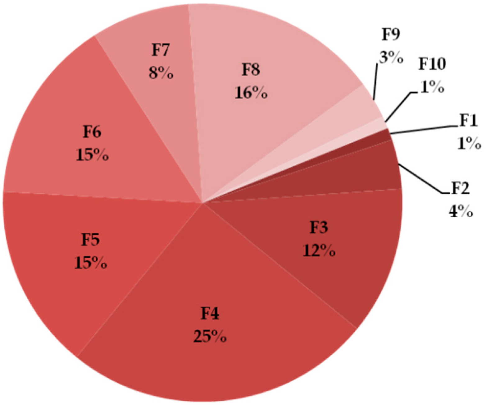
The percentage share of individual fractions in the collected material was estimated from the obtained chromatograms using the area under the curve (AUC). The largest part of proteins from the whole pool were found in fractions 4 and 8, the least in fractions 1–2 and 9–10 (Figure 2).
The SDS-PAGE technique was used to monitor the complexity of the fractions. Fractions 6–7 and 1 and 8 were separated into two and three bands, respectively. Fractions 2 and 10 consisted of more than three bands. In fractions 3, 4, 5, and 9, only one band is visible. However, in these samples, as well as in 6 and 8, the lowest bands migrated with the front of the electrophoresis, which means that there is a high probability that there is a mixture of low molecular weight proteins (Figure 3).

2.2. Identification of Proteins in Obtained Fractions

2.2.1. General Characteristics of the Obtained Fractions

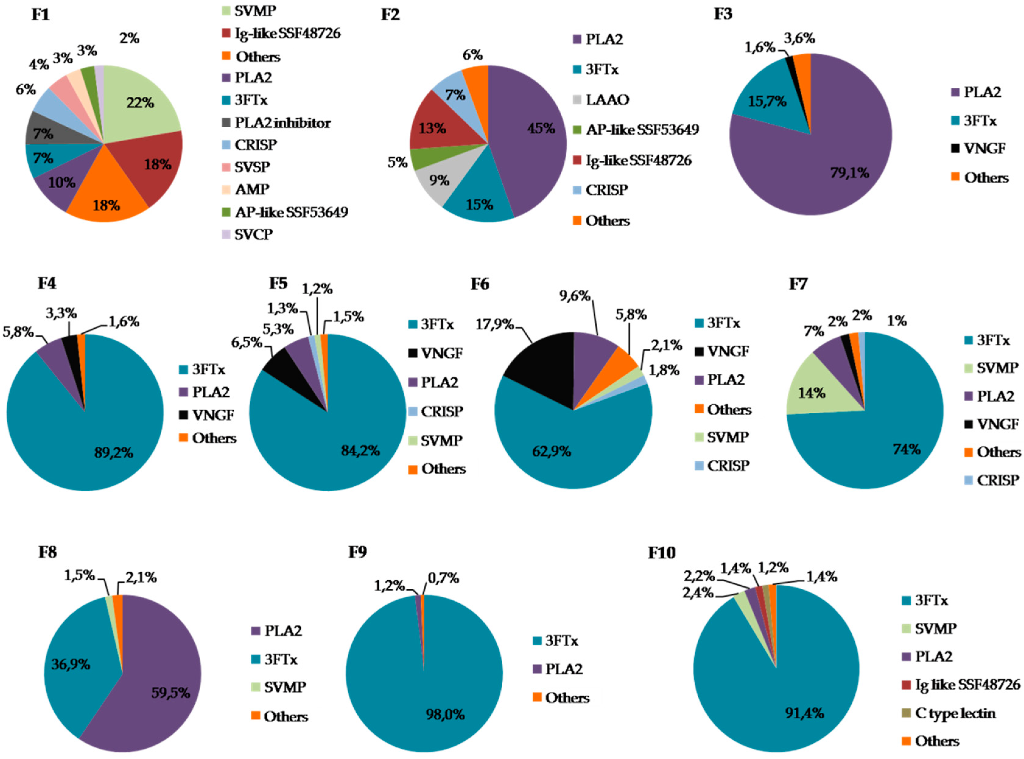
MS analysis indicated that in six of the obtained fractions, the predominant group of proteins was 3FTx (F4–F7, F9–F10). In three fractions (F2–F3, F8), the highest share of phospholipases A2 was observed, but in F2 it was below 50%. The largest number of different protein groups was found in fraction F1. In fractions F1 and F7, a significant share of SVMPs (snake venom metalloproteinases) was observed while in fractions F5 and F6, the highest percentage of VNGF (venom nerve growth factor) was detected (Figure 4). Detailed information on the identification of proteins in individual fractions can be found in the Supplementary Materials (Table S1).
In addition, a number of other low abundant proteins were detected. Table 1 shows those with a share exceeding 1% in at least one fraction.

2.2.2. Detailed Composition of the F2 Fraction

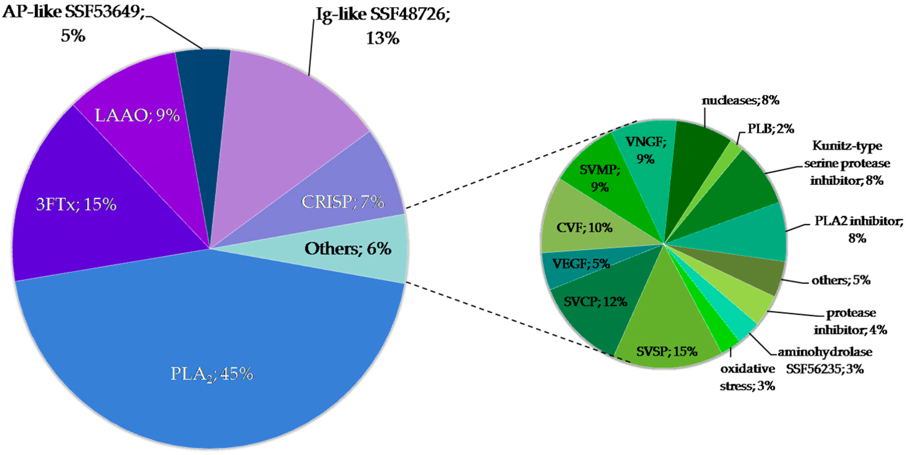
Of all the obtained fractions, only F2 showed antibacterial properties (see the next paragraph for details). The protein concentration in this fraction was estimated at 1.49 μg/μL. The main six components of F2 fraction are: PLA2s, 3FTxs, CRISPs, LAAOs, alkaline phosphatase-like proteins and Ig-like domain containing proteins. Among the low abundant proteins, three classes of proteases (SVMP, SVSP, and SVCP), CVF, VNGF, and enzyme inhibitors have the highest share (Figure 5).

2.2.3. Proteins with Ig-Like Domain

Overall, there were 38 identifications of proteins belonging to Ig-like superfamily SSF48726; however, only 23 hits were unique. Seventeen unique proteins were identified on the basis of mRNA sequences obtained from the venom glands of Micrurus species (15 identifications) or Boiga irregularis (2). The rest of them (six proteins) were identified on the basis of genomic sequences from Ophiophagus hannah. Two identified proteins were described as transmembrane proteins in the Uniprot database. In turn, two others were also classified as members of the superfamily SSF54452 belonging to MHC class I family. Most proteins from this group were identified in F2 and F1 (19 and 11, respectively). Details on the identifications are summarized in Table S2. The percentage share of the Ig-like domain-containing protein class in individual fractions is presented in Table S3.

2.3. Microbiological Tests

2.3.1. Determination of the Minimum Inhibitory Concentration (MIC)

All 10 fractions were tested for antibacterial properties. Only fraction F2 demonstrated antimicrobial activity against S. epidermidis ATCC 12228 (MIC = 37.25 µg/mL) and S. epidermidis ATCC 35984 (MIC = 9.3 µg/mL) strains. The MICs of 9 out of 10 fractions could not be determined, as these fractions were ineffective against S. epidermidis even at a concentration of 500 µg/mL. The minimum inhibitory concentration of fraction F2 was 4.6, 9.3, and 37.25 µg/mL, respectively, for clinical S. epidermidis strains 2346, 2452, and 2702. The results for the antibiotic inhibitory concentration, MIC, are shown in Table 2.

2.3.2. Synergy Testing

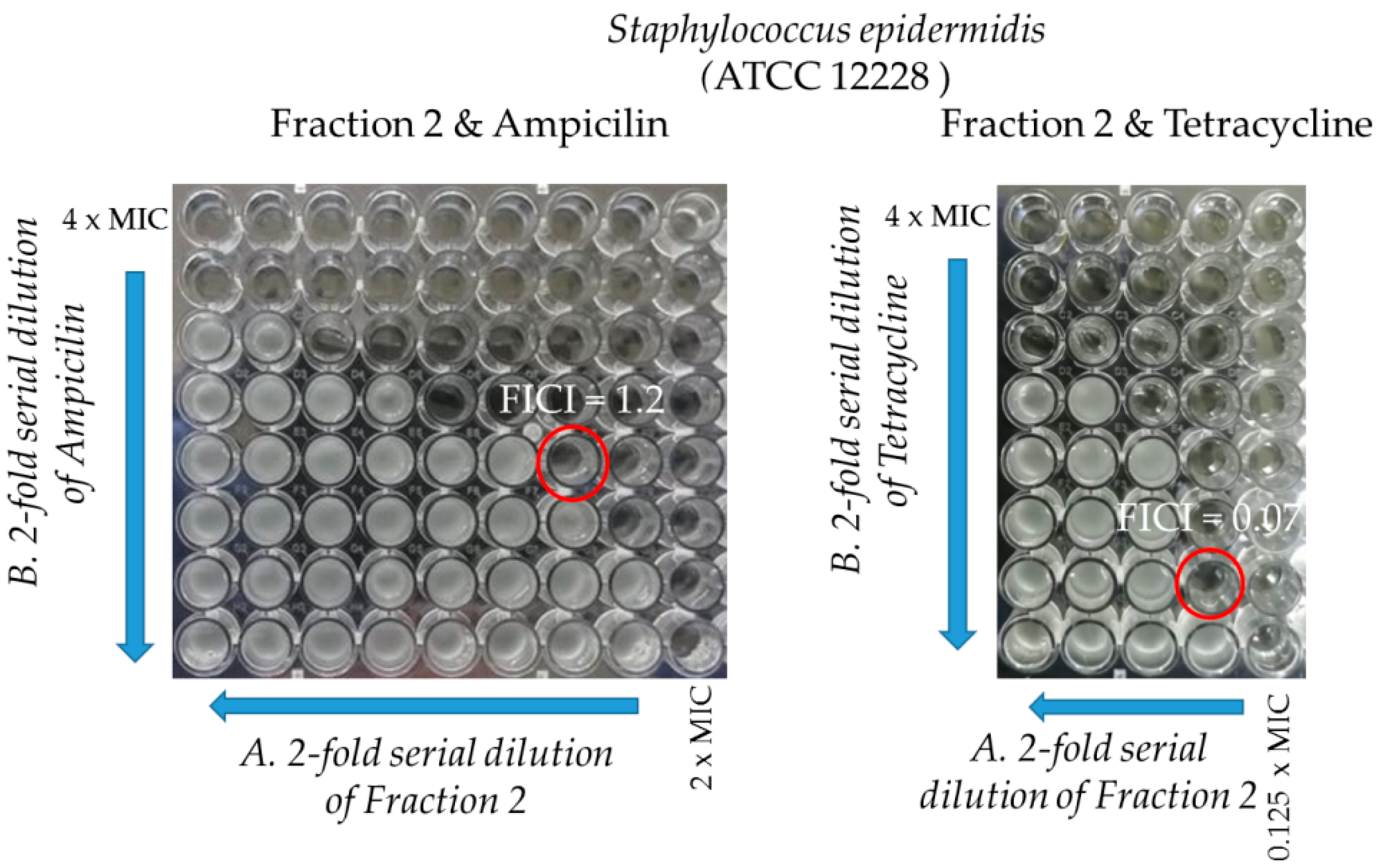
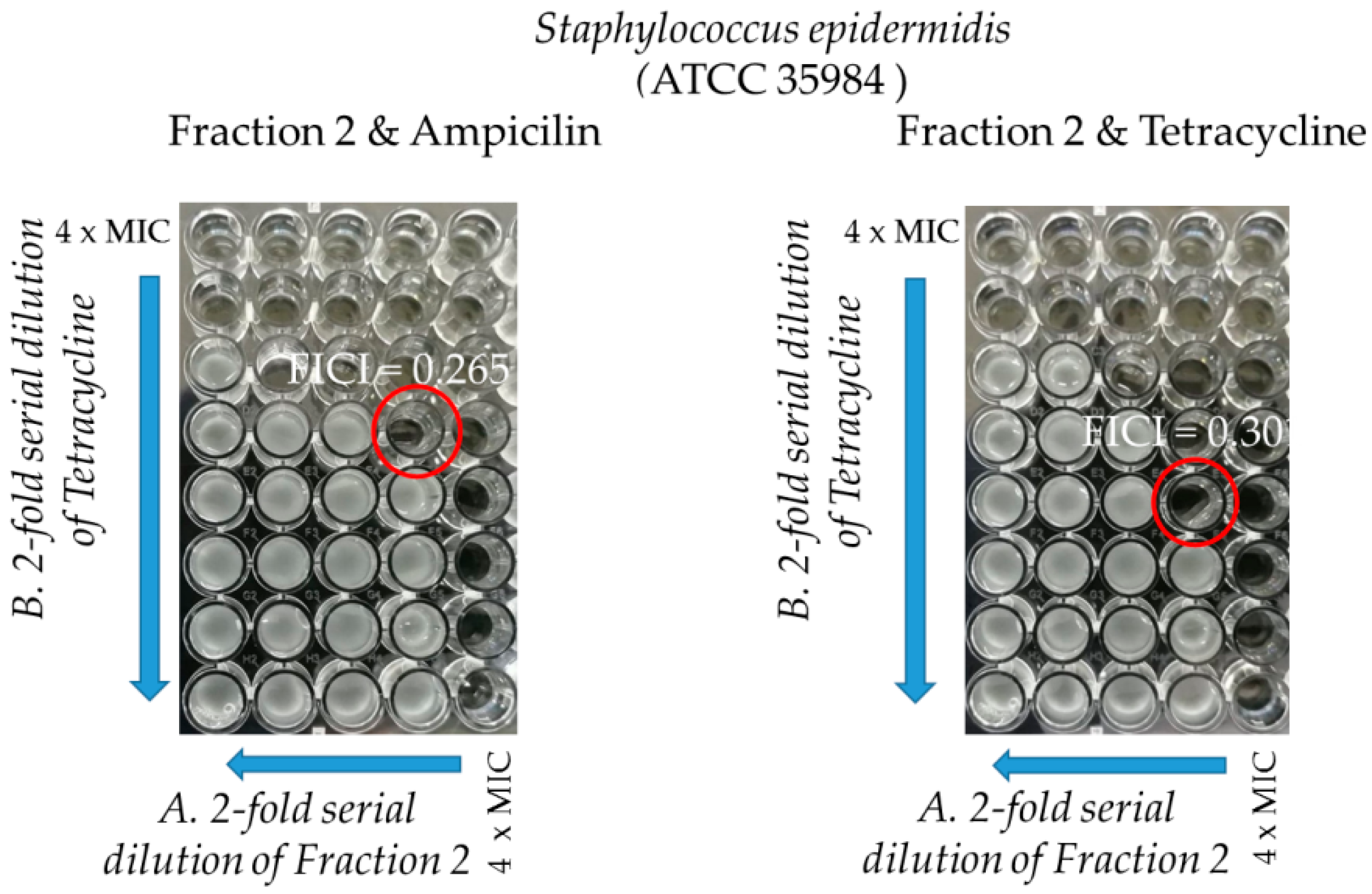
Selected antibacterial compounds combinations in double-dose response (checkerboard) experiments were used to determine the nature of their interaction. The checkerboard assay was conducted using fraction F2, ampicillin, and tetracycline against two studied standard S. epidermidis strains and the results are summarized in Table 3, Figure 6 and Figure 7. The checkerboard assay could not be performed on other three MRCNS S. epidermidis strains due to the limited amount of fraction F2.
Fraction F2 alone showed high antibacterial activity, with MICs between 4.6 and 37.25 µg/mL, against different S. epidermidis strains. In three out of four combinations with ampicillin (AMP) and tetracycline (TET) against two standard strains, the MICs of the fraction were up to 16 times reduced for the TET/fraction F2 combination against S. epidermidis ATCC 12228. It was also observed that in all combinations, the MICs of the antibiotics were also reduced in a concentration-dependent manner for fraction F2. The interaction between ampicillin and fraction F2 against S. epidermidis ATCC 12228 showed a neutral character while all other combinations demonstrated a synergistic interaction.

2.3.3. Anti-Biofilm Activity

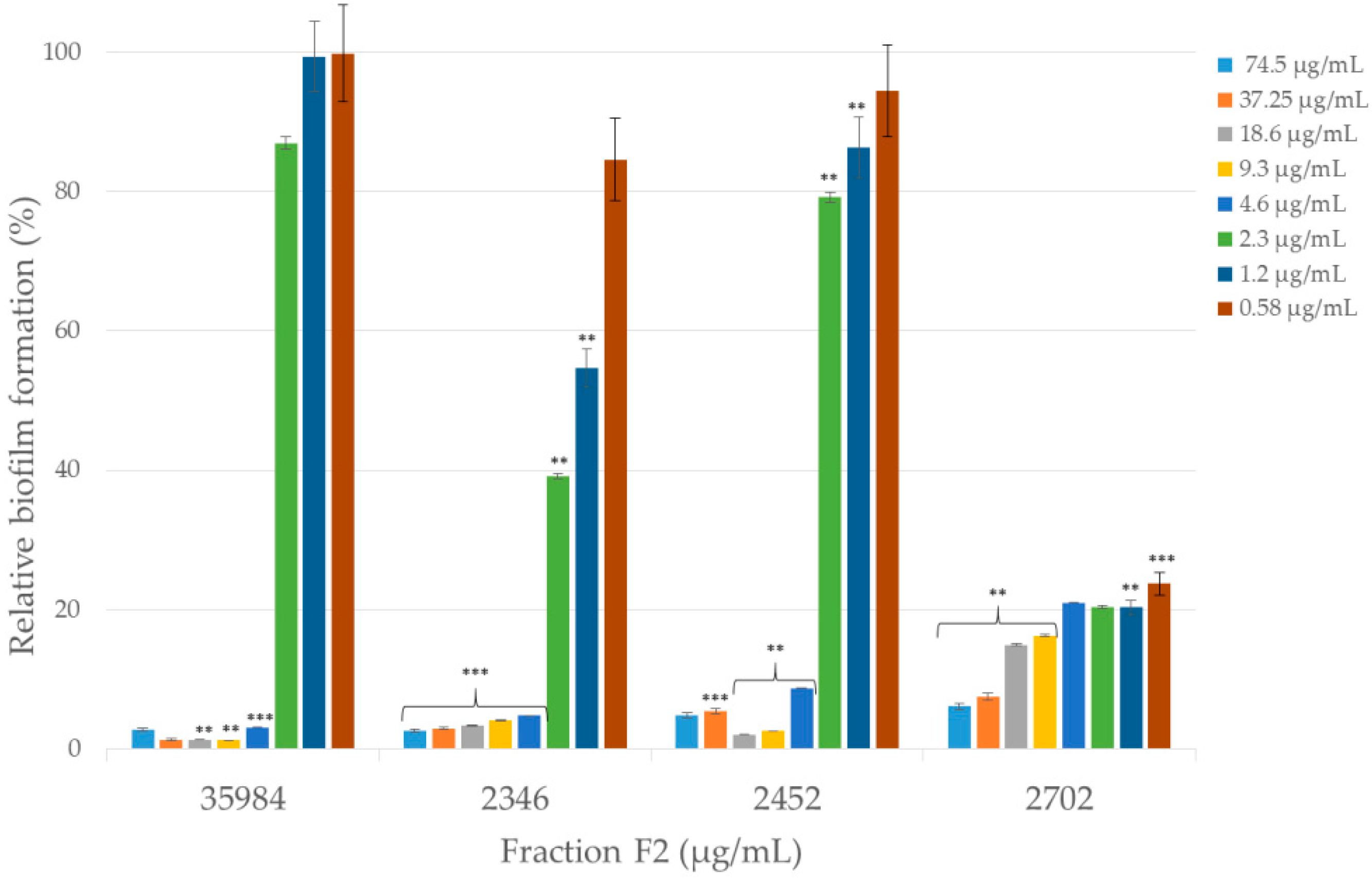
The effects of fraction F2 on the inhibition of biofilm formation were investigated. The biofilm of certified and clinical S. epidermidis strains was grown in the presence of a decreasing concentration of fraction F2. The results revealed that the amount of biofilm formed by various strains of S. epidermidis was differently affected upon the exposure of fraction F2 (Figure 8).
Fraction F2 at a concentration of 4.6 µg/mL inhibited the formation of 79.02% to 96.91% of the biofilm compared to the control (biofilm formation of certain certified and clinical strains without an anti-biofilm component). The most effective anti-biofilm activity for the lowest concentration (0.58 µg/mL) was demonstrated by fraction F2 against clinical S. epidermidis 2702 strain (76.3%) while the highest inhibition of biofilm formation was exhibited against certified ATCC 35984 S. epidermidis strain (98.8%).

3. Discussion

The activity of snake venom against Staphylococcus epidermidis has not been extensively studied so far. The only reports concern viperids, namely Calloselasma rhodostoma and Bothrops atrox [24] and Vipera ammodytes [25]. Bee venom has also been shown to be effective against skin bacteria, in addition to S. epidermidis, Cutibacterium acnes, and Streptococcus pyogenes [26]. Individual venom components are also effective against skin infections, e.g., snake cathelicidin BG-CATH [27], mucroporin-M1 from scorpion venom [28], or Lymnaea stagnalis snail peptides [29].
In our experiments, we proved the effectiveness of only one fraction, F2, both in direct bacteria elimination and inhibition of biofilm production. The five main components of this fraction are phospholipases A2, 3FTx proteins, L-amino acid oxidases, Ig-like proteins, and CRISPs. The group of proteins with the Ig-like domain has just been described by us for the first time, and so far, there are no functional studies on this group. Therefore, it is difficult to speculate on the participation of this group in the bactericidal effect. As for the other groups, reports indicate their antibacterial character [14]. What is very interesting is that the composition of F2 fraction is quite similar to F1, although there is no LAAOs but there are SVMPs and, more importantly, cathelicidin-like antimicrobial peptides, which have a wide spectrum of antibacterial activity and high efficiency [15,16]. Despite this, the F1 fraction did not show any antibacterial properties in our tests. We surmise the main reason for the lack of these properties may be the 10 times lower concentration of proteins in comparison to fraction F2.
Almost half of all proteins in the F2 fraction are phospholipases A2. Additionally, it is probably the most widely described group of snake venom proteins in terms of antibacterial properties [20,21,30,31,32]. The mechanism of action of these proteins is based on cell wall damage, pore generation in membranes, and their permeabilization [32]. Also, proteins with the three-finger motif (3FTxs) were described as having an antibacterial effect by interacting with the components of membranes and walls of bacteria and their destabilization [22,23,33]. However, the group that was widely described in terms of its antibacterial properties is LAAOs. Interestingly, this group of proteins was not detected during our previous analysis of the Naja ashei venom proteome [19]. This may indicate that the share of this group in the whole venom is negligible, but fractionation caused a significant reduction of the sample complexity, thus allowing for the identification of low abundant proteins. There are three hypotheses concerning the antibacterial mechanism of LAAO. The first one assumes that the oxidized form of cofactor present in these enzymes (FAD or FMN) may interact with amino acids, which in turn may affect the structure and function of nucleic acids, proteins, and membranes [34]. The second mechanism results from the fact that during the reaction carried out by these enzymes, hydrogen peroxide appears as a by-product and causes oxidative damage to the membranes [35] or DNA [36]. There is also a hypothesis that LAAOs can directly oxidize amino acids in proteins, thus causing damage [37]. Regardless of whether only one of these mechanisms takes place or all three, the undeniable fact remains that these proteins are extremely effective against bacteria [38,39,40,41]. In the context of the other results obtained from fractionation, the latter group of proteins seems to be the key component with antibacterial properties. This is due to the fact that it is the only fraction in which the amount of LAAO exceeds 1%. Nevertheless, it cannot be excluded that other components of this fraction, such as PLA2 or 3FTx described above, act together to create a synergistic antibacterial effect. It also seems probable that the right proportions of the individual components are also necessary to achieve the desired effect. On the basis of the results obtained, we can speculate that the mechanism of action responsible for the efficiency of the F2 fraction is probably due to damage to the bacterial cell wall and outflow of the cytosol content. However, in order to finally answer the question about which ingredient(s) are responsible for the antibacterial effect, detailed analyses of individual ingredients and their combinations are necessary.
There are three other interesting aspects of our experiments. Firstly, the F2 fraction shows synergistic effects with antibiotics (ampicillin and tetracycline). Despite the fact, that tetracycline and ampicillin belong to two different classes of antibiotics and use different mechanisms of action against bacteria, for both antibiotics, a similar effect for antimicrobial peptides was previously described [42,43,44] and for the whole venom of Bothrops moojeni [45]. The antibacterial effect of ampicillin is based on the inhibition of penicillin-binding proteins involved in cell wall synthesis, whereas tetracycline inhibits 30S subunit during protein synthesis [46]. In general, FIC smaller than MIC makes the protein or peptide factor less toxic and the antibiotic more effective at lower concentrations [44,47]. The mechanism of the creation of synergy is explained in many ways. It is possible, for example, that both substances have the same molecular target but do not compete for a binding site [48]. The second possibility is to block the same metabolic pathway at different stages [49,50]. In other cases, the second component somehow strengthens the action of the first component by facilitating access to the molecular target. This is how antibacterial peptides work, which cause permeabilization of the membrane, which in turn causes the release of antibiotics into the periplasm and cytoplasm of bacterial cells [42,51,52,53,54,55,56]. It is also known that Staphylococci have an efflux pump, which determines drug resistance to a large extent because it is responsible for removing from cells various types of substances, which are unnecessary for metabolism or harmful to cells [57]. Bothrops moojeni venom inhibits the action of such a pump in Staphylococcus aureus and thus, in combination with antibiotics, increases their effectiveness [45]. Perhaps, therefore, one of the components of the F2 fraction may also have similar properties. The synergistic effect of the F2 fraction with ampicillin and tetracycline obtained by us promises very good prospects for the future. As the promising strategies for solving the problem of drug resistance are, on the one hand, the search for new antibacterial factors and, on the other hand, the use of combined antibiotics with other compounds, such as peptides or proteins [58]. Therefore, it seems that this result will be a very important point for future research.
The second important aspect of the results obtained in this experiment is the ability of the F2 fraction to inhibit biofilm formation. S. epidermidis is genetically conditioned to live in the form of biofilm, which is proved by the downregulation of basic cellular processes, such as biosynthesis of nucleic acids, proteins, and wall-building elements [59]. This explains the relatively high resistance to antibiotics, which acts against actively growing cells [60,61]. These bacteria produce poly-γ-glutamic acid (PGA) and poly-N-acetylglucosamine (PNAG) exopolymers, which play a key role in the resistance of bacterial cells to human antibacterial peptides and avoidance of phagocytosis by neutrophils [62,63,64]. There are few reports describing the action of venom and its components against biofilm formation. Therefore, the information that the F2 fraction inhibits the biofilm of both certified and clinical drug-resistant strains is extremely important. Until now, it was known that the whole venom of Bothrops moojeni inhibits the formation of biofilm [45] as well as the lectin from Bothrops jaracussu [65,66]. In all three cases, the inhibition of the biofilm did not result in the destruction of cells or the slowing down of their growth. This is very important because, usually, biofilms are insensitive to antibiotics and the increase in the effectiveness of antibiotics is explained by the ability to inhibit biofilm formation [67,68,69], which could explain why the synergistic effect in the case of F2 fractions was more pronounced for the strain forming the biofilm (ATCC 35984).
The last very interesting discovery was the identification of proteins with the Ig-like domain. According to the latest data concerning Naja ashei, its venom is composed mostly of 3FTxs (69%) and PLA2s (27%). However, there are also small amounts of SVMPs (2.1%), VNGF (1%), CRISPs (0.7%), CVF (0.12%), and 5′-nucleotidases (0.014%) [19]. After fractionation, we were able to discover other groups of low abundant proteins that were previously described in other snake species, for example, L-amino acid oxidases or snake venom serine proteases (SVSPs) (Table 1). Moreover, we discovered the group of Ig-like domain-containing proteins, which, as far as we know, has not yet been described for snake venom. According to our calculations, this group constitutes less than 1% of all proteins collected during chromatography, i.e., less than 1% of all venom proteins of Naja ashei. It seems that this group is extremely low abundant, which could explain why it has not been noticed so far.
The assignment of the identified proteins to the Ig-like domain-containing protein group was made based on information from the superfamily database of structural and functional annotation [70] on the UniProt website. The assigned superfamily SSF48726 is a superfamily of immunoglobulins, which brings together beta proteins with the immunoglobulin-like beta-sandwich {48725} fold type. According to the integrated resource of protein families, domains, and functional sites, all of the identified proteins have immunoglobulin-like domain (IPR007110) and immunoglobulin-like fold IPR013783 [71]. Unfortunately, the Ig-like domain is one of the most widespread but also the most heterogeneous domains. Proteins with this fold differ in their cellular localization, amino acid sequence, and biological role [72]. Apart from the actual immunoglobulins, proteins possessing this domain also include, for example, enzymes [73], their inhibitors [74], transcription factors [75], or components of ion channels [76]. Moreover, what is very interesting is that some snake venom proteins also have this domain [77,78,79]. However, multiple sequence alignment by Clustal Omega showed that the similarity of these proteins to the identified Ig-like domain-containing proteins is low and does not exceed 30%. Given the variety of functions of proteins in this superfamily, it is difficult to speculate what their role in the venom of Naja ashei is, but at this stage of the study, it appears that the identified proteins are unlikely to be constituents of the immune system or blood contaminants, especially considering that they were mostly identified by transcripts from the venom glands [78,80,81] and that there are no reports that the actual immunoglobulins are synthesized there. Given the high heterogeneity of this family, it is very difficult to speculate what the function of these proteins is. Further research is needed to answer a number of questions: Do all the proteins identified in our studies have the same or similar function, do they play a role in envenoming, or do they have a function in the proper functioning of the venom glands? The obtained results suggest that Naja ashei venom may contain proteins not previously described in any of the known snake venoms and it cannot be ruled out that these proteins are more common (at least within the Naja genus).

4. Materials and Methods

4.1. Venom Fractionation

For the experiments, pooled venom of Naja ashei from two adult snakes (male and female) was used. Snakes were captured and officially imported from Kenya. Venom was extracted in the Pata breeding garden (Hlohovec, Slovakia), under the veterinary certificate No. CHEZ-TT-01. This breeding garden also serves as a quarantine station for imported animals and is an official importer of exotic animals from around the world, having the permission of the State Nature Protection of the Slovak Republic under the No. 03418/06, the trade with endangered species of wild fauna and flora and on amendments to certain laws under Law No. 237/2002 Z.z.
In total, 25 mg of crude venom were diluted to the volume of 1 mL with the use of 50 mM sodium acetate pH 5 (buffer A). Ion exchange chromatography was performed on an NGC Chromatography System (Bio-Rad, Hercules, CA, USA) using 6 mL Resource S column (GE Healthcare, Little Chalfont, UK) at 23 °C. For gradient preparation, two buffers were used: A—as described above, and B—50 mM sodium acetate pH 5 with 1M NaCl which served as an eluent. ChromLab software (Bio-Rad, Hercules, CA, USA) was used to monitor the parameters and develop the results.
After optimizing the method, the venom was separated according to the following scheme (Table 4):
Fractions of 1 mL were collected manually and those belonging to one peak were combined and concentrated as described below. To obtain sufficient material for further studies, five identical chromatographic separations were carried out. The initial comparison of the fraction content was made by SDS-PAGE electrophoresis on 13% resolving gels (with 5% stacking gels) according to the standard procedure [82] with a Roti®-Mark PRESTAINED molecular weight marker as a standard (Roth, Karlsruhe, Germany) using Mini-Protean II apparatus (Bio-Rad Laboratories, Inc., Hercules, CA, USA). Samples were prepared by mixing 20 µL of pre-concentrated fraction and 10 µL of buffer to SDS-PAGE, and after 5 min of boiling, the samples were put into gel slots. After electrophoresis, the gels were incubated overnight in staining solution with colloidal Coomassie Brilliant Blue G-250. The number of the bands in particular lines was determined using ImageJ software.
Finally, the peaks from individual chromatographic separations were combined and concentrated on the centrifuge filters Vivaspin 2 with membrane 3000 MWCO PES (Sartorius Stedim Lab Ltd., Stonehouse, UK). The final concentration of the protein in fractions was measured with 2-D Quant Kit (GE Healthcare, Little Chalfont, UK), using bovine serum albumin as a standard.

4.2. Sample Preparation for LC-MS/MS

After IEX separation, proteins from each sample (approximately 10) were dissolved in 50 mM ammonium bicarbonate pH 8. Such samples were subjected to acetone precipitation by mixing one volume of samples with six volumes of acetone, and after vortexing, they were incubated overnight at −20 °C. After centrifugation (16,000× g, 10 min), the obtained pellets were dissolved with 0.1% (v/v) RapiGest (Waters, Milford, MA, USA) in 50 mM ammonium bicarbonate pH 8, boiled and then cooled before in-solution digestion. The reduction was carried out with dithiothreitol (DTT) (final concentration: 5 mM) at 60 °C for 30 min, whereas alkylation was performed with iodoacetamide (IAA) (final concentration: 15 mM) for 30 min at room temperature in the dark. The samples were digested with trypsin (50:1 w/w) (Promega, Madison, WI, USA) for 18 h at 37 °C, and after that, trifluoroacetic acid (TFA) was added to a final concentration of 0.5% (v/v). Samples were incubated for 45 min at 37 °C and centrifuged for 20 min at 16,000× g. Tryptic peptides were then purified on C18 StageTips, which were prepared by packing 6 layers of Empore™ Octadecyl C18 extraction disk (3M, Maplewood, MN, USA) into a 0.2 mL pipette tip. Such columns were successively preconditioned by rinsing with 100% methanol, 60% acetonitrile (ACN) with 0.1% trifluoroacetic acid (TFA), and 0.1% TFA in water. Bound peptides were washed with 5% methanol in 0.1% TFA (1 time) and with 0.1% TFA/H2O (3 times) and then eluted with 60% ACN with 0.1% TFA. After every washing and elution step, columns were centrifuged for 5 min at 1000× g. Purified samples were evaporated in a vacuum centrifuge, and obtained peptides were dissolved in LC-MS grade water with 0.1% (v/v) TFA.

4.3. Protein Identification by LC-MS/MS

Approximately 0.6 μg of tryptic peptides from each sample were used for MS analysis. LC separation was conducted on a Dionex Ultimate 3000 Nano system (Thermo Fisher Scientific, Waltham, MA, USA) using Acclaim PepMap RSLC nanoViper C18 column (75 μm × 25 cm, 2 μm particles) (Thermo Fisher Scientific, Waltham, MA, USA) with 180 min ACN gradient (from 4% to 60%, in 0.1% formic acid). Chromatograph worked in on-line mode with Q ExactivePlusOrbitrap mass spectrometer (Thermo Scientific, Waltham, MA, USA). The analysis was conducted in data-dependent acquisition mode with survey scans acquired at a resolution of 70,000 at m/z 200 in MS mode, and 17,500 at m/z 200 in MS2 mode. The 15 most prominent peaks from each MS spectra were subjected to further fragmentation. Spectra were recorded in the scanning range of 300–2000 m/z in positive ion mode. Higher energy collisional dissociation (HCD) ion fragmentation was performed with normalized collision energies set to 25.
Peak lists obtained from MS/MS spectra were identified using 3 search engines: X!Tandem (ver. 2015.12.15.2), MS-GF+ (ver. 2018.04.09), and MyriMatch (ver. 2.2.140). The search was conducted using SearchGUI (ver. 3.3.16) [83]. Protein identification was conducted against a concatenated target/decoy UniProtKB Serpentes database. The decoy sequences were created by reversing the target sequences in SearchGUI. The identification settings were as follows: Trypsin, Specific, with a maximum of 2 missed cleavages; 10.0 ppm as MS1 and 0.02 Da as MS2 tolerances; fixed modifications: Carbamidomethylation of C, variable modifications: Oxidation of M, fixed modifications during refinement procedure: Carbamidomethylation of C, variable modifications during refinement procedure: Acetylation of protein N-term, Pyrolidone from E, Pyrolidone from Q, Pyrolidone from carbamidomethylated C.
Peptides and proteins were inferred from the spectrum identification results using PeptideShaker (ver. 1.16.42). Peptide spectrum matches (PSMs), peptides, and proteins were validated at a 2.5% false discovery rate (FDR) estimated using the decoy hit distribution. Hits marked by the software as “validated” were taken into consideration in further analysis, whereas, proteins labeled as “doubtful” were manually revised and based on the #PSMs, #Peptides and the obtained spectra, some of them were also included into data analysis.
Quantification of proteins was performed using the NSAF+ algorithm, which was implemented into PeptideShaker software [84]. Data was extracted into Excel and then the identified proteins were assigned to different groups/families. Final quantitative values for the whole protein group/family were calculated by summing individual NSAF+ values of proteins assigned to a given group.
The mass spectrometry data along with the identification results have been deposited to the ProteomeXchange Consortium via the PRIDE partner repository with the dataset identifier PXD015814 and 10.6019/PXD015814.

4.4. Antibacterial Activity

The following certified bacteria cultures used to test the antimicrobial activity were received from the Department of Biotechnology and Bioinformatics, Faculty of Chemistry, Rzeszow University of Technology (S. epidermidis ATTCC 12228, does not form a biofilm) and from Chair and Department of Medical Microbiology Medical University of Lublin (S. epidermidis ATCC 35984, form a biofilm). The anti-biofilm activity of tested fractions was evaluated with the use of three clinical Methicyllin-Resistant Coagulase-Negative Staphylococci (MRCNS) strains of (2346, 2452, 2702) obtained from the Department of Medical Laboratory Diagnostics of Provincial Specialist Hospital in Rzeszow.

4.4.1. Determination of Minimum Inhibitory Concentration (MIC)

The antibacterial activity of fractions (1–10) was evaluated by determination of the minimum inhibitory concentration (MIC, μg/mL) using the micro-broth dilution method, as described before [85] and according to the Clinical and Laboratory Standards Institute (CSLI) guidelines for S. epidermidis antimicrobial susceptibility testing [86]. Briefly, each bacterial strain was incubated in 37 °C in New Brunswick Innova 40 Shaker (Eppendorf AG, Hamburg, Germany) until turbidity of 0.5 McFarland’s standard (108 CFU/mL, colony-forming units per mL) was obtained. Series of two-fold dilutions of tested fractions (range from 19 to 500 μg/mL) were prepared in Muller Hinton Broth (MHB). Working bacterial cultures were diluted to final density 105 CFU/mL, added to prepared series of fraction’s dilutions, and incubated at 37 °C. After 24 h, bacterial growth in comparison with the positive control (the medium without antibacterial agents) was monitored. The MIC was defined as the lowest concentration of the antibacterial agent, which completely inhibited the visible growth of the microorganism. These results were confirmed by measurement of the optical density at 630 nm using BIO-RAD Microplate Reader. The experiment was carried out in triplicate. A positive (the medium without antibacterial agents) and negative control (no bacterial cultures added) of bacterial growth and solvent control were performed. An evaluation of the antibiotic susceptibility of each bacterial strain to ampicillin (AMP), tetracycline (TET), kanamycin (KAN), streptomycin (STR), and chloramphenicol (CF) was also performed by the micro-dilution method (range from 0.03 to 500 μg/mL) All reagents and bacterial cultures were prepared using Laminar Flow Cabinet ESCO Airstream.

4.4.2. Synergy Testing

Two standard S. epidermidis (ATCC12228 and ATCC35984) isolates were used to test interactions between two antibacterial agents (antibiotic and fraction F2) by the checkerboard assay [87,88] using the same medium and incubation conditions as described for MIC determinations. Two out of five antibiotics (ampicillin, tetracycline) were selected to the checkerboard assay due to: (i) Limited amount of fraction F2; (ii) the lowest (tetracycline) and the highest (ampicillin) obtained MIC values; (iii) different mechanisms of action of ampicillin (as beta lactam antibiotic) and tetracycline (as tetracycline antibiotic); and (iv) different activity of ampicillin (bactericidal) and tetracycline (bacteriostatic) [46]. Kanamycin and streptomycin were excluded due to resistance to at least one certified S. epidermidis strain. Compounds were usually tested in a range from 1/32 to 4 × MIC. The checkerboard assay was conducted on 96-well microtiter plates, where the first antimicrobial agent (fraction F2) in the combination was serially diluted 2-fold along the abscissa, whereas the second (antibiotic) was diluted along the ordinate. Starting concentrations for 2-fold dilution of compounds were selected on the basis of previously determined MIC. The initial concentration of fraction F2 in combination with both antibiotics was 37.25 and 74.5 µg/mL, respectively, against S. epidermidis 12228 and S. epidermidis 12228. Against S. epidermidis 12228, starting concentrations of ampicillin and tetracycline in combination with fraction 2 were 250 µg/mL. Ampicillin and tetracycline were used with an initial concentration of 500 and 0.49 µg/mL, respectively, against S. epidermidis 35984. The MIC of every antimicrobial agent in combination represented the lowest dilution that completely inhibited the growth of the bacterium. The interaction of the drugs in a combination was expressed quantitatively as a fractional inhibitory concentration (FIC) Index (FICI) and calculated for each drug combination using the following equation: FICI = FICA + FICB, where FICA = MIC of drug A in the combination/MIC of drug A alone, and FICB = MIC of drug B in the combination/ MIC of drug B alone. The FICI results were interpreted as synergistic (≤0.5), additive (>0.5 to ≤1), neutral (1–2), or antagonistic (≥2) [89].

4.4.3. Anti-Biofilm Activity

Staphylococcus epidermidis certified and three clinical strains were cultured overnight at 37 °C in MHB. Series of two-fold dilutions in MHB of fraction F2 and proper bacterial culture (105 CFU/mL) were added to 96-well polystyrene microtiter plates and incubated in 37 °C. The final concentrations of the tested compounds ranged from 0.58 to 149 μg/mL. The negative control was MHB medium, and the positive control (biofilm formation) was bacterial culture in MHB. After incubation, medium was removed from wells and washed twice with sterile phosphate-buffered saline (PBS) to remove the planktonic bacteria. Alive and adherent bacterial cells that usually formed biofilm in each well of the microtiter plate were stained with 3-(4,5-dimethyl-2-thiazolyl)-2,5 diphenyl-2H-tetrazolium bromide (MTT; 0.5% in PBS) for 2 h at 37 °C (protected from light) [90]. After incubation, the solution was removed, and bacterial biofilm was solubilized by DMSO and mixed for 15 min at room temperature in an INNOVA 40 Incubator Shaker. The absorbance was measured at 630 nm using a spectrophotometer (BIO-RAD Microplate Reader). No biofilm-producing S. epidermidis ATCC 12228 and high biofilm-producing S. epidermidis ATCC 35984 were used, respectively, as a negative and positive control. Tetracycline was used as the reference antimicrobial compound with the final concentration ranging from 0.03 to 500 μg/mL. The amount of biofilm inhibition was calculated relative to the amount of biofilm grown in the absence of anti-biofilm agent (defined as 100%) and the media sterility control (defined as 0%). Results from at least three separate biological replicates were averaged.

4.4.4. Statistical Analysis

The final results obtained from triplicate experiments were presented as means ± SD. STATISTICA v.12 (StatSoft, OK, USA) software was used to analyze the significances between data. p values were calculated using one-way ANOVA to compare the differences between each pair of groups. p value <0.05 was considered as significant.

5. Conclusions

Fractions of Naja ashei venom differing in protein composition were tested against certified and clinical strains of Staphylococcus epidermidis. Fraction F2 composed mostly with PLA2s, 3FTxs, CRISPs, LAAOs, alkaline phosphatase-like proteins, and Ig-like domain-containing proteins was the only one with antibacterial properties. It also showed a synergistic effect with antibiotics and was effective in the inhibition of biofilm formation. Among the F2 components, PLA2s, 3FTxs, and LAAOs were described before as potential components with bacteriostatic and bacteriocidal properties. The main thing that distinguishes this fraction from the others is the high content of L-amino acid oxidases and it is in this fact that the unique properties of this fraction are probably to be seen. Moreover, after separating Naja ashei venom into fractions, low-copy proteins with the Ig-like domain (SSF48726) were identified. The proteins belonging to this superfamily are characterized by a great variety of functions and sequences, but they are united by the type of folding. Most of the identification was made based on the transcript sequences from the venom glands, which suggests that these proteins are not related to the immune system but their function remains unexplained.

Supplementary Materials

The following are available online, Table S1: Protein identifications in fraction F2. Table S2: Identified proteins from Ig-like domain-containing protein group. Table S3: The percentage share of the Ig-like domain-containing protein class in individual fractions and total collected material.

Author Contributions

Conceptualization, A.B., E.C., J.L.; methodology, A.B., E.C., M.P. (Monika Pietrowska); investigation, A.B., E.C., K.K.H., M.P. (Monika Pietrowska), J.B., K.L.-S.; resources, V.P., M.P. (Monika Petrillova); writing—original draft preparation, A.B., E.C., K.K.H.; writing—review and editing, M.P. (Monika Pietrowska), K.L.-S.; funding acquisition, A.B., V.P., Ľ.L., J.L. All authors have read and agreed to the published version of the manuscript.

Funding

This research was funded by National Science Centre, Poland, grant number 2018/02/X/NZ6/00840 and by Slovak Research and Development Agency, Ministry of Education, Science, Research and Sport of the Slovak Republic, grant number APVV-17-0017.

Acknowledgments

We would like to thank Pawel Krawczyk from Bio-Rad Poland for his invaluable help at the stage of venom fractionation.

Conflicts of Interest

The authors declare no conflict of interest.

References

  1. Kloos, W.E.; Musselwhite, M.S. Distribution and persistence of Staphylococcus and Micrococcus species and other aerobic bacteria on human skin. Appl. Microbiol. 1975, 30, 381–385. [Google Scholar] [CrossRef] [Green Version]
  2. Dong, Y.; Speer, C.P.; Glaser, K. Beyond sepsis: Staphylococcus epidermidis is an underestimated but significant contributor to neonatal morbidity. Virulence 2018, 9, 621–633. [Google Scholar] [CrossRef] [PubMed] [Green Version]
  3. Rogers, K.L.; Fey, P.D.; Rupp, M.E. Coagulase-negative staphylococcal infections. Infect. Dis. Clin. N. Am. 2009, 23, 73–98. [Google Scholar] [CrossRef] [PubMed]
  4. De Lalla, F. Antimicrobial chemotherapy in the control of surgical infectious complications. J. Chemother. 1999, 11, 440–445. [Google Scholar] [CrossRef] [PubMed]
  5. Fey, P.D.; Olson, M.E. Current concepts in biofilm formation of Staphylococcus epidermidis. Future Microbiol. 2010, 5, 917–933. [Google Scholar] [CrossRef] [Green Version]
  6. Xu, K.D.; McFeters, G.A.; Stewart, P.S. Biofilm resistance to antimicrobial agents. Microbiology 2000, 146, 547–549. [Google Scholar] [CrossRef]
  7. Costerton, J.W.; Stewart, P.S.; Greenberg, E.P. Bacterial biofilms: A common cause of persistent infections. Science 1999, 284, 1318–1322. [Google Scholar] [CrossRef] [Green Version]
  8. Sieradzki, K.; Roberts, R.B.; Serur, D.; Hargrave, J.; Tomasz, A. Recurrent peritonitis in a patient on dialysis and prophylactic vancomycin. Lancet 1998, 351, 880–881. [Google Scholar] [CrossRef]
  9. Tammelin, A.; Domicel, P.; Hambraeus, A.; Ståhle, E. Dispersal of methicillin-resistant Staphylococcus epidermidis by staff in an operating suite for thoracic and cardiovascular surgery: Relation to skin carriage and clothing. J. Hosp. Infect. 2000, 44, 119–126. [Google Scholar] [CrossRef]
  10. Raad, I.; Alrahwan, A.; Rolston, K. Staphylococcus epidermidis: Emerging resistance and need for alternative agents. Clin. Infect. Dis. 1998, 26, 1182–1187. [Google Scholar] [CrossRef] [Green Version]
  11. Villari, P.; Sarnataro, C.; Iacuzio, L. Molecular epidemiology of Staphylococcus epidermidis in a neonatal intensive care unit over a three-year period. J. Clin. Microbiol. 2000, 38, 1740–1746. [Google Scholar] [CrossRef] [PubMed] [Green Version]
  12. Al Ahmadi, A.J.; Fathi, B.; Jamshidi, A.; Zolfagharian, H.; Mirakabbadi, A.Z. Investigation of the antibacterial effect of venom of the Iranian snake Echiscarinatus. Iran. J. Vet. Scitechnol. 2010, 2, 93–100. [Google Scholar]
  13. Perumal, S.R.; Stiles, B.G.; Franco, O.L.; Sethi, G.; Lim, L.H.K. Animal venoms as antimicrobial agents. Biochem. Pharmacol. 2017, 134, 127–138. [Google Scholar] [CrossRef] [PubMed]
  14. Bocian, A.; Hus, K.K. Antibacterial properties of snake venom components. Chem. Pap. 2019. [Google Scholar] [CrossRef] [Green Version]
  15. Azim, S.; McDowell, D.; Cartagena, A.; Rodriguez, R.; Laughlin, T.F.; Ahmad, Z. Venom peptides cathelicidin and lycotoxin cause strong inhibition of Escherichia coli ATP synthase. Int. J. Biol. Macromol. 2016, 87, 246–251. [Google Scholar] [CrossRef] [Green Version]
  16. Zhao, F.; Lan, X.Q.; Du, Y.; Chen, P.Y.; Zhao, J.; Zhao, F.; Lee, W.H.; Zhang, Y. King cobra peptide OH-CATH30 as a potential candidate drug through clinic drug-resistant isolates. Zool. Res. 2018, 39, 87–96. [Google Scholar]
  17. Koh, D.C.; Armugam, A.; Jeyaseelan, K. Snake venom components and their applications in biomedicine. Cell. Mol. Life Sci. 2006, 63, 3030–3041. [Google Scholar] [CrossRef]
  18. Wüster, W.; Broadley, D.G. Get an eyeful of this: A new species of giant spitting cobra from eastern and north-eastern Africa (Squamata: Serpentes: Elapidae: Naja). Zootaxa 2007, 1532, 51–68. [Google Scholar]
  19. Hus, K.; Buczkowicz, J.; Petrilla, V.; Petrillová, M.; Łyskowski, A.; Legáth, J.; Bocian, A. First Look at the venom of Naja ashei. Molecules 2018, 23, 609. [Google Scholar] [CrossRef] [Green Version]
  20. Okubo, B.M.; Silva, O.N.; Migliolo, L.; Gomes, D.G.; Porto, W.F.; Batista, C.L.; Ramos, C.S.; Holanda, H.H.; Dias, S.C.; Franco, O.L.; et al. Evaluation of an antimicrobial L-amino acid oxidase and peptide derivaties from Bothropoides mattogrosensis Pitviper venom. PLoS ONE 2012, 7, e33639. [Google Scholar] [CrossRef] [Green Version]
  21. Bacha, B.A.; Alonazi, M.A.; Elshikh, M.S.; Karray, A. A novel bactericidal homodimeric PLA2 group-I from Walterinnesia aegyptia venom. Int. J. Biolmacromol. 2018, 117, 1140–1146. [Google Scholar] [CrossRef] [PubMed]
  22. Chen, L.W.; Kao, P.H.; Fu, Y.S.; Hu, W.P.; Chang, L.S. Bactericidal effect of Naja nigricollis toxin γ is related to its membrane-damaging activity. Peptides 2011, 32, 1755–1763. [Google Scholar] [CrossRef] [PubMed]
  23. Kao, P.H.; Lin, S.R.; Chang, L.S. Differential binding to phospholipid bilayers modulates membrane-damaging activity of Naja najaatra cardiotoxins. Toxicon 2009, 54, 321–328. [Google Scholar] [CrossRef] [PubMed]
  24. Ferreira, B.L.; Santos, D.O.; Dos Santos, A.L.; Rodrigues, C.R.; De Freitas, C.C.; Cabral, L.M.; Castro, H.C. Comparative analysis of viperidae venoms antibacterial profile: A short communication for proteomics. Evid. Based. Complement. Altern. Med. 2011, 2011, 960267. [Google Scholar] [CrossRef] [Green Version]
  25. Iğci, N.; Nalbantsoy, A.; Erkan, L.G.; Akça, G.Y.; Yalçın, H.T.; Yalçın, M.; Göçmen, B. Screening of cytotoxic, anti-angiogenic, anti-tumorogenic and antimicrobial activities of Anatolian Vipera ammodytes (Nose-horned viper) venom. Turk. J. Biochem. 2016, 41, 483–491. [Google Scholar] [CrossRef]
  26. Hegazi, A.G.; EL-Feel, M.A.; Rahman, A.E.H.; Al-Fattah, A.M.A. Antibacterial activity of bee venom collected from Apis mellifera carniolan pure and hybrid races by two collection methods. Int. J. Currmicrobiol. Appl. Sci. 2015, 4, 141–149. [Google Scholar]
  27. Wang, Y.; Zhang, Z.; Chen, L.; Guang, H.; Li, Z.; Yang, H.; Li, J.; You, D.; Yu, H.; Lai, R. Cathelicidin-BF, a snake cathelicidin-derived antimicrobial peptide, could be an excellent therapeutic agent for acne vulgaris. PLoS ONE 2011, 6, e22120. [Google Scholar] [CrossRef] [Green Version]
  28. Dai, C.; Ma, Y.; Zhao, Z.; Zhao, R.; Wang, Q.; Wu, Y.; Cao, Z.; Li, W. Mucroporin, the first cationic host defence peptide from the venom of Lychas mucronatus. Antimicrob. Agents Chemother. 2018, 52, 3967–3972. [Google Scholar] [CrossRef] [Green Version]
  29. Gauri, S.S.; Bera, K.C.; Bhattacharyya, R.; Mandal, S.M. Identification of an antimicrobial peptide from large freshwater snail (Lymnaea stagnalis): Activity against antibiotics resistant Staphylococcus epidermidis. Int. J. Exp. Res. Rev. 2016, 2, 5–9. [Google Scholar]
  30. Toyama, M.H.; De Oliveira, D.G.; Beriam, L.O.S.; Novello, J.C.; Rodrigues-Simioni, L.; Marangoni, S. Structural, enzymatic and biological properties of new PLA2 isoform from Crotalus durissus terrificus venom. Toxicon 2003, 41, 1033–1038. [Google Scholar] [CrossRef]
  31. Vargas, L.J.; Londoño, M.; Quintana, J.C.; Rua, C.; Segura, C.; Lomonte, B.; Núñez, V. An acidic phospholipase A2 with antibacterial activity from Porthidium nasutum snake venom. Comp. Biochem. Physiol. 2012, 161, 341–347. [Google Scholar] [CrossRef]
  32. Perumal, S.R.; Kandasamy, M.; Gopalakrishnakone, P.; Stiles, B.G.; Rowan, E.G.; Becker, D.; Shanmugam, M.K.; Sethi, G.; Chow, V.T. Wound healing activity and mechanisms of action of an antibacterial protein from the venom of the eastern diamondback rattlesnake (Crotalus adamanteus). PLoS ONE 2014, 9, e80199. [Google Scholar]
  33. Chen, L.W.; Kao, P.H.; Fu, Y.S.; Lin, S.R.; Chang, L.S. Membrane-damaging activity of Taiwan cobra cardiotoxin 3 is responsible for its bactericidal activity. Toxicon 2011, 58, 46–53. [Google Scholar] [CrossRef]
  34. Izidoro, L.F.; Sobrinho, J.C.; Mendes, M.M.; Costa, T.R.; Grabner, A.N.; Rodrigues, V.M.; Da Silva, S.L.; Zanchi, F.B.; Zuliani, J.P.; Fernandes, C.F.; et al. Snake venom L-amino acid oxidases: Trends in pharmacology and biochemistry. Biomed. Res. Int. 2014, 196754. [Google Scholar] [CrossRef] [PubMed] [Green Version]
  35. Toyama, M.H.; Toyama, D.D.O.; Passero, L.F.D.; Laurenti, M.D.; Corbett, C.E.; Tomokane, T.Y.; Fonseca, F.V.; Antunes, E.; Joazeiro, P.P.; Beriam, L.O.; et al. Isolation of a new L-amino acid oxidase from Crotalus durissus cascavella venom. Toxicon 2006, 47, 47–57. [Google Scholar] [CrossRef] [PubMed]
  36. Braga, M.D.M.; Martins, A.M.C.; Amora, D.N.; De Menezes, D.B.; Toyama, M.H.; Toyama, D.O.; Marangoni, S.; Alves, C.D.; Barbosa, P.S.; De Sousa Alves, R.; et al. Purification and biological effects of l-amino acid oxidase isolated from Bothrops insularis venom. Toxicon 2008, 51, 199–207. [Google Scholar] [CrossRef] [PubMed]
  37. Ande, S.R.; Fussi, H.; Knauer, H.; Murkovic, M.; Ghisla, S.; Fröhlich, K.U.; Macheroux, P. Induction of apoptosis in yeast by L-amino acid oxidase from the Malayan pit viper Calloselasma rhodostoma. Yeast 2008, 25, 349–357. [Google Scholar] [CrossRef] [PubMed] [Green Version]
  38. Ciscotto, P.; De Avila, M.R.A.; Coelho, E.A.; Oliveira, J.; Diniz, C.G.; Farías, L.M.; De Carvalho, M.A.; Maria, W.S.; Sanchez, E.F.; Borges, A.; et al. Antigenic, microbicidal and antiparasitic properties of an l-amino acid oxidase isolated from Bothrops jararaca snake venom. Toxicon 2009, 53, 330–341. [Google Scholar] [CrossRef]
  39. Costa Torres, A.F.; Dantas, R.T.; Toyama, M.H.; DizFilho, E.; Zara, F.J.; De Queiroz, R.M.G.; Pinto, N.N.A.; De Oliveira, R.M.; De Oliveira, T.D.; Monteiro, H.S.; et al. Antibacterial and antiparasitic effects of Bothrops marajoensis venom and its fractions: Phospholipase A2 and L-amino acid oxidase. Toxicon 2010, 55, 795–804. [Google Scholar] [CrossRef]
  40. Lee, M.L.; Tan, N.H.; Fung, S.Y.; Sekaran, S.D. Antibacterial action of a heat-stable form of l-amino acid oxidase isolated from king cobra (Ophiophagus hannah) venom. Comp. Biochem. Physiol. 2011, 153, 237–242. [Google Scholar] [CrossRef]
  41. Vargas, L.J.; Quintana, J.C.; Pereañez, J.A.; Núñez, V.; Sanz, L.; Calvete, J. Cloning and characterization of an antibacterial L-amino acid oxidase from Crotalus durissus cumanensis venom. Toxicon 2013, 64, 1–11. [Google Scholar] [CrossRef] [PubMed]
  42. Yenugu, S.; Narmadha, G. The human male reproductive tract antimicrobial peptides of the HE2 family exhibit potent synergy with standard antibiotics. J. Pept. Sci. 2010, 16, 337–341. [Google Scholar] [CrossRef] [PubMed]
  43. Sanchez-Gomez, S.; Japelj, B.; Jerala, R.; Moriyon, I.; Fernandez, A.M.; Leiva, J.; Blondelle, S.E.; Andrä, J.; Brandenburg, K.; Lohner, K.; et al. Structural features governing the activity of lactoferricin-derived peptides that act in synergy with antibiotics against Pseudomonas aeruginosa in vitro and in vivo. Antimicrob. Agents Chemother. 2011, 55, 218–228. [Google Scholar] [CrossRef] [PubMed] [Green Version]
  44. Zharkova, M.S.; Orlov, D.S.; Golubeva, O.Y.; Chakchir, O.B.; Eliseev, I.E.; Grinchuk, T.M.; Shamova, O.V. Application of antimicrobial peptides of the innate immune system in combination with conventional antibiotics—A novel way to combat antibiotic resistance? Front. Cell. Infect. Microbiol. 2019, 9, 128. [Google Scholar] [CrossRef] [Green Version]
  45. Canhas, I.N.; Heneine, L.G.D.; Fraga, T.; De Assis, D.C.S.; Borges, M.H.; Chartone-Souza, E.; Nascimento, A.M.A. Antibacterial activity of different types of snake venom from the Viperidae family against Staphylococcus aureus. Acta Sci. Biol. Sci. 2017, 39, 309–319. [Google Scholar] [CrossRef] [Green Version]
  46. Hoerr, V.; Duggan, G.E.; Zbytnuik, L.; Poon, K.K.; Große, C.; Neugebauer, U.; Methling, K.; Löffler, B.; Vogel, H.J. Characterization and prediction of the mechanism of action of antibiotics through NMR metabolomics. BMC Microbiol. 2016, 16, 82. [Google Scholar] [CrossRef] [Green Version]
  47. Almaaytah, A.; Qaoud, M.T.; Abualhaijaa, A.; Al-Balas, Q.; Alzoubi, K.H. Hybridization and antibiotic synergism as a tool for reducing the cytotoxicity of antimicrobial peptides. Infect. Drug Resist. 2018, 11, 835–847. [Google Scholar] [CrossRef] [Green Version]
  48. Breitinger, H.-G. Drug synergy–mechanisms and methods of analysis. Toxic. Drug Test. 2012, 143–166. [Google Scholar]
  49. Jia, J.; Zhu, F.; Ma, X.; Cao, Z.; Li, Y.; Chen, Y.Z. Mechanisms of drug combinations: Interaction and network perspectives. Nat. Rev. Drug Discov. 2009, 8, 111–128. [Google Scholar] [CrossRef]
  50. Yeh, P.J.; Hegreness, M.J.; Aiden, A.P.; Kishony, R. Drug interactions and the evolution of antibiotic resistance. Nat. Rev. Microbiol. 2009, 7, 460–466. [Google Scholar] [CrossRef] [Green Version]
  51. Cassone, M.; Otvos, L., Jr. Synergy among antibacterial peptides and between peptides and small-molecule antibiotics. Expert Rev. Anti Infect. Ther. 2010, 8, 703–716. [Google Scholar] [CrossRef]
  52. Singh, A.P.; Prabha, V.; Rishi, P. Efficacy of cryptdin-2 as an adjunct to antibiotics from various generations against Salmonella. Indian J. Microbiol. 2014, 54, 323–328. [Google Scholar] [CrossRef] [PubMed] [Green Version]
  53. Feng, Q.; Huang, Y.; Chen, M.; Li, G.; Chen, Y. Functional synergy of alpha-helical antimicrobial peptides and traditional antibiotics against Gram-negative and Gram-positive bacteria in vitro and in vivo. Eur. J. Clinmicrobiol. Infect. Dis. 2015, 34, 197–204. [Google Scholar] [CrossRef] [PubMed]
  54. Gupta, K.; Singh, S.; Van Hoek, M.L. Short, synthetic cationic peptides have antibacterial activity against Mycobacterium smegmatis by forming pores in membrane and synergizing with antibiotics. Antibiotics 2015, 4, 358–378. [Google Scholar] [CrossRef] [PubMed] [Green Version]
  55. Khara, J.S.; Lim, F.K.; Wang, Y.; Ke, X.-Y.; Voo, Z.X.; Yang, Y.Y.; Lakshminarayanan, R.; Ee, P.L.R. Designing α-helical peptides with enhanced synergism and selectivity against Mycobacterium smegmatis: Discerning the role of hydrophobicity and helicity. Acta Biomater. 2015, 28, 99–108. [Google Scholar] [CrossRef]
  56. Soren, O.; Brinch, K.S.; Patel, D.; Liu, Y.; Liu, A.; Coates, A.; Hu, Y. Antimicrobial peptide novicidin synergizes with rifampin, ceftriaxone, and ceftazidime against antibiotic resistant Enterobacteriaceae in vitro. Antimicrob. Agents Chemother. 2015, 59, 6233–6240. [Google Scholar] [CrossRef] [Green Version]
  57. Costa, S.S.; Viveiros, M.; Amaral, L.; Couto, I. Multidrug efflux pumps in Staphylococcus aureus: An update. Open Microbiol. J. 2013, 7, 59–71. [Google Scholar] [CrossRef] [Green Version]
  58. Lin, L.; Nonejuie, P.; Munguia, J.; Hollands, A.; Olson, J.; Dam, Q.; Kumaraswamy, M.; Rivera, H., Jr.; Corriden, R.; Rohde, M.; et al. Synergizes with cationic antimicrobial peptides to exert bactericidal and therapeutic activity against highly multidrug-resistant gram-negative bacterial pathogens. EBioMedicine 2015, 2, 690–698. [Google Scholar] [CrossRef] [Green Version]
  59. Yao, Y.; Sturdevant, D.E.; Otto, M. Genome wide analysis of gene expression in Staphylococcus epidermidis biofilms: Insights into the pathophysiology of S. epidermidis biofilms and the role of phenol-soluble modulins in formation of biofilms. J. Infect. Dis. 2005, 191, 289–298. [Google Scholar] [CrossRef] [Green Version]
  60. Duguid, I.G.; Evans, E.; Brown, M.R.; Gilbert, P. Effect of biofilm culture upon the susceptibility of Staphylococcus epidermidis to tobramycin. J. Antimicrobiol. Chemother. 1992, 30, 803–810. [Google Scholar] [CrossRef]
  61. Duguid, I.G.; Evans, E.; Brown, M.R.; Gilbert, P. Growth-rate-independent killing by ciprofloxacin of biofilm-derived Staphylococcus epidermidis: Evidence for cell-cycle dependency. J. Antimicrobiol. Chemother. 1992, 30, 791–802. [Google Scholar] [CrossRef]
  62. Vuong, C.; Voyich, J.M.; Fischer, E.R.; Braughton, K.R.; Whitney, A.R.; De Leo, F.R.; Otto, M. Polysaccharide intercellular adhesin (PIA) protects Staphylococcus epidermidis against major components of the human innate immune system. Cell. Microbiol. 2004, 6, 269–275. [Google Scholar] [CrossRef]
  63. Kocianova, S.; Vuong, C.; Yao, Y.; Voyich, J.M.; Fischer, E.R.; DeLeo, F.R.; Otto, M. Key role of poly-γ-dl -glutamic acid in immune evasion and virulence of Staphylococcus epidermidis. J. Clin. Investig. 2005, 115, 688–694. [Google Scholar] [CrossRef]
  64. Kristian, S.A.; Birkenstock, T.A.; Sauder, U.; Mack, D.; Götz, F.; Landmann, R. Biofilm formation induces C3a release and protects Staphylococcus epidermidis from IgG and complement deposition and from neutrophil-dependent killing. J. Infect. Dis. 2008, 197, 1028–1035. [Google Scholar] [CrossRef] [Green Version]
  65. Klein, R.C.; Fabres-Klein, M.H.; De Oliveira, L.L.; Feio, R.N.; Malouin, F.; Ribon, A.O.B. A C-Type lectin from Bothrops jararacussu venom disrupts staphylococcal biofilms. PLoS ONE 2015, 10, e0120514. [Google Scholar] [CrossRef] [Green Version]
  66. Aguilar, A.P.; Onofre, T.S.; Fabres-Klein, M.H.; Klein, R.C.; Feio, R.N.; De Oliveira, M.T.A.; De Oliveira, B.R.A. Carbohydrate-independent biofilm effect of lectin BJcuL on Staphylococcus aureus. Microb. Pathog. 2019, 137, 103745. [Google Scholar] [CrossRef]
  67. Gopal, R.; Kim, Y.G.; Lee, J.H.; Lee, S.K.; Chae, J.D.; Son, B.K.; Seo, C.H.; Park, Y. Synergistic effects and antibiofilm properties of chimeric peptides against multidrug-resistant Acinetobacter baumannii strains. Antimicrob. Agents Chemother. 2014, 58, 1622–1629. [Google Scholar] [CrossRef] [Green Version]
  68. De La Fuente, N.C.; Reffuveille, F.; Mansour, S.C.; Reckseidler, Z.S.L.; Hernández, D.; Brackman, G.; Coenye, T.; Hancock, R.E. D-Enantiomeric peptides that eradicate wild-type and multidrug-resistant biofilms and protect against lethal Pseudomonas aeruginosa infections. Chem. Biol. 2015, 22, 196–205. [Google Scholar] [CrossRef]
  69. Bessa, L.J.; Eaton, P.; Dematei, A.; Plácido, A.; Vale, N.; Gomes, P.; Delerue, M.C.; Sa Leite, J.R.; Gameiro, P. Synergistic and antibiofilm properties of ocellatin peptides against multidrug-resistant Pseudomonas aeruginosa. Future Microbiol. 2018, 13, 151–163. [Google Scholar] [CrossRef] [Green Version]
  70. Gough, J.; Karplus, K.; Hughey, R.; Chothia, C. Assignment of homology to genome sequences using a library of hidden Markov models that represent all proteins of known structure. J. Mol. Biol. 2001, 313, 903–919. [Google Scholar] [CrossRef]
  71. Mitchell, A.L.; Attwood, T.K.; Babbitt, P.C.; Blum, M.; Bork, P.; Bridge, A.; Brown, S.D.; Chang, H.-Y.; El-Gebali, S.; Fraser, M.I.; et al. InterPro in 2019: Improving coverage, classification and access to protein sequence annotations. Nucleic Acids Res. 2019, 47, 351–360. [Google Scholar] [CrossRef] [Green Version]
  72. Halaby, D.M.; Poupon, A.; Mornon, J.-P. The immunoglobulin fold family: Sequence analysis and 3D structure comparisons. Protein Eng. 1999, 12, 563–571. [Google Scholar] [CrossRef] [PubMed] [Green Version]
  73. Watanabe, H.; Nishimoto, T.; Kubota, M.; Chaen, H.; Fukuda, S. Cloning, sequencing, and expression of the genes encoding an isocyclomaltooligosaccharide glucanotransferase and an alpha-amylase from a Bacillus circulans strain. Biosci. Biotechnol. Biochem. 2006, 70, 2690–2702. [Google Scholar] [CrossRef] [PubMed] [Green Version]
  74. Shia, S.; Stamos, J.; Kirchhofer, D.; Fan, B.; Wu, J.; Corpuz, R.T.; Santell, L.; Lazarus, R.A.; Eigenbrot, C. Conformational lability in serine protease active sites: Structures of hepatocyte growth factor activator (HGFA) alone and with the inhibitory domain from HGFA inhibitor-1B. J. Mol. Biol. 2005, 346, 1335–1349. [Google Scholar] [CrossRef] [PubMed]
  75. Ikeda, T.; Honjo, K.; Hirota, Y.; Onodera, T. Isolation of the chicken NF-kappa B p65 subunit-encoding cDNA and characterization of its products. Gene 1993, 133, 237–242. [Google Scholar]
  76. Isom, L.L.; De Jongh, K.S.; Patton, D.E.; Reber, B.F.X.; Offord, J.; Charbonneau, H.; Walsh, K.; Goldin, A.L.; Catterall, W.A. Primary structure and functional expression of the beta 1 subunit of the rat brain sodium channel. Science 1992, 256, 839–842. [Google Scholar] [CrossRef]
  77. Rehana, S.; Kini, R.M. Molecular isoforms of cobra venom factor-like proteins in the venom of Austrelaps superbus. Toxicon 2007, 50, 32–52. [Google Scholar] [CrossRef]
  78. Rokyta, D.R.; Lemmon, A.R.; Margres, M.J.; Aronow, K. The venom-gland transcriptome of the eastern diamondback rattlesnake (Crotalus adamanteus). BMC Genom. 2012, 13, 312. [Google Scholar] [CrossRef] [Green Version]
  79. Zeng, L.; Sun, Q.Y.; Jin, Y.; Zhang, Y.; Lee, W.H.; Zhang, Y. Molecular cloning and characterization of a complement-depleting factor from king cobra, Ophiophagus hannah. Toxicon 2012, 60, 290–301. [Google Scholar] [CrossRef]
  80. McGivern, J.J.; Wray, K.P.; Margres, M.J.; Couch, M.E.; Mackessy, S.P.; Rokyta, D.R. RNA-seq and high-definition mass spectrometry reveal the complex and divergent venoms of two rear-fanged colubrid snakes. BMC Genom. 2014, 15, 1061. [Google Scholar] [CrossRef] [Green Version]
  81. Aird, S.D.; Da Silva, N.J.; Qiu, L.; Villar-Briones, A.; Saddi, V.A.; Pires De Campos Telles, M.; Grau, M.L.; Mikheyev, A.S. Coral snake Venomics: Analyses, of venom gland transcriptomes and proteomes of six Brazilian taxa. Toxins 2017, 8, 9. [Google Scholar]
  82. Laemmli, U.K. Cleavage of structural proteins during the assembly of the head of bacteriophage T4. Nature 1970, 227, 680–685. [Google Scholar] [CrossRef] [PubMed]
  83. Barsnes, H.; Vaudel, M. SearchGUI: A highly adaptable common interface for proteomics search and de novo engines. J. Proteome Res. 2018, 17, 2552–2555. [Google Scholar] [CrossRef]
  84. Vaudel, M.; Burkhart, J.M.; Zahedi, R.P.; Oveland, E.; Berven, F.S.; Sickmann, A.; Martens, L.; Barsnes, H. PeptideShaker enables reanalysis of MS-derived proteomics data sets. Nat. Biotechnol. 2015, 33, 22–24. [Google Scholar] [CrossRef]
  85. Zapała, L.; Kosińska, M.; Woźnicka, E.; Byczyński, Ł.; Ciszkowicz, E.; Lecka-Szlachta, K.; Zapała, W.; Chutkowski, M. Comparison of spectral and thermal properties and antibacterial activity of new binary and ternary complexes of Sm(III), Eu(III) and Gd(III) ions with N-phenylanthranilic acid and 1,10-phenanthroline. Thermochim. Acta 2019, 671, 134–148. [Google Scholar] [CrossRef]
  86. Clinical and Laboratory Standards Institute. Methods for Determining Bactericidal Activity of Antimicrobial Agents. Approved Guideline; CLSI document M26-A; Clinical and Laboratory Standards Institute: Wayne, PA, USA, 1999; Volume 19. [Google Scholar]
  87. Spoorthi, N.J.; Vishwanatha, T.; Reena, V.; Divyashree, B.C.; Aishwarya, S.; Siddhalingeshwara, K.G.; Venugopal, N.; Ramesh, I. Antibiotic synergy test: Checkerboard method on multidrug resistant Pseudomonas aeruginosa. Int. Res. J. Pharm. 2011, 2, 196–198. [Google Scholar]
  88. Vázquez, R.; García, P. Synergy between two chimeric lysins to kill Streptococcus pneumoniae. Front. Microbiol. 2019, 10, 1251. [Google Scholar] [CrossRef] [Green Version]
  89. Sopirala, M.M.; Mangino, J.E.; Gebreyes, W.A.; Biller, B.; Bannerman, T.; Balada-Llasat, J.M.; Pancholi, P. Synergy testing by Etest, microdilution checkerboard, and time-kill methods for pan-drug-resistant Acinetobacter baumannii. Antimicrob. Agents Chemother. 2010, 54, 4678–4683. [Google Scholar] [CrossRef] [Green Version]
  90. Bielenica, A.; Drzewiecka-Antonik, A.; Rejmak, P.; Stefańska, J.; Koliński, M.; Kmiecik, S.; Lesyng, B.; Włodarczyk, M.; Pietrzyki, P.; Struga, M. Synthesis, structural and antimicrobial studies of type II topoisomerase targeted copper (II) complexes of 1,3-disubstituted thiourea ligands. J. Inorg. Biochem. 2018, 182, 61–70. [Google Scholar] [CrossRef]
Sample Availability: Not available.
Figure 1. Representative chromatogram obtained for the separation of Naja ashei venom on the Resource S column. The fractions considered in the next stages of the experiment are marked by numbers above the peaks.
Molecules 25 00293 g001
Figure 2. Percentage share of collected proteins in particular fractions from AUC analysis in ChromeLab.
Molecules 25 00293 g002
Figure 3. Representative SDS-PAGE gels of Naja ashei venom fractions. The numbers above the lines represent the fractions collected. The numbers below lines (red) indicate the number of bands defined in the ImageJ software. Weakly visible bands are marked with arrows on the gels.
Molecules 25 00293 g003
Figure 4. The percentage distribution of particular protein groups in each fraction estimated using semi-quantitative MS analysis. (SVMP—Snake venom metalloproteinase, Ig-like SSF48726—Immunoglobulin-like domain-containing protein; PLA2—Phospholipase A2, 3FTx—Three-finger toxin, CRISP—Cysteine-rich secretory protein, SVSP—Snake venom serine proteases, AMP—Antimicrobial peptide, AP-like SSF53649—Alkaline phosphatase-like protein, SVCP—Snake venom cysteine protease, LAAO—L-amino acid oxidase, VNGF—Venom nerve growth factor).
Molecules 25 00293 g004
Figure 5. Detailed percentage distribution of identified proteins in fraction F2. LAAO—L-amino acid oxidase, 3FTx—3 finger toxin, PLA2—phospholipase A2, AP—Alkaline phosphatase -like SSF53649, CRISP—cysteine-rich secretory protein, SVMP—snake venom metaloproteinase, SVSP—snake venom serine proteinase, SVCP—snake venom cysteine proteinase, CVF—cobra venom factor, VNGF—venom nerve growth factor, VEGF—vascular endothelial growth factor.
Molecules 25 00293 g005
Figure 6. Effect of the combination of fraction F2 with antibiotics: ampicillin (left) and tetracycline (right) against certified S. epidermidis ATCC 12228. FICI arrows indicate the direction of the decreasing concentration. Fraction F2 MIC = 37.25 µg/mL.
Molecules 25 00293 g006
Figure 7. Effect of the combination of fraction F2 with antibiotics: ampicillin (left) and tetracycline (right) against certified S. epidermidis ATCC 35984. FICI arrows indicate the direction of the decreasing concentration. Fraction F2 MIC = 9.3 µg/mL.
Molecules 25 00293 g007
Figure 8. Anti-biofilm activities of fraction F2 against standard S. epidermidis (ATCC 35984) and clinical strains (2346, 2452, 2702). Fraction F2 occurred in two-fold decreasing concentrations (74.5, 37.25, 18.6, 9.3, 4.6, 2.3, 1.2, and 0.58 µg/mL) for each bacterial strain. The error bars and asterisks represent the standard errors and statistical significance with the p-values: *** p < 0.001, ** p< 0.01.
Molecules 25 00293 g008
Table 1. Low abundant proteins in Naja ashei venom. (+) denotes fractions in which a given protein group was detected; (−) denotes fractions in which the presence of a given protein group was not observed.
Fraction
Protein12345678910
C-type lectin+
SVSP+++
SVCP+++
PLA2 inhibitor+++
AP-like SSF53649++
LAAO+++
Ig-like SSF48726++++
CRISP++++++++
VNGF++++++++++
AMP+
CVF+++++++++
Table 2. MIC of antibiotics and fraction F2 of Naja ashei venom on S. epidermidis certified and clinical strains.
AntibioticStaphylococcus epidermidis
ATCC 12228ATCC 35984234624522702
MIC µg/mL
Chloramphenicol7.815.61257.8125
Ampicillin62.525050062.57.8
Kanamycin1.9aa1.93.9
Streptomycinaa3.97.8250
Tetracycline62.50.121.962.50.24
Fraction F237.259.34.69.337.25
a—no inhibition of growth in the concentration range.
Table 3. MICs and FIC indexes of fraction F2 with antibiotics against S. epidermidis standard strains.
StrainsAmpicillin/FractionF2Tetracycline/FractionF2
MIC (µg/mL)
In Single UseIn CombinationFIC IndexIn Single UseIn CombinationFIC Index
ATCC 1222862.5/37.2515.6/37.251.262.5/37.253.9/2.30.07
ATCC 35984250/9.362.5/2.30.260.12/9.30.03/2.30.30
Table 4. N. ashei venom fractionation program.
StepSegmentInitial %BFinal %BFR (mL/min)CV
EquilibrationIsocratic0063
Sample applicationIsocratic0030.5
Column washIsocratic0063
ElutionIsocratic212164
Gradient2156630
Isocratic565663
Isocratic10010067
Column washIsocratic0065
B—buffer B (50 mM sodium acetate pH 5 with 1M NaCl), FR—flow rate, CV—column volume.