Article

Design, Synthesis, and Structure–Activity Relationships of Thiazole Analogs as Anticholinesterase Agents for Alzheimer’s Disease

by
Begüm Nurpelin Sağlık
1,2,
Derya Osmaniye
1,2,
Ulviye Acar Çevik
1,2,
Serkan Levent
1,2,
Betül Kaya Çavuşoğlu
3,
Yusuf Özkay
1,2 and
Zafer Asım Kaplancıklı
1,*
1
Department of Pharmaceutical Chemistry, Faculty of Pharmacy, Anadolu University, 26470 Eskişehir, Turkey
2
Doping and Narcotic Compounds Analysis Laboratory, Faculty of Pharmacy, Anadolu University, 26470 Eskişehir, Turkey
3
Department of Pharmaceutical Chemistry, Faculty of Pharmacy, Zonguldak Bülent Ecevit University, 67600 Zonguldak, Turkey
*
Author to whom correspondence should be addressed.
Molecules 2020, 25(18), 4312; https://doi.org/10.3390/molecules25184312
Submission received: 12 August 2020 / Revised: 10 September 2020 / Accepted: 18 September 2020 / Published: 20 September 2020
(This article belongs to the Special Issue Development of Novel Drugs for Alzheimer´s Disease)

Abstract

:
Dementia is a neurological condition commonly correlated with Alzheimer’s disease (AD), and it is seen with many other central nervous system (CNS) disorders. The restricted number of medications is not appropriate to offer enough relief to enhance the quality of life of patients suffering from this symptom; thus, all therapeutic choices should be carefully assessed. In this study, new thiazolylhydrazone derivatives (2a2l) were designed and synthesized based on the cholinergic hypothesis. Their chemical structures were confirmed by 1H NMR, 13C NMR, and HRMS spectrometric techniques. The ADME (absorption, distribution, metabolism, elimination) parameters of the synthesized compounds were predicted by using QikProp 4.8 software. It was concluded that all compounds presented satisfactory drug-like characteristics. Furthermore, their inhibitory activities against acetylcholinesterase (AChE) and butyrylcholinesterase (BChE) in vitro were also tested by modified the Ellman spectrophotometric method. According to the results, all compounds showed a weak inhibitory effect on BChE. On the other hand, most of the compounds (2a, 2b, 2d, 2e, 2g, 2i, and 2j) had a certain AChE inhibitory activity, and the IC50 values of them were calculated as 0.063 ± 0.003, 0.056 ± 0.002, 0.147 ± 0.006, 0.040 ± 0.001, 0.031 ± 0.001, 0.028 ± 0.001, and 0.138 ± 0.005 µM, respectively. Among these derivatives, compound 2i was found to be the most active agent in the series with an IC50 value of 0.028 ± 0.001 µM, which indicated an inhibition profile at a similar rate as the reference drug, donepezil. The potential binding modes of compounds 2a, 2b, 2e, 2g, and 2i with AChE were investigated and compared with each other by the molecular docking studies. The results showed that these compounds were strongly bound up with the AChE enzyme active site with the optimal conformations.

Graphical Abstract

1. Introduction

Alzheimer’s disease (AD), the most common type of dementia [1], is an irreversible, progressive brain disease that has destructive effects on memory and thinking [2]. The most frequent symptoms of AD are impairment in the formation of the short-term memory, problems in language skills, and attention deficits. Personality change, deterioration of visuospatial skills, anxiety, and depression are common during the course of the disease. Patients eventually lose the ability to be self-supportive and become bedridden. The survival of the disease is 8–10 years, and death usually occurs due to complications that result from immobility and malnutrition [3].
Although there are many hypotheses about the pathophysiology of the disease, its pathogenesis cannot be fully explained [4]. The cholinergic system [5], beta amyloid (Aβ) protein [6], tau protein, oxidative stress [7], inflammation [8], and toxic metal ions [9] are the main hypotheses that explain the causes and progression of AD. In line with these hypotheses, increasing cholinergic activity with cholinesterase inhibitory compounds [10], inhibiting Aβ aggregate formation [11], and reducing the hyperphosphorylation of tau protein [12] are the main treatment approaches for the disease. Studies for the treatment of AD have focused primarily on cholinesterase (ChE) inhibitors, which ensure the regulation of cholinergic conduction. The acetylcholine (ACh) is the main neurotransmitter of the cholinergic system, and its hydrolysis into choline and acetic acid is catalyzed by acetylcholinesterase (AChE). The cholinergic hypothesis is based on the presumption that the inhibition of AChE would prevent the hydrolysis of ACh, so that the level of ACh in cholinergic synapses is increased. Donepezil, galantimine, and rivastigmine [13] are cholinesterase inhibitors that have been used as drugs approved by the Food and Drug Administration (FDA) for AD [14]. However, the drugs used in the treatment of AD are only for relieving the symptoms of the disease, and a drug that stops the progression of the disease has not yet been developed. In addition, the currently used drugs have side effects that limit their clinical use. For this reason, scientific studies for the discovery of stronger drugs with fewer side effects for the treatment of AD remain important and continue intensely [15,16,17,18].
It is well-known that the AChE enzyme active site structure is important to the development of new ChE inhibitors. The catalytic anionic site (CAS) and the peripheral anionic binding site (PAS) form the structure of the AChE active site. Chemically, the aromatic groups of the compounds bind to the PAS region, whereas the moieties carrying a basic center interact with the CAS [19,20]. Dual binding inhibitors such as donepezil bind to both sites; therefore, these types of compounds can show a very potent AChE inhibition profile [21,22,23,24].
Heterocyclic rings are frequently used as the main structure in the design of biological compounds [25]. It has been demonstrated in several studies that compounds carrying a thiazole ring are effective against neurodegenerative disorders caused by low cholinergic neurotransmitter levels [26,27,28,29,30,31]. In this paper, we aim to present the new thiazole derivatives as ChE inhibitors. In accordance with this purpose, the thiazole ring and hydrazine moiety, which is thought to act as a basic center, are gathered structurally. So, novel 3-((2-(4-(4-(trifluoromethyl)phenyl)thiazol-2-yl)hydrazineylidene)methyl)-substituedphenyl derivatives were designed and synthesized. It is believed that the lipophilic 4-(4-(trifluoromethyl)phenyl)thiazole group can bind to the PAS region; however, the binding to the CAS of the enzyme active site can be provided by the benzylidenehydrazine moiety (Figure 1). Moreover, the hydroxyl and methoxy groups were selected as functional groups inspired by the methoxy groups in donepezil molecule (Scheme 1). Thus, it was aimed to discuss the contribution of functional groups such as these.

2. Materials and Methods

2.1. Chemistry

All the chemicals used in the synthesis were obtained from Merck (Darmstadt, Germany) or Sigma-Aldrich (St. Louis, MO, USA). A MP90 digital melting point apparatus (Mettler Toledo, OH, USA) was used to determine the melting points of the resulting compounds and was presented uncorrected. The Rf values of the synthesized compounds were measured using the solution system of petroleum ether:ethyl acetate (3:1). The obtained view of thin-layer chromatography was also added in the Supplementary Material Figure S7. 1H NMR and 13C NMR spectra were recorded by a Bruker 300 MHz and 75 MHz digital FT-NMR spectrometer (Bruker Bioscience, Billerica, MA, USA) in DMSO-d6, respectively. In the NMR spectra, splitting patterns were determined and recognized as follows: s: singlet, d: doublet, t: triplet, dd: double doublet, and m: multiplet. Coupling constants (J) were reported in units of Hertz (Hz). Mass spectra were recorded on an LCMS-IT-TOF (Shimadzu, Kyoto, Japan) equipped with a PDA detector (Supplementary Material Figure S8). Silica gel 60 F254 with thin-layer chromatography (Merck KGaA, Darmstadt, Germany) was used to check the purity of compounds.

2.1.1. General Procedure for the Synthesis of the Compounds

Synthesis of Substituted Thiosemicarbazones (1a1l)

A mixture of appropriately substituted benzaldehyde (0.016 mol) and thiosemicarbazide (1.456 g, 0.016 mol) was refluxed in EtOH (100 mL) for 3 h. After completion of the reaction, the mixture was cooled in an ice bath, and the precipitated product was filtered, dried, and recrystallized from the EtOH.

General Procedures of Target Compounds (2a2l)

Corresponding substituted thiosemicarbazone (1a1l) (0.001 mol) and 2-bromo-1-(4-(trifluoromethyl)phenyl)ethan-1-one (0.001 mol) in EtOH (40 mL) were refluxed for 4 h. The mixture was cooled in an ice bath, and the precipitated product was filtered, dried, and recrystallized from the EtOH.
3-[(2-{4-[4-(Trifluoromethyl)phenyl]thiazol-2-yl}hydrazineylidene)methyl]phenol (2a) Yield: 81%. Rf = 0.604. M.P.: 249.2–251.8 °C. Purity: 96.8%. 1H-NMR (300 MHz, DMSO-d6): 6.78–6.82 (1H, m, monosubstituted benzene), 7.06 (1H, d, J = 7.7 Hz, monosubstituted benzene), 7.12–7.13 (1H, m, monosubstituted benzene), 7.24 (1H, t, J = 7.8 Hz, monosubstituted benzene), 7.59 (1H, s), 7.78 (2H, d, J = 8.3 Hz, 1,4-disubstituted benzene), 7.98 (1H, s), 8.08 (2H, d, J = 8.0 Hz, 1,4-disubstituted benzene), 9.49 (1H, br.s., -OH), 12.23 (1H, br.s., -NH). 13C-NMR (75 MHz, DMSO-d6): δ = 106.9, 112.4, 117.2, 118.4, 119.4, 123.0, 124.8 (J = 269.9 Hz), 126.0 (J = 3.6 Hz), 126.5, 128.2 (J = 31.5 Hz), 130.3, 135.9, 138.7, 142.3, 149.2, 158.1, 168.9. HRMS (m/z): [M+H]+ calcd for C17H12N3OF3S: 364.0726; found: 364.0727.
4-[(2-{4-[4-(Trifluoromethyl)phenyl]thiazol-2-yl}hydrazineylidene)methyl]phenol (2b) Yield: 79%. Rf = 0.472. M.P.: 244.1–247.0 °C. Purity: 90.6%. 1H-NMR (300 MHz, DMSO-d6): 6.85 (2H, d, J = 8.6 Hz, 1,4-disubstituted benzene), 7.52 (2H, t, J = 8.7 Hz, 1,4-disubstituted benzene), 7.56 (1H, s), 7.78 (2H, d, J = 8.3 Hz, 1,4-disubstituted benzene), 7.99 (1H, s), 8.07 (2H, d, J = 8.1 Hz, 1,4-disubstituted benzene), 9.28 (1H, br.s., -OH), 12.04 (1H, br.s., -NH). 13C-NMR (75 MHz, DMSO-d6): δ = 106.6, 116.2, 124.8 (J = 270.5 Hz), 125.6, 126.0 (J = 3.7 Hz), 126.5, 128.4 (J = 31.3 Hz), 128.7, 138.5, 142.9, 148.8, 159.3, 169.1. HRMS (m/z): [M+H]+ calcd for C17H12N3OF3S: 364.0726; found: 364.0739.
3-[(2-{4-[4-(Trifluoromethyl)phenyl]thiazol-2-yl}hydrazineylidene)methyl]benzene-1,2-diol (2c) Yield: 75%. Rf = 0.417. M.P.: 234.2–236.8 °C. Purity: 98.5%. 1H-NMR (300 MHz, DMSO-d6): 6.72 (1H, t, J = 7.8 Hz, 1,2,3-trisubstituted benzene), 6.84 (1H, dd, J1 = 1.6 Hz, J2 = 7.8 Hz, 1,2,3-trisubstituted benzene), 7.12 (1H, dd, J1 = 1.6 Hz, J2 = 7.9 Hz, 1,2,3-trisubstituted benzene), 7.57 (1H, s), 7.78 (2H, d, J = 8.3 Hz, 1,4-disubstituted benzene), 8.08 (2H, d, J = 8.0 Hz, 1,4-disubstituted benzene), 8.36 (1H, s), 9.33 (1H, br.s., -OH), 12.25 (1H, br.s., -NH). 13C-NMR (75 MHz, DMSO-d6): δ = 106.5, 116.6, 117.3, 119.7, 120.9, 124.8 (J = 269.9 Hz), 126.0 (J = 3.6 Hz), 126.5, 128.0 (J = 31.4 Hz), 138.7, 141.1, 145.1, 146.0, 149.4, 168.6. HRMS (m/z): [M+H]+ calcd for C17H12N3O2F3S: 380.0675; found: 380.0675.
2-[2-(2-Methoxybenzylidene)hydrazineyl]-4-[4-(trifluoromethyl)phenyl]thiazole (2d) Yield: 78%. Rf = 0.833. M.P.: 223.0–225.9 °C. Purity: 92.4%. 1H-NMR (300 MHz, DMSO-d6): 3.84 (3H, s, -OCH3), 7.00 (1H, t, J = 7.6 Hz, 1,2-disubstituted benzene), 7.08 (1H, d, J = 8.1 Hz, 1,2-disubstituted benzene), 7.33–7.39 (1H, m, 1,2-disubstituted benzene), 7.56 (1H, s), 7.75 (2H, d, J = 8.5 Hz, 1,4-disubstituted benzene), 7.79–7.83 (1H, m, 1,2-disubstituted benzene), 8.04 (2H, d, J = 8.0 Hz, 1,4-disubstituted benzene), 8.38 (1H, s), 12.29 (1H, br.s., -NH). 13C-NMR (75 MHz, DMSO-d6): δ = 56.1, 106.9, 112.2, 121.2, 122.6, 124.7 (J = 265.2 Hz), 125.3, 126.0 (J = 3.6 Hz), 128.0 (J = 31.4 Hz), 128.9, 131.3, 137.7, 138.6, 149.1, 157.6, 168.9. HRMS (m/z): [M+H]+ calcd for C18H14N3OF3S: 378.0882; found: 378.0887.
2-[2-(3-Methoxybenzylidene)hydrazineyl]-4-[4-(trifluoromethyl)phenyl]thiazole (2e) Yield: 80%. Rf = 0.799. M.P.: 238.8–241.0 °C. Purity: 94.5%. 1H-NMR (300 MHz, DMSO-d6): 3.80 (3H, s, -OCH3), 6.94–6.98 (1H, m, 1,3-disubstituted benzene), 7.20–7.25 (2H, m, 1,4-disubstituted benzene), 7.35 (1H, t, J = 7.8 Hz, 1,3-disubstituted benzene), 7.59 (1H, s), 7.76 (1H, d, J = 8.3 Hz, 1,3-disubstituted benzene), 7.84 (1H, d, J = 8.4 Hz, 1,3-disubstituted benzene), 8.02 (1H, s), 8.06 (2H, d, J = 7.9 Hz, 1,4-disubstituted benzene), 12.35 (1H, br.s., -NH). 13C-NMR (75 MHz, DMSO-d6): δ = 55.5, 107.0, 111.6, 115.6, 119.3, 124.8 (J = 268.6 Hz), 126.0 (J = 3.6 Hz), 127.9 (J = 31.4 Hz), 128.9, 130.4, 136.1, 138.7, 141.8, 149.3, 159.9, 168.9. HRMS (m/z): [M+H]+ calcd for C18H14N3OF3S: 378.0882; found: 378.0887.
2-[2-(2,3-Dimethoxybenzylidene)hydrazineyl]-4-[4-(trifluoromethyl)phenyl]thiazole (2f) Yield: 83%. Rf = 0.826. M.P.: 212.5–214.9 °C. Purity: 96.7%. 1H-NMR (300 MHz, DMSO-d6): 3.76 (3H, s, -OCH3), 3.81 (3H, s, -OCH3), 7.03–7.08 (1H, m, 1,2,3-trisubstituted benzene), 7.09 (1H, d, J = 7.7 Hz, 1,2,3-trisubstituted benzene), 7.34–7.40 (1H, m, 1,2,3-trisubstituted benzene), 7.55 (1H, s), 7.74 (1H, d, J = 8.4 Hz, 1,4-disubstituted benzene), 7.80 (1H, d, J = 8.4 Hz, 1,4-disubstituted benzene), 8.03–8.07 (2H, m, 1,2,3-trisubstituted benzene, Ar-H), 8.32 (2H, d, J = 6.2 Hz, 1,2,3-trisubstituted benzene). 13C-NMR (75 MHz, DMSO-d6): δ = 56.1, 61.5, 93.4, 106.9, 114.1, 116.9, 124.5 (J = 265.4 Hz), 124.8, 126.5 (J = 3.6 Hz), 128.0 (J = 30.5 Hz), 128.8, 137.7, 138.6, 147.7, 149.1, 153.1, 167.6. HRMS (m/z): [M+H]+ calcd for C19H16N3O2F3S: 408.0988; found: 408.0989.
2-[2-(3,4-Dimethoxybenzylidene)hydrazineyl]-4-[4-(trifluoromethyl)phenyl]thiazole (2g) Yield: 77%. Rf = 0.479. M.P.: 198.6–200.3 °C. Purity: 90.3%. 1H-NMR (300 MHz, DMSO-d6): 3.78 (3H, s, -OCH3), 3.81 (3H, s, -OCH3), 7.00 (1H, d, J = 8.4 Hz, 1,3,4-trisubstituted benzene), 7.15–7.19 (1H, m, 1,3,4-trisubstituted benzene), 7.25–7.27 (1H, m, 1,3,4-trisubstituted benzene), 7.55 (1H, s), 7.75 (2H, d, J = 8.4 Hz, 1,4-disubstituted benzene), 7.99 (1H, s), 8.05 (2H, d, J = 8.0 Hz, 1,4-disubstituted benzene), 12.31 (1H, br.s., -NH). 13C-NMR (75 MHz, DMSO-d6): δ = 55.7, 55.9, 93.0, 106.7, 108.7, 112.0, 120.8, 124.7 (J = 263.6 Hz), 126.0 (J = 3.5 Hz), 127.9 (J = 31.6 Hz), 128.8, 138.6, 142.4, 146.6, 149.4, 150.6, 169.0. HRMS (m/z): [M+H]+ calcd for C19H16N3O2F3S: 408.0988; found: 408.0989.
2-[2-(Benzo[1,3]dioxol-5-ylmethylene)hydrazineyl]-4-[4-(trifluoromethyl)phenyl]thiazole (2h) Yield: 82%. Rf = 0.813. M.P.: 237.2–239.9 °C. Purity: 90.2%. 1H-NMR (300 MHz, DMSO-d6): 6.08 (2H, s, benzo[1,3]dioxol), 6.10 (1H, d, J = 8.0 Hz, benzo[1,3]dioxol), 7.10–7.13 (1H, m, benzo[1,3]dioxol), 7.23 (1H, d, J = 1.3 Hz, benzo[1,3]dioxol), 7.57 (1H, s), 7.76 (2H, d, J = 8.4 Hz, 1,4-disubstituted benzene), 7.96 (1H, s), 8.05 (2H, d, J = 8.0 Hz, 1,4-disubstituted benzene), 12.20 (1H, br.s., -NH). 13C-NMR (75 MHz, DMSO-d6): δ = 101.9, 105.0, 106.8, 109.0, 122.6, 124.8 (J = 269.9 Hz), 126.0 (J = 3.6 Hz), 126.5, 127.9 (J = 31.6), 128.9, 138.7, 141.9, 148.4, 148.9, 149.2, 169.0. HRMS (m/z): [M+H]+ calcd for C18H12N3O2F3S: 392.0675; found: 392.0681.
2-Methoxy-5-[(2-{4-[4-(trifluoromethyl)phenyl]thiazol-2-yl}hydrazineylidene)methyl]phenol (2i) Yield: 78%. Rf = 0.375. M.P.: 226.5–229.8 °C. Purity: 91.1%. 1H-NMR (300 MHz, DMSO-d6): 3.79 (3H, s, -OCH3), 6.94 (1H, d, J = 8.4 Hz, 1,3,4-trisubstituted benzene), 6.98–7.01 (1H, m, 1,3,4-trisubstituted benzene), 7.19–7.20 (1H, m, 1,3,4-trisubstituted benzene), 7.54 (1H, s), 7.75 (1H, d, J = 8.4 Hz, 1,4-disubstituted benzene), 7.82 (1H, d, J = 8.4 Hz, 1,4-disubstituted benzene), 7.93 (1H, s), 8.05 (2H, d, J = 8.2 Hz, 1,4-disubstituted benzene). 13C-NMR (75 MHz, DMSO-d6): δ = 55.9, 92.9, 106.6, 112.1, 112.3, 119.9, 124.8 (J = 269.1 Hz), 126.0 (J = 3.6 Hz), 127.5, 128.5 (J = 31.6 Hz), 128.8, 138.6, 142.7, 147.2, 149.7, 169.0. HRMS (m/z): [M+H]+ calcd for C18H14N3O2F3S: 394.0832; found: 394.0834.
4-Methoxy-2-[(2-{4-[4-(trifluoromethyl)phenyl]thiazol-2-yl}hydrazineylidene)methyl]phenol (2j) Yield: 75%. Rf = 0.688. M.P.: 252.8–255.1 °C. Purity: 93.2%. 1H-NMR (300 MHz, DMSO-d6): 3.72 (3H, s, -OCH3), 6.83–6.84 (2H, m, 1,2,5-trisubstituted benzene), 7.18–7.19 (1H, m, 1,2,5-trisubstituted benzene), 7.57 (1H, s), 7.76 (1H, d, J = 8.4 Hz, 1,4-disubstituted benzene), 8.06 (2H, d, J = 8.1 Hz, 1,4-disubstituted benzene), 8.31 (1H, s), 9.67 (1H, br.s., -OH), 12.26 (1H, br.s., -NH). 13C-NMR (75 MHz, DMSO-d6): δ = 55.7, 106.7, 109.9, 117.5, 117.6, 120.8, 124.3 (J = 270.3 Hz), 126.0 (J = 3.6 Hz), 126.5, 128.5 (J = 31.6 Hz), 138.7, 139.7, 149.4, 150.5, 152.7, 168.7. HRMS (m/z): [M+H]+ calcd for C18H14N3O2F3S: 394.0832; found: 394.0844.
4-[4-(Trifluoromethyl)phenyl]-2-[2-(3,4,5-trimethoxybenzylidene)hydrazineyl]thiazole (2k) Yield: 79%. Rf = 0.493. M.P.: 141.9–143.8 °C. Purity: 91.7%. 1H-NMR (300 MHz, DMSO-d6): 3.69 (3H, s, -OCH3), 3.83 (6H, s, -OCH3), 6.98 (2H, s), 7.59 (1H, s), 7.76 (2H, d, J = 8.4 Hz, 1,4-disubstituted benzene), 7.98 (1H, s), 8.06 (2H, d, J = 8.1 Hz, 1,4-disubstituted benzene), 12.29 (1H, br.s., -NH). 13C-NMR (75 MHz, DMSO-d6): δ = 56.2, 56.3, 60.5, 103.9, 106.9, 124.3 (J = 270.2 Hz), 126.0 (J = 3.6 Hz), 126.4, 128.4 (J = 36.1 Hz), 130.3, 138.7, 139.0, 141.9, 149.3, 153.6, 168.9. HRMS (m/z): [M+H]+ calcd for C20H18N3O3F3S: 438.1094; found: 438.1089.
2,6-Dimethoxy-4-[(2-{4-[4-(trifluoromethyl)phenyl]thiazol-2-yl}hydrazineylidene)methyl]phenol (2l) Yield: 76%. Rf = 0.208. M.P.: 248.0–250.1 °C. Purity: 93.8%. 1H-NMR (300 MHz, DMSO-d6): 3.81 (6H, s, -OCH3), 6.95 (2H, s), 7.54 (1H, s), 7.76 (2H, d, J = 8.4 Hz, 1,4-disubstituted benzene), 7.94 (1H, s), 8.06 (2H, d, J = 8.1 Hz, 1,4-disubstituted benzene), 8.84 (1H, br.s., -OH), 12.13 (1H, br.s., -NH). 13C-NMR (75 MHz, DMSO-d6): δ = 56.3, 56.4, 104.2, 106.5, 124.3 (J = 270.3 Hz), 125.0, 126.0 (J = 3.6 Hz), 126.4, 128.4 (J = 31.6 Hz), 137.7, 138.8, 142.6, 148.6, 149.4, 169.0. HRMS (m/z): [M+H]+ calcd for C19H16N3O3F3S: 424.0937; found: 424.0943.

2.2. Cholinesterase Enzymes Inhibition Assay

The in vitro AChE and BChE enzymes inhibition potencies of the synthesized compounds (2a2l) were evaluated according to the modified Ellman’s spectrophotometric method [32]. The reagents and materials used in the enzyme inhibition assay were supplied commercially from Sigma-Aldrich (St. Louis, MO, USA) and Fluka (Steinheim, Germany). The cholinesterase enzyme inhibition procedure was applied as previously reported in our research papers [33,34,35,36,37,38,39,40].
The percentage of inhibition results were displayed as the mean ± standard deviation (SD). Moreover, the IC50 values were calculated with the help of GraphPad ‘PRISM’ software (version 5.0) by using a dose–response curve achieved by plotting the percentage inhibition versus the log concentration.

2.3. Kinetic Studies of Enzyme Inhibition

The compound 2i, which was found to be the most effective derivative in the series, was included in the enzyme kinetics study to assign the type of inhibition. For this purpose, this compound was prepared at different concentrations (IC50, 2xIC50 and IC50/2). Moreover, a substrate (ATC) was used at various concentrations (600, 300, 150, 75, 37.5, and 18.75 µM). The enzyme kinetics assay was carried out as in our previous publications [33,34,35,36,37,38,39,40]. Lineweaver–Burk plots were formed using Microsoft Office Excel 2013. The Ki values of the compound were easily calculated from the second plot with a common intercept on the x-axis (corresponding to −Ki).

2.4. Prediction of ADME Parameters and BBB Permeability

In order to predict the pharmacokinetic profiles of synthesized compounds 2a2l, QikProp 4.8 software [41] was used, and their physicochemical parameters were calculated via the in silico method.

2.5. In Vitro BBB Permeability Assay

In order to observe the BBB crossing ability of the most active compound 2i, the parallel artificial membrane permeability assay (PAMPA) was performed as previously described [42].

2.6. Molecular Docking

A structure-based in silico procedure was applied to discover the binding modes of compounds 2a, 2b, 2d, 2e, 2g, and 2i to hAChE enzyme active site. The crystal structures of hAChE (PDB ID: 4EY7) [22], which was crystallized with donepezil, was retrieved from the Protein Data Bank server (www.pdb.org). The molecular docking procedure was applied as in our previously reported studies [33,34,35,36,37,38,39,40].

3. Results and Discussion

3.1. Chemistry

In this study, new compounds were synthesized that possessed thiazolylhydrazone moiety. The synthetic route to the obtained compounds 2a2l is presented in the Scheme 1. Substituted thiosemicarbazone derivatives were obtained by reacting the substituted aldehyde derivatives with thiosemicarbazide. The target compounds (2a2l) were synthesized by means of ring closure reaction using thiosemicarbazones (1a1l) and appropriate 2-bromoacetophenone.
The final compounds were purified, and their structures were characterized by spectroscopic methods (1H NMR, 13C NMR, and HRMS). In the 1H NMR spectrum, methinic protons had singlet peaks between 7.54 and 7.59 ppm. Thiazole NH exhibited broadened singlet peaks between 12.04 and 12.35 ppm. In the 13C NMR spectrum, peak splitting was observed due to the C-F coupling. All of the masses were in accordance with the estimated M+H values.

3.2. Anticholinesterase Enzymes Inhibition Assay

All of the attained 3-((2-(4-(4-(trifluoromethyl)phenyl)thiazol-2-yl)hydrazineylidene)methyl)-substituedphenyl derivatives were investigated with regard to their ability to inhibit the activity of cholinesterase enzymes using the in vitro modified Ellman’s spectrophotometric method previously described [33,34,35,36,37,38,39,40].
First, all of the synthesized compounds and reference agents, namely donepezil and tacrine, were subjected to enzyme inhibition assay and prepared at concentrations of 10−3 and 10−4 M; thus, the first step of the assay was completed. The enzyme activity results of this step are presented in Table 1. Next, the selected compounds that displayed an inhibition profile of more than 50% at a concentration of 10−4 M were further tested, together with reference agents, at concentrations of 10−5 to 10−9 M to perform the second step of enzyme inhibition assay. Therefore, the IC50 values of the selected compounds and reference agents were calculated. The percent inhibitions (at concentrations of 10−3–10−9 M) and IC50 values are given in Supplementary Material Table S1.
When all of the results were considered, it was observed that none of the synthesized compounds could pass the second step of the enzyme activity assay in terms of BChE enzyme. Only compounds 2a, 2b, 2d, 2e, 2g, 2i, and 2j exhibited more than 50% inhibitory activity at a concentration of 10−3 M against the BChE enzyme. All of the obtained compounds displayed selective inhibition on the AChE. All of the compounds exhibited more than 50% inhibitory activity at a concentration of 10−3 M, whereas compounds 2a, 2b, 2d, 2e, 2g, 2i, and 2j could pass the second step of AChE enzyme activity assay, and thus, they were subjected to the detailed inhibition assay. The IC50 values of compounds 2a, 2b, 2d, 2e, 2g, 2i, and 2j were calculated as 0.063 ± 0.003, 0.056 ± 0.002, 0.147 ± 0.006, 0.040 ± 0.001, 0.031 ± 0.001, 0.028 ± 0.001, and 0.138 ± 0.005 µM, respectively. It was seen that compounds 2a, 2b, 2e, 2g, and 2i had a better AChE enzyme inhibition profile than compounds 2d and 2j. Among these derivatives, compound 2i was found to be the most active agent in the series with an IC50 value of 0.028 ± 0.001 µM. It was seen that compound 2i showed an inhibition profile at a similar rate as the reference drug, donepezil (IC50 value = 0.021 ± 0.001 µM).
When the most active compounds 2a, 2b, 2e, 2g, and 2i were analyzed chemically, it was seen that these compounds carried the substituents at the 3rd or 4th or 3rd/4th positions of the phenyl ring. Compounds 2d and 2j, which showed a moderate inhibition profile, carried the substituents at the 2nd and 2nd/5th positions, respectively. On the other hand, the other inactive compounds 2c, 2f, 2h, 2k, and 2l carried the substituents at the 2nd/3rd or 3rd/4th/5th positions of the phenyl ring. It could be suggested that the substituents at the 3rd or 4th or 3rd/4th positions of the phenyl ring activated the compounds biologically. However, the 2nd, 2nd/3rd, 2nd/5th, and 3rd/4th/5th positions resulted in a decrease in enzyme inhibition. Specifically, it was believed that the 2nd position and carrying trisubstituents formed steric obstacles conformationally and thus, this situation results in important interactions belonging to these positions to not be created.

3.3. Kinetic Studies of Enzyme Inhibition

Enzyme kinetics studies were conducted to determine the mechanism of inhibition of AChE using a procedure similar to that of the inhibition assay for cholinesterase enzymes. These studies were performed with compound 2i, which was found to be the most potent agent. Linear Lineweaver–Burk graphs were used to estimate the type of inhibition of this compound. The velocity curves of the substrates were recorded in the absence and presence of compound 2i. This compound was prepared for enzyme kinetic studies at concentrations of IC50/2, IC50, and 2xIC50. In each case, the initial velocity measurements were obtained at different substrate (ATC) concentrations ranging from 600 to 18.75 µM. To calculate the Ki (intercept on the x-axis) values of this compound value, the secondary plots of slope (Km/Vmax) versus varying concentrations (0, IC50/2, IC50, and 2xIC50) were created. The graphical analyses of steady-state inhibition data for compound 2i are shown in Figure 2.
According to the Lineweaver–Burk plots, the type of inhibition consists of two general classes: reversible or irreversible. Mixed-type, uncompetitive, competitive, and noncompetitive inhibition types are included in the reversible inhibition [33,34,35,36,37,38,39,40]. As seen in the Lineweaver–Burk plot of compound 2i (Figure 2), a graph with lines that do not intersect at the x-axis or the y-axis was formed. This observation indicated that compound 2i was a reversible and mixed-type inhibitor with similar inhibition features as the substrates. Moreover, it was understood that this finding was compatible with the nature of enzyme inhibition of donepezil. It is known that donepezil has a mixed-type inhibition mechanism [43]. Furthermore, the Ki value of compound 2i was calculated as 0.025 µM with the help of a secondary plot.
It is known that reversible enzyme inhibition has advantages compared with the irreversible inhibition type, because non-covalent interactions such as hydrophobic interactions, ionic bonds, and hydrogen bonds between the substrate and the enzyme are in question in the reversible inhibition, and these interactions provide the rapid formation and easy breakage of the enzyme-inhibitor complex. Furthermore, reversible inhibitors have a lower risk of side effects than irreversible inhibitors owing to their non-covalent binding ability. Consequently, it can be said that compound 2i, whose inhibition type was determined to be reversible and mixed type, has a pharmaceutical significance and advantage in that it is an inhibitor candidate of the AChE enzyme.

3.4. Prediction of ADME Parameters and BBB Permeability

For a compound candidate to become a drug, it should have appropriate pharmacokinetics, in addition to possessing good pharmacological activity and a low toxicity profile. It is also beneficial if, along with good activity, the molecules to be exposed to advanced bioactivity tests have strong bioavailability. In this way, only compounds with strong potential and acceptable pharmacokinetic properties are chosen for research on drug development [44]. Estimates of the absorption, distribution, metabolism, and excretion (ADME) profiles of drug candidates have been made within the scope of computer-aided drug development studies throughout recent years. The ADME estimates provide early warning of developmental difficulties that may arise in the formulation, process development, and safety that will increase the time/cost of the development of the drug and delay clinical entry [45]. They also allow for the precise design and interpretation of drug discovery experiments [46]. Therefore, in this study, the ADME parameters of the synthesized compounds (2a2l) were evaluated using QikProp 4.8 software (Schrödinger Inc., New York, NY, USA) [41].
In addition to the ADME properties, drug-likeness properties were also estimated using QikProp. The drug-likeness of the compounds was assessed according to Lipinski’s rule of 5 and Jorgensen’s rule of 3 [47,48,49,50]. The calculated ADME parameters are presented in Table 2. It can be seen in Table 2 that all of the parameters are within the reference ranges. In keeping with the rules of 3 and 5, the obtained compounds (2a2l) were in accordance with the set parameters, as they did not cause more than one violation. Considering the results of the ADME parameter studies, the synthesized compounds were determined to have pharmacokinetic profiles that may be appropriate for clinical use.
Since the molecules are expected to show activity in the central nervous system (CNS) for anti-Alzheimer’s efficiency, when the results were analyzed in this respect, it was seen that the predicted CNS activity values of the compounds were between 0 and 1. The value of 1 on this scale represents positive activity in the CNS. In addition, the log P and log BB values, which are the guides for the compounds to cross the blood–brain barrier, were in the range of 3.641 to 5.273 and (−0.756) to 0.374, respectively, and they were within the recommended limits [51]. Therefore, it can be considered that the synthesized compounds can exceed the blood–brain barrier (BBB), which is very important for CNS-related drugs.
Considering the results of the ADME and BBB permeability studies, the synthesized compounds were determined to possess pharmacokinetic profiles that may be appropriate for clinical use.

3.5. In Vitro BBB Permeability Assay

The capability of BBB penetration is very important for CNS-related drugs. The most active derivative 2i was submitted to a PAMPA test to detect its BBB permeability (Pe). The results of this assay can be classified as mentioned in Table 3 [42,52,53]:
According to assay results and above classification, it was found that compound 2i showed good BBB penetration and thus its ability was evaluated as “CNS+”.

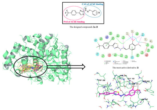
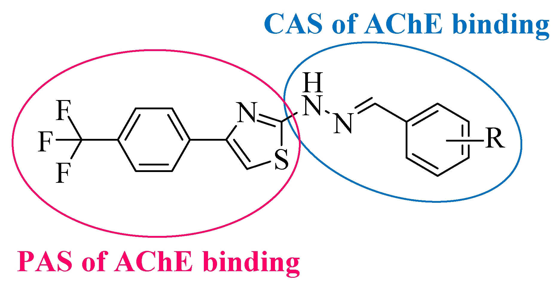
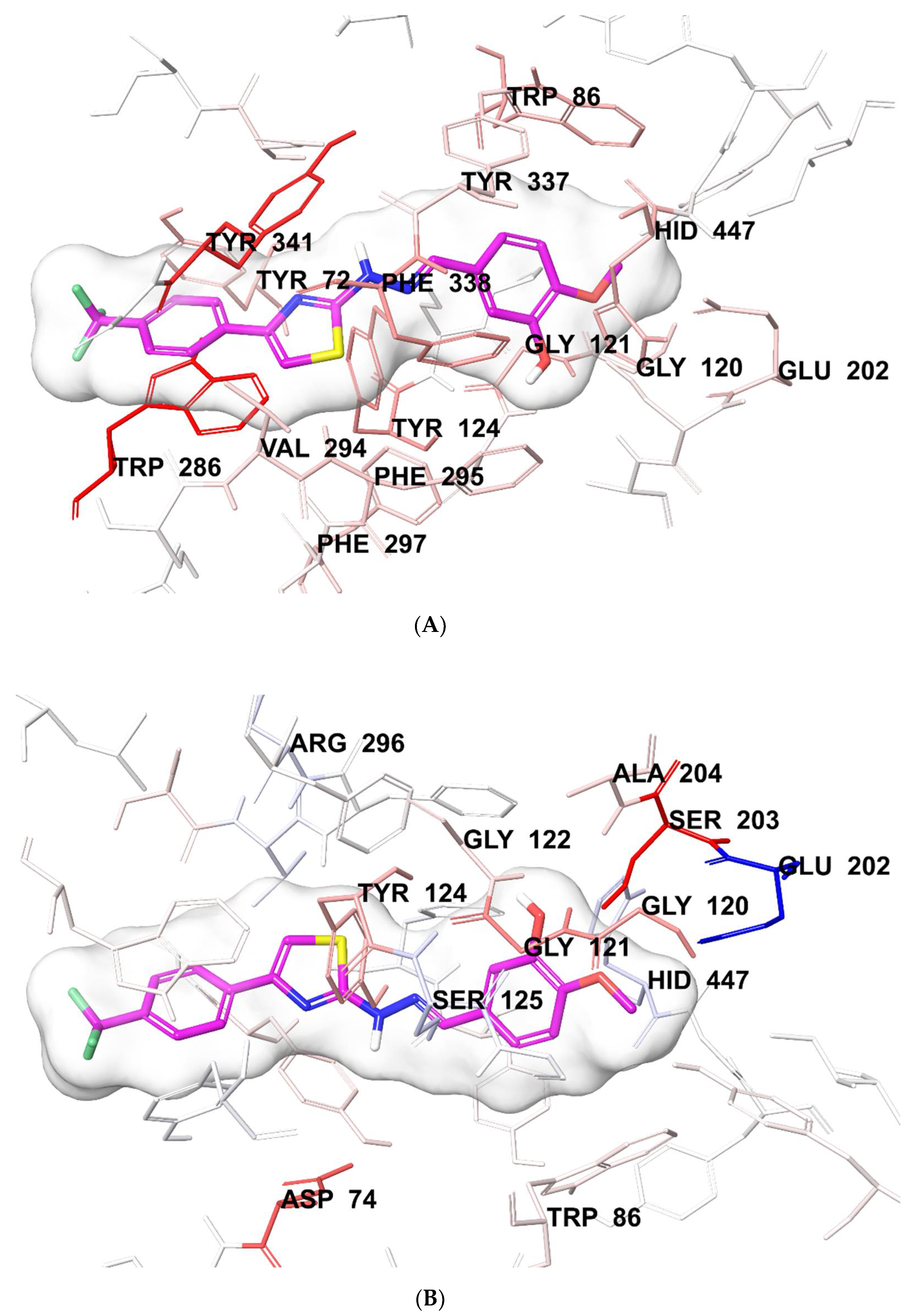
3.6. Molecular Docking

As described in the inhibition assay for cholinesterase enzymes, compounds 2a, 2b, 2e, 2g, and 2i were found to be the most active derivatives against the AChE enzyme in the series. Therefore, docking experiments were conducted to determine their inhibition ability in silico. Based on the docking experiments, further information into the binding modes of compounds 2a, 2b, 2e, 2g, and 2i and assessment of the impact of structural modifications on inhibitory activity against the AChE enzyme could be obtained. Studies were carried out using the X-ray crystal structure of Homo sapiens AChE (hAChE PDB ID:4EY7) [22] retrieved from the Protein Data Bank server (www.pdb.org). This X-ray crystal structure was selected because it is of human nature, has high resolution, and contains a donepezil molecule as the ligand. The rendered docking poses of compounds 2a, 2b, 2e, 2g, and 2i are given in Figure 3 and Figure 4, Supplementary Material Figures S1–S5.
According to the crystallographic X-ray structure of the AChE (PDB ID:4EY7), the active pocket enzyme consisted of two major binding sites, the CAS and PAS. The CAS contained the residues of amino acids Trp86, Tyr133, Glu202, Ser203, Tyr337, Phe338, and His447, while amino acids Tyr72, Asp74, Tyr124, Trp286, Phe295, and Tyr341 were identified in the PAS [54,55,56,57] (Figure 3). Donepezil interacts with both the CAS and PAS. Thus, it can be concordantly settled into the gorge due to its dual binding site functionality [58,59]. There is a π–π interaction between the benzyl of donepezil and the indole of Trp86. Moreover, the formation of cation–π interactions are in question between the protonated nitrogen atom of piperidine and the indole of Trp86 and the phenyl of Tyr337. Thus, the benzylpiperidine group of donepezil is strongly located in the CAS. The interactions related to the 1-indanone ring are important for binding to the PAS. In this context, Trp286 amino acid is very essential. The indole of this amino acid creates a π–π interaction with the 1-indanone ring. Furthermore, a hydrogen bond formation is observed between the carbonyl of the 1-indanone and the amine of Phe295 [22,23,24,60,61].
Supplementary Material Figure S1 presents the superimposition of compounds 2a, 2b, 2e, 2g, and 2i in the active region of the AChE. The 2- and 3-dimensional docking poses of compounds 2a, 2b, 2e, 2g, and 2i are given in Figure 4, Supplementary Material Figures S2–S5. As can be seen from the docking studies, the designed and synthesized compounds in this study can be located in the active region, similar to donepezil. According to these poses, the phenyl of the 4-(4-(trifluoromethyl)phenyl)thiazole group creates a π–π interaction with the indole of Trp286 in compounds 2e, 2g, and 2i. This π–π interaction with Trp286 was observed with the thiazole ring of compound 2a. For compound 2b, both of the phenyl and thiazole rings of the 4-(4-(trifluoromethyl)phenyl)thiazole group established π–π interactions with the phenyl of Tyr341. Moreover, in compound 2e, there was a π–π interaction between the thiazole ring and the phenyl of Tyr341. All of these interactions described provided insight into how compounds 2a, 2b, 2e, 2g, and 2i bound to the PAS.
When the interactions were analyzed in terms of binding to the CAS, a π–π interaction formation was seen between the substituted phenyl ring and the indole of Trp86 in compounds 2a and 2b. For compounds 2e, 2g, and 2i, this interaction was observed with the imidazole of Hid447. Furthermore, the phenyl ring of compound 2g formed the other π–π interactions with the phenyl of Phe338. Another important interaction of binding to the CAS was the formation of a hydrogen bond with the hydroxyl of Tyr124. This interaction was seen with the nitrogen of hydrazine moiety in compounds 2a and 2i.
The main chemical structural difference of compounds 2a, 2b, 2e, 2g, and 2i was the various substituents of the phenyl ring. Compounds 2a and 2b had hydroxyl at the 3rd and 4th positions of the phenyl, respectively. The hydroxyl of compound 2a created a hydrogen bond with the carbonyl of Asp74; however, two hydrogen bond formations were seen between the hydroxyl in compound 2b and the amino of Gly120 and the carbonyl of Glu202, respectively. Compound 2e bore a methoxy moiety at the 3rd position of the phenyl, which was different than that of compounds 2a and 2b. This methoxy established two hydrogen bonds with the amino of Gly121 and the hydroxyl of Ser203. There were two methoxy moieties at the 3rd and 4th positions of the phenyl in compound 2g. Each of them formed two hydrogen bonds with the hydroxyl of Ser203. Compound 2i contained hydroxyl and methoxy moieties at the 3rd and 4th positions of the phenyl, respectively. The hydroxyl of compound 2i created three hydrogen bonds with the hydroxyl of Ser203 and the amino groups of Gly121 and Gly122. Another hydrogen bond was found between the methoxy at the 4th position of the phenyl and the hydroxyl of Ser203.
Consequently, molecular docking studies have shown that the substituents at the 3rd and 4th positions of the phenyl ring contribute positively to enzyme activity. This result supported the finding that compounds 2g and 2i, which had disubstituents at the 3rd and 4th positions, displayed a more potent inhibition profile than compounds 2a, 2b, and 2e, which carried the substituents at the 3rd or 4th positions. Moreover, the reason why compound 2i was more active than compound 2g was that it had additional formations of hydrogen bonds belonging to the substituents at the 3rd and 4th positions.
Molecular docking studies were carried out in detail using the per-residue interaction panel of Glide to determine the contribution of van der Waals and electrostatic interactions in binding to the enzyme active region. Figure 5 presents the van der Waals and electrostatic interactions of compound 2i, which was found to be the most active agent. It can be seen that this compound had favorable van der Waals interactions with amino acids Tyr72, Trp86, Gly120, Gly121, Tyr124, Glu202, Trp286, Val294, Phe295, Phe297, Tyr337, Phe338, Tyr341, and Hid447, which are displayed in pink and red, as described in the Glide user guide [62]. Similarly, promising electrostatic contributions of compound 2i were determined with amino acids Asp74, Trp86, Gly120, Gly121, Gly122, Tyr124, Ser125, Glu202, Ser203, Ala204, Arg296, and Hid447. All of these findings explained why compound 2i exhibited a stronger inhibition profile than the other compounds.
Supplementary Material Figure S6 shows the docking pose of compound 2d, which displayed a moderate enzyme inhibitory activity in the series. This compound was subjected to molecular docking studies to explain why it was less active than the other compounds (2a, 2b, 2e, 2g, and 2i) and what caused this result chemically. When the docking poses of this compound were analyzed, it was seen that there were common interactions related to the 4-(4-(trifluoromethyl)phenyl)thiazole and benzylidenehydrazine groups in terms of binding to the CAS and PAS regions mentioned above. Compound 2d had a methoxy moiety at the 2nd position of the phenyl ring, which was different from that of the other compounds (2a, 2b, 2e, 2g, and 2i). However, the hydrogen bond formations observed at the 3rd and 4th positions of other compounds could not be detected at the 2nd position of this compound. The absence of additional interactions belonging to the substituents of the phenyl ring could clarify the decrease in the enzyme inhibitory activity. It was believed that the 2nd position presented a steric obstacle against interaction with any amino acid conformationally. Moreover, it was assumed that this position negatively affected the interaction of the 3rd/4th positions of the compounds, which had disubstituents (at the 2nd and 5th positions) and trisubstituents (at the 3rd, 4th, and 5th positions). Therefore, these compounds could only exhibit interactions related to common chemical structures and could not display remarkable enzyme inhibition profiles such as with compounds 2a, 2b, 2e, 2g, and 2i.

4. Conclusions

The designed 3-((2-(4-(4-(trifluoromethyl)phenyl)thiazol-2-yl)hydrazineylidene)methyl)-substituedphenyl derivatives as AChE inhibitors were successfully synthesized and analyzed in detail using 1H NMR, 13C NMR, and HRMS spectrometric techniques. It was also concluded that the synthesized compounds presented promising drug-like characteristics and ADME properties with the help of ADME predictions by using QikProp 4.8 software. Their in vitro enzyme inhibitory activity test results showed that none of the compounds exhibited significant enzyme inhibition against the BChE enzyme. Compounds 2a, 2b, 2e, 2g, and 2i containing the substituents at the 3rd or 4th or 3rd/4th positions of the phenyl ring displayed the highest AChE inhibitory activity with the IC50 values of 0.063 ± 0.003, 0.056 ± 0.002, 0.040 ± 0.001, 0.031 ± 0.001, and 0.028 ± 0.001 µM, respectively. This is thought to be caused by that the substituents at the 3rd or 4th or 3rd/4th positions of the phenyl ring, which activated the compounds biologically. Specially, compound 2i displayed a strong inhibitory activity against AChE (IC50 = 0.028 ± 0.001 µM), which had an inhibition profile at a similar rate as the reference drug, donepezil (IC50 value = 0.021 ± 0.001 µM). This superiority of this compound could be explained by the molecular modeling studies. According to the results of docking studies, compound 2i bounded to the active site by forming additional interactions via its substituent at the 3rd and 4th positions with Gly121, Gly122, and Ser203 amino acids. In summary, structural modifications can be further made on the basis of the new thiazolylhydrazone derivatives to seek compounds having higher inhibitory activity against ChE enzymes. All these findings encourage medicinal chemists to explore more structures based on thiazolylhydrazone derivatives for high-efficiency ChE inhibitors. New chemical modifications can be designed based on this paper so that novel effective derivatives may be subject to future studies. Hence, studies to develop new candidates that may be effective in Alzheimer’s disease can be followed rationally.

Supplementary Materials

The following are available online, Table S1. IC50 values of compounds 2a, 2b, 2d, 2e, 2g, 2i, 2j and donepezil against AChE. Figure S1. The superimposition pose of selected compounds in the enzyme active site (AChE PDB Code: 4EY7). Figure S2. The two- (A) and three-dimensional (B) interacting mode of compound 2a in the active region of AChE. The inhibitor and important residues in the active site of enzyme are presented by tube model and colored with grey and aquamarine, respectively (AChE PDB Code: 4EY7). Figure S3. The two- (A) and three-dimensional (B) interacting mode of compound 2b in the active region of AChE. The inhibitor and important residues in the active site of enzyme are presented by tube model and colored with blue and aquamarine, respectively (AChE PDB Code: 4EY7). Figure S4. The two- (A) and three-dimensional (B) interacting mode of compound 2e in the active region of AChE. The inhibitor and important residues in the active site of enzyme are presented by tube model and colored with orange and aquamarine, respectively (AChE PDB Code: 4EY7). Figure S5. The two- (A) and three-dimensional (B) interacting mode of compound 2g in the active region of AChE. The inhibitor and important residues in the active site of enzyme are presented by tube model and colored with dark green and aquamarine, respectively (AChE PDB Code: 4EY7). Figure S6. The two- (A) and three-dimensional (B) interacting mode of compound 2d in the active region of AChE. The inhibitor and important residues in the active site of enzyme are presented by tube model and colored with pink and aquamarine, respectively (AChE PDB Code: 4EY7). Figure S7. The thin-layer chromatography of the synthesized compounds. Figure S8. Purity of compound 2i. Figure S9. 1H NMR spectra of the compound 2i. Figure S10. 13C NMR spectra of the compound 2i. Figure S11. HRMS spectra of the compound 2i.

Author Contributions

Y.Ö. and Z.A.K. conceived and designed the experiments; B.N.S. performed the synthesis, activity tests and molecular docking studies; D.O., U.A.Ç. and B.K.Ç. performed the synthesis; D.O. and S.L. performed analysis studies; B.N.S., D.O., U.A.Ç., S.L., B.K.Ç., Y.Ö. and Z.A.K. wrote the paper. All authors have read and agreed to the published version of the manuscript.

Funding

This study was financially supported by Anadolu University Scientific Projects Fund, Project No: 2005S054.

Conflicts of Interest

The authors declare no conflict of interest.

References

  1. Kalaria, R.N.; Maestre, G.E.; Arizaga, R.; Friedland, R.P.; Galasko, D.; Hall, K.; Luchsinger, J.A.; Ogunniyi, A.; Perry, E.K.; Potocnik, F.; et al. Alzheimer’s disease and vascular dementia in developing countries: Prevalence, management, and risk factors. Lancet Neurol. 2008, 7, 812–826. [Google Scholar] [CrossRef] [Green Version]
  2. Rodgers, A.B. Alzheimer’s Disease: Unraveling the Mystery; Publication Number: 02-3782; National Institutes of Health Publications: Bethesda, MD, USA, 2002.
  3. Huber, A.; Stuchbury, G.; Bürkle, A.; Burnell, J.; Münch, G. Neuroprotective therapies for Alzheimer’s disease. Curr. Pharm. Des. 2006, 12, 705–717. [Google Scholar] [CrossRef] [PubMed]
  4. Smith, D.G.; Cappai, R.; Barnham, K.J. The redox chemistry of the Alzheimer’s disease amyloid β peptide. Biomembranes 2007, 768, 1976–1990. [Google Scholar] [CrossRef] [PubMed] [Green Version]
  5. Craig, L.A.; Hong, N.S.; McDonald, R.J. Revisiting the cholinergic hypothesis in the development of Alzheimer’s disease. Neurosci. Biobehav. Rev. 2011, 35, 1397–1409. [Google Scholar] [CrossRef] [Green Version]
  6. Hardy, J.; Selkoe, D.J. The amyloid hypothesis of Alzheimer’s disease: Progress and problems on the road to therapeutics. Science 2002, 297, 353–356. [Google Scholar] [CrossRef] [Green Version]
  7. Su, B.; Wang, X.; Nunomura, A.; Moreira, P.I.; Lee, H.G.; Perry, G.; Smith, M.A.; Zhu, X. Oxidative stress signaling in Alzheimer’s disease. Curr. Alzheimer Res. 2008, 5, 525–532. [Google Scholar] [CrossRef]
  8. Holmes, C. Systemic inflammation and a lzheimer’s disease. Neuropathol. Appl. Neurobiol. 2013, 39, 51–68. [Google Scholar] [CrossRef]
  9. Greenough, M.A.; Camakaris, J.; Bush, A.I. Metal dyshomeostasis and oxidative stress in Alzheimer’s disease. Neurochem. Int. 2013, 62, 540–555. [Google Scholar] [CrossRef]
  10. Bajda, M.; Więckowska, A.; Hebda, M.; Guzior, N.; Sotriffer, C.A.; Malawska, B. Structure-based search for new inhibitors of cholinesterases. Int. J. Mol. Sci. 2013, 14, 5608–5632. [Google Scholar] [CrossRef] [Green Version]
  11. Mason, J.M.; Kokkoni, N.; Stott, K.; Doig, A.J. Design strategies for anti-amyloid agents. Curr. Opin. Struct. Biol. 2013, 13, 526–532. [Google Scholar] [CrossRef]
  12. Shukla, V.; Skuntz, S.; Pant, H.C. Deregulated Cdk5 activity is involved in inducing Alzheimer’s disease. Arch. Med. Res. 2013, 43, 655–662. [Google Scholar] [CrossRef] [PubMed] [Green Version]
  13. Atri, A.; Frölich, L.; Ballard, C.; Tariot, P.N.; Molinuevo, J.L.; Boneva, N.; Windfeld, K.; Raket, L.L.; Cummings, J.L. Effect of idalopirdine as adjunct to cholinesterase inhibitors on change in cognition in patients with Alzheimer disease: Three randomized clinical trials. JAMA 2018, 319, 130–142. [Google Scholar] [CrossRef] [PubMed]
  14. Lahiri, D.K.; Farlow, M.R.; Greig, N.H.; Sambamurti, K. Current drug targets for Alzheimer’s disease treatment. Drug Dev. Res. 2002, 56, 267–281. [Google Scholar] [CrossRef]
  15. Bajda, M.; Guzior, N.; Ignasik, M.; Malawska, B. Current drug targets for Alzheimer’s disease treatment. Curr. Med. Chem. 2011, 18, 4949–4975. [Google Scholar] [CrossRef]
  16. Alagille, D.; DaCosta, H.; Baldwin, R.M.; Tamagnan, G.D. 2-Arylimidazo [2, 1-b] benzothiazoles: A new family of amyloid binding agents with potential for PET and SPECT imaging of Alzheimer’s brain. Bioorg. Med. Chem. Lett. 2011, 21, 2966–2968. [Google Scholar] [CrossRef]
  17. Gan, C.; Zhou, L.; Zhao, Z.; Wang, H. Benzothiazole Schiff-bases as potential imaging agents for β-amyloid plaques in Alzheimer’s disease. Med. Chem. Res. 2013, 22, 4069–4074. [Google Scholar] [CrossRef]
  18. Xie, Y.; Deng, S.; Chen, Z.; Yan, S.; Landry, D.W. Identification of small-molecule inhibitors of the Aβ–ABAD interaction. Bioorg. Med. Chem. Lett. 2006, 16, 4657–4660. [Google Scholar] [CrossRef]
  19. Youssef, K.M.; Fawzy, I.M.; El-Subbagh, H.I. N-substituted-piperidines as Novel Anti-alzheimer Agents: Synthesis, antioxidant activity, and molecular docking study. Future J. Pharm. Sci. 2018, 4, 1–7. [Google Scholar] [CrossRef]
  20. Silva, D.; Chioua, M.; Samadi, A.; Agostinho, P.; Garcão, P.; Lajarín-Cuesta, R.; de los Ríos, C.; Iriepa, I.; Moraleda, I.; Gonzalez-Lafuente, L.; et al. Synthesis, pharmacological assessment, and molecular modeling of acetylcholinesterase/butyrylcholinesterase inhibitors: Effect against amyloid-β-induced neurotoxicity. ACS Chem. Neurosci. 2013, 4, 547–565. [Google Scholar] [CrossRef] [Green Version]
  21. Lan, J.S.; Zhang, T.; Liu, Y.; Yang, J.; Xie, S.S.; Liu, J.; Miao, Z.Y.; Ding, Y. Design, synthesis and biological activity of novel donepezil derivatives bearing N-benzyl pyridinium moiety as potent and dual binding site acetylcholinesterase inhibitors. Eur. J. Med. Chem. 2017, 133, 184–196. [Google Scholar] [CrossRef]
  22. Cheung, J.; Rudolph, M.J.; Burshteyn, F.; Cassidy, M.S.; Gary, E.N.; Love, J.; Matthew, C.F.; Height, J.J. Structures of human acetylcholinesterase in complex with pharmacologically important ligands. J. Med. Chem. 2012, 55, 10282–10286. [Google Scholar] [CrossRef] [PubMed]
  23. Huang, C.S.; Tu, W.T.; Luo, M.; Shi, J.G. Molecular Docking and Design of Novel Heterodimers of Donepezil and Huperzine Fragments as Acetylcholinesterase Inhibitors. Chin. J. Struct. Chem. 2016, 35, 839–848. [Google Scholar]
  24. Vitorovic-Todorovic, M.D.; Koukoulitsa, C.; Juranic, I.O.; Mandic, L.M.; Drakulic, B.J. Structural Modifications of 4-Aryl-4-Oxo-2-Aminylbutanamides and Their Acetyl-and Butyrylcholinesterase Inhibitory Activity. Investigation of AChE-Ligand Interactions by Docking Calculations and Molecular Dynamics Simulations. Supplementary data for the article. Eur. J. Med. Chem. 2014, 81, 158–175. [Google Scholar] [CrossRef] [Green Version]
  25. Neochoritis, C.; Tsoleridis, C.A.; Stephanidou-Stephanatou, J. 1-Arylaminoimidazole-2-thiones as intermediates in the synthesis of imidazo [2,1-b][1,3,4] thiadiazines. Tetrahedron 2008, 64, 3527–3533. [Google Scholar] [CrossRef]
  26. Ibrar, A.; Khan, A.; Ali, M.; Sarwar, R.; Mehsud, S.; Farooq, U.; Halimi, M.A.S.; Khan, I.; Al-Harrasi, A. Combined in vitro and in silico studies for the anticholinesterase activity and pharmacokinetics of coumarinyl thiazoles and oxadiazoles. Front. Chem. 2018, 6, 61–72. [Google Scholar] [CrossRef] [PubMed] [Green Version]
  27. Tripathi, R.K.P.; Sasi, M.V.; Gupta, S.K.; Krishnamurthy, S.; Ayyannan, S.R. Design, synthesis, and pharmacological evaluation of 2-amino-5-nitrothiazole derived semicarbazones as dual inhibitors of monoamine oxidase and cholinesterase: Effect of the size of aryl binding site. J. Enzyme Inhib. Med. Chem. 2018, 33, 37–57. [Google Scholar] [CrossRef] [PubMed] [Green Version]
  28. Ma, Z.Y.; Yang, Q.; Zhang, Y.G.; Li, J.J.; Yang, G.L. Design, synthesis and evaluation of N-acyl-4-phenylthiazole-2-amines as acetylcholinesterase inhibitors. Acta Pharm. Sin. 2014, 49, 813–818. [Google Scholar]
  29. Tian, Y.F.; Chen, J.T.; Li, J.J.; Zhang, Y.C.; Cao, T.T.; Ma, Z.Y. Design, synthesis and evaluation of new L-proline derivatives as acetylcholinesterase inhibitors. Acta Pharm. Sin. 2015, 50, 719–724. [Google Scholar]
  30. Zhang, X.Z.; Xu, Y.; Jian, M.M.; Yang, K.; Ma, Z.Y. Synthesis, in vitro assays, molecular docking, theoretical ADMET prediction, and evaluation of 4-methoxy-phenylthiazole-2-amine derivatives as acetylcholinesterase inhibitors. Med. Chem. Res. 2019, 28, 1683–1693. [Google Scholar] [CrossRef]
  31. Xu, Y.; Jian, M.M.; Han, C.; Yang, K.; Bai, L.; Cao, F.; Ma, Z.Y. Design, synthesis and evaluation of new 4-arylthiazole-2-amine derivatives as acetylcholinesterase inhibitors. Bioorg. Med. Chem. Lett. 2020, 30, 126985. [Google Scholar] [CrossRef]
  32. Ellman, G.L.; Courtney, K.D.; Andres, V., Jr.; Feather-Stone, R.M. A new and rapid colorimetric determination of acetylcholinesterase activity. Biochem. Pharmacol. 1961, 7, 88–95. [Google Scholar] [CrossRef]
  33. Saglık, B.N.; Ilgın, S.; Ozkay, Y. Synthesis of new donepezil analogues and investigation of their effects on cholinesterase enzymes. Eur. J. Med. Chem. 2016, 124, 1026–1040. [Google Scholar] [CrossRef] [PubMed]
  34. Demir Özkay, Ü.; Can, Ö.D.; Sağlık, B.N.; Acar Çevik, U.; Levent, S.; Özkay, Y.; Ilgın, S.; Atlı, Ö. Design, synthesis, and AChE inhibitory activity of new benzothiazole-piperazines. Bioorg. Med. Chem. Lett. 2016, 26, 5387–5394. [Google Scholar] [CrossRef] [PubMed]
  35. Acar Çevik, U.; Levent, S.; Sağlık, B.N.; Özkay, Y.; Kaplancıklı, Z.A. Synthesis of Novel 4-(Dimethylaminoalkyl) piperazine-1-carbodithioa te Derivatives as Cholinesterase Inhibitors. Lett. Drug Des. Discov. 2017, 14, 528–539. [Google Scholar] [CrossRef]
  36. Levent, S.; Acar Çevik, U.; Sağlık, B.N.; Özkay, Y.; Can, Ö.D.; Demir Özkay, Ü.; Uçucu, Ü. Anticholinesterase activity screening of some novel dithiocarbamate derivatives including piperidine and piperazine moieties. Phosphorus Sulfur Silicon Relat. Elem. 2017, 192, 469–474. [Google Scholar] [CrossRef]
  37. Hussein, W.; Sağlık, B.N.; Levent, S.; Korkut, B.; Ilgın, S.; Özkay, Y.; Kaplancıklı, Z.A. Synthesis and biological evaluation of new cholinesterase inhibitors for Alzheimer’s disease. Molecules 2018, 23, 2033. [Google Scholar] [CrossRef] [Green Version]
  38. Tok, F.; Koçyiğit Kaymakçıoğlu, B.; Sağlık, B.N.; Levent, S.; Özkay, Y.; Kaplancıklı, Z.A. Synthesis and biological evaluation of new pyrazolone Schiff bases as monoamine oxidase and cholinesterase inhibitors. Bioorg. Chem. 2019, 84, 41–50. [Google Scholar] [CrossRef]
  39. Acar Çevik, U.; Sağlık, B.N.; Levent, S.; Osmaniye, D.; Kaya Çavuşoğlu, B.; Özkay, Y.; Kaplancıklı, Z.A. Synthesis and AChE-inhibitory activity of new benzimidazole derivatives. Molecules 2019, 24, 861. [Google Scholar] [CrossRef] [Green Version]
  40. Osmaniye, D.; Sağlık, B.N.; Acar Çevik, U.; Levent, S.; Kaya Çavuşoğlu, B.; Özkay, Y.; Kaplancıklı, Z.A.; Turan-Zitouni, G. Synthesis and AChE inhibitory activity of novel thiazolylhydrazone derivatives. Molecules 2019, 24, 2392. [Google Scholar] [CrossRef] [Green Version]
  41. QikProp, version 4.8; Schrödinger, LLC.: New York, NY, USA, 2016.
  42. Yan, G.; Hao, L.; Niu, Y.; Huang, W.; Wang, W.; Xu, F.; Liang, L.; Wang, C.; Jin, H.; Xu, P. 2-Substituted-thio-N-(4-substituted-thiazol/1H-imidazol-2-yl) acetamides as BACE1 inhibitors: Synthesis, biological evaluation and docking studies. Eur. J. Med. Chem. 2017, 137, 462–475. [Google Scholar] [CrossRef]
  43. Caliandro, R.; Pesaresi, A.; Cariati, L.; Procopio, A.; Oliverio, M.; Lamba, D. Kinetic and structural studies on the interactions of Torpedo californica acetylcholinesterase with two donepezil-like rigid analogues. J. Enzyme Inhib. Med. Chem. 2018, 33, 794–803. [Google Scholar] [CrossRef] [PubMed] [Green Version]
  44. Henchoz, Y.; Bard, B.; Guillarme, D.; Carrup, P.A.; Veuthey, J.L.; Martel, S. Analytical tools for the physicochemical profiling of drug candidates to predict absorption/distribution. Anal. Bioanal. Chem. 2009, 394, 707–729. [Google Scholar] [CrossRef] [PubMed]
  45. Kerns, E.H. High throughput physicochemical profiling for drug discovery. J. Pharm. Sci. 2001, 90, 1838–1858. [Google Scholar] [CrossRef] [PubMed]
  46. Kerns, E.H.; Di, L. Physicochemical profiling: Overview of the screens. Drug Discov. Today Technol. 2004, 1, 343–348. [Google Scholar] [CrossRef]
  47. Lipinski, C.A.; Franco, L.; Dominy, B.W.; Feeney, P.J. Experimental and computational approaches to estimate solubility and permeability in drug discovery and development settings. Adv. Drug Deliv. Rev. 1997, 23, 3–25. [Google Scholar] [CrossRef]
  48. Jorgensen, W.L.; Duffy, E.M. Prediction of drug solubility from structure. Adv. Drug Deliv. Rev. 2002, 54, 355–366. [Google Scholar] [CrossRef]
  49. Daina, A.; Michielin, O.; Zoete, V. SwissADME: A free web tool to evaluate pharmacokinetics, drug-likeness and medicinal chemistry friendliness of small molecules. Sci. Rep. 2017, 7, 1–13. [Google Scholar] [CrossRef] [Green Version]
  50. Dunitz, J.D.; Taylor, R. Organic fluorine hardly ever accepts hydrogen bonds. Chem-A Eur. J. Med. Chem. 1997, 3, 89–98. [Google Scholar] [CrossRef]
  51. Dvira, H.; Silman, I.; Harel, M.; Rosenberry, T.L.; Sussman, J.L. Acetylcholinesterase: From 3D structure to function. Chem. Biol. Interact. 2010, 187, 10–22. [Google Scholar] [CrossRef] [Green Version]
  52. Di, L.; Kerns, E.H.; Fan, K.; McConnell, O.J.; Carter, C.T. High throughput artificial membrane permeability assay for blood–brain barrier. Eur. J. Med. Chem. 2003, 38, 223–232. [Google Scholar] [CrossRef]
  53. Ning, X.; Guo, Y.; Wang, X.; Ma, X.; Tian, C.; Shi, X.; Zhu, R.; Cheng, C.; Du, Y.; Ma, Z.; et al. Design, synthesis, and biological evaluation of (e)-3, 4-dihydroxystyryl aralkyl sulfones and sulfoxides as novel multifunctional neuroprotective agents. J. Med. Chem. 2014, 57, 4302–4312. [Google Scholar] [CrossRef] [PubMed]
  54. Colovic, M.B.; Krstic, D.; Lazarevic-Pactil, T.; Bondzic, A.M.; Vasic, V. Acetylcholinesterase inhibitors: Pharmacology and toxicology. Curr. Neuropharmacol. 2013, 11, 315–335. [Google Scholar] [CrossRef] [PubMed] [Green Version]
  55. Atanasova, M.; Stavrakov, G.; Philipova, L.; Zheleva, D.; Yordanov, N.; Doytchinova, I. Galantamine derivatives with indole moiety: Docking, design, synthesis and acetylcholinesterase inhibitory activity. Bioorg. Med. Chem. 2015, 23, 5382–5389. [Google Scholar] [CrossRef] [PubMed]
  56. Wu, M.Y.; Esteban, G.; Brogi, S.; Shionoya, M.; Wang, I.; Campiani, G.; Unzeta, M.; Inokuchi, T.; Butini, S.; Marco-Contelles, J. Donepezil-like multifunctional agents: Design, synthesis, molecular modeling and biological evaluation. Eur. J. Med. Chem. 2015, 121, 864–879. [Google Scholar] [CrossRef]
  57. Genest, D.; Rochais, C.; Lecoutey, C.; Jana Sopkova-de Oliveira, S.; Ballandonne, C.; Butt-Gueulle, S.; Legay, R.; Since, M.; Dallemagne, P. Design, synthesis and biological evaluation of novel indano-and thiaindano-pyrazoles with potential interest for Alzheimer’s disease. Med. Chem. Commun. 2013, 4, 925–931. [Google Scholar] [CrossRef]
  58. Al-Rashid, Z.F.; Hsung, R.P. A computational view on the significance of E-ring in binding of (+)-arisugacin A to acetylcholinesterase. Bioorg. Med. Chem. 2015, 25, 4848–4853. [Google Scholar] [CrossRef] [Green Version]
  59. Alipour, M.; Khoobi, M.; Foroumadi, A.; Nadri, H.; Moradi, A.; Sakhteman, A.; Ghandi, M.; Shafiee, A. Novel coumarin derivatives bearing N-benzyl pyridinium moiety: Potent and dual binding site acetylcholinesterase inhibitors. Bioorg. Med. Chem. 2012, 20, 7214–7222. [Google Scholar] [CrossRef]
  60. Cheung, J.; Gary, E.N. Structures of human acetylcholinesterase bound to dihydrotanshinone I and territrem B show peripheral site flexibility. ACS Med. Chem. Lett. 2013, 4, 1091–1096. [Google Scholar] [CrossRef] [Green Version]
  61. Chiou, S.Y.; Huang, C.F.; Hwang, M.T.; Lin, G. Comparison of Active Sites of Butyrylcholinesterase and Acetylcholinesterase Based on Inhibition by Geometric Isomers of Benzene-di-N-Substituted Carbamates. J. Biochem. Molecular Toxicology 2009, 23, 303–308. [Google Scholar] [CrossRef]
  62. Glide; Version 7.1; Schrödinger, LLC.: New York, NY, USA, 2016.
Figure 1. The chemical parts of the target compounds 2a2l according to the binding to the catalytic anionic site (CAS) and peripheral anionic binding site (PAS) regions.
Molecules 25 04312 g001
Scheme 1. Synthesis way of the compounds 2a2l.
Molecules 25 04312 sch001
Figure 2. (A) Lineweaver–Burk plots for the inhibition of AChE by compound 2i. [S], substrate concentration (µM); V, reaction velocity (1/V (abs/min)−1). Inhibitor concentrations are shown at the left. (B) Secondary plot for the calculation of the steady-state inhibition constant (Ki) of compound 2i. Ki was calculated as 0.025 µM.
Molecules 25 04312 g002
Figure 3. The CAS and PAS regions of AChE enzyme along with the important amino acids.
Molecules 25 04312 g003
Figure 4. The two- (A) and three-dimensional (B) interacting mode of compound 2i in the active region of AChE. The inhibitor and important residues in the active site of enzyme are presented by a tube model colored with purple and aquamarine, respectively (AChE PDB Code: 4EY7).
Molecules 25 04312 g004
Figure 5. The van der Waals (A) and electrostatic interactions (B) of compound 2i with an active region of AChE. The active ligand has a lot of favorable van der Waals and electrostatic interactions. The residues are colored (blue, red, and pink) according to the distance from ligand by per-residue interaction panel.
Molecules 25 04312 g005
Table 1. % Inhibition and IC50 values of the synthesized compounds, donepezil and tacrine against AChE and BChE enzymes.
CompoundsAChE % InhibitionAChE
IC50 (µM)
BChE % InhibitionBChE
IC50 (µM)
10−3 M10−4 M10−3 M10−4 M
2a93.425 ± 1.65290.465 ± 1.3220.063 ± 0.00352.380 ± 0.93542.466 ± 0.978>100
2b90.285 ± 1.45182.151 ± 1.4780.056 ± 0.00255.488 ± 0.87540.541 ± 0.710>100
2c78.215 ± 1.02545.108 ± 0.971>10043.612 ± 0.95532.466 ± 0.721>1000
2d89.462 ± 2.04182.445 ± 1.6950.147 ± 0.00651.950 ± 0.99443.795 ± 0.895>100
2e90.611 ± 1.81585.387 ± 1.7300.040 ± 0.00160.150 ± 1.03941.575 ± 0.907>100
2f71.658 ± 1.39540.487 ± 0.890>10040.858 ± 0.89531.495 ± 0.820>1000
2g93.461 ± 1.63288.347 ± 1.6040.031 ± 0.00163.515 ± 1.15540.388 ± 0.845>100
2h77.561 ± 1.75841.947 ± 0.976>10045.795 ± 0.92634.306 ± 0.738>1000
2i95.207 ± 1.50292.130 ± 1.7980.028 ± 0.00165.015 ± 1.47041.518 ± 0.870>100
2j91.326 ± 2.10784.945 ± 1.3690.138 ± 0.00550.811 ± 0.98839.628 ± 0.865>100
2k65.298 ± 1.04540.825 ± 0.940>10040.171 ± 0.90225.631 ± 0.633>1000
2l62.797 ± 1.27735.369 ± 0.895>10038.654 ± 0.81523.208 ± 0.759>1000
Donepezil99.254 ± 2.10497.426 ± 1.8900.021 ± 0.001---
Tacrine---98.255 ± 1.89595.465 ± 1.3440.006 ± 0.0002
Table 2. Calculated ADME (absorption, distribution, metabolism, elimination) parameters of compounds 2a2l.
Comp.MWRBDMMVDHBAHBPSAlogPlogSPCacologBBPMDCKCNSPM%HOAVRFVRT
2a363.35756.3521073.11224.75057.6344.295−6.0851363.075−0.2925986.2320210001
2b363.35758.0871073.11224.75057.6344.295−6.0851363.095−0.2925986.3240210001
2c379.35666.6001090.47935.50077.3293.641−5.694602.692−0.7562477.7811310000
2d377.38357.2361122.80914.75042.0935.142−6.5964525.1170.29310,0001210011
2e377.38356.3131113.12814.75043.4335.066−6.2194525.1170.31310,0001210011
2f407.41065.3471189.49815.50050.4405.192−6.6554525.1080.23110,0001310011
2g407.41067.5351179.30915.50051.6245.098−6.3514525.1080.24510,0001310011
2h391.36747.0471084.67115.50053.6984.576−5.9044526.6660.37410,0001110001
2i393.38367.5511142.73725.50065.4474.388−6.2491454.418−0.3346420.9850310001
2j393.38366.8251132.21825.50064.3004.374−5.8901674.494−0.2207477.3110310001
2k437.43677.5961265.95516.25059.0895.273−6.6194525.1080.17710,0001410011
2l423.40975.8771207.46626.25073.2614.414−6.1771428.142−0.3866295.690410001
MW: Molecular weight RB: Number of rotatable bonds (recommended value: 0–15) DM: Computed dipole moment (recommended value: 1–12.5) MV: Total solvent-accessible volume (recommended value: 500–2000) DHB: Estimated number of hydrogen bond donors (recommended value: 0–6) AHB: Estimated number of hydrogen bond acceptors (recommended value: 2–20) PSA: Van der Waals surface area of polar nitrogen and oxygen atoms and carbonyl carbon atoms (recommended value: 7–200) logP: Predicted octanol/water partition coefficient (recommended value: −2–6.5) logS: Predicted aqueous solubility (recommended value: −6.5–0.5) PCaco: Predicted apparent Caco-2 cell permeability (recommended value: <25 poor, >500 great) logBB: Predicted brain/blood partition coefficient (recommended value: −3–1.2) PMDCK: Predicted apparent MDCK cell permeability (recommended value: <25 poor, >500 great) CNS: Predicted central nervous system activity on a −2 (inactive) to +2 (active) scale (recommended value: −2 (inactive), +2 (active)) PM: Number of likely metabolic reactions (recommended value: 1–8) %HOA: Predicted human oral absorption percent (recommended value: >80% is high, <25% is poor) VRF: Number of violations of Lipinski’s rule of five. The rules are: MW < 500, logP < 5, DHB ≤ 5, AHB≤ 10, Positive PSA value. VRT: Number of violations of Jorgensen’s rule of three. The three rules are: logS > −5.7, PCaco > 22 nm/s, PM < 7.
Table 3. Type of blood–brain barrier (BBB) penetration of compound 2i.
ClassificationType of BBB PermeationCompoundType of BBB Permeation
CNS+High BBB permeation
Pe (10−6 cm s−1) > 4.0
2iCNS+
High BBB permeation
CNS>−Low BBB permeation
Pe (10−6 cm s−1) < 2.0
CNS±BBB permeation uncertain
2.0 < Pe (10−6 cm s−1) < 4.0

