Article

Anti-Obesity and Gut Microbiota Modulation Effect of Secoiridoid-Enriched Extract from Fraxinus mandshurica Seeds on High-Fat Diet-Fed Mice

1
College of Food Science and Technology, Northwest University, 229 Taibai North Road, Xi’an 710069, China
2
Department of Pharmaceutical Engineering, College of Chemical Engineering, Northwest University, 229 Taibai North Road, Xi’an 710069, China
3
College of Chemical Engineering, Northwest University, 229 Taibai North Road, Xi’an 710069, China
4
Department of Food Science, Rutgers University, 65 Dudley Road, New Brunswick, NJ 08901, USA
*
Authors to whom correspondence should be addressed.
Molecules 2020, 25(17), 4001; https://doi.org/10.3390/molecules25174001
Submission received: 14 August 2020 / Revised: 31 August 2020 / Accepted: 1 September 2020 / Published: 2 September 2020
(This article belongs to the Special Issue Anti-obesity Effects of Natural Compounds)

Abstract

:
Previously we conducted a phytochemical study on the seeds of Fraxinus excelsior and isolated nine secoiridoid compounds with adipocyte differentiation inhibitory activity and peroxisome proliferator activated receptor alpha (PPARα) activation effects. However, the bioactive constituents and functions of Fraxinus mandshurica seeds have not been studied. In the present study, we investigated the secoiridoid compounds in F. mandshurica seed extract (FM) using column chromatography, 1H-NMR, 13C-NMR and HPLC-DAD methods. The pancreatic lipase inhibitory activities of isolated compounds were evaluated in vitro. Additionally, the anti-obesity and gut microbiota modulation effect of FM on high-fat diet-induced obesity in C57BL/6 mice were also studied in vivo. The results showed that 19 secoiridoids were isolated from FM and identified. The total content of secoiridoids in FM reached 181.35 mg/g and the highest content was nuzhenide (88.21 mg/g). All these secoiridoid compounds exhibited good pancreatic lipase inhibitory activity with inhibition rate ranged from 33.77% to 70.25% at the concentration of 100 μM. After obese mice were administrated with FM at 400 mg/kg.bw for 8 weeks, body weight was decreased by 15.81%. Moreover, FM could attenuate the lipid accumulation in serum and liver, relieve the damage in liver and kidney, and extenuate oxidative stress injury and inflammation caused by obesity in mice. FM could also modulate the structural alteration of gut microbiota in obese mice, increasing the proportion of anti-obesity gut microbiota (Bacteroidetes, Bacteroidia, S24-7 and Allobaculum), and reducing the proportion of obesogenic gut microbiota (Firmicutes and Dorea). This study suggests that F. mandshurica seeds or their secoiridoids may have potential for use as a dietary supplement for obesity management.

1. Introduction

With widespread social and lifestyle changes, obesity has become a serious public health issue. Obesity can cause many chronic diseases and metabolic disorders such as hyperlipidaemia [1], hypertension [2], cardiovascular diseases [3] type 2 diabetes [4] and certain cancers [5]. Currently, there are approximately 641 million obese individuals worldwide. China has the largest number of obese people and the prevalence of obesity in children and adolescents has been increasing [6]. By 2030, 49.5 million children will suffer from obesity, adversely affecting their physical and intellectual development [7,8]. Therefore, there is an urgent need to reduce both its prevalence and impact. Current strategies for obesity treatments including exercise, dietary control, life-style changes, prescription weight-loss medications and surgeries [9]. Among these options, appropriate diet and lifestyle changes plus moderate exercise are generally considered the most ideal ways to lose weight [10]. However, several clinical and epidemic studies have shown that maintaining long-term lifestyle modification is a great challenge to obese patients [11]. Pharmacotherapy can provide an easy way to accelerate weight loss, hence natural supplements derived from plants or synthetic chemicals have become a better choice for obesity treatment [12,13,14,15].
Fraxinus mandshurica Rupr., belonging to the Oleaceae family, is one of the most widely used hardwood trees in Northeast China, especially in the area of Changbai Mountain [16]. The dried bark of F. mandshurica has been used as traditional Chinese medicine for the treatment of inflammatory, fever, rheumatism arthritis and urinary retention [17]. Phytochemical investigations on F. mandshurica revealed that secoiridoids, phenylethanoids, hydroxycoumarins and lignans were its major constituents [18,19]. Among them, secoiridoids seem to be the most promising agents for obesity treatment. Ahn et al. have proved that four secoiridoid compounds from the stem barks of Fraxinus rhynchophylla exerted inhibitory effect on pancreatic lipase in a mixed mechanism of competitive and noncompetitive manners [20]. Nuzhenide and GI3, the primary secoiridoids in the extract from Fraxinus excelsior seed (FXE), could dose-dependently activate peroxisome proliferator-activated receptor alpha (PPARα) and inhibit preadipocyte differentiation in the 3T3-L1 cell model [21]. In addition, Ibarra et al. found that mice fed a high-fat diet containing 0.5% FXE had significantly lower body weight, omental and retroperitoneal fat levels at the end of the 16-week study compared to mice fed a high-fat diet alone [22]. The anti-obesity effect of FXE in obese Zucker rat model have also been studied. Results showed that chronic treatment with FXE at 100 mg/kg.bw per day for 5 weeks resulted in a significant decrease in glycemia (16.3%), triglyceridemia (33.4%) and body weight (8.1%) [23]. For the safety of FXE, in vitro, in vivo and clinical studies have clearly demonstrated that it is safe and well tolerated in healthy subjects [24].
F. mandshurica and F. excelsior, as two major species of Fraxinus genus, their seeds have similar chemical constituents [25]. Hence, we speculated that F. mandshurica seeds possessed beneficial effects against obesity. In this study, we investigated the secoiridoid compounds extracted from F. mandshurica seeds by MS, 1H-NMR, 13C-NMR and HPLC-DAD, and their pancreatic lipase inhibitory activity in vitro. In addition, the anti-obesity and gut microbiota modulation effects of secoiridoid-enriched extract obtained from the seeds of F. mandshurica on high-fat diet induced obese mice were studied. This study may provide references to the development of nutritional products of F. mandshurica seeds or their secoiridoids.

2. Results

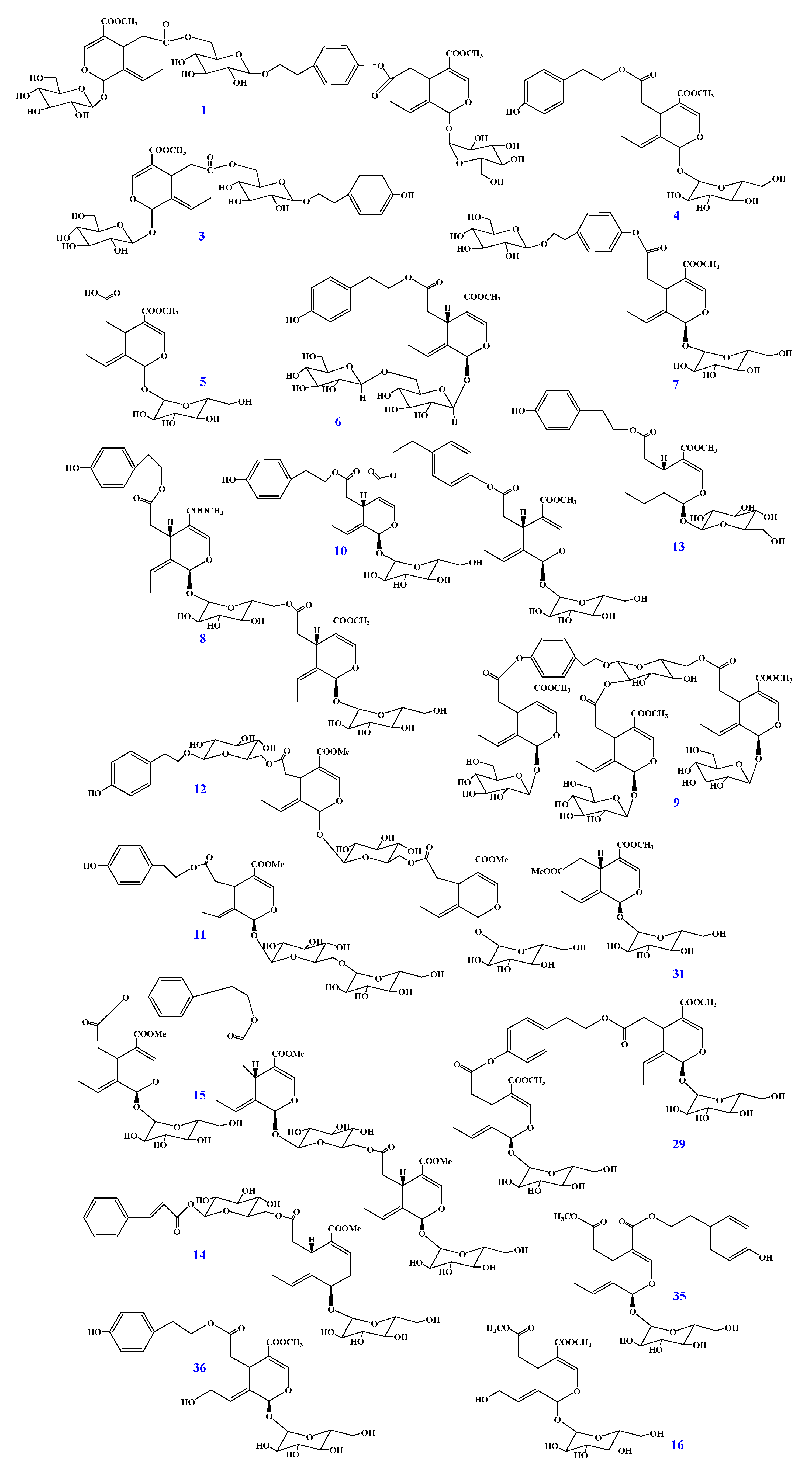
2.1. Identification of Secoiridoids in F. mandshurica Seeds

Nineteen secoiridoid compounds were first isolated from F. mandshurica seeds using a combination of column chromatography on silica gel, MCI GEL CHP-20P and polyamide. The structures of these secoiridoids were established by MS, 1H- and 13C-NMR and verified by comparing the NMR data (see the Supplementary Materials) with values reported in the literature. They were thus identified as GI3 (1) [26], nuzhenide (3) [27], ligstroside (4) [28], oleoside-11-methyl ester (5) [29], oleuricine A (6) [30], nicotiflorine (7) [31,32], jaspolyanoside (8) [33], oleopolynuzhenide A (9) [34], safghanoside G (10) [35], excelside B (11) [21], isooleonuezhenide (12) [36], lucidumoside A (13) [37], safghanoside A (14) [38], jaspolyoleoside B (15) [38], 10-hydroxoleoside-7,11-dimethyl ester (16) [39], GI5 (29) [40], oleoside-7,11-dimethyl ester (31) [41], isolignstroside (35) [42], 10-hydroxyligstroside (36) [43]. All the secoiridoids were found in F. mandshurica seeds in the form of glycosides. Their chemical structures are shown in Figure 1.

2.2. Quantitation of Secoiridoids in FM by HPLC Analysis

The quantitation results of nineteen secoiridoids in FM are shown in Table 1. All calibration curves exhibit excellent linear regressions with the determination coefficients (r2) ranging from 0.9986 to 1.0000. Total content of nineteen secoiridoids was 181.35 mg/g, the highest content was 88.21 mg/g (nuzhenide, 3) and the lowest was 0.22 mg/g (isolignstroside, 35).

2.3. Inhibitory Effect of Secoiridoids on Pancreatic Lipase

All secoiridoids were evaluated for their inhibitory effects on pancreatic lipase in vitro for the first time. The results are summarized in Table 1. Overall, the secoiridoids isolated from F. mandshurica seeds exhibited good pancreatic lipase inhibitory activity at the concentration of 100 μM. Oleopolynuzhenide A (9) showed the strongest inhibition among these secoiridoids, reaching 70.25%, and it was close to the positive control compound orlistat (73.11%). Further, by examining the structural characteristics of these secoiridoid compounds, we could conclude that compounds with two or three secoiridoid skeletons exhibit stronger inhibitory activity than compounds with only one basic skeleton. Specifically, inhibitory effects of GI3 (1), jaspolyanoside (8), oleopolynuzhenide A (9), safghanoside G (10), isooleonuezhenide (12), jaspolyoleoside B (15) and GI5 (29) were generally stronger than nuzhenide (3), ligstroside (4), oleoside-11-methyl ester (5), oleuricine A (6), nicotiflorine (7), excelside B (11), lucidumoside A (13), safghanoside A (14), 10-hydroxoleoside-7,11-dimethyl ester (16), oleoside-7,11-dimethyl ester (31), isolignstroside (35) and 10-hydroxyligstroside (36).

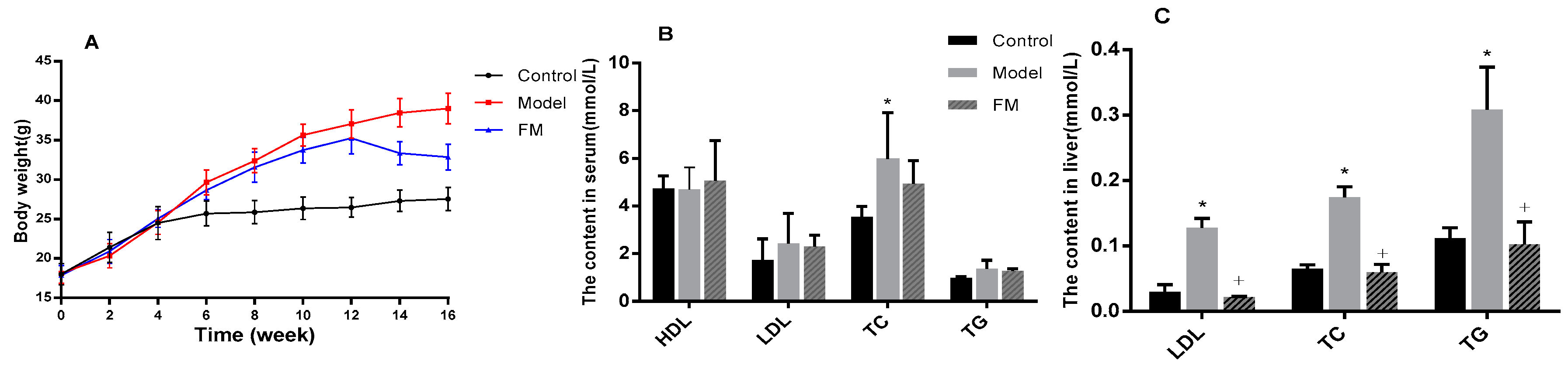
2.4. FM Declined the Body Weight of High-Fat Diet-Induced Obese Mice

Body weight was traced in this study and the results are displayed in Figure 2A. During the 16-week experiment period, all the mice were weighed every two weeks. At the end of the 8th week, the weight of the mice with high-fat diet (model group) reached 32.40 g, while the weight of the mice with low-fat diet (control group) were only 25.86 g. After administration with FM (400 mg/kg.bw) for the next 8 weeks, the body weight of the mice in FM group declined by 15.81% over the model group.

2.5. Effects of FM on TC, TG, LDL-C and HDL-C Levels in Serum and Liver

Results of TC, TG, LDL-C and HDL-C levels in serum are shown in Figure 2B. Compared to the control group, TC, TG and LDL-C levels of model group were increased by 68.4%, 39.4% and 39.7%, respectively. After treatment with FM at a dose of 400 mg/kg.bw for 8 weeks, the contents of TC, TG and LDL-C declined by 17.3%, 6.9% and 5.5% over the model group, respectively, and there was no significant difference (p > 0.05). Although high-fat diet caused a slight decrease of HDL-C content, FM consumption decreased HDL-C level compared to the model group (p > 0.05).
As shown in Figure 2C, the contents of TC, TG and LDL-C in the liver were significantly increased by high-fat diet feeding. A 167.5% increase of TC content, a 175.7% increase of TG content and a 331.8% increase of LDL-C content in liver were observed in the model group (p < 0.05). Notably, FM administration significantly decreased TC, TG and LDL-C levels by 65.9%, 66.8% and 83.0%, respectively, compared to the model group (p < 0.05).

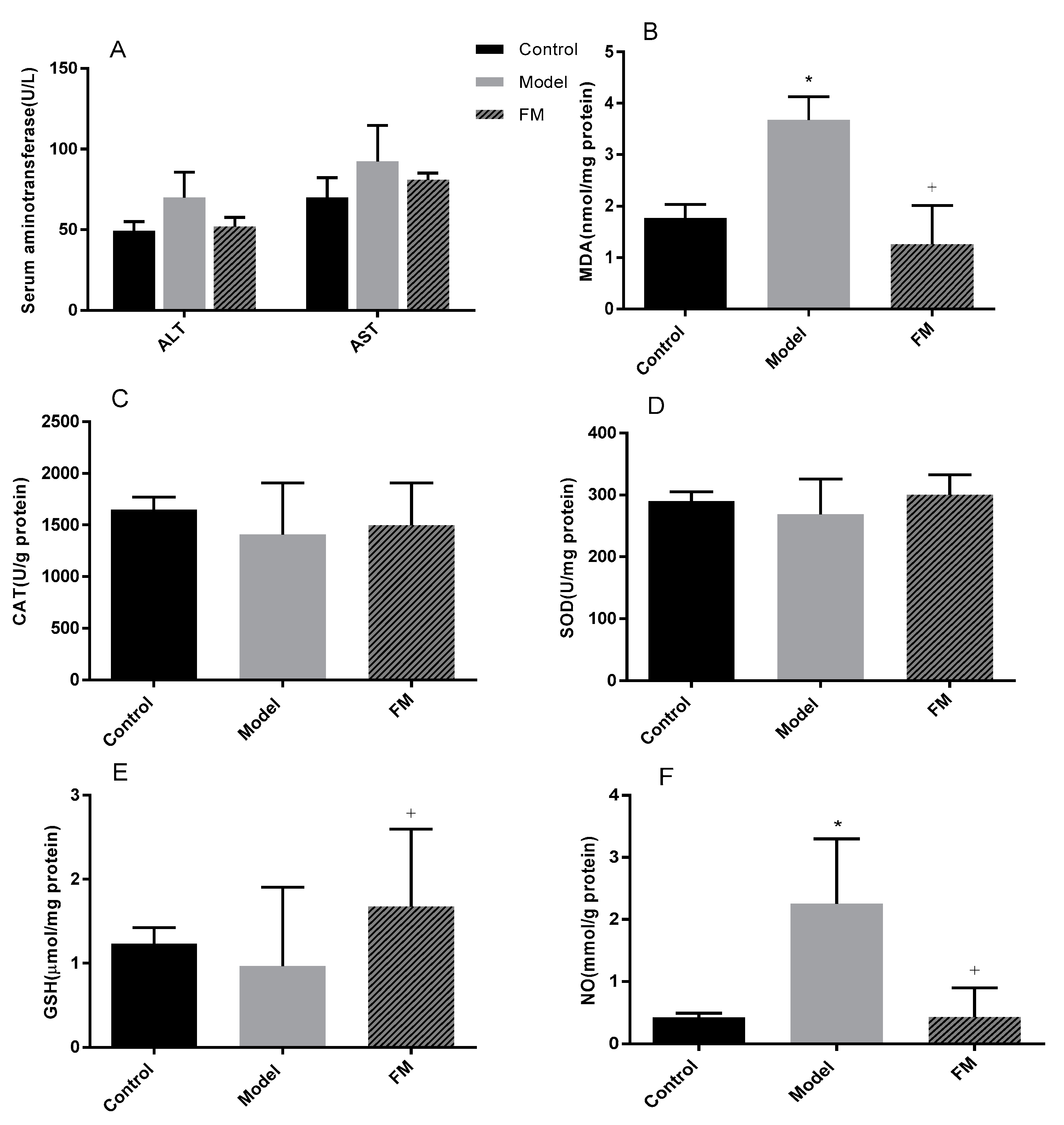
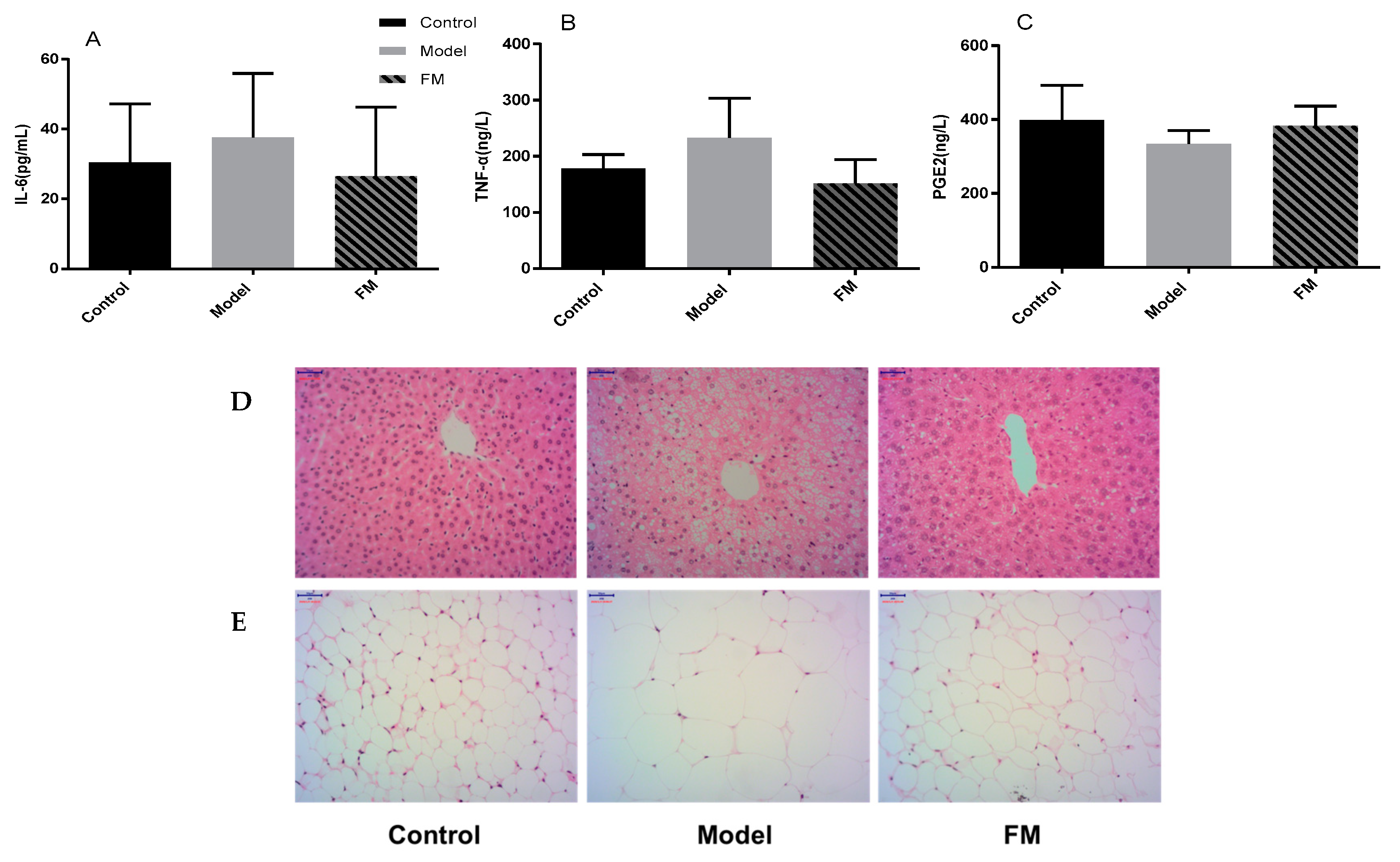
2.6. Effects of FM on Liver Injury, Oxidative Stress and Inflammatory Factors

As shown in Figure 3A, ALT and AST levels were increased by feeding of a high-fat diet. ALT and AST levels in the model group were 69.97 and 92.56 U/L, 1.42 and 1.32 times that of the control group, respectively. ALT and AST in the levels FM group were mitigated after treatment with FM, and there was no statistically significant difference (p > 0.05).
In this study, hepatic oxidative stress was evaluated and the results are summarized in Figure 3B–F. A 7.4% decrease of SOD level and a 14.4% decrease of CAT level were observed in the model group compared with the control group. After treatment with FM for 8 weeks inhibited the high-fat diet-induced decrease in SOD and CAT levels in mouse liver was inhibited, but there was no statistically significant difference. MDA and NO contents were increased by feeding of a high-fat diet. MDA and NO contents in the model group were 2.08 and 5.23 times that of the control group, respectively (p < 0.05). However, administration with FM significantly reduced MDA and NO productions compared with the model group (p < 0.05). GSH content was decreased in model group fed with high-fat diet, showing about a 21.76% decline compared with the control group (p > 0.05). Compared with the model group, FM consumption increased the GSH content by 73.74% (p < 0.05).
Inflammatory factors including IL-6, TNF-α and PGE2 were also determined in this study, and the results are displayed in Figure 4A–C Compared to the control group, high-fat diet induced a 28.47% increase of IL-6 level, a 30.77% increase of TNF-α level and a 16.09% decrease of PGE2 level in the model group (p > 0.05). Meanwhile, 400 mg/kg.bw of FM consumption induced a 29.57% decrease of IL-6 level, a 34.67% decrease of TNF-α level and a 14.46% increase of PGE2 level compared with the model group (p > 0.05).

2.7. Histopathological Examinations of Liver and Epididymal Adipose

Morphological analysis of liver and epididymal adipose were conducted. As shown in Figure 4D, mice in the control group fed with low-fat diet showed normal hepatic tissue with regular morphology of hepatic cells, while, mice in the model group fed with high-fat diet exhibited macrovesicular steatosis and cell ballooning degeneration. After administration with FM, pathological changes of mice in the FM group were effectively reversed. The results of morphological analysis of epididymal adipose are shown in Figure 4E, the adipocytes in the model group were much bigger than the control group, administration of FM alleviated the adverse change.

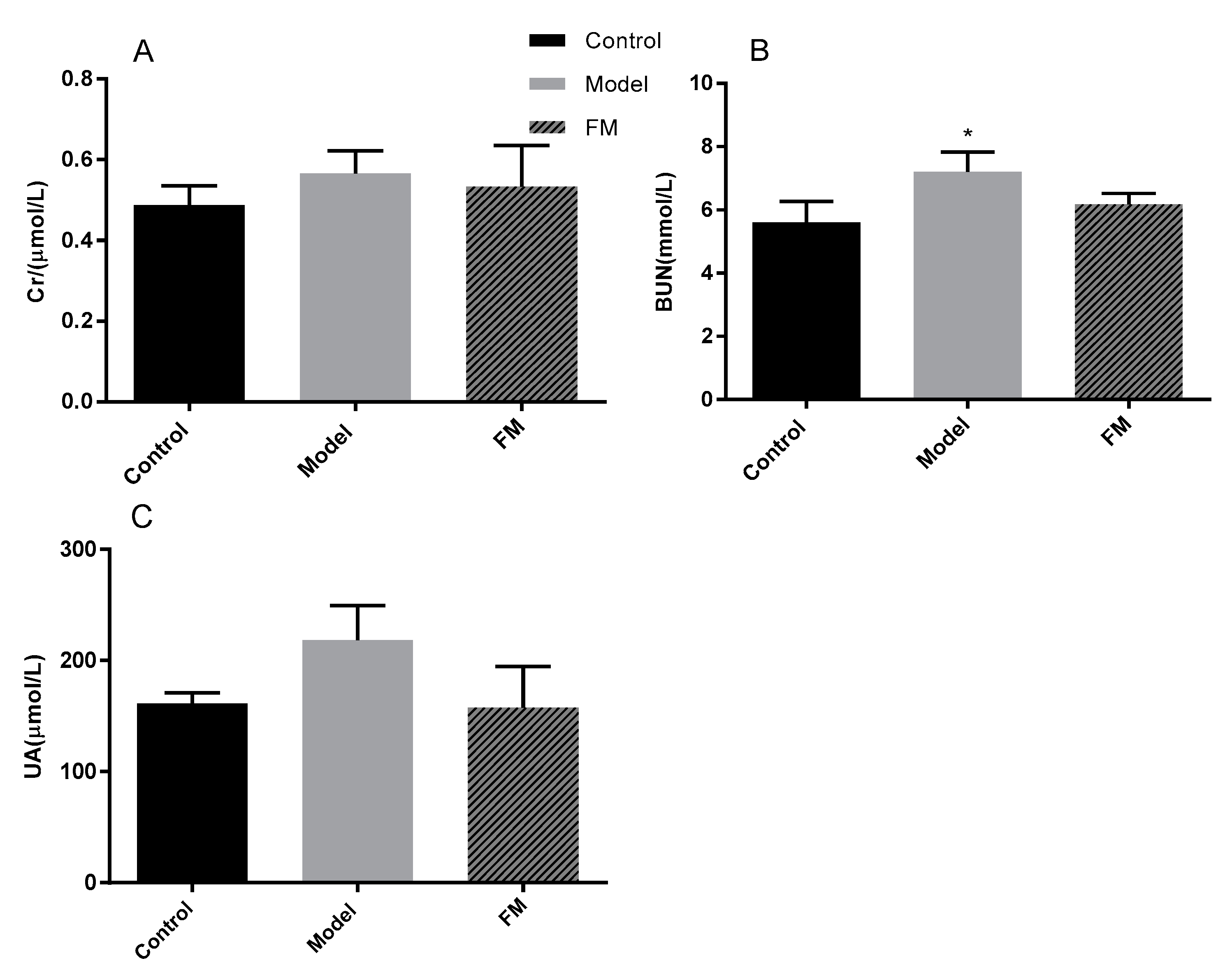
2.8. Effects of FM on Serum Cr, BUN and UA Levels

Cr, BUN and UA levels were determined to evaluate the kidney status of mice. As shown in Figure 5A–C, mice in the model group showed a significant increase of BUN level compared with the mice in the control group (p < 0.05). And high-fat diet induced a 16.05% increase of Cr level and a 35.41% increase of UA level in the model group compared with the control group, and there was no statistically significant difference (p > 0.05). After administration with FM, the contents of Cr, BUN and UA in the FM group were decreased, and there was no statistically significant difference (p > 0.05).

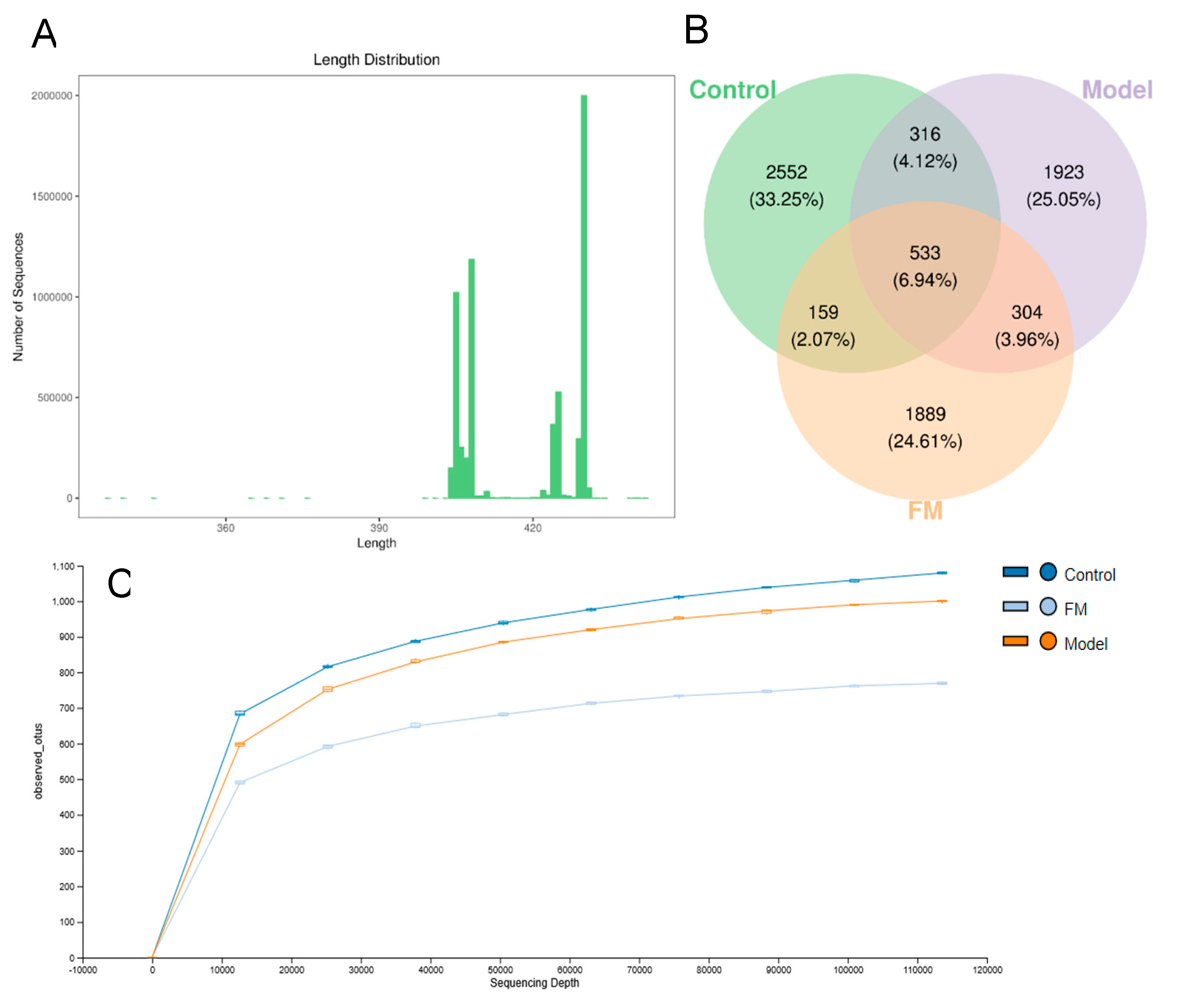
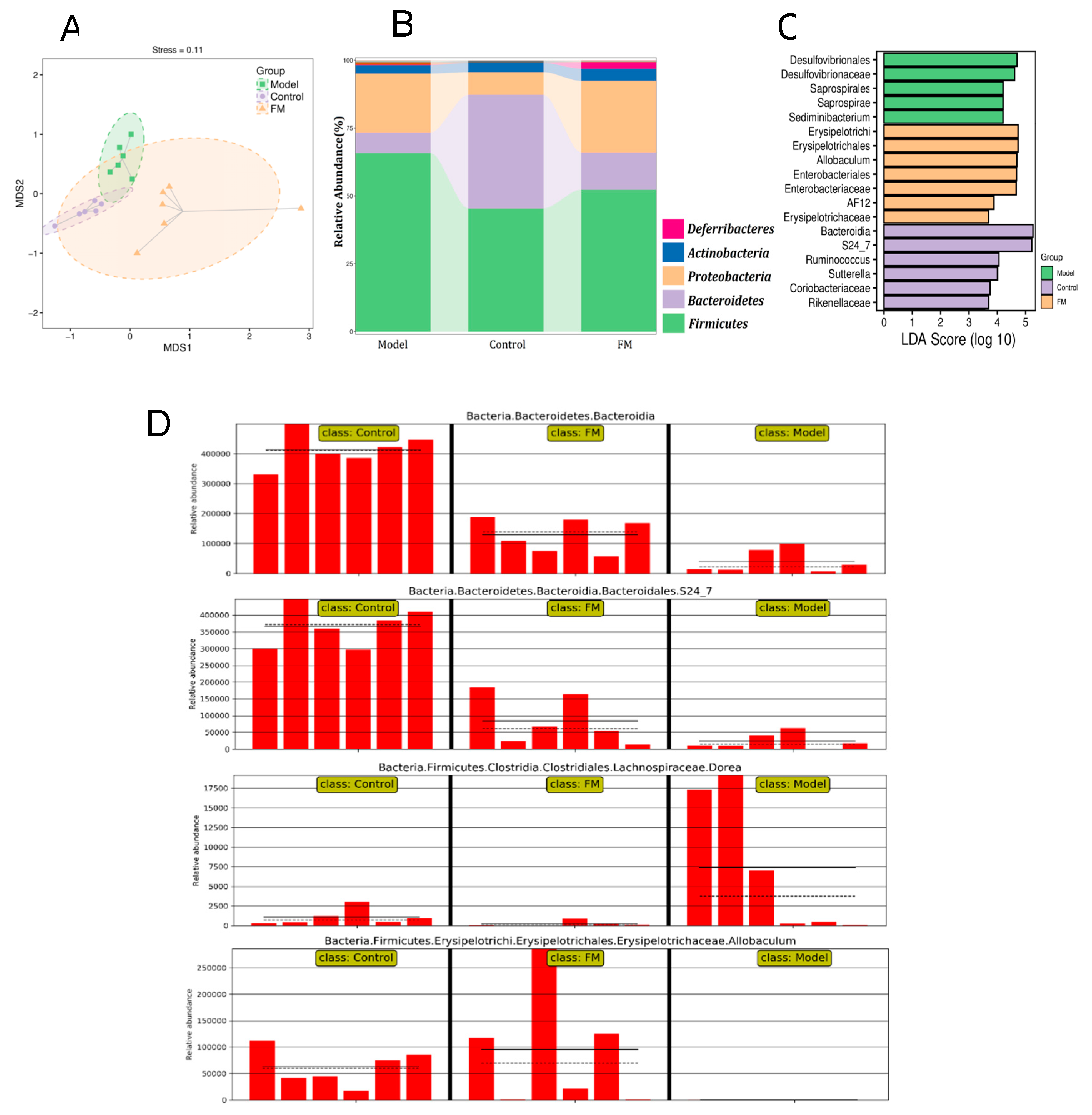
2.9. FM Modulates the Gut Microbiota in High-Fat Diet-Induced Obese Mice

To evaluate the impact of FM on the gut microbiota in mice, pyrosequencing of the variable region V3-V4 of the bacterial 16S rRNA gene was performed in this study. As shown in Figure 6A, a total of 3,682,083 sequences (400–450 base pairs in length) were obtained from 22 samples, including an average of 162,037 sequences in the control group, 172,220 sequences in the model group and 168,002 sequences in the FM group. The Venn diagram (Figure 6B) shows that the qualified sequences (>0.001%) were clustered into 7676 bacterial OTUs. Among them, 316 same OTUs were identified in the control and model groups, 159 same OTUs were found in the control and FM groups. Administration with FM decreased the shared OTUs in the mice of the control and model groups. From Figure 6C, we can conclude that rarefaction curves of three groups tend to be flat when the sequencing depth increases. This indicated that the sequencing results were sufficient to reflect the gut microbial diversity of the samples.
Non-metric multidimensional scaling (NMDS) analysis based on UniFrac distance was performed to evaluate the similarity of gut microbial communities between different groups. As shown in Figure 7A, the similarity was described by the distance matrix of different samples. Samples in different groups clustered separately, the sample distributions of the model group and FM group were far away from the control group. This indicated that consumption of a high-fat diet and FM changed the gut microbiota composition. And samples in the FM group exhibited a unique gut microbial structure. The stress value is 0.11 (<0.2), indicating that the analysis results were reliable. The relative abundance of major microbiota constituents at phylum level is shown in Figure 7B, Firmicutes, Bacteroidetes and Proteobacteria were the top three phyla. Compared to the control group, the high-fat diet caused an increase of Firmicutes and a decrease of Bacteroidetes in the model group, while, FM consumption alleviated these changes in the FM group. The impact of FM on the key community of gut microbiota was described by histogram of LDA value (Figure 7C). Bacteroidia, S24-7, Ruminococcus and Sutterella were the four main communities in the gut of the control mice. The high-fat diet induced a significant effect on Desulfovibrionales, Desulfovibrionaceae, Saprospirales, and Saprospirae in the gut of the model mice. However, FM consumption demonstrated a selective enrichment of Erysipelotrichi, Erysipelotrichales, Allobaculum, Enterobacteriales and Enterobacteriaceae. The relative abundance of gut microbial biomarkers of three groups are displayed in Figure 7D. To visually show the differences between three groups, the solid and dashed lines are used to identify the average and median relative abundance of the taxa in each group. In the control group, the abundance of Bacteroidia and S24-7 were high, however, high-fat diet induced significant increase of those in the model group. Adversely, the abundance of Dorea was low in the control group, and it was greatly increased by high-fat diet in the model group. Notably, compared with the control group, the high-fat diet completely inhibited the growth of Allobaculum. Treatment with FM could significantly alleviate the changes as mentioned above.

3. Discussion

Obesity and overweight are major contributors to the global burden of chronic diseases and complications [44]. Thus, edible and medicinal plants and their bioactive components helping people to fight the battle against obesity have been widely explored, such as the extracts of garcinia, green tea and white bean [13,14,15]. The present investigation showed that F. mandshurica seed extract containing secoiridoids reduced body weight and lipid levels in serum and liver, alleviated liver and kidney damage and regulated the gut microbiota in high-fat diet-induced obese mice.
Studies in recent years have demonstrated that secoiridoid-enriched extract from F. excelsior seed reduced gains in body weight, omental fat and retroperitoneal fat in obese C57BL/6J mice, and alleviated triglyceridemia, body weight gain and systolic blood pressure in SHR and Zucker rats [22,23]. In the present study, treatment with FM for 8 weeks reduced body weight in obese mice. Serum and liver TG, TC, LDL-C contents in high-fat diet-induced obese mice were increased and FM consumption reduced these lipid levels. Obesity can cause a decrease in HDL-C concentration and HDL-C dysfunction, in this study, the serum HDL-C content of mice treated with FM was higher than that of obese mice.
Obesity is usually associated with chronic metabolic diseases such as nonalcoholic fatty liver disease [45]. Ibarra et al. have proved that extract from F. excelsior seed lowered liver weight gains and the incidence of fatty livers in obese C57BL/6J mice [22]. ALT and AST levels are clinically considered as credible markers for monitoring liver injury or liver function [46,47]. The present investigation showed that high-fat diet induced increases of serum ALT and AST levels in obese mice, and after treatment with FM for 8 weeks, the above variations were alleviated.
Obesity usually accompanied by increased oxidative stress caused by the reactive oxygen species (ROS). High-fat diet can also produce ROS by changing oxygen metabolism, which further aggravates the oxidative stress in an obese body [48]. Finally, persistent oxidative stress leads to the consumption of antioxidants and the decreased levels of antioxidant enzymes such as CAT and SOD [49]. Younis et al. reported that F. xanthoxyloides leaf extract increased the level of hepatic antioxidant enzymes including CAT, SOD, peroxidase (POD), glutathione-S-transferase (GST) and glutathione reductase (GSR) in CCl4- induced hepatotoxic rats [50]. In the present study, administration of FM significantly increased the levels of CAT and SOD in high-fat diet-induced obese mice. Obesity-related mitochondrial dysfunction can cause increased NO synthesis [51]. After treatment with FM for 8 weeks, the NO content of high-fat diet-induced obese mice was significantly decreased and was close to that of the low-fat diet mice. Additionally, GSH content of high-fat diet induced obese mice was significantly increased after administration with FM. GSH can function as direct free-radical scavenger and reduce intracellular ROS, thereby protect the cell against toxicity and disease [52]. MDA is the main product of lipid peroxidation induced by ROS, its content in high-fat diet-induced obese mice was significantly decreased by the treatment of FM [53]. Inflammation is a manifestation of increased oxidative stress induced by obesity [48]. Recent study has indicated that ROS generated during fatty acid oxidation triggers inflammatory pathway involving TNF-α and IL-6 [54]. Younis et al. also proved that F. xanthoxyloides leaf extract had the ability to decrease the concentrations of TNF-α and IL-6 in air pouch exudate of rat [55]. In the present study, TNF-α and IL-6 levels were increased in high-fat diet induced obese mice, and administration of FM to obese mice for 8 weeks significantly decreased the TNF-α and IL-6 contents. PGE2, an anti-inflammatory lipid mediator derived enzymatically from arachidonic acid and catalyzed by cyclooxygenase (COX-1 and -2) and prostaglandin E synthase, which inhibits the differentiation and activation of inflammatory cells [56]. A decreased level of PGE2 have been observed in high-fat diet-induced obese mice, after treatment with FM for 8 weeks, the PGE2 content was increased. Overall, the above results illustrated that F. mandshurica seed extract significantly inhibited the obesity and alleviated liver injury, oxidative stress and inflammation induced by high-fat diet. The beneficial effects of F. mandshurica seed on obesity and obesity-related liver injury were also confirmed by conventional histological assessment of adipocytes and livers.
A report has demonstrated that obesity is associated with increased risk of developing chronic kidney disease [57]. The present study demonstrated that FM alleviated kidney injury as indicated by the decreased levels of serum Cr, BUN and UA in high-fat diet induced obese mice.
In recent years, the impact of gut microbiota on obesity has become a hot research topic. Generally, the gut microbiota consists of six bacterial phyla, including Firmicutes, Bacteroidetes, Protebacteria, Actinobacteria, Fusobacteria and Verrucomicrobia. Among them, Firmicutes and Bacteroidetes account for 90% and are closely related to obesity [58,59]. Hildebrandt et al. reported that a decrease in Bacteroidetes and an increase in Firmicutes were found in the high-fat diet induced obese mice, we also observed these alterations in obese mice in this study [60]. Interestingly, FM consumption can increase the abundance of Bacteroidetes and decrease the abundance of Firmicutes, the result preliminarily shows that FM can treat obesity by regulating the gut microbiota. Further, we found several gut microbial biomarkers between three groups, including Bacteroidia, S24-7, Dorea and Allobaculum. In this study, the decrease in relative abundance of Bacteroidia in high-fat diet mice agrees with a previous study [61]. After treatment with FM for 8 weeks, Bacteroidia were enriched and were negatively correlated with obesity phenotypes. S24-7, a bacterium that produces butyrate, has been shown to be beneficial to intestinal epithelial health and energy metabolism [62,63]. However, compared to the low-fat diet mice, the abundance of S24-7 in high-fat diet obese mice was significantly decreased, and FM consumption promoted an increase of S24-7 in obese mice. Studies have found that Dorea is positively correlated with body mass index (BMI), waist circumference and diastolic blood pressure. Additionally, a high abundance of Dorea was found in the intestines of patients with multiple sclerosis and inflammatory bowel disease, suggesting that a pro-inflammatory effect of this genus [64,65]. Compared with the low-fat diet mice, the abundance of Dorea in high-fat diet induced obese mice was significantly increased, and it could be obviously decreased by consumption of FM. It has been reported that Allobaculum was negatively associated with obesity, inflammation and insulin resistance [66]. Notably, in the present study, the Allobaculum of high-fat diet induced obese mice was completely inhibited, and the FM consumption effectively increases its abundance. The above results indicate that the modulation of gut microbiota caused by FM consumption has a therapeutic effect on obesity in mice.
To identify the bioactive constitutes of FM, extensive chromatographic and spectral analysis were used in the phytochemical study. Finally, we obtained nineteen secoiridoid compounds from the FM for the first time. The content of each secoiridoid in FM is quantified by a RP HPLC-DAD method and quantitative results ranged from 0.22 to 88.21 mg/g. Nuzhenide was the richest among these secoiridoids, which has been shown to activate PPARα and inhibit preadipocyte differentiation in the 3T3-L1 cell [21]. In the human body, fat is hydrolyzed by pancreatic lipase and then absorbed in the small intestine, therefore, pancreatic lipase inhibitors can be used to prevent/treat obesity [67]. Interestingly, nineteen secoiridoids isolated from FM exerted good pancreatic lipase inhibitory activity in vitro. Hence, the anti-obesity effect of FM should be attributed to the abundant secoiridoid compound. However, the mechanism of the anti-obesity effect of secoiridoids in FM was not clear and this will require more studies to be performed.

4. Materials and Methods

4.1. Preparation of F. Mandshurica Seed Extract

The dried seeds of F. mandshurica were purchased from Jilin City, Jilin Province, China in 17 November 2017. The botanical origins were identified by the corresponding author Prof. Naisheng Bai, and voucher specimens of F. mandshurica seeds (FM20171117) were deposited in Room 611, College of Food Science and Technology, Northwest University. The plant materials (30.0 kg) were cleaned and powdered (60–100 mesh), then percolated three times (24 h each time) with 75% ethanol-water solution at room temperature, the ratio of the material to solvent was 1:3 (w/v). The ethanol solution was directly concentrated under vacuum at less than 50 °C. Then the residue was dispersed in water and partitioned between methylene chloride three times. After removing solvent, the water portion was dried under vacuum below 50 °C to obtain powder extract (FM). Sample FM was subjected to the isolation and characterization of secoiridoids, HPLC analysis and animal study.

4.2. Extraction and Isolation of Secoiridoid Compounds from F. mandshurica Seeds

The FM (540 g) residue was chromatographically separated on a polyamide column (PA, 60–100 mesh, 2000 g) and eluted with an EtOH/H2O gradient solvent system. The effluent was combined after monitoring by TLC and seven major fractions (I–VII) were collected.
Sample I (94.3 g) was chromatographed on a PA column (60–100 mesh, 300 g) and the eluent was EtOH/H2O gradient solvent system, then three samples of different polarity were collected (I-1, I-2 and I-3). Compounds 9 (5.4 mg), 12 (25.0 mg) and 13 (13.2 mg) were isolated from I-1 and I-2 using a CHP20P column (80 g) and the eluent was MeOH/H2O system. I-3 was successively chromatographed on a CHP20P column (50 g, MeOH/H2O, 1:1), a silica gel column (200–300 mesh, 300 g, CH2Cl2/MeOH, 20:1–1:1) and a CHP20P column (50 g, MeOH/H2O, 1:1), to give pure compounds 11 (8.1 mg) and 7 (15.9 mg).
Sample II (44.1 g) was submitted to a silica gel column (200–300 mesh, 500 g, CH2Cl2/MeOH, 40:1–1:1) and three sub fractions were collected. Compound 15 (11.3 mg) was purified by a CHP20P column (100 g, MeOH/H2O, 0:1–1:0) from II-1. Isolation of compound 10 (9.5 mg) from II-2 was effected by a silica gel column (200–300 mesh, 500 g, CH2Cl2/MeOH, 20:1–1:1) and a CHP20P column (100 g, MeOH/H2O, 1:1). II-3 was repeatedly chromatographed on silica gel columns (300–400 mesh, 500 g, 20:1–1:1) to give pure compound 6 (28.3 mg)
Sample III (22.8 g) was chromatographed on a CHP20P column (500 g, MeOH/H2O, 0:1–1:0) to give two polar parts. Further, one part (III-1) was chromatographed on a silica gel column (300–400 mesh, 300 g, CH2Cl2/MeOH, 20:1–1:1) to afford pure compound 8 (18.1 mg); the other part (III-2) was chromatographed on a CHP20P column (100 g, MeOH/H2O, 1:1) to yield pure compound 14 (6.4 mg).
Sample IV (35.7 g) was successively separated by a PA column (100–200 mesh, 500 g, EtOH/H2O, 0:1–1:0), a silica gel column (200–300 mesh, 500 g, CH2Cl2/MeOH, 20:1–1:1) and a CHP20P column (300 g, MeOH-H2O, 0:1–1:0) to give pure compounds 4 (8.9 mg), 1 (11.3 mg) and 3 (16.7 mg).
Sample V (62.4 g) was repeatedly chromatographed on CHP20P columns (500 g, MeOH/H2O, 0:1–1:0) to afford pure compounds 16 (10.1 mg), 35 (6.7 mg) and 29 (14.1 mg).
Sample VI (78.6 g) was repeatedly chromatographed on silica gel column (200–300 mesh, 800 g, CH2Cl2/MeOH, 30:1–1:1) to give VI-1 and VI-2. Compound 5 (21.3 mg) was purified from VI-1 using a CHP20P column (500 g, MeOH/H2O, 0:1–1:0). The isolation of compound 36 (15.5 mg) from VI-2 was orderly effected by a silica gel column (200–300 mesh, 100 g, CH2Cl2/MeOH, 20:1–1:1) and a CHP20P column (100 g, MeOH/H2O, 2:1).
Sample VII (67.7 g) was submitted to repeated silica gel columns (300–400 mesh, 500 g, CH2Cl2/MeOH, 25:1–1:1) for the separation of compound 31 (17.3 mg).
The separation process of pure compounds is shown in Figure S1 (Supplementary Materials). These compounds were identified by MS, 1H- and 13C-NMR.

4.3. Quantification of Secoiridoids in FM by HPLC Analysis

HPLC analysis was performed on an Agilent 1260 LC Series instrument (Agilent Technologies, Santa Clara, CA, USA) connected to a G4212B DAD detector, using an Agilent Eclipse Plus C18 column (250 × 4.6 mm, 5 μm) with a flow rate of 1.0 mL/min, and column temperature was maintained at 30 °C. The mobile phase was composed of D (water) and C (acetonitrile) with a gradient elution: 0 min, 95% D; 0–8 min, 95–75% D; 8–18 min, 75–50% D; 18–28 min, 50–25% D; 28–40 min, 25–15% D; 40–52 min, 15–5% D; 52–58 min, 5% D; 58–60 min, 5–95% D; 60–70 min, 95% D. The chromatogram was monitored at a wavelength of 220 nm during the experiment. Nineteen chemical standards used in quantitative analysis were isolated from FM and purity of each standard compound was over 98% as monitored by HPLC analysis. The standard solution containing nineteen secoiridoids were prepared and diluted to five different concentrations. Calibration curve for each secoiridoid was constructed by plotting peak area versus corresponding concentration. The sample solution was obtained by dissolving FM in MeOH. Secoiridoids were identified by comparing their UV spectra and HPLC retention times of target peaks with those of standard compounds.

4.4. Pancreatic Lipase Inhibitory Activity of Secoiridoids In Vitro

Pancreatic lipase inhibitory activities of secoiridoids were determined as previously reported [68,69]. Briefly, the enzyme solution was prepared by the reconstitution of porcine pancreatic lipase (Sigma-Aldrich, St. Louis, MO, USA) in 0.1 M Tris-HCl buffer (pH = 8). Then, all isolated compounds (100 μM) and reference compound orlistat (100 μM) and different concentrations of FM were mixed with enzyme buffer and incubated for 20 min at 37 °C. After incubation, 10 mM p-nitrophenyl butyrate (p-NPB) was added at 37 °C to start the reaction. The absorbance at 405 nm was determined for 20 min with interval of 2 min using a BioTek Epoch microplate spectrophotometer (Winooski, VT, USA). The enzyme activity was represented by the absorbance growth slope (K) which was calculated by linear regression. The inhibitory rate was calculated by the formula below:
% Inhibition = (Kb − Ks)/Kb × 100
where Ks and Kb were the enzyme activity in the presence and absence of secoiridoids, respectively. Orlistat (Sigma-Aldrich) was used as a positive control. The IC50 values were determined by nonlinear regression with normalized dose-response fitting using Graphpad Prism 6 software (San Diego, CA, USA), which represent the concentrations of compounds at which 50% of the pancreatic lipase activity was inhibited.

4.5. Animal Experiments

4.5.1. Animals and Diets

Thirty male C57BL/6 mice (20 ± 2 g) were purchased from the Experimental Animal Center of Xi’an Jiaotong University. All the mice were maintained at temperature of 25 ± 2 °C, relative humidity of 50 ± 10% and subjected to a 12 h light/dark cycle for 7 days of adaptation. Then thirty mice were randomly assigned to 3 groups with 10 mice per group. All animal experimental protocols used in this study were approved by the Regulations of Experimental Animal Administration issued by the State Committee of Science and Technology of People’s Republic of China (SCXK 2012-003). Mice in control group were fed with a low-fat diet (LFD, 8% fat, 22% proteins and 70% carbohydrates). Mice in other two groups were given a high-fat diet (HFD, 45% fat, 18% proteins and 37% carbohydrates) [70]. All mice subsisted on the experimental diets until the end of the experiment. After 8 weeks of daily feeding, one of the high-fat diet group (FM group) was given 400 mg/kg.bw FM by intragastric administration, another high-fat diet group was named as model group. After 16 weeks of treatment, all the mice were food-deprived for 10 h and decapitated. Whole blood was obtained by cardiac puncture. Liver, colon and epididymal adipose were separated and harvested. All samples were stored at −80 °C.

4.5.2. Serum and Hepatic Biochemical Analysis

The levels of high density lipoprotein cholesterol (HDL-C), low density lipoprotein cholesterol (LDL-C), total cholesterol (TC), triglyceride (TG), alanine aminotransferase (ALT), aspartate aminotransferase (AST), creatinine (Cr), blood urea nitrogen (BUN) and uric acid (UA) in serum samples were measured with commercial diagnostic kits (Nanjing Jiancheng Bioengineering Institute, Nanjing, China).
Livers were immediately excised from sacrificed mice and washed with ice-cold physiological saline three times to remove the blood, fat and connective tissue. Then the liver from dried filter paper was weighed and homogenized with ice-cold physiological saline using a Dounce tissue grinder (Kimble, Vineland, NJ, USA) with 20,000 rpm for 10 s. Finally, the homogenate was centrifuged at 3000 rpm for 20 min at 4 °C and the supernatant was used for hepatic biochemical analysis. Malondialdehyde (MDA), catalase (CAT), superoxide dismutase (SOD), glutathione (GSH) and nitric oxide (NO) levels were determined using diagnostic kits (Nanjing Jiancheng Bioengineering Institute, Nanjing, China). The levels of tumor necrosis factor (TNF-α), prostaglandin E2 (PGE2) and interleukin-6 (IL-6) were detected using commercial ELISA kits (Shanghai Fusheng, Shanghai, China). In addition, the supernatant sample was used for determination of TG and TC contents. A multifunctional plate reader (Infinite M200Pro, TECAN, Männedorf, Switzerland) was used for the quantitative measurements.

4.5.3. Histopathological Examinations of Liver and Epididymal Adipose

Liver and epididymal adipose tissues was immersed in 10% neutral formalin solution and embedded in paraffin. Then processed by standard procedures, paraffin-embedded section at 5 μm thickness was cut and stained with hematoxylin and eosin (H&E).

4.6. Gut Microbiota Analysis

Six samples of colonic contents per group were randomly selected for Illumina sequencing and statistical analysis of the bacterial 16S rRNA gene. Total DNA was extracted using the Fast SPIN extraction kits (MP Biomedicals, Santa Ana, CA, USA). Quantification of DNA was measured by a NanoDrop ND-1000 spectrophotometer (Thermo Fisher Scientific, Waltham, MA, USA) and the quality of DNA was evaluated by 1.2% agarose gel electrophoresis. The V3-V4 hypervariable regions of 16S rRNA genes were amplified using primers 338-forward (5′-ACTCCTACGGGAGGCAGCA-3′) and 806-reverse (5′-GGACTACHVGGGTWTCTAAT-3′). Quantification of the purified PCR amplicons was checked by PicoGreendsDNA Assay Kit (Invitrogen, Carlsbad, CA, USA). Then the purified PCR products were pooled in equimolar and paired-end sequenced (2 × 300 bp) on an Illlumina MiSeq platform with MiSeq Reagent Kit V3 (600 cycles) (Shanghai Personal Biotechnology Co., Ltd., Shanghai, China). The OTU (operational taxonomic unit) relative abundances were hierarchically clustered and visualized by the R software (v3.5.1) gplots package, and a LEfSe (linear discriminant analysis effect size) analysis coupled with the Kruskal-Wallis and Wilcoxon rank sum tests was performed.

4.7. Statistical Analysis

All experiments were performed in triplicate and results were represented as mean ± SD. Differences between control and treated groups were assessed by Duncan’s multiple range test after SAS one-way ANOVA, version 8.1 (SAS Institute, Cary, NC, USA). p < 0.05 was significantly different.

5. Conclusions

In conclusion, FM could reduce the body weight of obese mice, attenuate the lipid accumulation in serum and liver, relieve the damage in liver and kidney, and extenuate oxidative stress injury and inflammation caused by obesity. In addition, FM could modulate the structural alteration of gut microbiota in obesity mice, increase the proportion of anti-obesity gut microbiota (Bacteroidetes, Bacteroidia, S24-7 and Allobaculum), and reduce the proportion of obesogenic gut microbiota (Firmicutes and Dorea). The anti-obesity and gut microbiota modulation effect could be associated with the abundant secoiridoid compounds in FM. The phytochemical study of FM resulted in the isolation of nineteen secoiridoid compounds. And these compounds exhibited good pancreatic lipase inhibitory activity in vitro. In summary, this study suggests that F. mandshurica seed or its secoiridoids may have potential for use as a dietary supplement for controlling obesity.

Supplementary Materials

The following are available online, Figure S1: The separation process of F. mandshurica seeds, MS, 1H NMR and 13C NMR data of compounds.

Author Contributions

Conceptualization: N.B., C.-T.H.; Study design: S.G., H.Z.; Data Curation: S.G., H.Z., Z.M.; Figures: S.Z., M.L., X.R.; Writing Original Draft Preparation: S.G.; Review and Editing: C.-T.H., Z.Z. All authors have read and agreed to the published version of the manuscript.

Funding

This research was funded by National Natural Science Foundation of China (Grant No. 31570348).

Acknowledgments

The authors wish to thank the National Natural Science Foundation of China (No. 31570348) for funding this study.

Conflicts of Interest

The authors declare no conflict of interest.

References

  1. Saltiel, A.R.; Kahn, C.R. Insulin signaling and the regulation of glucose and lipid metabolism. Nature 2001, 414, 799–806. [Google Scholar] [CrossRef] [PubMed]
  2. Rahmouni, K.; Correia Correia, L.G.; Haynes, W.G.; Mark, A.L. Obesity-associated hypertension-New insights into mechanisms. Hypertension 2005, 45, 9–14. [Google Scholar] [CrossRef] [PubMed]
  3. Bastien, M.; Poirier, P.; Lemieux, I.; Despres, J.P. Overview of epidemiology and contribution of obesity to cardiovascular disease. Prog. Cardiovasc. Dis. 2014, 56, 369–381. [Google Scholar] [CrossRef] [PubMed]
  4. Tobias, D.K.; Pan, A.; Jackson, C.L.; O’Reilly, E.J.; Ding, E.L.; Willett, W.C.; Manson, J.E.; Hu, F.B. Body-mass index and mortality among adults with incident type 2 diabetes. N. Engl. J. Med. 2014, 370, 233–244. [Google Scholar] [CrossRef] [Green Version]
  5. Vucenik, I.; Stains, J.P. Obesity and cancer risk: Evidence, mechanisms, and recommendations. Ann. N. Y. Acad. Sci. 2012, 1271, 37–43. [Google Scholar] [CrossRef]
  6. Zhang, C.; Zhang, J.; Liu, W.; Chen, X.; Liu, Z.; Zhou, Z. Improvements in humoral immune function and glucolipid metabolism after laparoscopic sleeve gastrectomy in patients with obesity. Surg. Obes. Relat. Dis. 2019, 15, 1455–1463. [Google Scholar] [CrossRef]
  7. An, R.; Shen, J.; Yang, Q.; Yang, Y. Impact of built environment on physical activity and obesity among children and adolescents in China: A narrative systematic review. J. Sport Health Sci. 2019, 8, 153–169. [Google Scholar] [CrossRef]
  8. Zhao, Y.L.; Ma, R.M.; Lao, T.T.; Chen, Z.; Du, M.Y.; Liang, K.; Huang, Y.K.; Zhang, L.; Yang, M.H.; Sun, Y.H.; et al. Maternal gestational diabetes mellitus and overweight and obesity in offspring: A study in Chinese children. J. Dev. Orig. Health Dis. 2015, 6, 479–484. [Google Scholar] [CrossRef]
  9. Chandrasekaran, C.V.; Vijayalakshmi, M.A.; Prakash, K.; Bansal, V.S.; Meenakshi, J.; Amit, A. Review article: Herbal approach for obesity management. Am. J. Plant Sci. 2012, 3, 1003–1014. [Google Scholar] [CrossRef] [Green Version]
  10. Apovian, C.M.; Aronne, L.J.; Bessesen, D.H.; McDonnell, M.E.; Murad, M.H.; Pagotto, U.; Ryan, D.H.; Still, C.D. Pharmacological management of obesity: An endocrine society clinical practice guideline. J. Clin. Endocr. Metab. 2015, 100, 342–362. [Google Scholar] [CrossRef]
  11. Thomas, J.G.; Bond, D.S.; Phelan, S.; Hill, J.O.; Wing, R.R. Weight-loss maintenance for 10 years in the national weight control registry. Am. J. Prev. Med. 2014, 46, 17–23. [Google Scholar] [CrossRef] [PubMed]
  12. Sjostrom, L.; Rissanen, A.; Andersen, T.; Boldrin, M.; Golay, A.; Koppeschaar, H.P.; Krempf, M. Randomised placebo-controlled trial of orlistat for weight loss and prevention of weight regain in obese patients. Lancet 1998, 352, 167–173. [Google Scholar] [CrossRef]
  13. Barrett, M.L.; Udani, J.K. A proprietary alpha-amylase inhibitor from white bean (Phaseolus vulgaris): A review of clinical studies on weight loss and glycemic control. Nut. J. 2011, 10, 24. [Google Scholar] [CrossRef] [PubMed] [Green Version]
  14. Jayawardena, R.; Sooriyaarachchi, P.; Ranasinghe, P.; Perera, A.; Hills, A.P. Availability and composition of weight-loss supplements in Sri Lanka. Nutr. Diet. 2020, 77, 247–252. [Google Scholar] [CrossRef] [PubMed]
  15. Kim, S.Y.; Oh, M.R.; Kim, M.G.; Chae, H.J.; Chae, S.W. Anti-obesity effects of Yerba Mate (Ilex paraguariensis): A randomized, double-blind, placebo-controlled clinical trial. BMC Complement. Altern. Med. 2015, 15, 338. [Google Scholar] [CrossRef] [Green Version]
  16. Chen, Y.J.; Xue, G.; Liu, F.Z.; Gong, X.L. Immunosuppressive effect of extracts from leaves of Fraxinus mandshurica Rupr. Bioengineered 2017, 3, 212–216. [Google Scholar] [CrossRef] [Green Version]
  17. Chen, Y.J.; Zhang, H.G.; Li, X. Phenylethanoid glycosides from the bark of Fraxinus mandshurica. Chem. Nat. Compd. 2009, 45, 330–332. [Google Scholar] [CrossRef]
  18. Eyles, A.; Jones, W.; Riedl, K.; Cipollini, D.; Schwartz, S.; Chan, K.; Herms, D.A.; Bonello, P. Comparative phloem chemistry of Manchurian (Fraxinus mandshurica) and two North American ash species (Fraxinus americana and Fraxinus pennsylvanica). J. Chem. Ecol. 2007, 33, 1430–1448. [Google Scholar] [CrossRef]
  19. Chen, Y.J.; Wang, H.T.; He, L.P.; Zhang, H.G.; Yin, J.L. Study on chemical constituents from bark of Fraxinus mandshurica Rupr. Chin. Pharm. J. 2009, 44, 1133–1136. [Google Scholar]
  20. Ahn, J.H.; Shin, E.J.; Liu, Q.; Kim, S.B.; Choi, K.M.; Yoo, H.S.; Hwang, B.Y.; Lee, M.K. Secoiridoids from the stem barks of Fraxinus rhynchophylla with pancreatic lipase inhibitory activity. Nat. Prod. Res. 2013, 27, 1132–1135. [Google Scholar] [CrossRef]
  21. Bai, N.; He, K.; Ibarra, A.; Bily, A.; Roller, M.; Chen, X.; Ruhl, R. Iridoids from Fraxinus excelsior with adipocyte differentiation-inhibitory and PPAR-α activation activity. J. Nat. Prod. 2010, 73, 2–6. [Google Scholar] [CrossRef] [PubMed]
  22. Ibarra, A.; Bai, N.; He, K.; Bily, A.; Cases, J.; Roller, M.; Sang, S. Fraxinus excelsior seed extract FraxiPureTM limits weight gains and hyperglycemia in high-fat diet-induced obese mice. Phytomedicine 2011, 18, 479–485. [Google Scholar] [CrossRef] [PubMed]
  23. Montó, F.; Arce, C.; Noguera, M.A.; Ivorra, M.D.; Flanagan, J.; Roller, M.; Issaly, N.; D’Ocon, P. Action of an extract from the seeds of Fraxinus excelsior L. on metabolic disorders in hypertensive and obese animal models. Food Funct. 2014, 5, 786–796. [Google Scholar] [CrossRef] [PubMed]
  24. Flanagan, J.; Meyer, M.; Pasamar, M.A.; Ibarra, A.; Roller, M.; Alvarez, I.; Genoher, N.; Leiva, S.; Gomez-Garc’ıa, F.; Alcaraz, M.; et al. Safety evaluation and nutritional composition of a Fraxinus excelsior seed extract, FraxiPure™. Food Chem. Toxicol. 2013, 53, 10–17. [Google Scholar] [CrossRef]
  25. Kostova, I.; Iossifova, T. Chemical components of Fraxinus species. Fitoterapia 2007, 78, 85–106. [Google Scholar] [CrossRef]
  26. LaLonde, R.T.; Wong, C.; Tsai Amy, I.M. Polygluosidic metabolites of Oleaceae. The chain sequence of oleoside aglucon, tyrosol, and glucose units in three metabolites from Fraxinus americana. J. Am. Chem. Soc. 1976, 98, 3007–3013. [Google Scholar] [CrossRef]
  27. Servili, M.; Baldioli, M.; Selvaggini, R.; Macchioni, A.; Montedoro, G.F. Phenolic compounds of Olive fruit: One-and two-dimensional nuclear magnetic resonance characterization of nuzhenide and its distribution in the constitutive parts of fruit. J. Agric. Food Chem. 1999, 47, 12–18. [Google Scholar] [CrossRef]
  28. Takenaka, Y.; Tanahashi, T.; Shintaku, M.; Sakai, T.; Nagakura, N. Secoiridoid glucosides from Fraxinus Americana. Phytochemistry 2000, 55, 275–284. [Google Scholar] [CrossRef]
  29. Zhao, G.Q.; Xia, J.J.; Dong, J.X. Glycosides from flowers of Jasminum officinale L. var. grandiflorum. Acta Pharm. Sin. 2007, 42, 1066–1069. [Google Scholar]
  30. Wang, X.F.; Li, C.; Shi, Y.P.; Di, D.L. Two new secoiridoid glycosides from the leaves of Olea europaea L. J. Asian Nat. Prod. Res. 2009, 11, 940–944. [Google Scholar] [CrossRef]
  31. Gao, B.B.; She, G.M.; She, D.M. Chemical constituents and biological activities of plants from the genus Ligustrum. Chem. Biodivers. 2013, 10, 96–128. [Google Scholar] [CrossRef] [PubMed]
  32. Takao, T.; Hiroko, W.; Atsuko, I. Five secoiridoid glucosides from F. formosana. Phytochemistry 1992, 32, 133–136. [Google Scholar]
  33. Tanahashi, T.; Takenaka, Y.; Akimoto, M.; Okuda, A. Six secoiridoid glucosides from Jasminum polyanthum. Chem. Pharm. Bull. 1997, 45, 367–372. [Google Scholar] [CrossRef]
  34. Aoki, S.; Honda, Y.; Kikuchi, T.; Miura, T.; Sugawara, R.; Yaoita, Y.; Kikuchi, M.; Machida, K. Six new secoiridoids from the dried fruits of Ligustrum lucidum. Chem. Pharm. Bull. 2012, 60, 251–256. [Google Scholar] [CrossRef] [Green Version]
  35. Takenaka, Y.; Okazaki, N.; Tanahashi, T.; Nagakura, N.; Nishi, T. Secoiridoid and iridoid glucosides from Syringa afghanica. Phytochemistry 2002, 59, 779–787. [Google Scholar] [CrossRef]
  36. Fu, G.M.; Ip, F.C.; Pang, H.H.; Ip, N.Y. New secoiridoid glucosides from Ligustrum lucidum induce ERK and CREB phosphorylation in cultured cortical neurons. Planta Med. 2010, 76, 998–1003. [Google Scholar] [CrossRef]
  37. He, Z.D.; Dong, H.; Xu, H.X.; Ye, W.C.; Sun, H.D.; HayBut, P.P. Secoiridoid constituents from the fruits of Ligustrum lucidum. Phytochemistry 2001, 56, 327–330. [Google Scholar] [CrossRef]
  38. Dinda, B.; Debnath, S.; Harigaya, Y. Naturally occurring secoiridoids and bioactivity of naturally occurring iridoids and secoiridoids. A review, part 2. Chem. Pharm. Bull. 2007, 38, 689–728. [Google Scholar] [CrossRef] [Green Version]
  39. Sugiyama, M.; Machida, K.; Matsuda, N.; Kikuchi, M. A secoiridoid glycoside from Osmanthus asiaticus. Phytochemistry 1993, 34, 1169–1170. [Google Scholar] [CrossRef]
  40. Tanahashi, T.; Takenaka, Y.; Nagakura, N. Two dimeric secoiridoid glucosides from Jasminum polyanthum. Phytochemistry 1996, 41, 1341–1345. [Google Scholar] [CrossRef]
  41. Ouyang, X.L.; Wei, L.X.; Wang, H.S.; Pan, Y.M. Antioxidant activity and phytochemical composition of Osmanthus fragrans’ pulps. S. Afr. J. Bot. 2015, 98, 162–166. [Google Scholar] [CrossRef]
  42. Zheng, D.H.; Ueda, S.; Inoue, K.; Akaji, M.; Fujita, T.; Ren, Y.C. Secoiridoid glucosides from Fraxinus malacophylla. Phytochemistry 1993, 35, 177–181. [Google Scholar] [CrossRef]
  43. Wu, Z.B.; Liu, Y.; Tian, S.S.; Wen, C. Chemical constituents of the stem bark of Fraxinus rhynchophylla. Chem. Nat. Compd. 2014, 49, 1162–1163. [Google Scholar] [CrossRef]
  44. Nan-Nong, S.; Tsung-Yen, W.; Chi-Fai, C. Natural dietary and herbal products in anti-obesity treatment. Molecules 2016, 21, 1351. [Google Scholar]
  45. Cheng, N.; Chen, S.N.; Liu, X.Y.; Zhao, H.A.; Cao, W. Impact of Schisandra Chinensis bee pollen on nonalcoholic fatty liver disease and gut microbiota in high fat diet induced obese mice. Nutrients 2019, 11, 346. [Google Scholar] [CrossRef] [Green Version]
  46. Giannini, E.G.; Testa, R.; Savarino, V. Liver enzyme alteration: A guide for clinicians. Can. Med. Assoc. J. 2005, 172, 367–379. [Google Scholar] [CrossRef] [Green Version]
  47. Recknagel, R.O.; Glende, E.A.; Dolak, J.A.; Waller, R.L. Mechanisms of carbon tetrachloride toxicity. Pharmacol. Ther. 1989, 43, 139–154. [Google Scholar] [CrossRef]
  48. Fernández-Sánchez, A.; Madrigal-Santillán, E.; Bautista, M.; Esquivel-Soto, J.; Morales-González, Á.; Esquivel-Chirino, C.; Durante-Montiel, I.; Sánchez-Rivera, G.; Valadez-Vega, C.; Morales-González, J.A. Inflammation, oxidative stress, and obesity. Int. J. Mol. Sci. 2011, 12, 3117–3132. [Google Scholar] [CrossRef] [Green Version]
  49. Amirkhizi, F.; Siassi, F.; Minaie, S.; Djalali, M.; Rahimi, A.; Chamari, M. Is obesity associated with increased plasma lipid peroxidación and oxidative stress in women? ARYA Atheroscler. 2007, 2, 189–192. [Google Scholar]
  50. Younis, T.; Khan, M.R.; Sajid, M. Protective effects of Fraxinus xanthoxyloides (Wall.) leaves against CCl4 induced hepatic toxicity in rat. BMC Complement. Altern. Med. 2016, 16, 407. [Google Scholar] [CrossRef]
  51. Martínez-Morúa, A.; Soto-Urquieta, M.G.; Franco-Robles, E.; Zúñiga-Trujillo, I.; Ramírez-Emiliano, J. Curcumin decreases oxidative stress in mitochondria isolated from liver and kidneys of high-fat diet-induced obese mice. J. Asian Nat. Prod. Res. 2013, 15, 905–915. [Google Scholar]
  52. Romao, P.R.T.; Tovar, J.; Fonseca, S.G.; Moraes, R.H.; Cruz, A.K.; Hothersall, J.S.; Noronha-Dutra, A.A.; Ferreira, S.H.; Cunha, F.Q. Glutathione and the redox control system trypanothione/trypanothione reductase are involved in the protection of Leishmania spp. against nitrosothiol-induced cytotoxicity. Braz. J. Med. Biol. Res. 2006, 39, 355–363. [Google Scholar] [CrossRef]
  53. Wu, D.F.; Cederbaum, A.I. Oxidative stress and alcoholic liver disease. Semin. Liver Dis. 2009, 29, 141–154. [Google Scholar] [CrossRef] [PubMed]
  54. Sikaris, K. The clinical biochemistry of obesity. Clin. Biochem. Rev. 2004, 25, 165–181. [Google Scholar] [PubMed]
  55. Younis, T.; Khan, M.R.; Sajid, M.; Majid, M.; Zahra, Z.; Shah, N.A. Fraxinus xanthoxyloides leaves reduced the level of inflammatory mediators during in vitro and in vivo studies. BMC Complement. Altern. Med. 2016, 16, 230. [Google Scholar] [CrossRef] [PubMed] [Green Version]
  56. Chen, R.; Cao, Y.W.; Tian, Y.; Gu, Y.F.; Lu, H.X.; Zhang, S.Q.; Xu, H.X.; Su, Z.L. PGE2 ameliorated viral myocarditis development and promoted IL-10-producing regulatory B cell expansion via MAPKs/AKT-AP1 axis or AhR signaling. Cell. Immunol. 2020, 347, 104025. [Google Scholar] [CrossRef] [PubMed]
  57. Edward, N. Obesity and chronic kidney disease. Curr. Opin. Pediatr. 2018, 30, 241–246. [Google Scholar]
  58. Woting, A.; Blaut, M. The intestinal microbiota in metabolic disease. Nutrients 2016, 8, 202. [Google Scholar] [CrossRef]
  59. Tremaroli, V.; Backhed, F. Functional interactions between the gut microbiota and host metabolism. Nature 2012, 489, 242–249. [Google Scholar] [CrossRef]
  60. Hildebrandt, M.A.; Hoffmann, C.; Scott, A.; Sherrill–Mix, S.A.; Keilbaugh, S.A.; Hamady, M.; Chen, Y.Y.; Knight, R.; Ahima, R.; Bushman, F.; et al. High-fat diet determines the composition of the murine gut microbiome independently of obesity. Gastroenterology 2009, 137, 1716–1724. [Google Scholar] [CrossRef] [Green Version]
  61. Liu, D.M.; Wen, B.B.; Zhu, K.; Luo, Y.; Li, J.; Li, Y.H.; Lin, H.Y.; Huang, J.A.; Liu, Z.H. Antibiotics-induced perturbations in gut microbial diversity influence metabolic phenotypes in a murine model of high-fat diet-induced obesity. Appl. Microbiol. Biotechnol. 2019, 103, 5269–5283. [Google Scholar] [CrossRef] [PubMed]
  62. Villanueva-Millan, M.; Perez-Matute, P.; Oteo, J. Gut microbiota: A key player in health and disease. A review focused on obesity. J. Physiol. Biochem. 2015, 71, 509–525. [Google Scholar] [CrossRef] [PubMed]
  63. Wei, G.H.; Ye, Y.; Yan, X.; Chao, X.Y.; Yang, F.; Wang, M.Y.; Zhang, W.C.; Yuan, C.X.; Zeng, Q.M. Effect of banana pulp dietary fibers on metabolic syndrome and gut microbiota diversity in high-fat diet mice. J. Food Biochem. 2020, e13362. [Google Scholar] [CrossRef]
  64. Shahi, S.K.; Freedman, S.N.; Mangalam, A.K. Gut microbiome in multiple sclerosis: The players involved and the roles they play. Gut Microbes 2017, 8, 607–615. [Google Scholar] [CrossRef]
  65. Wang, Y.; Ames, N.P.; Tun, H.M.; Tosh, S.M.; Jones, P.J.; Ames, N.P. High molecular weight barley β-glucan alters gut microbiota toward reduced cardiovascular disease risk. Front. Microbiol. 2016, 7, 129. [Google Scholar] [CrossRef] [PubMed] [Green Version]
  66. Wang, J.; Wang, P.; Li, D.T.; Hu, X.S.; Chen, F. Beneficial effects of ginger on prevention of obesity through modulation of gut microbiota in mice. Eur. J. Nutr. 2020, 59, 699–718. [Google Scholar] [CrossRef]
  67. Sacks, F.M.; Lichtenstein, A.H.; Wu, J.H.; Appel, L.J.; Creager, M.A.; Kris-Etherton, P.M.; Miller, M.; Rimm, E.B.; Rudel, L.L.; Robinson, J.G.; et al. Dietary fats and cardiovascular disease: A presidential advisory from the American heart association. Circulation 2017, 136, 1–23. [Google Scholar] [CrossRef]
  68. Jo, Y.H.; Kim, S.B.; Ahn, J.H.; Turk, A.; Kwon, E.B.; Kim, M.O.; Hwang, B.Y.; Lee, M.K. Xanthones from the stems of Cudrania tricuspidata and their inhibitory effects on pancreatic lipase and fat accumulation. Bioorg. Chem. 2019, 92, 103234. [Google Scholar] [CrossRef]
  69. Qiu, X.L.; Zhang, Q.F. Chemical profile and pancreatic lipase inhibitory activity of Sinobambusa tootsik (Sieb.) Makino leaves. Peer J. 2019, 7, e7765. [Google Scholar] [CrossRef] [Green Version]
  70. Speakman, J.R. Use of high-fat diets to study rodent obesity as a model of human obesity. Int. J. Obes. 2019, 43, 1491–1492. [Google Scholar] [CrossRef] [Green Version]
Sample Availability: Samples of the compounds are not available from the authors.
Figure 1. Chemical structures of secoiridoids in F. mandshurica seeds.
Molecules 25 04001 g001
Figure 2. Effects of FM on body weight (A), TC, TG, LDL-C and HDL-C levels in serum (B) and TC, TG and LDL-C levels in liver (C). * p < 0.05 vs. control group. + p < 0.05 vs. model group.
Molecules 25 04001 g002
Figure 3. Effects of FM on serum ALT and AST activities (A), hepatic MDA content (B), CAT activity (C), SOD activity (D), GSH (E) and NO (F) contents. * p < 0.05 vs. control group. +p < 0.05 vs. model group.
Molecules 25 04001 g003
Figure 4. Effects of FM on serum IL-6 (A), TNF-α (B) and PGE2 (C) contents. * p < 0.05 vs. control group. + p < 0.05 vs. model group. Pathological sections of liver tissue (D) and epididymal adipose (E) in mice (H&E staining × 200).
Molecules 25 04001 g004
Figure 5. Effects of FM on serum Cr (A), BUN (B) and UA (C) levels. * p < 0.05 vs. control group.
Molecules 25 04001 g005
Figure 6. Sequence number and length of samples (A), Venn diagram for describing the common and unique OTUs (B) and rarefaction curve of OTUs of samples (C).
Molecules 25 04001 g006
Figure 7. The NMDS analysis (A), the relative abundance (%) of bacterial at phylum level in different groups (B), the significantly different species in three groups (C) and the relative abundance the of gut microbial biomarkers in three groups (D).
Molecules 25 04001 g007
Table 1. Contents and pancreatic lipase inhibitory activity of secoiridoids.
No.NameCalibration Curves aR2Content b
(mg/g)
Inhibition% b
(100 μM)
IC50 b
(μM for Compounds)
(mg/L for FM Extract)
1GI3Y = 481.08 + 6.30X0.99849.17 ± 0.0553.42 ± 6.3395.36 ± 3.12
3NuzhenideY = 667.40 + 4.41X0.999988.21 ± 0.1943.45 ± 3.28120.97 ± 4.94
4LigstrosideY = 40.34 + 13.69X0.998616.88 ± 0.1149.67 ± 4.25100.59 ± 2.74
5Oleoside-11-methyl esterY = 153.23 + 3.97X0.99912.90 ± 0.0348.53 ± 5.13102.94 ± 4.13
6Oleuricine AY = 32.99 + 2.35X0.99970.42 ± 0.0141.41 ± 1.95126.27 ± 0.87
7NicotiflorineY = −65.95 + 4.79X0.99997.37 ± 0.1037.35 ± 6.34135.84 ± 4.98
8JaspolyanosideY = 108.89 + 0.36X0.99953.19 ± 0.0262.41 ± 3.9866.46 ± 2.20
9Oleopolynuzhenide AY = 42.78 + 2.25X1.00000.62 ± 0.0370.25 ± 4.5654.60 ± 2.38
10Safghanoside GY = 91.52 + 2.39X1.00005.55 ± 0.0960.16 ± 5.7867.91 ± 3.49
11Excelside BY = 169.19 + 2.31X0.99993.22 ± 0.0249.98 ± 4.33100.06 ± 2.26
12IsooleonuezhenideY = 81.21 + 3.72X0.99990.89 ± 0.1257.62 ± 4.8390.48 ± 3.67
13Lucidumoside AY = 261.83 + 3.66X0.99976.11 ± 0.0147.69 ± 8.36102.37 ± 5.02
14Safghanoside AY = 130.90 + 1.87X0.99964.81 ± 0.1033.77 ± 6.14193.81 ± 4.60
15Jaspolyoleoside BY = −1.75 + 0.09X0.99991.33 ± 0.0553.32 ± 7.3196.27 ± 4.91
1610-hydroxoleoside-7,11-dimethyl esterY = −1578.72 + 15.65X0.99935.31 ± 0.0143.57 ± 3.09117.42 ± 1.38
29GI5Y = −2.04 + 1.72X0.99998.91 ± 0.0158.77 ± 6.0190.51 ± 2.24
31Oleoside-7,11-dimethyl esterY = −26.33 + 15.08X0.999810.79 ± 0.0250.11 ± 5.9799.78 ± 3.88
35IsolignstrosideY = 235.57 + 4.07X0.99990.22 ± 0.0352.07 ± 2.9998.87 ± 2.85
3610-hydroxyligstrosideY = 6236.70 + 1.94X0.99915.45 ± 0.0136.35 ± 5.27167.72 ± 3.51
PositiveOrlistat///73.11 ± 3.2348.92 ± 2.44
ExtractFM/ //80.29 ± 1.28
a Y is the value of peak area, X is the value of the compound’s concentration (μg/mL). b Data are represented as the mean ± SD (n = 3).

