Article

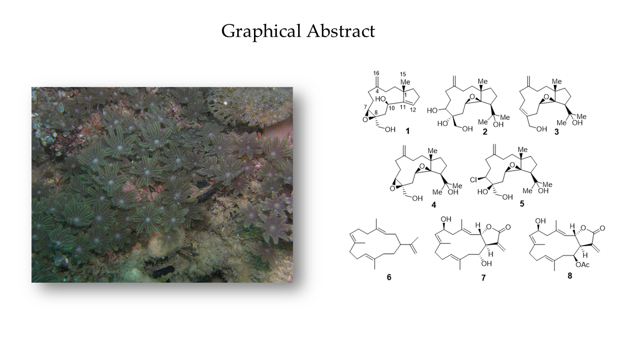
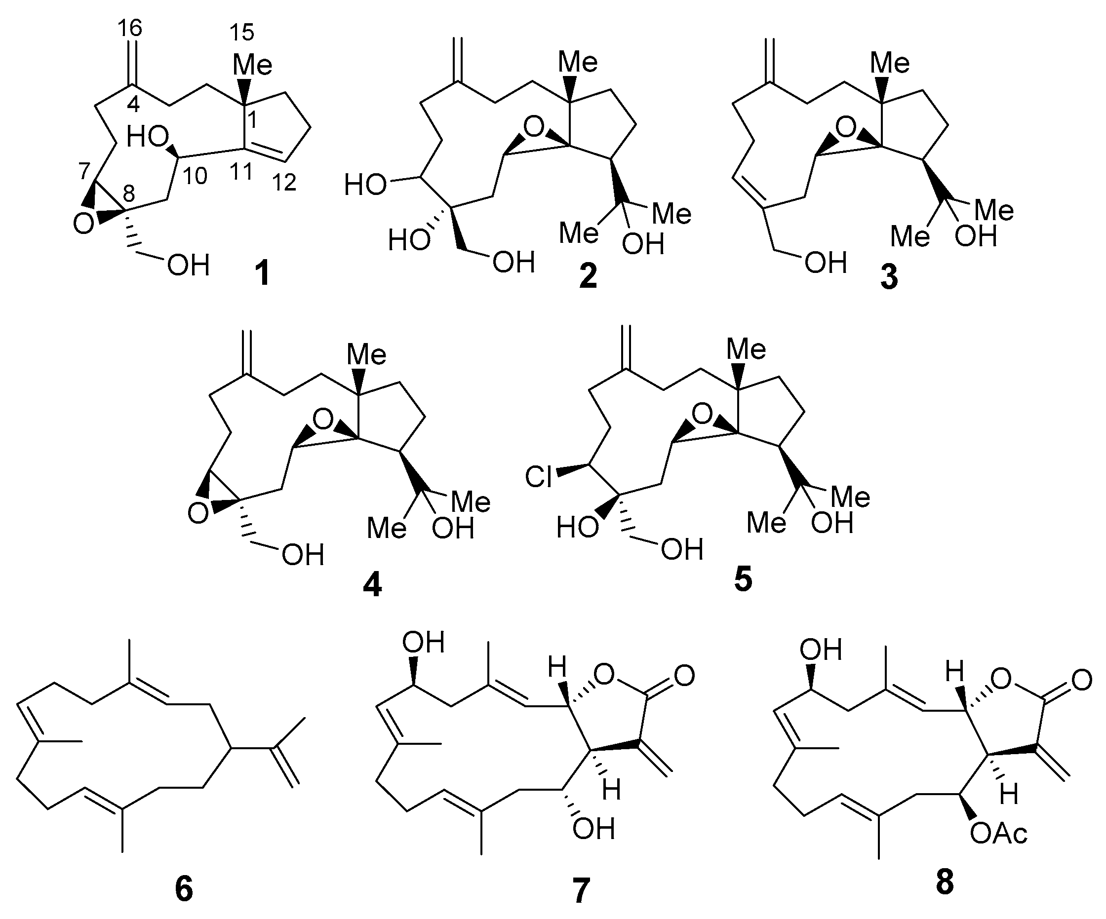
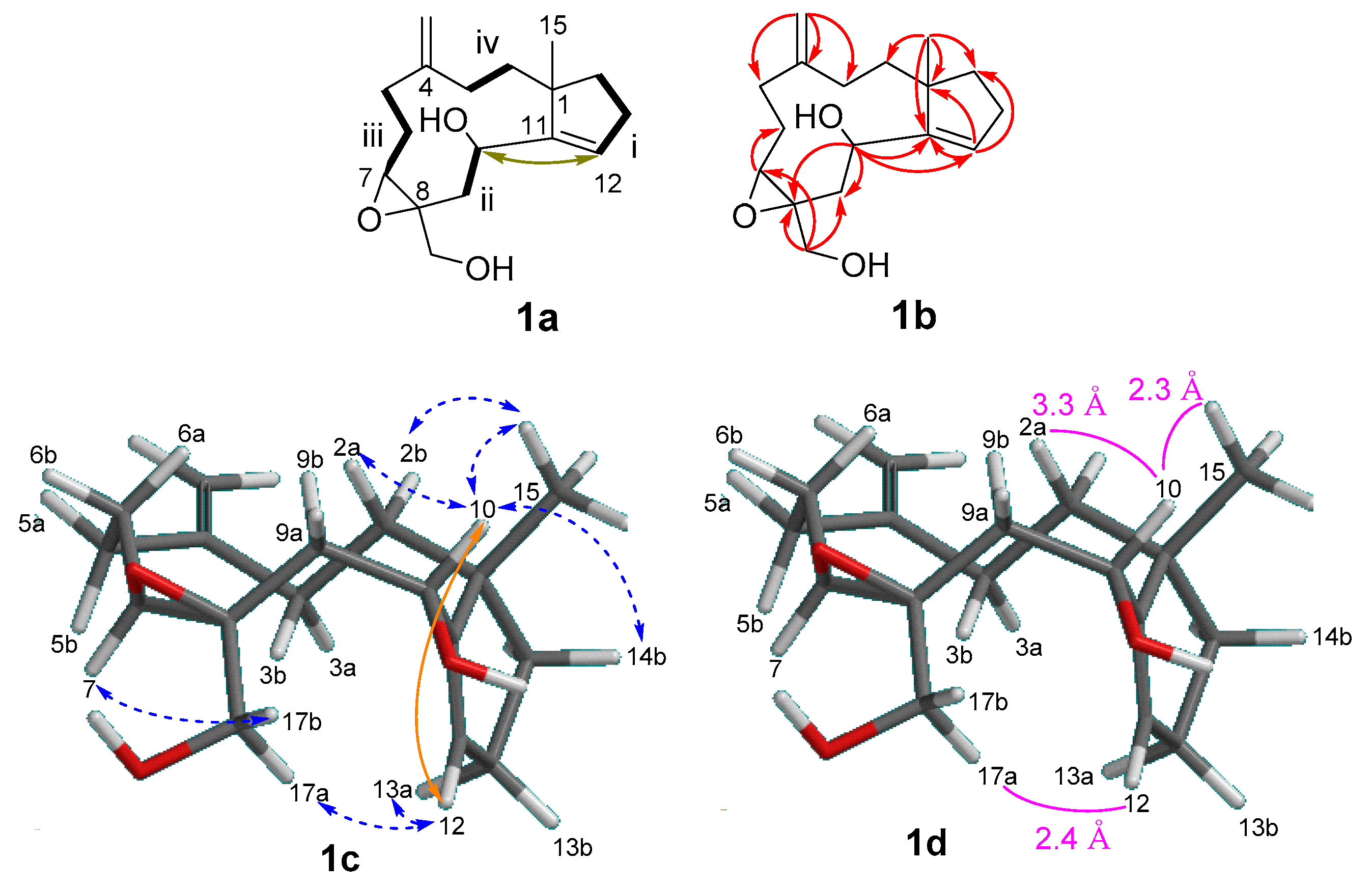
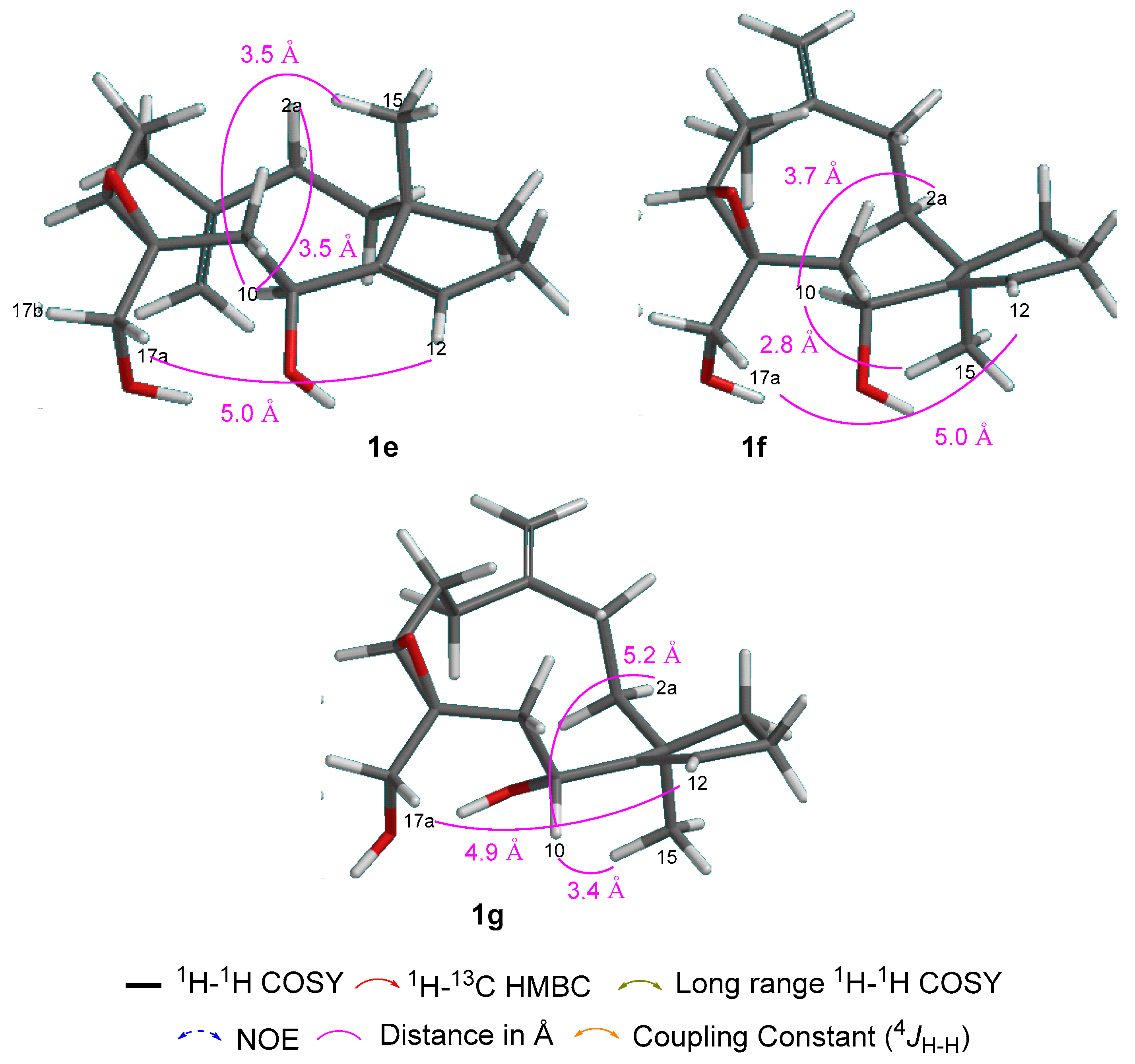
Sangiangols A and B, Two New Dolabellanes from an Indonesian Marine Soft Coral, Anthelia sp.

1
Department of Chemistry, Faculty of Mathematics and Natural Sciences, IPB University, Bogor 16680, Indonesia
2
Tropical Biopharmaca Research Center, IPB University, Bogor 16128, Indonesia
3
Department of Chemistry, Biology and Marine Science, University of the Ryukyus, Nishihara, Okinawa 903-0213, Japan
*
Author to whom correspondence should be addressed.
Molecules 2020, 25(17), 3803; https://doi.org/10.3390/molecules25173803
Submission received: 20 July 2020 / Revised: 14 August 2020 / Accepted: 18 August 2020 / Published: 21 August 2020
(This article belongs to the Section Natural Products Chemistry)

Abstract

:
A new, rare trinor-dolabellane diterpenoid, sangiangol A (1), and one new dolabellane diterpenoid, sangiangol B (2), together with known cembranes and dolabellanes (38), were isolated from the ethyl acetate layer of an extract of an Indonesian marine soft coral, Anthelia sp. Compounds 18 exhibited moderate cytotoxicity against an NBT-T2 cell line (0.5–10 µg/mL). The structures of the new compounds were determined by analyzing their spectra and a molecular modelling study. A possible biosynthetic pathway for sangiangols A (1) and B (2) is presented. Cytotoxicity requires two epoxide rings or a chlorine atom, as in 4 (stolonidiol) and 5 (clavinflol B).

Graphical Abstract

1. Introduction

Soft corals produce numerous, structurally diverse, biologically active terpenoids [1]. More specifically, Indonesian alcyonaceans are rich sources of diterpenoids with a variety of molecular skeletons. From 1970–2017, eight diterpenoid skeletons (briarane, cladiellane, seco-cladiellane, cembrane, nor-cembrane, dolabellane, flexibilane, and xenicane) were discovered in 11 genera of Indonesian alcyonaceans [2]. Among them, cembrane and briarane skeletons comprise a majority of the known diterpenoids in soft corals globally [2,3]. To date, soft corals of the genus Anthelia, family Xeniidae, have been shown to contain one type of sesquiterpenoid [4], three types of diterpenoids (xenicane [5], dolabellane [6], a C24-acetoacetylated diterpenoid [7,8]), and one type of steroid [9] with cytotoxic activity against various cell lines [4,5,6,7,8,9]. Moreover, the dolabellane stolonidiol (4), was identified as a promising candidate against Alzheimer’s disease after its mode of action in HEK293 cells was determined [10]. In our continuing study of metabolites of Indonesian Anthelia [4,6], we isolated known diterpenoids 38 and new dolabellanes, named sangiangols A (1) and B (2), the structures of which are the subject of this article.

2. Results and Discussion

A sample of the soft coral Anthelia sp., collected at Banten (BTN) in northwestern Java, was thoroughly extracted with acetone. After concentration, the residue was partitioned between EtOAc and H2O. The EtOAc extract showed significant cytotoxicity against NBT-T2 cells at 1 μg/mL. Thus, it was chromatographed on silica gel, followed by normal or reversed phase HPLC to afford two new molecules, 1 and 2, along with known compounds 38 (Figure 1).
Sangiangol A (1) was obtained as an optically active oil, [α]D27 + 20. Its molecular formula is C17H26O3 by HRESIMS and NMR (Table 1), indicating five degrees of unsaturation. Two compounds were identified as olefins (δC 153.0 (C); 127.0 (CH) (δH 5.88 brt, J = 2.5); 150.1 (C); 111.4 (CH2) (δH 4.70 s, 4.76 s)) and three others were assigned to a trisubstituted epoxide (δC 61.3 (CH) (δH 3.06 t, J = 6.5); δC 63.7 (C)) and a bicyclic structure. IR absorption at 3310 cm−1 and at 1714 and 1040 cm−1 suggested the presence of hydroxy and exomethylene groups, respectively.
Four spin systems iiv (1a; Figure 2) were disclosed by inspecting 1H–1H COSY cross peaks: (i) a trisubstituted double bond next to two methylenes (δH 5.88, 2.31, 2.10, 1.84, 1.63; H-12 to H-14), (ii) an oxymethine connected to a methylene (δH 2.20, 4.40; H-9 to H-10), (iii) the epoxide methine next to two methylenes (δH 2.42, 2.22, 1.73, 3.06; H-5 to H-7), and (iv) two methylenes (δH 1.63, 1.50, 2.10, 1.73; H-2 to H-3). A small coupling (4JH-H 1.3 Hz) between H-10 and H-12 with a COSY cross peak supported the presence of an allylic alcohol in Figure 2 (1a). HMBC correlations for H-10/C-8, 9, 11, 12 and H-12/C-1, 10, 11, 13, 14 confirmed the connection of spin systems i and ii. The trisubstituted epoxide was placed at C-7 and C-8, connecting spin systems ii, iii, and a primary alcohol (δH 3.38, 4.07) by observing HMBC correlations for H-7/C-6, 17; H-10/C-8; H-9/C-7, 8, 17; H-17a,b/C-7, 8, 9. Spin systems iii and iv were connected through an exomethylene (δH 4.70 s, 4.76 s; δC 153.0, 111.4) placed at C-4, as HMBC correlations H-16a,b/C-3, 4, 5 were observed. Finally, correlations from H3-15 to C-1, 2, 11, 14 supported the connection of spin systems i and iv, confirming the planar structure of 1 as a trinor-dolabellane diterpenoid.
The relative stereochemistry of 1 was tentatively assigned as follows, based on positive NOEs (1c; Figure 2). By observing a strong NOE between H-7 and H-17b (1c; Figure 2), chirality at the epoxide was revealed to be 7S*, 8S*, as in similar structural units [6,11]. Therefore, four possible stereoisomers—1c (1S*, 7S*, 8S*, 10R*), 1e (1S*, 7S*, 8S*, 10S*), 1f (1R*, 7S*, 8S*, 10S*), and 1g (1R*, 7S*, 8S*, 10R*)—were further considered. Figure 2 shows the energy-minimized conformations of 1c and 1e1g after molecular mechanics (MMFF) calculation. Of four possibilities, 1c was the most likely structure because a positive NOE was observed between H-17a and H-12 within a reasonable distance (Figure 2), while other candidates were expected to have longer distances (~5 Å). The angular methyl H-15 at δH 1.09 partially supported this conformation, showing NOEs for H-15/H-2b, 10 (Figure 2). The more downfield-shifted signal for the C-15 (δ 1.09) of 1 compared to that (δ 0.85) of the stolonidiol of 4 [6,11] may be due to the absence of the epoxide ring and the presence of an olefin at the ring junction.
Using HRESIMS and NMR, it was determined that sangiangol B (2), [α]D27 + 15, has the molecular formula C20H34O5, with an additional oxygen and two hydrogen atoms compared to stolonidiol (4). Furthermore, four degrees of unsaturation in 2 indicated a similarity to clavinflol B (5) [6,12], a chlorohydrin analog. A detailed 2D NMR (Figure 3) analysis of 2 revealed that the major differences between 2, 4, and 5 were the chemical shifts at C-7 (δH 3.56, d (11.4); δC 72.9 for 2 and δH 3.96, d (11.5); δC 67.2 for 5) [6,12] and at C-8 (δC 75.3 for 2 and 63.2 for 4) [6,11] (Figure 4). With the NMR chemical shifts and high-resolution mass spectrometry (HRMS) data, compound 2 contained 1,2-diol at C-7 and C-8 for an epoxide in stolonidiol (4) or for a chlorohydrin in clavinflol B (5) [6,12]. Key HMBC correlations for H-7/C-8, 17; H-17/C-7, 8; H-9/C-7; H-10/C-8 further confirmed the position of the diol, establishing the planar structure.
Of the six stereocenters of sangiangol B (2), three can be confirmed as 1S, 11R, and 12S by comparing the 13C-NMR data for the cyclopentane moiety (C-1, C-11~14) with those of 3, 4, and 5 (Figure 4) [6,11,12] and by observing the rotation value [α]D-37.9 of co-isolated stolonidiol (4) and the value [α]D-31 of that reported in 4, the absolute stereochemistry of which was established by X-ray crystallography [11]. Chirality at C-10 was shown to have the same R configuration as stolonidiol (4), based on a positive NOE between H-10 and H-15. Among the four possible structures—2d: 7R,8R, 2e: 7S,8S, 2f: 7R,8S, and 2g: 7S,8R—the distances for H-10/H-7 and H-10/H-17b with energy-minimized conformations were compared, as in Figure 3. Both 2e and 2f were eliminated due to the relatively long distances of H-10/H-17b. However, as two candidates, 2d and 2g, accorded with the spectral data, the configuration at C-8 was then revealed, while only C-7 remained to be solved.
Furthermore, the biosynthesis of stolonidiol-related molecules can be proposed, as in Figure 5. Geranylgeranyl pyrophosphate (GGPP) is a well-known starting material for diterpenoids [13]. Sangiangol A (1) could be derived from stolonidiol (4) through a series of degradation and epoxide ring-opening reactions, while sangiangol B (2) could be derived from sangiangol C (3) through an epoxidation reaction. Moreover, sangiangol C (3) could be the precursor of stolonidiol (4). Unfortunately, attempts to prepare α-methoxy-α-trifluoromethylphenylacetic acid (MTPA) esters for the determination of the absolute configurations of both molecules failed because of their instability and the small quantities of these compounds available.
All isolated compounds (18) were evaluated for cytotoxicity against NBT-T2 rat bladder epithelial cells (Table 2). New entities 1 and 2 showed weak toxicity at 5 and 10 μg/mL, respectively, while known molecules 38 showed moderate and weak toxicity at 10, 1, 0.5, 10, 1, and 10 μg/mL, respectively. From the structure–activity relationship of stolonidiol derivatives, two epoxide rings or a chlorine atom are required for their cytotoxicity, as in 4 (stolonidiol) and 5 (clavinflol B).

3. Materials and Methods

3.1. General Methods

The optical rotations were obtained with a JASCO P-1010 digital polarimeter. The 1H and 13C-NMR spectra were recorded on a JEOL α 500 FT NMR spectrometer. The chemical shifts were expressed in δ (ppm) and the coupling constants (J) in Hz. The electrospray ionization mass spectrometry (ESIMS) data were obtained on a PE QSTAR mass spectrometer and the infrared (IR) spectra were recorded on a DR 8020 Shimadzu spectrophotometer. The HPLC was performed on a Hitachi L-6000 pump equipped with a Shodex RI-101 monitor and a Hitachi L-4000 UV detector, using a Cosmosil 5C18AR-II (5 µm) or a Mightysil RP-18 (5 µm) column. Merck silica gel 60 (0.063–0.20 mm) was used for column chromatography. The analytical thin layer chromatography (TLC) was performed on commercial silica gel 60 F254 visualized with vanillin–EtOH-1% H2SO4. All solvents used were reagent grade.

3.2. Animal Material

A marine soft coral (AA-C31) was collected from Krakatau Island, Banten, Indonesia at 10–15 m depth by hand, while scuba diving. It was then stored in EtOH. The specimen was identified as Anthelia sp. by one of us (J.T.).

3.3. Extraction and Isolation

The fresh soft coral specimen (wet weight, 121 g) stored in EtOH was extracted four times using Me2CO (4 × 150 mL). The four solutions were pooled and concentrated under vacuum, and the resulting residue was partitioned between EtOAc and H2O to obtain a cytotoxic lipophilic extract (1.57 g, NBT-T2 1 μg/mL). The whole extract was separated on a Si gel 60 column by eluting stepwise with hexane–EtOAc–MeOH to afford 19 fractions. The second fraction (9.7 mg) was further separated using normal phase silica HPLC (hexane 100%) to give cembrane A (6) [13] (2.3 mg). The eighth fraction was purified on reversed phase HPLC to give sangiangol C (3) [14] (1.0 mg). The ninth fraction was repeatedly separated by HPLC (first, reversed-phase C18 (RP18), MeOH–H2O, 5:2; second, Si 60, CH2Cl2–EtOAc, 7:3) to afford kericembrenolide E (7) [15] (2.7 mg), stolonidiol (4) [6,11] (3.5 mg), and clavinflol B (5) [6,12] (1.3 mg). The tenth fraction was subfractionated on RP18 HPLC to give cembrenolide (8) [16] (1.7 mg), sangiangol A (1) (1.3 mg), and stolonidiol (4) (103.7 mg). Finally, the most polar fraction was repeatedly separated on reversed-phase HPLC (first, RP18: MeOH–H2O, 2:1 second, RP18, MeOH–H2O, 4:5) to give sangiangol B (2) (1.4 mg).

3.3.1. Sangiangol A (1)

Colorless oil; [α]D27 + 20 (c 0.09, CHCl3); IR (KBr) νmax 3418, 2965, 1683, 1645, 1456, 1378, 1168, 1064 cm−1; 1H and 13C-NMR (see Table 1 and Table 2); HRESIMS m/z 301.1658 [M + Na]+ (calculated (calcd) for C17H26O3Na 301.1779).

3.3.2. Sangiangol B (2)

Colorless oil; [α]D27 + 15 (c 0.14, CHCl3); IR (KBr) νmax 3418, 2965, 1645, 1456, 1378, 1168, 1064 cm−1; 1H and 13C-NMR (see Table 1 and Table 2); HRESIMS m/z 377.2230 [M + Na]+ (calcd for C20H34O5Na 377.2304).

3.4. Cytotoxicity Assay

NBT-T2 rat bladder epithelial cells (BRC-1370) purchased from RIKEN (Tsukuba, Ibaraki, Japan) were cultured under a standard protocol using Dulbecco’s modified Eagle’s medium (DMEM). The cells were seeded in 1 mL of modified Eagle’s media supplemented with 10% heat-inactivated fetal bovine serum, streptomycin, amphotericin B, and glutamic acid. The cells were exposed to graded concentrations of the new and known compounds, as well as their fractions at 37 °C, for 72 h and observed under a microscope to observe the effects at 48 and 72 h.

Author Contributions

Conceptualization, N.H., A.M. and J.T.; methodology, N.H., A.M. and J.T.; validation, N.H., A.M. and J.T.; investigation, N.H., A.M. and J.T.; data curation, N.H., A.M. and J.T.; writing—original draft preparation, N.H., A.M. and J.T.; writing—review and editing, N.H., A.M. and J.T. All authors have read and agreed to the published version of the manuscript.

Funding

This research was partially funded by the Ministry of Research and Technology-National Research and Innovation (RISTEK–BRIN) of the Republic of Indonesia grant number (1769/IT3.11/PN/2018, 4175/IT3.L1/PN/2019, 4014/IT3.L1/PN/2020) and Ministry of Education, Culture, Sports, Science and Technology (MEXT) of Japan.

Acknowledgments

We thank the Ministry of Research and Technology–National Research and Innovation (RISTEK–BRIN) of the Republic of Indonesia for its support of N.H. through its excellent basic university research program (1769/IT3.11/PN/2018, 4175/IT3.L1/PN/2019, 4014/IT3.L1/PN/2020). We also thank Masaki Kita of Nagoya University for performing a database search.

Conflicts of Interest

The authors declare no conflict of interest.

References

  1. Caroll, A.R.; Copp, B.R.; Davis, R.A.; Keyzers, R.A.; Prinsep, M.R. Marine natural products. Nat. Prod. Rep. 2020, 37, 175–223. [Google Scholar] [CrossRef] [PubMed]
  2. Hanif, N.; Murni, A.; Tanaka, C.; Tanaka, J. Marine natural products from Indonesian waters. Mar. Drugs 2019, 17, 364. [Google Scholar] [CrossRef] [PubMed] [Green Version]
  3. Berrue, F.; Kerr, R.G. Diterpenes from gorgonian corals. Nat. Prod. Rep. 2009, 26, 681–710. [Google Scholar] [CrossRef] [PubMed]
  4. Hanif, N.; Murni, A.; Yamauchi, M.; Higashi, M.; Tanaka, J. A new trinor-guaine sesquiterpene from and Indonesian soft coral Anthelia sp. Nat. Prod. Commun. 2015, 10, 1907–1910. [Google Scholar] [PubMed] [Green Version]
  5. Coval, S.J.; Scheuer, P.J.; Matsumoto, G.K.; Clardy, J. Two new xenicin diterpenoid from the octocoral Anthelia edmonsoni. Tetrahedron 1984, 40, 3823–3828. [Google Scholar] [CrossRef]
  6. Murni, A.; Hanif, N.; Tanaka, J. A new cytotoxic dolabellane from the Indonesian soft coral Anthelia sp. Indones. J. Chem. 2013, 13, 216–220. [Google Scholar] [CrossRef] [Green Version]
  7. Green, D.; Carmely, S.; Benayahu, Y.; Kashman, Y. Antheliolide A & B: Two new C24-acetoacetylated diterpenoids of the soft coral Anthelia glauca. Tetrahedron Lett. 1988, 29, 1605–1608. [Google Scholar]
  8. Smith, A.B.; Carrol, P.J.; Kashman, Y.; Green, D. Revised structure of antheliolides A and B. Tetrahedron Lett. 1989, 30, 3363–3364. [Google Scholar] [CrossRef]
  9. Sjsstrand, U.; Bohlin, L.; Fisher, L.; Colin, M.; Djerassi, C. Minor and trace sterol from marine invertebrate 28: Novel polyhydroxylated sterol from soft coral Anthelia glauca. Steroids 1981, 38, 347–354. [Google Scholar] [CrossRef]
  10. Mason, J.W.; Schmid, C.L.; Bohn, L.M.; Roush, W.R. Stolonidiol: Synthesis, target identification, and mechanism for choline acetyltransferase activation. J. Am. Chem. Soc. 2017, 139, 5865–5869. [Google Scholar] [CrossRef] [PubMed]
  11. Mori, K.; Iguchi, K.; Yamada, N.; Yamada, Y.; Inouye, Y. Stolonidiol, a new marine diterpenoid with a strong cytotoxic activity from the Japanese soft coral. Tetrahedron Lett. 1987, 28, 5673–5676. [Google Scholar] [CrossRef]
  12. Shen, Y.; Pan, Y.; Ko, C.; Kuo, Y.; Chen, C. New dolabellanes from the Taiwanese soft coral Clavularia inflate. J. Chin. Chem. Soc. 2003, 50, 471–476. [Google Scholar] [CrossRef]
  13. Ireland, C.; Faulkner, D.J.; Finer, J.; Clardy, J. A novel diterpene from Dollabella californica. J. Am. Chem. Soc. 1976, 98, 4664–4665. [Google Scholar] [CrossRef] [PubMed]
  14. Vanderah, D.J.; Rutledge, N.; Schmitz, F.J.; Ciereszko, L.S. Marine natural products: Cembrene-A and cembrene-C from a soft coral, Nephthea species. J. Org. Chem. 1978, 43, 1614–1616. [Google Scholar] [CrossRef]
  15. Kobayashi, M.; Son, B.W.; Kyogoku, Y.; Kitagawa, I. Kericembrenolides A, B, C, D, and E, five new cytotoxic cembrenolides from the Okinawan soft coral Clavularia koellikeri. Chem. Pharm. Bull. 1986, 34, 2306–2309. [Google Scholar] [CrossRef] [PubMed]
  16. Iwashima, M.; Matsumoto, Y.; Takahashi, H.; Iguchi, K. New marine cembrane-type diterpenoids from the Okinawan soft coral Clavularia koellikeri. J. Nat. Prod. 2000, 63, 1647–1652. [Google Scholar] [CrossRef] [PubMed]
Sample Availability: Not available.
Figure 1. Chemical structures of dolabellane-type molecules (15) and cembrane-type molecules (68).
Molecules 25 03803 g001
Figure 2. Key: COSY (1a), HMBC (1b), NOE (1c), correlations and a long-range coupling constant (4JH-H), as well as the distance between atoms (1d), with a computer-generated model of 1 (energy minimized: 1S*, 7S*, 8S*, 10R* for (1c); 1S*, 7S*, 8S*, 10S* for (1e); 1R*, 7S*, 8S*, 10S* for (1f); 1R*, 7S*, 8S*, 10R* for (1g), obtained from calculations with molecular mechanics MMFF).
Molecules 25 03803 g002aMolecules 25 03803 g002b
Figure 3. Key: COSY (2a), HMBC (2b), NOE (2c), correlations and distance between atoms (2d) with a computer-generated model of 2 (energy-minimized 1S, 7R, 8R, 10R, 11R, 12S for (2c); 1S, 7S, 8S, 10R, 11R, 12S for (2e); 1S, 7R, 8S, 10R, 11R, 12S for (2f); 1S, 7S, 8R, 10R, 11R, 12S for (2g), obtained from calculations with MMFF).
Molecules 25 03803 g003
Figure 4. Comparative analysis of 13C chemical shifts between 2 and 3, 4, 5.
Molecules 25 03803 g004
Figure 5. A possible biosynthetic pathway for sangiangols A (1) and B (2).
Molecules 25 03803 g005
Table 1. 1H NMR data for compounds 1 and 2 in CDCl3.
PositionSangiangol A (1)Sangiangol B (2)
δC *mult.δH **J in HzδC *mult.δH **J in Hz
150.2C 44.7C
2a38.4CH21.63m ***42.8CH21.97m
2b 1.50ddd (14.4, 11.6, 7.2) 1.25m ***
3a29.6CH22.10m ***25.0CH22.09dt (15.4, 10.0)
3b 1.73m *** 1.64dd (13.3, 10.0)
4150.1C 149.3C
5a30.8CH22.42dt (14.6, 5.4)34.3CH22.45td (13.6, 4.6)
5b 2.22dd (14.6, 7.7) 2.29brdd (13.6, 4.6)
6a24.4CH21.73m ***27.4CH21.79m
6b 1.73m *** 1.50m
761.3CH3.06t (6.5)72.9CH3.56d (11.4)
863.7C 75.3C
9a36.0CH22.20dd (15.7, 3.0)33.6CH21.95dd (4.5, 2.7)
9b 2.00dd (15.7, 6.3) 1.92dd (5.5, 1.6)
1065.1CH4.40dd (6.3, 1.3)54.5CH3.02dd (5.4, 2.7)
11153.0C 76.7C
12127.0CH5.88brt (2.5)50.2CH2.18d (10.8, 2.0)
13a29.7CH22.31m ***27.8CH21.89m
13b 2.10m *** 1.60m
14a36.7CH21.84ddd (12.7, 9.0, 7.1)39.0CH21.79m
14b 1.63m *** 1.74m
1527.4CH31.09s24.0CH30.84s
16a111.4CH24.76s112.7CH24.96s
16b 4.70s 4.79s
17a67.2CH24.07d (12.2)67.2CH23.89d (11.4)
17b 3.38d (12.2) 3.52d (11.4)
18 75.2C
19 29.6CH31.21s
20 26.0CH31.26s
* Assigned by DEPT and 2D NMR (HSQC and HMBC) experiments. ** Assigned by 2D NMR (COSY, HSQC, and HMBC) experiments. *** Overlapping signals.
Table 2. Cytotoxicity of compounds 18 against NBT-T2 rat bladder epithelial cells.
CompoundConcentration (µg/mL)
15
210
310
41
50.5
610
71
810

