Article

Synthesis and Biological Activity of New 7-Amino-oxazolo[5,4-d]Pyrimidine Derivatives

1
Department of Organic Chemistry, Faculty of Pharmacy, Wroclaw Medical University, 211A Borowska Street, 50-556 Wroclaw, Poland
2
Department of Experimental Therapy, Institute of Immunology and Experimental Therapy, Polish Academy of Sciences, 12 Rudolf Weigl Street, 53-114 Wroclaw, Poland
3
Department of Drug Technology, Faculty of Pharmacy, Wroclaw Medical University, 211A Borowska Street, 50-556 Wroclaw, Poland
4
Faculty of Chemistry, University of Wroclaw, 14 Joliot-Curie, 50-383 Wroclaw, Poland
*
Author to whom correspondence should be addressed.
Molecules 2020, 25(15), 3558; https://doi.org/10.3390/molecules25153558
Submission received: 17 July 2020 / Revised: 29 July 2020 / Accepted: 3 August 2020 / Published: 4 August 2020
(This article belongs to the Section Medicinal Chemistry)

Abstract

:
The synthesis of a series of novel 7-aminooxazolo[5,4-d]pyrimidines 5, transformations during their synthesis and their physicochemical characteristics have been described. Complete detailed spectral analysis of the intermediates 24, the N′-cyanooxazolylacetamidine by-products 7 and final compounds 5 has been carried out using MS, IR, 1D and 2D NMR spectroscopy. Theoretical research was carried out to explain the privileged formation of 7-aminooxazolo[5,4-d]pyrimidines in relation to the possibility of their isomer formation and the related thermodynamic aspects. Additionally, the single-crystal X-ray diffraction analysis for 5h was reported. Ten 7-aminooxazolo[5,4-d]pyrimidines 5 (SCM110) were biologically tested in vitro to preliminarily evaluate their immunological, antiviral and anticancer activity. Compounds SCM5 and SCM9 showed the best immunoregulatory profile. The compounds displayed low-toxicity and strongly inhibited phytohemagglutinin A-induced proliferation of human peripheral blood lymphocytes and lipopolysaccharide-induced proliferation of mouse splenocytes. Compound SCM9 caused also a moderate suppression of tumor necrosis factor α (TNF-α) production in a human whole blood culture. Of note, the compounds also inhibited the growth of selected tumor cell lines and inhibited replication of human herpes virus type-1 (HHV-1) virus in A-549 cell line. Molecular investigations showed that the compounds exerted differential changes in expression of signaling proteins in Jurkat and WEHI-231 cell lines. The activity of SCM5 is likely associated with elicitation of cell signaling pathways leading to cell apoptosis. The compounds may be of interest in terms of therapeutic utility as inhibitors of autoimmune disorders, virus replication and antitumor agents.

1. Introduction

The oxazolo[5,4-d]pyrimidine system was synthesized for the first time by Clapp as a derivative, i.e., 5-(ethylsulfanyl)-2-phenyl[1,3]oxazolo[5,4-d]pyrimidine (Structure I in Figure 1), which was described and revealed in 1905 by Johnson [1].
Since that time, many derivatives of oxazolo[5,4-d]pyrimidine system have been synthesized and extensively researched for their potential biological activity. Some oxazolo[5,4-d]pyrimidines are well known as adenosine receptor antagonists (Structure II in Figure 1) [2], inhibitors of receptor tyrosine kinase (Structure III in Figure 1) [3], inhibitors of adenosine kinase (Structure IV in Figure 1) [4], effective inhibitors of ricin [5] and Shiga toxin inhibitors (Structure V in Figure 1) [6]. The antiviral (Structure VI in Figure 1) [7], anti-angiogenic (Structure VII in Figure 1) [8] and immunosuppressive (Structure VIII in Figure 1) [9] activity of some of them has been also revealed. Besides, some oxazolo[5,4-d]pyrimidines can also activate the caspase cascade (Structure IX in Figure 1) [10] and inhibit ubiquitin-activating enzymes (E1 enzymes), particularly NEDD8-activating enzyme (NAE) (Structure X in Figure 1) [11], which may result in their antineoplastic activity.
In our previous search for novel potent immunomodulatory, anti-inflammatory and anticancer agents, many of five-membered heterocycles with two heteroatoms, such as 5-amino-3-methyl-4-isoxazolecarboxylic acid [12], 5-amino-3-methyl-4-isothiazolecarboxylic acid [13,14,15] and 4-amino-1-methyl-5-imidazolecarboxylic acid derivatives [16] were designed and synthesized. The described results revealed that the isoxazole moiety was particularly responsible for the immunological activity of these compounds. These derivatives exhibited a variety of activities, such as immunosuppressive [17,18,19,20,21], immunostimulatory [22,23] and anti-inflammatory [24,25].
The aim of this work was the synthesis of 7-aminooxazolo[5,4-d]pyrimidines series with 5-amino-3-methyl-isoxazol-4-yl substituent at position 2 of their structure to find novel derivatives exhibiting potential immunosuppressive activity. It is worth noting that oxazolo[5,4-d]pyrimidine system is structurally similar to purine analogs of naturally occurring nucleic acid bases. Such a structure may, in theory, condition antitumor and/or antiviral activity, so for this purpose, several in vitro immunological and antitumor assays have been designed and carried out. Finally, on the basis of the obtained results, an attempt to establish a plausible mechanism of the biological action of these compounds has been made.

2. Results and Discussion

2.1. Chemistry

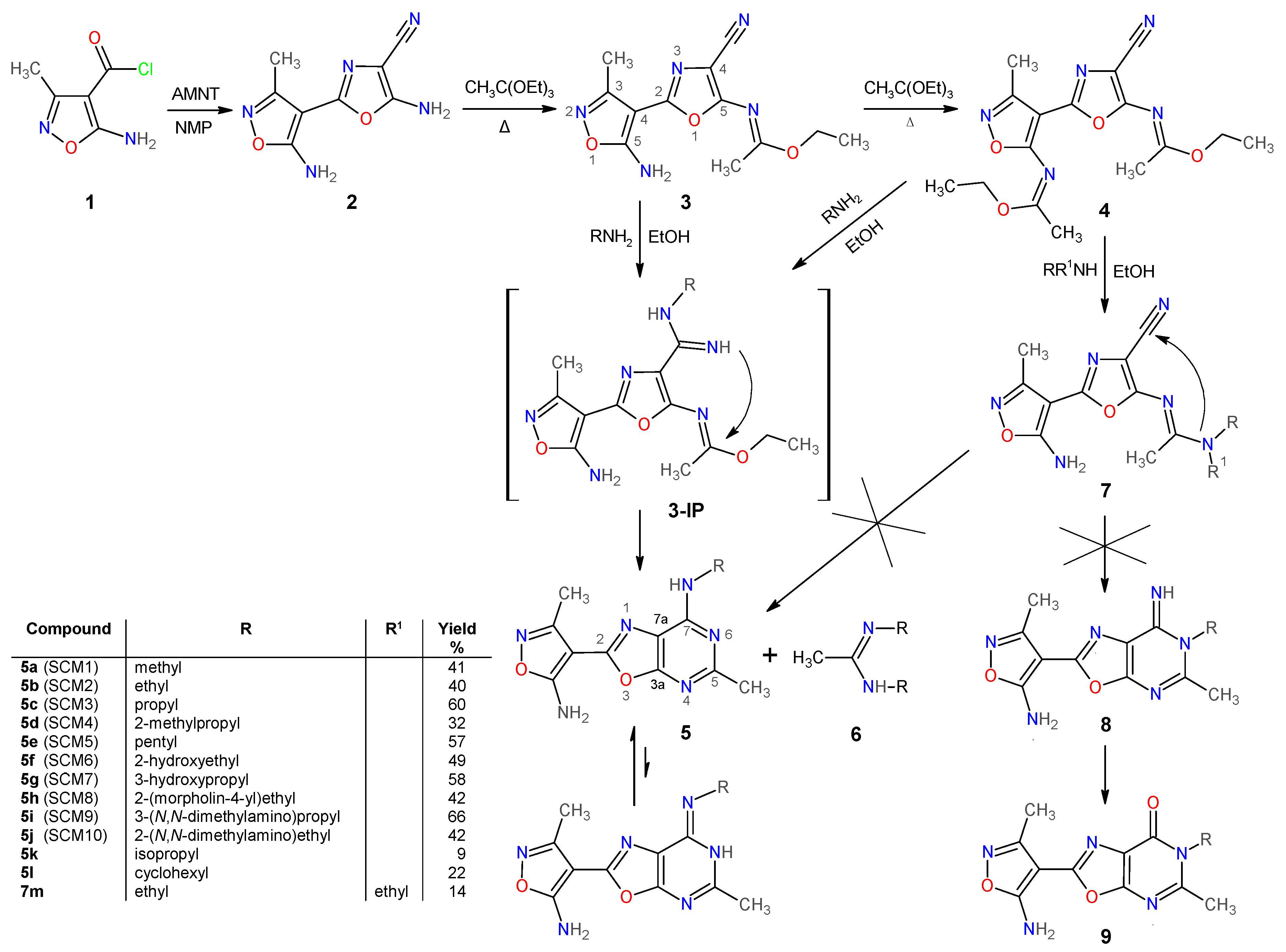
2.1.1. Synthesis and Some Physicochemical Properties of New 7-Aminooxazolo[5,4-d]Pyrimidines 5

In general, there are two known main methods of synthesis of oxazolo[5,4-d]pyrimidines, namely, through cyclization of the pyrimidine ring on existing, appropriately substituted, oxazole derivative or through cyclization of the oxazole ring on pyrimidine derivative [26]. For the first time, this system was synthesized just by using the second method [1], where 2-mercapto-5-benzoylamino-4-hydroxypyrimidine was cyclized with phosphoryl trichloride (POCl3). Herein, the described series of new oxazolo[5,4-d]pyrimidines 5 is synthesized according to the synthetic pathway outlined in Scheme 1.
The following reagents and substrates have been used in the synthesis of the considered compounds: starting materials, such as esters of 2-cyano-3-alkoxy-2-butenoate acids, which were used to synthesize known 5-amino-3-methyl-4-isoxazolecarboxylic acid [27], were prepared according to the highly efficient methods precisely described in [28]. 5-Amino-3-methylisoxazole-4-carbonyl chloride (1, Scheme 1) was also prepared using the method described earlier [12]. Similarly to the method described by Freeman and Kim [29], 5-amino-2-(5-amino-3-methylisoxazol-4-yl)-oxazole-4-carbonitrile (2) was obtained in the reaction, which was conducted in 1-methyl-2-pyrrolidinone (NMP) at room temperature, between the commercially available aminomalononitrile tosylate (AMNT) and compound 1. Then, ethyl N-{4-cyano-2-[5-(1-ethoxyethylidene)amino-3-methylisoxazol-4-yl]-oxazol-5-yl}ethanimidate (4) was obtained through the intermediate imidate, i.e., ethyl N-[2-(5-amino-3-methylisoxazol-4-yl)-4-cyanooxazol-5-yl]ethanimidate (3), by heating of oxazole 2 in an excess of triethyl orthoacetate under reflux. It is necessary to emphasize that the compound 3 has one and the product 4 has two centers of Z-E isomerism, respectively, so there is a possibility that, theoretically, two in the case of 3 or four single isomers in the case of 4 or their mixtures can be created, respectively. Anyway, a spectral analysis revealed that only one isomer was formed in both cases for the compounds 3 and 4. We have not determined which one, as this fact seems to be of little importance for a course of the next stage of the synthesis. The reactions of the imidate 4 with a 30% ethanolic solution of the appropriate aliphatic amines, conducted at room temperature, resulted in a series of final oxazolo[5,4-d]pyrimidines 5. It should be noted that the molecule of compound 4 has three reactive centers with lowered electron density that are susceptible to attack by nucleophilic agents, the nitrile carbon and the carbons of both imidate moieties. Therefore, three possible competitive reactions can occur and result in various products. Our research indicates that the desired compounds 5 form via the labile intermediate amidines 3-IP that are formed by the nucleophilic addition of the corresponding amine to the nitrile group of the compound 4 or 3. In this stage, an excess of the primary aliphatic amine as a strong nucleophile agent causes the removal of the ethoxyethylidene moiety (in the case of the compound 4) from the amino group of the isoxazole ring with simultaneous cyclization to the pyrimidine ring, while acetamidines 6 (only in the case of primary amines) are produced as by-products. However, secondary aliphatic amines (such as N,N-diethylamine, R, R1 = Et) did not give the expected products 5 under these conditions but N′-cyanooxazolylacetamidine 7 (7m, R,R1 = Et for N,N-diethylamine) in low yield (≈14%) and the mixture of product of the decomposition of the compound 4, i.e., the compounds 2 and 3. It is worth mentioning that the 1H-NMR spectrum (recorded at 297.8 K ≈ 25 °C) of N′-cyanooxazolylacetamidine 7m (for a detailed description of its spectrum, see Section 3.1.2 of “Preparation and experimental properties of compounds 25, 7” and for the spectrum, see Figure S76 in the Supplementary Material) shows that the corresponding protons of the N-ethyl groups are not magnetically equivalent. This suggests that inhibition of rotation around the bond between the quaternary acetamidine carbon atom and the nitrogen atom occur. This supposition has been supported with the theoretically predicted rotational barrier that has been estimated to be about 23 kcal·mol−1 (see Section 3.1.4. Computational details). A similar energy barrier is needed for free rotation of the N,N-dimethyl group in a known useful aprotic polar solvent, N,N-dimethylformamide (DMF), for which the experimentally estimated energy barrier is about 21 kcal·mol−1 [30]. In DMF, this inhibition of rotation is caused by fact that bond between carbonyl carbon and nitrogen has partial double bond character, which is connected with a significant contribution of the imidic acid tautomer to the resonance structure. In the case of N′-cyanooxazolylacetamidine 7m, the reason could be similar.
Primary aliphatic amines with rigid bulky steric hindrance (with a secondary aliphatic radical like isopropyl or cyclohexyl) usually gave the mixture of products (like 2, 5 and 6) with very low abundance of the oxazolo[5,4-d]pyrimidines 5. In the cases of isopropylamine and cyclohexylamine, the yield of appropriate pure oxazolo[5,4-d]pyrimidines 5 was about 9% (5k) and 22% (5l), respectively. However, in the case of t-butylamine, only the products of decomposition of substrate 4 were isolated. Similarly, in the case of aromatic amines (such as aniline, R = Ph, R1 = H and p-toluidine, R = p-MePh, R1 = H), neither oxazolo[5,4-d]pyrimidines 5 nor N′-cyanooxazolyl-acetamidines 7 were observed to be formed. In these cases, the imidate 4 underwent only decomposition to the mixture of the compounds 3 and 2 under these conditions, so only the mixture of all of them was isolated. This is probably caused by the much less nucleophilicity of the aromatic amine nitrogen, making aromatic amines behave inertly under these reaction conditions.
It is necessary to underline that during the course of all the reactions of the imidate 4 with amines, the creation of some quantity of the substrate 2 and the intermediate imidate 3 was observed as a result of decomposition of the compound 4. Besides, it appears that compound 4 is unstable in ethanolic solution and slowly decomposes through the intermediate imidate 3 to the substrate 2. The isolated, from reaction mixture, intermediate imidate 3 has been also undergone the reactions with the appropriate aliphatic amines (like in the case of the imidate 4) giving the same final oxazolo[5,4-d]pyrimidines 5 as in the case of the imidate 4, which, in addition to spectral analysis, supports and chemically proves the molecular structure of the compound 3 (for example, preparation of the compound 5i, see Method C in Section 3.1.2 “Preparation and experimental properties of compounds 25, 7”).
Theoretically predicted heats (enthalpies ΔfH298) of formation, opposite to binding energies, of N′-cyanooxazolylacetamidines 7 (see calculation details in 3.1.4. Computational details), are greater than the ones of structurally corresponding isomeric oxazolo[5,4-d]pyrimidines 5, which means that oxazolo[5,4-d]pyrimidines 5 should be more thermodynamically stable than N′-cyanooxazolyl-acetamidines 7. This points out that the observed cases of formation of N′-cyanooxazolylacetamidines 7 are rather kinetically and/or sterically controlled, especially taking into account that N′-cyanooxazolylacetamidines 7 were formed only in the case of reaction with secondary amines, like N,N-diethylamine, for example. Besides, the formation of N′-cyanooxazolylacetamidines 7 suggests that final 7-aminooxazolo[5,4-d]pyrimidines 5 can be produced through only unstable intermediate amidines 3-IP that is formed as a result of the nucleophilic addition of appropriately active amine to cyano group of the imidate 4 or the intermediate imidate 3 because only these amidines 3-IP can cyclize into oxazolo[5,4-d]pyrimidines 5 contrary to N′-cyanooxazolylacetamidines 7. Theoretically, N′-cyanooxazolylacetamidines 7 could cyclize into N-6-substituted-oxazolo[5,4-d]pyrimidin-7-imines 8 but only in the cases where R1 = H (see Scheme 1). Formation of such similar imines 8 have been previously described by Ohtsuka [31]. However, imines 8 should be rather unstable, especially, in the presence of water (moisture) which can lead them to decompose into N-6-substituted-oxazolo[5,4-d]pyrimidin-7-ones 9. Actually, the formation of imines 8 and/or oxazolo[5,4-d]pyrimidin-7-ones 9 was not observed under the applied reaction conditions during the conducted research. Besides, this fact is also supported on the basis of the theoretical research on the example of the compounds 5a and 5c, for a simulation carried out for single isolated molecule in a vacuum environment. The calculated difference of energy (ΔE298) between thermal-corrected energies of the considered compounds (5a and 5c) and their respective isoforms (8a and 8c) points out that the compounds 5a and 5c should be more thermodynamically stable by about 13 kcal·mol−1 (Estab = −ΔE298 = 13.06 and 13.23 kcal·mol−1) than the theoretical compounds 8a and 8c, respectively (see calculation details in 3.1.4. Computational details). The calculations for simulated ethanol solutions of considered molecules conducted with the Conductor-like Polarizable Continuum Model (CPCM) [32] give similar results, i.e., Estab = 12.43 and = 12.83 kcal·mol−1 for the compounds 5a and 5c, respectively. It can be seen that the electrostatic field generated by ethanol molecules has very little effect on the stabilization of the imino isomers 8 compared to vacuum. Finally, the 7-aminooxazolo[5,4-d]pyrimidine structure of the final products 5 were unquestionably proven by X-ray crystallography on the basis of single-crystal X-ray diffraction measurement of the compound 5h.
It should be also noted that compounds 5 may exhibit amine-imine tautomerism (see Scheme 1), similar to purines and pyrimidine bases (e.g., adenine and cytosine), due to the mobility of the proton of the amino group at the seventh position of the oxazolopyrimidine ring. Therefore, there is probability that this proton may easily jump to nitrogen at the sixth position of the pyrimidine ring of the oxazolopyrimidine system due to the fact that the nitrogen atom of amino group at the seventh position, the nitrogen atom of the pyrimidine ring at the sixth position and the carbon atom at seventh position form a semi-cyclic amidine moiety. Theoretical estimation (on the example of the compound 5h) of free enthalpy (ΔGtrans(298) = 10.748 kcal·mol−1) and the equilibrium constant (Ktrans = [imine]/[amine] ≈ 1.323 × 10⁻⁸ proton transition as a result of amine-imine tautomerism suggests that the amine tautomer of the compound 5h should dominate with a huge advantage in the state of thermodynamic equilibrium, which could have been expected because of partial loss of aromaticity by oxazolopyrimidine ring system of imine tautomer. It can be figured out that one molecule of imine tautomer of the compound 5h occurs with over 75.5 million molecules of its amine tautomer in the equilibrium (see calculation details in 3.1.4. Computational details). For ethanol environment simulation, the results (ΔGtrans(298) = 10.752 kcal·mol−1 and Ktrans ≈ 1.313 × 10⁻⁸) are very similar (insignificant difference) to those of a vacuum one what would suggests that that electric field generating by the solvent does not influence on the equilibrium in this case. Unfortunately, the CPCM calculations do not take into account the possibility that hydrogen bonds may form between the protic solvent, which is ethanol, and the molecules of the considered compounds, which is a disadvantage of this method.
Taking into account some chemical instability of the compound 4 and the formation of by-products (2, 6 and 7) during the reaction, the yields (calculated for purified products) of expected 7-aminooxazolo[5,4-d]pyrimidines 5 have ranged from about 9% (for isopropyl 5k derivatives) to 66% (for 3-(N,N-dimethylamino)propyl derivative 5i) depending on the primary aliphatic amine used. Since the compound 4 slowly decomposes in ethanol solution, reactions with some amines in aprotic carbon tetrachloride (CCl4) were also carried out (see Method B for the compound 5c and 5i in Section 3.1.2 “Preparation and experimental properties of compounds 25, 7”). However, it appeared that the yields of final products 5 are much lower in this case. For example, for the crude compound 5i (R = 3-(N,N-dimethylamino)propyl) and 5c (R = propyl), the yield was only 31% and 13%, respectively, which indicates that CCl4 has proven to be a worse solvent than ethanol to carry out for these reactions. Therefore, research on the selection of optimal conditions for these reactions will be continued in order to obtain better yields. The structures of all the new compounds were proven by ESI-MS, IR, 1H, 13C-NMR and 2D NMR COSY spectroscopy, with single-crystal X-ray diffraction measurement (on the example of the compound 5h) and, additionally, through appropriate chemical transformations. 2D NMR spectra were particularly useful in the unambiguous assignment of chemical shifts to the corresponding protons and to some quaternary carbon atoms. The investigated compounds 5 are highly melting (with decomposition) solids, insoluble in chloroform and water, slightly soluble in methanol and ethanol but well soluble in DMSO (at room temperature).

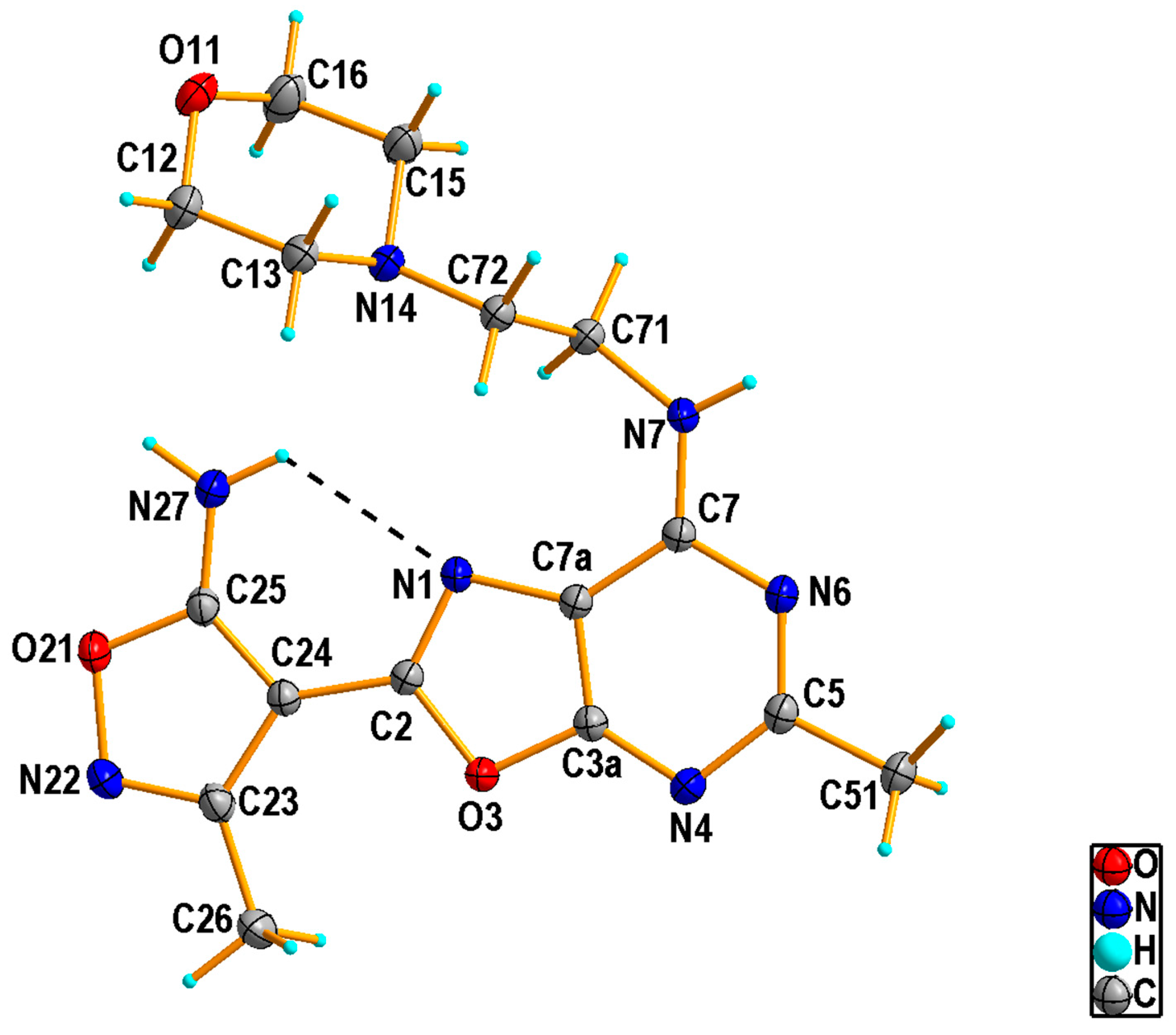
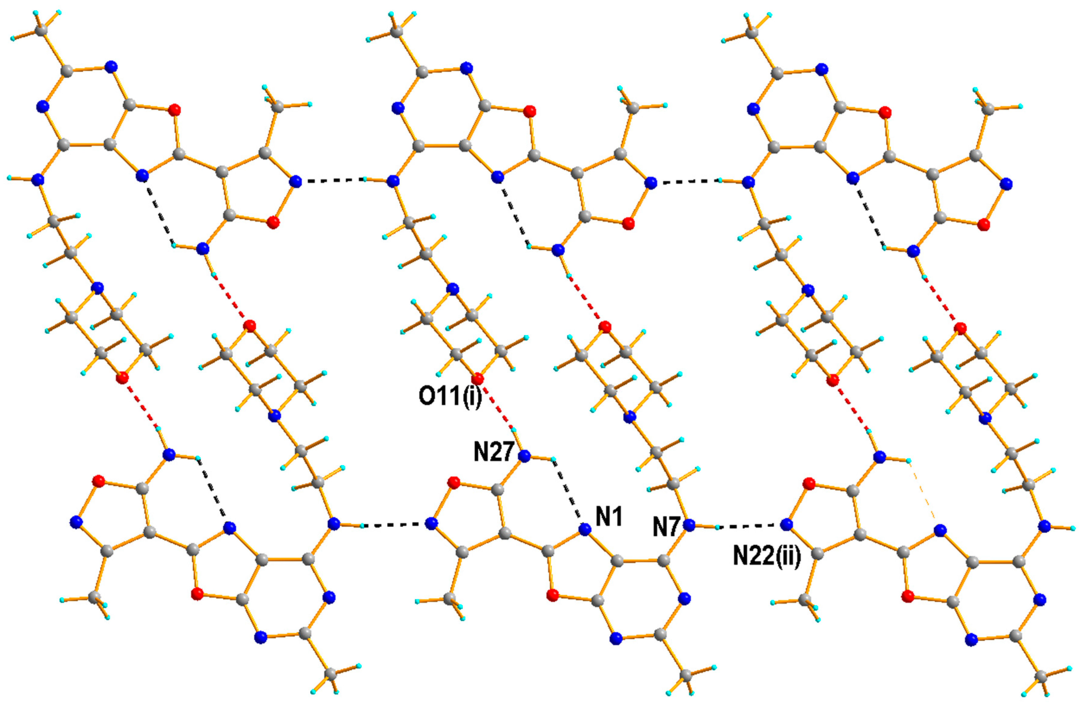
2.1.2. Crystal Structure of the Compound 5h

The structure of 5h was confirmed unambiguously by X-ray single-crystal analysis. Selected geometrical parameters and hydrogen bonds are given in Tables S25 and S26 (Supplementary Materials). 5h crystallizes in the P 1 ¯ space group, with one molecule in the asymmetric unit. The molecular conformation of each molecule is stabilized by an intramolecular N—H···N hydrogen bond [N···N = 2.889 (2) Å] (Table S2), which closes six-membered ring (forming S(6) motif), as shown in Figure 2.
The oxazolo[5,4-d]pyrimidine system is essentially planar [r.m.s. deviation 0.007 Å] and forms a dihedral angle of 0.936(11)° with the 1,2-oxazole group [r.m.s. deviation 0.002 Å] attached to atom C2. Additionally, due to the quite short C2–C24 bond length [1.426(2) Å], one can conclude that the aromatic systems are conjugated. However, there was a major localization of the double bonds in the pyrimidine ring of the oxazolo[5,4-d]pyrimidine system. The C4–N3a bond length is 1.322(2) Å against 1.347(2) and 1.358(2) Å for the C5-N4 and C7–N6 bonds, respectively. This difference is also observed in the C–C bond distances [1.407(2) and 1.372(2) Å for C7–C7a and C3a–C7a, respectively]. These values in connection with the shortening of the C7-N7 bond [1.341(2) Å] may be associated with the partial aromaticity loss by the oxazolo[5,4-d]pyrimidine ring system. The morpholine ring adopts a chair conformation. The orientation of the chain substituent attached to the atom C7 with respect to the plane defined by oxazolo[5,4-d]pyrimidine ring system may be reflected in C71-N7-C7-C7a and C7-N7-C71-C72 torsion angles of −9.0(3)° and 86.5(2)°, respectively.
The packing of 5h is mainly dictated by N—H···O and N—H···N hydrogen bonds (Table S2). The nitrogen atom (N27) from the amine group acts as hydrogen-bond donor via H atoms to the oxygen atom (O11) from the morpholine ring of adjacent molecules, forming the N27—H27···O11i hydrogen bond of 2.889(2) Å [symmetry code: (i) −x, −y + 1, −z], which leads to a dimer formation. On the other hand, molecules are linked through N7—H7···N22ii hydrogen bond of 2.959(3) Å [symmetry code: (ii) x + 1, y − 1, z] between the second amine group as a donor and the nitrogen atom of the 1,2-oxazole ring as an acceptor. The combination of these interactions results a ladder-like chain structure (Figure 3).

2.2. Biological Activity of Some New 7-Aminooxazolo[5,4-d]Pyrimidines 5a5j (SCM110)

The ten compounds 5a5j subjected to biological testing have been labelled as SCM110 (see Scheme 1).

2.2.1. Effects of Tested Compounds on Viability of A-549 Reference Cell Line, Lymphocyte Proliferation and Growth of Tumor Cell Lines

Estimation of Cytotoxicity of Tested Compounds on A-549 Reference Cell Line

Selection of therapeutically useful compounds, in order that they could be potential drugs, depends not only on their significant biological actions but also on bioavailability and possibly lowest cytotoxic properties. The compounds were therefore evaluated for their cytotoxic action by application of a reference A-549 cell line at a concentration range of 62.5–15.6 µM using respective dilutions of the solvent (DMSO) in the culture medium as control cultures (Table 1).

Influence of the Tested Compounds on Lymphocyte Proliferation

The ability to suppress mitogen-induced lymphocyte proliferation by the compounds SCM110 was evaluated in the model of PHA-induced proliferation of human PBMC, in which the compounds exhibited differentiated suppressive activity with best results for SCM5 and SCM9 (Table 2). At concentration of 50 µM, the suppressive acitivity of SCM5, SCM9 and teriflunomide were 69.8%, 60.7% and 37.9%, respectively. Therefore, these two compounds with strongest suppressive effects with regard to PHA-induced PBMC proliferation at a lower concentration range were selected for the subsequent more advanced experiments.
The tested compounds SCM5 and SCM9 appeared to be even more efficient in inhibition of LPS-stimulated mouse splenocytes (Figure 4) which reflects B cell proliferation [33]. This suppressive effect was more profound in comparison with that one of teriflunomide.

Influence of the Tested Compounds on Growth of Tumor Cell Lines

To evaluate potential antitumor activity of the compounds, A-431 epidermal, HT-29 colon and L-1210 leukemia cell lines (Table 3) were used. Cisplatin was the reference anticancer drug in the study. The results showed that the compounds displayed differential ability to inhibit growth of these cell lines. The best results were observed for HT-29 cell line where SCM9 caused (still significant) 30% growth inhibition at a concentration of 12.5 μM, albeit the activity was weaker than the one of reference cisplatin.

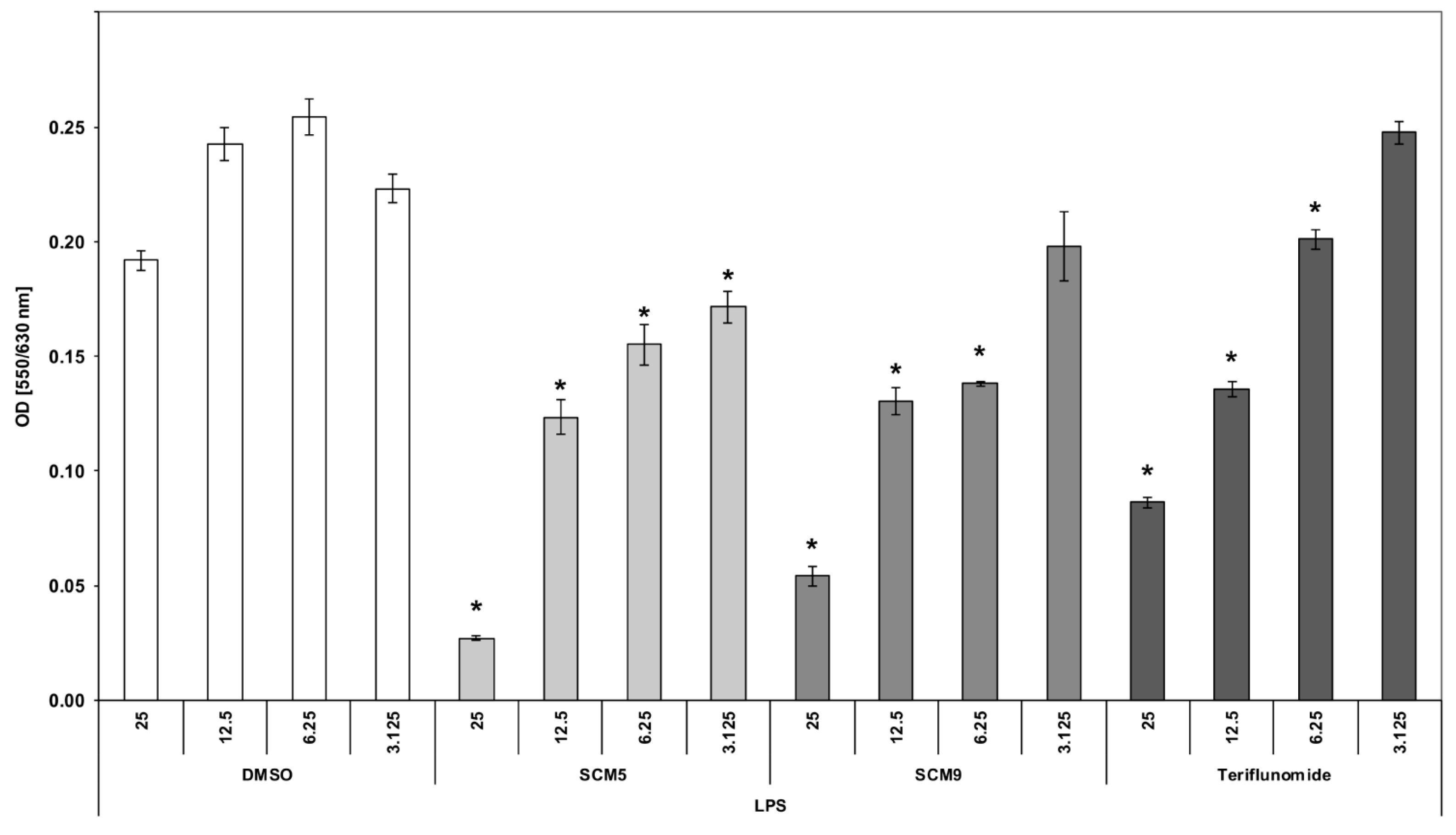
2.2.2. Effect of the Compounds on TNF α Production by LPS-Stimulated Human Whole Blood Cultures

Testing the ability of the compounds to inhibit LPS-induced TNF α production in the human whole blood culture (Table 4) revealed that SCM5 was inactive whereas SCM9 demonstrated dose-dependent suppression with a significant inhibition only at 25 μM concentration.

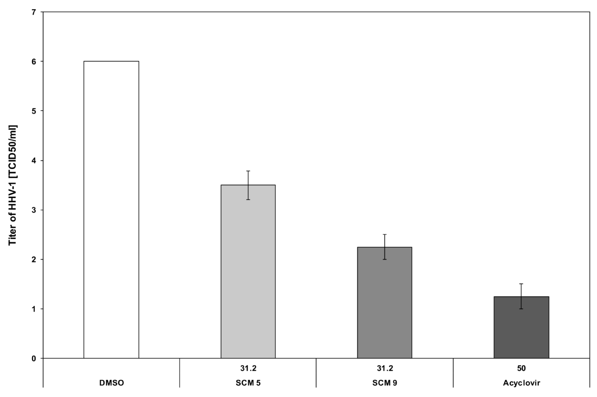
2.2.3. Antiviral Properties of the Compounds

The next aim of this study was to examine the antiviral activity of SCM5 and SCM9 in vitro against HHV-1. The treatment of A-549 cells infected with HHV-1 with SCM compounds resulted in significant reductions in virus yields, reaching up to a 3.5-log10 decrease for SCM5 and a 2.2-log10 decrease for SCM9 in comparison with appropriate DMSO control as it is shown in Figure 5. A 4-log10 reduction of virus titer demonstrates the ability of the tested compounds to inactivation of viruses to a level acceptable by the European Standards. In addition, SCM9, and to a lesser degree SCM5, significantly enhanced the viability of A-549 cells infected with the virus (data are not shown).
A-549 cells were infected with HHV-1 and after 1h of virus absorption at 37 °C, inoculum of virus was removed and the infected cells were incubated for 48 h with SCM5, SCM9 compounds (31.2 µM) and DMSO as the solvent control. Acyclovir (50 µM) was used as a reference drug. The viral titer was expressed with reference to the TCID50 value, which is based on the cytopathic effect caused by this virus in approximately 50% of infected cells. The results, which were obtained from quadruplicate determinations (mean ±SE), are presented as the log10 TDIC50/mL values in relation with appropriate DMSO control. The level of virus was defined according to the requirements presented in PN-EN 14476:2013+A1:2015.

2.2.4. Influence of the Compounds on Expression of Signaling Molecules in Cell Lines

The researched compounds were tested for their influence on expression of signaling molecules playing roles in apoptosis in Jurkat T cell model. The expression of the following signaling proteins was analyzed: caspases3, 7, 8, 9, Bcl-2, Fas, NFκB and p53. Of interest, the compounds did not alter expression of these proteins except one instance, namely, SCM5 caused a complete block in Bcl-2 expression (data are not shown). Since the expression of MAP kinases may also predict a fate of cells (activation, differentiation or apoptosis) the expression of ERK, JNK and p38 in Jurkat cells have been we additionally measured. The results (Table 5) show that the compounds have differently affected expression of signaling molecules. SCM5 strongly suppressed the expression of ERK1, ERK2, JNK and to a lesser degree p38 subunits. On the other hand, the compound SCM9 did not significantly affect the expression of ERK, but elevated the expression of JNK, p38α and p38β.
The determination of MAP kinase expression in WEHI-231 cells, which were exposed to the compounds, revealed no significant changes in the case of SCM9 with except 2-fold increase in the level of p38δ (Table 6). On the other hand, SCM5 caused a strong decrease in expression of ERK-1, ERK-2 and to a lesser degree of JNK and p38 subunits. No significant changes were also observed with regard to MAP kinase expression in WEHI 213 cells in the case of SCM9 apart from 2× increase in p38δ (Table 6). More marked changes were noted in the case of SCM5 which increased the expression levels of p38γ (2.7×) and p38δ (3.7).
In contrast, more significant changes in expression of signaling molecules, associated with apoptosis, were found in WEHI-231 cells incubated with SCM5. The changes (Table 7) included strong elevations in expression of caspase 8 (8×), caspase 9 (23×), NFκB (17.9×), Bcl-2 (10×) and Fas (8.8×). SCM9, on the other hand, did not significantly change the expression of these molecules.
The results, presented above, revealed in vitro immunosuppressive activity of the selected compounds (SCM5 and SCM9), which was, amongst the other, manifested by inhibition of cell proliferation and tumor cell growth. Both compounds exhibited the stronger antiproliferative effect on B cells in the mouse model than on T cells in the human peripheral blood cell population. Interestingly, only the compound SCM9 inhibited LPS-induced TNF α production which suggests its anti-inflammatory character. The changes in the expression of signaling molecules by SCM9 in Jurkat cells, such as 2× increase in JNK and an increase in p38α may indicate a proapoptotic action of the compound towards T cells associated with changes in MAP kinase expression [34,35]. However, it is intriguing that the signaling pathways induced by the compound did not involve caspase, NFκB, BcL-2 and Fas pathway despite very strong immunosuppressive properties of SCM9 towards WEHI-231. It is possible that the suppressive and anti-inflammatory action of SCM9 may engage still another signaling pathway, which could not be identified by the limited application of signaling molecules used in this study. In the case of SCM5, its immunosuppressive mechanism of action is more much easier to explain taking into consideration elicitation of signaling pathways by the compound. Firstly, the compound abrogated cell activation as evidenced by a strong, although differential decrease of expression of all MAP kinases, such as: ERK1 and ERK2, JNK and p38 subunits, which concerted involvement in cell differentiation and activation is essential [36]. More importantly, in the model of WEHI-231 cells, the compound induced strong amplification in expression of all molecules involved in apoptosis such as: caspase 8 [37], caspase 9 [38], NFκB [37,38], Bcl-2 [39] and Fas [40]. The level of cytotoxicity against A-549 cells at concentration of 62.5 µM is in accordance with our results on cell signaling showing that the proapoptotic action of SCM5 is more evident than that of SCM9. Of interest, the antiproliferative activity of both compounds on B cells, was stronger than this one observed with PBMC (predominantly T cells), since it was also high at lower concentration. For example, SCM5 at 25 µM/mL concentration, inhibited PBMC and splenocyte proliferation by 28% and 81.5%, respectively. This phenomenon may be of importance when potential therapeutic applications of the compounds are considered, for example in autoimmune disorders, particularly involving B cells and in neoplastic diseases. The ability of the compounds to interfere with the virus replication would require further research in terms of mechanism of action. Interestingly, stimulation of expression of p38 γ and p38 δ in WEHI 231 cells by the compounds may have significance in inhibition of virus replication [41]. Such properties, comparable to that of Acyclovir®, may enable application possibility of SCM9 as a potential antiviral drug on condition further advanced and positive pharmacological research.
The preliminary analysis of structure/activity relationship of the considered compounds 5a5j (SCM1-10) led us to the following general conclusions. Changes of substituents at position 7 in the structure of 7-aminooxazolo[5,4-d]pyrimidines 5 have resulted in varying degrees of biological activity. Amongst all the compounds that were biologically tested, the most active compounds, 5e (SCM5) and 5i (SCM9), have lipophilic chains with five linearly arranged non-hydrogen atoms, i.e., 7-N-pentyl and 7-(N,N-dimethylamino)propyl, respectively. In the opposition, there are the compounds with more hydrophilic substituents, such as 2-(morpholin-4-yl)ethyl in a molecule of the compound 5h (SCM8) or hydroxylalkyl ones in a molecule of the compounds 5f (SCM6) and 5g (SCM7), that showed no significant biological activity in the conducted tests.

3. Experimental

3.1. Chemistry

3.1.1. Materials and Methods

Commercially available reagents, i.e., aminomalononitrile tosylate (AMNT), triethyl orthoacetate and aliphatic amines were purchased from following suppliers such as TCI (Tokyo, Japan) and Sigma-Aldrich (Merck Group, Darmstadt, Germany), and were used without further purification. Thin layer chromatography (TLC) analyses were conducted using Alugram SIL G/UV 254nm plates (Macherey-Nagel, Düren, Germany) and visualized by ultraviolet (UV) light at 254 nm (UV A. KRÜSS Optronic GmbH, Hamburg, Germany). Column chromatography was carried out using Silicagel 60 and the solvent system of eluent was composed of chloroform and ethyl acetate (9:1 or 1:1) or chloroform and methanol (9:1). Melting points were measured on uniMELT 2 apparatus (LLG, Meckenheim, Germany) and were uncorrected. Electrospray Ionization Mass Spectroscopy (ESI-MS) spectra were recorded with a compactTM Electrospray Ionisation-Quadrupole-Time of Flight (ESI-Q-TOF) apparatus (Bruker Daltonik GmbH, Bremen, Germany). All compounds for ESI-MS experiments were dissolved in methanol and detected as positive ions. Theoretical monoisotopic masses of ions were calculated (calc.) using Bruker Compass Data Analysis 4.2 software (Bruker Daltonik GmbH, Bremen, Germany). The Attenuated Total Reflectance IR (ATR-FT-IR) spectra (4000–450 cm−1) were recorded on a Nicolet iS50 FT-IR spectrophotometer (Thermo Fisher Scientific Inc., Waltham, MA, USA). using clean solid forms of the compounds (on the diamond crystal surface, 32 scans, resolution: 1 cm−1, measurement temperature: 20–25 °C). IR Spectra were recorded with ATR intensity correction. It is known, that the relative intensity of bands in an ATR spectrum increases with wavelength causing the distortion of relative peak intensities in comparison with the classical transmission experiment [42]. Omnic Specta software (Thermo Fisher Scientific Inc., Waltham, MA, USA) was used for IR spectra analysis. Baselines of all IR spectra were corrected with autocorrection (fit order 2) and then all the spectra were normalized. 1H-NMR (300.15 MHz), 13C-NMR (75.47 MHz, broadband full decoupling method), 1H-1H COSY (300.15 MHz), 1H-13C 2D COSY NMR (channel F2: 300.15 MHz, channel F1: 75.47 MHz), 2D 1H-13C HSQC (Heteronuclear Single Quantum Coherence = Heteronuclear Single Quantum Correlation, channel F2: 300.15 MHz, channel F1: 75.47 MHz) and 2D 1H-13C HMBC (Heteronuclear Multiple Bond Correlation) spectra were recorded using 5 mm tubes and concentration about 20 mg of tested compounds in 0.6 mL (≈90–130 mM solutions) of appropriate deuterated solvent (such as dimethyl sulfoxide (DMSO-d6), chloroform (CDCl3) or methanol (CD3OD) with a Bruker ARX-300 spectrometer (Bruker Analytische Messtechnik GmbH, Rheinstetten, Germany). 2D Spectra enabled the unambiguous assignment of chemical shifts to the corresponding protons and to some quaternary carbon atoms. The reported data contain chemical shift in parts per million (ppm) units, signal multiplicities (abbreviations used: br—broad, s—singlet, d—doublet; t—triplet, q—quartet, m—multiplet), and the number of protons. The values of coupling constant were reported as J in Hz. All the NMR, IR and MS measurements were carried out in the Laboratory of Elemental Analysis and Structural Research, Faculty of Pharmacy, Wroclaw Medical University.

3.1.2. Preparation and Experimental Properties of Compounds 25 and 7

5-Amino-2-(5-Amino-3-Methyl-Isoxazol-4-yl)-Oxazole-4-Carbonitrile (2)

The reaction was carried out according to the protocol described by Freeman and Kim with slight modifications [29]. A solution of aminomalononitrile tosylate (AMNT, 0.8 g, 3.2 mmol) in 1-methyl-2-pyrrolidinone (NMP, 8.0 mL) was stirred with freshly prepared 5-amino-3-methylisoxazole-4-carbonyl chloride (1.02 g, 6.4 mmol, 2 equiv) at room temperature for 7 days. The course of the reaction was controlled by TLC. The reaction was conducted as long as the starting reagent AMNT was present. Then, the final mixture was cooled to 5–10 °C and diluted with ice-cold water. The formed precipitate was filtered off and dried to give the crude compound 2 as a purple solid that was crystallized from methanol. Yield 0.306 g, 47.2%; m.p. 235–237 °C (dec.); HR-ESI-MS: m/z[u/e] calc. for C8H7N5O2Na [M + Na]+ 228.0491, found 228.0499 (see Figure S1 in the Supplementary Material); ATR- FTIR: νmax (cm−1) 3369, 3159 (NH2), 2210 (CN), 1636, 1592, 1534, 1488, 1062 (see Figure S24 in the Supplementary Material); 1H-NMR (DMSO-d6, 300 MHz): δ (ppm) 2.24 (s, 3H, methyl group of isoxazole ring), 7.40 (brs, 2H, -NH2), 7.58 (brs, 2H, -NH2) (see Figure S22 in the Supplementary Material); 13C-NMR (DMSO-d6, 75.5 MHz): δ (ppm) 11.3, 81.2, 82.4, 115.8, 145.3, 157.1, 160.7, 168.2 (see Figure S23 in the Supplementary Material).

Ethyl N-{4-Cyano-2-[5-(1-Ethoxyethylidene)Amino-3-Methylisoxazol-4-yl]-Oxazol-5-yl}Ethanimidate (4)

Compound 3 (2.54 g, 7.35 mmol) and triethyl orthoacetate (32 mL) were heated with stirring at boiling temperature (145 °C) for 15 h. On completion of the reaction as monitored by TLC, the excess of triethyl orthoacetate was evaporated under vacuum. The crude product was purified by chromatography on silica gel (CHCl3:EtOAc, 9:1) to give compound 4 as a yellow solid. Yield 3.93 g, 91.9%; m.p. 81–83 °C (dec.); ESI-MS: m/z[u/e] calc. for C16H20N5O4 [M + H]+ 346.1510, found 346.1445 (see Figure S3 in the Supplementary Material); ATR-FTIR: νmax (cm−1) 2994, 2231 (CN), 1659, 1635, 1464, 1378, 1298, 1043 (see Figure S30 in the Supplementary Material); 1H-NMR (CDCl3, 300 MHz): δ (ppm) 1.37 (t, 3J = 7 Hz, 3H, methyl of ethoxyl group of ethanimidate moiety in isoxazole ring), 1.39 (t, 3J = 7 Hz, 3H, methyl of ethoxyl group of ethanimidate moiety in oxazole ring), 2.10 (s, 3H, methyl group of ethanimidate moiety in isoxazole ring), 2.24 (s, 3H, methyl group of ethanimidate moiety in oxazole ring), 2.50 (s, 3H, methyl group of isoxazole ring), 4.36 (q, 3J = 7 Hz, 2H, methylene of ethoxyl group of ethanimidate moiety in isoxazole ring); 4.37 (q, 3J = 7 Hz, 2H, methylene of ethoxyl group of ethanimidate moiety in oxazole ring) (see Figure S28 in the Supplementary Material). 13C-NMR (CDCl3, 75.4 MHz): δ (ppm) 12.42, 13.84, 13.87, 19.20, 19.24, 64.07, 64.31, 93.58, 100.07, 113.70, 150.62, 158.30, 159.49, 168.14, 168.87, 169.10 (see Figure S29 in the Supplementary Material).

Ethyl N-[2-(5-Amino-3-Methylisoxazol-4-yl)-4-Cyanooxazol-5-yl]Ethanimidate (3)

Method A (in ethanol)
Compound 4 (0.3 g, 0.87 mmol) and ethanol (6 mL) were heated with stirring at boiling temperature (78 °C) for 26 h. On completion of the reaction as monitored by TLC, the ethanol was evaporated under vacuum. The crude product was purified by chromatography on silica gel to give the compound 3 as the beige solid. Yield 0.037 g, 15.5%; m.p. 181–182 °C (dec.); ESI-MS: m/z[u/e] calc. for C12H13N5O3Na [M + Na]+ 298.0911, found 298.0975 (see Figure S2 in the Supplementary Material); ATR-FTIR: νmax (cm−1) 3389, 3129, 2992, 2230 (CN), 1640, 1611, 1382, 1294, 1032, 953, 744 (see Figure S27 in the Supplementary Material); 1H-NMR (CDCl3, 300 MHz): δ (ppm) 1.38 (t, 3J = 7.1 Hz, 3H, methyl of ethoxyl group of ethanimidate moiety in the oxazole ring), 2.27 (s, 3H, methyl group of ethanimidate moiety in the oxazole ring), 2.41 (s, 3H, methyl group of isoxazole ring), 4.36 (q, 3J = 7.1 Hz, 2H, methylene of ethoxyl group of ethanimidate moiety in the oxazole ring), 6.03 (s, 2H, -NH2 of isoxazole ring) (see Figure S25 in the Supplementary Material); 13C-NMR (CDCl3, 75.5 MHz): δ (ppm) 11.7, 13.9, 19.3, 64.3, 83.3, 99.2, 113.7, 152.4, 157.4, 157.5, 168.1, 168.6 (see Figure S26 in the Supplementary Material).

2-(5-Amino-3-Methylisoxazol-4-yl)-7-N-Methylamino-5-Methyloxazolo[5,4-d]Pyrimidine (5a, SCM1)

Method A (in ethanol)
To compound 4 (0.2 g, 0.58 mmol), a 30% solution of methylamine in ethanol (1 mL) was added in one portion. The reaction mixture was cooled to 5–10 °C and stirred for 0.5 h. When the reaction was complete, as shown by TLC, the precipitate formed was filtered off, dried and crystallized from ethanol. The compound 5a (0.062 g, 41.0%) was obtained as a white crystals: m.p. 246–247 °C (dec.); HR-ESI-MS: m/z[u/e] calc. for C11H13N6O2 [M + H]+ 261.1094, found 261.1081 (see Figure S4 in the Supplementary Material); ATR-FTIR: νmax (cm−1) 3365, 3300, 3120, 1662, 1617, 1532, 1322, 1200, 1054 (see Figure S34 in the Supplementary Material); 1H-NMR (DMSO-d6, 300 MHz): δ (ppm) 2.34 (s, 3H, methyl group of isoxazole moiety), 2.50 (s, 3H, signal covered with solvent, methyl group at position 5 of the oxazolopyrimidine system), 3.53 (s, 3H, N-methyl group), 7.26 (brs, 1H, -NH), 7.87 (brs, 2H,-NH2) (see Figure S31 in the Supplementary Material); 13C-NMR (DMSO-d6, 75.5 MHz): δ (ppm) 11.3 (carbon of methyl group of the isoxazole moiety), 23.4 (carbon of methyl group at position 5 of the oxazolopyrimidine system), 31.9 (carbon of N-methyl group at position 7 of the oxazolopyrimidine system), 81.6 (quaternary carbon C4 of the isoxazole ring), 116.7 (quaternary bridgehead carbon C7a of the oxazolopyrimidine system), 153.6, 154.4, 154.9, 157.0 (quaternary carbon C5 of the oxazolopyrimidine system), 157.2 (quaternary carbon C3 of the isoxazole ring), 169.2 (quaternary carbon C5 of the isoxazole ring) (see Figure S32 in the Supplementary Material). 1H-13C 2D COSY NMR spectrum of 5a (see Figure S33 in the Supplementary Material).

2-(5-Amino-3-Methylisoxazol-4-yl)-7-N-Ethylamino-5-Methyloxazolo[5,4-d]Pyrimidine (5b, SCM2)

Method A (in ethanol)
To compound 4 (0.2 g, 0.58 mmol), a 30% solution of ethylamine in ethanol (1 mL) was added in one portion. The reaction mixture was cooled to 5–10 °C and stirred for 0.5 h. When the reaction was complete, as shown by TLC, the formed precipitate was filtered off, dried and crystallized from ethanol. The compound 5b (0.063 g, 39.6%) was obtained as a white crystals: m.p. 245–245.5 °C (dec.); HR-ESI-MS: m/z[u/e] calc. for C12H15N6O2 [M + H]+ 275.1251, found 275.1246 (see Figure S5 in the Supplementary Material); ATR-FTIR: νmax (cm−1) 3410, 3084, 1669, 1639, 1533, 1316, 1169, 1040 (see Figure S38 in the Supplementary Material); 1H-NMR (DMSO-d6, 300 MHz): δ (ppm) 1.24 (t, 3J = 7.0 Hz, 3H), 2.33 (s, 3H, methyl group of isoxazole moiety), 2.55 (s, 3H, methyl group at position 5 of the oxazolopyrimidine system), 4.17 (q, 3J = 6.9 Hz, 2H, N-methylene group of the ethyl chain), 7.23 (brs, 1H, -NH), 7.86 (brs, 2H, -NH2) (see Figure S35 in the Supplementary Material); 13C-NMR (DMSO-d6, 75.5 MHz): δ (ppm) 11.4, 12.2, 22.8, 39.8 (signal covered with solvent, carbon of N-methylene group of ethyl chain at position 7 of the oxazolopyrimidine system), 81.6 (quaternary carbon C4 of the isoxazole ring), 117.0 (quaternary bridgehead carbon C7a of the oxazolopyrimidine system), 153.4, 153.8, 155.1, 156.3 (quaternary carbon 5 of the oxazolopyrimidine system), 157.2 (quaternary carbon C3 of the isoxazole ring), 169.4 (quaternary carbon C5 of the isoxazole ring) (see Figure S36 in the Supplementary Material). 1H-13C 2D HSQC spectrum of 4b (see Figure S37 in the Supplementary Material).

2-(5-Amino-3-Methylisoxazol-4-yl)-7-N-Propylamino-5-Methyloxazolo[5,4-d]Pyrimidine (5c, SCM3)

Method A (in ethanol)
To compound 4 (0.2 g, 0.58 mmol), a 30% solution of propylamine in ethanol (1 mL) was added in one portion. The reaction mixture was cooled to 5–10 °C and stirred for 1 h. Then, the mixture was warmed to room temperature and maintained with stirring for 1.5 h. When the reaction was complete, as shown by TLC, the formed precipitate was filtered off, dried and crystallized from ethanol. The compound 5c (0.104 g, 60.5%) was obtained as a white solid: m.p. 209–210 °C (dec.); HR-ESI-MS: m/z[u/e] calc. for C13H17N6O2 [M + H]+ 289.1408, found 289.1407 (see Figure S6 in the Supplementary Material); ATR-FTIR: νmax (cm−1) 3397, 3306, 2979, 1684, 1662, 1617, 1599, 1443, 1353, 1181, 1043, 792 (see Figure S41 in the Supplementary Material); 1H-NMR (DMSO-d6, 300 MHz): δ (ppm) 0.93 (t, 3J32 = 7.3 Hz, 3H, terminal chain methyl), 1.69 (m, 3J23 = 7.3 Hz, 3J21 = 7.8 Hz, 2H, middle chain methylene), 2.33 (s, 3H, methyl group of the isoxazole moiety), 2.54 (s, 3H, methyl group at position 5 of the oxazolopyrimidine system), 4.05 (t, 3J12 = 7.8 Hz, 2H, chain methylene connected with amine nitrogen at the 7th position), 7.25 (brs, 1H, -NH), 7.87 (brs, 2H, -NH2) (see Figure S39 in the Supplementary Material); 13C-NMR (DMSO-d6, 75.5 MHz): δ (ppm) 11.03 (carbon of terminal methyl of propyl chain at position 7 of the oxazolopyrimidine system), 11.39 (carbon of methyl group of the isoxazole moiety), 19.91 (carbon of middle methylene group of propyl chain at position 7 of the oxazolopyrimidine system), 22.95 (carbon of methyl group at position 5 of the oxazolopyrimidine system), 46.28 (carbon of N-methylene group of the propyl chain), 81.58 (carbon C4 of the isoxazole ring), 117.06 (quaternary bridgehead carbon C7a of the oxazolopyrimidine system), 153.70, 153.75, 154.84, 156.38 (quaternary carbon C5 of the oxazolopyrimidine system), 157.23 (quaternary carbon C3 of the isoxazole ring), 169.33 (quaternary carbon C5 of the isoxazole ring) (see Figure S40 in the Supplementary Material).
Method B (in tetrachloromethane)
To compound 4 (0.14 g, 0.40 mmol), a 30% solution of propylamine in CCl4 (1 mL) was added in one portion. The reaction mixture was cooled to 5–10 °C and stirred for 1 h. Then, the mixture was warmed to room temperature and maintained with stirring for 47 h. When the reaction was complete, as shown by TLC, the formed precipitate was filtered off. The solution after filtration was evaporated in a vacuum and the residue was dilution with ethanol. The formed precipitate was filtered off and dried to give the compound 5c as a white solid (0.015 g, 12.8%): m.p. 208–209 °C (dec.); ESI-MS: m/z[u/e] calc. for C13H17N6O2 [M + H]+ 289.1408, found 289.1454 (see Figure S7 in the Supplementary Material). Other spectral analyzes (IR, NMR) confirm that the product is identical to that obtained by Method A.

2-(5-Amino-3-Methylisoxazol-4-yl)-7-N-(2-Methylpropyl)Amino-5-Methyloxazolo[5,4-d]Pyrimidine (5d, SCM4)

Method A (in ethanol)
To compound 4 (0.2 g, 0.58 mmol), a 30% solution of 2-methylpropylamine in ethanol (1 mL) was added in one portion. The reaction mixture was cooled to 5–10 °C and stirred for 1 h. Then, the mixture was warmed to room temperature and maintained with stirring for 3.5 h. When the reaction was complete, as shown by TLC, the formed precipitate was filtered off, dried and crystallized from ethanol. The compound 5d (0.057 g, 32.5%) was obtained as a white solid: m.p. 183–184 °C (dec.); HR-ESI-MS: m/z[u/e] calc. for C14H19N6O2 [M + H]+ 303.1564, found 303.1556 (see Figure S8 in the Supplementary Material); ATR-FTIR: νmax (cm−1) 3388, 3301, 3146, 2963, 1663, 1616, 1538, 1188, 1034, 818 (see Figure S46 in the Supplementary Material); 1H-NMR (DMSO-d6, 300 MHz): δ (ppm) 0.90 (d, 3J32 = 6.6 Hz, 6H, two terminal methyl groups of the isobutyl chain), 2.30 (m, 3J23 ≈ 6.9 Hz, 3J21 ≈ 4.4 Hz 1H, isobutyl methanetriyl (methine) group, signal partially covered and disturbed by the isoxazole methyl group), 2.33 (s, 3H, methyl group of the isoxazole moiety), 2.52 (s, 3H), 3.98 (br apparent singlet, 2H, chain methylene connected with amine nitrogen at the 7th position), 7.33 (brs, 1H, -NH), 7.88 (brs, 2H, -NH2) (see Figure S44 in the Supplementary Material); 13C-NMR (DMSO-d6, 75.5 MHz): δ (ppm) 11.4, 19.7, 23.5, 26.1, 50.9, 81.6, 117.2, 153.7, 154.3, 154.7, 156.7, 157.2, 169.3 (see Figure S45 in the Supplementary Material).

2-(5-Amino-3-Methylisoxazol-4-yl)-7-N-Pentylamino-5-Methyloxazolo[5,4-d]Pyrimidine (5e, SCM5)

Method A (in ethanol)
To compound 4 (0.2 g, 0.58 mmol), a 30% solution of pentylamine in ethanol (1 mL) was added in one portion. The reaction mixture was cooled to 5–10 °C and stirred for 1 h. Then, the mixture was warmed to room temperature and maintained with stirring for 1.5 h. When the reaction was complete, as shown by TLC, the formed precipitate was filtered off, dried and crystallized from ethanol. The compound 5e (0.104 g, 56.8%) was obtained as a white solid: m.p. 188–189 °C (dec.); HR-ESI-MS: m/z[u/e] calc. for C15H21N6O2 [M + H]+ 317.1720, found 317.1717 (see Figure S9 in the Supplementary Material); ATR-FTIR: νmax (cm−1) 3407, 2959, 1664, 1616, 1592, 1521, 1350, 1178, 1043, 790 (see Figure S49 in the Supplementary Material); 1H-NMR (DMSO-d6, 300 MHz): δ (ppm) 0.89 (t, 5J ≈ 6.5 Hz, 3H, terminal methyl group of the pentyl chain), 1.30–1.35 (m, 4H, two methylene groups of pentyl chain), 1.66 (m, 2H, methylene group of the pentyl chain), 2.33 (s, 3H, methyl group of the isoxazole moiety), 2.53 (s, 3H), 4.08 (t, 3J ≈ 7.8 Hz, 2H, methylene group of the pentyl chain connected with amine nitrogen at position 7), 7.23 (brs, 1H, -NH), 7.86 (brs, 2H, -NH2) (see Figure S47 in the Supplementary Material); 13C-NMR (DMSO-d6, 75.5 MHz): δ (ppm) 11.4, 13.8, 21.8, 22.9, 26.2, 28.5, 44.7, 81.6, 117.0, 153.7, 153.8, 154.8, 156.3, 157.2, 169.3 (see Figure S48 in the Supplementary Material).

2-(5-Amino-3-Methylisoxazol-4-yl)-7-N-(2-Hydroxyethyl)Amino-5-Methyloxazolo[5,4-d]Pyrimidine (5f, SCM6)

Method A (in ethanol)
To compound 4 (0.2 g, 0.58 mmol), a 30% solution of ethanolamine in ethanol (1 mL) was added in one portion. The reaction mixture was cooled to 5–10 °C and stirred for 1 h. Then, the mixture was warmed to room temperature and maintained with stirring for 1.5 h. When the reaction was complete, as shown by TLC, the formed precipitate was filtered off, dried and crystallized from ethanol. The compound 5f (0.083 g, 49.4%) was obtained as a white solid: m.p. 251–252 °C (dec.); HR-ESI-MS: m/z[u/e] calc. for C12H15N6O3 [M + H]+ 291.1200, found 291.1195 (see Figure S10 in the Supplementary Material); ATR-FTIR: νmax (cm−1) 3324, 3259, 1662, 1622, 1592, 1531, 1442, 1352, 1270, 1166, 1050 (see Figure S52 in the Supplementary Material); 1H-NMR (DMSO-d6, 300 MHz): δ (ppm) 2.34 (s, 3H, methyl group of the isoxazole moiety), 2.59 (s, 3H, methyl group at position 5 of the oxazolopyrimidine system), 3.72 (t, 3J = 5.2 Hz, 2H, methylene group connected with the amine nitrogen at position 7), 4.20 (t, 3J = 5.4 Hz, 2H, methylene group connected hydroxyl group), 5.03 (brs, 1H, -OH), 7.23 (brs, 1H, -NH), 7.87 (brs, 2H, -NH2) (see Figure S50 in the Supplementary Material); 13C-NMR (DMSO-d6, 75.5 MHz): δ (ppm) 11.2, 23.5, 47.3, 57.7, 81.6, 117.0, 153.5, 154.0, 154.8, 157.1, 157.2, 169.2 (see Figure S51 in the Supplementary Material).

2-(5-Amino-3-Methylisoxazol-4-yl)-7-N-(3-Hydroxypropyl)Amino-5-Methyloxazolo[5,4-d]Pyrimidine (5g, SCM7)

Method A (in ethanol)
To compound 4 (0.2 g, 0.58 mmol), a 30% solution of 3-aminopropan-1-ol in ethanol (1 mL) was added in one portion. The reaction mixture was cooled to 5–10 °C and stirred for 1 h. Then, the mixture was warmed to room temperature and maintained with stirring for 1.5 h. When the reaction was complete, as shown by TLC, the formed precipitate was filtered off, dried and crystallized from ethanol. The compound 5g (0.103 g, 58.5%) was obtained as a white solid: m.p. 224–226 °C (dec.); HR-ESI-MS: m/z[u/e] calc. for C13H17N6O3 [M + H]+ 305.1357, found 305.1352 (see Figure S11 in the Supplementary Material); ATR-FTIR: νmax (cm−1) 3243, 3140, 1663, 1616, 1594, 1538, 1351, 1168, 1086, 1050, 795 (see Figure S55 in the Supplementary Material); 1H-NMR (DMSO-d6, 300 MHz): δ (ppm) 1.84 (m, 3J ≈ 6.3–6.5 Hz, 2H, middle methylene group of the propyl chain), 2.34 (s, 3H, methyl group of the isoxazole moiety), 2.56 (s, 3H, methyl group at position 5 of the oxazolopyrimidine system), 3.44 (heavily distorted triplet t, 3J is difficult to estimate, 2H, methylene group connected with amine nitrogen at position 7 of the oxazolopyrimidine system), 4.19 (t, 3J = 6.9 Hz, 2H, methylene group connected hydroxyl group), 4.98 (brs, 1H, -OH), 7.24 (brs, 1H, -NH), 7.88 (brs, 2H, -NH2) (see Figure S53 in the Supplementary Material); 13C-NMR (DMSO-d6, 75.5 MHz): δ (ppm) 11.4, 22.9, 30.2, 42.4, 57.7, 81.6, 117.0, 153.8, 154.3, 155.1, 156.6, 157.2, 169.4 (see Figure S54 in the Supplementary Material).

2-(5-Amino-3-Methylisoxazol-4-yl)-7-N-[2-(Morpholin-4-yl)Ethyl]amino-5-Methyloxazolo[5,4-d]Pyrimidine (5h, SCM8)

Method A (in ethanol)
To compound 4 (0.2 g, 0.58 mmol), a 30% solution of 2-(morpholin-4-yl)ethanamine in ethanol (1 mL) was added in one portion. The reaction mixture was cooled to 5–10 °C and stirred for 1 h. Then, the mixture was warmed to room temperature and maintained with stirring for 3.5 h. When the reaction was complete, as shown by TLC, the formed precipitate was filtered off, dried and crystallized from ethanol. The compound 5h (0.087 g, 41.8%) was obtained as a white solid: m.p. 207–209.5 °C (dec.); HR-ESI-MS: m/z[u/e] calc. for C16H22N7O3 [M + H]+ 360.1779, found 360.1768 (see Figure S12 in the Supplementary Material); ATR-FTIR: νmax (cm−1) 3332, 2960, 2801, 1661, 1615, 1593, 1532, 1352, 1282, 1166, 1115, 1054, 1009, 868, 782 (see Figure S58 in the Supplementary Material); 1H-NMR (DMSO-d6, 300 MHz): δ (ppm) 2.33 (s, 3H, methyl group of the isoxazole moiety), 2.46 (t, 3J ≈ 4.5 Hz, 4H, two morpholine ring methylene group connected directly with the morpholine ring nitrogen), 2.59 (s, 3H, methyl group at position 5 of the oxazolopyrimidine system), 2.61 (t, 3J ≈ 6.8 Hz, 2H, methylene group of ethyl chain directly connected with the morpholine ring nitrogen), 3.56 (t, 3J ≈ 4.5 Hz, 4H, two morpholine ring methylene group connected directly with the morpholine ring oxygen), 4.23 (t, 3J ≈ 6.5 Hz, 2H, methylene group of the ethyl chain directly connected with the amine nitrogen at position 7 of the oxazolopyrimidine system), 7.26 (brs, 1H, -NH), 7.87 (brs, 2H, -NH2) (see Figure S56 in the Supplementary Material); 13C-NMR (DMSO-d6, 75.5 MHz): δ (ppm) 11.4 (carbon of the methyl group of the isoxazole moiety), 23.2 (carbon of the methyl group at position 5 of the oxazolopyrimidine system), 42.5 (carbon of the ethyl chain methylene group directly connected with the amine nitrogen at position 7 of the oxazolopyrimidine system), 53.6 (carbons of two morpholine ring methylene group connected directly with the morpholine ring nitrogen), 54.9 (carbon of the ethyl chain methylene group directly connected with the morpholine ring nitrogen), 66.2 (carbons of two morpholine ring methylene group connected directly with the morpholine ring oxygen), 81.6 (quaternary carbon C4 of the isoxazole ring), 117.0 (quaternary bridgehead carbon C7a of the oxazolopyrimidine system), 153.7 (quaternary bridgehead carbon C3a of the oxazolopyrimidine system, uncertain assignment), 153.8 (quaternary carbon C2 of the oxazolopyrimidine system), 154.8 (quaternary carbon C7 of the oxazolopyrimidine system, uncertain assignment), 156.8 (quaternary carbon C5 of the oxazolopyrimidine system), 157.2 (quaternary carbon C3 of the isoxazole ring), 169.3 (quaternary carbon C5 of the isoxazole ring) (see Figure S57 in the Supplementary Material).

2-(5-Amino-3-Methylisoxazol-4-yl)-7-N-[3-(N,N-Dimethylamino)Propyl]Amino-5-Methyloxazolo[5,4-d]Pyrimidine (5i, SCM9)

Method A (in ethanol)
To compound 4 (0.2 g, 0.58 mmol), a 30% solution of N,N-dimethylpropane-1,3-diamine in ethanol (1 mL) was added in one portion. The reaction mixture was cooled to 5–10 °C and stirred for 1 h. Then, the mixture was warmed to room temperature and maintained with stirring for 4 h. When the reaction was complete, as shown by TLC, the formed precipitate was filtered off, dried and crystallized from ethanol. The compound 5i (0.127 g, 66.1%) was obtained as a white solid: m.p. 209.5–210 °C (dec.); HR-ESI-MS: m/z[u/e] calc. for C15H22N7O2 [M + H]+ 332.1829, found 332.1821 (see Figure S13 in the Supplementary Material); ATR-FTIR: νmax (cm−1) 3469, 3364, 2774, 1674, 1615, 1595, 1529, 1440, 1346, 1183, 1041 (see Figure S61 in the Supplementary Material); 1H-NMR (DMSO-d6, 300 MHz): δ (ppm) 1.82 (m, 3J23 = 6.6 Hz, 3J21 = 7.6 Hz, 2H, middle methylene group of the propyl chain), 2.14 (s, 6H, two methyls of the dimethylamino group), 2.28 (t, 3J32 = 6.6 Hz, 2H, methylene of the propyl chain directly connected to nitrogen of the dimethylamino group), 2.33 (s, 3H, methyl group of the isoxazole moiety), 2.55 (s, 3H, methyl group at position 5 of the oxazolopyrimidine system), 4.11 (t, 3J12 = 7.5 Hz, 2H, methylene of the propyl chain directly connected to the amine nitrogen at position 7 of the oxazolopyrimidine system), 7.23 (brs, 1H, -NH), 7.86 (brs, 2H, -NH2) (see Figure S59 in the Supplementary Material); 13C-NMR (DMSO-d6, 75.5 MHz): δ (ppm) 11.4, 22.9, 24.4, 43.5, 45.1, 56.3, 81.6, 117.1, 153.6, 153.8, 154.8, 156.4, 157.2, 169.3 (see Figure S60 in the Supplementary Material).
Method B (in tetrachloromethane)
To compound 4 (0.2 g, 0.58 mmol), 30% solution of N,N-dimethylpropane-1,3-diamine in CCl4 (1 mL) was added in one portion. The reaction mixture was cooled to 5–10 °C and stirred for 1 h. Then, the mixture was warmed to room temperature and maintained with stirring for 47 h. When the reaction was complete, as shown by TLC, the reaction mixture was evaporated in a vacuum and purified by chromatography on silica gel (CHCl3: AcOEt, 9:1). The compound 5i (0.06 g, 30.6%) was obtained as a beige solid: m.p. 202–204 °C (dec.); HR-ESI-MS: m/z [M + H]+ calc. for C15H22N7O2 332.1829, found 332.1824 (see Figure S14 in the Supplementary Material). Other spectral analyses (IR, NMR) confirmed that the product is identical to that obtained by Method A.
Method C
To compound 3 (0.05 g, 0.18 mmol), a 30% solution of N,N-dimethylpropane-1,3-diamine in ethanol (0.25 mL) was added in one portion. The reaction mixture was cooled to 5–10 °C and stirred for 1 h. Then, the mixture was warmed to room temperature and maintained with stirring for 4 h. When the reaction was complete, as shown by TLC, the formed precipitate was filtered off, dried and crystallized from ethanol. The compound 5i (0.007 g, 11.6%) was obtained as a white solid: m.p. 210–211 °C (dec.); ESI-MS: m/z[u/e] calc. for C15H22N7O2 [M + H]+ 332.1829, found 332.1848 (see Figure S15 in the Supplementary Material). Other spectral analyses (IR, NMR) confirmed that the product is identical to that obtained by Methods A and B.

2-(5-Amino-3-Methylisoxazol-4-yl)-7-N-[2-(N,N-Dimethylamino)Ethyl]amino-5-Methyloxazolo[5,4-d]Pyrimidine (5j, SCM10)

Method A (in ethanol)
To compound 4 (0.2 g, 0.58 mmol), a 30% solution of N,N-dimethylethane-1,2-diamine in ethanol (1 mL) was added in one portion. The reaction mixture was cooled to 5–10 °C and stirred for 1 h. Then, the mixture was warmed to room temperature and maintained with stirring for 2 h. When the reaction was complete, as shown by TLC, the formed precipitate was filtered off, dried and crystallized from ethanol. The compound 5j (0.078 g, 42.4%) was obtained as a white solid: m.p. 222.5–223 °C (dec.); HR-ESI-MS: m/z[u/e] calc. for C14H20N7O2 [M + H]+ 318.1673, found 318.1664 (see Figure S16 in the Supplementary Material); ATR-FTIR: νmax (cm−1) 3226, 2832, 2780, 1665, 1616, 1593, 1529, 1450, 1352, 1184, 1027 (see Figure S69 in the Supplementary Material); 1H-NMR (DMSO-d6, 300 MHz): δ (ppm) 2.22 (s, 6H, dimethylamino group), 2.33 (s, 3H, methyl group of the isoxazole moiety), 2.54 (heavily distorted and partly covered with solvent triplet t, 3J21 is difficult to estimate, 2H, methylene of the propyl chain directly connected to the nitrogen of the dimethylamino group) 2.58 (s, 3H, methyl group at position 5 of the oxazolopyrimidine system), 4.20 (t, 3J12 = 6.7 Hz, 2H, methylene of the ethyl chain directly connected to the amine nitrogen at position 7 of the oxazolopyrimidine system), 7.25 (brs, 1H, -NH), 7.87 (brs, 2H, -NH2) (see Figure S67 in the Supplementary Material); 13C-NMR (DMSO-d6, 75.5 MHz): δ (ppm) 11.4, 23.1, 43.1, 45.5, 55.8, 81.6, 117.0, 153.6, 153.8, 154.7, 156.8, 157.2, 169.3 (see Figure S68 in the Supplementary Material).

2-(5-Amino-3-Methylisoxazol-4-yl)-7-N-Ispropylamino-5-Methyloxazolo[5,4-d]Pyrimidine (5k)

Method A (in ethanol)
To compound 4 (0.29 g, 0.84 mmol), a 30% solution of isopropylamine in ethanol (1 mL) was added in one portion. The reaction mixture was cooled to 5–10 °C and stirred for 1 h. Then, the mixture was warmed to room temperature and maintained with stirring for 3.5 h. When the reaction was complete, as shown by TLC, the formed precipitate was filtered off. The solution after filtration was evaporated in a vacuum and crystallized from ethanol. The compound 5k (0.022 g, 9.1%) was obtained as a beige solid: m.p. 161–162 °C (dec.); HR-ESI-MS: m/z[u/e] calc. for C13H17N6O2 [M + H]+ 289.1408, found 289.1396 (see Figure S17 in the Supplementary Material); ATR-FTIR: νmax (cm−1) 3085, 2925, 1633, 1604, 1315, 1042, 744 (see Figure S72 in the Supplementary Material); 1H-NMR (DMSO-d6, 300 MHz): δ (ppm) 1.62 (d, 3J = 6.7 Hz, 6H, two methyl groups of the isopropyl chain), 2.32 (s, 3H, methyl group of the isoxazole moiety), 2.54 (s, 3H, methyl group at position 5 of the oxazolopyrimidine system), 4.69 (br m, very low intensity, 1H, methanetriyl (methine) group of the isopropyl chain), 7.32 (brs, 1H, -NH), 7.85 (brs, 2H, -NH2) (see Figure S70 in the Supplementary Material); 13C-NMR δ (ppm) 11.4 (carbon of methyl group of the isoxazole moiety), 18.5 (carbons of the two methyl groups of the isopropyl moiety), 24.3 (carbon of the methyl group at position 5 of the oxazolopyrimidine system), 53.4 (methanetriyl (methine) group of the isopropyl chain), 81.6 (C4 of the isoxazole ring), 118.1 (quaternary bridgehead carbon C7a of the oxazolopyrimidine system), 153.6, 154.1, 154.5 (three quaternary carbons C2, C3a and C7 of the oxazolopyrimidine system), 156.6 (quaternary carbon C5 of the oxazolopyrimidine system), 157.2 (quaternary carbon C3 of the isoxazole ring), 169.3 (quaternary carbon C5 of the isoxazole ring) (see Figure S71 in the Supplementary Material).

2-(5-Amino-3-Methylisoxazol-4-yl)-7-N-Cyclohexylamino-5-Methyloxazolo[5,4-d]Pyrimidine (5l)

Method A (in ethanol)
To compound 4 (0.3 g, 0.87 mmol), a 30% solution of cyclohexylamine in ethanol (1 mL) was added in one portion. The reaction mixture was cooled to 5–10 °C and stirred for 1 h. Then, the mixture was warmed to room temperature and maintained with stirring for 24 h. When the reaction was complete, as shown by TLC, the formed precipitate was filtered off and washed with chloroform. CHCl3-insoluble solid was filtered off. The filtrate was evaporated in a vacuum and purified by chromatography on silica gel (CHCl3:MeOH, 9:1). Compound 5l (0.064 g, 22.4%) was obtained as a light beige solid: m.p. 206-208 °C (dec.); HR-ESI-MS: m/z[u/e] calc. for C16H21N6O2 [M + H]+ 329.1720, found 329.1706 (see Figure S19 in the Supplementary Material); ATR-FTIR: νmax (cm−1) 3299, 3122, 2936, 1675, 1646, 1533, 1431, 1317, 1164, 1046 (see Figure S75 in the Supplementary Material); 1H-NMR (CDCl3, 300 MHz): δ (ppm) 1.21–1.82 (two multiplets, 8H, methylene protons of the cyclohexyl ring), 2.32 (s, 3H, methyl group of the isoxazole moiety), 2.62 (s, 3H, methyl group of the acetamidine moiety), 2.98 (brs, 2H, -CH2-, methylene protons connected to C4 of the cyclohexyl ring), 4.22 (br m, low intensity, 1H, methanetriyl (methine) group of the cyclohexyl ring), 7.80 (low intensity, 1H, -NH), 7.96 (brs, 2H, -NH2) (see Figure S73 in the Supplementary Material); 13C-NMR (DMSO-d6, 75.5 MHz): δ (ppm) 11.4 (carbon of methyl group of the isoxazole moiety), 24.5 (carbon of methyl group at position 5 of the oxazolopyrimidine system), 25.9, 26.5, 29.0 (carbons of cyclohexyl methylene groups -CH2-), 62.1 (methanetriyl (methine) carbon (-CH-N) of the N-cyclohexyl group at position 7 of the oxazolopyrimidine system), 81.5 (quaternary carbon C4 of the isoxazole ring), 118.3 (quaternary bridgehead carbon C7a of the oxazolopyrimidine system), 154.0, 154.3, 154.9 (three quaternary carbons C2, C3a and C7 of the oxazolopyrimidine system), 156.8 (quaternary carbon C5 of the oxazolopyrimidine system), 157.2 (quaternary carbon C3 of the isoxazole ring), 169.6 (quaternary carbon C5 of the isoxazole ring) (see Figure S74 in the Supplementary Material).

N’-[2-(5-Amino-3-Methylisoxazol-4-yl)-4-Cyanooxazol-5-yl]-N,N-Diethylacetamidine (7m)

To compound 4 (0.15 g, 0.43 mmol), a 50% solution of N,N-diethylamine in ethanol (1 mL) was added in one portion. The reaction mixture was cooled to 5–10 °C and stirred for 1 h. Then, the mixture was warmed to room temperature and maintained with stirring for 4 days. When the reaction was complete, as shown by TLC, the reaction mixture was evaporated and purified by chromatography on silica gel (CHCl3:EtOAc, 1:1). Compound 7m (0.019 g, 14.5%) was obtained as a white solid: m.p. 98–99 °C (dec.); HR-ESI-MS: m/z[u/e] calc. for C28H36N12O4Na [2M + Na]+ 627.2875, found 627.2885 (see Figure S24 in the Supplementary Material); ATR-FTIR: νmax (cm−1) 3409, 3118, 2976, 2934, 2215 (CN), 1632, 1600, 1559, 1429, 1359, 1291, 1026 (see Figure S78 in the Supplementary Material); 1H-NMR (CDCl3, 300 MHz): δ (ppm) 1.23 (t, J = 7.1Hz, 3H, CH3 connected with CH2 at 3.62 ppm), 1.25 (t, J = 7.2Hz, 3H, CH3 connected with CH2 at 3.42 ppm), 2.26 (s, 3H. methyl group of the acetamidine moiety, located in space closer to N-ethyl group that is characterized with following chemical shifts 1.25 (-CH3) and 3.65 (-CH2-) ppm), 2.41 (s, 3H, methyl group of the isoxazole moiety), 3.42 (q, J = 7.1Hz, 2H, -CH2-), 3.65 (q, J = 7.0Hz, 2H, -CH2-), 5.96 (brs, 2H, NH2) (see Figure S76 in the Supplementary Material); 13C-NMR (CDCl3): δ (ppm) 11.7 (CH3, isoxazole), 12.4 (CH3CH2N, methyl carbon connected with the methylene carbon at 43.24 ppm), 14.0 (CH3CH2N, methyl carbon connected with the methylene carbon at 43.97 ppm), 16.7 (CH3, acetamidine), 43.1 (N-CH2CH3), 43.8 (N-CH2CH3), 83.8 (quaternary carbon C4 of the isoxazole ring), 94.6 (quaternary carbon C4 of the oxazole ring), 115.8 (CN), 150.3 (quaternary carbon C2 of the oxazole ring), 157.7 (quaternary carbon C5 of the oxazole ring), 160.3 (quaternary carbon C3 of the isoxazole ring), 161.2 (quaternary carbon of the acetamidine moiety), 168.1 (quaternary carbon C5 of the isoxazole ring) (see Figure S77 in the Supplementary Material).

3.1.3. Single Crystal X-ray Diffraction Measurement of the Compound 5h

The single crystals of the oxazolo[5,4-d]pyrimidine derivative 5h suitable for X-ray diffraction analysis were obtained as follows: a small amount of the compound 5h was dissolved in methanol and the solution was stirred for about 10 min at 40 °C to ensure homogeneity and then cooled. After dissolving, the solution sample was sealed with a perforated film and placed under ambient conditions for slow evaporation. During the recrystallization, 3–4 drops of 2-propanol and methanol were added several times. Colorless needle-shaped crystals, suitable for X-ray diffraction experiments, were obtained after several weeks.
The X-ray diffraction data for 5h were obtained with an Xcalibur automated four-circle diffractometer with Ruby CCD camera using Enhance Source Cu-Kα radiation (λ = 1.5418 Å). The data were collected at 200(2) K by using an Oxford-Cryosystems cooler. Data collection, cell refinement, data reduction and analysis were carried out with CrysAlisPro [43]. Diffraction data have been corrected for absorption effects by multiple scans [43]. The crystal structure was solved using SHELXS [44]. Refinements were performed using SHELXL-2014/7 [45] based on F2 through the full-matrix least-squares method. All H atoms were found in difference Fourier maps, and in the final refinement cycle they were repositioned in their calculated positions and refined using a riding model, with the length of C–H = 0.98–0.99 Å and with Uiso(H) = 1.2 Ueq(C) for CH2 and 1.5 Ueq(C) for CH3; and N-H = 0.88 Å and with Uiso(H) = 1.2 Ueq(N) for NH and NH2. All non-hydrogen atoms were refined with anisotropic atomic displacement parameters. The structure plots for 5h were prepared with DIAMOND [46].
CCDC No. 2,005,200 contains the supplementary crystallographic data for this paper. These data can be obtained free of charge via www.ccdc.cam.ac.uk, or from the Cambridge Crystallographic Data Centre, 12 Union Road, Cambridge CB2 1EZ, UK; fax: (+44) 1223-336-033; or e-mail: [email protected].
Crystal data for 5h
C16H21N7O3, M = 359.40, colorless needle, crystal dimensions: 0.14 × 0.02 × 0.01 mm; triclinic, space group P 1 ¯ ; a = 8.524 (2) Å, b = 10.246 (3) Å, c = 10.775 (3) Å, α = 83.65 (5)°, β = 89.60 (4)°, γ = 69.34 (5)°; V = 874.6 (5) Å3; T = 200(2) K; Z = 2; ρcalc = 1.365 g cm−3; μ = 0.82 mm−1 (for Cu-Kα, λ = 1.54184 Å); F(000) = 380; reflections collected = 6213; reflections independent = 3137 [Rint = 0.027]; reflections observed = 2549 [I > 2σ(I)]; θ range 4.1–67.8°; h, k, l range: −7 ≤ h ≤ 10, −12 ≤ l ≤ 11, −12 ≤ k ≤ 12; parameters = 237; restraints = 0; R1 = 0.039; wR2 = 0.103 [F2 > 2σ(F2)]; GooF = S = 1.02; largest difference in peak and hole, Δρmax and Δρmin = 0.27 and −0.18 e Å−3; absorption corrections: empirical correction (multi scan), transmission factors: Tmin = 0.924, Tmax = 1.000.

3.1.4. Computational Details

A full geometry optimization and the thermochemistry of the following compounds has been performed on the basis of ab initio quantum mechanical DFT (density functional theory) method using the B3LYP [47,48] hybrid density functional with the extended 6-311++G(df,pd) basis set:
(a)
5a, 5c and their respective theoretical isoforms 8a, 8c;
(b)
5h and its imine tautomer;
(c)
N′-Cyanooxazolylacetamidine 7m and its corresponding theoretical structurally isomeric 7-N,N-diethyloxazolo[5,4-d]pyrimidine;
(d)
Theoretical N′-cyanooxazolylacetamidine 7 (where R = Me, R1 = H) (see Scheme 1)
The molecules of the compounds 5a, 5c, 8a, 8c, 5h, 7m and their theoretical isomers (tautomers) were treated as single isolated molecules in vacuum environment. Additionally, in the case of the compounds 5a, 5c, 8a, 8c, 5h and its imine tautomer, the Conductor-like Polarizable Continuum Model (CPCM) [32] was used for simulation of ethanol environment with Gaussian 16 default set of parameters (Scaling factor for van der Waals radius for all atoms, i.e., alpha = 1.1). All the calculations were computed using the Gaussian 2016 revision C.01 software [49]. To possibly get most accurate approximation the following keywords, i.e., Fopt = (Tight,CalcAll), SCF = (Direct,VeryTight) and Integral(Grid = SuperFineGrid) were used in the calculations. The confirmation that the final structure and presented results correspond to a minimum on the potential energy surface is the absence of imaginary wavenumbers in the results of frequency calculations. The thermochemistry calculations were carried out for standard conditions of temperature and pressure (i.e., T = 298.15 K and p = 1 atm). Stabilization energy (presented here as Estab = −ΔE = 13.06 kcal mol−1 for vacuum conditions) of the compound 5a relative to its isomeric 8a was calculated as the difference (ΔE298 = −13.06 kcal·mol−1) between thermal-corrected energies (E = Eelec. + ZPE + Etrans. + Erot. + Evib.) of the compound 5a and its isoform 8a. Taking into account the ethanol environment, the appropriate values of Estab are 12.43 kcal·mol−1 for the compounds (5a versus 8a). For the compound 5c and its isomeric 8c, ΔE298 = −13.23 kcal·mol−1, which gives Estab = 13.23 kcal·mol−1 for vacuum and ΔE298 = −12.83 kcal·mol−1, which gives Estab = 12.83 kcal·mol−1 for ethanol environment.
Free enthalpy (ΔGtrans(298) = 10.748 kcal·mol−1) of transition of proton between amine-imine tautomers of the compound 5h was calculated as difference between free enthalpy (ΔfGimine) of formation of its imine tautomer and free enthalpy (ΔfGamine) of formation of its amine tautomer. Because an equilibrium constant K is related to the standard free enthalpy change ΔG° through the following expression ΔG° = −RT·lnK, where R is molar gas constant (1.98720425864083·10−3 kcal·K−1·mol−1) and T is absolute temperature (298.15 K), thus, using this relationship, the equilibrium constant of amine-imine transition Ktrans(298), which express a ratio of moles (or molar concentration) of imine tautomer to moles (or molar concentration) of amine one, can be easily estimated from ΔGtrans(298) = −RT·ln Ktrans, it follows that Ktrans(298) ≈ 1.323 × 10⁻⁸ which can be expressed that one molecule of less stable imine tautomer of the compound 5h falls to (occurs with) over 75,500,000 (75.5 million) molecules of more stable amine tautomer of the compound 5h in the equilibrium. Taking into account the ethanol environment, the appropriate values are ΔGtrans(298) = 10.752 kcal·mol−1 and Ktrans(298) ≈ 1.313 × 10⁻⁸ which can be expressed that one molecule of less stable imine tautomer of the compound 5h falls to (occurs with) over 7,600,000 (76 million) molecules of more stable amine tautomer of the compound 5h in the equilibrium.
Theoretically estimated enthalpies of formation (ΔfH298) at 298.15 K in gas phase for N′-cyanooxazolylacetamidine 7m and its corresponding theoretical 7-N,N-diethylaminooxazolo[5,4-d]pyrimidine isomer are 62.2 kcal·mol−1 and 38.7 kcal·mol−1 respectively. For compound 5a (where R = Me) and its corresponding theoretical N′-cyanooxazolylacetamidine 7 (where R = Me, R1 = H), appropriate values of enthalpies of formation (ΔfH298) are as following 38.4 kcal mol−1 and 63.4 kcal mol−1, respectively.
Potential Energy Surface (PES) was scanned through the change of dihedral angle CH3-C-N-CH2CH3 of acetamidine moiety of N′-cyanooxazolylacetamidine 7m in the full range of −180–180° as a result of rotation about C-N bond to find highest energy conformers, which were needed to estimate the rotational energy barrier. As a result, two maxima of energy were found, which are connected with two conformers, for which appropriate dihedral angle CH3-C-N-CH2CH3 was as follows −80.839° and 105.162°, respectively. Predicted energy (Erot = 23.4 kcal·mol−1) needed to overcome a rotational barrier to internal free rotation around bond CH3C—N(Et)2 of acetamidine moiety at fifth position of oxazole ring of N′-cyanooxazolylacetamidine 7m has been calculated as difference between free enthalpy of most energetically unstable (where appropriate dihedral angle CH3-C-N-CH2CH3 is −80.839°) and most energetically stable conformer (where appropriate dihedral angle is −3.838°). Details on how to calculate enthalpies and Gibbs free energy of formation with using Gaussian can be found in the paper “Thermochemistry in Gaussian” [50].

3.2. Experimental Biological Section

3.2.1. General Information Concerning Preparation for Tests and Statistics

Cell Lines and Reagents

WEHI-231—a murine B cell line (ATCC, CRL-1702), Jurkat—a human T cell line (ATCC, TIB-152), A-549—a human adenocarcinoma lung cells (ATCC CCL185), L-1210 (ATCC® CCL-219™)—a mouse leukemia, HT-29 (ATCC® HTB-38™)—a human colon tumor and A-431 (ATCC® CRL-1555™)—a human epidermal cell lines derived from the Collection of Cell Lines of The Institute of Immunology and Experimental Therapy (Wrocław, Poland). Dulbecco′s Modified Eagle’s Medium (DMEM) was obtained from the American Type Culture Collection—ATCC (Manassas, VA, USA), RPMI-1640 and Hanks’ medium were purchased from Biowest (Nuaillé, France). Fetal calf serum (FCS) was from HyClone (Pittsburgh, PA, USA). Other reagents such as L-glutamine, penicillin and streptomycin solution, 2-mercaptoethanol, lipopolysaccharide (LPS) from Escherichia coli strain O111:B4 (L4130), teriflunomide (SML0936) and MTT (3-[4,5-dimethylthiazol-2-yl]-2,5-diphenyltetrazoliumbromide) were purchased from Sigma-Aldrich (St. Louis, MO, USA). Human herpes virus type-1 (HHV-1), McIntyre strain, Herpesviridae, DNA enveloped virus was from the Laboratory of Virology, Institute of Immunology and Experimental Therapy. TNF α levels in the supernatants were determined by ELISA kit from eBioscience (cat. 88-7346-88, Thermo Fisher Scientific Inc., Waltham, MA, USA).

Mice

Two month-old, specific pathogen free BALB/c male mice were provided by the Center of Experimental Medicine, Medical University (Białystok, Poland), The mice were kept in 12/12 h light/dark cycles with a free access to filtered tap water and commercial food. The animals were used only as organ donors what does not require approval of ethics committee according to Directive 2010/63/EU of the European Union Parliament and of the Council of 22 September 2010 on the protection of animals used for scientific purposes.

Preparation of the Oxazolo[5,4-d]Pyrimidine SCM Derivatives for In Vitro Experiments

The compounds SCM110 were initially dissolved in DMSO (6.25 mM). Then, the solutions were further diluted in the culture medium. In parallel, respective DMSO solutions were also prepared.

Statistics

The results were subjected to statistical analysis using analysis of variance (one-way ANOVA) in STATISTICA 7 for Windows (TIBCO Software Inc., Palo Alto, CA, USA). Brown-Forsyth′s test was used to determine the homogeneity of variance between groups. When the variance was homogenous, analysis of variance (one-way ANOVA) was applied, followed by post hoc comparisons with the Tukey’s test to estimate the significance of the difference between groups. Nonparametric data were evaluated with the Kruskal-Wallis analysis of variance. Significance was determined at p < 0.05. The results are presented as mean values ±SE. Two/three independent assays were conducted and the results are presented as mean values from quadruplicate determinations ±SE, taking into account only one representative assay.

3.2.2. Biological Tests

Determination of the Toxicity of the SCM Compounds against the A-549 Cell Line

The cytotoxicity of the compounds was determined by measuring growth of human lung epithelial A-549 cells (Criteria of toxicity effect see Table 8). The cells were suspended in a density of 2 × 10⁵/mL in a culture medium, consisting of RPMI-1640 with the addition of 2% FCS, 100 U/mL of penicillin, 100 μg/mL streptomycin, and 2 mM L-glutamine (referred to below as “the culture medium”). The compounds were tested at a concentration range of 62.5–15.6 μM. The test was performed in 96-flat bottom plates containing 2 × 10⁴/100 µL /well cells incubated initially for 24 h in a cell culture incubator. Then, the supernatants were removed and appropriate dilutions of the compounds (0.2 mL) were added to the cells in a cell culture medium. The cultures were incubated for 72 h followed by determination of cell viability by a colorimetric MTT method measuring optical density (OD). In parallel, control cultures containing appropriate dilutions of the solvent (DMSO) were also incubated. The results are given in percentage inhibition in relation with appropriate DMSO controls (see Table 1). Data were shown as mean ±SE.

Isolation of the Peripheral Blood Mononuclear Cells (PBMCs)

Venous blood from a single donor was withdrawn into heparinized syringes and diluted twice with phosphate buffered saline (PBS). PBMCs were isolated by centrifugation on a Ficoll-Uropoline gradient (density 1.077 g/mL) at 400× g for 20 min at 4 °C. The interphase cells were then washed three times with Hanks’ medium and re-suspended in the culture medium at a density of 2 × 10⁶ cells/mL.

PHA-Induced Proliferation of Human PBMC

PBMC were distributed into 96-well flat-bottom plates in 100 μL aliquots (2 × 10⁵ cells/well). PHA was added at a concentration of 5 μg/mL. The compounds were tested at the following concentrations of: 50, 25 and 12.5 μM. DMSO at appropriate dilutions served as the control. After three days of incubation in a cell culture incubator, the proliferative response of the cells was determined by colorimetric MTT [51]. The results were obtained as optical density (OD) at 550/630 nm values and presented as mean values from quadruplicate determinations ±SE.

LPS-Induced Proliferation of Mouse Splenocytes

The spleens were pressed against a plastic screen into 0.83% NH4Cl solution to lyze erythrocytes (5 min incubation at room temperature). The cells were then washed twice with Hanks’ medium, passed through a glass wool column to remove debris, and re-suspended in the culture medium. The cells were then distributed into 96-well flat-bottom tissue culture plates at a density of 2 × 10⁵/100 µL/well. 50 μg/mL of LPS was applied to induce cell proliferation. The compounds were added to the cultures at concentrations of: 25, 12.5, 6.25 and 3.125 μM. After a 3-day incubation, the cell proliferation was determined using a colorimetric MTT colorimetric assay. The results are presented as the mean OD (Optical density) at 550/630 nm from quadruplicate determinations ±SE.

LPS-Induced TNF-α Production in Whole Blood Cell Cultures

Human whole blood was diluted 10 times with RPMI-1640 medium and distributed to 24-well culture plates in 1 mL aliquots. The cultures were stimulated with LPS (100 ng/mL), and the studied compounds were added at concentrations of: 25, 10 and 1 μM. The control cultures contained DMSO in appropriate concentrations. After an overnight incubation, the supernatants were harvested and frozen at −80 °C until cytokine determination. TNF α levels in the supernatants were determined by ELISA kit and the results are shown as percentage of inhibition versus DMSO control (mean values ±SE).

Growth Inhibition of Tumor Cell Lines

The cells were re-suspended in the culture medium and distributed into 96-well flat-bottom plates. L-1210 was present at 1.5 × 10⁴ cells/well while HT-29 and A-431 at 2.5 × 10⁴ cells/well. The preparations were added to the wells at the concentration range of 50–12.5 μM. Cisplatin was used as a reference drug in the same concentrations. After 3-day incubation in a cell culture incubator, the proliferation was determined using MTT colorimetric method. The data are presented as a mean OD (Optical density) values from quadruplicate wells ±SE.

Antiviral Activity of the Compounds

In order to determine the antiviral activities, SCM5 and SCM9 compounds were tested using A-549 cells infected with HHV-1. Briefly, the A-549 cells were seeded in 96-well culture plates at a density of 1 × 10⁵ cells/mL and then incubated in 37 °C in a humidified atmosphere containing CO2 for 24h to produce a semi-confluent monolayer. Afterwards, A-549 cells were infected with HHV-1. After incubation for 1h in 37 °C/CO2 atmosphere to allow virus adsorption to the cells, the medium (virus inoculum) was removed and tested compounds or control solvent (DMSO) were added to the infected cells. Acyclovir was used as a reference drug. After 48 h incubation, the supernatants were collected and stored at −80 °C for virus titration using a standard Tissue Culture Infective Dose 50 (TCID50) method with a two-fold serial dilution. Briefly, the supernatants were diluted and adsorbed to A-549 cells at density 1 × 10⁵ cells/mL in 96-well plates. The plates were incubated in 37 °C/CO2 atmosphere. After 48 h of incubation viral cytopathic effect (CPE) was analyzed in an inverted microscope. The TCID50 was calculated by determining the end-point dilution of virus where 50% of the infected cells display CPE. The antiviral activity of tested compounds was determined by comparing the logarithmic reduction factor (log 10) of the viral titer with DMSO control. The antiviral activity virus was defined according to the requirements presented in PN-EN 14476:2013+A1:2015.

Colorimetric MTT Assay for Cell Growth and Kill

Briefly, 25 µL of MTT (5 mg/mL) stock solution was added per well at the end of cell incubation period and the plates were incubated for additional 3 h in a cell culture incubator. Then, 100 µL of the extraction buffer (20% SDS with 50% DMF, pH 4.7) was added. After an overnight incubation the optical density (OD) was measured at 550 nm with the reference wavelength of 630 nm in a Dynatech 5000 spectrophotometer (Dynatech Laboratories Inc., Alexandria, VA, USA).

Cultures of Jurkat and WEHI-231 Cells and Total RNA Isolation

WEHI-231 cells were cultured in medium consisting of DMEM supplemented with 10% FCS, l-glutamine, 2-mercaptoethanol and antibiotics, at a density of 106 cells/mL. Jurkat cells were cultured in RMPI 1640 supplemented with 10% FCS, l-glutamine, and antibiotics, at a density of 105 cells/mL. The cells cultures were exposed overnight to 10 μg/mL of the compounds. Then, the cells were centrifuged at 300× g for 10 min.
Total RNA isolation was performed with TRIzol Reagent (Ambion, Austin, TX, USA) accordingly to manufacturer’s recommendations. The cell pellet (2 × 10⁶ cells) was resuspended in 1 mL of TRIzol reagent, shaken, incubated for 10 min at room temperature (RT), supplemented with 0.2 mL of chloroform, shaken vigorously for 15 s, incubated for 3 min at RT and centrifuged at 12,000× g for 15 min at 4 °C. The water phase was collected, transferred to a new tube, supplemented with 0.5 mL of isopropanol, incubated at RT for 10 min and centrifuged at 12,000× g for 10 min at 4 °C. The RNA pellet was washed with 1 mL of 75% ethanol, dried in air and dissolved in 20–30 μL of sterile diethylpyrocarbonate-treated Mili-Q water. RNA samples were stored at −20 °C.

Reverse Transcription

Single stranded complementary DNA (cDNA) was synthesized with oligo (dT)12-18 primers from 5 μg of total RNA using a VerteKit (Novazym, Poznan, Poland) according to the manufacturer’s instruction.

Quantification of Gene Expression by Real Time PCR

Expression of genes for GAPDH, caspases 3, 7, 8 and 9, Bcl-2, Fas, NFκB1 and p53 was measured using AmpliQ 5×HOT EvaGreen® qPCR Mix Plus (noROX) (Novazym) in “The CFX Connect Real-Time PCR Detection System” (BIORAD, Hercules, CA, USA) starting the reaction with 15 min of preincubation at 95 °C followed by 35 amplification cycles consisting of: denaturation in 95 °C for 15 s; annealing in 53 °C for 30 s and elongation in 72 °C for 15 s. GAPDH was used as a housekeeping gene for arbitrary unit calculation for every tested gene. The sequences of primers are enclosed in the Supplementary Material (Table S27).

Supplementary Materials

The Supplementary Materials are available online. Such as detailed analysis of ESI-MS spectra, visualizations of IR and NMR spectra and other additional information associated with this paper can be found in the Supplementary Material file (pdf). CCDC 2005200 contains the supplementary crystallographic data for this paper. These data can be obtained free of charge via www.ccdc.cam.ac.uk/structures/ (or from the Cambridge Crystallographic Data Centre, 12, Union Road, Cambridge CB2 1EZ, UK; fax: +44 1223 336033) or e-mail: [email protected].

Author Contributions

Conceptualization, A.S.-Ć., A.R., M.Z. and M.M.; Methodology, A.S.-Ć., A.R., I.B. and M.Z.; Formal Analysis, A.S.-Ć., A.R.; Investigation, A.S.-Ć., A.R., M.M., I.B., A.P., J.A., E.Z., M.K., I.K. and M.Z.; Writing—Original Draft Preparation, A.S.-Ć., A.R., M.M., I.B. and M.Z.; Writing—Review & Editing, A.S.-Ć.,A.R., M.M., I.B. and M.Z.; Visualization, A.S.-Ć., A.R., J.A., E.Z., M.K. and I.K.; Supervision, A.R., M.Z. and M.M.; Funding Acquisition, M.M. All authors have read and agreed to the published version of the manuscript.

Funding

The investigation was supported by grant of Wroclaw Medical University (number SUB.D090.19.002) and statuary grant from Hirszfeld Institute of Immunology and Experimental Therapy, Wroclaw, #504-1/2019. The computations have been carried out using resources provided by Wroclaw Centre for Networking and Supercomputing (grant No. 305).

Acknowledgments

The authors especially thank Hanna Czapor-Irzabek and Łukasz Fajfrowski from the Laboratory of Elemental Analysis and Structural Research (Faculty of Pharmacy of Wroclaw Medical University) for performing HR-ESI-MS and NMR measurements.

Conflicts of Interest

The authors declare no conflict of interest.

References

  1. Johnson, T.B. Researches on pyrimidines: 2-ethylmercapto-5-amino-6-oxypyrimidine. Am. Chem. J. 1905, 34, 191–204. [Google Scholar]
  2. Block, M.H.; Harrison, A.; Hargreaves, R.B. Preparation of Furyl-Substituted Purines, Oxazolopyrimidines and Pteridines as Adenosine Antagonist. European Patent EP544445A2, 2 June 1993. [Google Scholar]
  3. Martin-Kohler, A.; Widmer, J.; Bold, G.; Meyer, T.; Séquin, U.; Traxler, P. Furo [2,3-d] pyrimidines and oxazolo [5,4-d] pyrimidines as inhibitors of receptor tyrosine kinases (RTK). Helv. Chim. Acta 2004, 87, 956–975. [Google Scholar] [CrossRef]
  4. Bauser, M.; Delapierre, G.; Hauswald, M.; Flessner, T.; D’Urso, D.; Hermann, A.; Beyreuther, B.; De Vry, J.; Spreyer, P.; Reissmüller, E.; et al. Discovery and opitimalization of 2-aryl oxazolo-pyrimidines as adenosine kinase inhibitors using liquid phase paralel synthesis. Bioorg. Med. Chem. Lett. 2004, 14, 1997–2000. [Google Scholar] [CrossRef] [PubMed]
  5. Robertus, J.; Kerwin, S.M.; Yan, X. Ricin Inhibitors and Methods for Use Thereof. WO Patent WO2001073438A1, 4 October 2001. [Google Scholar]
  6. Miller, D.J.; Ravikumar, K.; Shen, H.; Suh, J.-K.; Kerwin, S.M.; Robertus, J.D. Structure-based design and characterization of novel platforms for ricin and Shiga toxin inhibition. J. Med. Chem. 2002, 45, 90–98. [Google Scholar] [CrossRef] [PubMed] [Green Version]
  7. De Jonghe, S.; Gao, L.-J.; Herdewijn, P.; Herman, J.; Jang, M.; Leyssen, P.; Louat, T.; Neyts, J.; Pannecouque, C.; Vanderhoydonck, B. Preparation of Bicyclic Heterocycles as Antiviral Agents. WO Patent WO2011147753A1, 1 December 2011. [Google Scholar]
  8. Liu, J.; Deng, Y.H.; Yang, L.; Chen, Y.; Lawali, M.; Sun, L.P.; Liu, Y. CPU-12, a novel synthesized oxazolo [5,4-d] pyrimidine derivative, showed superior anti-angiogenic activity. J. Pharmacol. Sci. 2015, 129, 9–17. [Google Scholar] [CrossRef] [PubMed] [Green Version]
  9. Jang, M.-Y.; Lin, Y.; De Jonghe, S.; Gao, L.-J.; Vanderhoydonck, B.; Froeyen, M.; Rozenski, J.; Herman, J.; Louat, T.; Van Belle, K.; et al. Discovery of 7-N-piperazinylthiazolo [5,4-d] pyrimidine analogues as a novel class of immunosuppressive agents with in vivo biological activity. J. Med. Chem. 2011, 54, 655–668. [Google Scholar] [CrossRef]
  10. Cai, S.X.; Kemnitzer, W.E.; Sirisoma, S.; Zhang, H.-Z. N-Aryl-Isoxazolopyrimidin-4-Amines and Related Compounds as Activators of Caspases and Inducers of Apoptosis and the Use Thereof. WO Patent WO2008057402A2, 15 May 2008. [Google Scholar]
  11. Claiborne, C.F.; Critchley, S.; Langston, S.P.; Olhava, E.J.; Peluso, S.; Weatherhead, G.S.; Vyskocil, S.; Visiers, I.; Mizutani, H.; Cullis, C. Heteroaryl Compounds Useful as Inhibitors of E1 Activating Enzymes. WO Patent WO2008019124A1, 14 February 2008. [Google Scholar]
  12. Regiec, A.; Wojciechowski, P.; Pietraszko, A.; Mączyński, M. Infrared spectra and other properties predictions of 5-amino-3-methyl-4-isoxazolecarbohydrazide with electric field simulation using CPC model. J. Mol. Struct. 2018, 1161, 320–338. [Google Scholar] [CrossRef]
  13. Regiec, A.; Machoń, Z.; Międzybrodzki, R.; Szymaniec, S. New isothiazole derivatives: Synthesis, reactivity, physicochemical properties and pharmacological activity. Arch. Pharm. Chem. Life Sci. 2006, 339, 401–413. [Google Scholar] [CrossRef]
  14. Lipnicka, U.; Regiec, A.; Sułkowski, E.; Zimecki, M. New amides of 5-acylamino-3-methyl-4-isothiazolecarboxylic acid and their immunotropic activity. Arch. Pharm. Chem. Life Sci. 2005, 338, 322–328. [Google Scholar] [CrossRef]
  15. Lipnicka, U.; Regiec, A.; Machoń, Z. Synthesis and cytotoxic activity of new isothiazolo [5,4-d] pyrimidine derivatives. Pharmazie 1994, 40, 642–646. [Google Scholar]
  16. Regiec, A.; Mastalarz, H.; Międzybrodzki, R.; Śmietańska, K.; Jasztold-Howorko, R. N-Arylamides of 4-amino-1-methyl-5-imidazolecarboxylic acid-their synthesis and biological activity. Lett. Drug Des. Discov. 2006, 3, 192–199. [Google Scholar] [CrossRef]
  17. Mączyński, M.; Zimecki, M.; Taraszkiewicz, M.; Ryng, S. Synthesis, immunological activity and computational study of 5-amino-3-methyl-4-isoxazolecarboxylic acid semicarbazides and thiosemicarbazides. Acta Pol. Pharm. 2008, 65, 543–549. [Google Scholar] [PubMed]
  18. Mączyński, M.; Ryng, S.; Artym, J.; Kocięba, M.; Zimecki, M.; Brudnik, K.; Jodkowski, J.T. New lead structures in the isoxazole system: Relationship between quantum chemical parameters and immunological activity. Acta Pol. Pharm. 2014, 71, 71–83. [Google Scholar] [PubMed]
  19. Drynda, A.; Mączyński, M.; Ryng, S.; Obmińska-Mrukowicz, B. In vitro immunomodulatory effects of 5-amino-3-methyl-4-isoxazolecarboxylic acid hydrazide on the cellular immune response. Immunopharmacol. Immunotoxicol. 2014, 36, 150–157. [Google Scholar] [CrossRef] [PubMed]
  20. Drynda, A.; Obmińska-Mrukowicz, B.; Mączyński, M.; Ryng, S. The effect of 5-amino-3-methyl-4-isoxazolecarboxylic acid hydrazide on lymphocyte subsets and humoral immune response in SRBC-immunized mice. Immunopharmacol. Immunotoxicol. 2015, 37, 148–157. [Google Scholar] [CrossRef]
  21. Mączyński, M.; Artym, J.; Kocięba, M.; Sochacka-Ćwikła, A.; Drozd-Szczygieł, E.; Ryng, S.; Zimecki, M. Synthesis and immunoregulatory properties of selected 5-amino-3-methyl-4 isoxazolecarboxylic acid benzylamides. Acta Pol. Pharm. 2016, 73, 1201–1211. [Google Scholar]
  22. Zimecki, M.; Artym, J.; Kocięba, M.; Obmińska-Mrukowicz, B.; Mączyński, M.; Ryng, S. Restoration of immune system function is accelerated in immunocompromised mice by the B-cell-tropic isoxazole R-11. Pharmacol. Rep. 2012, 64, 403–411. [Google Scholar] [CrossRef]
  23. Zimecki, M.; Mączyński, M.; Artym, J.; Ryng, S. Closely related isoxazoles may exhibit opposite immunological activities. Acta Pol. Pharm. 2008, 65, 793–794. [Google Scholar]
  24. Mączyński, M.; Artym, J.; Kocięba, M.; Kochanowska, I.; Ryng, S.; Zimecki, M. Anti-inflammatory properties of an isoxazole derivative—MZO-2. Pharmacol. Rep. 2016, 68, 894–902. [Google Scholar] [CrossRef]
  25. Zimecki, M.; Ryng, S.; Mączyński, M.; Chodaczek, G.; Kocięba, M.; Kuryszko, J.; Kaleta, K. Immunosuppressory activity of an isoxazolo [5,4-e] triazepine-compound RM-33 II. Effects on the carrageenan-induced inflammation. Pharmacol. Rep. 2006, 58, 236–241. [Google Scholar]
  26. De Coen, L.M.; Roman, B.I.; Movsisyan, M.; Heugebaert, T.S.A.; Stevens, C.V. Synthesis and biological activity of oxazolopyrimidines. Eur. J. Org. Chem. 2018, 19, 2148–2166. [Google Scholar] [CrossRef]
  27. Shaw, G.; Sugowdz, G. The hydrogenation of 5-aminoizooxazoles. A new synthesis of pyrimidines. J. Chem. Soc. 1954, 665–668. [Google Scholar] [CrossRef]
  28. Regiec, A.; Gadziński, P. New Methods for Preparing of Esters of 2-Cyano-3-Alkoxy-2-Butenoate Acids. Polish Patent PL216770, 30 May 2014. (Chemical Abstracts CAN 165:274203). [Google Scholar]
  29. Freeman, F.; Kim, D.S.H.L. Preparation of 2-alkyl- and 2-aryl-5-amino-4-cyano-1,3-oxazoles. Tetrahedron Lett. 1989, 30, 2631–2632. [Google Scholar] [CrossRef]
  30. Günther, H. NMR Spectroscopy. An Introduction; John Wiley and Sons: Chichester, UK, 1980; pp. 18 and 266. [Google Scholar]
  31. Ohtsuka, Y. Study on oxazolopyrimidines. VI. Formation of 3-substitutes xanthines via 7(6H)-iminooxazolopirymidines. Bull. Chem. Soc. Jpn. 1973, 46, 506–509. [Google Scholar] [CrossRef] [Green Version]
  32. Cossi, M.; Rega, N.; Scalmani, G.; Barone, V. Energies, structures, and electronic properties of molecules in solution with the C-PCM solvation model. J. Comp. Chem. 2003, 24, 669–681. [Google Scholar] [CrossRef] [PubMed]
  33. Williamson, S.I.; Wannemuehler, M.J.; Jirillo, E.; Pritchard, D.G.; Michalek, S.M.; McGhee, J.R. LPS regulation of the immune response: Separate mechanisms for murine B cell activation by lipid A (direct) and polysaccharide (macrophage-dependent) derived from Bacteroides LPS. J. Immunol. 1984, 133, 2294–2300. [Google Scholar] [PubMed]
  34. Harrington, E.O.; Smeglin, A.; Parks, N.; Newton, J.; Rounds, S. Adenosine induces endothelial apoptosis by activating protein tyrosine phosphatase: A possible role of p38alpha. Am. J. Physiol. Lung Cell. Mol. Physiol. 2000, 279, L733–L742. [Google Scholar] [CrossRef]
  35. Wu, Y.J.; Su, T.R.; Dai, G.F.; Su, J.H.; Liu, C.I. Flaccidoxide-13-acetate-induced apoptosis in human bladder cancer cells is through activation of p38/JNK, mitochondrial dysfunction, and endoplasmic reticulum stress regulated pathway. Mar. Drugs 2019, 17, 287. [Google Scholar] [CrossRef] [Green Version]
  36. Sutherland, C.L.; Heath, A.W.; Pelech, S.L.; Young, P.R.; Gold, M.R. Differential activation of the ERK, JNK, and p38 mitogen-activated protein kinases by CD40 and the B cell antigen receptor. J. Immunol. 1996, 157, 3381–3390. [Google Scholar]
  37. Chaudhary, P.M.; Eby, M.T.; Jasmin, A.; Kumar, A.; Liu, L.; Hood, L. Activation of the NF-kappaB pathway by caspase 8 and its homologs. Oncogene 2000, 19, 4451–4460. [Google Scholar] [CrossRef] [Green Version]
  38. Zhao, Y.; Lei, M.; Wang, Z.; Qiao, G.; Yang, T.; Zhang, J. TCR-induced, PKC-θ-mediated NF-κB activation is regulated by a caspase-8-caspase-9-caspase-3 cascade. Biochem. Biophys. Res. Commun. 2014, 450, 526–531. [Google Scholar] [CrossRef] [PubMed] [Green Version]
  39. Fan, M.; Goodwin, M.; Vu, T.; Brantley-Finley, C.; Gaarde, W.A.; Chambers, T.C. Vinblastine-induced phosphorylation of Bcl-2 and Bcl-XL is mediated by JNK and occurs in parallel with inactivation of the Raf-1/MEK/ERK cascade. J. Biol. Chem. 2000, 275, 29980–29985. [Google Scholar] [CrossRef] [PubMed] [Green Version]
  40. Juo, P.; Kuo, C.J.; Yuan, J.; Blenis, J. Essential requirement for caspase-8/FLICE in the initiation of the Fas-induced apoptotic cascade. Curr. Biol. 1998, 8, 1001–1008. [Google Scholar] [CrossRef] [Green Version]
  41. Gutierrez-Sanmartin, D.; Varela-Ledo, E.; Aguilera, A.; Romero-Yuste, S.; Romero-Jung, P.; Gomez-Tato, A.; Regueiro, B.J. Implication of p38 mitogen-activated protein kinase isoforms (alpha, beta, gamma and delta) in CD4+ T-cell infection with human immunodeficiency virus type I. J. Gen. Virol. 2008, 89, 1661–1671. [Google Scholar] [CrossRef]
  42. Nunn, S.; Nishikida, K. Advanced ATR Correction Algorithm; Thermo Fisher Scientific Inc.: Madison, WI, USA, 2008; pp. 1–4. [Google Scholar]
  43. CrysAlis, Pro; Rigaku Oxford Diffraction: Yarnton, UK, 2018.
  44. Sheldrick, G.M. A short history of SHELX. Acta Crystallogr. Sect. A 2008, 64, 112–122. [Google Scholar] [CrossRef] [Green Version]
  45. Sheldrick, G.M. Crystal structure refinement with SHELXL. Acta Crystallogr. Sect. C 2015, 71, 3–8. [Google Scholar] [CrossRef]
  46. Brandenburg, K. Diamond. Version 3.2i; Crystal Impact GbR: Bonn, Germany, 2012. [Google Scholar]
  47. Becke, A.D. Density-functional thermochemistry. IV. A new dynamical correlation functional and implications for exact-exchange mixing. J. Chem. Phys. 1996, 104, 1040–1046. [Google Scholar] [CrossRef]
  48. Lee, C.T.; Yang, W.T.; Parr, R.G. Development of the Colle-Salvetti correlation-energy formula into a functional of the electron density. Phys. Rev. B 1988, 37, 785–789. [Google Scholar] [CrossRef] [Green Version]
  49. Frisch, M.J.; Trucks, G.W.; Schlegel, H.B.; Scuseria, G.E.; Robb, M.A.; Cheeseman, J.R.; Scalmani, G.; Barone, V.; Petersson, G.A.; Nakatsuji, H.; et al. Gaussian 16 (Revision, A.03); Gaussian, Inc.: Wallingford, CT, USA, 2016. [Google Scholar]
  50. Ochterski, J.W. Thermochemistry in Gaussian; Gaussian Inc.: Pittsburgh, PA, USA, 2000; Volume 264, pp. 1–19. [Google Scholar]
  51. Hansen, M.B.; Nielsen, S.E.; Berg, K. Reexamination and further development of a precise and rapid dye method for measuring cell growth/cell kill. J. Immunol. Methods 1989, 119, 203–210. [Google Scholar] [CrossRef]
Sample Availability: Samples of the compounds are not available from the authors.
Figure 1. Selected oxazolo[5,4-d]pyrimidines showing biological activity.
Molecules 25 03558 g001
Scheme 1. Transformations occurring during the synthesis of 7-aminooxazolo[5,4-d]pyrimidines 5.
Molecules 25 03558 sch001
Figure 2. X-ray crystal structure of 5h, with atom-numbering scheme. Displacement ellipsoids are drawn at the 30% probability level. H atoms are shown as small spheres of arbitrary radii.
Molecules 25 03558 g002
Figure 3. Crystal packing diagram of 5h, showing dominating N—H···N and N—H···O hydrogen bond interactions, viewed along the a axis. (The dashed lines indicates N—H···N (black) and N—H···O (red) hydrogen bonds. Symmetry codes: (i) −x, −y + 1, −z; (ii) x + 1, y−1, z.).
Molecules 25 03558 g003
Figure 4. The effect of SCM compounds on LPS-induced mouse splenocyte proliferation. The compounds were added to the cultures at concentrations of 25, 12.5, 6.25 and 3.125 μM, and LPS (50 μg/mL) as inducer of cell proliferation. After 3-day incubation, the cell proliferation was determined using a colorimetric MTT assay. Teriflunomide served as a reference drug. The results are presented as the OD values from quadruplicate determinations (mean ±SE). * indicates statistical significance when p < 0.05, versus DMSO control.
Molecules 25 03558 g004
Figure 5. The virus-inhibiting properties of SCM compounds against human HHV-1 in A-549 cell line.
Molecules 25 03558 g005
Table 1. The cytotoxic effect of SCM compounds on A-549 cell line. The cells were treated with different concentrations (62.5, 31.2 and 15.6 µM) of SCM compounds for 72 h. The raw results, which were obtained as OD (Optical Density) values from quadruplicate wells, were then transformed into the final results that are presented as % of dead cells (mean values ±SE) in relation with appropriate DMSO controls. The level of toxicity was defined according to the requirements presented in EN ISO 10993-5:2009(see Table 8).
Concentration [µM]
62.531.215.6
CompoundMean±SEGrade
Toxicity
Mean±SEGrade
Toxicity
Mean±SEGrade
Toxicity
SCM16.10.80
lack
3.01.50
lack
2.51.40
lack
SCM20.50.50
lack
0.00.00
lack
0.00.00
lack
SCM315.61.91
weak
14.31.41
weak
6.20.30
lack
SCM418.01.81
weak
1.20.80
lack
0.50.50
lack
SCM531.80.52
moderate
8.33.00
lack
2.01.20
lack
SCM615.20.21
weak
4.90.40
lack
4.32.20
lack
SCM714.31.21
weak
8.20.60
lack
2.62.60
lack
SCM816.52.71
weak
15.30.81
weak
12.71.51
weak
SCM915.70.71
weak
7.51.10
lack
0.10.10
lack
SCM 101.91.00
lack
1.20.50
lack
0.20.20
lack
Table 2. The effects of the compounds on PHA-induced proliferation of PBMC. The compounds were added to the cultures at concentrations of 50, 25 and 12.5 μM. The proliferative response of the cells in 72 h culture was determined by colorimetric MTT method. Teriflunomide was used as a reference drug. The results are presented as the OD (Optical Density) values from quadruplicate determinations (mean ±SE).
CompoundConcentration [µM]
502512.5
Optical DensityInhibitionOptical DensityInhibitionOptical DensityInhibition
Mean ±SE[%]Mean ±SE[%]Mean ±SE[%]
DMSO0.364 0.0100.388 0.0000.385 0.010
SCM10.312*0.0114.30.353 0.019.00.401 0.01−4.2
SCM20.259*0.0128.80.370 0.014.60.380 0.011.3
SCM30.238*0.0134.60.365 0.015.90.382 0.000.8
SCM40.193*0.0047.00.341*0.0112.10.383 0.010.5
SCM50.110*0.0069.80.279*0.0028.10.377 0.012.1
SCM60.351 0.003.60.383 0.011.30.371 0.013.6
SCM70.336 0.017.70.365 0.015.90.352 0.018.6
SCM80.297*0.0118.40.350 0.009.80.361 0.016.2
SCM90.143*0.0060.70.278*0.0028.40.322*0.0116.4
SCM100.228*0.0137.40.360 0.017.20.371 0.013.6
Teriflunomide0.226*0.0137.90.228*0.0041.20.291*0.0024.4
* indicates statistical significance when p < 0.05, versus DMSO control. The proliferation inhibition expressed in % was calculated with following formula Inh% = (1 − ODcomp./ODDMSO) × 100.
Table 3. Effect of SCM compounds on growth of tumor lines. The compounds were added to the cultures at concentrations of 50, 25 and 12.5 μM. The cell lines were incubated for 3 days and cell viability was determined using a MTT colorimetric method. The data are presented as optical density (OD) values from quadruplicate wells ±SE. Control cell cultures contained cisplatin (dissolved in medium only).
Concentration [μM]
Cell lineCompound502512.5
Optical DensityInhibitionOptical DensityInhibitionOptical DensityInhibition
Mean ±SE[%]Mean ±SE[%]Mean ±SE[%]
A-431Control1.020 0.020
DMSO control0.459 0.01600.809 0.02400.875 0.0200
SCM50.396 0.01313.70.557*0.01531.10.688*0.00921.4
SCM90.478 0.022−4.10.641*0.02720.80.726*0.00617.0
Cisplatin0.022*0.00097.80.025*0.00197.60.066*0.00293.5
HT-29Control1.147 0.031
DMSO control0.685 0.01700.871 0.02801.131 0.0390
SCM50.483*0.01329.50.630*0.03927.70.853*0.03424.6
SCM90.301*0.01056.10.533*0.02138.80.789*0.00430.2
Cisplatin0.197*0.00982.80.451*0.01260.70.675*0.02641.2
L-1210Control0.769 0.008
DMSO control1.172 0.02900.910 0.03601.160 0.0510
SCM50.308*0.00773.70.834 0.0508.41.008*0.01013.1
SCM90.411*0.01564.90.894 0.0181.81.043 0.02810.1
Cisplatin0.018*0.00097.70.020*0.00097.40.030*0.00196.1
* indicates statistical significance when p < 0.05, versus DMSO control. The growth cancer cell inhibition expressed in % was calculated with following formula Inh% = (1 − ODcomp./ODDMSO) × 100.
Table 4. The effects of SCM compounds on TNF α production by LPS-stimulated human whole blood cultures. Human whole blood cultures were incubated overnight with the compounds at concentrations of 25, 10 and 1 μM and 100 ng/mL of LPS. TNF α levels in the supernatants were determined by ELISA kit. The results are shown as percentage of inhibition versus DMSO control (mean values from three independent experiments ±SE).
CompoundConcentration [µM]% Inhibition
Meanp±SE
SCM525- -
10- -
1- -
SCM92521.3*2.7
106.9 3.1
1- -
* indicates statistical significance when p < 0.05, versus DMSO control; (-) denotes lack of inhibition.
Table 5. Effect of SCM compounds on MAP kinase expression in Jurkat cells. Glyceraldehyde-3-phosphate dehydrogenase (GAPDH) was used as a housekeeping gene to normalize values obtained for the target genes. The dimensionless values presented in the table reflect relative fold changes in response of the cells to SCM compounds, when the GAPDH gene expression was a control and assumed as equal to one.
CompoundERK-1ERK-2p38αp38βp38γp38δJNK
SCM50.090.130.840.480.330.540.35
SCM90.800.681.451.400.930.672.04
Table 6. Effect of SCM compounds on MAP kinase expression in WEHI 231 cells. For description see Table 5 legend.
CompoundERK-1ERK-2p38αp38βp38γp38δJNK
SCM 51.040.851.381.202.683.741.20
SCM 91.010.520.651.011.361.980.65
Table 7. Effect of SCM compounds on expression of selected signaling molecules in WEHI 231 cells. For description see Table 5 legend.
CompoundCasp-3Casp-8Casp-9NFκBBcl-2Fas
SCM 50.978.2023.1317.9110.328.82
SCM 91.160.800.860.620.740.61
Table 8. Criteria of toxicity effect based on changes in cell morphology (according to: EN ISO 10993-5:2009. Biological evaluation. Part 5: Test for in vitro cytotoxicity. International Organization for Standardization, Geneva, Switzerland, 2009).
GradeToxicityCell Morphology
0lackDiscrete intracytoplasmic granules, no evidence cell lysis, lack of inhibition of cell growth
1weakNot more than 20% of rounded, shrunk cells, separating from the substrate without densities of cytoplasm, individual cells disrupted
2moderateNot more than 50% of rounded cells, no evidence of granules, vast cell lysis and empty spaces between cells
3averageNot more than 70% of rounded cells, cells underwent lysis
4strongAlmost completely or completely damage cell culture

