Article

Phytochemical Characterization and In Vitro Antioxidant Properties of Four Brassica Wild Species from Italy

1
CREA Research Centre for Engineering and Agro-Food Processing, via G. Venezian 26, 20133 Milan, Italy
2
CREA Research Centre for Animal Production and Aquaculture, viale Piacenza 29, 26900 Lodi, Italy
3
Department of Chemistry, University of Pavia, viale Taramelli 10, 27100 Pavia, Italy
4
CNR Istituto per i Sistemi Agricoli e Forestali del Mediterraneo (ISAFoM), via Empedocle 58, 95128 Catania, Italy
5
Dipartimento di Agricoltura, Alimentazione e Ambiente, Università degli Studi di Catania, via Valdisavoia 5, 95123 Catania, Italy
*
Author to whom correspondence should be addressed.
Molecules 2020, 25(15), 3495; https://doi.org/10.3390/molecules25153495
Submission received: 29 June 2020 / Revised: 27 July 2020 / Accepted: 28 July 2020 / Published: 31 July 2020
(This article belongs to the Special Issue Natural Products in Italy)

Abstract

:
In the present study, we evaluated for the first time the variability of antioxidant traits of four Brassica wild species: B. incana, B. macrocarpa, B. villosa, and B. rupestris. The content of the main water-soluble antioxidants (phenolics, ascorbic acid, and total biothiols) and the in vitro antioxidant potential (1,1-diphenyl-2-picrylhydrazil (DPPH) and superoxide anion scavenging capacity) were investigated. A total of 28 polyphenolic compounds were identified by LC/MS and quantitated by HPLC/DAD analysis. Kaempferol and quercetin derivatives were the most abundant phenolics compared to hydroxycinnamoyl gentiobiosides. In the ten populations, phenolics ranged from 163.9 to 533.9 mg/100 g dry weight (d.w.), ascorbic acid from 7.6 to 375.8 mg/100 g d.w., and total biothiols from 0.59 to 5.13 mg/100 g d.w. The different classes of phytochemicals were separated using solid-phase extraction at increasing methanol concentrations, and the antioxidant power of fractionated extracts was evaluated. The superoxide anion scavenging activity was significantly correlated to phenolics, particularly to flavonol derivatives, while DPPH was mainly related to ascorbic acid content. The present findings improve the knowledge of the phytochemical composition of Italian Brassica wild species by showing the great diversity of phytochemicals among populations and highlighting their importance as a valuable genetic resource for developing new cultivars with improved bioactive content.

1. Introduction

Within the broad genetic differences in Brassica genus, crop wild relatives (CWRs) are worth considering for their potential economic importance [1]. Wild species have survived and adapted to large historic climate variability and therefore may possess valuable genetic information for improving crop performance in the face of climate change [2]. Additionally, wild species have great potential as sources of bioactive compounds, because adaptation to challenging environments has led the plants to devote greater resources to synthesizing their own chemical defense mechanisms by increasing specialized secondary metabolites, including antioxidants [3].
In their work, Sánchez-Mata et al. [4] provided evidence that several wild and underutilized vegetables of the Mediterranean area showed a high content of bioactive compounds. In this context, Brassicaceae wild Mediterranean species could be of great interest as sources of nutraceuticals, such as glucosinolates and phenolics, and could be used in developing new cultivars with enhanced accumulation of phytochemicals and proposed as new types of health-promoting foods. In the remote past, Brassica CWRs intercrossed for several centuries in the Mediterranean basin. However, during the domestication process of several B. oleracea crops, the great variability of antioxidant traits was reduced. For this reason, Brassica CWRs were the subject of recent research aiming to evaluate and exploit their plasticity in traits [5].
In Brassica, phenolic compounds are widely considered to be the most important specialized metabolites with antioxidant activity, responsible for the beneficial effects on human health [6]. Hydroxycinnamic acids, such as caffeic, ferulic, sinapic, and p-coumaric acid, are among the most widespread phenolics in these plants, in which they are differently conjugated to organic acids, such as quinic and malic acid, and sugars, such as sophorose and gentiobiose [7,8,9]. Extensive studies on different varieties of Brassica species have disclosed variations in their composition [10,11,12]. Moreover, Brassica species are also rich in flavonols, with kaempferol, quercetin, and isorhamnetin as the main aglycones. They are mainly O-glycosylated at the 3- and 7-position of the C-ring of the aglycone with glucose, sophorose, or sophorotriose and often acylated with different hydroxycinnamic acids [13]. The compositional profile of flavonoids has been shown to vary among Brassica species [14,15,16,17,18].
The importance of consuming Brassica vegetables is also related to their high content of ascorbic acid (AsA) [7] as well as other water-soluble antioxidants, such as thiols [19,20,21]. Thiols are sulfur-containing compounds that have sulfhydryl functional groups (–SH), which play a crucial role in protecting cells from oxidative damage. Biologically derived thiols, such as glutathione, cysteine, and homocysteine, are often called biothiols and are present in plants at various concentrations. Limited and contrasting data exist about the content of biothiols in Brassica species. Demirkol et al. [19] reported a quantity of glutathione in broccoli of 0.2 mg/100 g fresh weight (fw), while Zacharis et al. [21] reported higher amounts in broccoli and Brussels sprouts (18.4 mg/100 g fw). On the other hand, Shigenaga et al. [20] revealed a reduced glutathione content of 30 mg/100 g fw in broccoli florets. All of the mentioned phytochemicals are important contributors of antioxidant potential, as reported in a number of studies carried out on Brassica species [6,18,22,23].
With the aim to preserve the agrobiodiversity of wild species, the Brassica Working Group of the European Cooperative Program for Genetic Resources (ECPGR), managed by Bioversity International, paid special attention to evaluating Brassica wild species (n = 9) including B. incana, B. macrocarpa, B. rupestris, and B. villosa, mainly grown in Southern Italy (Sicily) during the EU GENRES057 AEGRO project [24]. In a preliminary study, some wild Brassica species were examined for their glucosinolates, ascorbic acid, and total phenol and carotenoid content, including an evaluation of their antioxidant capacity [25].
As a continuation of a previous investigation [25], the present work was aimed at investigating the main water-soluble antioxidants (phenolics, ascorbic acid, and total biothiols) in ten populations of four B. oleracea wild species collected in Italy (B. incana, B. macrocarpa, B. villosa, B. rupestris) and to evaluate their antioxidant capacity in vitro. The relationships between the classes of phytochemicals and their antioxidant capacity are also presented and discussed.

2. Results and Discussion

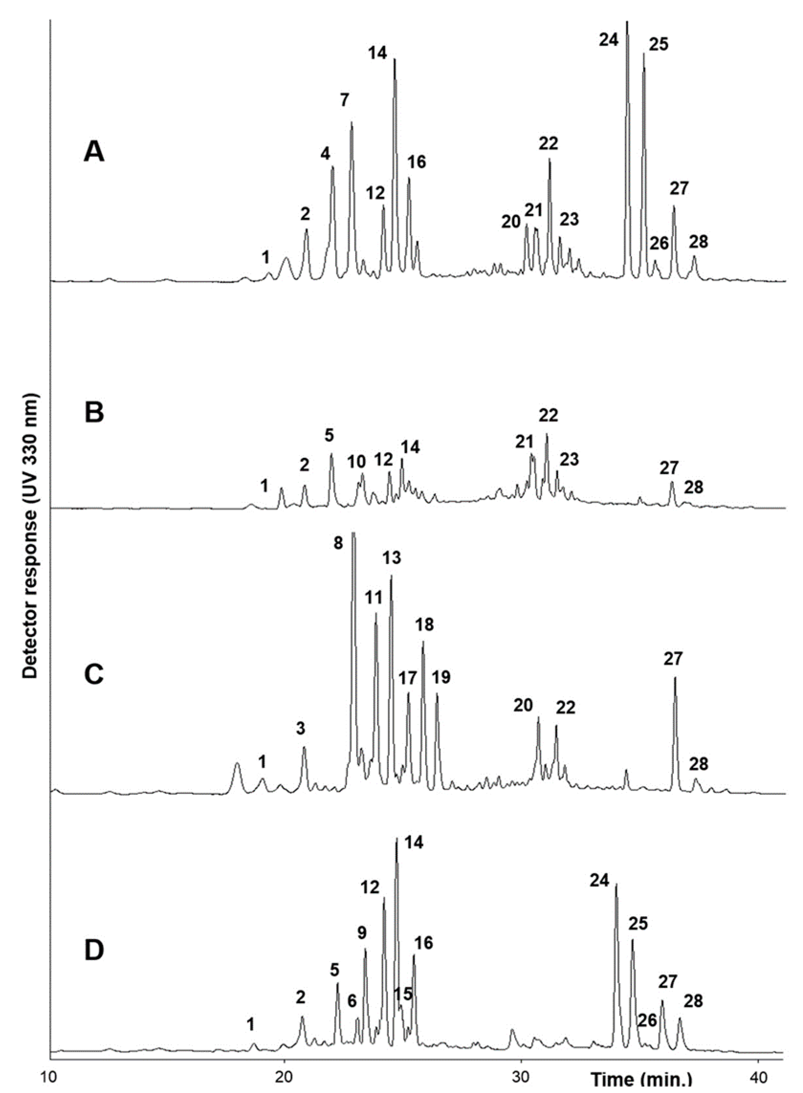
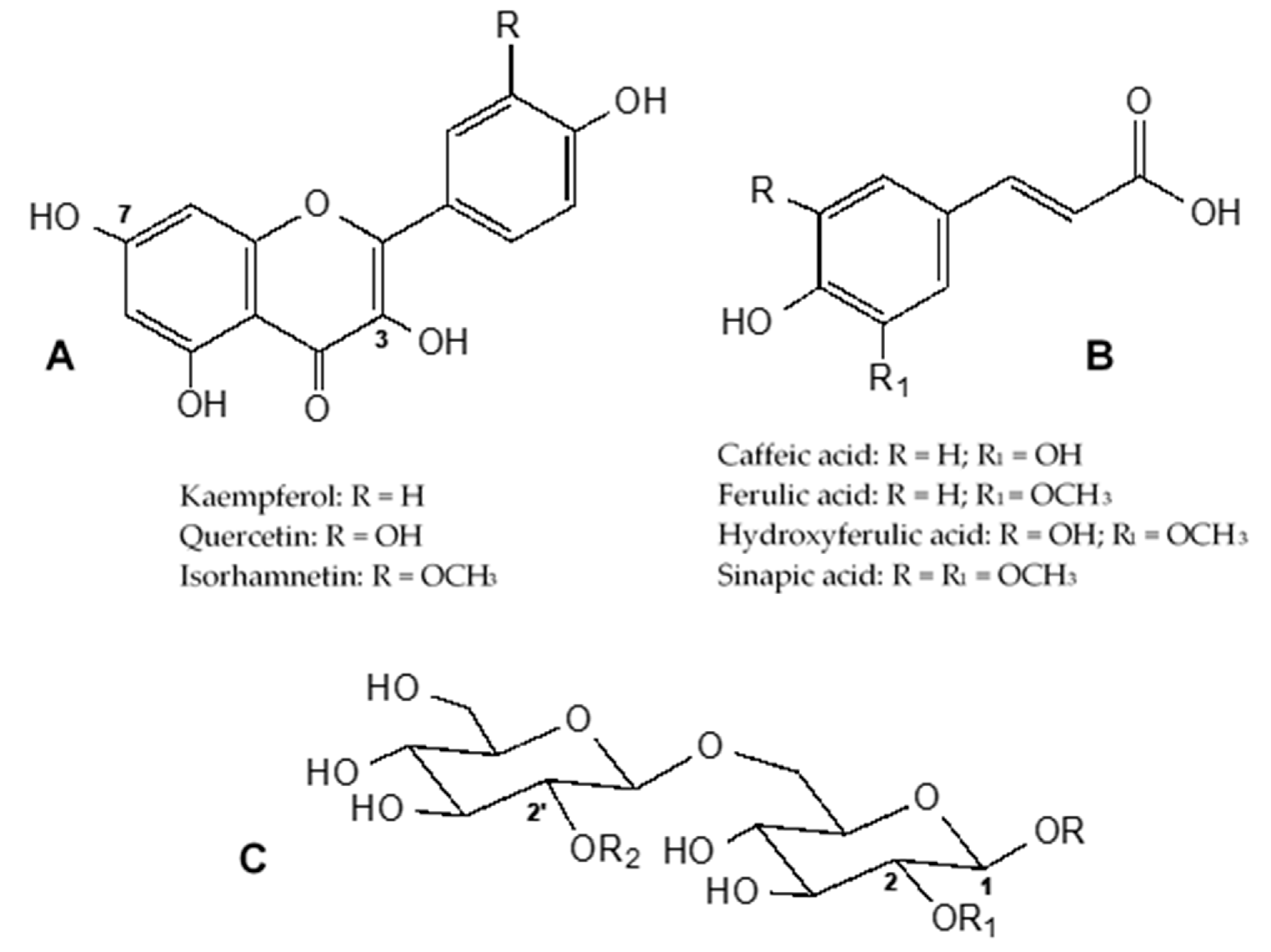
2.1. Phenolic Content and Composition

The UPLC/DAD analysis showed the presence of a high number of phenolic compounds, as shown in Figure 1, and their UV spectra indicate that most of them were esters of hydroxycinnamic acid derivatives. All detected compounds are listed in Table 1. Their chemical structures are shown in Figure 2.
From the UPLC/MS/MS study, different groups of compounds were detected, including flavonoids, mostly represented by hydroxycinnamic acid esters of kaempferol and quercetin glycosides (compounds 123), and hydroxycinnamic acyl glycosides, with a predominance of sinapoyl gentiobiosides (compounds 2428). Key fragment ions and other MS observations were also used for this evaluation. In positive ion mode, only the [M + H]+ ion was observed, while the [M − H] and MSn ions were registered in the negative ion mode. For all detected compounds, a loss of 162 m/z was indicative of the presence of a hexose (glucose) and of 324 m/z the presence of a di-hexose (sophorose). Moreover, for hydroxycinnamic acid esters of flavonoid glycosides, losses of 162 m/z, 176 m/z, 176 m/z, and 206 m/z are also indicative of the presence of caffeic acid, ferulic acid, hydroxyferulic acid, and sinapic acid, respectively. For hydroxycinnamic acyl glycosides, a loss of 194 m/z is indicative of the presence of ferulic acid and of 224 m/z, the presence of sinapic acid [11,15,16,26,27,28,29]. As an example, the negative ion electrospray MS2 spectra of hydroxycinnamic acid esters of flavonoid glycosides 6, 8, and 16 are reported in Figure S1, and the negative ion electrospray MS2 spectra of hydroxycinnamoyl gentiobiosides 24, 25, and 28 are reported in Figure S2.
All compounds detected in the Brassica samples used in this investigation are listed in Table 1 and were previously identified and reported in several Brassica spp. [10,11,12,13,15,16,17,23,26,27,28,29,30,31]. Quantitative evaluation was performed by HPLC using an external standard method (see experimental section), and their amounts are also reported in Table 1. Great variability in their qualitative and quantitative composition can be seen. As a general trend, in all Brassica samples, kaempferol is the most detected flavonol compared to quercetin, and sinapoyl and feruloyl derivatives are the most abundant hydroxycinnamoyl esters compared to caffeoyl and hydroxyferuloyl derivatives.
Kaempferol synapoyl glucoside 14 (67.5–81.3 mg/100 g d.w.), kaempferol caffeoyl glucoside 7 (43.5–62.5 mg/100 g d.w.), and kaempferol feruloyl glucoside 4 (52.3–99.7 mg/100 g d.w.) were the most abundant flavonol derivatives in both the samples of B. incana. Kaempferol feruloyl glucoside 21 (30.7 ± 1.8 mg/100 g d.w.) and kaempferol glucosides 2 and 5 (23.7 ± 6.6 and 28.1 ± 1.9 mg/100 g d.w., respectively) were the most abundant compounds detected in B. macrocarpa.
B. rupestris extracts were characterized by a higher amount of quercetin sinapoyl glucoside 8 (12.5–73.7 mg/100 g d.w.), kaempferol feruloyl glucoside 11 (45.4–74.8 mg/100 g d.w.), and kaempferol synapoyl glucoside 13 (19.5–62.0 mg/100 g d.w.).
A higher amount of kaempferol synapoyl glucoside 12 (24.8–86.1 mg/100 g d.w.) and 14 (25.8–81.4 mg/100 g d.w.), quercetin sinapoyl glucoside 9 (11.6–76.1 mg/100 g d.w.), and compound 2, kaempferol-3-O-diglucoside-7-O-diglucoside (46.9–80.4 mg/100 g d.w.), were the most abundant flavonol derivatives detected in B. villosa extracts.
A higher amount of hydroxycinnamoyl gentiobiosides (compounds 2428) was found in B. incana and B. villosa compared to B. rupestris and B. macrocarpa, in which only two hydroxycinnamoyl gentiobiosides were detected (compounds 27 and 28). Among gentiobiosides, compound 24 (1,2-disinapoyl gentiobiose) and 25 (1-sinapoyl-2-feruloyl gentiobiose) were the most abundant in B. incana. In B. villosa, compound 27 (1,2,2′-trisinapoyl gentiobiose) was the most abundant in BL and DC, while compound 24 was predominant in BX. In BY5, we observed similar amounts of compounds 2427 and a low content of compound 28.
So far, no study has yet been carried out reporting the LC-MS characterization of the phenolic content of Brassica wild species, with the exception of B. incana, which was analyzed in recent work by Miceli et al. [31]. Compared to Miceli et al. [31], our extracts of B. incana contained more quercetin and kaempferol derivatives and five different hydroxycinnamoyl gentiobiosides, while isorhamnetin derivatives were not detected, indicating that different environmental growing conditions can affect the phenolic composition.

2.2. Quantification of Phytochemicals and their Fractionation

The content of water-soluble phytochemicals in raw extracts and fractions obtained after chromatographic separation of the 10 wild Brassica spp. under investigation (see experimental section) is reported in Table 2.
Figure S3 reports an example of the HPLC chromatograms of B. rupestris (BU15) raw extract (Figure S3A) and the two fractions, F50 (Figure S3B) and F80 (Figure S3C), obtained after chromatographic separation. These data confirmed good separation of the compounds and revealed that the F50 fraction was mostly constituted by flavonol glycosides (peaks 123, Figure S3B), while the F80 fraction contained mostly hydroxycinnamoyl gentiobiosides (peaks 2428, Figure S3C). The F17 fraction did not contain any detectable phenol (data not shown); only ascorbic acid was found. Biothiols were detected only in raw extracts.
As reported in Table 2, B. incana samples showed the highest content of phenolics (531.0 ± 4.2 mg/100 g d.w. and 533.9 ± 23.0 mg/100 g d.w. in BN and BW, respectively) of which flavonol derivatives represent 70–80% of the total. B. villosa samples showed a quite constant phenolic content, in the range of 451.6–464.4 mg/100 g d.w., with flavonol derivatives representing 60–75% of the total. In raw extract of B. rupestris, phenolics ranged from 237.6 to 444.5 mg/100 g d.w., with about 90% of flavonol derivatives, while the B. macrocarpa sample showed the lowest phenolic content (163.9 ± 1.8 mg/100 g d.w.), of which flavonol derivatives represent 94% of the total. It is interesting to note that the wild Brassica species investigated here are characterized by a higher content of flavonol derivatives with respect to gentiobiosides, as shown in Table 2. It has been reported that in other Brassica species like broccoli, gentiobiosides were the most representative phenolic compounds [7,10].
Besides the importance of flavonols as powerful antioxidants [18], their health-related properties are also associated with their capacity to prevent AsA degradation by reverting the ascorbic radical to AsA and inhibiting the enzyme ascorbate oxidase [32].
The amounts of AsA and total thiols are also shown in Table 2. The highest AsA content was detected in B. villosa BX (375.8 mg/100 g d.w.) and in B. incana BN (355.7 mg/100 g d.w.). The same genotypes also showed the highest total thiol content, at 5.13 and 3.83 mg/100 g d.w., respectively. In BU5, BW, and BL, AsA ranged from 167.0 to 249.2 mg/100 g d.w., while BU9, BY5, and DC had the lowest AsA content (7.6–38.2 mg/100 g d.w.). BB, BY5, and DC were characterized by the lowest total thiol content, ranging from 0.59 to 0.83 mg/100 g d.w. Besides raw extracts, AsA was also detected in F17 fractions, although in lower amounts. The reduction of AsA content in F17 fractions was inversely related to its amount in the original raw extracts, with higher losses in samples that had the lowest AsA content in the raw extract, such as BY5 and DC (see Table 2).
Brassica crops have been intensively studied for their content of secondary metabolites, mainly phenolic compounds. Broccoli (B. olearcea L. italica) is also reported as a good source of ascorbic acid and thiols [33]. However, there is a lack of knowledge regarding the ascorbic acid and thiol content of Brassica wild species, and the present study attempts to fill this gap. The present findings show that Brassica wild species contained higher AsA and thiol content compared to other Brassica crops, such as broccoli [19,34], thus representing an importance source of bioactive compounds with potential positive effects on human health. In fact, AsA and thiols (particularly glutathione and cysteine) are widely recognized among the most effective antioxidants able to protect against various oxidative stress-related diseases like cancers cardiovascular disease and aging [34,35].

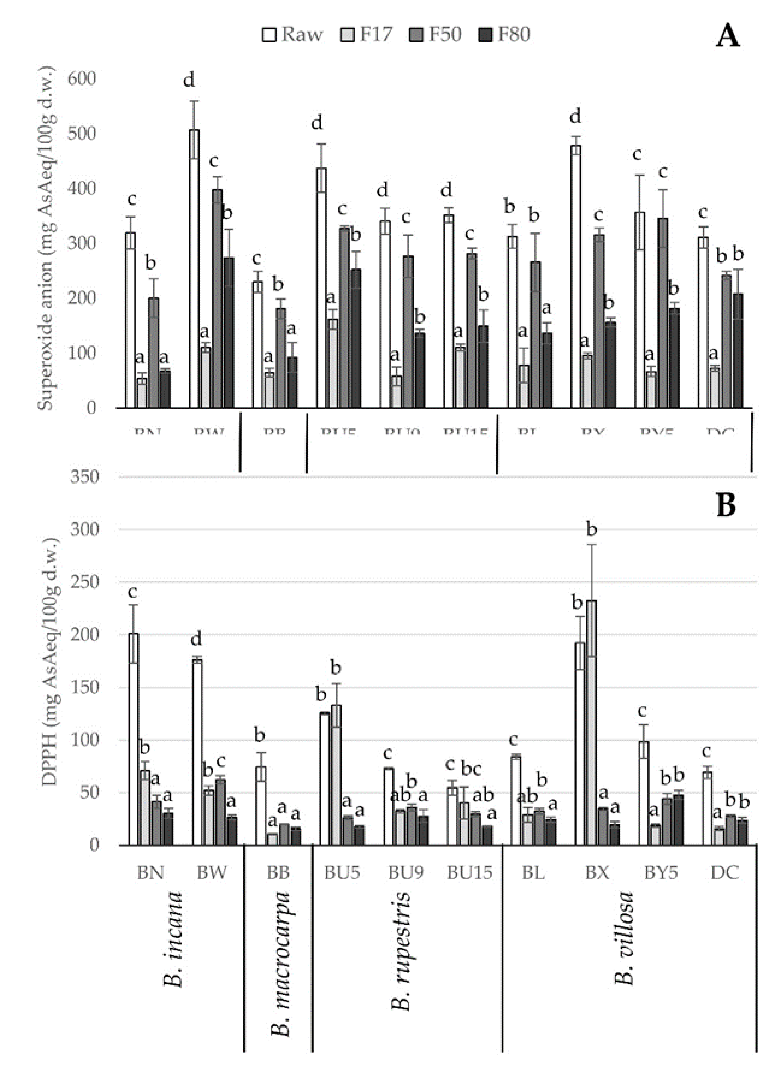
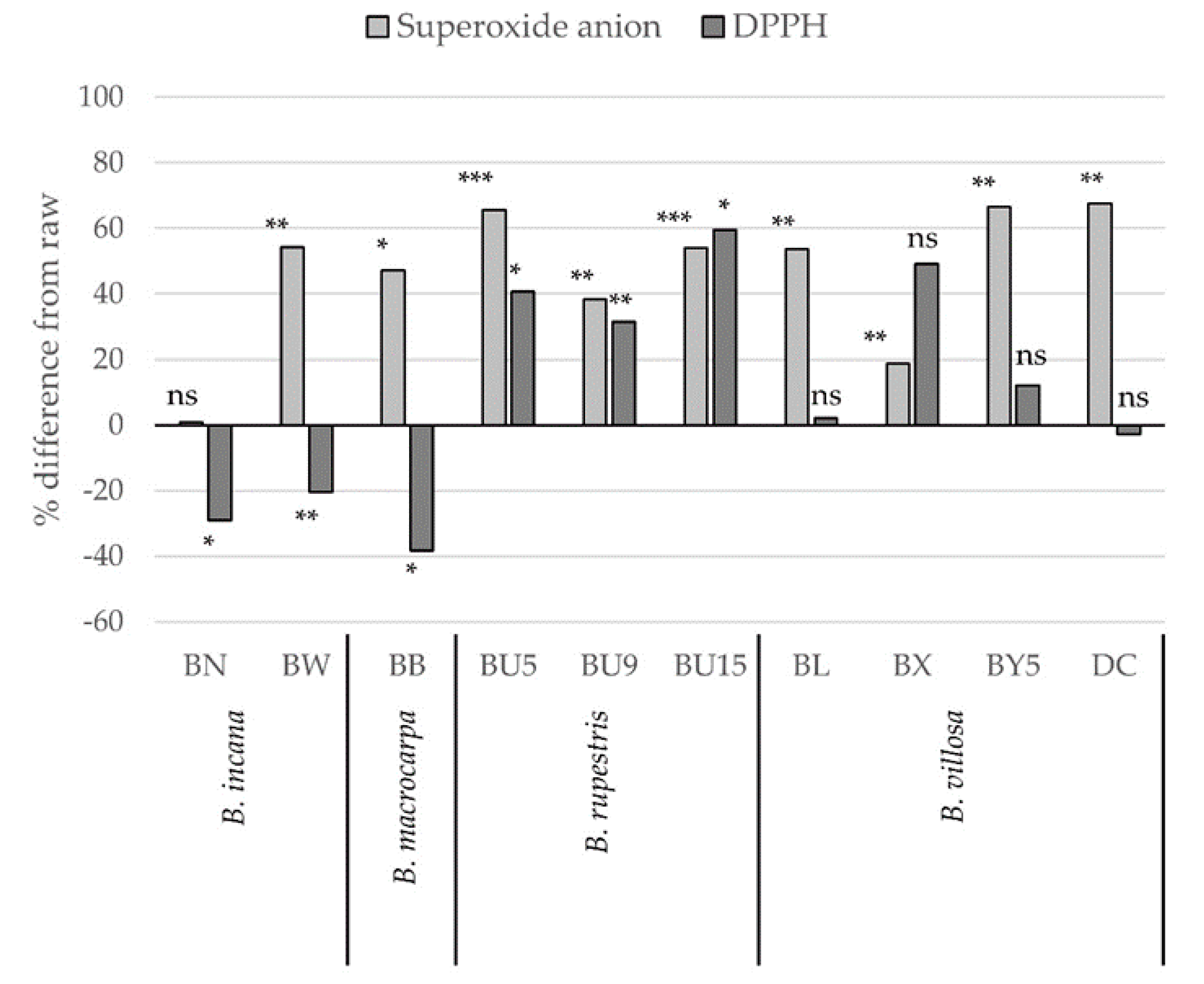
2.3. Antioxidant Capacity

Figure 3 shows the antioxidant capacity of the raw extracts and the three obtained fractions (F17, F50, and F80), evaluated by superoxide anion (Figure 3A) and DPPH (Figure 3B) methods.
With regard to the superoxide anion quenching capacity (Figure 3A), BW, BU5, and BX had the highest values (around 500 mg AsA eq/100 g d.w.), while other Brassica wild populations had slightly lower values (300–400 mg AsA eq/100 g d.w.). The BB sample showed the lowest superoxide scavenging capacity (250 mg AsA eq/100 g d.w.). The three fractions (F17, F50, and F80) contributed differently to the overall superoxide scavenging capacity, as shown in Figure 3A. F17, which only contains ascorbic acid, always had lower values with respect to F50 and, in most cases, F80, with the exception of three of the ten populations: BN, BB, and BL. These results indicate that phenolic compounds in F50 and F80 fractions were primarily responsible for the superoxide scavenging capacity. These results are in accordance with the study of Tabart et al. [36], which reported that the scavenging capacity against superoxide anion was strongly related to phenolic compounds. Indeed, the superoxide scavenging activity of raw extracts was significantly correlated with the total phenolic content (r = 0.53, p < 0.001) and particularly with flavonol derivatives (r = 0.58, p < 0.001), which were mainly retained in the F50 fraction. In fact, the superoxide scavenging activity of raw extracts was significantly correlated to the scavenging capacity of the F50 fraction (r = 0.77, p < 0.001), while the F17 and F80 fractions were less correlated (r = 0.59 for both fractions, p < 0.05).
Some authors studied the influence of glycosylated and acylated flavonol derivatives on the antioxidant activity of kale (B. oleracea var. sabellica) and showed that flavonol derivatives possessed higher antioxidant capacity than gentiobiosides [17,37]. Moreover, among flavonols, quercetin and its derivatives seem to be more capable of scavenging free radicals than kaempferol, because of the catechol structure of the B-ring [38,39]. This finding explains the higher superoxide scavenging capacity of the F50 fraction, which contained mainly flavonol derivatives.
The results of DPPH scavenging capacity of the raw extracts and the three obtained fractions (F17, F50, and F80) are shown at the bottom of Figure 3. The raw extracts of genotypes BN, BW, and BX showed the highest values (around 200 mg AsA eq/100 g d.w.), whereas the other populations ranged from 54.7 (BU15) to 125.4 (BU5) mg AsA eq/100 g d.w. The DPPH scavenging capacity of raw samples was primarily dependent on AsA content (r = 0.82, p < 0.001) and thiols (r = 0.76, p < 0.001), whereas flavonol derivatives contributed to a lesser extent (r = 0.35, p < 0.05). With regard to the contributions of single fractions to total DPPH scavenging capacity, Figure 3B shows that F17 had higher DPPH values compared to F50 and F80 in BN, BU5, and BX. In the other populations, the DPPH scavenging capacity of the three fractions was similar. In BY5 and DC, which were distinguished by the lowest AsA content (Table 2), the DPPH values were lower in F17 than in F50 and F80. These results suggest a strong dependence of DPPH scavenging capacity on AsA, which indeed characterized the F17 fraction. The primary role of AsA in determining DPPH scavenging capacity was already observed in other Brassica extracts [10,40]. In line with this observation, linear regression analysis showed a good relationship between DPPH values of raw extract and F17 fraction (r = 0.67, p < 0.0001), while a lower correlation was found with F50 fraction (r = 0.38, p < 0.05). These findings are in line with the rationale that AsA is the fastest scavenger of DPPH, as it reacts rapidly with DPPH, reaching a steady state in less than 1 min [41], corresponding to the reaction time used in the present work.
As a whole, the results of antioxidant capacity determined by the two tests highlight that Brassica species with high phenolic and AsA content, as in the case of BN, BW, BU5, and BX, are the ones with the greatest potential positive effects on human health.
Figure 4 shows the percent differences with respect to raw extracts (assumed equal to 100%) of superoxide anion and DPPH scavenging activity obtained by the sum of the values of the three fractions (F17, F50, and F80) in the ten Brassica wild species. Summing the antioxidant capacity values of each fraction, we observed that in most cases, the superoxide and DPPH scavenging capacity was significantly higher compared to raw extracts. Only in the case of B. incana (BN and BW) and B. macrocarpa (BB), the lower DPPH scavenging capacity based on the sum of the three fractions was probably related to the loss of AsA during the fractionation process (around 40–50% of the total; see Table 2) and the high dependency of DPPH scavenging capacity on AsA content itself. Since the levels of antioxidant metabolites (AsA, thiols, and phenols) were obviously higher in raw samples compared to the level obtained by summing the content in each fraction (see Table 2), we can hypothesize a form of antagonism among the phytochemicals of single fractions when mixed. Indeed, it is difficult to establish a clear relationship between the antioxidant content in a complex matrix and the antioxidant activity of a single compound. For example, while Marin et al. [32] observed a positive effect of flavonoid on ascorbic acid regeneration, other authors [42] showed that quercetin may act as a pro-oxidant in the presence of thiols, possibly because of an induced increase of reactive oxygen species and the formation of harmful thiol radicals. Experiments with different antioxidant assays and model solutions with mixed antioxidants gave extremely different responses in terms of antagonism, synergism, and additive effect [43,44,45].
The results of the multilinear regression (MLR) analysis indicated that the concentration of phenolics and AsA, but not thiols, could be a good predictor of DPPH scavenging capacity, since the total variance explained by the model was 69% (Figure S4). By contrast, the same was not true for the superoxide scavenging capacity, where the total variance explained by the model was equal to 24% (data not shown). The correlation between predicted and measured values of DPPH showed highly significant values with a coefficient of determination (R2) of 0.799 in calibration (Cal) and 0.693 in cross-validation (CV). Low values and results of MLR prediction were found for superoxide scavenging activity (R2 Cal = 0.416; R2 CV = 0.239). These findings confirm the strong dependence of DPPH scavenging capacity on AsA content and suggest the need to analyze not only phenols but also AsA when determining the total antioxidant capacity of a complex matrix.

3. Materials and Methods

3.1. Plant Material

Seeds of wild populations of B. incana (Figures S5.1 and S5.2), B. macrocarpa (Figures S5.3 and S5.4), B. rupestris (Figures S5.5 and S5.6), and B. villosa (Figures S5.7 and S5.8) belonging to the B. oleracea complex species (number of chromosomes = 9) were collected in different areas of Sicily during AEGRO projects in 2006–2010. B. incana is mainly present along the eastern and northeastern Sicilian coast, B. macrocarpa is an endemic species of the Egadi Islands, B. rupestris is widespread along the northern coast of Sicily, mainly in Western areas, and B. villosa grows in the central-western inland areas and along the northwestern coast. In addition, one population of B. incana and one of B. rupestris collected in the Lazio and Calabria regions, respectively, were also investigated. Voucher specimens of each population are maintained at the Department of Agriculture, Food and Environment of the University of Catania. The complete list of accessions is shown in Table 3. Brassica populations were grown in the experimental farm of the University of Catania (37°27′ N, 15°40′ E, 10 m a.s.l.). Eight months after transplanting, leaf samples were collected and immediately frozen and lyophilized. The freeze-dried samples were ground to a fine powder at low temperature and stored at –20 °C.

3.2. Extraction and Fractionation

Sample extraction and fractionation were performed according to Rodríguex et al. [46], with modifications. The raw extract was obtained by adding 1 g of lyophilized powder to 15 mL of MeOH/0.03 N HCl (1:1). The mixture was shaken at room temperature for two hours and then centrifuged at 25,000× g at 4 °C for 20 min. The supernatant was filtered on glass wool and stored at −80 °C. Fractionation of raw extracts was performed on 1.2 cm diameter cartridges filled with 2 g of ICN Biomedicals C18 adsorbent resin (32–63 µm, 60 A) preconditioned with 3 × 5 mL of 0.03 N HCl. The sample (4 mL) was diluted three-fold with 0.03 N HCl and then loaded at a flow rate of 1 mL/min. The sample was eluted with 4 mL of three solutions at increasing MeOH concentration, thus obtaining three fractions: (1) F17, eluted with 17% MeOH in 0.03 N HCl; (2) F50, eluted with 50% MeOH in 0.03 N HCl; and (3) F80, eluted with 80% MeOH in 0.03 N HCl.
Further elution with increasing MeOH up to 100% did not reveal any other compound, as confirmed by HPLC analysis. The three collected fractions were then stored at –80 °C until analysis. Each sample was analyzed in triplicate.

3.3. LC/UV–DAD/ESI–MS Analysis

LC-MS analysis was performed on raw extracts using a Jasco UPLC-system combined with a Thermo LTQ. A Thermo Xcalibur Qual Browser was used for the acquisition and processing of mass data. The UV-vis chromatograms were analyzed with a Jasco Chemstation (ChromNAV, Jasco, Mary’s Court, Easton, MD, USA). Chromatographic analysis was carried out on an Acquity UPLC BEH C18 column (1.7 µm; 50 × 2.1 mm, Phenomenex, Torrence, CA, USA). All analyzed samples were dissolved in MeOH/H2O (9:1) at a concentration of 20–30 ppm. The column was set at 35 °C. The mobile phase for UPLC analysis was 0.1% formic acid in water and acetonitrile. The samples were spectrophotometrically checked by reading at eight wavelengths (205 nm to 275 nm). Elution was performed with a flow rate of 0.3 mL/min according to the following gradient method: injection volume 5 µL; linear gradient starting from 95% aqueous, gradually to 25% of acetonitrile over 30 min, gradually to 70% of acetonitrile within 40 min, gradually to 100% of acetonitrile over 50 min, and then isocratic flow for 5 min. Mass spectrometry data were acquired in positive and negative ion mode. All analyses were carried out using an ESI ion source with the following settings: capillary voltage, 3100 V; sheath gas (He), aux gas (He), and sweep gas (He) heated at 275 °C and introduced with a source heater temperature of 80 °C. Full scan spectra were acquired over the range of 100–2000 m/z. Automated MS/MS was performed by isolating the base peaks (molecular ions) using an isolation width of 2.0 m/z, normalized collision energy of 25 V, threshold set at 500, and ion charge control on, with max acquisition time set at 300 ms. All compounds were tentatively identified by comparing their retention times and mass spectral data obtained in positive and negative ion mode with those of standard compounds or with compounds previously reported in the literature for Brassica spp. [11,15,16,26,27,28,29,30].

3.4. HPLC-DAD Analyses

The phenolic composition of raw and fractionated extracts was also evaluated by HPLC-DAD using a Jasco system equipped with a diode array detector (MD-910, Jasco, Mary’s Court, Easton, MD, USA). The pump (PU-980, Jasco, Mary’s Court, Easton, MD, USA) was coupled with a ternary gradient unit (LG-1580-02, Jasco, Mary’s Court, Easton, MD, USA). The analytical data were evaluated using a software management system for chromatographic data (ChromNAV, Jasco, Mary’s Court, Easton, MD, USA). The separation was performed by a reversed phase C18 Purospher Star column (250 × 4 mm, 5 µ, Merck, Darmstadt, Germany) was coupled with a ternary gradient unit (LG-1580-02, Jasco, Mary’s Court, Easton, MD, USA). The flow rate was 0.6 mL/min, the injection volume was 10 μL, and the column temperature was maintained at 42 °C. The mobile phase consisted of water with 5% of glacial acetic acid (solvent A) and acetonitrile with 5% of glacial acetic acid (solvent B). The gradients were as follows (A/B): 95/5 from 0–10 min, 95/5 to 85/15 in 10 min, 85/15 to 70/30 in 10 min, 70/3 for 10 min, 70/30 to 95/5 in 10 min, and 95/5 for 10 min. Peak identification was based on LC-MS data. Quantitation of detected compounds was performed at 330 nm by an external standard method using available commercial standards (purity HPLC ≥98%) of kaempferol-3-O-glucoside (y = 134,103x – 103,094, R2 = 0.9942) and quercetin-3-O-glucoside (y = 112,896x + 58,728, R2 = 0.9959) for flavonol derivatives (peaks 123) and sinapic acid acyl-β-d-glucoside (y = 194,026x + 13,9401, R2 = 0.9960) for hydroxycinnamoyl gentiobiosides (peaks 2428). For calibration curves, all standards were injected in triplicate in the range of 4 to 40 µg/mL.

3.5. Ascorbic Acid Analysis

Ascorbic acid was analyzed in raw extracts and in the three fractions following the method described by Picchi et al. [47], with slight modifications. For sample preparation, 200 µL of extract was treated with 800 µL of cold 3% metaphosphoric acid, homogenized, centrifuged at 25,000× g for 5 min at 4 °C, and immediately analyzed. The analytical column was a 250 × 6 mm, 5 µ Intersil ODS-3, maintained at 30 °C. The isocratic elution was performed using orthophosphoric acid 0.02 M as mobile phase, at a flow rate of 0.7 mL/min. Samples of 20 μL were injected and monitored at 254 nm. The concentration of ascorbic acid was calculated from the experimental peak area by analytical interpolation in a standard calibration curve (range 1.0–100.0 mg/100 mL) and was expressed as mg/100 g dry weight (d.w.). Ascorbic acid was detected in raw extracts and in F17 fractions, while it was not found in F50 and F80 fractions.

3.6. Analysis of Thiol Groups

The determination of non-protein –SH groups in the extracts and fractions was performed according to Hawrylak and Szymanska [48], with some modifications. An aliquot of 0.1 mL of sample was added to 0.8 mL of 1 M sodium phosphate buffer (pH 8.0) and 0.1 mL of 10 mM dithionitrobenzoic acid (DTNB). The absorbance was then read at 415 nm. The amount of biothiols was calculated from a standard curve made for l-cysteine, and the data are reported as mg/100 g d.w. Total thiol groups were only detected in raw extracts.

3.7. Antioxidant Capacity Measurement

The antioxidant capacity potential was measured using 1,1-diphenyl-2-picrylhydrazil (DPPH) radical quenching and superoxide anion radical scavenging. All measurements were performed by electronic paramagnetic resonance (EPR) using a MiniScope MS200 (Magnettech, Berlin, Germany). The protocol was detailed in Picchi et al. [47]. The calibration was made with ascorbic acid solution at known concentration and the results were reported as equivalent of ascorbic acid/100 g d.w.

3.8. Statistical Analysis

The analyses were carried out using three biological replicates. The phytochemical data of raw extracts were subjected to one-way analysis of variance and comparisons among means were performed according to the least significant difference (LSD) test. For each species, the antioxidant activity of the four extracts (raw, F17, F50, F80) was compared according to the LSD test. Significant differences were accepted at the minimum probability level of p < 0.05. A multilinear regression (MLR) analysis was carried out using the data of raw extracts to assess a possible relationship between the concentrations of all antioxidants analyzed in this paper (flavonol derivatives, hydroxycinnamoyl gentiobiosides, ascorbic acid, and thiols) and their overall antioxidant capacity, as measured using the DPPH and superoxide scavenging test. All measured antioxidants (independent variables) were considered in the statistical development of the MLR model. The procedure included the following steps: (1) extraction of raw data of antioxidants (X block variables); (2) extraction of measured values of antioxidant activity (DPPH or superoxide scavenging activity, Y block variables); (3) application of preprocessing algorithms to X and Y; (4) application of the MLR multivariate technique for modelling and testing; and (5) calculation of efficiency parameter of prediction. The stability and validity of the model were tested using Venetian blind cross-validation. For variable selection, a stepwise procedure was utilized. The model includes a calibration phase and a validation phase, with residual errors calculated for both root mean square error in calibration (RMSEC) and cross-validation (RMSECV). The predictive ability of the model was tested with the coefficient of determination (R2) between observed and predicted values.

4. Conclusions

The ten Mediterranean wild relatives of B. oleracea considered here were evaluated for the first time for their content of main water-soluble antioxidants, namely ascorbic acid, phenolics, and total biothiols. The results showed great variability in terms of the qualitative and quantitative composition of these metabolites. LC/MS investigation of polyphenolics showed that the greatest constituents were represented by hydroxycinnamoyl glycosides of the flavonols kaempferol and quercetin and by hydroxycinnamoyl gentiobiosides. The evaluation of the in vitro antioxidant activity on fractionated extracts contributes to clarifying the relationship between bioactive compounds of Brassica wild species and their antioxidant capacity. The results highlight the importance of consuming Brassica wild species because of their high content of flavonol derivatives, ascorbic acid, and thiols with respect to commonly cultivated Brassica genotypes. These metabolites are known to be very powerful antioxidants, indicating that consuming Brassica wild species may have an important impact on human health. Besides the great diversity of phytochemicals among populations and their good antioxidant potential, the Italian Brassica wild relatives may represent a valuable genetic source for developing new cultivars with improved bioactive metabolite content. Future work aimed at studying the in vivo antioxidant potential of Brassica wild species could help to confirm the high nutritional value of these edible plants and recognize their importance in the human diet for their health properties because of the capacity of effectively keeping under control the production of reactive oxygen species.

Supplementary Materials

The following are available online. Figure S1: Negative ion electrospray MS2 mass spectra of hydroxycinnamic acid esters of flavonol glycosides: (A) quercetin 3-O-sinapoyldiglucoside-7-O-glucoside (6); (B) kaempferol 3-O-feruloyldiglucoside-7-O-diglucoside (16); (C) quercetin 3-O-sinapoyltriglucoside-7-O-diglucoside (8). Figure S2: Negative ion electrospray MS2 mass spectra of hydroxycinnamoyl gentiobiosides: (A) 1,2-disinapoyl gentiobioside (24); (B) 1-sinapoyl-2-feruloyl gentiobioside (25); (C) 1,2-disinapoyl-2-feruloyl gentiobioside (28). Figure S3: Example of chromatograms acquired by HPLC-DAD (330 nm) after solid-phase extraction. See paragraph 3.4 for details. (A) Raw extract; (B) F50 fraction characterized by the predominance of flavonol-caffeoyl and kaempferol-sinapoyl derivatives; (C) F80 fraction characterized by the predominance of hydroxycinnamoyl gentiobiosides. For peak numbering, see Table 1. Figure S4: Scatter plot of measured vs. MLR prediction for DPPH scavenging activity, with root mean square error (RMSE), bias, and coefficient of determination (R2) in calibration (C, Cal) and cross-validation (CV). See paragraph 3.8. for details. Figure S5: Characteristics of the four Brassica wild species evaluated in the present work. Whole plant (left) and leaves (right) of B. incana (1-2), B. macrocarpa (3-4), B. rupestris (5-6), and B. villosa (7-8).

Author Contributions

Conceptualization, V.P. and R.L.S.; data curation, V.P. and A.T.; formal analysis, V.P. and R.L.S.; funding acquisition, F.B.; investigation, V.P., R.L.S., A.T., and F.D.; methodology, R.L.S. and A.T.; project administration, F.B.; resources, S.A., S.T. (Stefania Toscano), and S.T. (Simone Treccarichi); supervision, R.L.S. and F.B.; visualization, V.P., A.T., and F.D.; writing—original draft, V.P., R.L.S., and A.T.; writing—review and editing, V.P., R.L.S., and F.B. All authors have read and agreed to the published version of the manuscript.

Funding

This research was funded by EU HORIZON 2020, Breeding for Resilient, Efficient and Sustainable Organic Vegetable (BRESOV) production project, grant number 774244.

Conflicts of Interest

The authors declare no conflict of interest.

References

  1. Rakow, G. Species origin and economic importance of Brassica. In Brassica. Biotechnology in Agriculture and Forestry; Pua, E.C., Douglas, C.J., Eds.; Springer: Berlin, Germany, 2004; Volume 54, pp. 3–11. [Google Scholar] [CrossRef]
  2. Bojórquez-Velázquez, E.; Velarde-Salcedo, A.J.; de León-Rodríguez, A.; Jimenez-Islas, H.; Pérez-Torres, J.L.; Herrera-Estrella, A.; Espitia-Rangel, E.; de la Rosa, A.P.B. Morphological, proximal composition, and bioactive compounds characterization of wild and cultivated amaranth (Amaranthus spp.) species. J. Cereal Sci. 2011, 83, 222–228. [Google Scholar] [CrossRef]
  3. Aguinagalde, I.; Gomez-Campo, C.; Sanchez-Yelamo, M.D. A chemosystematic survey on wild relatives of Brassica oleracea L. Bot. J. Linn. Soc. 1992, 109, 57–67. [Google Scholar] [CrossRef]
  4. Sánchez-Mata, M.C.; Cabrera Loera, R.D.; Morales, P.; Fernández-Ruiz, V.; Cámara, M.; Díez Marqués, C.; Pardo-de-Santayana, M.; Tardío, J. Wild vegetables of the Mediterranean area as valuable sources of bioactive compounds. Genet. Resour. Crop. Evol. 2012, 59, 431–443. [Google Scholar] [CrossRef]
  5. Castillo-Lorenzo, E.; Finch-Savage, W.E.; Seal, C.E.; Pritchard, H.W. Adaptive significance of functional germination traits in crop wild relatives of Brassica. Agric. Meteorol. 2019, 264, 343–350. [Google Scholar] [CrossRef]
  6. Podsędek, A.; Sosnowska, D.; Redzynia, M.; Anders, B. Antioxidant capacity and content of Brassica oleracea dietary antioxidants. Int. J. Food Sci. Technol. 2006, 41, 49–58. [Google Scholar] [CrossRef]
  7. Vallejo, F.; Tomás-Barberán, F.A.; Garciá-Viguera, C. Health-promoting compounds in broccoli as affected by refrigerated transport and retail sale period. J. Agric. Food. Chem. 2003, 51, 3029–3034. [Google Scholar] [CrossRef]
  8. Cartea, M.E.; Francisco, M.; Soengas, P.; Velasco, P. Phenolic compounds in Brassica vegetables. Molecules 2011, 16, 251–280. [Google Scholar] [CrossRef]
  9. Podsędek, A. Natural antioxidants and antioxidant capacity of Brassica vegetables: A review. LWT 2007, 40, 1–11. [Google Scholar] [CrossRef]
  10. Heimler, D.; Vignolini, P.; Dini, M.G.; Vincieri, F.F.; Romani, A. Antiradical activity and polyphenol composition of local Brassicaceae edible varieties. Food Chem. 2006, 99, 464–469. [Google Scholar] [CrossRef]
  11. Francisco, M.; Moreno, D.A.; Cartea, M.E.; Ferreres, F.; García-Viguera, C.; Velasco, P. Simultaneous identification of glucosinolates and phenolic compounds in a representative collection of vegetable Brassica rapa. J. Chromatogr. A 2009, 1216, 6611–6619. [Google Scholar] [CrossRef]
  12. Gratacoós-Cubarsí, M.; Ribas-Agustí, A.; García-Regueiro, J.A.; Castellari, M. Simultaneous evaluation of intact glucosinolates and phenolic compounds by UPLC-DAD-MS/MS in Brassica oleracea L. var. botrytis. Food Chem. 2010, 121, 257–263. [Google Scholar] [CrossRef]
  13. Nielsen, J.K.; Olsen, C.E.; Petersen, M.K. Acylated flavonol glycosides from cabbage leaves. Phytochemistry 1993, 34, 539–544. [Google Scholar] [CrossRef]
  14. Llorach, R.; Espin, J.C.; Tomás-Barberán, F.A.; Ferreres, F. Valorization of cauliflower (Brassica oleracea L. var. botrytis) by-products as a source of antioxidant phenolics. J. Agric. Food. Chem. 2003, 51, 2181–2187. [Google Scholar] [CrossRef] [PubMed]
  15. Lin, L.Z.; Harnly, J.M. Identification of the phenolic components of Collard greens, Kale and Chinese broccoli. J. Agric. Food. Chem. 2009, 57, 7401–7408. [Google Scholar] [CrossRef]
  16. Lin, L.Z.; Harnly, J.M. Phenolic component profiles of Mustard greens, Yu Choy and 15 other Brassica vegetables. J. Agric. Food. Chem. 2010, 58, 6850–6857. [Google Scholar] [CrossRef] [PubMed] [Green Version]
  17. Fiol, M.; Adermann, S.; Neugart, S.; Rohn, S.; Mügge, C.; Schreiner, M.; Krumbein, A.; Kroh, L.W. Highly glycosylated and acylated flavonols isolated from kale (Brassica oleracea var. sabellica)—Structure–antioxidant activity relationship. Food Res. Int. 2012, 47, 80–89. [Google Scholar] [CrossRef]
  18. Soengas, P.; Cartea, M.E.; Francisco, M.; Sotelo, T.; Velasco, P. New insights into antioxidant activity of Brassica crops. Food Chem. 2012, 134, 725–733. [Google Scholar] [CrossRef] [Green Version]
  19. Demirkol, O.; Adams, C.; Ercal, N. Biologically important thiols in various vegetables and fruits. J. Agric. Food. Chem. 2004, 52, 8151–8154. [Google Scholar] [CrossRef]
  20. Shigenaga, T.; Yamauchi, N.; Funamoto, Y.; Shigyo, M. Effects of heat treatment on an ascorbate–glutathione cycle in stored broccoli (Brassica oleracea L.) florets. Postharvest Biol. Technol. 2005, 38, 152–159. [Google Scholar] [CrossRef]
  21. Zacharis, C.K.; Tzanavaras, P.D.; Zotou, A. Ethyl propiolate as a post-column derivatization reagent for thiols: Development of a green liquid chromatographic method for the determination of glutathione in vegetables. Anal. Chim. Acta 2011, 690, 122–128. [Google Scholar] [CrossRef]
  22. Sousa, C.; Valentão, P.; Ferreres, F.; Seabra, R.M.; Andrade, P.B. Tronchuda cabbage (Brassica oleracea L. var. costata DC): Scavenger of reactive nitrogen species. J. Agric. Food. Chem. 2008, 56, 4205–4211. [Google Scholar] [CrossRef] [PubMed]
  23. Upadhyay, R.; Sehwag, S.; Singh, S.P. Antioxidant activity and polyphenol content of Brassica oleracea varieties. Int. J. Veg. Sci. 2016, 22, 353–363. [Google Scholar] [CrossRef]
  24. Branca, F.; Ragusa, L.; Tribulato, A.; Di Gaetano, C.; Calì, F. Genetic relationships of Brassica vegetables and wild relatives in Southern Italy determined by five SSR. Acta Hortic. 2013, 1005, 189–196. [Google Scholar] [CrossRef]
  25. Branca, F.; Ragusa, L.; Tribulato, A.; Bagatta, M.; Lo Scalzo, R.; Picchi, V. Evaluation of sicilian wild brassica species (n=9) for glucosinolate profile and antioxidant compounds. Acta Hortic. 2013, 1005, 181–188. [Google Scholar] [CrossRef]
  26. Ferreres, F.; Sousa, C.; Pereira, D.M.; Valentao, P.; Taveira, M.; Martins, A.; Pereira, J.A.; Seabra, R.M.; Andrade, P.B. Screening of antioxidant phenolic compounds produced by in vitro shoots of Brassica oleracea L. var. costata DC. Comb. Chem. High Throughput Screen. 2009, 12, 230–240. [Google Scholar] [CrossRef]
  27. Lin, L.Z.; Sun, J.; Chen, P.; Harny, J. UHPLC-PDA-ESI/HRMS/MSn analysis of anthocyanins, flavonol, glucosides, and hydroxycinnamic acid derivatives in red mustard greens (Brassica juncea Coss variety). J. Agric. Food. Chem. 2011, 59, 12059–12072. [Google Scholar] [CrossRef] [Green Version]
  28. Velasco, P.; Francisco, M.; Moreno, D.A.; Ferreres, F.; Garcia-Viguera, C.; Cartea, M.E. Phytochemical fingerprinting of vegetable Brassica oleracea and Brassica napus by simultaneous identification of glucosinolates and phenolics. Phytochem. Anal. 2011, 22, 144–152. [Google Scholar] [CrossRef] [Green Version]
  29. Yang, S.C.; Arasu, M.V.; Chun, J.H.; Jang, Y.S.; Lee, Y.H.; Kim, I.H.; Lee, K.T.; Hong, S.T.; Kim, S.J. Identification and determination of phenolic compounds in rapeseed meals (Brassica napus L.). J. Agric. Chem. Env. 2015, 4, 14–23. [Google Scholar] [CrossRef] [Green Version]
  30. Romani, A.; Vignolini, P.; Isolani, L.; Ieri, F.; Heimler, D. HPLC-DAD/MS characterization of flavonoids and hydroxycinnamic derivatives in turnip tops (Brassica rapa L. subsp. sylvestris L.). J. Agric. Food. Chem. 2006, 54, 1342–1346. [Google Scholar] [CrossRef] [Green Version]
  31. Miceli, N.; Cavò, E.; Ragusa, M.; Cacciola, F.; Mondello, L.; Dugo, L.; Acquaviva, R.; Malfa, G.A.; Marino, A.; D’Arrigo, M.; et al. Brassica incana Ten. (Brassicaceae): Phenolic constituents, antioxidant and cytotoxic properties of the leaf and flowering top extracts. Molecules 2020, 25, 1461. [Google Scholar] [CrossRef] [Green Version]
  32. Marín, F.R.; Frutos, M.J.; Pérez-Alvarez, J.A.; Martinez-Sánchez, F.; del Río, J.A. Flavonoids as nutraceuticals: Structural related antioxidant properties and their role on ascorbic acid preservation. Stud. Nat. Prod. Chem. 2002, 26, 741–778. [Google Scholar] [CrossRef]
  33. Raseetha, S.; Leong, S.Y.; Burritt, D.J.; Indrawati, O. Understanding the degradation of ascorbic acid and glutathione in relation to the levels of oxidative stress biomarkers in broccoli (Brassica oleracea L. italica cv. Bellstar) during storage and mechanical processing. Food Chem. 2013, 138, 1360–1369. [Google Scholar] [CrossRef] [PubMed]
  34. Davey, M.V.; Van Montagu, M.; Inzé, D.; Sanmartin, M.; Kanellis, A.; Smirnoff, N.; Benzie, I.J.J.; Strain, J.J.; Favell, D.; Fletcher, J. Plant L-ascorbic acid: Chemistry, function, metabolism, bioavailability and effects of processing. J. Sci. Food Agric. 2000, 80, 825–860. [Google Scholar] [CrossRef]
  35. Mills, B.J.; Stinson, C.T.; Liu, M.C.; Lang, C.A. Glutathione and cyst(e)ine profiles of vegetables using high performance liquid chromatography with dual electrochemical detection. J. Food Comp. Anal. 1997, 10, 90–101. [Google Scholar] [CrossRef]
  36. Tabart, J.; Kevers, C.; Pincemail, J.; Defraigne, J.O.; Dommes, J. Comparative antioxidant capacities of phenolic compounds measured by various tests. Food Chem. 2009, 113, 1226–1233. [Google Scholar] [CrossRef]
  37. Zietz, M.; Weckmüller, A.; Schmidt, S.; Rohn, S.; Schreiner, M.; Krumbein, A.; Kroh, L.W. Genotypic and climatic influence on the antioxidant activity of flavonoids in kale (Brassica oleracea var. sabellica). J. Agric. Food Chem. 2010, 58, 2123–2130. [Google Scholar] [CrossRef]
  38. Rice-Evans, C.A.; Miller, N.J.; Paganga, G. Structure-antioxidant activity relationships of flavonoids and phenolic acids. Free Radic. Biol. Med. 1996, 20, 933–956. [Google Scholar] [CrossRef]
  39. Rösch, D.; Bergmann, M.; Knorr, D.; Kroh, L.W. Structure antioxidant efficiency relationships of phenolic compounds and their contribution to the antioxidant activity of Sea Buckthorn juice. J. Agric. Food Chem. 2003, 51, 4233–4239. [Google Scholar] [CrossRef]
  40. Lo Scalzo, R.; Picchi, V.; Migliori, C.A.; Campanelli, G.; Leteo, F.; Ferrari, V.; Di Cesare, L.F. Variations in the phytochemical contents and antioxidant capacity of organically and conventionally grown Italian cauliflower (Brassica oleracea L. subsp. botrytis): Results from a three-year field study. J. Agric. Food Chem. 2013, 61, 10335–10344. [Google Scholar] [CrossRef]
  41. Brand-Williams, W.; Cuvelier, M.E.; Berset, C. Use of a free radical method to evaluate antioxidant activity. LWT 1995, 28, 25–30. [Google Scholar] [CrossRef]
  42. Fujisawa, S.; Kadoma, Y. Anti- and pro-oxidant effects of oxidized quercetin, curcumin or curcumin-related compounds with thiols or ascorbate as measured by the induction period method. In Vivo 2006, 20, 39–44. [Google Scholar] [PubMed]
  43. Hajimehdipoor, H.; Shahrestani, R.; Shekarchi, M. Investigating the synergistic antioxidant effects of some flavonoid and phenolic compounds. Res. J. Pharm. 2014, 1, 35–40. [Google Scholar]
  44. Hidalgo, M.; Sánchez-Moreno, C.; de Pascual-Teresa, S. Flavonoid–flavonoid interaction and its effect on their antioxidant activity. Food Chem. 2010, 121, 691–696. [Google Scholar] [CrossRef]
  45. Kurin, E.; Mučaji, P.; Nagy, M. In vitro antioxidant activities of three red wine polyphenols and their mixtures: An interaction study. Molecules 2012, 17, 14336–14348. [Google Scholar] [CrossRef] [Green Version]
  46. Rodríguex, I.; Llompart, M.P.; Cela, R. Solid-phase extraction of phenols. J. Chromatogr. A 2000, 885, 291–304. [Google Scholar] [CrossRef]
  47. Picchi, V.; Migliori, C.A.; Lo Scalzo, R.; Campanelli, G.; Ferrari, V.; di Cesare, L.F. Phytochemical content in organic and conventionally grown Italian cauliflower. Food Chem. 2012, 130, 501–509. [Google Scholar] [CrossRef]
  48. Hawrylak, B.; Szymanska, M. Selenium as a sulphydrylic group inductor in plant. Cell. Mol. Biol. Lett. 2004, 9, 329–336. [Google Scholar]
Sample Availability: Samples of the compounds are not available from the authors.
Figure 1. UPLC chromatogram (330 nm) of raw extracts of four Brassica species: (A) B. incana, (B) B. macrocarpa, (C) B. rupestris, and (D) B. villosa. For compound identification, see Table 1.
Molecules 25 03495 g001
Figure 2. Chemical structure of phenolic compounds identified in wild Brassica samples: (A) flavonoids, (B) hydroxycinnamic acids, and (C) gentiobiosides.
Molecules 25 03495 g002
Figure 3. Antioxidant capacity of raw extracts and F17, F50, and F80 fractions of ten populations of Brassica wild species measured as (A) superoxide anion and (B) DPPH scavenging capacity. Letters above bars within each population indicate significantly different means according to Tukey’s test (p < 0.05).
Molecules 25 03495 g003
Figure 4. Percent differences with respect to raw extracts (assumed equal to 100%) of superoxide anion and DPPH scavenging activity obtained by the sum of values of three fractions (F17, F50, F80) in ten Brassica wild species. * p < 0.05, ** p < 0.01, *** p < 0.001, ns, p > 0.05, according to Tukey’s test.
Molecules 25 03495 g004
Table 1. Content (mg/100 g d.w.) and composition of tentatively identified polyphenols in wild Brassica spp. used in this investigation.
#UV λmax (nm)[M + H]+/ [M − H]-MS2 (m/z) (%)Compound aB.incana B. macrocarpa B. rupestris B. villosa
BNBW BB BU5BU9BU15 BLBXBY5DC
1235, 263, 319951/949625(100); 301(5)Q-3-O-diglucoside-
7-O-diglucoside
2.3 ± 0.09.6 ± 0.1 18.1 ± 0.1 6.0 ± 0.469.2 ± 10.72.5 ± 0.02 13.0 ± 0.23.6 ± 0.35.7 ± 0.66.6 ± 0.0
2239, 263, 338935/933701(100); 285(2)K-3-O-triglucoside-
7-O-glucoside
51.2 ± 1.93.3 ± 0.5 23.7 ± 6.6 --- 50.2 ± 3.146.9 ± 2.780.4 ± 8.666.9 ± 3.1
3237, 255, 3301275/12731111(100); 949(43); 787(68); 301(1)Q-3-O-caffeoyltriglucoside-
7-O-diglucoside
-- - 15.1 ± 1.159.8 ± 9.118.8 ± 7.4 ----
4239, 267, 3311127/1125801(100); 609(27); 285(1)K-3-O-hydroxyferuloyl
diglucoside-7-O-diglucoside
52.3 ± 1.999.7 ± 1.3 - --- ----
5235, 263, 330935/933609(100);285(2)K-3-O-diglucoside-
7-O-diglucoside
-- 28.1 ± 1.9 --- 52.9 ± 3.036.0 ± 2.216.3 ± 2.050.1 ± 1.3
6239, 267, 331995/993831(100); 787(88); 625(56); 301(1)Q-3-O-sinapoyldiglucoside-
7-O-glucoside
-- - --- 16.5 ± 0.59.1 ± 1.221.2 ± 2.130.7 ± 0.1
7243, 267, 3311097/1095771(100); 609(17)K-3-O-caffeoyldiglucoside-
7-O-diglucoside
62.5 ± 2.143.5 ± 2.2 - --- ----
8245, 3401319/13171111(100); 993(72); 787(87); 301(2)Q-3-O-sinapoyltriglucoside-
7-O-diglucoside
-- - 54.0 ± 2.373.7 ± 9.612.5 ± 0.0 ----
9237, 3391157/1155949(100); 831(32); 625(35); 301(1)Q-3-O-sinapoyldiglucoside-
7-O-diglucoside
-- - --- 76.1 ± 2.438.9 ± 2.417.7 ± 2.611.6 ± 0.5
10235, 340951/949787(100); 625(52); 301(18)Q-3-O-caffeoyldiglucoside-
7-O-glucoside
-- 9.2 ± 0.6 --- ----
11253, 3471289/12871111(93); 963(75); 787(100); 301(1)K-3-O-feruloyltriglucoside-
7-O-diglucoside
-- - 45.4 ± 12.362.1 ± 9.374.8 ± 0.2 ----
12239, 267, 331979/977815(100); 771(2); 609(7); 285(2)K-3-O-sinapoyldiglucoside-
7-O-glucoside
27.7 ± 0.328.2 ± 0.7 7.0 ± 0.1 --- 40.0 ± 1.386.1 ± 6.124.8 ± 3.731.3 ± 0.6
13239, 267, 3301303/13011095(7); 977(100); 771(75); 285(2)K-3-O-sinapoyltriglucoside-
7-O-diglucoside
-- - 62.0 ± 2.219.5 ± 2.936.7 ± 0.4 ----
14243, 267, 3311141/1139815(100); 609(28); 285(1)K-3-O-sinapoyldiglucoside-
7-O-diglucoside
81.3 ± 1.867.5 ± 0.7 7.7 ± 0.4 --- 27.1 ± 0.981.4 ± 4.437.1 ± 3.925.8 ± 1.3
15239, 264, 331949/947785(100); 609(7); 285(2)K-3-O-feruloyldiglucoside-
7-O-glucoside
-- - --- 21.5 ± 0.610.1 ± 1.243.3 ± 4.232.1 ± 0.1
16239, 267, 3311111/1109785(100); 609(21); 285(1)K-3-O-feruloyldiglucoside-
7-O-diglucoside
38.5 ± 1.125.3 ± 1.5 - --- 34.3 ± 1.530.9 ± 1.930.6 ± 3.042.3 ± 0.2
17239, 265, 3321273/12711095(10); 947(100); 771(64); 285(3)K-3-O-feruloyltriglucoside-
7-O-diglucoside
-- - 19.5 ± 0.126.2 ± 4.016.7 ± 0.1 ----
18239, 263, 343935/933771(100); 609(10); 285(5)K-3-O-caffeoyldiglucoside-
7-O-glucoside
-- - 29.4 ± 0.339.0 ± 5.818.6 ± 0.3 ----
19239, 253, 353965/963801(100); 625(9); 301(3)Q-3-O-feruloyldiglucoside-
7-O-glucoside
-- - 12.6 ± 0.131.9 ± 4.810.6 ± 0.2 ----
20239, 269 sh, 3291495/14931169(100); 609(39); 285(7)K-3-O-sinapoylhydroxyferuloyl triglucoside-7-O-diglucoside10.4 ± 0.320.0 ± 2.1 - 19.1 ± 0.220.0 ± 2.911.1 ± 0.0 ----
21239, 269 sh, 3301465/14631139(100); 609(21); 285(3)K-3-O-feruloylhydroxyferuloyl triglucoside-7-O-diglucoside5.6 ± 0.231.3 ± 2.7 30.7 ± 1.8 --- ----
22239, 267, 3311509/15071301(7); 1183(100); 609(27); 285(5)K-3-O-disinapoyltriglucoside-
7-O-diglucoside
22.4 ± 1.353.0 ± 2.7 18.2 ± 1.5 18.9 ± 0.324.0 ± 3.816.2 ± 1.8 ----
23239, 267 sh, 3271479/14771153(100); 947(25); 609(33); 285(4)K-3-O-feruloylsinapoyl
triglucoside-7-O-diglucoside
16.6 ± 0.437.7 ± 3.0 10.9 ± 1.3 --- ----
24240, 331777 b/753529(100)1,2-disinapoyl gentiobioside57.2 ± 0.051.3 ± 2.4 - --- 27.8 ± 0.750.6 ± 2.439.4 ± 3.717.1 ± 0.2
25240, 327747 b/723529(38); 499(100)1-sinapoyl-2-feruloyl gentiobioside55.6 ± 0.755.6 ± 1.3 - --- 15.5 ± 0.136.9 ± 3.648.7 ± 3.933.3 ± 0.7
26239, 294 sh, 327717 b/693499(100)1,2-diferuloyl gentiobioside9.0 ± 0.01.6 ± 0.8 - --- 0.94 ± 0.00.7 ± 0.045.4 ± 4.232.4 ± 0.8
27240, 327983 b/959735(100); 511(21)1,2,2′-trisinapoyl gentiobioside20.9 ± 0.75.4 ± 1.2 8.9 ± 0.7 34.5 ± 0.528.1 ± 4.018.3 ± 0.3 55.1 ± 3.219.5 ± 0.945.7 ± 4.266.3 ± 1.8
28239, 327953 b/929735(98); 705(100); 511(13); 481(7)1,2′-disinapoyl-2-feruloyl gentiobioside14.8 ± 0.10.98 ± 0.0 1.3 ± 0.1 3.4 ± 0.047.6 ± 1.10.75 ± 0.0 33.7 ± 0.512.8 ± 2.58.3 ± 0.75.2 ± 0.5
aK, kaempferol; Q, quercetin. b[M + Na]+ for gentiobiosides.
Table 2. Content of total water-soluble antioxidants (mg/100 g d.w.) of wild Brassica spp. Flavonol derivatives, hydroxycinnamoyl derivatives, and total phenols were detected in raw, F50, and F80 extracts. Ascorbic acid (AsA) was detected in raw and F17. Biothiols were present only in raw extracts.
B.incana B. macrocarpa B. rupestris B. villosa
BNBW BB BU5BU9BU15 BLBXBY5DC
FlavonolRaw370.6 ± 6.8 g419.0 ± 17.2 h 153.7 ± 0.9 a 282.0 ± 13.7 c430.2 ± 30.2 h218.6 ± 10.5 b 325.5 ± 6.1 de343.0 ± 22.4 ef277.0 ± 61.5 c297.4 ± 5.7 cd
derivativesF50284.6 ± 38.1275.1 ± 5.0 109.1 ± 6.4 262.1 ± 10.7152.8 ± 21.0166.9 ± 2.6 344.3 ± 17.6338.4 ± 10.5101.3 ± 19.2159.0 ± 19.2
F80n.d.69.9 ± 7.7 27.3 ± 2.7 33.2 ± 2.1 33.5 ± 4.2 n.d. 57.5 ± 17.038.7 ± 6.876.2 ± 16.48.3 ± 0.3
Hydroxy-Raw157.4 ± 1.5 e114.9 ± 5.8 c 10.2 ± 0.8 a 38.0 ± 13.7 b37.5 ± 2.6 b19.0 ± 0.3 a 133.0 ± 4.5 d120.5 ± 9.4 cd187.4 ± 33.4 f154.2 ± 2.6 e
cinnamoylF509.3 ± 0.321.6 ± 1.8 1.7 ± 0.2 3.2 ± 0.48.8 ± 0.81.0 ± 0.2 10.7 ± 2.510.2 ± 2.224.8 ± 2.018.1 ± 8.0
gentiobiosidesF80113.0 ± 13.393.7 ± 7.3 8.9 ± 0.6 32.4 ± 0.251.6 ± 7.718.3 ± 0.6 91.1 ± 4.2121.0 ± 3.0181.0 ± 20.4116.4 ± 4.2
Total phenolsRaw531.0 ± 4.2 e533.9 ± 23.0 e 163.9 ± 1.8 a 319.9 ± 13.2 c 444.5 ± 65.6 d 237.6 ± 10.7 b 454.7 ± 5.3 d 463.5 ± 31.8 d 464.4 ± 94.9 d 451.6 ± 8.3 d
AsARaw355.7 ± 60.8 f249.2 ± 42.6 e 103.9 ± 17.8 b 214.8 ± 36.7 de30.1 ± 12.2 a119.8 ± 20.5 bc 167.0 ± 28.5 cd375.8 ± 64.2 f7.6 ± 1.3 a38.2 ± 6.5 a
F17122.8 ± 3.596.5 ± 3.9 37.0 ± 10.5 101.5 ± 6.514.6 ± 0.270.0 ± 7.2 76.4 ± 21.9131.0 ± 9.11.1 ± 0.15.7 ± 2.1
%*- 65.5- 61.3 - 64.4 - 52.8- 51.6- 41.6 - 54.2- 65.1- 85.2- 85.1
BiothiolsRaw3.83 ± 1.02 d1.88 ± 0.5 c 0.59 ± 0.16 a 1.19 ± 0.45 abc1.7 ± 0.0 bc1.02 ± 0.27 abc 0.89 ± 0.24 abc5.13 ± 1.37 e0.67 ± 0.18 ab0.83 ± 0.22 ab
Values are reported as mean ± standard deviation. Significantly different means in a row are indicated with different letters (p < 0.001). %* indicates percent reduction of AsA in F17 compared to raw extract.
Table 3. List of wild Brassica populations evaluated in this work.
SpeciesCodeOriginCoordinatesAccession Code
B. incanaBNAgnone Bagni (SR)37°19′ N 15°05′ EUNICT4419
BWSortino (SR)37°10′ N 15°20′ EUNICT4158
B. macrocarpaBBFavignana (TP)37°55′ N 12°19′ EUNICT4801
B. rupestrisBU5Roccella Valdemone (ME)37°56′ N 15°10′ EUNICT3405
BU9Ragusa Ibla (RG)36°56′ N 14°45′ EUNICT3458
BU15Stilo (RC)38°29′ N 16°28′ EUNICT3411
B. villosaBLCaltabellotta (AG)37°34′ N 13°13′ EUNICT5031
BXMarianopoli (CL)37°36′ N 13°55′ EUNICT3944
BY5Agnone Bagni (SR)37°19′ N 15°05′ EUNICT4581
DCLago Albano (RM)41°45′ N12°39′ EUNICT4202

