Article

Vasodilatory Effect of Phellinus linteus Extract in Rat Mesenteric Arteries

1
Department of Physiology, College of Medicine, Brain Korea 21 PLUS Project for Medical Sciences, Yonsei University, 50 Yeonseiro, Seodaemun-gu, Seoul 03722, Korea
2
Department of Food and Biotechnology, Korea University, 145 Anam-ro, Seongbuk-gu, Seoul 02841, Korea
*
Authors to whom correspondence should be addressed.
Molecules 2020, 25(14), 3160; https://doi.org/10.3390/molecules25143160
Submission received: 23 June 2020 / Revised: 8 July 2020 / Accepted: 9 July 2020 / Published: 10 July 2020

Abstract

:
Phellinus linteus is a well-known medicinal mushroom that is widely used in Asian countries. In several experimental models, Phellinus linteus extracts were reported to have various biological effects, including anti-inflammatory, anti-cancer, hepatoprotective, anti-diabetic, neuroprotective, and anti-angiogenic activity. In the present study, several bioactive compounds, including palmitic acid ethyl ester and linoleic acid, were identified in Phellinus linteus. The intermediate-conductance calcium-activated potassium channel (IKCa) plays an important role in the regulation of the vascular smooth muscle cells’ (VSMCs) contraction and relaxation. The activation of the IKCa channel causes the hyperpolarization and relaxation of VSMCs. To examine whether Phellinus linteus extract causes vasodilation in the mesenteric arteries of rats, we measured the isometric tension using a wire myograph. After the arteries were pre-contracted with U46619 (a thromboxane analogue, 1 µM), Phellinus linteus extract was administered. The Phellinus linteus extract induced vasodilation in a dose-dependent manner, which was independent of the endothelium. To further investigate the mechanism, we used the non-selective K+ channel blocker tetraethylammonium (TEA). TEA significantly abolished Phellinus linteus extract-induced vasodilation. Thus, we tested three different types of K+ channel blockers: iberiotoxin (BKca channel blocker), apamin (SKca channel blocker), and charybdotoxin (IKca channel blocker). Charybdotoxin significantly inhibited Phellinus linteus extract-induced relaxation, while there was no effect from apamin and iberiotoxin. Membrane potential was measured using the voltage-sensitive dye bis-(1,3-dibutylbarbituric acid)-trimethine oxonol (DiBAC4(3)) in the primary isolated vascular smooth muscle cells (VSMCs). We found that the Phellinus linteus extract induced hyperpolarization of VSMCs, which is associated with a reduced phosphorylation level of 20 KDa myosin light chain (MLC20).

Graphical Abstract

1. Introduction

Resistance arteries are blood vessels that have a small diameter in the microcirculation that contributes to regulation of blood pressure and the distribution of cardiac output within the tissues and organs to meet their physiological demands [1]. The arterial wall consists of a single layer of endothelial cells, vascular smooth muscle cells, elastic fibers, and other extracellular matrix elements. Vascular smooth muscle cells (VSMCs) play significant roles in the functioning of arteries [2]. VSMCs cause the contraction and relaxation of the vascular wall and hence contribute to the regulation of blood flow.
Smooth muscle cells of resistance arteries express various ion channels, including calcium-activated potassium channels (Kca channels) [3]. The Kca channels are a group of abundant K+ channels that are activated by increase in intracellular Ca2+ concentration, including the small-conductance calcium-activated potassium channel (SKca), the intermediate-conductance calcium-activated potassium channel (IKca), and large-conductance calcium-activated potassium (BKca) channels. The activation of Kca channels enables K+ efflux, resulting in hyperpolarization and causing vasodilation. Thus, Kca channel activity is one of the major determinants for vascular tone [4].
Phellinus linteus is a well-known medicinal mushroom that is widely used in Korea, Japan, China, and other Asian countries [5]. In several experimental models, it has been reported that Phellinus linteus extract contains various phenolic compounds that exert various biological effects, including anti-inflammatory [6,7], anti-cancer [8,9], hepatoprotective [10,11], anti-diabetic [12,13], and neuroprotective [14,15] effects. Recently, it has been shown that Phellinus linteus exhibited anti-angiogenic activity in mice [16,17]. Although the biological activities of Phellinus linteus extract have been widely reported, the vascular effect of Phellinus linteus extract has not been investigated. Thus, in the present study, we investigated whether Phellinus linteus extract has effects on the mesenteric resistance arteries of rats, and if so, what the underlying mechanisms were.

2. Results

2.1. Gas Chromatograms of the Compounds in Phellinus linteus Extract

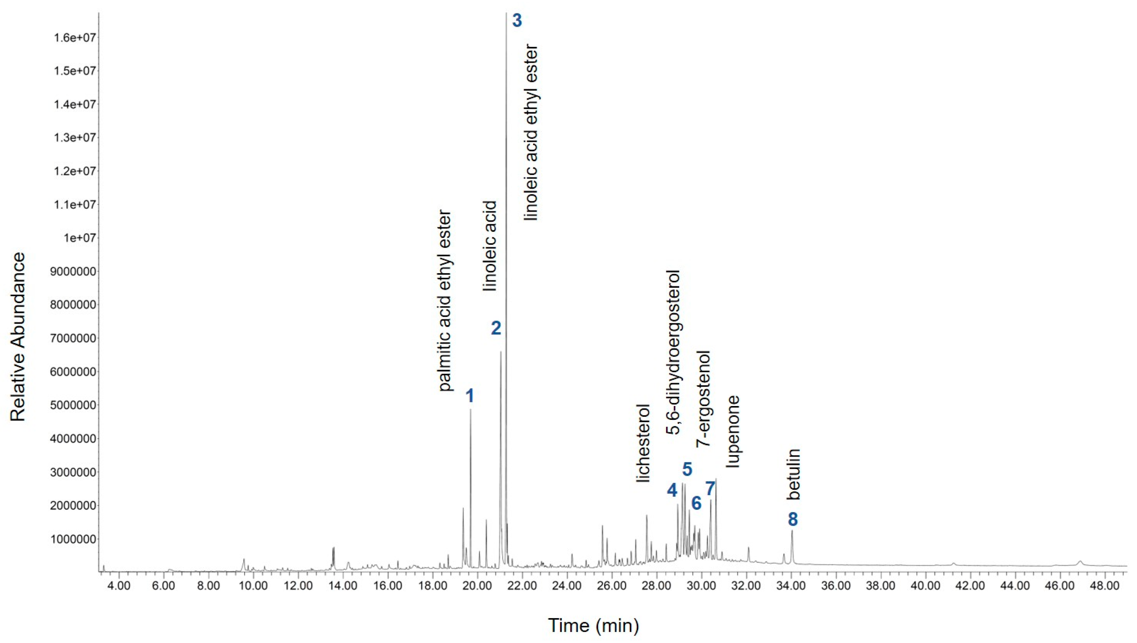
The gas chromatogram of the compounds identified in the sample of Phellinus linteus extract is demonstrated in Figure 1. The identities of eight compounds were determined, along with their retention time (Table 1). The compounds identified based on the gas chromatography–mass spectrometry (GC/MS) analysis include palmitic acid ethyl ester, linoleic acid, linoleic acid ethyl ester, lichesterol, 5,6-dihydroergosterol, 7-ergostenol, lupenone, and betulin (Table 1).

2.2. Effect of Phellinus linteus Extract on Agonist-Induced Contraction in Mesenteric Arteries of Rats

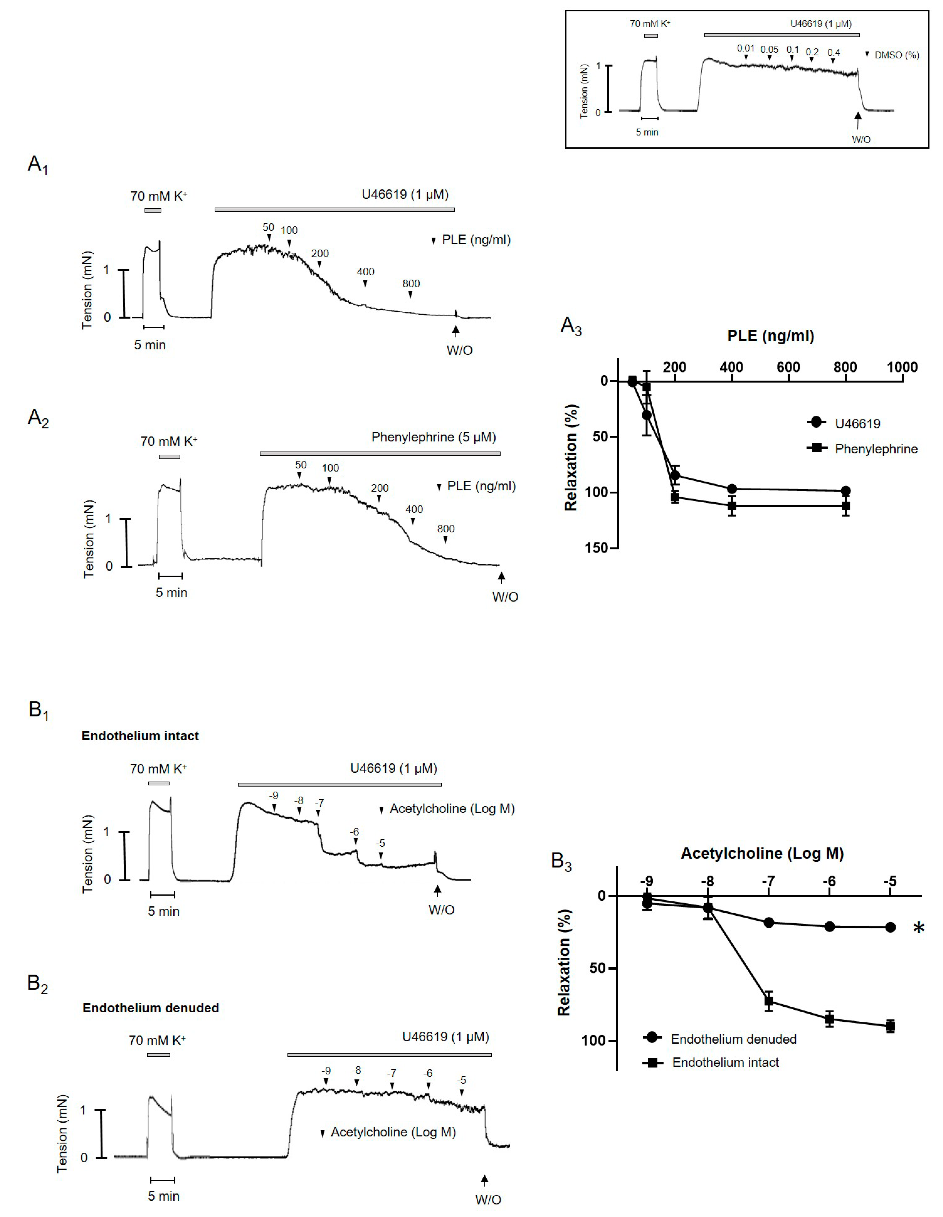
Phellinus linteus extract induced relaxation in a dose-dependent manner in the rats’ mesenteric arteries pre-contracted with U46619 (1 μM) and phenylephrine (5 μM) (Figure 2(A1,A2)). There was no difference in vasodilatory effect of Phellinus linteus extract between U46619- and phenylephrine-induced contraction (Figure 2(A3)). The vehicle, dimethyl dulfoxide (DMSO, maximum of 0.4%) had no significant effect on the U46619-induced contraction (Figure 2 inset). To compare the effect of Phellinus linteus extract with another vasodilator, aceylcholine was administered in a U46619-induced contraction (Figure 3). Acetylcholine induced dose-dependent relaxation in a U46619-induced contraction in endothelium-intact mesenteric arteries (Figure 3(B1)), which was significantly abolished by endothelium removal (Figure 3(B2,B3)). These results suggested that Phellinus linteus extract can act as a vasodilator in the mesenteric arteries of rats.

2.3. Phellinus linteus Extract-Induced Endothelium-Independent Relaxation

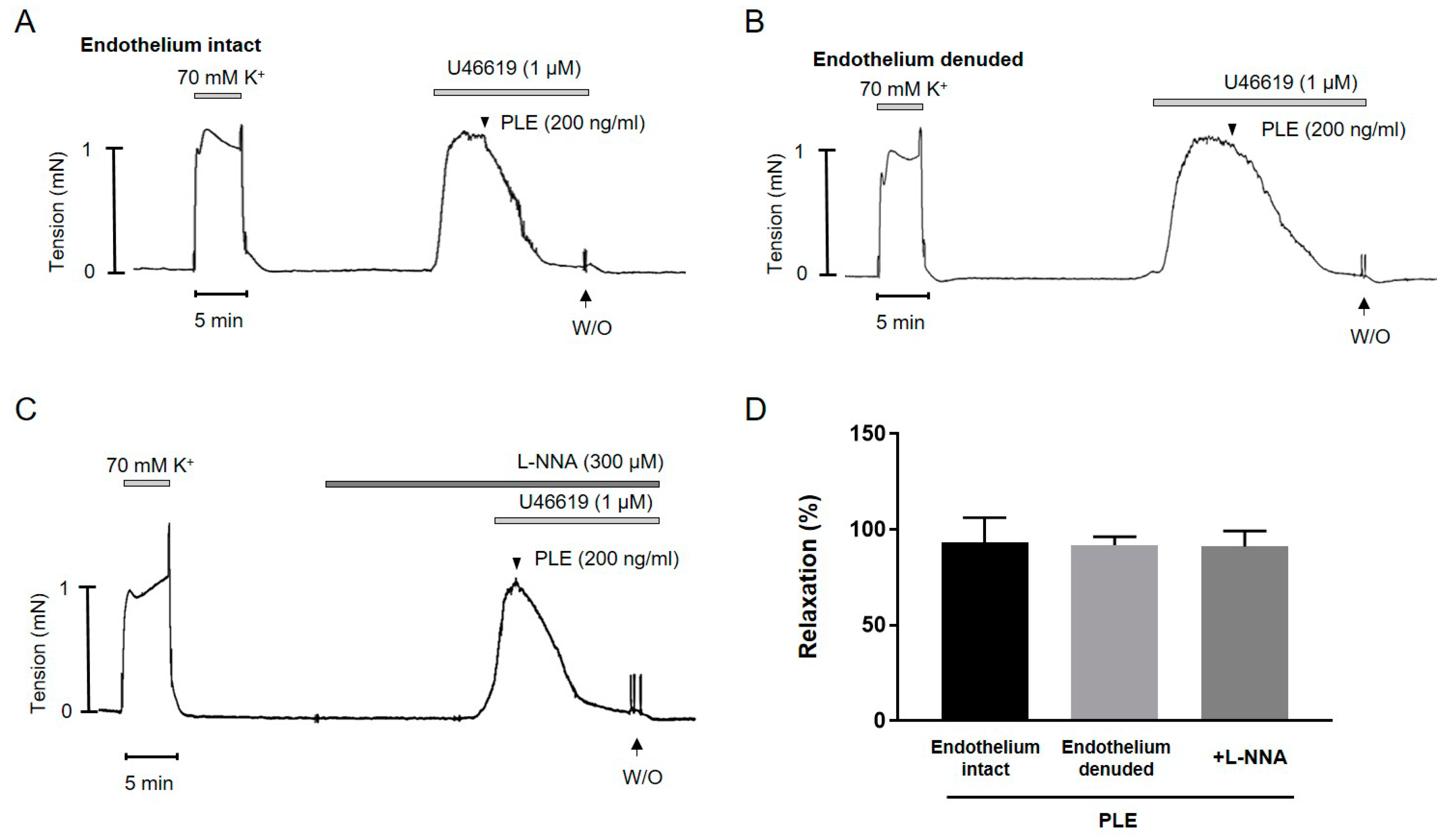
To investigate the underlying mechanisms of Phellinus linteus extract-induced relaxation, Phellinus linteus extract was applied in endothelium-intact and endothelium-denuded mesenteric arteries (Figure 3A,B). There was no significant difference between endothelium-intact and endothelium-denuded mesenteric arteries. To confirm the effect of Phellinus linteus extract on the endothelium, the mesenteric arteries were pre-incubated with the endothelial nitric oxide synthase (eNOS) inhibitor nomega–nitro–l–arginine (l–NNA, 300 μM) for 20 min before being contracted with U46619 (1 μM, Figure 3C). The l–NNA did not affect the Phellinus linteus extract-induced relaxation, indicating that the relaxation effect of Phellinus linteus extract was not related to the endothelium. This result suggests that Phellinus linteus extract-induced relaxation is endothelium-independent.

2.4. Inhibition of Phellinus linteus Extract-Induced Relaxation by K+ Channel Blockers

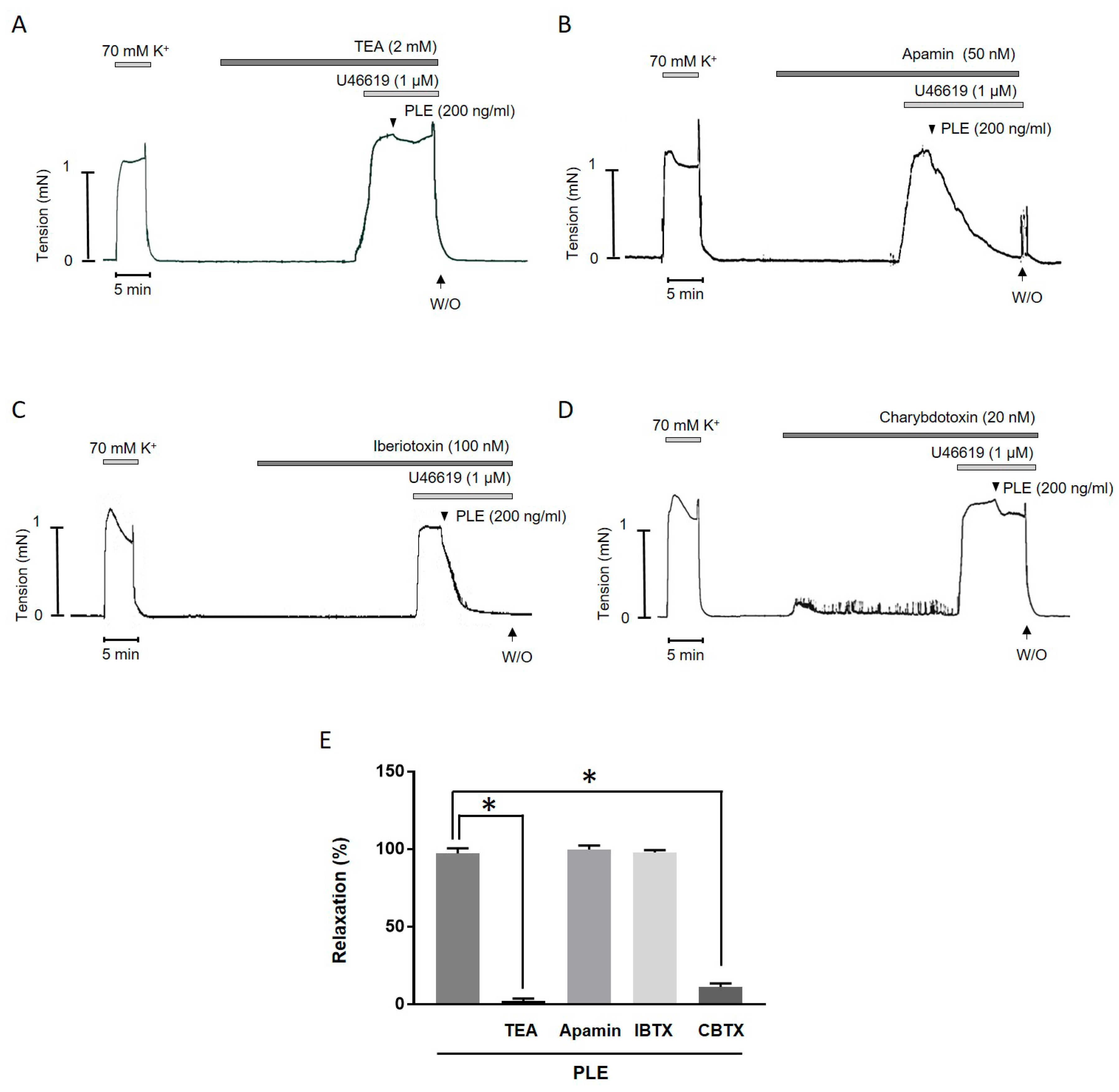
To clarify the underlying mechanisms of the Phellinus linteus extract-induced relaxation, the mesenteric arteries were incubated with tetraethylammonium (TEA, 2 mM, Figure 4A), apamin (50 nM, Figure 4B), iberiotoxin (100 nM, Figure 4C), or charybdotoxin (20 nM, Figure 4D) for 20 min, and then Phellinus linteus extract was added. The non-selective K+ channel blocker, TEA, and the IKca blocker, charybdotoxin, significantly inhibited Phellinus linteus extract-induced relaxation, while the SKca channel blocker, the apamin, and the BKca channel blocker iberiotoxin did not affect the Phellinus linteus extract-induced vasodilation (Figure 4E). These results indicate that the IKca channel is involved in the relaxation induced by Phellinus linteus extract.

2.5. Effect of Phellinus linteus Extract on the Membrane Potential and Phosphorylation of 20 KDa Myosin Light Chain (MLC20)

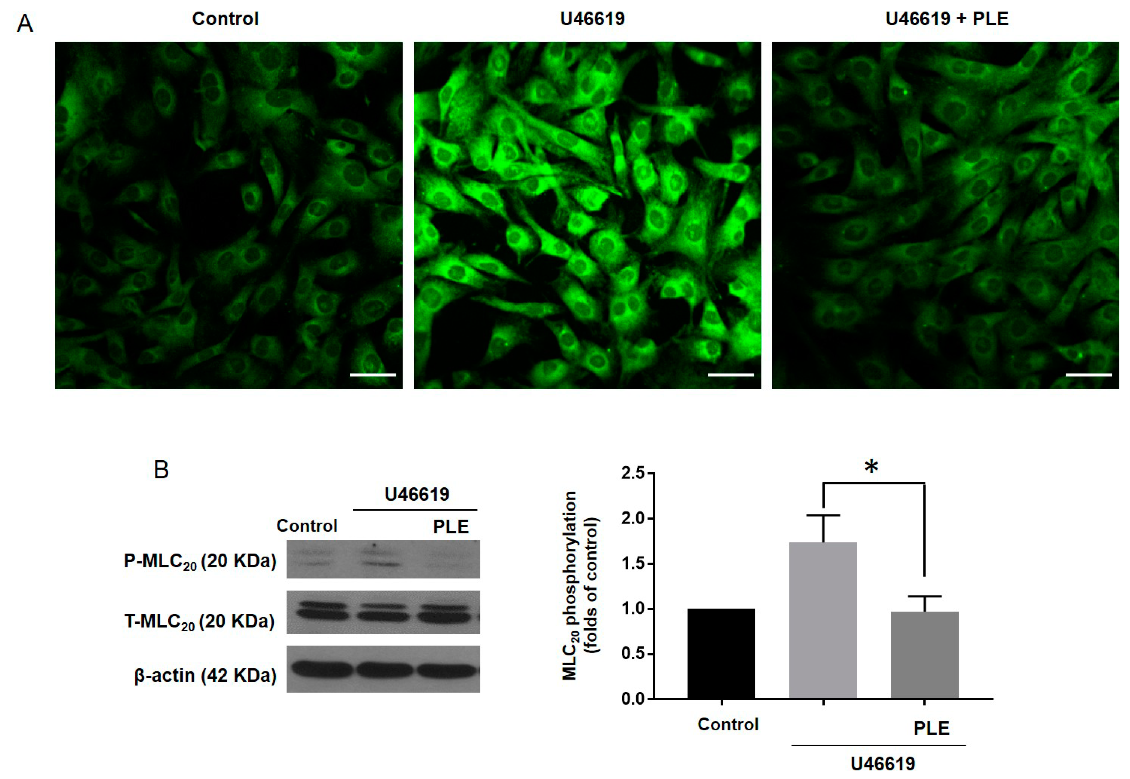
To clarify whether Phellinus linteus extract-induced relaxation was produced by hyperpolarization in VSMCs, we measured the membrane potential using the voltage sensitive dye bis-(1,3-dibarbituric acid)-trimethine oxanol (DiBAC4(3)) and obtained confocal images. The application of U46619 (1 µM) increased the fluorescence intensity of the membrane potential in VSMCs compared to the control group. In the presence of Phellinus linteus extract (200 ng/mL), U46619 did not increase the fluorescence intensity of the membrane potential in the VSMCs (Figure 5A). To investigate whether the Phellinus linteus extract-induced relaxation was caused by the decreased phosphorylation of MLC20, we measured the phosphorylation and the expression level of MLC20 in the VSMCs (Figure 5). The administration of U46619 increased the phosphorylation level of MLC20 in the VSMCs compared to the control group. In the presence of Phellinus linteus extract, U46619 did not increase the phosphorylation level of MLC20 (Figure 5B). These results suggested that Phellinus linteus extract induced hyperpolarization and inhibited the subsequent phosphorylation of MLC20 in U46619-treated VSMCs.

3. Discussion

In this study, we investigated the vasodilatory effect of Phellinus linteus extract on the mesenteric arteries of rats. We found that this effect was independent of the endothelium. The mechanism of vasodilation induced by the Phellinus linteus extract involved the IKCa channel. The non-selective K+ channel blocker TEA and the specific IKCa channel blocker charybdotoxin inhibited the relaxation induced by the Phellinus linteus extract. We also found that the Phellinus linteus extract induced hyperpolarization of the VSMCs, which caused a decrease of the phosphorylated MLC20 and subsequent vasodilation of the mesenteric arteries.
In several experimental models, it has been reported that Phellinus linteus extract has several strong biological activities, such as anti-oxidative, immune-modulating, hypoglycemic, and hepatoprotective effect [18,19,20,21]. However, the vascular effect of Phellinus linteus extract has not been assessed. Thus, the present study is the first investigation to explore the vascular effect of this extract in resistance arteries. The vasodilatory effect of Phellinus linteus extract remained in endothelium-denuded mesenteric arteries of rats and in endothelium-intact arteries in the presence of l–NNA. Thus, the relaxation caused by the extract was independent of the endothelium. We found in the Phellinus linteus extract various compounds, including palmitic acid ethyl ester, linoleic acid, linoleic acid ethyl ester, lichesterol, 5,6-dihydroergosterol, 7-ergostenol, lupenone, and betulin. We assume that linoleic acid could play a critical role in the relaxation induced by Phellinus linteus extract since it has been reported that linoleic acid induced relaxation and hyperpolarization in the coronary arteries of pigs [22]. However, further studies are needed to clarify the exact compound that causes relaxation.
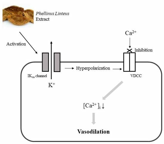
Vascular smooth muscle contraction is initiated by an elevation of intracellular Ca2+, which can result from either extracellular Ca2+ influx through calcium channels, or Ca2+ release from sarcoplasmic reticulum (SR). Extracellular Ca2+ influx via the voltage gated Ca2+ channel can be evoked by membrane depolarization. Inhibition of the K+ channels contributes to membrane depolarization. By contrast, activation of the K+ channels enables K+ efflux and leads to membrane hyperpolarization, which contributes to the closure of the voltage-dependent Ca2+ channels to block the influx of extracellular Ca2+, thereby inducing relaxation of the smooth muscle cells [23].
It is well known that at least four different types of K+ channels are expressed in VSMCs, including KCa channels, voltage-gated K+ (Kv) channels, ATP-sensitive K+ (KATP) channels, and inward-rectifier K+ channels [24]. KCa channels play a functional role by coupling the increase of intracellular Ca2+ to the hyperpolarization of the membrane potential. This feature enables KCa channels to play key roles in regulating cell excitability and K+ homeostasis [25]. The KCa channels are divided into three subfamilies that include small conductance KCa (SKCa) channels, large or big KCa (BKCa), and intermediate KCa (IKCa). It has been reported that the IKCa channel seems to be involved in the VSMCs proliferation [26] and vasodilation of porcine coronary arteries [27].
In the present study, we found that Phellinus linteus extract-induced relaxation was abolished in the presence of the non-selective K+ channel inhibitor TEA. Thus, we assumed that the K+ channel is involved in the extract-induced vasodilation. To delineate which K+ channel is involved in the Phellinus linteus extract-induced relaxation, we used specific blockers of BKca (iberiotoxin), SKca (apamin), and IKca (charybdotoxin) channels. Interestingly, charybdotoxin significantly inhibited Phellinus linteus extract-induced relaxation, while apamin and iberiotoxin did not affect the extract-induced vasodilation. Charybdotoxin is known to block several KCa channels [28], however it is also known to specifically block the IKca channel in a concentration between 20 and 300 nM [29,30,31]. Additionally, in the present study we used a low dose (20 nM) of charybdotoxin to block only the IKca channel. Thus, we assume that the IKca channel could be involved in Phellinus linteus extract-induced vasodilation. To confirm the effect of the Phellinus linteus extract, we measured the membrane potential with the voltage-sensitive dye DiBAC4(3) in the primary isolated VSMCs. The membrane potential is one of the major contributors to the contractile activity of VSMCs. The activation of the IKCa channel and subsequent K+ efflux leads to membrane hyperpolarization, which causes vasodilation. We found that Phellinus linteus extract induced hyperpolarization of VSMCs. This result is consistent with the previous report that showed that Phellinus linteus extract induced hyperpolarization in a concentration-dependent manner in monocytes [15].
The increase of intracellular Ca2+ concentration activates Ca2+/calmodulin-dependent myosin light chain kinase (MLCK) and the phosphorylation of 20 KDa myosin light chain (MLC20) and thus induces smooth muscle contraction. The decrease of intracellular Ca2+ induces dephosphorylation of the MLC20 and thus smooth muscle relaxation [32,33]. Therefore, the phosphorylation of MLC20 is considered to be a key regulation step in smooth muscle contraction and relaxation. In the present study, we found that phosphorylated MLC20 is decreased in Phellinus linteus extract-treated VSMCs. These results indicate that the relaxation induced by Phellinus linteus extract involves hyperpolarization via IKca channel activation and subsequent decrease in MLC20 phosphorylation.
In conclusion, Phellinus linteus extract induces endothelium-independent relaxation in the mesenteric arteries of rats, and this effect involves the opening of IKCa channels, thereby hyperpolarizing the VSMCs. Thus, the IKCa channel may be a cellular target for the vasodilatory effects of Phellinus linteus extract in the vasculature.

4. Methods

All experiments were performed according to the Guide for the Care and Use of Laboratory Animals, published by the US National Institutes of Health (NIH publication No. 85-23, 2011) and were approved by the Ethics Committee and the Institutional Animal Care and Use Committee of Yonsei University College of Medicine (Approval number: 2019-0278).

4.1. Phellinus linteus Extract Preparation

Dried Phellinus linteus was purchased from the Gyeong-dong medicinal herb market (Seoul, Korea). The sample (1 kg) was ground into a powder and mixed in ethanol (5 L) by shaking for 24 h at 125 rpm (1.57× g). The ethanol extract was filtered through No. 42 filter paper (Whatman International Ltd., Middlesex, UK) with five replicates, and evaporated in a rotary evaporator (Eyela, Tokyo, Japan) under reduced pressure at 37 °C.

4.2. Gas Chromatography–Mass Spectrometry (GC/MS) Analysis

GC/MS analysis was performed by an Agilent 7890B gas chromatograph, equipped with a 5977A mass selective detector quadrupole mass spectrometer system (Palo Alto, CA, USA). The DB-5 MS capillary column (30 m × 0.25 mm i.d., 0.25 μm film thickness, 5% diphenyl-95% dimethylsiloxane phase) was obtained from J&W Scientific (Folsom, CA, USA). The GC oven temperature was maintained at 60 °C for 3 min, and then ramped to 320 °C at 10 °C per min. The sample was injected in the split mode, at a splitting ratio of 1:30. The temperatures of the GC injection port and MS interface were set at 300 °C. The mass selective detector was run in the electron impact (EI) mode, with an electron energy at 70 eV. The mass spectrometer was operated in the full scan mode between 40 and 600 amu. For the identification of the compounds, EI mass spectral library search (Wiley registry 7n edition, Wiley Science Solutions, Hoboken, NJ, USA) was used.

4.3. Tissue Preparation

In this experiment, 12-week-old male Sprague Dawley rats were used. The rats were sacrificed with isoflurane (5%), followed by CO2 inhalation. To confirm death, the rats were carefully checked for several signs, such as no response to toe pinch, no palpable heartbeat, and color change opacity in the eyes. After we confirmed death, the heart was excised immediately and then the mesenteric artery beds were removed and placed in ice-cold Krebs–Henseleit (K–H) solution (composition in mM: NaCl, 119; CaCl2, 2.5; NaHCO3, 25; MgSO4, 1.2; KH2PO4, 1.2; KCl, 4.6; and glucose, 11.1). Connective tissues and adipose tissues were removed under an optical microscope (model SZ-40, Olympus, Tokyo, Japan). The second or third branches of the mesenteric arteries (200–250 μm, inner diameter) were isolated and cut into 2–3 mm segments for subsequent analysis.

4.4. Isometric Tension Recording

The mesenteric artery segments were mounted in a myograph chamber (DMT, Arhaus, Denmark) for recording of isometric tension. Briefly, two steel wires (40 µm in diameter) were inserted into the lumen of the artery and then mounted according to the methods previously described [34]. After a 30-min equilibration period in a K–H solution bubbled with 5% CO2 + 95% O2 at 37 °C, the arteries were stretched to their optimal lumen diameter for active tension development. Vessel contractility was tested by exposure to a high K+ (70 mM) solution. Where required, the endothelium was mechanically denuded by rubbing the inner surface of an arterial segment with a wire. Removal of the endothelium was confirmed by the absence of relaxation from acetylcholine (10 µM) in the U46619 (thromboxane analogue, 1 µM) pre-contracted artery. After another wash step, the rings of the mesenteric arteries were pre-contracted with U46619 (1 µM), and at the steady maximal contraction, cumulative dose-response curves were obtained for the Phellinus linteus extract. To determine the involvement of the endothelium, the arteries were pre-incubated with l–NNA (300 µM), a nitric oxide synthase inhibitor, for 20 min before being contracted with U46619 (1 μM). To determine the effects of the Kca channel blockers on vasodilation, the arterial segments were pre-treated with TEA (2 mM), iberiotoxin (100 nM), charybdotoxin (20 nM), or apamin (50 nM) for 20 min, then U46619 was administered.

4.5. Isolation and Culture of Vascular Smooth Muscle cells (VSMCs)

Vascular smooth muscle cells (VSMCs) were obtained as previously described [35]. Briefly, the aortas were excised, and fat and connective tissues were removed. And the lumen of the aorta was gently rubbed for removal of the endothelium. The aortas were cut into small segments and transferred into a tube containing elastase (0.5 mg/mL, Calbiochem, San Diego, CA, USA) and collagenase (1 mg/mL, Worthington Biomedical Corporation, Lakewood Township, NJ, USA) in Dulbecco’s Modified Eagle Medium (DMEM, Gibco, Waltham, MS, USA) at 37 °C for 30 min. After trituration and centrifugation, the cells were seeded in culture dishes (Corning, New York, NY, USA) and cultivated in DMEM supplemented with 10% fetal bovine serum (FBS, Gibco), 100 IU/mL penicillin (Gibco), and 100 μg/mL streptomycin (Gibco) at 37 °C, 5% CO2 with a humidified atmosphere. The early passage cells (between 2 and 4) were used.

4.6. Western Blot Analysis

The cultured VSMCs were frozen in liquid nitrogen after treatment with U46619, with or without Phellinus linteus extract. VSMCs were homogenized in an ice-cold lysis buffer, as described previously [36]. Western blot analysis was performed for total MLC20 and phosphorylated MLC20 (1:1000 dilution; Cell signaling, Boston, MA, USA). The blots were stripped and then reprobed with the β-actin antibody (1:2000 dilution; Santa Cruz Biotechnology, Santa Cruz, CA, USA) to verify equal loading between the samples.

4.7. Determination of Membrane Potential of Vascular Smooth Muscle Cells by Confocal Microscopy

The cells were seeded in a plate coated with poly–l–lysine for 24 h, then incubated with membrane potential-sensitive fluorescent dye DiBAC4(3) (5 μM) for 20 min. U46619 (1 μM) and Phellinus linteus extract (200 ng/mL) were added to the cells for 10 min, and the cells were washed with PBS 2–3 times. The cells were then fixed with formaldehyde (4%).

4.8. Drugs

The following drugs were used: U46619 (Tocris Bioscience, Ellisville, MO, USA), acetylcholine (Sigma-Aldrich, St Louis, MO, USA), DiBAC4(3) (Biotium, Fremont, CA, USA), and general laboratory reagents (Sigma-Aldrich, St Louis, MO, USA).

4.9. Statistical Analysis

Results were expressed as mean ± SD. One-way or two-way ANOVA was used to compare each parameter when appropriate. Comparisons between groups were performed with t-tests when the ANOVA test was statistically significant. Values of p < 0.05 were considered significant. Differences between specified groups were analyzed using the Student t test (2-tailed) for comparing two groups, with p < 0.05 considered statistically significant.

Author Contributions

All the work was done in the laboratory of Y.-H.L. in the department of physiology at Yonsei University College of Medicine. S.-K.C. designed the experiments, contributed data acquisition, and wrote the manuscript. Y.K., C.E.H., and S.B. performed the analysis. S.J.C. and D.-H.S. provided Phellinus linteus extract. Y.-H.L. contributed to the analysis and interpretation of the data and revised the work critically. All authors approved the final version of the manuscript. All persons designated as authors qualify for authorship, and all those who qualify for authorship are listed.

Funding

This work was supported by the National Research Foundation of Korea (NRF) with funding from the Ministry of Education, Science and Technology granted to S.-K.C. (2018R1D1A1B07041820) and Y.-H.L. (2019R1F1A1061771). The funders had no role in study design, data collection and analysis, decision to publish, or preparation of the manuscript.

Acknowledgments

We thank Ahn (Korea Basic Science Institute, Western Seoul Center) for helpful discussions about the data analysis.

Conflicts of Interest

The authors declare no conflicts of interest.

References

  1. Tykocki, N.R.; Boerman, E.M.; Jackson, W.F. Smooth Muscle Ion Channels and Regulation of Vascular Tone in Resistance Arteries and Arterioles. Compr. Physiol. 2017, 7, 485–581. [Google Scholar] [CrossRef] [PubMed] [Green Version]
  2. Jaminon, A.; Reesink, K.D.; A Kroon, A.; Schurgers, L.J. The Role of Vascular Smooth Muscle Cells in Arterial Remodeling: Focus on Calcification-Related Processes. Int. J. Mol. Sci. 2019, 20, 5694. [Google Scholar] [CrossRef] [Green Version]
  3. Brayden, J.E. POTASSIUM CHANNELS IN VASCULAR SMOOTH MUSCLE. Clin. Exp. Pharmacol. Physiol. 1996, 23, 1069–1076. [Google Scholar] [CrossRef] [PubMed]
  4. LeDoux, J.; Werner, M.E.; Brayden, J.E.; Nelson, M.T. Calcium-Activated Potassium Channels and the Regulation of Vascular Tone. Physiology 2006, 21, 69–78. [Google Scholar] [CrossRef] [PubMed]
  5. Chen, H.; Tian, T.; Miao, H.; Zhao, Y.-Y. Traditional uses, fermentation, phytochemistry and pharmacology of Phellinus linteus: A review. Fitoterapia 2016, 113, 6–26. [Google Scholar] [CrossRef] [PubMed]
  6. Hu, T.; Lin, Q.; Guo, T.; Yang, T.; Zhou, W.; Deng, X.; Yan, J.-K.; Luo, Y.; Ju, M.; Luo, F. Polysaccharide isolated from Phellinus linteus mycelia exerts anti-inflammatory effects via MAPK and PPAR signaling pathways. Carbohydr. Polym. 2018, 200, 487–497. [Google Scholar] [CrossRef]
  7. Kim, B.-C.; Jeon, W.-K.; Hong, H.-Y.; Jeon, K.-B.; Hahn, J.-H.; Kim, Y.-M.; Numazawa, S.; Yosida, T.; Park, E.-H.; Lim, C.-J. The anti-inflammatory activity of Phellinus linteus (Berk. & M.A. Curt.) is mediated through the PKCδ/Nrf2/ARE signaling to up-regulation of heme oxygenase-1. J. Ethnopharmacol. 2007, 113, 240–247. [Google Scholar] [CrossRef]
  8. Mei, Y.; Zhu, H.; Hu, Q.; Liu, Y.; Zhao, S.; Peng, N.; Liang, Y. A novel polysaccharide from mycelia of cultured Phellinus linteus displays antitumor activity through apoptosis. Carbohydr. Polym. 2015, 124, 90–97. [Google Scholar] [CrossRef]
  9. Chai, Y.; Wang, G.; Fan, L.; Zhao, M. A proteomic analysis of mushroom polysaccharide-treated HepG2 cells. Sci. Rep. 2016, 6, 23565. [Google Scholar] [CrossRef]
  10. Wang, H.; Wu, G.; Park, H.-J.; Jiang, P.P.; Sit, W.-H.; Van Griensven, L.J.; Wan, J.M. Protective effect of Phellinus linteus polysaccharide extracts against thioacetamide-induced liver fibrosis in rats: A proteomics analysis. Chin. Med. 2012, 7, 23. [Google Scholar] [CrossRef] [Green Version]
  11. Huang, S.C.; Wang, P.W.; Kuo, P.C.; Hung, H.Y.; Pan, T.L. Hepatoprotective Principles and Other Chemical Constituents from the Mycelium of Phellinus linteus. Molecules 2018, 23, 1705. [Google Scholar] [CrossRef] [PubMed] [Green Version]
  12. Kim, H.M.; Kang, J.S.; Kim, J.Y.; Park, S.-K.; Kim, H.S.; Lee, Y.J.; Yun, J.; Hong, J.T.; Kim, Y.; Han, S.-B. Evaluation of antidiabetic activity of polysaccharide isolated from Phellinus linteus in non-obese diabetic mouse. Int. Immunopharmacol. 2010, 10, 72–78. [Google Scholar] [CrossRef]
  13. Park, J.M.; Lee, J.S.; Song, J.E.; Sim, Y.C.; Ha, S.-J.; Hong, E.K. Cytoprotective Effect of Hispidin against Palmitate-Induced Lipotoxicity in C2C12 Myotubes. Molecules 2015, 20, 5456–5467. [Google Scholar] [CrossRef] [PubMed]
  14. Choi, D.J.; Cho, S.; Seo, J.Y.; Lee, H.B.; Park, Y.I. Neuroprotective effects of the Phellinus linteus ethyl acetate extract against H2O2-induced apoptotic cell death of SK-N-MC cells. Nutr. Res. 2016, 36, 31–43. [Google Scholar] [CrossRef] [PubMed]
  15. Van Griensven, L.J.L.D.; Verhoeven, H.A. Phellinus linteus polysaccharide extracts increase the mitochondrial membrane potential and cause apoptotic death of THP-1 monocytes. Chin. Med. 2013, 8, 25. [Google Scholar] [CrossRef] [Green Version]
  16. Lee, Y.S.; Kim, Y.H.; Shin, E.K.; Kim, D.H.; Lim, S.S.; Lee, J.-Y.; Kim, J.-K. Anti-angiogenic activity of methanol extract of Phellinus linteus and its fractions. J. Ethnopharmacol. 2010, 131, 56–62. [Google Scholar] [CrossRef]
  17. Song, Y.S.; Kim, S.-H.; Sa, J.-H.; Jin, C.; Lim, C.-J.; Park, E.-H. Anti-angiogenic, antioxidant and xanthine oxidase inhibition activities of the mushroom Phellinus linteus. J. Ethnopharmacol. 2003, 88, 113–116. [Google Scholar] [CrossRef]
  18. Shon, Y.-H.; Nam, K.-S. Inhibition of cytochrome P450 isozymes in rat liver microsomes by polysaccharides derived from Phellinus linteus. Biotechnol. Lett. 2003, 25, 167–172. [Google Scholar] [CrossRef]
  19. Fan, G.; Jian, D.; Sun, M.; Zhan, Y.; Sun, F. Endogenous and Exogenous Calcium Involved in the Betulin Production from Submerged Culture of Phellinus linteus Induced by Hydrogen Sulfide. Appl. Biochem. Biotechnol. 2015, 178, 594–603. [Google Scholar] [CrossRef]
  20. Nam, S.W.; Baek, J.T.; Kang, S.B.; Lee, D.S.; Kim, J.I.; Cho, S.H.; Park, S.-H.; Han, J.-Y.; Ahn, B.M.; Kim, J.K.; et al. A case of the hepatocellular carcinoma during the pregnancy and metastasis to the left atrium. Korean J. Hepatol. 2005, 11, 381–385. [Google Scholar]
  21. Liu, Y.; Wang, C.; Li, J.; Mei, Y.; Liang, Y. Hypoglycemic and Hypolipidemic Effects of Phellinus Linteus Mycelial Extract from Solid-State Culture in A Rat Model of Type 2 Diabetes. Nutrients 2019, 11, 296. [Google Scholar] [CrossRef] [PubMed] [Green Version]
  22. Pomposiello, S.I.; Alva, M.; Wilde, D.W.; Carretero, O.A. Linoleic acid induces relaxation and hyperpolarization of the pig coronary artery. Hypertension 1998, 31, 615–620. [Google Scholar] [CrossRef] [PubMed] [Green Version]
  23. Jackson, W.F. Potassium Channels in Regulation of Vascular Smooth Muscle Contraction and Growth. Adv. Pharmacol. 2016, 78, 89–144. [Google Scholar] [CrossRef] [PubMed] [Green Version]
  24. Cheng, J.; Wen, J.; Wang, N.; Wang, C.; Xu, Q.; Yang, Y. Ion Channels and Vascular Diseases. Arter. Thromb. Vasc. Boil. 2019, 39, e146–e156. [Google Scholar] [CrossRef]
  25. Kshatri, A.; González-Hernández, A.J.; Giraldez, T. Physiological Roles and Therapeutic Potential of Ca2+ Activated Potassium Channels in the Nervous System. Front. Mol. Neurosci. 2018, 11, 258. [Google Scholar] [CrossRef]
  26. Bi, D.; Toyama, K.; Lemaître, V.; Takai, J.; Fan, F.; Jenkins, D.P.; Wulff, H.; Gutterman, D.D.; Park, F.; Miura, H. The Intermediate Conductance Calcium-activated Potassium Channel KCa3.1 Regulates Vascular Smooth Muscle Cell Proliferation via Controlling Calcium-dependent Signaling. J. Biol. Chem. 2013, 288, 15843–15853. [Google Scholar] [CrossRef] [Green Version]
  27. Dalsgaard, T.; Kroigaard, C.; Misfeldt, M.; Bek, T.; Simonsen, U. Openers of small conductance calcium-activated potassium channels selectively enhance NO-mediated bradykinin vasodilatation in porcine retinal arterioles. Br. J. Pharmacol. 2010, 160, 1496–1508. [Google Scholar] [CrossRef] [Green Version]
  28. Hermann, A.; Erxleben, C. Charybdotoxin selectively blocks small Ca-activated K channels in Aplysia neurons. J. Gen. Physiol. 1987, 90, 27–47. [Google Scholar] [CrossRef]
  29. Sánchez-Carranza, O.; Torres-Rodríguez, P.; Darszon, A.; Treviño, C.L.; López-González, I. Pharmacology of hSlo3 channels and their contribution in the capacitation-associated hyperpolarization of human sperm. Biochem. Biophys. Res. Commun. 2015, 466, 554–559. [Google Scholar] [CrossRef]
  30. Logsdon, N.J.; Kang, J.; Togo, J.A.; Christian, E.P.; Aiyar, J. A Novel Gene, hKCa4, Encodes the Calcium-activated Potassium Channel in Human T Lymphocytes. J. Boil. Chem. 1997, 272, 32723–32726. [Google Scholar] [CrossRef] [Green Version]
  31. Anderson, A.J.; Harvey, A.L.; Rowan, E.G.; Strong, P.N. Effects of charybdotoxin, a blocker of Ca2+ -activated K + channels, on motor nerve terminals. Br. J. Pharmacol. 1988, 95, 1329–1335. [Google Scholar] [CrossRef] [Green Version]
  32. Ip, K.; Sobieszek, A.; Solomon, D.; Jiao, Y.; Paré, P.; Seow, C. Physical Integrity of Smooth Muscle Myosin Filaments is Enhanced by Phosphorylation of the Regulatory Myosin Light Chain. Cell. Physiol. Biochem. 2007, 20, 649–658. [Google Scholar] [CrossRef]
  33. Deng, M.; Ding, W.; Min, X.; Xia, Y. MLCK-independent phosphorylation of MLC20 and its regulation by MAP kinase pathway in human bladder smooth muscle cells. Cytoskeleton 2010, 68, 139–149. [Google Scholar] [CrossRef] [PubMed] [Green Version]
  34. Sun, J.; Yang, G.M.; Tao, T.; Wei, L.S.; Pan, Y.; Zhu, M.S. Isometric Contractility Measurement of the Mouse Mesenteric Artery Using Wire Myography. J. Vis. Exp. 2018, 20, e58064. [Google Scholar] [CrossRef] [PubMed]
  35. Choi, S.-K.; Kwon, Y.; Byeon, S.; Haam, C.E.; Lee, Y.-H. AdipoRon, adiponectin receptor agonist, improves vascular function in the mesenteric arteries of type 2 diabetic mice. PLoS ONE 2020, 15, e0230227. [Google Scholar] [CrossRef] [PubMed]
  36. Choi, S.-K.; Ahn, D.S.; Lee, Y.-H. Comparison of contractile mechanisms of sphingosylphosphorylcholine and sphingosine-1-phosphate in rabbit coronary artery. Cardiovasc. Res. 2008, 82, 324–332. [Google Scholar] [CrossRef] [PubMed] [Green Version]
Sample Availability: Not available.
Figure 1. Gas chromatogram of the compounds in Phellinus linteus extract.
Molecules 25 03160 g001
Figure 2. Phellinus linteus extract induces vasodilation in mesenteric arteries of rats. (A1A3), data showing responses to cumulative administration of Phellinus linteus (50 ng/mL–800 ng/mL) on U46619 (A1) and phenylephrine (A2)-induced contraction. Statistical analysis of the relaxation response to Phellinus linteus (A3). (B1B3), data showing responses to cumulative administration of acetylcholine (10−9 M–10−5 M) on U46619-induced contraction in endothelium intact (B1) and endothelium denuded (B2) mesenteric arteries. Statistical analysis of the relaxation response to acetylcholine (B3). Inset, representative trace showing responses to vehicle DMSO (0.01–0.4%). Mean ± SD (n = 5). * p < 0.05 for endothelium intact vs. endothelium denuded. (PLE: Phellinus linteus extract, W/O: wash out).
Molecules 25 03160 g002
Figure 3. Involvement of endothelium in Phellinus linteus extract-induced relaxation. (A) Relaxation by Phellinus linteus extract in endothelium intact mesenteric artery pre-contracted with U46619 (1 μΜ). (B) Relaxation by Phellinus linteus extract in endothelium denuded mesenteric artery pre-contracted with U46619 (1 μΜ). (C) Relaxation by Phellinus linteus extract in mesenteric artery in the presence of l–NNA (300 μM). (D) Statistical analysis of the relaxation response of Phellinus linteus extract. Relaxation of arteries is expressed as the percentage of the contraction induced by U46619 (1 μΜ). Mean ± SD. (n = 5). (l–NNA: nomega–nitro–l–arginine).
Molecules 25 03160 g003
Figure 4. Involvement of K+ channel in Phellinus linteus extract-induced relaxation. (A) Effect of Phellinus linteus extract in the mesenteric artery pre-contracted with U46619 (1 μΜ) in the presence of TEA (2 mM). (B) Effect of Phellinus linteus extract in the mesenteric artery pre-contracted with U46619 (1 μΜ) in the presence of apamin (50 nM). (C) Effect of Phellinus linteus extract in the mesenteric artery pre-contracted with U46619 (1 μΜ) in the presence of iberiotoxin (100 nM). (D) Effect of Phellinus linteus extract in the mesenteric artery pre-contracted with U46619 (1 μΜ) in the presence of charybdotoxin (20 nM). (E) Statistical analysis of the relaxation response of Phellinus linteus extract in the presence of various blockers. Relaxation of arteries is expressed as the percentage of the contraction induced by U46619 (1 μΜ). Mean ± SD (n = 5). * p < 0.05 for control versus TEA or charybdotoxin. (TEA: tetraethylammonium, IBTX: iberiotoxin, CBTX: charybdotoxin).
Molecules 25 03160 g004
Figure 5. Effect of Phellinus linteus extract on the membrane potential and phosphorylation of 20 KDa myosin light chain (MLC20). (A) Representative images of fluorescence intensity of DiBAC4(3) in control VSMCs, VSMCs treated with U46619 (1 μΜ), and VSMCs co-treated with U46619 (1 μΜ) and Phellinus linteus extract (200 ng/mL) (scale bar: 50 μm). (B) Representative western blot analysis and quantitative data for phosphorylated MLC20 (P–MLC20) and total MLC20 (T–MLC20) in control VSMCs, VSMCs treated with U46619 (1 μΜ), and VSMCs co-treated with U46619 (1 μΜ) and Phellinus linteus extract (200 ng/mL). * p < 0.05 for VSMCs treated with U46619 vs. VSMCs co-treated with U46619 and Phellinus linteus extract. (n = 4).
Molecules 25 03160 g005
Table 1. Bioactive compounds detected in Phellinus linteus extract. (PK: peak, RT: retention time).
PKRT (min)Tentative CompoundsTotal (%)
119.68palmitic acid ethyl ester3.48
221.03linoleic acid10.39
321.27linoleic acid ethyl ester15.86
428.93lichesterol2.59
529.255,6-dihydroergosterol3.26
629.697-ergostenol2.80
730.40lupenone3.24
834.03betulin2.19