Review

RNA Granules: A View from the RNA Perspective

Homewood Campus, Department of Biology, Johns Hopkins University, 3400 N. Charles Street, Baltimore, MD 21218, USA
*
Author to whom correspondence should be addressed.
Molecules 2020, 25(14), 3130; https://doi.org/10.3390/molecules25143130
Submission received: 30 May 2020 / Revised: 29 June 2020 / Accepted: 7 July 2020 / Published: 8 July 2020
(This article belongs to the Special Issue Frontiers in RNA Structure)

Abstract

:
RNA granules are ubiquitous. Composed of RNA-binding proteins and RNAs, they provide functional compartmentalization within cells. They are inextricably linked with RNA biology and as such are often referred to as the hubs for post-transcriptional regulation. Much of the attention has been given to the proteins that form these condensates and thus many fundamental questions about the biology of RNA granules remain poorly understood: How and which RNAs enrich in RNA granules, how are transcripts regulated in them, and how do granule-enriched mRNAs shape the biology of a cell? In this review, we discuss the imaging, genetic, and biochemical data, which have revealed that some aspects of the RNA biology within granules are carried out by the RNA itself rather than the granule proteins. Interestingly, the RNA structure has emerged as an important feature in the post-transcriptional control of granule transcripts. This review is part of the Special Issue in the Frontiers in RNA structure in the journal Molecules.

1. Importance of RNA Granules

The self-assembly of RNA-binding proteins (RBPs) and RNAs by phase separation into predominantly spherical, non-membrane bound condensates called RNA granules is a phenomenon observed across species. These granules form in distinct cellular locations and compartmentalize biomolecules. This compartmentalization creates cellular asymmetries, may enhance biological reactions, and promote molecular interactions required for cell growth and development. Phase separation imparts liquid-like properties to these assemblies, allowing granules to rapidly condense and dissolve depending on the environment in which they form and enables the exchange of granule components with the granule environment [1].
Research of the past few decades uncovered a variety of RNA granules. Some, such as Processing bodies (P-bodies) and stress granules (Figure 1A,B) are wide spread, while others such as germ granules (Figure 1C–F) are highly specialized and form only in a subset of cells. Given that they enrich mRNAs, RNA granules have been mainly associated with post-transcriptional regulation. Increasing evidence suggests that these condensates appear at the center of diverse biological processes and are even associated with various human diseases (reviewed in [2]). For example, P-body homeostasis appears to regulate stem cell potency by alternating the chromatin structure of these cells [3]. In addition, P-bodies are involved in post-transcriptional regulation of the huntingtin protein in Huntington’s disease [4]. Stress granules have been found to promote cancer cell survival (reviewed in [5]) and may also regulate viral replication upon infection (reviewed in [6]). In Drosophila melanogaster, mutations in germ granule components induce sterility of the progeny (reviewed in [7]) whereas mutations of P granule components in Caenorhabditis elegans induce piRNA-initiated transgenerational silencing of genes involved in RNA interference [8]. Several features are conserved among diverse RNA granules, including their morphology, how they form, and what protein families enrich in them. Despite these similarities RNA granules have distinct biological roles. Studying different RNA granules therefore not only deepens our understanding of these condensates but also provides fundamental new insights into human disease.

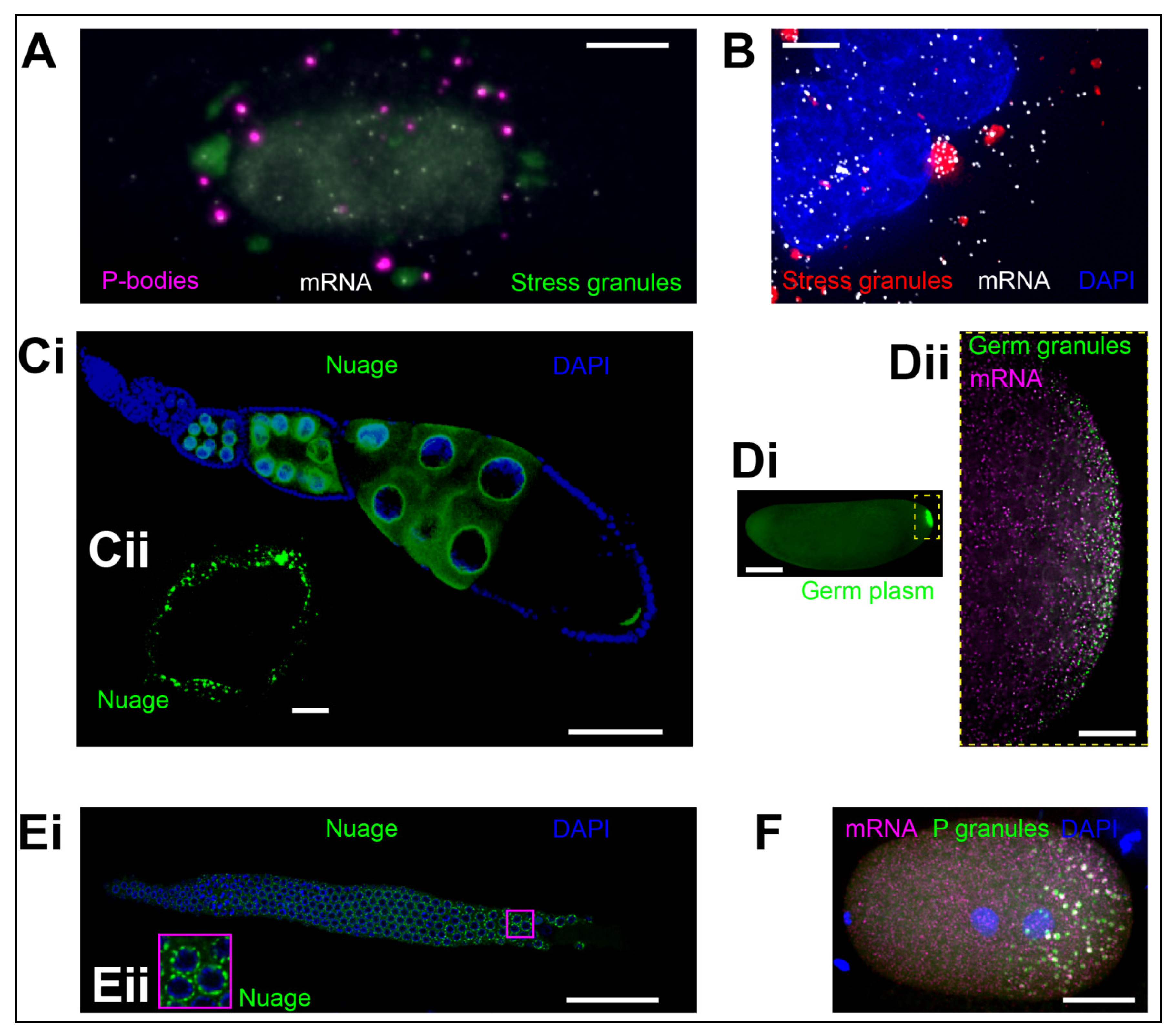
2. Types of RNA Granules and Their Functions

2.1. P-Bodies

P-bodies have been first described in yeast cells in 2003 [9] and have since been intensely studied. Several observations indicated that these RNA granules could be primary sites of mRNA degradation. First, purification and imaging of P-bodies revealed that most proteins residing within them are involved in mRNA decay (i.e., XRN1, DCP1), mRNA surveillance (i.e., UPF1, SMG5), translation regulation (i.e., eIF4E, DDX6), and miRNA pathways (i.e., AGO1 and AGO2) (reviewed in [10,11]). Second, inhibiting mRNA decay by blocking XRN1, a highly conserved eukaryotic exoribonuclease responsible for the 5′-3′ degradation of cytoplasmic mRNAs [12], increases the size and the number of P-bodies, as well as the decay intermediates within P-bodies [9,13,14]. Third, P-bodies are not observed when deadenylation, the first step in mRNA decay pathway is blocked, but are restored after this blockage is removed [15].
P-bodies have long been seen as one of the cornerstones of RNA biology capable of organizing reactions fundamental to the physiology of a cell. However, a closer look challenges this view. Live cell imaging of single, fluorescently-labeled mRNAs revealed that in human epithelial (HeLa) cells mRNAs that enter P-bodies do not necessarily degrade in them [16]. Furthermore, a transcriptome-wide analysis revealed that 5′ truncated RNAs are mostly absent from purified P-bodies [17] and that kinetics of degradation and fates of different mRNAs within P-bodies vary such that some transcripts are degraded whereas others remain stably enriched in P-bodies [18,19]. In some instances, mRNAs that accumulate in P-bodies appear to be better protected from decay rather than being subjected to it [18]. Perhaps most surprising is the observation that most transcripts decay just as likely within granules as they do outside [16,17,20,21]. Thus, the P-body formation appears to be a consequence rather than a cause of RNA-mediated silencing [20]. Indeed, readily observable P-bodies do not form in budding yeast under normal conditions without a notable effect on mRNA turnover and instead appear when cells enter stationary growth or when they experience nutrient deprivation [13]. In addition, inhibition of mRNA decay induces P-body formation in vivo indicating that undegraded transcripts are necessary for their formation [13], further questioning the idea that P-bodies are required for mRNA turnover.
P-bodies could instead simply store transcripts and direct their flow through a translational cycle. For example, mRNAs appear to move between the polysome-depleted, P-body-enriched state and polysome-bound, cytoplasmic state [17,22,23,24,25,26]. This observation raises a possibility that P-bodies could facilitate translation re-initiation of repressed mRNAs by releasing them into the translation pool upon removal of cellular stress. However, this idea has similarly been challenged by demonstrating that decay rates of mRNAs, which accumulate in P-bodies were similar to decay rates of mRNAs that remain outside [21]. Therefore, the main function of P-bodies remains elusive and could instead be dependent on the cellular contexts and environmental conditions. Lastly, and perhaps speculatively, P-bodies could simply form as a manifestation of liquid-liquid phase separation without providing a particular function in regulating RNA biology.

2.2. Stress Granules

Stress granules form when messenger ribonucleoproteins (mRNPs) become stalled in translation initiation and can be induced by various cellular stresses such as heat shock, hypoxia, endoplasmic reticulum (ER) stress, arsenite poisoning, and viral infections [27,28,29,30,31]. They contain small ribosomal subunits (40S), translation initiation factors (i.e., eIF2, eIF3, eIF4A, eIF4G), and specific (marker) RBPs such as T-cell restricted intracellular antigen 1 (TIA-1), TIA-1-related protein (TIAR), and RasGAP SH3-domain binding protein 1 (G3BP1) and 2 (G3BP2) [32,33], which are largely depleted from P-bodies [17]. Unlike P-bodies, where over 70% of the proteome comprises RBPs, almost 50% of stress granule proteins are neither RBPs nor prion-like [34]. Stress granules tend to accumulate translationally-repressed mRNAs and disassemble quickly upon stress removal [35,36]. Due to their physical association with P-bodies (Figure 1A) and their potential to store translationally-repressed transcripts, stress granules have been hypothesized to function as triage sites that store mRNAs during cellular stress and direct them towards P-bodies for degradation or back into the cytoplasm for translation once the stress is relieved [33].
Purification and single molecule imaging techniques have also been applied to investigate the regulation of mRNAs in stress granules. These techniques revealed that while 185 mRNAs strongly accumulate in stress granules, most cellular transcripts enrich only at low levels (up to 30% of a given mRNA) [21,29,37]. Furthermore, after recovery from stress, those mRNAs that accumulated in stress granules became as efficiently translated as those that remained outside, indicating that accumulation of transcripts in stress granules does not destine them for a particular translational regulation [21]. In addition, the exchange of mRNAs between stress granules and P-bodies appears to be highly infrequent [21], indicating that stress granules might not serve as triage sites for all cellular transcripts. Perhaps most surprising is the fact that at least one mRNA, ATF4 (an mRNA whose protein codes for a transcription factor that mounts a stress response in mammalian cells), translates abundantly and equally efficiently in stress granules as outside [38]. These data suggest that stress granules might not be always required for translational repression during stress. Indeed, U2OS cells lacking G3BP1 and 2, two redundant stress granule nucleators, do not form stress granules during sodium arsenite-induced oxidative stress but still exhibit stress-induced translational arrest [39]. What function these mysterious condensates perform in regulating RNA biology remains unclear.

2.3. Germ Granules

Germ granules form only in germ cells and appear inextricably linked with the germ cell biology. First described in the beetle Calligrapha punctate by Robert Hegner over 100 years ago [40], these condensates appear to regulate post-transcriptional processes to direct the germ cell fate across metazoans. Some germ granule components such as a DEAD-box RNA helicase Vasa (DDX4 in mammals and GLH-1 in C. elegans) and nanos (nos) mRNA are essential germ granule components and conserved across species (reviewed in [7,41]) while others such as Oskar, MEG-3/4, and Bucky ball, core granule proteins that nucleate germ granules in Drosophila, C. elegans, and Danio rerio, respectively are species-specific [42,43,44,45]. Germ granules are unique in that they associate with the same cell lineage throughout the development of an organism but can transform their morphology and function. For example, during early development, germ granules referred to as nuage envelop the nuclei within the germ cell lineage (Figure 1Ci,ii,Ei,ii) [46]. Nuage is thought to be involved in the biogenesis of small RNAs. It appears tightly associated with nuclear pores and both nuclear pore complex-like FG repeat components and mRNAs are found within these perinuclear germ granules [41,46,47,48]. During early embryogenesis, however, germ granules form in the germ plasm, a specialized cytoplasm that in D. melanogaster and C. elegans accumulates at the posterior pole of the early zygote (Figure 1Di,ii,F) (reviewed in [7,41]). These germ granules, also called polar granules (Drosophila) or P granules (C. elegans), enrich mRNAs whose protein products are required for the specification of the germ cell fate and thus temporally and spatially instruct the formation of the germ cell linage in these two organisms (reviewed in [7,41]).

2.3.1. Germ Granules of the Early Embryo

Super-resolution microscopy revealed that in the early Drosophila embryo, the core germ granule proteins are homogeneously mixed within granules [49] and display liquid-like as well as gel-like properties [50]. The germ plasm and its germ granules (Figure 1Di,ii) are critically required for the establishment of germ cells as strong mutations of their core proteins inhibit granule formation and give rise to sterile progeny [51,52]. These granules are also polysome-bound [53] suggesting that germ granules could be sights of translational activity. Indeed, their purification revealed that they are composed of diverse RBPs involved in translational control, mRNA turnover, as well as RNA splicing [54,55]. Nearly 200 maternally deposited mRNAs such as Cyclin B (CycB), nos, polar granule component (pgc), and germ-cell-less (gcl) enriched in these granules [56], where they translate to control the formation (gcl), specification (nos), transcriptional silencing (pgc), and division (CycB) of the primordial germ cells (reviewed in [7,57]). While direct evidence for translational control in Drosophila germ granules is lacking, genetic and imaging data indicate that mRNAs that are granule-enriched predominantly translate within granules. For instance, nos, which codes for a translational regulator Nanos required for body patterning of the early embryo, as well as proliferation and migration of germ cells (reviewed in [7]), contains an evolutionary conserved translational control element (TCE) within its 3′UTR, which folds into a well-defined two stem RNA structure [58,59]. The TCE binds Smaug, a protein that prevents translation of unlocalized nos [60]. Upon localization into germ granules, Oskar, which is almost exclusively found in germ granules [50], prevents deadenylation and translational repression of nos imposed by Smaug [61]. A posterior-anterior gradient of Nanos protein thus forms, which instructs the formation of abdominal segments within the developing embryo [62,63]. In the absence of germ granules, the translational repression of nos is not relieved and the lack of Nanos causes embryonic lethality [62,63].
In contrast to polar granules of Drosophila, the protein components of P granules of the early C. elegans embryo are structured. Here, the gel-like shell formed by two intrinsically disordered proteins MEG-3 and its redundant partner MEG-4 envelopes the liquid-like core composed of PGL-1 and -3 proteins [64,65,66]. Purification of P granules identified 492 maternally-provided mRNAs as P granule-enriched, including nos-1 and 2 [67]. These two mRNAs are required for the specification of the germ cell fate in the worm [68]. However, unlike mRNAs enriched in Drosophila polar granules, mRNAs in P granules in C. elegans are translationally repressed and only translate once they become released from P granules [67]. In addition, P granules are dispensable for the establishment of germ cells in C. elegans and their removal results in only approximately 30% reduction in the fecundity of the offspring [69].

2.3.2. Nuage

Nuage is involved with the production of small, piwi protein-interacting RNAs (piRNAs) that, in Drosophila, mediate transposon silencing (reviewed in [70]) and, in C. elegans, the self/non-self recognition of germline mRNAs (reviewed in [71]). During self/non-self recognition (a mechanism that allows the worm to distinguish foreign RNAs from its own), the nuage harbors transcripts required for RNA-mediated interference. In the absence of nuage, these transcripts become dispersed through the germ cell cytoplasm where they are recognized by the piRNA machinery and degraded [8]. In addition, small 22G RNAs that arise from this piRNA-mediated degradation accumulate and feed-back into the nuclei to further suppress transcription of respective mRNAs, a phenomenon that is trans-generationally inherited [8]. Therefore, nuage appears to act as an mRNA export center that sequesters mRNAs susceptible to degradation by the piRNA machinery and protects them from piRNA-initiated transgenerational silencing [8].
In addition, different nuage functions appear to be segregated to different granules. For instance, in C. elegans, P granules, Z granules, and mutator foci associate into PZM assemblages at the nuclei of germ cells. These tri-condensate assemblages are believed to be involved in RNAi inheritance, with localized functions of mRNA surveillance, RNA marking for memory storage, and transposon surveillance in P granules, Z granules, and mutator foci, respectively [72].
Despite the different functions of P-bodies, stress granules and germ granules in post-transcriptional regulation, fundamental features are nevertheless shared among these RNA granules. Perhaps most surprising is the fact that many granule properties are instructed by the transcripts themselves rather than the granule proteins, as discussed below.

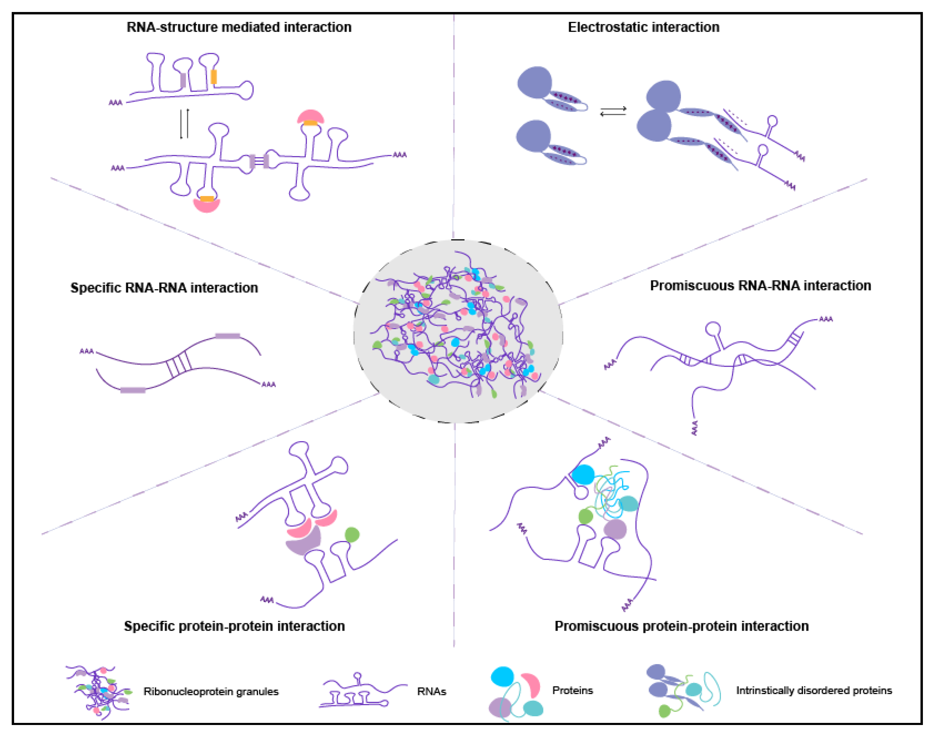
3. Formation of RNA Granules: The Role of the RNA

RNA granules form when the concentration of a protein which nucleates them reaches a critical concentration that triggers condensation [1]. Intrinsic properties of granule proteins including RNA-binding domains, intrinsically disordered regions, amino acid composition, and their capacity to engage in multiple interactions with neighboring molecules (valency) govern the physical properties of phase separation [73,74]. In addition, environmental factors such as molecular crowding, temperature, pH, and osmolarity also contribute by modulating the critical concentration required for condensation [75]. Notably, RNAs are active regulators rather than passive passengers in granule formation. In vitro, a small amount of RNA initially promotes protein droplet formation and afterwards begins dissolving the droplets once the concentration of the RNA reaches a threshold [76,77]. RNAs change physical properties of condensates (i.e., viscosity), augment granule assembly in vitro and in vivo [13,44,67,76,77,78,79,80,81,82,83,84], determine the three dimensional structure of granules in an RNA concentration-dependent manner [49,85], and can even instruct the morphology of granules by their ability to engage in intermolecular RNA-RNA interactions [86].

3.1. The Role of Long and Ribosome-Free RNAs

Long and translationally-repressed (exposed) transcripts tend to enrich in RNA granules (Table 1) [17,29,37,67,87]. These RNAs can have more conformational states [88] and could also engage in more protein-RNA interactions, promiscuous or sequence-specific intermolecular RNA-RNA base-pairing, or non-Watson-Crick interactions (Figure 2) [79,83,89,90,91,92]. Electrostatic interactions between proteins and RNA could also enhance granules formation (Figure 2). These interactions have been widely studied in viral nucleocapsid proteins, where viral proteins act as RNA chaperones that mask negative charges of nucleic acids. Recent fluorescence resonance energy transfer (FRET) experiments demonstrated that RNA chaperones act as macromolecular counterions that facilitate packing of negatively charged nucleic acids [93]. Therefore, electrostatic interactions may play a similar role in granule assembly. For example, G3BP1 RBP, a core stress granule protein [29,32,37] has three intrinsically disorder regions termed IDR1, IDR2, and IDR3. IDR1 is highly negative charged, IDR2 is slightly positively charged while IDR3 is an arginine-glycine (RG)-rich RNA-binding domain and net positively charged [94]. By using genetic mutants, Yang et al. found that IDR1 is inhibitory for stress granule formation while IDR3 augments it [94]. Since the electrostatic charges of RNAs and proteins could be shielded within tightly packed RNA condensates (see below), it is possible that the granule-associated proteins and RNAs have different conformations within and outside the granules. How RNA and protein conformations affect granule assembly and whether such interactions are mainly driven by electrostatic forces applicable to diverse granules requires further experimentation.

3.2. The Role of RNA Structure

Interestingly, the RNA structure is emerging as one of the principal regulators of phase separation (Figure 2). Highly structured RNAs tend to have more interactions with RBPs than less structured transcripts and can rearrange the composition of protein aggregates [95]. The secondary structure determines whether an mRNA will be recruited into the condensate and whether it will promote the assembly of distinct RNA granules within a cell [79]. In addition, secondary structures expose or mask the sequences for intermolecular RNA-RNA interactions [79] further influencing condensation. RNA structures can change upon nucleotide modifications, such as N6-methyladenosine (m6A), the most common mRNA modification [96]. In mouse embryonic stem cells, m6A impacts the RNA structure by inducing the transition from paired to unpaired RNA strands [97] and exposes RNA sequences to protein binding [98]. In addition, in vitro m6A enhances phase separation of proteins that bind m6A [78] indicating that RNA modifications or secondary structures these modifications induce contribute to the specificity of phase separation. Therefore, it is not surprising that mammalian stress granules enrich m6A-modified mRNAs [99,100] and that the levels of m6A modification in stress granules are approximately 50% higher than outside of granules [78]. Similarly, Drosophila of many polar granule-enriched mRNAs are similarly m6A modified [101]. Thus, it is possible that modified mRNAs may act as scaffolds to help granule proteins condense. Indeed, the YTH domain-containing family (YTHDF) proteins that bind m6A modifications enrich in mammalian stress granules and promote their formation [99]. The diverse RNA-protein and RNA-RNA interactions driven by exposed RNA sequences, structures, and modifications could create a highly interconnected molecular network that would in turn increase the stability of granules (Figure 2). Indeed, in vitro experiments and live imaging of Drosophila germ granules demonstrated that RNAs are more stably associated within granules than the granule proteins themselves [80,85]. RNAs could therefore constitute the stable component of granules capable of tuning the phase separation in cells.

4. Enrichment of RNAs to Granules: RNA Could Self-Recruit

4.1. Mechanisms of Enrichment

Enrichment of mRNAs to most RNA granules is passive. For instance, live imaging of nos in Drosophila oocytes demonstrated that this mRNA localizes to germ granules by a diffusion-entrapment mechanism and does not involve mRNA transport via molecular motors [102]. Characterization of purified RNA condensates also revealed that non-translating mRNAs and some long non-coding (lncRNAs) make up the transcriptome of stress granules and P-bodies, as well as germ granules in C. elegans (Table 1) [17,18,29,37,67,103,104]. While mRNAs in Drosophila polar granules appear to translate, a genetic analysis revealed that mRNAs recruited to Drosophila germ granules also tend to be translationally-repressed [58,59,87,105]. These data suggest that translational repression is a major determinant in the recruitment of RNAs to diverse RNA granules (Table 1).
Recently, single molecule imaging in U2OS cells provided direct evidence for these observations. Moon et al., demonstrated that translating mRNAs cannot become stably recruited to stress granules until the ribosomes have run off of the mRNA and translation has ceased [106]. These techniques also revealed that within P-bodies in U2OS cells, miRNAs are bound to their targets but when these targets were modified such that they no longer base-paired with repressive miRNAs, mRNA localization and enrichment to P-bodies decreased significantly [104]. Thus, ribosome dissociation from an mRNA is a pre-requisite for stable recruitment of most RNAs to P-bodies and stress granules [14,28,29,106,107]. However, not all translationally repressed mRNAs are recruited to stress granules [21,29] and at least one mRNA, ATF4, is stably associated with stress granules despite being translated within them [38]. This finding suggests a surprising new aspect of mRNA regulation in this mysterious RNA condensate. Similarly, translational repression correlates with RNA enrichment to germ granules in Drosophila, but it is not a requirement for enrichment. For instance, CycB is recruited to polar granules even though it translates outside of granules [108]. In addition, sucrose density gradient experiments revealed that unlocalized nanos remains associated with polysomes even though it is translationally-repressed [109] indicating that ribosome-free mRNA is not required for enrichment of nos to Drosophila polar granules. In support of this observation, deletion of the nos TCE abolishes the spatial control of translation of nos without affecting its ability to enrich in polar granules (Table 1) [59,110,111]. Finally, not all long mRNAs or long non-coding RNAs (lncRNAs) enrich in granules. Some lncRNAs interact with P-bodies only transiently and are largely depleted from the P-body transcriptome [17,104]. Thus, translational repression may be one of the mechanisms, but not a sole requirement for recruitment of RNAs to RNA granules.

4.2. Efficiency of Enrichment

RNA enrichment in condensates is an inefficient process. Up to four percent of a particular mRNA is enriched in Drosophila germ granules [49,110], which comprise about 0.01% of the embryo volume [49], while the rest of the mRNA remains dispersed through the embryo. Similarly, only a small fraction of mRNAs accumulates in P-bodies and stress granules, with the bulk remaining dispersed in the cytoplasm [21,29,37]. Additionally, the transcriptome of a particular RNA granule changes depending on the cell type or the environmental condition. For example, stress granules recruit translationally-repressed transcripts, however, the transcriptome accumulated in stress granules, which form during ER stress is somewhat different from the one that accumulates in stress granules during arsenite poisoning or heat stress [37]. In addition, during ER stress, almost 99% of mRNAs become strongly translationally repressed and yet only about 20% of these enrich in stress granules [37]. Finally, P-bodies that form in non-stressed conditions and stress granules recruit different sets of translationally-repressed mRNAs: P-body transcriptome is composed of mRNAs entirely devoid of ribosomal components while mRNAs that enrich in stress granules can still be bound by 40S ribosomal subunits (reviewed in [11]). Interestingly, during stress, P-body transcriptome shifts to resemble the transcriptome of stress granules [112], suggesting that environmental conditions can influence the mRNA composition of RNA granules. These data further support the idea that mechanisms other than translational regulation could contribute to the efficiency and the specificity of RNA recruitment to granules [8,37].

4.3. Specificity of Enrichment

Despite seeming permissiveness, mRNA enrichment to RNA granules is nevertheless highly selective. For example, in Drosophila, the 3′UTR is the major determinant of mRNA enrichment to polar granules [87,105,111], suggesting that these RNA segments carry particular sequences that mediate interaction of mRNAs with polar granules. Indeed, of the nearly 6000 genes expressed in the early Drosophila embryo [113] only up to 200 enrich at the posterior pole [56]. Similarly in C. elegans, of the 8890 genes expressed in the early embryo [114], fewer than 500 distinct mRNAs enrich in P granules [68]. In human U2OS cells, only 185 mRNAs enrich more than 50% efficiently in stress granules [29]. Interestingly, polar granule, P granule, and stress granules are composed of RBPs capable of interacting indiscriminately with the majority of cellular transcripts [50,54,115] and therefore, individually, these RBPs cannot provide the specificity for mRNA enrichment observed in vivo. In Drosophila, in vivo crosslinking and immunoprecipitation (CLIP) assays combined with in vitro RNA-binding analysis identified the germ granule nucleator Oskar as a non-canonical RNA binding protein capable of interacting with a few germ granule mRNAs such as nos, pgc, and gcl with a low affinity and weak sequence specificity [116,117]. Similarly, the P granule nucleator MEG-3 in C. elegans is also an RBP that interacts with mRNAs without sequence specificity and “prefers” longer transcripts with low ribosome occupancy [68]. To ensure selective mRNA recruitment, the RNA binding could be combinatorial involving multiple RBPs or could instead involve the RNA itself. Indeed, data suggests that mRNA enrichment to Drosophila germ granules and mammalian stress granules could in part occur through mRNA self-recruitment, where mRNAs anchored within granules could recruit additional mRNAs independently of granule proteins [92,118]. The mechanism for self-recruitment is unknown. However, intermolecular RNA-RNA interactions mediated by RBPs or by direct Watson-Crick base pairing could provide one mechanism. Longer, non-translating mRNAs with exposed sequences may provide more valency for RNA-protein and RNA-RNA interactions and increase the likelihood of self-recruitment. In support of this hypothesis, RNA helicases that unwind secondary structures and base-paired RNA stretches modulate RNA condensation in vitro and in vivo and affect the transcriptome of stress granules in U2OS cells in vivo [92]. RNA self-recruitment could also increase the efficiency of enrichment by eliminating the competition for entrapment by granule proteins. Thus, via self-recruitment, mRNAs could directly influence the transcriptome of RNA granules and thus the function of the granule.

4.4. Conserved RNA Properties of Diverse RNA Granules

Characteristics of RNAs enriched in P-bodies, stress granules, and germ granules (Table 1) pertain to other RNA granules as well. For example, bacterial RNP-bodies (BR-bodies) “prefer” long and non-translating mRNAs [119]. Similarly, fragile X mental-retardation protein FMRP, an mRNA-binding protein that forms granules in mammalian neurons [120,121], also selects for longer, translationally-repressed mRNAs [122]. In addition, mRNAs that encode membrane proteins use AU-rich elements located in their 3′UTRs to enrich TIS-granules (membraneless organelle associated with the endoplasmatic reticulum) [123], similar to the mRNA enrichment mechanism found in Drosophila polar granules [87,105,111]. Thus, aside from their morphology and biophysical properties, RNA enrichment principles seem conserved among diverse RNA granules.

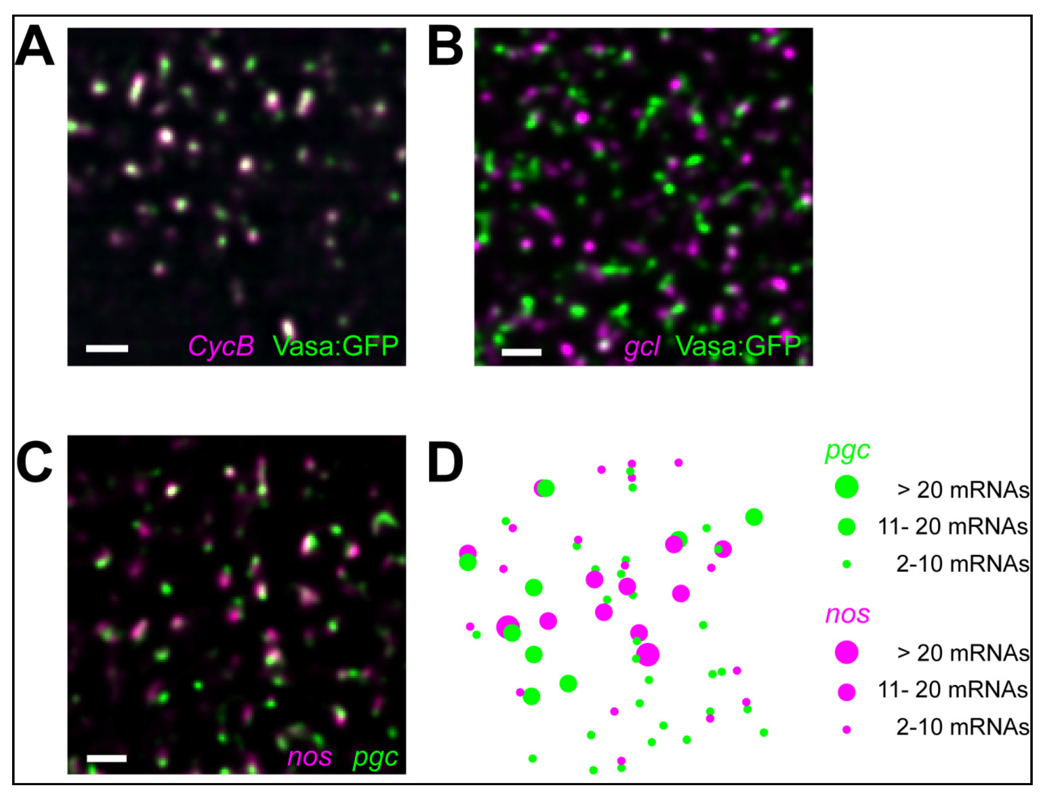
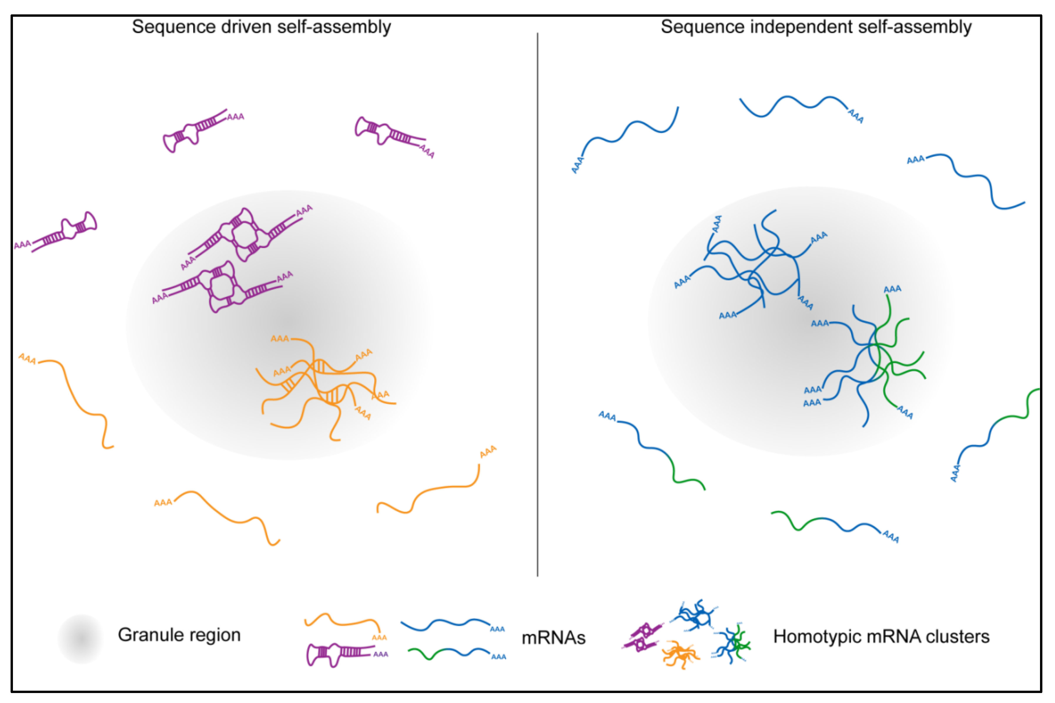
5. RNA Organization in RNA Granules: RNAs Self-Organize

5.1. Role of trans RNA-RNA Interactions

RNAs can spatially-organize independently of RBPs using intermolecular RNA-RNA interactions. Drosophila bicoid (bcd) and oskar (osk) mRNAs, the RNA genome of the human immunodeficiency virus (HIV) base-pair with each other in vitro and in vivo in RNA sequence-dependent manner [91,126,127,128,129]. Organization of bcd and osk into multi-mRNP transport particles drives their enrichment to the opposite poles of the embryo and helps establish the body plan of the developing organism. However, genetic mutations, which drive inappropriate RNA-RNA interactions cause neurodegeneration. For instance, over-expanded CUG repeats in the 3′ untranslated region (UTR) of the myotonic dystrophy protein kinase (DMPK) gene are thought to engage in multivalent base-pairing, thus driving the formation of nuclear RNA inclusions [91]. These inclusions are the causative agent of Myotonic Dystrophy 1 (DM1), driving progressive muscle weakening and premature death [130,131].
RNA assemblies can also form via RNA base stacking, which can happen both with and without the Watson-Crick base pairing [132]. Within the adjacent base-pairings, the bases may stack on each other either within the same strand or the opposite strand, possibly helping to stabilize the RNA dimerization as demonstrated for HIV dimers, bacterial Host factor-I bound RNAs as well as bicoid mRNA dimers [132,133,134]. The G-quadruplex, on the other hand, is based on the interactions among the aromatic rings of the guanines via Hoogsteen-type hydrogen bonding. Depending on the orientation of each guanine base, guanine base-stacking in the G-quadplex may alter the RNA geometry, RNA thermodynamics and translation [135,136] and also facilitate RNA phase separation both in vitro and in vivo [137,138,139].
Increasing evidence suggests that intermolecular RNA-RNA interactions also play a major role in the formation of RNA granules. Non Watson-Crick, non-specific intermolecular RNA:RNA, as well as sequence-specific intermolecular RNA-RNA interactions can promote homotypic and heterotypic RNA assemblies [79,89,90,91,92]. For instance, in vitro polyA and polyU RNAs heterotypically co-assemble while polyC and polyU de-mix into individual homotypic droplets that touch each other with a clearly defined border [92]. Thus, RNAs that are more likely to base pair with each other are also more likely recruited to the same condensate where they co-assemble [91,92]. In vitro, these base-paired RNAs adopt a rigid, highly compacted and poorly mobile architecture within the aggregate [91].
In vivo, mRNAs in Drosophila germ granules also self-assemble into homotypic clusters [85], which contain multiple mRNPs derived from the same gene (Figure 3A–D) [49,125]. Homotypic clusters from different genes co-exist and de-mix from each other within the same granule (Figure 3C,D) with more abundant clusters (those that contain multiple transcripts per cluster) residing in the center of the granule and less abundant ones residing at the granule periphery (Figure 3A,B) [49,85]. The concentration of mRNAs within homotypic assemblies is on average 4000-fold higher compared to the outside [85] indicating that mRNAs within clusters are highly compacted. Transcripts that accumulate in stress granules are also highly compacted and form a much smaller footprint within granules than in the cytoplasm [27,140]. These data suggest that localization into granules likely induces a conformational change in the RNA structure that could promote intermolecular RNA-RNA interactions allowing compaction. However, unlike polyU, A, G, and C RNAs and mRNAs with expanded repeats, which co-assemble via sequence-specific base-pairing (Figure 3D) [91,92], homotypic clusters in Drosophila germ granules self-assemble in an RNA sequence-independent manner without detectable sequence-specific intermolecular RNA-RNA interactions (Figure 4) [85]. These homotypic mRNA assemblies display a much reduced mobility compared to the proteins that form germ granules [85] raising a possibility that instead multiple sequence-independent, non-Watson-Crick, as well as RNA-protein interactions could stabilize mRNAs within a highly compacted cluster environment. Interestingly, super resolution microscopy revealed that despite an increased compaction within homotypic clusters, multiple non-specific RNA-RNA interactions are insufficient to spatially-constrict mRNAs to the same degree as a single, sequence-driven trans RNA-RNA interaction [85].

5.2. Homotypic mRNA Self-Assembly

It is unclear how mRNAs could self-assemble homotypically. In Drosophila, genetic experiments using chimeric reporters failed to identify mappable mRNP regions required to generate the specificity of homotypic assembly. Instead, small perturbations in the primary RNA sequence allowed chimeric transcripts to de-mix from the endogenous counterparts (Figure 4), arguing that a global property of the mRNP rather than a particular RNA feature alone or in combination likely provides the specificity for homotypic self-assembly in vivo [85]. In this model, mRNPs with different miscible properties specified by associated proteins, RNA modifications and structures could control de-mixing. Here, an mRNP could “read” the sum of all of its characteristics to discriminate between transcripts rather than rely on one single determinant such as an RNA fold. An implication of this model is that transcripts, which would share their primary sequences but became distinctly modified, structured or protein-bound, would also sort into distinct clusters. An average length of a Drosophila transcript is 3058 bases (estimate from BioNumbers [141]), allowing an mRNA to adopt a variety of secondary structures, carry multiple RNA modifications, and become bound by diverse proteins, all of which could influence the miscibility of an mRNA. These features could also change in response to cellular stimuli. For example, translating ribosomes could uncouple paired RNA duplexes. This is feasible given that transcripts that enrich in Drosophila germ granules translate once they become localized [87,105]. While mRNA self-assembly is not driven by granule proteins, these proteins are nevertheless critical for clustering as they provide a platform for the mRNAs to self-recognize. By entrapping transcripts and highly concentrating them, the granule proteins could act as crowding agents that increase the probability of transcripts to self-assemble and alter the properties of enriched mRNA [79]. For instance, recruitment of nucleic acids into phase separated granules melts double-stranded duplexes and therefore changes their structural properties [142], a process that could enable assembly of distinct RNA droplets within the same cell [79]. Thus, mRNA self-assembly could be a consequence of modified mRNA properties induced by granule proteins, allowing the mRNAs to organize specifically in granules and not outside.

5.3. Role of mRNA Self-Assemblies

mRNA assemblies have been observed in diverse RNA condensates, including germ granules in Drosophila [49,125] and zebrafish [143,144], P-bodies in Drosophila oocytes [145], in mammalian stress granules [29], and P-bodies [104]. Thus, homotypic assembly is an inherent property of mRNAs, revealed when they become sufficiently crowded, such as upon their enrichment in RNA condensates. Assembly could be functionally important. Just as assemblies locally increase mRNA concentration [85], they may also increase the concentration of important RNA regulators and thereby alter the post-transcriptional dynamics of granule mRNAs. For example, clustering could release mRNAs from translational repression or induce it, and even augment their translational efficiency by retaining ribosomes or translational regulators within assemblies to increase the likelihood of additional rounds of translation. Many Drosophila germ granule-enriched mRNAs are subjected to rapid degradation outside of germ granules and stabilized inside granules [113,146] and similar principles described above could also apply to increase the stability of granule-enriched transcripts. Given high compaction within granules that could promote trans RNA–RNA interactions (Figure 2) [27,85,140], mRNA assemblies could also act as structural components that would help form and maintain granules [13,80,81,89,147,148].
It is unclear why some RNA assemblies, such as those that form in RNA granules appear benign or perhaps beneficial while others, such as those manifested in DM1, cause disease. The mRNA assemblies observed in RNA granules form within a proteinaceous granule environment, which could prevent possible harmful RNA aggregation observed for toxic RNA inclusions in DM1. Indeed, RNA helicases are core components of diverse RNA granules, which could dissolve potentially harmful RNA-RNA interactions that may form within highly compacted RNA assemblies. Alternatively, the location within a cell where mRNAs assemble could also be relevant. For instance, if the granule RNAs self-assembled in the nucleus rather than in the cytoplasm, as is the case with RNA inclusions in DM1, perhaps they too could become pathogenic. Future experimentation is needed to elucidate the role of RNA self-assemblies in normal cell growth as well as in disease.

6. Outlook

Enrichment of RNAs in RNA granules provides compartmentalization of post-transcriptional processes within a cell. The role of many RNA granules in post-transcriptional regulation remains unclear and requires further investigation. Surprisingly, RNA itself regulates many aspects of its own biology within RNA granules, including its recruitment to and organization within granules as well as stabilization of granules. From this perspective, RNA granules remind us of the primordial world in which the RNA was the “working” molecule that organized fundamental reactions. Thus, intriguingly, RNA granules could be seen as the remnants of the early world, in which the RNA takes center stage and carries out important functions, while the granule proteins provide the supporting role, enabling the RNAs to do their work. As for the past few decades, these mysterious condensates continue to surprise us and undoubtedly, future discoveries will reveal fascinating and fundamental new roles of granule RNAs in post-transcriptional regulation.

Author Contributions

All authors contributed to writing and editing the manuscript. S.T. and T.T. provided the figures. All authors have read and agreed to the published version of the manuscript.

Funding

This work was supported by the NICHD K99HD088675 grant awarded to T.T.

Acknowledgments

We thank Madeline Cassani, Jeffrey Chao, Andrew Folkmann, John K. Kim, Yumi Kim, Jessica Kirshner, Stephanie Moon, Sua Myong, and Geraldine Seydoux for providing images of RNA granules and for critically reading the manuscript.

Conflicts of Interest

The authors declare no conflict of interest.

References

  1. Hyman, A.A.; Weber, C.A.; Julicher, F. Liquid-liquid phase separation in biology. Annu. Rev. Cell Dev. Biol. 2014, 30, 39–58. [Google Scholar] [CrossRef] [PubMed] [Green Version]
  2. Shukla, S.; Parker, R. Hypo- and Hyper-Assembly Diseases of RNA-Protein Complexes. Trends Mol. Med. 2016, 22, 615–628. [Google Scholar] [CrossRef] [PubMed] [Green Version]
  3. Di Stefano, B.; Luo, E.C.; Haggerty, C.; Aigner, S.; Charlton, J.; Brumbaugh, J.; Ji, F.; Rabano Jimenez, I.; Clowers, K.J.; Huebner, A.J.; et al. The RNA Helicase DDX6 Controls Cellular Plasticity by Modulating P-Body Homeostasis. Cell Stem Cell 2019, 25, 622–638 e613. [Google Scholar] [CrossRef] [PubMed] [Green Version]
  4. Savas, J.N.; Makusky, A.; Ottosen, S.; Baillat, D.; Then, F.; Krainc, D.; Shiekhattar, R.; Markey, S.P.; Tanese, N. Huntington’s disease protein contributes to RNA-mediated gene silencing through association with Argonaute and P bodies. Proc. Natl. Acad. Sci. USA 2008, 105, 10820–10825. [Google Scholar] [CrossRef] [PubMed] [Green Version]
  5. Anderson, P.; Kedersha, N.; Ivanov, P. Stress granules, P-bodies and cancer. Biochim. Biophys. Acta 2015, 1849, 861–870. [Google Scholar] [CrossRef] [PubMed] [Green Version]
  6. Valiente-Echeverria, F.; Melnychuk, L.; Mouland, A.J. Viral modulation of stress granules. Virus Res. 2012, 169, 430–437. [Google Scholar] [CrossRef] [PubMed]
  7. Trcek, T.; Lehmann, R. Germ Granules in Drosophila. Traffic 2019, 20, 650–660. [Google Scholar] [CrossRef] [Green Version]
  8. Ouyang, J.P.T.; Folkmann, A.; Bernard, L.; Lee, C.Y.; Seroussi, U.; Charlesworth, A.G.; Claycomb, J.M.; Seydoux, G. P Granules Protect RNA Interference Genes from Silencing by piRNAs. Dev. Cell 2019, 50, 716–728 e716. [Google Scholar] [CrossRef]
  9. Sheth, U.; Parker, R. Decapping and decay of messenger RNA occur in cytoplasmic processing bodies. Science 2003, 300, 805–808. [Google Scholar] [CrossRef] [Green Version]
  10. Eulalio, A.; Behm-Ansmant, I.; Izaurralde, E. P bodies: At the crossroads of post-transcriptional pathways. Nat. Rev. Mol. Cell Biol. 2007, 8, 9–22. [Google Scholar] [CrossRef]
  11. Standart, N.; Weil, D. P-Bodies: Cytosolic Droplets for Coordinated mRNA Storage. Trends Genet. 2018, 34, 612–626. [Google Scholar] [CrossRef] [PubMed]
  12. Nagarajan, V.K.; Jones, C.I.; Newbury, S.F.; Green, P.J. XRN 5′-->3′ exoribonucleases: Structure, mechanisms and functions. Biochim. Biophys. Acta 2013, 1829, 590–603. [Google Scholar] [CrossRef] [PubMed] [Green Version]
  13. Teixeira, D.; Sheth, U.; Valencia-Sanchez, M.A.; Brengues, M.; Parker, R. Processing bodies require RNA for assembly and contain nontranslating mRNAs. RNA 2005, 11, 371–382. [Google Scholar] [CrossRef] [PubMed] [Green Version]
  14. Cougot, N.; Babajko, S.; Seraphin, B. Cytoplasmic foci are sites of mRNA decay in human cells. J. Cell Biol. 2004, 165, 31–40. [Google Scholar] [CrossRef]
  15. Zheng, D.; Ezzeddine, N.; Chen, C.Y.; Zhu, W.; He, X.; Shyu, A.B. Deadenylation is prerequisite for P-body formation and mRNA decay in mammalian cells. J. Cell Biol. 2008, 182, 89–101. [Google Scholar] [CrossRef] [PubMed] [Green Version]
  16. Horvathova, I.; Voigt, F.; Kotrys, A.V.; Zhan, Y.; Artus-Revel, C.G.; Eglinger, J.; Stadler, M.B.; Giorgetti, L.; Chao, J.A. The Dynamics of mRNA Turnover Revealed by Single-Molecule Imaging in Single Cells. Mol. Cell 2017, 68, 615–625 e619. [Google Scholar] [CrossRef] [Green Version]
  17. Hubstenberger, A.; Courel, M.; Benard, M.; Souquere, S.; Ernoult-Lange, M.; Chouaib, R.; Yi, Z.; Morlot, J.B.; Munier, A.; Fradet, M.; et al. P-Body Purification Reveals the Condensation of Repressed mRNA Regulons. Mol. Cell 2017, 68, 144–157 e145. [Google Scholar] [CrossRef] [Green Version]
  18. Wang, C.; Schmich, F.; Srivatsa, S.; Weidner, J.; Beerenwinkel, N.; Spang, A. Context-dependent deposition and regulation of mRNAs in P-bodies. Elife 2018, 7, e29815. [Google Scholar] [CrossRef]
  19. Aizer, A.; Kalo, A.; Kafri, P.; Shraga, A.; Ben-Yishay, R.; Jacob, A.; Kinor, N.; Shav-Tal, Y. Quantifying mRNA targeting to P-bodies in living human cells reveals their dual role in mRNA decay and storage. J. Cell Sci. 2014, 127, 4443–4456. [Google Scholar] [CrossRef] [Green Version]
  20. Eulalio, A.; Behm-Ansmant, I.; Schweizer, D.; Izaurralde, E. P-body formation is a consequence, not the cause, of RNA-mediated gene silencing. Mol. Cell Biol. 2007, 27, 3970–3981. [Google Scholar] [CrossRef] [Green Version]
  21. Wilbertz, J.H.; Voigt, F.; Horvathova, I.; Roth, G.; Zhan, Y.; Chao, J.A. Single-Molecule Imaging of mRNA Localization and Regulation during the Integrated Stress Response. Mol. Cell 2019, 73, 946–958 e947. [Google Scholar] [CrossRef] [PubMed] [Green Version]
  22. Brengues, M.; Parker, R. Accumulation of polyadenylated mRNA, Pab1p, eIF4E, and eIF4G with P-bodies in Saccharomyces cerevisiae. Mol. Biol. Cell 2007, 18, 2592–2602. [Google Scholar] [CrossRef] [Green Version]
  23. Brengues, M.; Teixeira, D.; Parker, R. Movement of eukaryotic mRNAs between polysomes and cytoplasmic processing bodies. Science 2005, 310, 486–489. [Google Scholar] [CrossRef] [PubMed] [Green Version]
  24. Lui, J.; Castelli, L.M.; Pizzinga, M.; Simpson, C.E.; Hoyle, N.P.; Bailey, K.L.; Campbell, S.G.; Ashe, M.P. Granules harboring translationally active mRNAs provide a platform for P-body formation following stress. Cell Rep. 2014, 9, 944–954. [Google Scholar] [CrossRef] [PubMed]
  25. Patel, P.H.; Barbee, S.A.; Blankenship, J.T. GW-Bodies and P-Bodies Constitute Two Separate Pools of Sequestered Non-Translating RNAs. PLoS ONE 2016, 11, e0150291. [Google Scholar] [CrossRef] [PubMed]
  26. Kedersha, N.; Stoecklin, G.; Ayodele, M.; Yacono, P.; Lykke-Andersen, J.; Fritzler, M.J.; Scheuner, D.; Kaufman, R.J.; Golan, D.E.; Anderson, P. Stress granules and processing bodies are dynamically linked sites of mRNP remodeling. J. Cell Biol. 2005, 169, 871–884. [Google Scholar] [CrossRef] [Green Version]
  27. Adivarahan, S.; Livingston, N.; Nicholson, B.; Rahman, S.; Wu, B.; Rissland, O.S.; Zenklusen, D. Spatial Organization of Single mRNPs at Different Stages of the Gene Expression Pathway. Mol. Cell 2018, 72, 727–738 e725. [Google Scholar] [CrossRef] [Green Version]
  28. Kedersha, N.; Cho, M.R.; Li, W.; Yacono, P.W.; Chen, S.; Gilks, N.; Golan, D.E.; Anderson, P. Dynamic shuttling of TIA-1 accompanies the recruitment of mRNA to mammalian stress granules. J. Cell Biol. 2000, 151, 1257–1268. [Google Scholar] [CrossRef]
  29. Khong, A.; Matheny, T.; Jain, S.; Mitchell, S.F.; Wheeler, J.R.; Parker, R. The Stress Granule Transcriptome Reveals Principles of mRNA Accumulation in Stress Granules. Mol. Cell 2017, 68, 808–820 e805. [Google Scholar] [CrossRef]
  30. Mahboubi, H.; Stochaj, U. Cytoplasmic stress granules: Dynamic modulators of cell signaling and disease. Biochim. Biophys. Acta Mol. Basis Dis. 2017, 1863, 884–895. [Google Scholar] [CrossRef]
  31. White, J.P.; Lloyd, R.E. Regulation of stress granules in virus systems. Trends Microbiol. 2012, 20, 175–183. [Google Scholar] [CrossRef] [PubMed]
  32. Tourriere, H.; Chebli, K.; Zekri, L.; Courselaud, B.; Blanchard, J.M.; Bertrand, E.; Tazi, J. The RasGAP-associated endoribonuclease G3BP assembles stress granules. J. Cell Biol. 2003, 160, 823–831. [Google Scholar] [CrossRef] [PubMed]
  33. Kedersha, N.; Anderson, P. Stress granules: Sites of mRNA triage that regulate mRNA stability and translatability. Biochem. Soc. Trans. 2002, 30, 963–969. [Google Scholar] [CrossRef] [PubMed] [Green Version]
  34. Jain, S.; Wheeler, J.R.; Walters, R.W.; Agrawal, A.; Barsic, A.; Parker, R. ATPase-Modulated Stress Granules Contain a Diverse Proteome and Substructure. Cell 2016, 164, 487–498. [Google Scholar] [CrossRef] [PubMed] [Green Version]
  35. Wheeler, J.R.; Matheny, T.; Jain, S.; Abrisch, R.; Parker, R. Distinct stages in stress granule assembly and disassembly. Elife 2016, 5, e18413. [Google Scholar] [CrossRef]
  36. Wang, B.; Maxwell, B.A.; Joo, J.H.; Gwon, Y.; Messing, J.; Mishra, A.; Shaw, T.I.; Ward, A.L.; Quan, H.; Sakurada, S.M.; et al. ULK1 and ULK2 Regulate Stress Granule Disassembly Through Phosphorylation and Activation of VCP/p97. Mol. Cell 2019, 74, 742–757 e748. [Google Scholar] [CrossRef]
  37. Namkoong, S.; Ho, A.; Woo, Y.M.; Kwak, H.; Lee, J.H. Systematic Characterization of Stress-Induced RNA Granulation. Mol. Cell 2018, 70, 175–187 e178. [Google Scholar] [CrossRef] [Green Version]
  38. Mateju, D.; Eichenberger, B.; Eglinger, J.; Roth, G.; Chao, J.A. Single-molecule imaging reveals translation of mRNAs localized to stress granules. BioRxiv 2020. [Google Scholar] [CrossRef] [Green Version]
  39. Kedersha, N.; Panas, M.D.; Achorn, C.A.; Lyons, S.; Tisdale, S.; Hickman, T.; Thomas, M.; Lieberman, J.; McInerney, G.M.; Ivanov, P.; et al. G3BP-Caprin1-USP10 complexes mediate stress granule condensation and associate with 40S subunits. J. Cell Biol. 2016, 212, 845–860. [Google Scholar] [CrossRef] [Green Version]
  40. Hegner, R.W. Effects of removing germ-cell determinants from the eggs of some chrysomelid beetles. Preliminary Report. Biol. Bull. Mar. Biol. Lab. Woods Hole 1908, 16, 19–26. [Google Scholar] [CrossRef]
  41. Voronina, E.; Seydoux, G. The C. elegans homolog of nucleoporin Nup98 is required for the integrity and function of germline P granules. Development 2010, 137, 1441–1450. [Google Scholar] [CrossRef] [Green Version]
  42. Ephrussi, A.; Dickinson, L.K.; Lehmann, R. Oskar organizes the germ plasm and directs localization of the posterior determinant nanos. Cell 1991, 66, 37–50. [Google Scholar] [CrossRef]
  43. Markussen, F.H.; Michon, A.M.; Breitwieser, W.; Ephrussi, A. Translational Control of Oskar Generates Short Osk, the Isoform That Induces Pole Plasm Assembly. Development 1995, 121, 3723–3732. [Google Scholar] [PubMed]
  44. Smith, J.; Calidas, D.; Schmidt, H.; Lu, T.; Rasoloson, D.; Seydoux, G. Spatial patterning of P granules by RNA-induced phase separation of the intrinsically-disordered protein MEG-3. Elife 2016, 5, e21337. [Google Scholar] [CrossRef] [PubMed]
  45. Bontems, F.; Stein, A.; Marlow, F.; Lyautey, J.; Gupta, T.; Mullins, M.C.; Dosch, R. Bucky ball organizes germ plasm assembly in zebrafish. Curr. Biol. 2009, 19, 414–422. [Google Scholar] [CrossRef] [Green Version]
  46. Pitt, J.N.; Schisa, J.A.; Priess, J.R. P granules in the germ cells of Caenorhabditis elegans adults are associated with clusters of nuclear pores and contain RNA. Dev. Biol. 2000, 219, 315–333. [Google Scholar] [CrossRef] [Green Version]
  47. Updike, D.L.; Hachey, S.J.; Kreher, J.; Strome, S. P granules extend the nuclear pore complex environment in the C. elegans germ line. J. Cell Biol. 2011, 192, 939–948. [Google Scholar] [CrossRef] [Green Version]
  48. Sheth, U.; Pitt, J.; Dennis, S.; Priess, J.R. Perinuclear P granules are the principal sites of mRNA export in adult C. elegans germ cells. Development 2010, 137, 1305–1314. [Google Scholar] [CrossRef] [Green Version]
  49. Trcek, T.; Grosch, M.; York, A.; Shroff, H.; Lionnet, T.; Lehmann, R. Drosophila germ granules are structured and contain homotypic mRNA clusters. Nat. Commun. 2015, 6, 7962. [Google Scholar] [CrossRef] [Green Version]
  50. Kistler, K.E.; Trcek, T.; Hurd, T.R.; Chen, R.; Liang, F.X.; Sall, J.; Kato, M.; Lehmann, R. Phase transitioned nuclear Oskar promotes cell division of Drosophila primordial germ cells. Elife 2018, 7, e37949. [Google Scholar] [CrossRef]
  51. Arkov, A.L.; Wang, J.Y.; Ramos, A.; Lehmann, R. The role of Tudor domains in germline development and polar granule architecture. Development 2006, 133, 4053–4062. [Google Scholar] [CrossRef] [Green Version]
  52. Thomson, T.; Lasko, P. Drosophila tudor is essential for polar granule assembly and pole cell specification, but not for posterior patterning. Genesis 2004, 40, 164–170. [Google Scholar] [CrossRef] [PubMed]
  53. Mahowald, A.P. Fine Structure of Pole Cells and Polar Granules in Drosophila Melanogaster. J. Exp. Zool. 1962, 151, 201–215. [Google Scholar] [CrossRef]
  54. Voronina, E.; Seydoux, G.; Sassone-Corsi, P.; Nagamori, I. RNA Granules in Germ Cells. Cold Spring Harb. Perspect. Biol. 2011, 3, a002774. [Google Scholar] [CrossRef] [PubMed] [Green Version]
  55. Hurd, T.R.; Herrmann, B.; Sauerwald, J.; Sanny, J.; Grosch, M.; Lehmann, R. Long Oskar Controls Mitochondrial Inheritance in Drosophila melanogaster. Dev. Cell 2016, 39, 560–571. [Google Scholar] [CrossRef] [PubMed] [Green Version]
  56. Frise, E.; Hammonds, A.S.; Celniker, S.E. Systematic image-driven analysis of the spatial Drosophila embryonic expression landscape. Mol. Syst. Biol. 2010, 6, 345. [Google Scholar] [CrossRef]
  57. Lehmann, R. Germ Plasm Biogenesis-An Oskar-Centric Perspective. Curr. Top. Dev. Biol. 2016, 116, 679–707. [Google Scholar]
  58. Dahanukar, A.; Wharton, R.P. The Nanos gradient in Drosophila embryos is generated by translational regulation. Gene Dev. 1996, 10, 2610–2621. [Google Scholar] [CrossRef] [Green Version]
  59. Gavis, E.R.; Lunsford, L.; Bergsten, S.E.; Lehmann, R. A conserved 90 nucleotide element mediates translational repression of nanos RNA. Development 1996, 122, 2791–2800. [Google Scholar]
  60. Smibert, C.A.; Wilson, J.E.; Kerr, K.; Macdonald, P.M. smaug protein represses translation of unlocalized nanos mRNA in the Drosophila embryo. Genes Dev. 1996, 10, 2600–2609. [Google Scholar] [CrossRef] [Green Version]
  61. Zaessinger, S.; Busseau, I.; Simonelig, M. Oskar allows nanos mRNA translation in Drosophila embryos by preventing its deadenylation by Smaug/CCR4. Development 2006, 133, 4573–4583. [Google Scholar] [CrossRef] [Green Version]
  62. Asaoka-Taguchi, M.; Yamada, M.; Nakamura, A.; Hanyu, K.; Kobayashi, S. Maternal Pumilio acts together with Nanos in germline development in Drosophila embryos. Nat. Cell Biol. 1999, 1, 431–437. [Google Scholar] [CrossRef] [PubMed]
  63. Lehmann, R.; Nusslein-Volhard, C. The maternal gene nanos has a central role in posterior pattern formation of the Drosophila embryo. Development 1991, 112, 679–691. [Google Scholar]
  64. Putnam, A.; Cassani, M.; Smith, J.; Seydoux, G. A gel phase promotes condensation of liquid P granules in Caenorhabditis elegans embryos. Nat. Struct. Mol. Biol. 2019, 26, 220–226. [Google Scholar] [CrossRef] [PubMed]
  65. Brangwynne, C.P.; Eckmann, C.R.; Courson, D.S.; Rybarska, A.; Hoege, C.; Gharakhani, J.; Julicher, F.; Hyman, A.A. Germline P granules are liquid droplets that localize by controlled dissolution/condensation. Science 2009, 324, 1729–1732. [Google Scholar] [CrossRef] [PubMed]
  66. Hanazawa, M.; Yonetani, M.; Sugimoto, A. PGL proteins self associate and bind RNPs to mediate germ granule assembly in C. elegans. J. Cell Biol. 2011, 192, 929–937. [Google Scholar] [CrossRef] [PubMed] [Green Version]
  67. Lee, C.S.; Putnam, A.; Lu, T.; He, S.; Ouyang, J.P.T.; Seydoux, G. Recruitment of mRNAs to P granules by condensation with intrinsically-disordered proteins. Elife 2020, 9, e52896. [Google Scholar] [CrossRef]
  68. Lee, C.S.; Lu, T.; Seydoux, G. Nanos promotes epigenetic reprograming of the germline by down-regulation of the THAP transcription factor LIN-15B. Elife 2017, 6, e30201. [Google Scholar] [CrossRef]
  69. Gallo, C.M.; Wang, J.T.; Motegi, F.; Seydoux, G. Cytoplasmic partitioning of P granule components is not required to specify the germline in C. elegans. Science 2010, 330, 1685–1689. [Google Scholar] [CrossRef] [Green Version]
  70. Huang, X.; Fejes Toth, K.; Aravin, A.A. piRNA Biogenesis in Drosophila melanogaster. Trends Genet. 2017, 33, 882–894. [Google Scholar] [CrossRef] [Green Version]
  71. Kasper, D.M.; Gardner, K.E.; Reinke, V. Homeland security in the C. elegans germ line: Insights into the biogenesis and function of piRNAs. Epigenetics 2014, 9, 62–74. [Google Scholar] [CrossRef] [Green Version]
  72. Wan, G.; Fields, B.D.; Spracklin, G.; Shukla, A.; Phillips, C.M.; Kennedy, S. Spatiotemporal regulation of liquid-like condensates in epigenetic inheritance. Nature 2018, 557, 679–683. [Google Scholar] [CrossRef] [PubMed]
  73. Feric, M.; Vaidya, N.; Harmon, T.S.; Mitrea, D.M.; Zhu, L.; Richardson, T.M.; Kriwacki, R.W.; Pappu, R.V.; Brangwynne, C.P. Coexisting Liquid Phases Underlie Nucleolar Subcompartments. Cell 2016, 165, 1686–1697. [Google Scholar] [CrossRef] [Green Version]
  74. Wang, J.; Choi, J.M.; Holehouse, A.S.; Lee, H.O.; Zhang, X.; Jahnel, M.; Maharana, S.; Lemaitre, R.; Pozniakovsky, A.; Drechsel, D.; et al. A Molecular Grammar Governing the Driving Forces for Phase Separation of Prion-like RNA Binding Proteins. Cell 2018, 174, 688–699 e616. [Google Scholar] [CrossRef] [PubMed] [Green Version]
  75. Brangwynne, C.P. Phase transitions and size scaling of membrane-less organelles. J. Cell Biol. 2013, 203, 875–881. [Google Scholar] [CrossRef] [PubMed] [Green Version]
  76. Banerjee, P.R.; Milin, A.N.; Moosa, M.M.; Onuchic, P.L.; Deniz, A.A. Reentrant Phase Transition Drives Dynamic Substructure Formation in Ribonucleoprotein Droplets. Angew. Chem. Int. Ed. Engl. 2017, 56, 11354–11359. [Google Scholar] [CrossRef]
  77. Maharana, S.; Wang, J.; Papadopoulos, D.K.; Richter, D.; Pozniakovsky, A.; Poser, I.; Bickle, M.; Rizk, S.; Guillen-Boixet, J.; Franzmann, T.M.; et al. RNA buffers the phase separation behavior of prion-like RNA binding proteins. Science 2018, 360, 918–921. [Google Scholar] [CrossRef] [PubMed] [Green Version]
  78. Ries, R.J.; Zaccara, S.; Klein, P.; Olarerin-George, A.; Namkoong, S.; Pickering, B.F.; Patil, D.P.; Kwak, H.; Lee, J.H.; Jaffrey, S.R. m(6)A enhances the phase separation potential of mRNA. Nature 2019, 571, 424–428. [Google Scholar] [CrossRef]
  79. Langdon, E.M.; Qiu, Y.; Ghanbari Niaki, A.; McLaughlin, G.A.; Weidmann, C.A.; Gerbich, T.M.; Smith, J.A.; Crutchley, J.M.; Termini, C.M.; Weeks, K.M.; et al. mRNA structure determines specificity of a polyQ-driven phase separation. Science 2018, 360, 922–927. [Google Scholar] [CrossRef] [Green Version]
  80. Zhang, H.; Elbaum-Garfinkle, S.; Langdon, E.M.; Taylor, N.; Occhipinti, P.; Bridges, A.A.; Brangwynne, C.P.; Gladfelter, A.S. RNA Controls PolyQ Protein Phase Transitions. Mol. Cell 2015, 60, 220–230. [Google Scholar] [CrossRef] [Green Version]
  81. Schwartz, J.C.; Wang, X.; Podell, E.R.; Cech, T.R. RNA seeds higher-order assembly of FUS protein. Cell Rep. 2013, 5, 918–925. [Google Scholar] [CrossRef] [Green Version]
  82. Fuller, G.G.; Han, T.; Freeberg, M.A.; Moresco, J.J.; Ghanbari Niaki, A.; Roach, N.P.; Yates, J.R., 3rd; Myong, S.; Kim, J.K. RNA promotes phase separation of glycolysis enzymes into yeast G bodies in hypoxia. Elife 2020, 9, e48480. [Google Scholar] [CrossRef] [PubMed]
  83. Rhine, K.; Vidaurre, V.; Myong, S. RNA Droplets. Annu. Rev. Biophys. 2020, 49, 247–265. [Google Scholar] [CrossRef] [PubMed] [Green Version]
  84. Fernandes, N.; Buchan, J.R. RPS28B mRNA acts as a scaffold promoting cis-translational interaction of proteins driving P-body assembly. Nucleic Acids Res. 2020, 48, 6265–6279. [Google Scholar] [CrossRef] [PubMed]
  85. Trcek, T.; Douglas, T.E.; Grosch, M.; Yin, Y.; Eagle, W.V.I.; Gavis, E.R.; Shroff, H.; Rothenberg, E.; Lehmann, R. Sequence-Independent Self-Assembly of Germ Granule mRNAs into Homotypic Clusters. Mol. Cell 2020, 78, 941–950.e1. [Google Scholar] [CrossRef]
  86. Ma, W.; Zhen, G.; Xie, W.; Mayr, C. Unstructured mRNAs form multivalent RNA-RNA interactions to generate TIS granule networks. Biorxiv 2020. [Google Scholar] [CrossRef]
  87. Rangan, P.; DeGennaro, M.; Jaime-Bustamante, K.; Coux, R.X.; Martinho, R.G.; Lehmann, R. Temporal and spatial control of germ-plasm RNAs. Curr. Biol. 2009, 19, 72–77. [Google Scholar] [CrossRef] [PubMed] [Green Version]
  88. Ding, Y.; Chan, C.Y.; Lawrence, C.E. RNA secondary structure prediction by centroids in a Boltzmann weighted ensemble. RNA 2005, 11, 1157–1166. [Google Scholar] [CrossRef] [Green Version]
  89. Van Treeck, B.; Protter, D.S.W.; Matheny, T.; Khong, A.; Link, C.D.; Parker, R. RNA self-assembly contributes to stress granule formation and defining the stress granule transcriptome. Proc. Natl. Acad. Sci. USA 2018, 115, 2734–2739. [Google Scholar] [CrossRef] [Green Version]
  90. Eisenberg, H.; Felsenfeld, G. Studies of the temperature-dependent conformation and phase separation of polyriboadenylic acid solutions at neutral pH. J. Mol. Biol. 1967, 30, 17–37. [Google Scholar] [CrossRef]
  91. Jain, A.; Vale, R.D. RNA phase transitions in repeat expansion disorders. Nature 2017, 546, 243–247. [Google Scholar] [CrossRef] [Green Version]
  92. Tauber, D.; Tauber, G.; Khong, A.; Van Treeck, B.; Pelletier, J.; Parker, R. Modulation of RNA Condensation by the DEAD-Box Protein eIF4A. Cell 2020, 180, 411–426. [Google Scholar] [CrossRef]
  93. Holmstrom, E.D.; Liu, Z.; Nettels, D.; Best, R.B.; Schuler, B. Disordered RNA chaperones can enhance nucleic acid folding via local charge screening. Nat. Commun. 2019, 10, 2453. [Google Scholar] [CrossRef] [PubMed]
  94. Yang, P.; Mathieu, C.; Kolaitis, R.M.; Zhang, P.; Messing, J.; Yurtsever, U.; Yang, Z.; Wu, J.; Li, Y.; Pan, Q.; et al. G3BP1 Is a Tunable Switch that Triggers Phase Separation to Assemble Stress Granules. Cell 2020, 181, 325–345 e328. [Google Scholar] [CrossRef] [PubMed]
  95. Sanchez de Groot, N.; Armaos, A.; Grana-Montes, R.; Alriquet, M.; Calloni, G.; Vabulas, R.M.; Tartaglia, G.G. RNA structure drives interaction with proteins. Nat. Commun. 2019, 10, 3246. [Google Scholar] [CrossRef] [PubMed]
  96. Jia, G.; Fu, Y.; He, C. Reversible RNA adenosine methylation in biological regulation. Trends Genet. 2013, 29, 108–115. [Google Scholar] [CrossRef] [Green Version]
  97. Spitale, R.C.; Flynn, R.A.; Zhang, Q.C.; Crisalli, P.; Lee, B.; Jung, J.W.; Kuchelmeister, H.Y.; Batista, P.J.; Torre, E.A.; Kool, E.T.; et al. Structural imprints in vivo decode RNA regulatory mechanisms. Nature 2015, 519, 486–490. [Google Scholar] [CrossRef] [Green Version]
  98. Liu, N.; Zhou, K.I.; Parisien, M.; Dai, Q.; Diatchenko, L.; Pan, T. N6-methyladenosine alters RNA structure to regulate binding of a low-complexity protein. Nucleic Acids Res. 2017, 45, 6051–6063. [Google Scholar] [CrossRef] [Green Version]
  99. Fu, Y.; Zhuang, X. m6A-binding YTHDF proteins promote stress granule formation by modulating phase separation of stress granule proteins. BioRxiv 2020. [Google Scholar] [CrossRef] [Green Version]
  100. Anders, M.; Chelysheva, I.; Goebel, I.; Trenkner, T.; Zhou, J.; Mao, Y.; Verzini, S.; Qian, S.B.; Ignatova, Z. Dynamic m(6)A methylation facilitates mRNA triaging to stress granules. Life Sci. Alliance 2018, 1, e201800113. [Google Scholar] [CrossRef] [Green Version]
  101. Kan, L.; Grozhik, A.V.; Vedanayagam, J.; Patil, D.P.; Pang, N.; Lim, K.S.; Huang, Y.C.; Joseph, B.; Lin, C.J.; Despic, V.; et al. The m(6)A pathway facilitates sex determination in Drosophila. Nat. Commun. 2017, 8, 15737. [Google Scholar] [CrossRef]
  102. Forrest, K.M.; Clark, I.E.; Jain, R.A.; Gavis, E.R. Temporal complexity within a translational control element in the nanos mRNA. Development 2004, 131, 5849–5857. [Google Scholar] [CrossRef] [PubMed] [Green Version]
  103. Padron, A.; Iwasaki, S.; Ingolia, N.T. Proximity RNA Labeling by APEX-Seq Reveals the Organization of Translation Initiation Complexes and Repressive RNA Granules. Mol. Cell 2019, 75, 875–887 e875. [Google Scholar] [CrossRef]
  104. Pitchiaya, S.; Mourao, M.D.A.; Jalihal, A.P.; Xiao, L.; Jiang, X.; Chinnaiyan, A.M.; Schnell, S.; Walter, N.G. Dynamic Recruitment of Single RNAs to Processing Bodies Depends on RNA Functionality. Mol. Cell 2019, 74, 521–533 e526. [Google Scholar] [CrossRef] [PubMed]
  105. Gavis, E.R.; Lehmann, R. Translational regulation of nanos by RNA localization. Nature 1994, 369, 315–318. [Google Scholar] [CrossRef]
  106. Moon, S.L.; Morisaki, T.; Khong, A.; Lyon, K.; Parker, R.; Stasevich, T.J. Multicolour single-molecule tracking of mRNA interactions with RNP granules. Nat. Cell Biol. 2019, 21, 162–168. [Google Scholar] [CrossRef]
  107. Andrei, M.A.; Ingelfinger, D.; Heintzmann, R.; Achsel, T.; Rivera-Pomar, R.; Luhrmann, R. A role for eIF4E and eIF4E-transporter in targeting mRNPs to mammalian processing bodies. RNA 2005, 11, 717–727. [Google Scholar] [CrossRef] [PubMed] [Green Version]
  108. Kadyrova, L.Y.; Habara, Y.; Lee, T.H.; and Wharton, R.P. Translational control of maternal Cyclin B mRNA by Nanos in the Drosophila germline. Development 2007, 134, 1519–1527. [Google Scholar] [CrossRef] [Green Version]
  109. Clark, I.E.; Wyckoff, D.; Gavis, E.R. Synthesis of the posterior determinant Nanos is spatially restricted by a novel cotranslational regulatory mechanism. Curr. Biol. 2000, 10, 1311–1314. [Google Scholar] [CrossRef] [Green Version]
  110. Bergsten, S.E.; Gavis, E.R. Role for mRNA localization in translational activation but not spatial restriction of nanos RNA. Development 1999, 126, 659–669. [Google Scholar] [PubMed]
  111. Gavis, E.R.; Curtis, D.; Lehmann, R. Identification of cis-acting sequences that control nanos RNA localization. Dev. Biol. 1996, 176, 36–50. [Google Scholar] [CrossRef] [Green Version]
  112. Matheny, T.; Rao, B.S.; Parker, R. Transcriptome-Wide Comparison of Stress Granules and P-Bodies Reveals that Translation Plays a Major Role in RNA Partitioning. Mol. Cell Biol. 2019, 39. [Google Scholar] [CrossRef]
  113. Thomsen, S.; Anders, S.; Janga, S.C.; Huber, W.; Alonso, C.R. Genome-wide analysis of mRNA decay patterns during early Drosophila development. Genome Biol. 2010, 11, R93. [Google Scholar] [CrossRef] [PubMed] [Green Version]
  114. Baugh, L.R.; Hill, A.A.; Slonim, D.K.; Brown, E.L.; Hunter, C.P. Composition and dynamics of the Caenorhabditis elegans early embryonic transcriptome. Development 2003, 130, 889–900. [Google Scholar] [CrossRef] [PubMed] [Green Version]
  115. Zheng, J.; Gao, M.; Huynh, N.; Tindell, S.J.; Vo, H.D.; McDonald, W.H.; Arkov, A.L. In vivo mapping of a dynamic ribonucleoprotein granule interactome in early Drosophila embryos. FEBS Open Bio. 2016, 6, 1248–1256. [Google Scholar] [CrossRef] [PubMed]
  116. Jeske, M.; Bordi, M.; Glatt, S.; Muller, S.; Rybin, V.; Muller, C.W.; Ephrussi, A. The Crystal Structure of the Drosophila Germline Inducer Oskar Identifies Two Domains with Distinct Vasa Helicase- and RNA-Binding Activities. Cell Rep. 2015, 12, 587–598. [Google Scholar] [CrossRef] [PubMed] [Green Version]
  117. Yang, N.; Yu, Z.; Hu, M.; Wang, M.; Lehmann, R.; Xu, R.M. Structure of Drosophila Oskar reveals a novel RNA binding protein. Proc. Natl. Acad. Sci. USA 2015, 112, 11541–11546. [Google Scholar] [CrossRef] [Green Version]
  118. Niepielko, M.G.; Eagle, W.V.I.; Gavis, E.R. Stochastic Seeding Coupled with mRNA Self-Recruitment Generates Heterogeneous Drosophila Germ Granules. Curr. Biol. 2018, 28, 1872–1881 e1873. [Google Scholar] [CrossRef] [Green Version]
  119. Al-Husini, N.; Tomares, D.T.; Pfaffenberger, Z.J.; Muthunayake, N.S.; Samad, M.A.; Zuo, T.; Bitar, O.; Aretakis, J.R.; Bharmal, M.M.; Gega, A.; et al. BR-Bodies Provide Selectively Permeable Condensates that Stimulate mRNA Decay and Prevent Release of Decay Intermediates. Mol. Cell 2020, 78, 670–682 e678. [Google Scholar] [CrossRef]
  120. Antar, L.N.; Dictenberg, J.B.; Plociniak, M.; Afroz, R.; Bassell, G.J. Localization of FMRP-associated mRNA granules and requirement of microtubules for activity-dependent trafficking in hippocampal neurons. Genes Brain Behav. 2005, 4, 350–359. [Google Scholar] [CrossRef]
  121. De Diego Otero, Y.; Severijnen, L.A.; van Cappellen, G.; Schrier, M.; Oostra, B.; Willemsen, R. Transport of fragile X mental retardation protein via granules in neurites of PC12 cells. Mol. Cell Biol. 2002, 22, 8332–8341. [Google Scholar] [CrossRef] [Green Version]
  122. Greenblatt, E.J.; Spradling, A.C. Fragile X mental retardation 1 gene enhances the translation of large autism-related proteins. Science 2018, 361, 709–712. [Google Scholar] [CrossRef] [PubMed] [Green Version]
  123. Ma, W.; Mayr, C. A Membraneless Organelle Associated with the Endoplasmic Reticulum Enables 3’UTR-Mediated Protein-Protein Interactions. Cell 2018, 175, 1492–1506 e1419. [Google Scholar] [CrossRef] [PubMed] [Green Version]
  124. Souquere, S.; Mollet, S.; Kress, M.; Dautry, F.; Pierron, G.; Weil, D. Unravelling the ultrastructure of stress granules and associated P-bodies in human cells. J. Cell Sci. 2009, 122, 3619–3626. [Google Scholar] [CrossRef] [Green Version]
  125. Little, S.C.; Sinsimer, K.S.; Lee, J.J.; Wieschaus, E.F.; Gavis, E.R. Independent and coordinate trafficking of single Drosophila germ plasm mRNAs. Nat. Cell Biol. 2015, 17, 558–568. [Google Scholar] [CrossRef] [PubMed] [Green Version]
  126. Ferrandon, D.; Koch, I.; Westhof, E.; Nusslein-Volhard, C. RNA-RNA interaction is required for the formation of specific bicoid mRNA 3′ UTR-STAUFEN ribonucleoprotein particles. EMBO J. 1997, 16, 1751–1758. [Google Scholar] [CrossRef] [Green Version]
  127. Jambor, H.; Brunel, C.; Ephrussi, A. Dimerization of oskar 3′ UTRs promotes hitchhiking for RNA localization in the Drosophila oocyte. RNA 2011, 17, 2049–2057. [Google Scholar] [CrossRef] [Green Version]
  128. Clever, J.L.; Wong, M.L.; Parslow, T.G. Requirements for kissing-loop-mediated dimerization of human immunodeficiency virus RNA. J. Virol. 1996, 70, 5902–5908. [Google Scholar] [CrossRef] [Green Version]
  129. Marquet, R.; Baudin, F.; Gabus, C.; Darlix, J.L.; Mougel, M.; Ehresmann, C.; Ehresmann, B. Dimerization of human immunodeficiency virus (type 1) RNA: Stimulation by cations and possible mechanism. Nucleic Acids Res. 1991, 19, 2349–2357. [Google Scholar] [CrossRef] [Green Version]
  130. Kumar, A.; Agarwal, S.; Agarwal, D.; and Phadke, S.R. Myotonic dystrophy type 1 (DM1): A triplet repeat expansion disorder. Gene 2013, 522, 226–230. [Google Scholar] [CrossRef]
  131. Mateos-Aierdi, A.J.; Goicoechea, M.; Aiastui, A.; Fernandez-Torron, R.; Garcia-Puga, M.; Matheu, A.; Lopez de Munain, A. Muscle wasting in myotonic dystrophies: A model of premature aging. Front. Aging Neurosci. 2015, 7, 125. [Google Scholar] [CrossRef]
  132. Bou-Nader, C.; Zhang, J. Structural Insights into RNA Dimerization: Motifs, Interfaces and Functions. Molecules 2020, 25, 2881. [Google Scholar] [CrossRef]
  133. Ennifar, E.; Walter, P.; Ehresmann, B.; Ehresmann, C.; Dumas, P. Crystal structures of coaxially stacked kissing complexes of the HIV-1 RNA dimerization initiation site. Nat. Struct. Biol. 2001, 8, 1064–1068. [Google Scholar] [CrossRef] [PubMed]
  134. Schulz, E.C.; Seiler, M.; Zuliani, C.; Voigt, F.; Rybin, V.; Pogenberg, V.; Mucke, N.; Wilmanns, M.; Gibson, T.J.; Barabas, O. Intermolecular base stacking mediates RNA-RNA interaction in a crystal structure of the RNA chaperone Hfq. Sci. Rep. 2017, 7, 9903. [Google Scholar] [CrossRef] [PubMed] [Green Version]
  135. Lech, C.J.; Heddi, B.; Phan, A.T. Guanine base stacking in G-quadruplex nucleic acids. Nucleic Acids Res. 2013, 41, 2034–2046. [Google Scholar] [CrossRef] [PubMed] [Green Version]
  136. Varshney, D.; Spiegel, J.; Zyner, K.; Tannahill, D.; Balasubramanian, S. The regulation and functions of DNA and RNA G-quadruplexes. Nat. Rev. Mol. Cell Biol. 2020, 1–16. [Google Scholar] [CrossRef]
  137. Zhang, Y.; Yang, M.; Duncan, S.; Yang, X.; Abdelhamid, M.A.S.; Huang, L.; Zhang, H.; Benfey, P.N.; Waller, Z.A.E.; Ding, Y. G-quadruplex structures trigger RNA phase separation. Nucleic Acids Res. 2019, 47, 11746–11754. [Google Scholar] [CrossRef]
  138. Fay, M.M.; Anderson, P.J.; Ivanov, P. ALS/FTD-Associated C9ORF72 Repeat RNA Promotes Phase Transitions In Vitro and in Cells. Cell Rep. 2017, 21, 3573–3584. [Google Scholar] [CrossRef] [Green Version]
  139. Van Treeck, B.; Parker, R. Emerging Roles for Intermolecular RNA-RNA Interactions in RNP Assemblies. Cell 2018, 174, 791–802. [Google Scholar] [CrossRef] [Green Version]
  140. Khong, A.; Parker, R. mRNP architecture in translating and stress conditions reveals an ordered pathway of mRNP compaction. J. Cell Biol. 2018, 217, 4124–4140. [Google Scholar] [CrossRef] [Green Version]
  141. Adams, M.D.; Celniker, S.E.; Holt, R.A.; Evans, C.A.; Gocayne, J.D.; Amanatides, P.G.; Scherer, S.E.; Li, P.W.; Hoskins, R.A.; Galle, R.F.; et al. The genome sequence of Drosophila melanogaster. Science 2000, 287, 2185–2195. [Google Scholar] [CrossRef] [Green Version]
  142. Nott, T.J.; Craggs, T.D.; Baldwin, A.J. Membraneless organelles can melt nucleic acid duplexes and act as biomolecular filters. Nat. Chem. 2016, 8, 569–575. [Google Scholar] [CrossRef] [PubMed]
  143. Roovers, E.F.; Kaaij, L.J.T.; Redl, S.; Bronkhorst, A.W.; Wiebrands, K.; de Jesus Domingues, A.M.; Huang, H.Y.; Han, C.T.; Riemer, S.; Dosch, R.; et al. Tdrd6a Regulates the Aggregation of Buc into Functional Subcellular Compartments that Drive Germ Cell Specification. Dev. Cell 2018, 46, 285–301 e289. [Google Scholar] [CrossRef] [Green Version]
  144. Eno, C.; Hansen, C.L.; Pelegri, F. Aggregation, segregation, and dispersal of homotypic germ plasm RNPs in the early zebrafish embryo. Dev. Dyn. 2019, 248, 306–318. [Google Scholar] [CrossRef]
  145. Weil, T.T.; Parton, R.M.; Herpers, B.; Soetaert, J.; Veenendaal, T.; Xanthakis, D.; Dobbie, I.M.; Halstead, J.M.; Hayashi, R.; Rabouille, C.; et al. Drosophila patterning is established by differential association of mRNAs with P bodies. Nat. Cell Biol. 2012, 14, 1305–1313. [Google Scholar] [CrossRef] [PubMed] [Green Version]
  146. Bashirullah, A.; Halsell, S.R.; Cooperstock, R.L.; Kloc, M.; Karaiskakis, A.; Fisher, W.W.; Fu, W.; Hamilton, J.K.; Etkin, L.D.; Lipshitz, H.D. Joint action of two RNA degradation pathways controls the timing of maternal transcript elimination at the midblastula transition in Drosophila melanogaster. EMBO J. 1999, 18, 2610–2620. [Google Scholar] [CrossRef] [PubMed]
  147. Lin, Y.; Protter, D.S.; Rosen, M.K.; Parker, R. Formation and Maturation of Phase-Separated Liquid Droplets by RNA-Binding Proteins. Mol. Cell 2015, 60, 208–219. [Google Scholar] [CrossRef] [PubMed] [Green Version]
  148. Saha, S.; Weber, C.A.; Nousch, M.; Adame-Arana, O.; Hoege, C.; Hein, M.Y.; Osborne-Nishimura, E.; Mahamid, J.; Jahnel, M.; Jawerth, L.; et al. Polar Positioning of Phase-Separated Liquid Compartments in Cells Regulated by an mRNA Competition Mechanism. Cell 2016, 166, 1572–1584 e1516. [Google Scholar] [CrossRef] [PubMed] [Green Version]
Figure 1. (A) Processing bodies (P-bodies) (magenta, marked by DDX6 fused with TagRFP) interacting with stress granules (green, marked by G3BP1 fused with GFP) in U2OS cells during stress. mRNAs (a transgenic mRNA genetically-tagged with MS2:MCP-Halo) are shown as white dots. Image: Courtesy of Jeff A. Chao (FMI). (B) Stress granules (red; marked by G3BP1 fused with GFP) accumulate AHNAK mRNA (white, hybridized with smFISH probes) in U2OS cells during stress. Nuclei are labeled with a DAPI stain. Image: Courtesy of Stephanie Moon (University of Michigan) and Roy R. Parker (University of Colorado, Boulder). (Ci,ii) Nuage (green, marked by Vasa fused with GFP) abutting the nurse cell nuclei (blue, marked by DAPI) in Drosophila oocytes. (ii) Shows a close-up of a single nucleus from a different oocyte from the one shown in (i). (Di,ii) Germ granules (polar granules) (ii) form within germ plasm (i) at the posterior pole of the early Drosophila embryo (green; marked by Vasa fused with GFP). mRNAs such as gcl (magenta, hybridized with smFISH probes) enrich in these granules. (Ei,ii) Nuage ((ii) magenta square: Close-up of three nuclei) (green, marked anti-CSR-1 antibody) abutting the C. elegans germ cell nuclei (blue, marked by DAPI). Image: Courtesy of Jessica Kirshner and John K. Kim (Johns Hopkins University). (F) mRNAs in P granules are enriched in the C. elegans one cell zygote. P granules enrich polyA RNA (green) and nos-2 mRNA (magenta), both marked with smFISH probes. DNA is stained with DAPI (blue). Image: Courtesy of Madeline Cassani, Andrew Folkmann, and Geraldine Seydoux (John Hopkins School of Medicine). Scale bar in (A,B,Cii) is 5 µm, in (Dii,F) is 10 µm, in (Ci,Ei) is 50 µm, and in (Di) is 100 µm.
Molecules 25 03130 g001
Figure 2. Proposed intermolecular interactions among RNAs and proteins within RNA granules. RNA secondary structures can mask/expose the specific RNA sequences or binding motifs to neighboring RNAs or RBPs. The electrostatic interactions may facilitate RNA-protein interactions in crowded environments, and the proteins can change the conformations when the RNA binds. In addition, intermolecular RNA-RNA and protein-protein interactions can be specific or promiscuous. Intermolecular RNA-RNA and RNA-protein interactions depicted in this model were mostly studied on mRNAs, however similar principles may apply to other RNAs enriched in RNA granules such as piRNAs, microRNAs, or lncRNAs.
Molecules 25 03130 g002
Figure 3. (A,B) CycB and gcl mRNA clusters (magenta; hybridized with smFISH probes) occupy distinct positions within Drosophila germ granules (green, labeled by Vasa fused with GFP). (C,D) Homotypic clusters contain multiple nos or pgc mRNAs (magenta and green, respectively hybridized with smFISH probes), do not have a defined stoichiometry and de-mix from each other within the same granule [85]. Scale bar in all is 1 µm.
Molecules 25 03130 g003
Figure 4. Proposed models showing the mechanisms of mRNA self-organization in RNA granules using sequence-dependent or sequence-independent assembly. The later mechanism was demonstrated for homotypic mRNA clusters in Drosophila germ granules. Here, mRNAs distinguish between endogenous mRNAs and its derivatives and de-mix to form distinct homotypic clusters [85].
Molecules 25 03130 g004
Table 1. Characteristics of RNAs enriched in RNA granules.
RNA GranuleEnriched RNAs (Examples)Depleted RNAs (Examples)Prefer Long mRNAs?Prefer Translationally Repressed mRNAs?
P-bodiesTranslationally repressed mRNAs [17]18S and 28S rRNAs [17]N/AYes
Stress granulesmRNAs with longer coding regions and 3′UTRs [29,37]
lncRNAs [29]
18S rRNA [124]
Membrane-associated mRNAs [29]
gapdh [29]
28S rRNA [124]
YesYes
P granules
(C. elegans)
Long mRNAs with low ribosome occupancy [67]
Germ-cell specific mRNAs [67]
N/AYesYes
Polar granules
(D. melanogaster)
Germ-cell specific mRNAs [49,87,125]oskar [49,125]N/AYes, but with exceptions [58,59,87,105,108,109,110,111]

