Article

Biological Activities of Selected Plants and Detection of Bioactive Compounds from Ardisia elliptica Using UHPLC-Q-Exactive Orbitrap Mass Spectrometry

by
Pei Lou Wong
1,
Nurul Azila Fauzi
1,
Siti Norhamimah Mohamed Yunus
1,
Nur Ashikin Abdul Hamid
2,
Siti Zulaikha Abd Ghafar
1,
Awanis Azizan
2,
Nur Khaleeda Zulaikha Zolkeflee
2 and
Faridah Abas
1,2,*
1
Department of Food Science, Faculty of Food Science and Technology, Universiti Putra Malaysia, Serdang 43400, Selangor, Malaysia
2
Laboratory of Natural Products, Institute of Bioscience, Universiti Putra Malaysia, Serdang 43400, Selangor, Malaysia
*
Author to whom correspondence should be addressed.
Molecules 2020, 25(13), 3067; https://doi.org/10.3390/molecules25133067
Submission received: 1 May 2020 / Revised: 4 June 2020 / Accepted: 4 June 2020 / Published: 6 July 2020
(This article belongs to the Special Issue Natural Product Pharmacology and Medicinal Chemistry II)

Abstract

:
Plants and plant-based products have been used for a long time for medicinal purposes. This study aimed to determine the antioxidant and anti-α-glucosidase activities of eight selected underutilized plants in Malaysia: Leucaena leucocephala, Muntingia calabura, Spondias dulcis, Annona squamosa, Ardisia elliptica, Cynometra cauliflora, Ficus auriculata, and Averrhoa bilimbi. This study showed that the 70% ethanolic extract of all plants exhibited total phenolic content (TPC) ranging from 51 to 344 mg gallic acid equivalent (GAE)/g dry weight. A. elliptica showed strong 2,2-diphenyl-1-picrylhydrazyl (DPPH) and nitric oxide (NO) scavenging activities, with half maximal inhibitory concentration (IC50) values of 2.17 and 49.43 μg/mL, respectively. Most of the tested plant extracts showed higher inhibition of α-glucosidase enzyme activity than the standard, quercetin, particularly A. elliptica, F. auriculata, and M. calabura extracts with IC50 values of 0.29, 0.36, and 0.51 μg/mL, respectively. A total of 62 metabolites including flavonoids, triterpenoids, benzoquinones, and fatty acids were tentatively identified in the most active plant, i.e., A. elliptica leaf extract, by using ultra-high-performance liquid chromatography (UHPLC)–electrospray ionization (ESI) Orbitrap MS. This study suggests a potential natural source of antioxidant and α-glucosidase inhibitors from A. elliptica.

1. Introduction

Malaysia is a country that is recognized for its diverse flora and fauna. Various species of plants, animals, and microorganisms offer Malaysians an extensive source of nutritious food and medicines. Furthermore, the antioxidant activities of different parts of plants, including roots, leaves, stalks, flowers, fruits, and seeds were studied. Acknowledgement of the potential medicinal benefits of local plants along with the development of modern technology motivated researchers, pharmacists, and physicians to explore Malaysian biodiversity. In addition to commonly consumed local herbs and fruits, some underutilized local species have the potential to act as alternative sources of micronutrients, vitamins, and health-promoting secondary plant metabolites [1]. These species include “petai belalang” (Leucaena leucocephala (Lam.) de Wit (Fabaceae)), “ceri hutan” (Muntingia calabura L. (Muntingiaceae)), “kedondong” (Spondias dulcis Parkinson (Anacardiaceae)), “nona” (Annona squamosal L. (Annonaceae)), “mata ayam” (Ardisia elliptica Thunb. (Primulaceae)), “katak puru” (Cynometra cauliflora L. (Fabaceae)), “ara” (Ficus auriculata Lour. (Moraceae)), “belimbing buluh” (Averrhoa bilimbi L. (Oxalidaceae)), and others. However, the biological activity and the chemical profile of these underutilized plants remain unknown. Therefore, this study was conducted to fill in the current research gap existing for these plants.
The prevalence of Malaysian adults suffering from diabetes mellitus increased from 11.6% in 2006 to 15.2% in 2011; the rate is projected to reach 21.6% by 2020 [2]. Previous studies showed correlations between oxidative stress and diabetes [3]. Human bodies rely on endogenous and exogenous antioxidants to minimize the cellular damage and stress caused by free radicals by maintaining redox balance. Bouayed and Bohn [4] stated that antioxidants from fruits, vegetables, and other sources play a significant role in assisting the endogenous antioxidant defense system, which includes superoxide dismutase, catalase, and glutathione peroxidase, in preventing oxidative stress.
Diabetic patients suffer from an abnormal increase of blood glucose level after meal ingestion, a condition commonly known as postprandial hyperglycemia. α-Glucosidase, which is located in the epithelium of the small intestine, is one of the enzymes responsible for carbohydrate digestion. Postprandial hyperglycemia can be reduced through several means such as by suppressing α-glucosidase activity, thereby delaying the carbohydrate hydrolysis and glucose absorption by the cells [5]. Triggle and Ding [6] reported that synthetic drugs, such as metformin, sulfonylureas, thiazolidinediones, and other α-glucosidase inhibitors (including acarbose and miglitol, which were introduced as treatment for diabetes and are also known for their undesirable side effects) increased cardiovascular risk and induction of hepatotoxicity. Since modern medical treatments encourage the use of plant-based functional foods and drugs, particularly in diabetes treatment, numerous studies were conducted in the quest for effective hypoglycemic agents. Kumar et al. [7] suggested that natural α-glucosidase inhibitors from plant sources, including flavonoids, alkaloids, terpenoids, anthocyanins, glycosides, and phenolic compounds, are effective in inhibiting the activity of α-glucosidase. Therefore, this study aimed to determine the total phenolic content (TPC), as well as antioxidant (2,2-diphenyl-1-picrylhydrazyl (DPPH) and nitric oxide (NO) free radical scavenging) and anti-α-glucosidase activities, of the leaves of selected underutilized Malaysian plants. This study provides the first detailed metabolite profile of the most active extract, i.e., Ardisia elliptica, by using ultra-high-performance liquid chromatography (UHPLC)-electrospray ionization (ESI) Orbitrap MS.

2. Results and Discussion

2.1. Total Phenolic Content (TPC) of the Selected Plant Extracts

The current study showed that all leaf extracts had high TPC concentrations ranging from 50.90 ± 0.69 to 344.17 ± 10.80 mg gallic acid equivalent (GAE)/g crude extract (Table 1). The leaf extract of C. cauliflora had the highest phenolic content, followed by that of A. elliptica and A. squamosa (253.10 ± 1.19 and 199.62 ± 7.40 mg GAE/g crude extract, respectively), while the leaf extract of S. dulcis had the lowest phenolic content. A lower TPC value for S. dulcis was also reported by Rahman et al. [8]. Unlike other species from the Spondias family, this particular species was not thoroughly studied, probably due to its low phenolic content. The TPCs of L. leucocephala, M. calabura, and F. auriculata were not significantly different (p > 0.05), with values of 175.75 ± 3.48, 172.32 ± 3.39, and 167.15 ± 2.04 mg GAE/g crude extract, respectively, followed by the leaf extract of A. bilimbi at 97.50 ± 3.46 mg GAE/g crude extract. Variations in the applied extraction system might influence the phenolic content evaluated in plant extracts. Ethanol was believed to be able to extract more phenolic compounds compared to acetone, water, and methanol [9]. Methanolic A. squamosa and C. cauliflora leaf extracts were reported to have lower TPC compared to current study [10,11], while the 50% ethanolic M. calabura extract was found to retain higher TPC compared to absolute ethanol and water extracts [12]. Meanwhile, soaking of A. elliptica leaves in 95% methanol yielded a lower TPC compared to the present study which employed sonication-assisted extraction [13]. Furthermore, soaking of A. bilimbi leaves in 70% ethanol was found to result in higher TPC compared to the current extract [14]. Despite the choice of organic solvents used and the water content present in the extraction, the level of phenolic compounds produced in plant tissue might be affected by environmental factors, climatic factors, and soil nutrients [15].

2.2. DPPH and NO Free Radical Scavenging Activity of the Selected Plant Extracts

Two in vitro antioxidant assays were used to investigate the antioxidant potential of selected leaf extracts, which using DPPH and NO free radical scavenging assays. A. elliptica and C. cauliflora leaf extracts showed stronger antioxidant capability than the standard quercetin in the DPPH assay, whereas none of the leaf extracts showed higher activity than the standards quercetin and gallic acid in the NO assay. In addition, no similar trend was observed in free radical scavenging activities when the DPPH and NO assays were compared (Table 1). A. elliptica, C. cauliflora, and M. calabura extracts showed high activity in DPPH assay with half maximal inhibitory concentration (IC50) values of 2.17 ± 0.08, 2.88 ± 0.05, and 4.67 ± 0.21 µg/mL, respectively. On the other hand, the plant extracts that showed strong capability in scavenging NO radicals were A. elliptica and M. calabura extracts, with IC50 values of 49.43 ± 0.18 and 59.40 ± 3.39 µg/mL, respectively. However, C. cauliflora showed weak inhibition in the NO assay, with an IC50 value of 118.62 ± 3.44 µg/mL. This might be due to the slight difference in mechanism between both assays. DPPH radicals were scavenged by antioxidants that act as a hydrogen donor [16]. On the other hand, two possible pathways are available to scavenge NO radicals: one is the removal of hydrogen from antioxidants, and the other is by receiving a single-electron transfer from the NO radical to form an antioxidant cation, followed by an oxidation process [17].
Despite the divergence of mechanism of both antioxidant assays, the current study showed that A. elliptica leaf extract had the greatest antioxidant potential among all tested plants in the DPPH and NO scavenging assays, with IC50 values of 2.17 ± 0.08 and 49.43 ± 0.18 µg/mL, respectively (Table 1). Pearson correlation was employed to evaluate the association between TPC and antioxidant activities of the active extracts. A. elliptica and C. cauliflora demonstrated week and moderate positive correlation between TPC and the DPPH assay with R values of 0.27 and 0.46, respectively. M. calabura showed strong positive correlation (R value = 0.87), which suggests that phenolic compounds in M. calabura contribute to the DPPH assay as reported by previous study [12]. Furthermore, it is worth to note that C. cauliflora demonstrated a strong negative correlation with R value = −0.99 between TPC and the NO assay. Other types of metabolites such as tannin, terpenoids, and saponins found in C. cauliflora leaves might provide a positive effect on the bioactivities [18]. In addition, A. elliptica showed a strong positive correlation with R value = 0.87 between TPC and the NO assay, implying that secondary metabolites as phenolic compounds may be responsible for the plants’ antioxidant capability, which supported the corresponding high TPC values in A. elliptica leaf extract [4,19].

2.3. Anti-α-Glucosidase Activity of the Selected Plant Extracts

Among the tested plant samples, S. dulcis showed the weakest anti-α-glucosidase activity followed by A. bilimbi, with IC50 values of 45.52 ± 2.18 and 26.91 ± 0.58 µg/mL, respectively (Table 1). The remaining leaf extracts had potent enzyme inhibition activity with IC50 values between 0.29 ± 0.01 and 6.62 ± 0.19 µg/mL. The IC50 values shown by most of the leaf extracts were equal to or lower than that of the positive control, quercetin, suggesting the potential for the use of plant-based material as an anti-diabetic agent. A. elliptica, F. auriculata, M. calabura, and C. cauliflora leaf extracts showed high activity for the anti-α-glucosidase enzymatic reaction without a statistical difference (p > 0.05), with IC50 values ranging from 0.29 ± 0.01 to 0.90 ± 0.02 µg/mL. Pearson correlation analysis showed that the C. cauliflora extract demonstrated strong positive correlation between TPC and anti-α-glucosidase activity (R = 0.84), while A. elliptica and M. calabura extracts showed moderate positive correlation with R values of 0.71 and 0.59, respectively. The high content of phenolic compounds could contribute to the activity of these plants owing to the notable high TPC observed in the current study [20]. Apart from phenolic compounds, the presence of plant steroids, triterpenoids, and other nitrogenous compounds was also responsible for anti-diabetic effects [7]. The complexity of plant metabolites anticipated the simultaneous interactions of various phytoconstituents either synergistically or antagonistically on the bioactivities [21]. Therefore, the findings of this study further strengthen previous claims regarding the anti-diabetic potential of these plants.

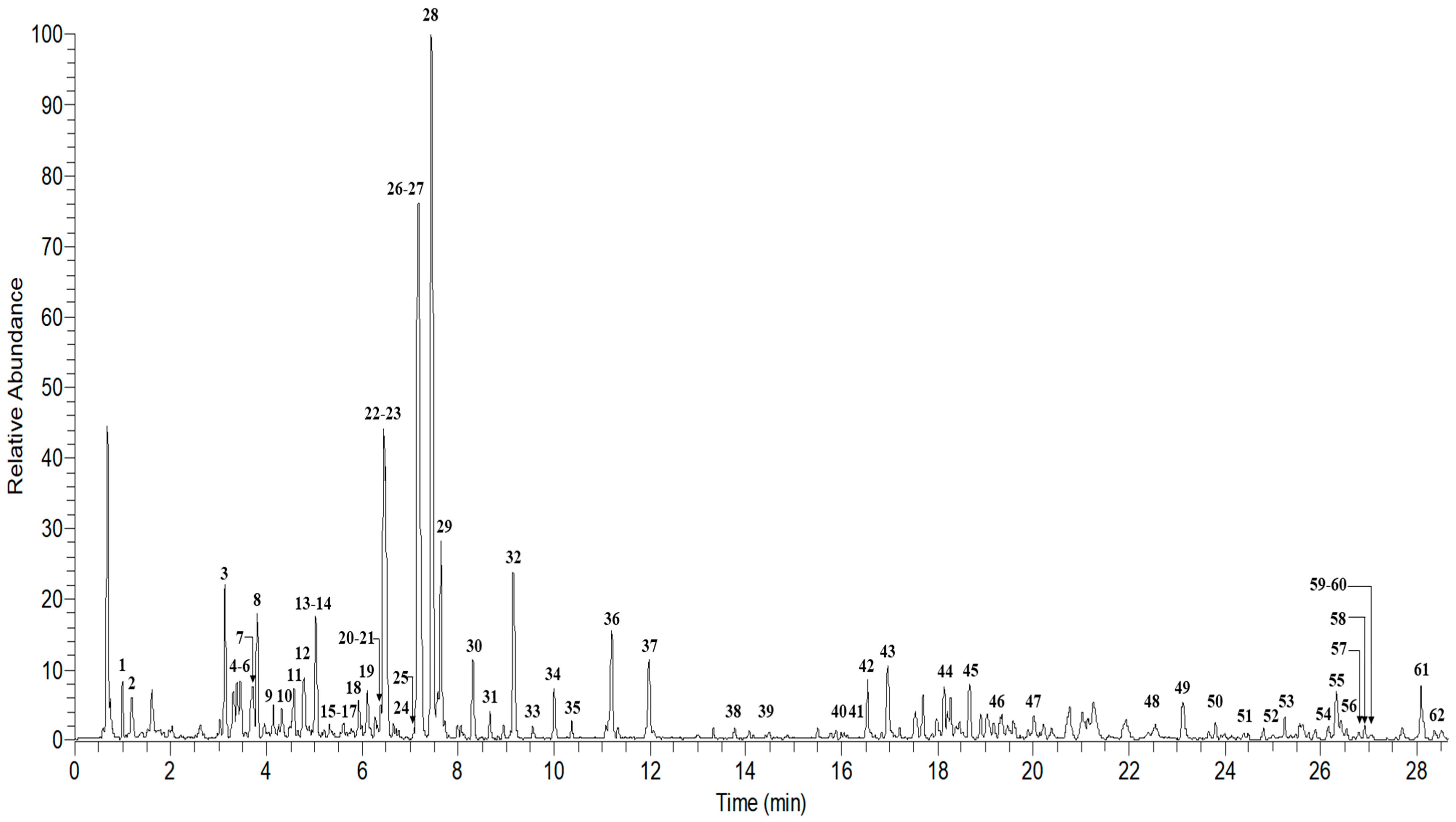
2.4. UHPLC–MS/MS Analysis of Ardisia Elliptica

Based on biological activity results, A. elliptica was identified as the most potent extract that showed antioxidant and anti-α-glucosidase activities. In view of the limited published information regarding the metabolite profile, further phytochemical characterization of the active extract was performed by using UPLC-ESI-Orbitrap MS/MS. A total of 62 metabolites were tentatively characterized in A. elliptica leaf extract (Table 2). The identification of the metabolites was based on the comparison with the literature and several online mass databases (Knapsack, Metabolomics Workbench, Human Metabolome Database, PubChem, and MassBank). The total ion chromatogram showed the major components observed between 0 and 30 min (Figure 1). Flavonoids accounted for 63% of the 62 tentatively identified metabolites, which may contribute to the high TPC value of A. elliptica leaf extract. Flavonoids such as quercetin, kaempferol, myricetin, and catechin derivatives, which are known to be abundant in plant matrices, could be identified in A. elliptica leaf extract. Other types of metabolites identified include fatty acid derivatives, benzoquinones, triterpenoids, phenolic lipids, and phenol ester.
A total of 10 metabolites were tentatively identified as quercetin derivatives in A. elliptica leaf extracts, with the signature aglycone fragment ion at m/z 301 (peak 33) and the characteristic fragment ions at m/z 271 and 151 in their MS/MS spectra [20]. Peak 7 was tentatively identified as quercetin 3-O-(2″-O-galloyl)-rutinoside with a deprotonated molecule at m/z 761.1343 [M − H]. Further fragmentation analysis of the compound showed fragmentation ions at m/z 610, 301, and 169, indicating the presence of rutin, quercetin, and gallic acid units. Peak 15 showed a deprotonated molecule at m/z 755.2025 and a fragment ion at 301, showing the loss of two rhamnosyl and one glucosyl moieties. Peak 15 was provisionally identified as quercetin 3-O-(2,6-di-O-rhamnosylglucoside) according to the comparison of previous MS/MS data [19]. Data presented in Table 2 indicated that peak 18 with m/z 595.1293 yielded fragment ion at m/z 301 attributed to the quercetin aglycone formed by the loss of a lathyrosyl residue (294 u). Meanwhile, peak 21 was tentatively identified as rutin with a deprotonated molecule at m/z 609.1450 and gave quercetin aglycone at m/z 301 due to the loss of a rutinoside residue (308 u). Similar to peak 23 at m/z 380.9911, loss of a sulfate residue (80 u) yielded a fragment ion at m/z 301. Peaks 24, 25, and 28 were identified as quercetin-3-O-glucoside, quercetin-3-O-arabinoside, and quercetin-3-O-rhamnoside, based on the deprotonated molecules at m/z 463.0875, 433.0768, and 447.0917, respectively. Based on the mass spectrum, the radical aglycone anion (m/z 300) also could be observed from the regular homolytic cleavage of glycosidic bonds in the deprotonated quercetin ion (m/z 301) by negative ion mode. The relative abundance of radical anions is affected by the collision energy with a relative increase in collision energy leading to a relative increase of radical anion [22]. Peak 20 was conditionally identified as quercetin 3-O-α-l-rhamnoside-7-O-β-d-glucoside with a deprotonated molecule at m/z 609.1451 with fragmentation ions at m/z 463, 447, and 301, which resulted from the removal of rhamnosyl residue (146 u) and glucose (162 u) moieties [19].
Peaks 16, 17, 19, and 22 were identified as myricetin derivatives based on the presence of fragmentation ion at m/z 316, which corresponds to the myricetin aglycone fragment in the MS/MS spectra [19]. Peaks 16, 17, 19, and 22 were tentatively assigned as myricetin-3-O-glucoside, myricetin-3-O-rutinoside, myricetin-3-O-arabinoside, and myricetin-3-O-rhamnoside, based on the deprotonated molecules at m/z 479.0816, 625.1396, 449.0713, and 463.0869 corresponding to the loss of sugar moieties, respectively. The radical myricetin anion (m/z 316) observed instead of m/z 317 might be due to the scission of glycosidic bonds as a result of high collision energy in the system [22].
Peak 13 showed a deprotonated molecule at m/z 289.1802 with fragment ions at m/z 245, 179, and 137, suggesting that the presence of catechin corresponded to the reported data [19]. Peak 3 was tentatively identified as catechin 6-C-glucoside based on deprotonated molecule at m/z 451.3394 and gave fragment ion at m/z 289 attributed to catechin aglycone formed by the loss of a glucosyl moiety (162 u). Epigallocatechin-3-gallate and its isomer were assigned as peaks 14 and 31 at m/z 457.0766 based on fragment ions observed at m/z 305 and m/z 169, which correspond to epigallocatechin and gallic acid moieties, respectively.
Peaks 26, 30, and 34 were identified as kaempferol derivatives. Peak 26 was conditionally identified as kaempferol with a deprotonated molecule at 285.0395 with the characteristic fragment ions at m/z 257, 213, and 187 [19]. Peak 30 was identified as kaempferol-3-O-rhamnoside based on the removal of rhamnosyl moiety. Peak 34 was assigned as rhamnocitrin 3-O-sulfate, and fragmentations indicated that the removal of a sulfate group (80 u) yielded a rhamnocitrin aglycone moiety (m/z 299), with further loss of a methyl radical (15 u) giving the fragment ion at m/z 285.
Apart from quercetin, myricetin, catechin, and kaempferol derivatives, other flavonoid signals could be detected in A. elliptica leaf extract. Peak 4 was tentatively identified as 5,7-dimethoxyflavone, with a deprotonated molecule at m/z 281.0331 and fragmentation ions at m/z 219 and 201, due to the loss of two methoxy groups and one hydroxyl group. Peak 8 showed a deprotonated molecule at m/z 549.1448 and fragmentation ion at m/z 269, suggesting the presence of a formononetin unit. Peak 10 was tentatively identified as 5,2′,4′,5′-tetrahydroxy-3-(3-hydroxy-3-methylbutyl)-6″,6″-dimethylpyrano[2″,3″:7,8]flavone, commonly named KB-2, with a deprotonated molecule at m/z 453.1605 and fragment ion at m/z 367 due to the elimination of C5H11O [23].
Furthermore, theasinensin A (Peak 11) could be detected with a deprotonated molecule at m/z 913.1451 and fragment ions at m/z 743 and 575 due to the loss of gallic acid moieties (169 u) [24]. Peaks 12, 27, and 32 were respectively assigned as apigenin 7-sulfate, luteolin 7-sulfate, and isorhamnetin 3-sulfate (persicarin), with the elimination of a sulfate moiety (80 u), yielding the apigenin fragment ion at m/z 270, luteolin fragment ion at m/z 285, and isorhamnetin fragment ion at m/z 315 [25,26]. Several significant fragment ions were used to distinguish flavone sulfate (peak 27) from flavonol sulfate (peak 34) derivatives at m/z 105, 133, and 178 which are characteristic of flavone fragmentation. Furthermore, due to the additional hydroxyl group, fragment ions at m/z 110, 151, 162, and 211 are characteristic of flavonol [27]. Peak 29 was conditionally detected as 7,2′-dihydroxyflavone 7-glucoside, with a deprotonated molecule at m/z 415.1962 and a fragment ion at m/z 252 due to the loss of a glucosyl moiety (162 u). Peak 35 was tentatively assigned as isoscutellarein 4′-methyl ether 8-(2″-sulfatoglucoside) with the elimination of sulfate and glucosyl moieties, which resulted in fragment ions at m/z 461 and 299. Thonningianin B (peak 42) could be identified with a deprotonated molecule at m/z 721.3632 and fragment ion at m/z 569 due to the elimination of C7H4O4. Moreover, gossypetin 8-glucoside-3-sulfate, 6-chlorocatechin, acerosin, and cycloheterophyllin could also be detected in A. elliptica leaf extract, and they were assigned as peaks 45, 49, 50, and 53, respectively. Interestingly, few metabolites that were reported in genus Ardisia could be tentatively identified in the currently studied leaf extract, including flavonoids, triterpenoids, benzoquinones, fatty acid derivatives, phenolic lipids, and phenol ester. Oxycoccicyanin or peonidin-3-glucoside was assigned as peak 9 with the [M − 2H] ion at m/z 461.1610 and a fragment ion at m/z 300 due to the elimination of a glucosyl moiety. Reported triterpenoids could be detected in the current leaf extract, including ardisianoside D (peak 40) with a deprotonated molecule at m/z 883.4165. The fragment ion at m/z 456 could be observed due to the loss of arabinose, xylose, and glucose moieties [28].
Furthermore, peak 46 was assigned as friedelan-3-one with a deprotonated molecule at m/z 425.2304, consistent with reported data at m/z 271 and 245 due to the loss of C11H20 and C13H26, respectively [29]. Alpha-amyrin (peak 58) could also be detected based on fragment ions of a dehydrated deprotonated molecule at m/z 407, with residues of C21H33, C19H29, and C16H24 at m/z 285, 257, and 216 respectively, which corresponded with the reported MS/MS data of alpha-amyrin [30]. Several benzoquinone derivatives were also tentatively identified in A. elliptica leaf extract at peaks 41, 44, 47, 54, 57, 59, and 60. Ardisianone A (peak 41) was conditionally identified with a deprotonated molecule at m/z 345.1830, as well as fragmentation ions at m/z 306 (loss of C3H7), 292 (loss of C4H9), and 192 [31]. Peak 44 showed a deprotonated molecule at m/z 293.2114 with fragment ions at 275 (loss of H2O), as well as 155 and 141 (benzoquinone unit with 2 hydroxyl groups), which are characteristic of embelin [32]. Ardisiaquinones which were previously reported in Ardisia genus could be detected in the current leaf extract. Peak 47 was conditionally identified as ardisiaquinone G with a deprotonated molecule at m/z 571.2880, as well as characteristic fragment ions at m/z 530 and 487 due to the loss of two acetyl units (84 u); the fragment ion at m/z 390 implied the two fully substituted benzoquinone rings present in ardisiaquinone G [33]. Ardisiaquinone D was assigned as peak 57 with a deprotonated molecule at m/z 541.3524 and product ions at m/z 526 and 511 due to the loss of two methyl groups, while m/z 359 and 183 showed the presence of a benzoquinone ring. The demethylation of metabolite 57 yielded ardisiaquinone A (peak 59) with a deprotonated molecule at m/z 527.3369, as well as fragment ions at m/z 514 and 499, due to the elimination of two methyl groups, whereas m/z 191, 165, and 151 implied the presence of benzoquinone rings [34]. Peak 60 was identified as ardisiaquinone J with a deprotonated molecule at m/z 529.3524 and fragmentation patterns of m/z 514 (loss of methyl group), 499 (loss of methoxy group), and 347 (loss of one benzoquinone ring), which corresponded to the reported MS/MS data of ardisiaquinone J [35].
Peak 54 was assigned as maesaquinone which can be found in the Primulaceae family. Maesaquinone showed a deprotonated molecule at m/z 417.2634 with fragment ions at m/z 401 (loss of hydroxyl group), 335 (loss of C6H11), and 193, suggesting the presence of a benzoquinone ring [34]. Ardisiphenol B (peak 38) was one of the phenol esters with a deprotonated molecule at m/z 375.1778 and fragmentation patterns of m/z 333 (elimination of acetyl group) and 207 (elimination of acetyl group and C9H17), which aligned with previously reported MS/MS data of this metabolite [36]. Peak 39 was conditionally identified as ardisinol II with a deprotonated molecule at m/z 289.1803, as well as characteristic fragmentation ions at m/z 245 (loss of C3H7), 161, 148, and 123 (loss of C12H23), which was consistent with reported MS/MS data [37].
Cornudentanone (peak 48) was conditionally identified with a deprotonated molecule at m/z 377.2329 and fragmentation patterns at m/z 359, 335 (loss of acetyl group), 316 (loss of ester group, C2H3O2), and 152, suggesting the presence of a benzoquinone ring. Peak 51 showed a deprotonated molecule at m/z 385.2741 and fragment ions at m/z 268 and 176, suggesting the presence of a hydroxyl benzoyl ion with a C6H12 aliphatic chain, and m/z 153 (presence of trihydroxyl benzoyl ion), which was then tentatively identified as ardisinone E [38]. Alkenylresorcinols such as bilobol (peak 52) could also be identified with a deprotonated molecule at m/z 317.2477, as well as fragment ions at m/z 300 (loss of hydroxyl moiety), 231 (loss of C6H13 aliphatic chain), 192 (loss of C9H17 aliphatic chain), 178, and 151, which aligned with previously reported MS/MS data [39]. Peak 62 was tentatively assigned as ardisenone with fragmentation patterns at m/z 316 (loss of C10H11O3), 278 (loss of C13H16O3), 205 (loss of C18H25O3), 181, and 169 with a deprotonated molecule at m/z 495.2625 [40].
Phenolic derivatives such as monogalloylglucose could be observed in A. elliptica leaf extract. Monogalloylglucose (peak 2) could be detected with a deprotonated molecule at m/z 331.0662 and fragment ion at m/z 169, suggesting the presence of a gallic acid unit due to the loss of a glucosyl moiety [19]. Peak 1 was identified with a deprotonated molecule at m/z 191.0184 and fragment ions at m/z 173, 129, and 111 (loss of hydroxyl and carbon dioxide moieties), which are characteristic of citric acid [41]. 1,5-Dibutyl methyl hydroxycitrate (peak 61) could be identified with a deprotonated molecule at m/z 333.2426 and fragmentation patterns at m/z 279 (loss of C4H9), 186, and 134 [42]. Peak 55 showed a deprotonated molecule at m/z 331.2267 with fragment ions at m/z 313 (loss of hydroxyl moiety) and 287 (loss of CHO2), which was tentatively identified as Gibberellin A4. Moreover, berberine was conditionally assigned as peak 56 which showed a deprotonated molecule at m/z 335.1342 with fragment ions at m/z 332, 317 (loss of hydroxyl moiety), and 279, which showed the transition of C20H18NO4 to C16H24NO3 [43]. Fatty acid derivatives could also be detected at peaks 36 and 37 with signature fragmentation patterns at m/z 229, 211, and 171 [41]. With the aid of phytochemical characterization, it provides a view of the antioxidant and anti-α-glucosidase potential of 70% ethanolic A. elliptica leaf extract. A large number of flavonoids such as quercetin, catechin, and kaempferol derivatives present in the extract were proven to play a role as antioxidant and anti-α-glucosidase agents [44,45]. Other types of compounds such as triterpenoids and benzoquinones might also contribute synergistically to the overall biological activities.

3. Materials and Methods

3.1. Chemicals and Reagents

Folin–Ciocalteu reagent and absolute ethanol were purchased from Merck (Darmstadt, Germany). Sodium carbonate, DPPH, α-glucosidase enzyme, p-nitrophenyl-α-d-glucopyranose (PNPG), glycine, phosphate-buffered saline, sodium nitroprusside (SNP), and other standard compounds used, including gallic acid and quercetin, were purchased from Sigma-Aldrich Co. (St. Louis, MO, USA). LC-MS-grade methanol, purified water, formic acid (FA), and dimethyl sulfoxide (DMSO) were supplied by Fisher Scientific (Geel, Belgium).

3.2. Plant Collection and Sample Preparation

Leucaena leucocephala (MFI 0079/19), Muntingia calabura (SK 3345/18), Spondias dulcis (MFI 0065/19), Annona squamosa (SK 2956/16), Ardisia elliptica (MFI 0054/19), Cynometra cauliflora (SK 1757/2011), Ficus auriculata (MFI 0146/19), and Averrhoa bilimbi (MFI 0139/19) were collected from Sri Serdang, Selangor in March 2018. The plants used in this study were identified by Dr. Mohd Firdaus Ismail, an in-house botanist at the Biodiversity Unit, Institute of Bioscience, Universiti Putra Malaysia. The leaves were separated from the stem and cleaned of any impurities with a clean tissue. The leaves were subjected to air drying treatment at room temperature in the shade until a constant weight was achieved [46]. The dried leaves were then ground into fine powder using a laboratory blender (Waring Commercial, Torrington, CT, USA) and stored at 4 °C for further analysis.

3.3. Sample Extraction

Sample extraction was done using the method of Mediani et al. [46] with slight modification. Briefly, 10 g of the plant sample was weighed and soaked in 100 mL of 70% ethanol and subjected to sonication at a controlled temperature (26–40 °C) using a Thermo-10D Ultrasonic Cleaner (Fisher Scientific, Waltham, MA, USA) for 1 h. The mixture was then filtered using Whatman filter paper No. 1 (GE Healthcare, Pittsburgh, PA, USA), and the crude extract was vacuum-evaporated using a rotary evaporator. The process was repeated twice using the residue of filtration to achieve maximum yield. The crude extracts were weighed and freeze-dried in a ScanVac CoolSafe Freeze dryer TM (Labogene, Lynge, Denmark). Freeze-dried samples were stored at 4 °C until further analysis.

3.4. Total Phenolic Content (TPC) Determination

The TPC was determined using the modified method by Zhang et al. [47]. A volume of 20 μL gallic acid, which was used as the standard, was mixed with 100 μL of Folin-Ciocalteu reagent in a 96-well plate. The mixture was left for 5 min until the addition of 80 μL of 7.5% sodium carbonates to each well. The plate was then incubated in the dark at room temperature for 30 min. The absorbance was measured at 750 nm using a Tecan Infinite F200 micro-plate reader (Tecan Group Ltd., Männedorf, Switzerland) in triplicate measurement. The same procedure was repeated using test samples to replace the standard. The gallic acid standard curve obtained was used to calculate the phenolic content of leaf extracts, which was expressed as mg of gallic acid equivalent per gram of crude extract (mg·GAE/g).

3.5. DPPH Free Radical Scavenging Assay

The assay was done using the method of Wan et al. [48] in a 96-well plate using serial dilutions of 50 µL of test sample (330–40 µg/mL). A volume of 100 µL of DPPH was then mixed with the serial diluted test samples. Then, the mixture was incubated for 30 min in the dark at room temperature. The absorbance was measured at 515 nm using a micro-plate reader in triplicate measurement. The scavenging capacity (SC) of the leaf extract was calculated as SC% = [(A0 − As)/A0] × 100, where A0 is the absorbance of reagent blank, whereas As is the absorbance of test samples. The result was conveyed as IC50 value, signifying the concentration of sample required to scavenge 50% of DPPH free radicals. Quercetin (positive control) was used in this assay.

3.6. Nitric Oxide (NO) Scavenging Assay

Based on the method used by Tsai et al. [49], the NO scavenging assay was done on a 96-well plate. Then, 60 µL of 10 mM SNP in phosphate-buffered saline was mixed with 60 µL of test samples (330–5.16 µg/mL) in a 96-well plate and incubated for 150 min. Then, 60 µL of freshly prepared Griess reagent was mixed with the test samples before the absorbance was measured at 550 nm using a micro-plate reader. Gallic acid was used as positive control, and the results were reported as IC50.

3.7. Anti-α-Glucosidase Assay

Anti-α-glucosidase assay was done as previously reported by Lee et al. [19]. The enzyme reaction was achieved using PNPG as the substrate and α-glucosidase enzyme, which were dissolved in 50 mM sodium phosphate buffer. Quercetin was used as the positive control. The test samples were prepared at concentrations of 500, 30, 25, 15, 3, and 1 µg/mL in accordance with the preliminary data obtained through screening analysis. Then, six serial dilutions were done. Thereafter, 10 µL of α-glucosidase enzyme was pipetted into a mixture of 10 µL of test sample and 130 µL of 30 mM phosphate buffer in a 96-well micro-plate. The negative control was prepared by substituting the sample with solvent, whereas blank solvent and blank sample were prepared by 140 µL of 30 mM sodium phosphate buffer with 10 µL of solvent, and 140 µL of 30 mM sodium phosphate buffer with 10 µL of test sample, respectively. Then, the mixture was incubated for 5 min at room temperature. The reaction was started by the adding 50 µL of PNPG substrate into each well of test sample, as well as into the negative and positive controls, while the remaining wells received 50 µL of 30 mM sodium phosphate buffer. The mixture was then incubated for 15 min at room temperature. The reaction was ceased with the addition of 50 µL of 2 M glycine (pH 10). The absorbance was then measured at 405 nm using a micro-plate reader in triplicate measurement. The percentage inhibition of the test sample was calculated as % = [(an − as)/an] × 100%, where an is the absorbance difference value between negative control and the blank, and as is the absorbance difference value between the sample and the blank. The result was expressed as IC50 value in µg/mL.

3.8. UHPLC–MS/MS Analysis

Based on the method used by Abd Ghafar et al. [50], the UHPLC-MS/MS analysis slight adjustment was done. The UHPLC-MS/MS spectrum of the active extract was acquired using a Thermo ScientificTM Q ExactiveTM Hybrid Quadrupole-Orbitrap mass spectrometer equipped with an electrospray ionization (ESI) source coupled with an auto-sampler and surveyor UHPLC binary pump (Thermo Fisher Scientific, Bremen, Germany). Phytochemical separation was done using an Acquity UPLC HSS T3 column (1.8 µm, 2.1 × 150 mm). The mobile phase used in the separation was LC-MS-grade water (solvent A) and acetonitrile (solvent B), each consisting of 0.1% FA. The programmed gradient was initiated with 5% to 100% solvent B from 0.5 to 30 min, and the solvent system was delivered at a flow rate of 0.4 mL/min. The sample was prepared in 10 mg/mL with an injection volume of 2 µL. Negative ion mode was done in full scan mass spectra acquisition from 150–1500 m/z with collision-induced dissociation (CID) energy of 30%. The mass spectra were collected and processed using Thermo Xcalibur Qual Browser software 4.0 (Thermo Fisher Scientific Inc., Waltham, MA, USA).

3.9. Statistical Analysis

The results of TPC, DPPH and NO scavenging, and anti-α-glucosidase activities were shown as means of three replicates ± standard deviation. One-way ANOVA with Tukey’s post hoc test was done to evaluate the significant effect of the factors at a confidence level of 95%. MS Excel (Microsoft, Redmond, WA, USA) and Minitab 17 (Minitab Inc., State College, PA, USA) software was used in the statistical calculation and Pearson correlation analysis.

4. Conclusions

The results of the current study illustrated that Ardisia elliptica, Muntingia calabura, and Cynometra cauliflora exhibited strong antioxidant and anti-α-glucosidase activities. A. elliptica showed the most potent activity among all tested plants. A total of 62 metabolites were tentatively characterized in A. elliptica 70% ethanolic leaf extract including flavonoids, benzoquinones, and triterpenoids which might contribute to the significant biological activities. To the best of our knowledge, this study provides the first detailed metabolite profile of A. elliptica by using UHPLC-ESI-Orbitrap MS. The findings in this work suggest that the leaves of A. elliptica could serve as a potential natural source of antioxidant and anti-diabetic agents. However, extensive studies are required to examine the safety and efficacy of their pharmacological properties for the utilization of alternative remedies for disease, particularly diabetes.

Author Contributions

P.L.W. and N.A.F., writing—original draft preparation, plant collection, data collection, and experimentation; S.N.M.Y., plant collection, data collection, and methodology; N.A.A.H., investigation and methodology; S.Z.A.G., data curation and methodology; A.A., methodology and statistical analysis; N.K.Z.Z., validation and methodology; F.A., conceptualization, project administration, reviewing and editing, and supervision. All authors have read and agreed to the published version of the manuscript.

Funding

This research was supported by a grant from Universiti Putra Malaysia (UPM/700/2/1/GPB/2017/9597400) under a Putra High Impact Grant Scheme.

Acknowledgments

The authors wish to thank Universiti Putra Malaysia for the facilities. The first author also gratefully acknowledges the support from Universiti Putra Malaysia for funding her study under the Graduate Research Fellowship scheme.

Conflicts of Interest

The authors have no conflicts of interest to disclose.

References

  1. IPGRI. Neglected and Underutilized Plant Species: Strategic Action Plan of the International Plant Genetic Resources Institute; International Plant Genetic Resources Institute: Rome, Italy, 2002; pp. 1–30. [Google Scholar]
  2. Feisul, M.I.; Azmi, S. National diabetes registry report. Minist. Health Malays. 2013, 1, 2009–2012. [Google Scholar]
  3. Trigwell, S.M.; Radford, P.M.; Page, S.R. Islet glutamic acid decarboxylase modified by reactive oxygen species is recognized by antibodies from patients with type 1 diabetes mellitus. Clin. Exp. Immunol. 2001, 126, 242–249. [Google Scholar] [CrossRef] [PubMed]
  4. Bouayed, J.; Bohn, T. Exogenous antioxidants-Double-edged swords in cellular redox state: Health beneficial effects at physiologic doses versus deleterious effects at high doses. Oxid. Med. Cell Longev. 2010, 3, 228–237. [Google Scholar] [CrossRef] [PubMed]
  5. Kim, K.Y.; Nam, K.A.; Kurihara, H.; Kim, S.M. Potent α-glucosidase inhibitors purified from the red alga Grateloupia elliptica. Phytochemistry 2008, 69, 2820–2825. [Google Scholar] [CrossRef] [PubMed]
  6. Triggle, C.R.; Ding, H. Cardiovascular impact of drugs used in the treatment of diabetes. Ther. Adv. Chronic Dis. 2014, 5, 245–268. [Google Scholar] [CrossRef] [PubMed] [Green Version]
  7. Kumar, S.; Narwal, S.; Kumar, V.; Prakash, O. α-glucosidase inhibitors from plants: A natural approach to treat diabetes. Pharmacogn. Rev. 2011, 5, 19–29. [Google Scholar] [CrossRef] [Green Version]
  8. Rahman, M.M.; Khan, F.E.; Das, R.; Hossain, M.A. Antioxidant activity and total phenolic content of some indigenous fruits of Bangladesh. Int. Food Res. J. 2016, 23, 2399–2404. [Google Scholar]
  9. Do, Q.D.; Angkawijaya, A.E.; Tran-Nguyen, P.L.; Huynh, L.H.; Soetaredjo, F.E.; Ismadji, S.; Ju, Y.H. Effect of extraction solvent on total phenol content, total flavonoid content, and antioxidant activity of Limnophila aromatica. J. Food Drug Anal. 2014, 22, 296–302. [Google Scholar] [CrossRef] [Green Version]
  10. Mariod, A.A.; Abdelwahab, S.I.; Elkheir, S.; Ahmed, J.M.; Fauzi, P.N.M.; Chuen, C.S. Antioxidant activity of different parts from Annona squamosa, and Catunaregam nilotica methanolic extract. Acta Sci. Pol. Technol. Aliment. 2012, 11, 249–257. [Google Scholar]
  11. Rabeta, M.S.; Nur Faraniza, R. Total phenolic content and ferric reducing antioxidant power of the leaves and fruits of Garcinia atrovirdis and Cynometra cauliflora. Int. Food Res. J. 2013, 20, 1691–1696. [Google Scholar]
  12. Zolkeflee, N.K.Z.; Isamail, N.A.; Maulidiani, M.; Abdul Hamid, N.A.; Ramli, N.S.; Azlan, A.; Abas, F. Metabolite variations and antioxidant activity of Muntingia calabura leaves in response to different drying methods and ethanol ratios elucidated by NMR-based metabolomics. Phytochem. Anal. 2020, in press, 1–15. [Google Scholar] [CrossRef]
  13. Al-Abd, N.M.; Nor, Z.M.; Mansor, M.; Zajmi, A.; Hasan, M.S.; Azhar, F.; Kassim, M. Phytochemical constituents, antioxidant and antibacterial activities of methanolic extract of Ardisia elliptica. Asian Pac. J. Trop. Biomed. 2017, 7, 569–576. [Google Scholar] [CrossRef]
  14. Rahardhian, M.R.R.; Murti, B.T.; Wigati, D.; Suharsanti, R.; Putri, C.N. Solvent concentration effect on total flavonoid and total phenolic contents of Averrhoa bilimbi leaf extract. Pharmaciana 2019, 9, 137–144. [Google Scholar] [CrossRef]
  15. Sampaio, B.L.; Edrada-Ebel, R.; Da Costa, F.B. Effect of the environment on the secondary metabolic profile of Tithonia diversifolia: A model for environmental metabolomics of plants. Sci. Rep. 2016, 6, 1–11. [Google Scholar] [CrossRef] [PubMed] [Green Version]
  16. Liang, N.; Kitts, D. Antioxidant property of coffee components: Assessment of methods that define mechanism of action. Molecules 2014, 19, 19180–19208. [Google Scholar] [CrossRef] [Green Version]
  17. Sumanont, Y.; Murakami, Y.; Tohda, M.; Vajragupta, O.; Matsumoto, K.; Watanabe, H. Evaluation of the nitric oxide radical scavenging activity of manganese complexes of curcumin and its derivative. Biol. Pharm. Bull. 2004, 27, 170–173. [Google Scholar] [CrossRef] [Green Version]
  18. Ado, M.A.; Abas, F.; Ismail, I.S.; Ghazali, H.M.; Shaari, K. Chemical profile and antiacetylcholinesterase, antityrosinase, antioxidant and α-glucosidase inhibitory activity of Cynometra cauliflora L. leaves. J. Sci. Food Agric. 2014, 95, 635–642. [Google Scholar] [CrossRef]
  19. Lee, S.Y.; Mediani, A.; Ismail, I.S.; Maulidiani; Abas, F. Antioxidants and α-glucosidase inhibitors from Neptunia oleracea fractions using 1H NMR-based metabolomics approach and UHPLC-MS/MS analysis. BMC Complem. Altern. Med. 2019, 19, 1–15. [Google Scholar] [CrossRef]
  20. Gavillán-Suárez, J.; Aguilar-Perez, A.; Rivera-Ortiz, N.; Rodríguez-Tirado, K.; Figueroa-Cuilan, W.; Morales-Santiago, L.; Martínez-Montemayor, G.; Cubano, L.A.; Martínez-Montemayor, M.M. Chemical profile and in vivo hypoglycemic effects of Syzygium jambos, Costus speciosus and Tapeinochilos ananassae plant extracts used as diabetes adjuvants in Puerto Rico. BMC Complem. Altern. Med. 2015, 15, 1–15. [Google Scholar] [CrossRef] [Green Version]
  21. Abdul-Hamid, N.A.; Maulidiani, M.; Mediani, A.; Yahya, U.I.I.; Ismail, I.S.; Tham, C.L.; Shadid, K.; Abas, F. Physicochemical characteristics, nutritional composition, and phytochemical profiles of nine Algerian date palm fruit (Phoenix dactylifera L.) varieties. J. Food Biochem. 2018, 42, e12663. [Google Scholar] [CrossRef]
  22. Hvattum, E.; Ekeberg, D. Study of the collision-induced radical cleavage of flavonoid glycosides using negative electrospray ionization tandem quadrupole mass spectrometry. J. Mass Spectrom. 2003, 38, 43–49. [Google Scholar] [CrossRef]
  23. Fujimoto, Y.; Zhang, X.; Kirisawa, M.; Uzawa, J.; Sumatra, M. New flavones from Artocarpus communis forst. Chem. Pharm. Bull. 1990, 38, 1787–1789. [Google Scholar] [CrossRef] [Green Version]
  24. Hatano, T.; Kusuda, M.; Hori, M.; Shiota, S.; Tsuchiya, T.; Yoshida, T. Theasinensin A, a tea polyphenol formed from (−)-epigallocatechin gallate, suppresses antibiotic resistance of methicillin-resistant Staphylococcus aureus. Planta Med. 2003, 69, 984–989. [Google Scholar] [CrossRef] [PubMed]
  25. Kwak, J.Y.; Seok, J.K.; Suh, H.J.; Choi, Y.H.; Hong, S.S.; Kim, D.S.; Boo, Y.C. Anti-melanogenic effects of luteolin 7-sulfate isolated from Phyllospadix iwatensis Makino. Brit. J. Dermatol. 2016, 175, 501–511. [Google Scholar] [CrossRef] [PubMed]
  26. Teles, Y.C.F.; Souza, M.S.R. Sulphated flavonoids: Biosynthesis, structures, and biological activities. Molecules 2018, 23, 480. [Google Scholar] [CrossRef] [Green Version]
  27. Cuyckens, F.; Ma, Y.L.; Pocsfalvi, G.; Claeysi, M. Tandem mass spectral strategies for the structural characterization of flavonoid glycosides. Analusis 2000, 28, 888–895. [Google Scholar] [CrossRef] [Green Version]
  28. Chang, X.; Li, W.; Jia, Z.; Satou, T.; Fushiya, S.; Koike, K. Biologically active triterpenoid saponins from Ardisia japonica. J. Nat. Prod. 2007, 70, 179–187. [Google Scholar] [CrossRef] [PubMed]
  29. Hisham, A.; Jayakumar, G.; Fujimoto, Y.; Hara, N. 1β,15α-dihydroxyfriedelan-3-one, a triterpene from Salacia beddomei. Phytochemistry 1996, 43, 843–845. [Google Scholar] [CrossRef]
  30. Naumoska, K.; Vovk, I. Analysis of triterpenoids and phytosterols in vegetables by thin-layer chromatography coupled to tandem mass spectrometry. J. Chromatogr. A 2015, 1381, 229–238. [Google Scholar] [CrossRef]
  31. Fukuyama, Y.; Kiriyama, Y.; Okino, J.; Kodama, M.; Iwaki, H.; Hosozawa, S.; Matsui, K. Naturally occuring 5-lipoxygenase inhibitor. II. Structures and syntheses of ardisianones A and B, and maesanin, alkenyl-1,4-benzoquinones from the rhizome of Ardisia japonica. Chem. Pharm. Bull. 1993, 41, 561–565. [Google Scholar] [CrossRef] [PubMed] [Green Version]
  32. Joy, B.; Sivadasan, R.; Emilia, A.T.; John, M.; Sobhan, P.K.; Seervi, M.; Santhoshkumar, T.R. Lysosomal destabilization and Cathepsin B contributes for cytochrome c release and caspase activation in embelin-induced apoptosis. Mol. Carcinogen. 2010, 49, 324–336. [Google Scholar] [CrossRef]
  33. Yang, L.; Khoo-beattie, C.; Goh, K.; Chng, B.; Yoganathan, K.; Lai, Y.; Butler, M.S. Ardisiaquinones from Ardisia teysmanniana. Phytochemistry 2001, 58, 1235–1238. [Google Scholar] [CrossRef]
  34. Ogawa, H.; Natori, S. Hydroxybenzoquinones from Myrsinaceae plants. The structures of 2-hydroxy-5-methoxy-3-pentadecenylbenzoquinone and ardisiaquinones A, B and C from Ardisia spp. Chem. Pharm. Bull. 1968, 16, 1709–1720. [Google Scholar] [CrossRef] [Green Version]
  35. Ndonsta, B.L.; Tatsimo, J.S.N.; Csupor, D.; Forgo, P.; Berkecz, R.; Berenyi, A.; Tene, M.; Molnar, J.; Zupko, I.; Hohmann, J.; et al. Alkylbenzoquinones with antiproliferative effect against human cancer cell lines from stem of Ardisia kivuensis. Phytochem. Lett. 2011, 4, 227–230. [Google Scholar] [CrossRef]
  36. Sumino, M.; Sekine, T.; Ruangrungsi, N.; Ikegami, F. Ardisiphenols A-C, novel antioxidants from the fruits of Ardisia colorata. Chem. Pharm. Bull. 2001, 49, 1664–1665. [Google Scholar] [CrossRef] [Green Version]
  37. Sun, W.Y.; Zong, Q.; Gu, R.L.; Pan, B.C. Synthesis of ardisinol II. Synthesis 1998, 11, 1619–1622. [Google Scholar] [CrossRef]
  38. Zheng, Y.; Deng, Y.; Wu, F. Ardisinones A-E, novel diarylundecanones from Ardisia arborescens. J. Nat. Prod. 2004, 67, 1617–1619. [Google Scholar] [CrossRef]
  39. Ndontsa, B.L.; Dongmo, F.L.M.; Tala, M.F.; Wabo, K.; Zeng, G.; Tan, N.; Tane, P. A new cytotoxic alkenylresorcinol from Embelia schimperi. Rec. Nat. Prod. 2013, 8, 37–40. [Google Scholar]
  40. David Horgen, F.; Guinaudeau, H.; Pezzuto, J.M.; Soejarto, D.D.; Farnsworth, N.R. Isolation and structure elucidation of ardisenone: A new, cytotoxic alkenylphenol from Ardisia iwahigensis. J. Nat. Prod. 1997, 60, 533–535. [Google Scholar] [CrossRef]
  41. Taamalli, A.; Arráez-Román, D.; Abaza, L.; Iswaldi, I.; Fernández-Gutiérrez, A.; Zarrouk, M.; Segura-Carretero, A. LC-MS-based metabolite profiling of methanolic extracts from the medicinal and aromatic species Mentha pulegium and Origanum majorana. Phytochem. Analysis 2015, 26, 320–330. [Google Scholar] [CrossRef]
  42. Mackeen, M.M.; Mooi, L.Y.; Amran, M.; Mat, N.; Lajis, N.H.; Ali, A.M. Noncytotoxic and antitumour-promoting activities of garcinia acid esters from Garcinia atroviridis Griff. ex T. Anders (Guttiferae). Evid. Based Compl. Alt. 2012, 2012, 1–5. [Google Scholar] [CrossRef] [Green Version]
  43. Scazzocchio, F.; Cometa, M.F.; Tomassini, L.; Palmery, M. Antibacterial activity of Hydrastis canadensis extract and its major isolated alkaloids. Planta Med. 2001, 67, 561–564. [Google Scholar] [CrossRef]
  44. Li, Y.Q.; Zhou, F.C.; Gao, F.; Bian, J.S.; Shan, F. Comparative evaluation of quercetin, isoquercetin and rutin as inhibitors of α-Glucosidase. J. Agric. Food Chem. 2009, 57, 11463–11468. [Google Scholar] [CrossRef]
  45. Atoui, A.K.; Mansouri, A.; Boskou, G.; Kefalas, P. Tea and herbal infusions: Their antioxidant activity and phenolic profile. Food Chem. 2005, 89, 27–36. [Google Scholar] [CrossRef]
  46. Mediani, A.; Abas, F.; Tan, C.; Khatib, A. Effects of different drying methods and storage time on free radical scavenging activity and total phenolic content of Cosmos Caudatus. Antioxidants 2014, 3, 358–370. [Google Scholar] [CrossRef] [Green Version]
  47. Zhang, Q.; Zhang, J.; Shen, J.; Silva, A.; Dennis, D.A.; Barrow, C.J. A simple 96-well microplate method for estimation of total polyphenol content in seaweeds. J. Appl. Phycol. 2006, 18, 445–450. [Google Scholar] [CrossRef] [Green Version]
  48. Wan, C.; Yuan, T.; Cirello, A.L.; Seeram, N.P. Antioxidant and α-glucosidase inhibitory phenolics isolated from highbush blueberry flowers. Food Chem. 2012, 135, 1929–1937. [Google Scholar] [CrossRef]
  49. Tsai, P.; Tsai, T.; Yu, C.; Ho, S. Comparison of NO-scavenging and NO-suppressing activities of different herbal teas with those of green tea. Food Chem. 2007, 103, 181–187. [Google Scholar] [CrossRef]
  50. Abd Ghafar, S.Z.; Mediani, A.; Maulidiani; Ramli, N.Z.; Abas, F. Antioxidant, α-glucosidase, and nitric oxide inhibitory activities of Phyllanthus acidus and LC–MS/MS profile of the active extract. Food Bioscience 2018, 25, 134–140. [Google Scholar] [CrossRef]
Sample Availability: Samples of the compounds are available from the authors.
Figure 1. Total ion chromatogram of 70% ethanolic leaves extract of A. elliptica.
Molecules 25 03067 g001
Table 1. Total phenolic content (TPC), 2,2-diphenyl-1-picrylhydrazyl (DPPH) and nitric oxide (NO) free radical scavenging, and anti-α-glucosidase activities of the extracts. IC50—half maximal inhibitory concentration; GAE—gallic acid equivalent.
Leaves Extracts/StandardTotal Phenolic Content
(mg GAE/g Sample)
IC50 Value (µg/mL)
DPPH Free Radical Scavenging ActivityNO Free Radical Scavenging ActivityAnti-α-Glucosidase Activity
Leucaena leucocephala175.75 ± 3.48 d8.67 ± 0.29 d65.57 ± 4.57 b6.62 ± 0.19 c
Muntingia calabura172.32 ± 3.39 d4.67 ± 0.21 c59.40 ± 3.39 b0.51 ± 0.01 a
Spondias dulcis50.90 ± 0.69 f14.22 ± 0.82 e301.66 ± 23.06 f45.52 ± 2.18 e
Annona squamosa199.62 ± 7.40 c5.00 ± 0.20 c109.02 ± 3.18 c3.59 ± 0.18 b
Ardisia elliptica253.10 ± 1.19 b2.17 ± 0.08 a49.43 ± 0.18 b0.29 ± 0.01 a
Cynometra cauliflora344.17 ± 10.80 a2.88 ± 0.05 ab118.62 ± 3.44 cd0.90 ± 0.02 a
Ficus auriculata167.15 ± 2.04 d5.06 ± 0.35 c169.65 ± 1.53 e0.36 ± 0.02 a
Averrhoa bilimbi97.50 ± 3.46 e16.80 ± 0.04 f134.33 ± 2.46 d26.91 ± 0.58 d
Quercetin-3.55 ± 0.28 b15.85 ± 0.58 a6.62 ± 0.03 c
Gallic acid--15.41 ± 0.63 a-
The results are expressed as means ± standard deviation. Means with different superscript letters are significantly different (p < 0.05) among leaf extracts. “-” indicates that the particular activities were not measured because of irrelevance to the compound.
Table 2. Mass spectral characteristics and tentative identification of compounds present in 70% ethanolic leaves extract of A. elliptica.
Peak No.Retention Time, MinExact MassDeprotonated Molecule
[M − H] (m/z)
DeltaMS/MS Fragment IonsTentative IdentificationMolecular Formula
11.00192.0197191.01840.0013173.0080, 129.0180, 111.0074, 87.0073Citric acidC6H8O7
21.19332.0671331.06620.0009312.1069, 271.0454, 241.0342, 211.0238, 169.0130MonogalloylglucoseC13H16O10
33.14452.4087451.33940.0693302.5710, 289.0708, 210.2854, 151.2638(+)-Catechin 6-C-glucosideC21H24O11
43.31282.0817281.03310.0486239.1548, 219.1387, 207.1384, 201.1277, 165.09045,7-DimethoxyflavoneC17H14O4
53.40282.0817281.03290.0488239.1548, 219.1387, 207.1384, 201.1277, 165.09045,7-Dimethoxyflavone isomerC17H14O4
63.45282.0817281.03280.0489239.1548, 219.1387, 207.1384, 201.1277, 165.09045,7-Dimethoxyflavone isomerC17H14O4
73.70762.1571761.13430.0228610.1257, 301.0352, 169.0131Quercetin 3-O-(2″-O-galloyl)-rutinosideC34H34O20
83.80550.1402549.14480.0046429.1028, 369.0819, 339.0715, 309.0611, 269.0662Formononetin 7-O-(2″-p-hydroxybenzoylglucoside)C29H26O11
94.14463.1168461.1610 *0.0442314.0427, 300.1083, 287.0562, 255.0286Oxycoccicyanin/Peonidin-3-glucosideC22H23O11
104.34454.1555453.16050.0050386.9793, 367.0700, 301.0338, 284.0323, 176.0435KB-2/5,2″,4′,5′-Tetrahydroxy-3-(3-hydroxy-3-methylbutyl)-6″,6″-dimethylpyrano[2″,3″:7,8]flavoneC25H26O8
114.57914.1469913.14510.0018761.1329, 743.1249, 609.1331, 591.1135, 575.0852, 453.0853Theasinensin AC44H34O22
124.79350.0024349.05910.0567269.6351, 241.0011, 227.0375, 152.0433Apigenin 7-sulfateC15H10O8S
134.95290.0790289.18020.1012245.0812, 203.0703, 179.0335, 137.0230, 123.0437CatechinC15H14O6
145.02458.0776457.07660.0010331.0454, 305.0666, 287.0562, 269.0456, 193.0132, 169.0131(−)-epigallocatechin-3-gallateC22H18O11
155.61756.2040755.20250.0015489.1044, 301.0315, 300.0270, 271.0243, 255.0294, 178.9978Quercetin 3-O-(2,6-di-O-rhamnosylglucoside)C33H40O20
165.71480.0831479.08160.0015316.0219, 287.0189, 271.0241, 178.9979, 151.0025Myricetin-3-O-glucosideC21H20O13
175.76626.1410625.13960.0014478.0751, 317.0288, 316.0212, 271.0243, 178.9976Myricetin-3-O-rutinosideC27H30O17
185.93596.1305595.12930.0012463.0802, 301.0349, 300.0271, 283.0230, 271.0244, 178.9975Quercetin 3-lathyrosideC26H28O16
196.11450.0726449.07130.0013316.0220, 287.0198, 271.0241, 178.9975Myricetin-3-O-arabinosideC20H18O12
206.32610.1461609.14510.0010463.0797, 447.0925, 301.0349, 300.0247Quercetin 3-O-α-l-rhamnoside-7-O-β-d-glucosideC27H30O16
216.38610.1461609.14500.0011301.0339, 300.0269, 271.0244, 178.9973, 151.0026Quercetin-3-O-rutinoside (Rutin)C27H30O16
226.45464.0882463.08690.0013316.0218, 287.0193, 178.9977, 151.0026Myricetin-3-O-rhamnosideC21H20O12
236.47381.9922380.99110.0011301.0349, 300.0247, 283.0245, 271.0238, 257.0445, 229.0496, 193.0133Quercetin 3-O-sulfateC15H10O10S
246.66464.0882463.08750.0007301.0339, 300.0270, 271.0244, 255.0293, 178.9976, 151.0024Quercetin-3-O-glucosideC21H20O12
256.95434.0776433.07680.0008301.0346, 300.0271, 271.0243, 255.0291, 178.9982, 151.0023Quercetin-3-O-arabinosideC20H18O11
267.18286.0405285.03950.0012257.0450, 213.0545, 187.0391, 163.0021KaempferolC15H10O6
277.19365.9973364.99610.0010285.0400, 267.0294, 255.0291, 241.0501, 229.0500, 213.0548, 178.4121, 133.0279, 105.6355Luteolin 7-sulfateC15H10O9S
287.44448.0933447.09170.0016301.0339, 300.0271, 271.0243, 255.0291, 178.9975, 151.0024Quercetin-3-O-rhamnosideC21H20O11
297.64416.1035415.19620.0927252.1096, 238.9105, 177.2131, 123.08047,2′-Dihydroxyflavone 7-glucosideC21H20O9
308.31432.0984431.09700.0014285.0393, 284.0321, 255.0293, 227.0341Kaempferol-3-O-rhamnosideC21H20O10
318.66458.0776457.07660.0010331.0454, 305.0666, 287.0562, 269.0456, 193.0132, 169.0131Epigallocatechin-3-gallate isomerC22H18O11
329.15396.0079395.00640.0015315.0607, 272.0317, 259.0608, 151.0027Persicarin/Isorhamnetin 3-sulfateC16H12O10S
339.57302.0354301.03480.0006273.0403, 178.9974, 151.0024, 121.0281QuercetinC15H10O7
3410.00380.0129379.01170.0012299.0555, 284.0321, 257.0403, 243.0656, 228.0399, 211.0385, 162.5436, 151.0027, 110.0001Rhamnocitrin 3-O-sulfateC16H12O9S
3510.37542.0658541.06440.0014461.1082, 314.0426, 299.0188, 271.0243, 256.0363, 158.7938Isoscutellarein 4′-methyl ether 8-(2′-sulfatoglucoside)C22H22O14S
3611.20328.2177327.21710.0006229.1440, 211.1331, 171.1015Trihydroxy octadecadienoic acidC18H32O5
3711.98328.2177327.21710.0006229.1440, 211.1331, 171.1015Trihydroxy octadecadienoic acid isomerC18H32O5
3813.78376.2541375.17780.0763333.6364, 330.1770, 329.1730, 307.1919, 235.1334, 207.0993Ardisiphenol BC23H36O4
3914.49290.2173289.18030.0370245.1902, 161.9148, 148.7701, 123.0794Ardisinol IIC19H30O2
4016.00884.5061883.41650.0896837.4141, 559.1864, 456.2514, 397.1332, 277.2172Ardisianoside DC46H76O16
4116.37346.2497345.18300.0667306.9802, 292.2949, 192.5377Ardisianone AC22H34O3
4216.54722.1410721.36320.2222675.3585, 569.1615, 415.1444, 400.9850, 305.0875, 277.2165Thonningianin BC35H30O17
4316.96722.1410721.36310.2221675.3580, 569.1614, 415.1447, 400.9853, 305.0875, 277.2166Thonningianin B isomerC35H30O17
4418.14294.1758293.21140.0356275.2011, 155.1072, 141.1270, 127.1115, 121. 1009EmbelinC17H26O4
4518.67560.0399559.12690.0870354.6981, 286.8783, 228.8837, 121.6282Gossypetin 8-glucoside-3-sulfateC21H20O16S
4619.33426.3789425.23040.1485271.0612, 245.0811, 203.0705, 177.0179, 151.0386Friedelan-3-one C30H50O
4720.00572.2621571.28800.0259530.8586, 487.1684, 391.2236, 255.2324, 241.0111, 223.0012Ardisiaquinone GC31H40O10
4822.76378.2334377.23290.0005359.2229, 335.2515, 316.2326, 152.0106CornudentanoneC22H34O5
4923.12324.0328323.11620.0834279.2323, 265.8335, 216.0093, 184.01946-ChlorocatechinC15H13ClO6
5023.80360.0772359.06120.0160317.2494, 315.2661, 245.0694, 211.2592AcerosinC18H16O8
5124.48386.2093385.27410.0648268.6892, 176.3903, 153.4738Ardisinone EC23H30O5
5225.02318.2558317.24770.0081300.0236, 231.3264, 192.0054, 178.9974, 151.0025BilobolC21H34O2
5325.24502.1919501.32110.1292486.2979, 473.2810, 456.2825, 443.2783, 435.2527CycloheterophyllinC30H30O7
5426.18418.3083417.26340.0449401.6784, 375.2524, 335.2505, 308.5951, 193.0859MaesaquinoneC26H42O4
5526.33332.1551331.22670.0716313.2373, 287.2391, 254.8929, 225.6182, 213.1681Gibberellin A4C19H24O5
5626.54336.1163335.13420.0179332.2440, 317.2474, 305.2462, 279.2698, 230.0207BerberineC20H18NO4
5726.80542.2879541.35240.0645526.3293, 511.3054, 493.2956, 359.2590, 183.6019Ardisiaquinone DC31H42O8
5826.93426.3861425.23000.1489407.0769, 381.0991, 339.0862, 257.0452, 216.0410, 167.2586Alpha-amyrinC30H50O
5927.05528.2723527.33690.0646514.3290, 499.3061, 191.0708, 165.0543, 151.0386Ardisiaquinone AC30H40O8
6027.08530.2952529.35240.0572514.3292, 499.3058, 481.2948, 453.2976, 347.2579, 225.7453Ardisiaquinone JC30H42O8
6128.09334.1555333.24260.0871279.2678, 186.7317, 134.03641,5-Dibutyl methyl hydroxycitrateC15H26O8
6228.37496.2824495.26250.0199316.1812, 278.8960, 205.1232, 181.0499, 169.0134ArdisenoneC30H40O6
* Represented [M − 2H] ion was observed.

