
Comparing Zwitterionic and PEG Exteriors of Polyelectrolyte Complex Micelles

 ,
,  ,
, 
Abstract
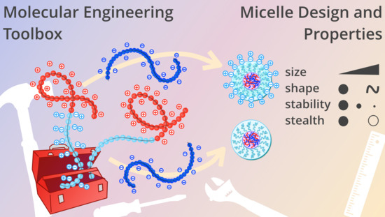
1. Introduction
2. Materials and Methods
2.1. Materials
2.2. RAFT Synthesis of PEG-PVBTMA, PAA and PMPC-PVBTMA
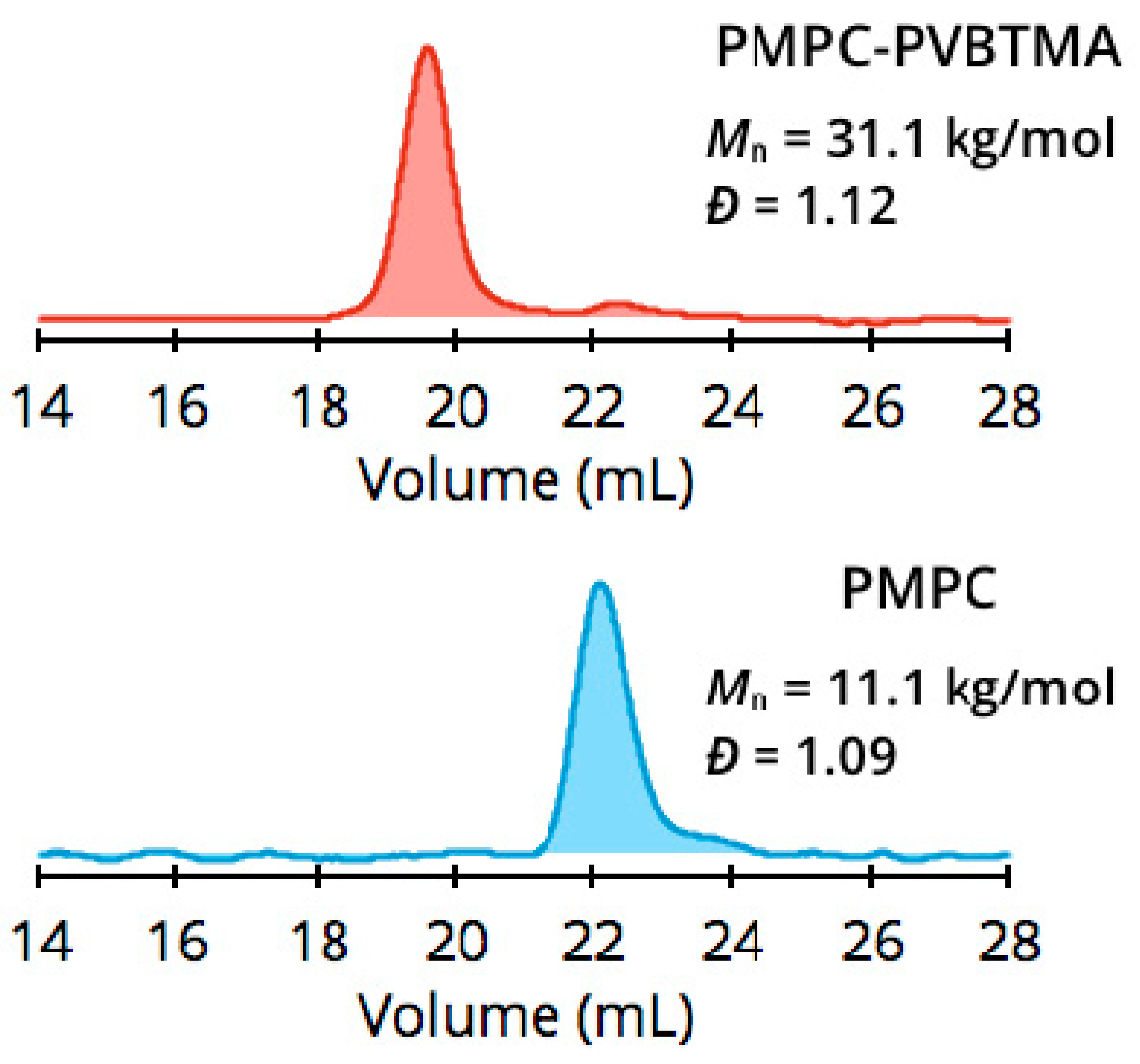
2.3. Size-Exclusion Chromatography with Multiangle Light Scattering (SEC-MALS)
2.4. Proton Nuclear Magnetic Resonance (1H NMR) Spectroscopy
2.5. Thermogravimetric Analysis (TGA)
2.6. Micelle Preparation
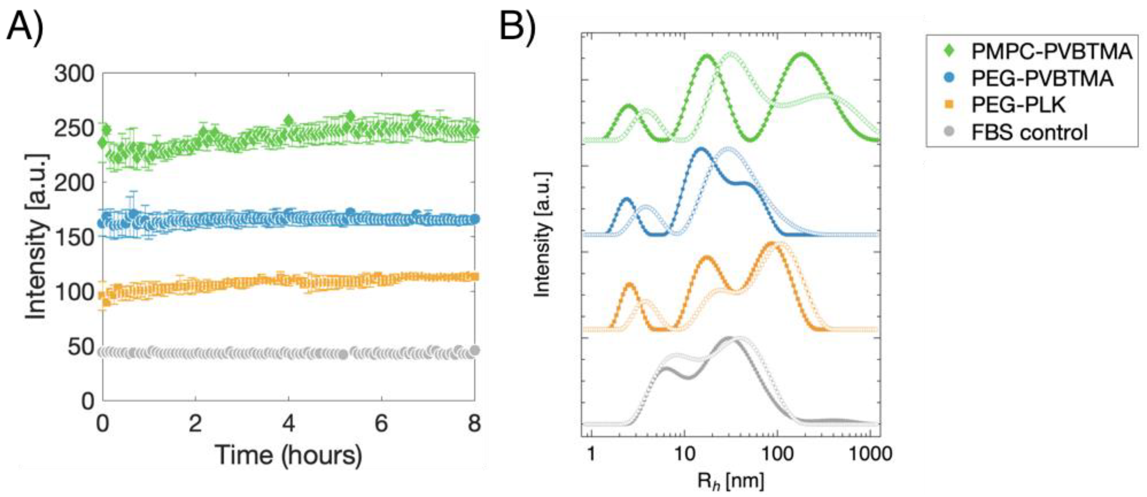
2.7. Dynamic Light Scattering (DLS)
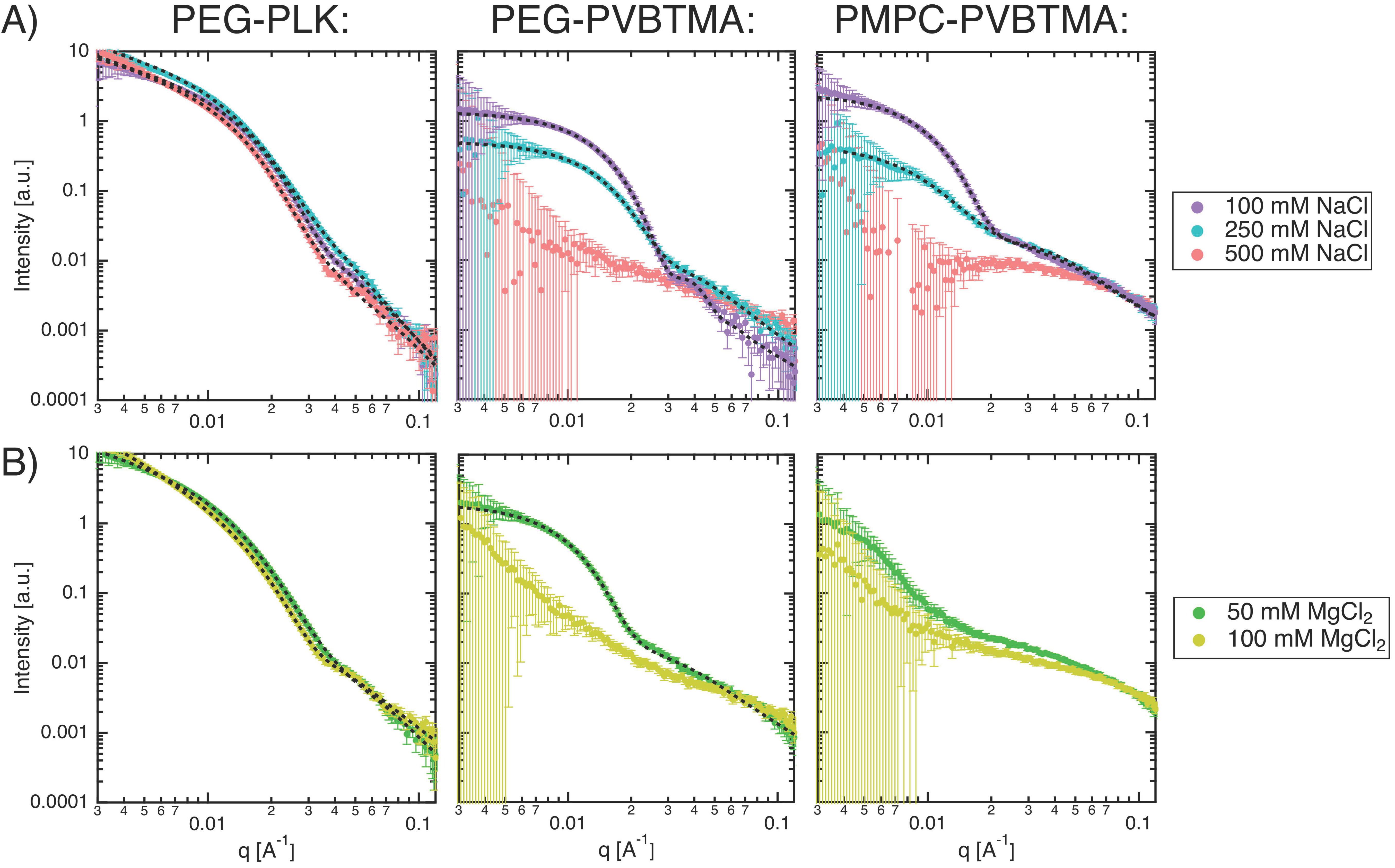
2.8. Small-Angle X-ray Scattering (SAXS)
2.9. Micelle Salt Dependence
2.10. Micelle Stability Tests
3. Results and Discussion
3.1. PMPC-PVBTMA Synthesis and Characterization
3.2. Polyelectrolyte Complex Micelle (PCM) and Zwitterionic PCM (zPCM) Assembly, Salt Resistance, and Stability
3.3. PCMs and zPCMs in Biologically Relevant Conditions
4. Conclusions
Supplementary Materials
Author Contributions
Funding
Acknowledgments
Conflicts of Interest
References
- Muthukumar, M. 50th Anniversary Perspective: A Perspective on Polyelectrolyte Solutions. Macromolecules 2017, 50, 9528–9560. [Google Scholar] [CrossRef] [PubMed]
- Sing, C.E. Micro-to macro-phase separation transition in sequence-defined coacervates. J. Chem. Phys. 2020, 152, 024902. [Google Scholar] [CrossRef] [PubMed]
- Acar, H.; Ting, J.M.; Srivastava, S.; LaBelle, J.L.; Tirrell, M.V. Molecular engineering solutions for therapeutic peptide delivery. Chem. Soc. Rev. 2017, 46, 6553–6569. [Google Scholar] [CrossRef] [PubMed]
- Voets, I.K.; de Keizer, A.; Stuart, M.A.C. Complex coacervate core micelles. Adv. Colloid Interface Sci. 2009, 147–148, 300–318. [Google Scholar] [CrossRef] [PubMed]
- Marciel, A.B.; Chung, E.J.; Brettmann, B.K.; Leon, L. Bulk and nanoscale polypeptide based polyelectrolyte complexes. Adv. Colloid Interface Sci. 2017, 239, 187–198. [Google Scholar] [CrossRef] [PubMed]
- Brangwynne, C.P.; Tompa, P.; Pappu, R.V. Polymer physics of intracellular phase transitions. Nat. Phys. 2015, 11, 899–904. [Google Scholar] [CrossRef]
- Cooper, C.L.; Dubin, P.L.; Kayitmazer, A.B.; Turksen, S. Polyelectrolyte–protein complexes. Curr. Opin. Colloid Interface Sci. 2005, 10, 52–78. [Google Scholar] [CrossRef]
- Horn, J.; Kapelner, R.; Obermeyer, A. Macro-and Microphase Separated Protein-Polyelectrolyte Complexes: Design Parameters and Current Progress. Polymers 2019, 11, 578. [Google Scholar] [CrossRef]
- Harada, A.; Kataoka, K. Effect of Charged Segment Length on Physicochemical Properties of Core−Shell Type Polyion Complex Micelles from Block Ionomers. Macromolecules 2003, 36, 4995–5001. [Google Scholar] [CrossRef]
- van der Kooij, H.M.; Spruijt, E.; Voets, I.K.; Fokkink, R.; Cohen Stuart, M.A.; van der Gucht, J. On the Stability and Morphology of Complex Coacervate Core Micelles: From Spherical to Wormlike Micelles. Langmuir 2012, 28, 14180–14191. [Google Scholar] [CrossRef]
- Lemmers, M.; Voets, I.K.; Cohen Stuart, M.A.; Der Gucht, J.V. Transient network topology of interconnected polyelectrolyte complex micelles. Soft Matter 2011, 7, 1378–1389. [Google Scholar] [CrossRef]
- Srivastava, S.; Andreev, M.; Levi, A.E.; Goldfeld, D.J.; Mao, J.; Heller, W.T.; Prabhu, V.M.; de Pablo, J.J.; Tirrell, M.V. Gel phase formation in dilute triblock copolyelectrolyte complexes. Nat. Commun. 2017, 8, 14131. [Google Scholar] [CrossRef] [PubMed]
- Heo, T.-Y.; Kim, I.; Chen, L.; Lee, E.; Lee, S.; Choi, S.-H. Effect of Ionic Group on the Complex Coacervate Core Micelle Structure. Polymers 2019, 11, 455. [Google Scholar] [CrossRef] [PubMed]
- Rahalkar, A.; Wei, G.; Nieuwendaal, R.; Prabhu, V.M.; Srivastava, S.; Levi, A.E.; de Pablo, J.J.; Tirrell, M.V. Effect of temperature on the structure and dynamics of triblock polyelectrolyte gels. J. Chem. Phys. 2018, 149, 163310. [Google Scholar] [CrossRef]
- Knop, K.; Hoogenboom, R.; Fischer, D.; Schubert, U.S. Poly (ethylene glycol) in Drug Delivery: Pros and Cons as Well as Potential Alternatives. Angew. Chem. Int. Ed. 2010, 49, 6288–6308. [Google Scholar] [CrossRef]
- Ishihara, T.; Takeda, M.; Sakamoto, H.; Kimoto, A.; Kobayashi, C.; Takasaki, N.; Yuki, K.; Tanaka, K.-I.; Takenaga, M.; Igarashi, R.; et al. Accelerated Blood Clearance Phenomenon Upon Repeated Injection of PEG-modified PLA-nanoparticles. Pharm. Res. 2009, 26, 2270–2279. [Google Scholar] [CrossRef]
- Ishihara, T.; Maeda, T.; Sakamoto, H.; Takasaki, N.; Shigyo, M.; Ishida, T.; Kiwada, H.; Mizushima, Y.; Mizushima, T. Evasion of the Accelerated Blood Clearance Phenomenon by Coating of Nanoparticles with Various Hydrophilic Polymers. Biomacromolecules 2010, 11, 2700–2706. [Google Scholar] [CrossRef]
- Pelegri-O’Day, E.M.; Lin, E.-W.; Maynard, H.D. Therapeutic Protein–Polymer Conjugates: Advancing Beyond PEGylation. J. Am. Chem. Soc. 2014, 136, 14323–14332. [Google Scholar] [CrossRef]
- Van Bruggen, C.; Hexum, J.K.; Tan, Z.; Dalal, R.J.; Reineke, T.M. Nonviral Gene Delivery with Cationic Glycopolymers. Acc. Chem. Res. 2019, 52, 1347–1358. [Google Scholar] [CrossRef]
- Elsabahy, M.; Wooley, K.L. Design of polymeric nanoparticles for biomedical delivery applications. Chem. Soc. Rev. 2012, 41, 2521–3012. [Google Scholar] [CrossRef]
- Burke, P.A.; Pun, S.H.; Reineke, T.M. Advancing Polymeric Delivery Systems Amidst a Nucleic Acid Therapy Renaissance. ACS Macro Lett. 2013, 2, 928–934. [Google Scholar] [CrossRef] [PubMed]
- Zou, H.; Wang, Z.; Feng, M. Nanocarriers with tunable surface properties to unblock bottlenecks in systemic drug and gene delivery. J. Control. Release 2015, 214, 121–133. [Google Scholar] [CrossRef] [PubMed]
- Hoang Thi, T.T.; Pilkington, E.H.; Nguyen, D.H.; Lee, J.S.; Park, K.D.; Truong, N.P. The Importance of Poly(ethylene glycol) Alternatives for Overcoming PEG Immunogenicity in Drug Delivery and Bioconjugation. Polymers 2020, 12, 298. [Google Scholar] [CrossRef] [PubMed]
- Blackman, L.D.; Gunatillake, P.A.; Cass, P.; Locock, K.E.S. An introduction to zwitterionic polymer behavior and applications in solution and at surfaces. Chem. Soc. Rev. 2019, 6, 757–770. [Google Scholar] [CrossRef]
- Schlenoff, J.B. Zwitteration: Coating Surfaces with Zwitterionic Functionality to Reduce Nonspecific Adsorption. Langmuir 2014, 30, 9625–9636. [Google Scholar] [CrossRef]
- Ishihara, K. Revolutionary advances in 2-methacryloyloxyethyl phosphorylcholine polymers as biomaterials. J. Biomed. Mater. Res. 2019, 107, 933–943. [Google Scholar] [CrossRef]
- Kitano, H.; Sudo, K.; Ichikawa, K.; Ide, M.; Ishihara, K. Raman Spectroscopic Study on the Structure of Water in Aqueous Polyelectrolyte Solutions. J. Phys. Chem. B 2000, 104, 11425–11429. [Google Scholar] [CrossRef]
- Ladd, J.; Zhang, Z.; Chen, S.; Hower, J.C.; Jiang, S. Zwitterionic Polymers Exhibiting High Resistance to Nonspecific Protein Adsorption from Human Serum and Plasma. Biomacromolecules 2008, 9, 1357–1361. [Google Scholar] [CrossRef]
- Goda, T.; Ishihara, K.; Miyahara, Y. Critical update on 2-methacryloyloxyethyl phosphorylcholine (MPC) polymer science. J. Appl. Polym. Sci. 2015, 132, 41766. [Google Scholar] [CrossRef]
- Sae-ung, P.; Kolewe, K.W.; Bai, Y.; Rice, E.W.; Schiffman, J.D.; Emrick, T.; Hoven, V.P. Antifouling Stripes Prepared from Clickable Zwitterionic Copolymers. Langmuir 2017, 33, 7028–7035. [Google Scholar] [CrossRef]
- He, Q.; Qiao, Y.; Mandia, D.J.; Gan, S.; Zhang, H.; Zhou, H.; Elam, J.W.; Darling, S.B.; Tirrell, M.V.; Chen, W. Enrichment and Distribution of Pb2+ Ions in Zwitterionic Poly (cysteine methacrylate) Brushes at the Solid-Liquid Interface. Langmuir 2019, 35, 17082–17089. [Google Scholar] [CrossRef] [PubMed]
- Lewis, A.; Tang, Y.; Brocchini, S.; Choi, J.-W.; Godwin, A. Poly (2-methacryloyloxyethyl phosphorylcholine) for Protein Conjugation. Bioconjugate Chem. 2008, 19, 2144–2155. [Google Scholar] [CrossRef] [PubMed]
- Jackson, M.A.; Werfel, T.A.; Curvino, E.J.; Yu, F.; Kavanaugh, T.E.; Sarett, S.M.; Dockery, M.D.; Kilchrist, K.V.; Jackson, A.N.; Giorgio, T.D.; et al. Zwitterionic Nanocarrier Surface Chemistry Improves siRNA Tumor Delivery and Silencing Activity Relative to Polyethylene Glycol. ACS Nano 2017, 11, 5680–5696. [Google Scholar] [CrossRef] [PubMed]
- Cao, Z.; Zhang, G. Dynamics of polyzwitterions in salt-free and salt solutions. Phys. Chem. Chem. Phys. 2015, 17, 27045–27051. [Google Scholar] [CrossRef]
- Georgiev, G.S.; Kamenska, E.B.; Vassileva, E.D.; Kamenova, I.P.; Georgieva, V.T.; Iliev, S.B.; Ivanov, I.A. Self-Assembly, Antipolyelectrolyte Effect, and Nonbiofouling Properties of Polyzwitterions. Biomacromolecules 2006, 7, 1329–1334. [Google Scholar] [CrossRef]
- Shao, Q.; Jiang, S. Molecular Understanding and Design of Zwitterionic Materials. Adv. Mater. 2014, 27, 15–26. [Google Scholar] [CrossRef]
- Kobayashi, M.; Terayama, Y.; Kikuchi, M.; Takahara, A. Chain dimensions and surface characterization of superhydrophilic polymer brushes with zwitterion side groups. Soft Matter 2013, 9, 5138–5148. [Google Scholar] [CrossRef]
- Morozova, S.; Hu, G.; Emrick, T.; Muthukumar, M. Influence of Dipole Orientation on Solution Properties of Polyzwitterions. ACS Macro Lett. 2016, 5, 118–122. [Google Scholar] [CrossRef]
- Wang, J.; Yuan, S.; Zhang, Y.; Wu, W.; Hu, Y.; Jiang, X. The effects of poly (zwitterions)s versus poly(ethylene glycol) surface coatings on the biodistribution of protein nanoparticles. Biomater. Sci. 2016, 4, 1351–1360. [Google Scholar] [CrossRef]
- Keefe, A.J.; Jiang, S. Poly (zwitterionic) protein conjugates offer increased stability without sacrificing binding affinity or bioactivity. Nat. Chem. 2011, 4, 59–63. [Google Scholar] [CrossRef]
- Miyamoto, D.; Watanabe, J.; Ishihara, K. Effect of water-soluble phospholipid polymers conjugated with papain on the enzymatic stability. Biomaterials 2004, 25, 71–76. [Google Scholar] [CrossRef]
- Lowe, A.B.; McCormick, C.L. Synthesis and Solution Properties of Zwitterionic Polymers. Chem. Rev. 2002, 102, 4177–4190. [Google Scholar] [CrossRef] [PubMed]
- Lobb, E.J.; Ma, I.; Billingham, N.C.; Armes, S.P.; Lewis, A.L. Facile Synthesis of Well-Defined, Biocompatible Phosphorylcholine-Based Methacrylate Copolymers via Atom Transfer Radical Polymerization at 20 °C. J. Am. Chem. Soc. 2001, 123, 7913–7914. [Google Scholar] [CrossRef] [PubMed]
- Hsiue, G.-H.; Lo, C.-L.; Cheng, C.-H.; Lin, C.-P.; Huang, C.-K.; Chen, H.-H. Preparation and characterization of poly(2-methacryloyloxyethyl phosphorylcholine)-block-poly(D,L-lactide) polymer nanoparticles. J. Polym. Sci. A Polym. Chem. 2007, 45, 688–698. [Google Scholar] [CrossRef]
- Matsuda, Y.; Kobayashi, M.; Annaka, M.; Ishihara, K.; Takahara, A. Dimension of poly (2-methacryloyloxyethyl phosphorylcholine) in aqueous solutions with various ionic strength. Chem. Lett. 2006, 35, 1310–1311. [Google Scholar] [CrossRef]
- Topham, P.D.; Sandon, N.; Read, E.S.; Madsen, J.; Ryan, A.J.; Armes, S.P. Facile Synthesis of Well-Defined Hydrophilic Methacrylic Macromonomers Using ATRP and Click Chemistry. Macromolecules 2008, 41, 9542–9547. [Google Scholar] [CrossRef]
- Bhuchar, N.; Deng, Z.; Ishihara, K.; Narain, R. Detailed study of the reversible addition–fragmentation chain transfer polymerization and co-polymerization of 2-methacryloyloxyethyl phosphorylcholine. Polym. Chem. 2011, 2, 632–639. [Google Scholar] [CrossRef]
- Nakaura, H.; Kawamura, A.; Miyata, T. Reductively Responsive Gel Capsules Prepared Using a Water- Soluble Zwitterionic Block Copolymer Emulsifier. Langmuir 2018, 35, 1413–1420. [Google Scholar] [CrossRef]
- Takahashi, R.; Narayanan, T.; Yusa, S.-I.; Sato, T. Kinetics of Morphological Transition between Cylindrical and Spherical Micelles in a Mixture of Anionic–Neutral and Cationic–Neutral Block Copolymers Studied by Time-Resolved SAXS and USAXS. Macromolecules 2018, 51, 3654–3662. [Google Scholar] [CrossRef]
- Ting, J.M.; Wu, H.; Herzog-Arbeitman, A.; Srivastava, S.; Tirrell, M.V. Synthesis and Assembly of Designer Styrenic Diblock Polyelectrolytes. ACS Macro Lett. 2018, 7, 726–733. [Google Scholar] [CrossRef]
- Wu, H.; Ting, J.M.; Yu, B.; Jackson, N.E.; Meng, S.; de Pablo, J.J.; Tirrell, M.V. Spatiotemporal Formation Kinetics of Polyelectrolyte Complex Micelles with Millisecond Resolution. ChemRxiv 2019, 1–8. [Google Scholar] [CrossRef]
- Wu, H.; Ting, J.M.; Weiss, T.M.; Tirrell, M.V. Interparticle Interactions in Dilute Solutions of Polyelectrolyte Complex Micelles. ACS Macro Lett. 2019, 8, 819–825. [Google Scholar] [CrossRef]
- Wu, H.; Ting, J.M.; Tirrell, M.V. Mechanism of Dissociation Kinetics in Polyelectrolyte Complex Micelles. Macromolecules 2020, 53, 102–111. [Google Scholar] [CrossRef]
- Wu, H.; Ting, J.M.; Werba, O.; Meng, S.; Tirrell, M.V. Non-equilibrium phenomena and kinetic pathways in self-assembled polyelectrolyte complexes. J. Chem. Phys. 2018, 149, 163330. [Google Scholar] [CrossRef]
- Marras, A.E.; Vieregg, J.R.; Ting, J.M.; Rubien, J.D.; Tirrell, M.V. Polyelectrolyte Complexation of Oligonucleotides by Charged Hydrophobic—Neutral Hydrophilic Block Copolymers. Polymers 2019, 11, 83. [Google Scholar] [CrossRef]
- Moad, G. RAFT Polymerization to Form Stimuli-Responsive Polymers. Polym. Chem. 2017, 8, 177–219. [Google Scholar] [CrossRef]
- Jakes, J. Regularized Positive Exponential Sum (REPES) Program-A Way of Inverting Laplace Transform Data Obtained by Dynamic Light Scattering. Collect. Czech. Chem. Commun. 1995, 60, 1781–1797. [Google Scholar] [CrossRef]
- Ilavsky, J.; Jemian, P.R. Irena: Tool suite for modeling and analysis of small-angle scattering. J. Appl. Cryst. 2009, 42, 347–353. [Google Scholar] [CrossRef]
- Marras, A.E.; Vieregg, J.R.; Tirrell, M.V. Assembly and Characterization of Polyelectrolyte Complex Micelles. J. Vis. Exp. 2020, 157, e60894. [Google Scholar] [CrossRef]
- Dreiss, C.C.A. Wormlike Micelles: Where Do We Stand? Recent Developments, Linear Rheology and Scattering Techniques. Soft Matter 2007, 3, 956–970. [Google Scholar] [CrossRef]
- Koppel, D.E. Analysis of Macromolecular Polydispersity in Intensity Correlation Spectroscopy: The Method of Cumulants. J. Chem. Phys. 1972, 57, 4814–4820. [Google Scholar] [CrossRef]
- Krogstad, D.V.; Lynd, N.A.; Miyajima, D.; Gopez, J.; Hawker, C.J.; Kramer, E.J.; Tirrell, M.V. Structural Evolution of Polyelectrolyte Complex Core Micelles and Ordered-Phase Bulk Materials. Macromolecules 2014, 47, 8026–8032. [Google Scholar] [CrossRef]
- Lueckheide, M.; Vieregg, J.R.; Bologna, A.J.; Leon, L.; Tirrell, M.V. Structure–Property Relationships of Oligonucleotide Polyelectrolyte Complex Micelles. Nano Lett. 2018, 18, 7111–7117. [Google Scholar] [CrossRef] [PubMed]
- Yin, L.; Dalsin, M.C.; Sizovs, A.; Reineke, T.M.; Hillmyer, M.A. Glucose-Functionalized, Serum-Stable Polymeric Micelles from the Combination of Anionic and RAFT Polymerizations. Macromolecules 2012, 45, 4322–4332. [Google Scholar] [CrossRef]
- Galmarini, S.; Hanusch, U.; Giraud, M.; Cayla, N.; Chiappe, D.; von Moos, N.; Hofmann, H.; Maurizi, L. Beyond Unpredictability: The Importance of Reproducibility in Understanding the Protein Corona of Nanoparticles. Bioconjugate Chem. 2018, 29, 3385–3393. [Google Scholar] [CrossRef]
- Gupta, M.N.; Roy, I. How Corona Formation Impacts Nanomaterials as Drug Carriers. Mol. Pharm. 2020, 17, 725–737. [Google Scholar] [CrossRef]
- Huang, J.; Morin, F.J.; Laaser, J.E. Charge-Density-Dominated Phase Behavior and Viscoelasticity of Polyelectrolyte Complex Coacervates. Macromolecules 2019, 52, 4957–4967. [Google Scholar] [CrossRef]
- Tabandeh, S.; Leon, L. Engineering Peptide-Based Polyelectrolyte Complexes with Increased Hydrophobicity. Molecules 2019, 24, 868. [Google Scholar] [CrossRef]
- Blaiszik, B.; Chard, K.; Pruyne, J.; Ananthakrishnan, R.; Tuecke, S.; Foster, I. The Materials Data Facility: Data Services to Advance Materials Science Research. JOM 2016, 68, 2045–2052. [Google Scholar] [CrossRef]
- Blaiszik, B.; Ward, L.; Schwarting, M.; Gaff, J.; Chard, R.; Pike, D.; Chard, K.; Foster, I. A Data Ecosystem to Support Machine Learning in Materials Science. MRS Commun. 2019, 9, 1125–1133. [Google Scholar] [CrossRef]
- Ting, J.M.; Marras, A.E.; Mitchell, J.D.; Campagna, T.; Tirrell, M.V. Dataset for Comparing Zwitterionic and PEG Exteriors of Polyelectrolyte Complex Micelles. Mater. Data Facil. 2020. [Google Scholar] [CrossRef]
Sample Availability: Data is available through the Materials Data Facility. doi:10.18126/n4un-6usf. |


| System a | Mn, SECb (kg/mol) | DPcharged, NMR c | DPcharged, SEC c | Đd | Tde (°C) |
|---|---|---|---|---|---|
| PEG5K-PLK47 | 13.0 | 47 | 44 | 1.06 | 314, 420 |
| PEG10K-PLK93 | 25.6 | 93 | 85 | 1.06 | 320, 404 |
| PEG5K-PVBTMA53 | 18.3 | 53 | 61 | 1.12 | 199, 378 |
| PEG10K-PVBTMA100 | 29.1 | 100 | 88 | 1.17 | 212, 366 |
| PMPC5K-PVBTMA50 | 17.8 | 50 | 59 | 1.12 | 211, 461 |
| PMPC10K-PVBTMA97 | 31.1 | 97 | 93 | 1.10 | 218, 460 |
| PAA50 | 4.9 | 50 | 50 | 1.11 | N/A |
| Sample | Rh, cumulanta (nm) | PDI b | Rh, REPESc (nm) | SD Rh, REPES d (nm) |
|---|---|---|---|---|
| PEG5K-PLK47/PAA50 | 16.4 ± 0.1 | 0.18 ± 0.03 | 18.6 | 2.8 |
| PEG10K-PLK93/PAA50 | 21.1 ± 0.2 | 0.11 ± 0.03 | 22.8 | 1.3 |
| PEG5K-PVBTMA53/PAA50 | 20.9 ± 0.8 | 0.28 ± 0.07 | 24.1 | 4.1 |
| PEG10K-PVBTMA100/PAA50 | 27.2 ± 0.7 | 0.26 ± 0.04 | 29.4 | 6.1 |
| PMPC5K-PVBTMA50/PAA50 | 40.0 ± 1.4 | 0.22 ± 0.01 | 50.7 | 11.4 |
| PMPC10K-PVBTMA97/PAA50 | 42.0 ± 1.6 | 0.23 ± 0.03 | 52.9 | 11.7 |
| Sample | [NaCl] (mM) | RGuiniera (nm) | PDI b | Aspect Ratio c |
|---|---|---|---|---|
| PEG5K-PLK47/PAA50 | 100 | 7.2 | 0.14 | – |
| 250 | 6.3 | 0.22 | – | |
| 500 | 8.7 | 0.13 | – | |
| PEG10K-PLK93/PAA50 | 100 | 10.8 | 0.08 | – |
| 250 | 10.7 | 0.15 | – | |
| 500 | 11.8 | 0.10 | – | |
| PEG5K-PVBTMA53/PAA50 | 100 | 12.3 | 0.03 | 1.4 |
| 250 | 9.8 | 0.23 | 1.03 | |
| 500 | – | – | – | |
| PEG10K-PVBTMA100/PAA50 | 100 | 13.7 | 0.01 | 1.65 |
| 250 | 12.9 | 0.07 | 1.24 | |
| 500 | – | – | – | |
| PMPC5K-PVBTMA50/PAA50 | 100 | 17.7 | 0.03 | 1.69 |
| 250 | 16.2 | 0.09 | 1.17 | |
| 500 | – | – | – | |
| PMPC10K-PVBTMA97/PAA50 | 100 | 19.4 | 0.05 | 1.36 |
| 250 | 10.0 | 0.44 | 1.11 | |
| 500 | – | – | – |
© 2020 by the authors. Licensee MDPI, Basel, Switzerland. This article is an open access article distributed under the terms and conditions of the Creative Commons Attribution (CC BY) license (http://creativecommons.org/licenses/by/4.0/).
Share and Cite
Ting, J.M.; Marras, A.E.; Mitchell, J.D.; Campagna, T.R.; Tirrell, M.V. Comparing Zwitterionic and PEG Exteriors of Polyelectrolyte Complex Micelles. Molecules 2020, 25, 2553. https://doi.org/10.3390/molecules25112553
Ting JM, Marras AE, Mitchell JD, Campagna TR, Tirrell MV. Comparing Zwitterionic and PEG Exteriors of Polyelectrolyte Complex Micelles. Molecules. 2020; 25(11):2553. https://doi.org/10.3390/molecules25112553
Chicago/Turabian StyleTing, Jeffrey M., Alexander E. Marras, Joseph D. Mitchell, Trinity R. Campagna, and Matthew V. Tirrell. 2020. "Comparing Zwitterionic and PEG Exteriors of Polyelectrolyte Complex Micelles" Molecules 25, no. 11: 2553. https://doi.org/10.3390/molecules25112553
APA StyleTing, J. M., Marras, A. E., Mitchell, J. D., Campagna, T. R., & Tirrell, M. V. (2020). Comparing Zwitterionic and PEG Exteriors of Polyelectrolyte Complex Micelles. Molecules, 25(11), 2553. https://doi.org/10.3390/molecules25112553